a quantitative study of the distribution of neurons projecting to the precentral motor cortex in the...
TRANSCRIPT

THE JOURNAL OF COMPARATIVE NEUROLOGY 259:424-444 (1987)
A Quantitative Study of the Distribution of Neurons Projecting to the recentral
Motor Cortex in the Monkey (M. fascicularis)
S. GHOSH, C. BRINKMAN, AND R. PORTER Experimental Neurology Unit, The John Curtin School of Medical Research,
Australian National University, Canberra City, ACT 2601 Australia
ABSTRACT The relative numbers and locations of neurons projecting to the “fore-
limb” region of the precentral motor cortex were studied in three monkeys by using the retrograde transport of horseradish peroxidase. Within the forelimb area of the motor cortex itself, there are extensive and profuse interconnections. However, regions within this area receive afferents from very few neurons in other parts of the motor cortex representing hindlimb or head movements. Most of the motor cortical representation of the forelimb in the anterior bank of the central sulcus is devoid of callosal connections.
In both the ipsilateral and contralateral hemispheres, the premotor (lateral area 6 ) and supplementary motor (medial area 6 ) areas dominate quantitatively the inputs to the motor cortical representation of the fore- limb. The afferents from the premotor area are restricted and come from a region immediately behind the arcuate spur and adjacent parts of the supe- rior and inferior limbs of the arcuate sulcus in the floor, caudal bank, and caudal lip of that sulcus. From the supplementary motor area (SMA), afferents originate from its whole rostrocaudal extent. Thalamic nuclear regions projecting to a restricted zone in the anterior bank of the central sulcus are recipients of cerebellar and somatosensory outputs. Involvement of more anterior parts of the motor cortex by the tracer labels thalamocorti- cal cells, which are targets of pallidal output also. Within the first somato- sensory cortex, cytoarchitectonic areas 1, 2, and 3a project to area 4. The projection from area 3a may provide one pathway by which short-latency peripheral inputs, especially from muscles, reach the motor cortex.
Key words: premotor area, supplementary motor area, thalamocortical cells, ipsilateral corticocortical cells, callosal cells
Studies of the activity of individual neurons in the pri- mary motor cortex (Brodmann’s area 4) of conscious mon- keys have shown that such activity may be closely related to both the preparation for and the execution of movements (Evarts, ’81). It is also clear that the postarcuate premotor area and the supplementary motor area (medial part of area 6) are closely associated with these activities (Wiesen- danger, ’81; Brinkman and Porter, ’83). These areas project bilaterally to the precentral motor cortex (MI), and symmet- rical areas of the frontal lobes project to the motor cortex of each hemisphere (Muakkassa and Stick, ’79; Matsumura and Kubota, ’79; Kunzle, ’78). However, quantitative data regarding afferents from these areas to the motor cortex,
0 1987 ALAN R. LISS, INC.
both in relation to other projection areas and to precentral topography, are lacking. Such information would be useful in understanding corticocortical relationships and the ana- tomical basis of motor activity. Within the premotor cortex, the region of the caudal bank of the arcuate spur is found to project to the “forelimb” motor area. However, neurons
Accepted September 3, 1986. Address reprint requests to Professsor R. Porter, Experimental
Neurology Unit, The John Curtin School of Medical Research, Australian National University, GPO Box 334 Canberra City, ACT 2601 Australia.
INPUTS TO PRIMATE MOTOR CORTEX 425
related to movement tasks performed by the forelimb have been located behind the arcuate %pur" (Brinkman and Porter, '83) as well as caudal to the superior limb of the arcuate sulcus (Wise and Mauritz, '85). Therefore, more extensive regions of the premotor area may be involved in control of forelimb movement. Activity of neurons in the premotor area may also be related t o motor set and re- sponses to visual signals (Wise and Mauritz, '85). Activities of neurons in the supplementary motor area (SMA) are related to movement performance and recognition of visual signals (Brinkman and Porter, '79) as well as movement planning (Roland, '84) and bimanual coordination (Brink- man, '84). These studies and stimulation experiments (Mac- pherson et al., '82) show that a very large proportion of the SMA may be involved in the control of forelimb muscula- ture. In both the premotor and supplementary motor areas, most neurons are related to niovements performed by either hand in contrast to the primary motor cortex, where the relationship is mostly contralateral (Porter, '83).
Asanuma et al. ('83a) have defined a region in the thala- mus comprising the nuclei VPL,, VL,, and X, which is characterized by a loose, cell-sparse appearance and which contains small cells interspersed among larger cells. Cells of this region of the thalamus are retrogradely labeled following HRP injections in the motor cortex. These cells are targets of projections from the deep cerebellar nuclei (Asanuma et al., '83b) and from ascending somatosensory pathways in the spinal cord (Horne and Tracey, '79; Lemon and Van der Burg, '79). It has been stated that the nucleus VPL, projects mainly to the more caudal regions of area 4, whereas the nuclei VL, and X project to rostra1 parts of area 4 (Jones et al., '79b) as well as to arcuate premotor (Kiinzle, '78) and supplementary motor areas (Wiesendan- ger and Wiesendanger, '85). Thus, there may be a consid- erable overlap of thalamic territories projecting to the motor, premotor, and supplementary motor areas (see Schell and Strick, '84) and the quantitation of thalamocortical inputs to area 4 could help to describe the extent of this overlap.
The corticocortical projection from the somatosensory cor- tex to the motor cortex arises mainly from area 2 (Jones et al., '78). Although connections have been reported between areas 3a and 4 in the cat (Zarzecki and Asanuma, '791, they have been described only rarely in monkeys (Godschalk et al., '84). It is also not clear whether short-latency peripheral responses recorded in the motor cortex are relayed in the somatic sensory area (Evarts, '81). These responses have been shown to persist unchanged when area 2 and adjacent parts of areas 1 and 5 have been cooled (Brinkman et al., '85). Similarly, surface potentials recorded from the motor area following stimulation of peripheral nerves do not change in amplitude or latency following ablation of the postcentral somatosensory cortex (Malis et al., '52). In all these experiments, area 3a, which has been shown to re- ceive afferents from muscle receptors (Phillips et al., '71), has been left intact. Thus, area 3a could be a potential relay for short-latency somatosensory stimuli to the motor cortex.
Jones et al. ('78) found that corticocortical cells projecting from somatosensory, motor, and parietal areas were aligned in mediolaterally oriented, discontinuous strips of variable length but relatively constant width (0.5 to 1 mm). Similar strips were also formed by callosal cells of the somatosen- sory and motor areas (Jones et al., '79a). On the other hand, Jenny ('79) noted that callosal projections of the motor cortex were oriented in anteroposteriorly directed strips in
the contralateral niotor cortex. Such striplike patterns of connections may affect the quantitation of connections us- ing small injections of tracer, as has been attempted in this paper, because of variable involvement of the strips. These errors may be minimized by avoiding large gaps between the cortical sections analyzed.
There are no precise, quantitative descriptions of the dis- tribution of cells projecting within the motor cortex itself. After making lesions of the full thickness of the motor cortex using fine needles, Gatter and Powell ('78) described a cylindrical zone of diameter 400 pm to 600 pm surround- ing the lesion, which showed intense finely granulated de- generation, whereas a further zone of 4 to 6 mm diameter (greater anteroposteriorly than mediolaterally) showed a moderate degree of terminal and fiber degeneration. Even greater connectivity in the mediolateral dimension within the motor cortex has been described by Pandya and Vignolo ('71), implying interconnections between lower limb, trunk, upper limb, and face regions of representation. Such con- nections have been denied by other studies (Matelli et al., '84; Jones et al., '78; Godschalk et al., '84). It was expected that the present quantitative study would help to detail these connections more precisely.
MATERIALS AND METHODS Three adult crab-eating monkeys (Macaca fmcicularis),
MI, M2, and Ms, weighing 1.92 Kg, 1.66 Kg, and 4.03 Kg, respectively, were used for the experiments. The animals were anesthetized using ketamine (10 mgkg) and xylazine (1 mgkg), injected intramuscularly and supplemented at regular intervals during surgery. Antibiotics were admin- istered routinely.
HRP injection A solution of 50% horseradish peroxidase (HRP Sigma)
and 5% lysolecithin in saline was used for the injections. The anesthetized animal was fixed in a sterotaxic frame and a trephine hole made over the precentral primary mo- tor cortical representation of the forelimb in the cerebral cortex. The dura was reflected and a Hamilton syringe (1 p1 or 5 p1) containing the HRP solution was lowered onto the cerebral surface using a micromanipulator. In animal MI, a single injection of 0.6 pl of HRP was made in the anterior bank of the central sulcus, behind the spur of the arcuate sulcus (left hemisphere). In animal M2, a similar injection was made in the right hemisphere. In animal M3, 3 injec- tions of HRP, each of 0.6 pl, were made in the left hemi- sphere starting in the anterior bank of the central sulcus behind the arcuate spur and continuing at 1 mm intervals medially. It was hoped that in animal M3 a major extent of the "forelimb" region in area 4 would be injected with the tracer. In all cases the needle of the Hamilton syringe was angled posteriorly and the injections made at a depth of 3 mm in the cortex of the anterior bank of the central sulcus. Since our injections were made in the anterior bank of the central sulcus, we did not attempt to t ry to stimulate the surface of the brain to obtain electroanatomical correlates of movements evoked. It was also thought inadvisable to make repeated tracks in the cortex for intracortical micro- stimulation. The injections were made slowly (0.1 p1 every 5 minutes) and after completion of the injection, the needle was withdrawn gradually (1 mm every 5 minutes). The dura and skin were then sutured and the animal returned to the cage. In addition to these animals, the brain of a fourth monkey (M. fmcicularis), M4, was available for study. In
426 S. GHOSH, C. BRINKMAN, AND R. PORTER
Figure 1
INPUTS TO PRIMATE MOTOR CORTEX 427
this animal 3 injections, each of 0.3 pl, of 2% HRP-WGA (Sigma) had been made in the motor cortex, in the same area as in animal M3.
Histological procedure Forty-eight hours after the HRP injection, the animals
were perfused through the heart, initially with one liter of saline followed by 2 to 3 liters of a mixture of glutaralde- hyde (2.5%) and paraformaldehyde (1%) in phosphate buffer. The cranium and underlying dura were removed and the head soaked in cold fixative overnight. Following fixation, the brains were removed from the skull, photographed, cut
VPL".
Abbreviations
inferior limb of arcuate sulcus inferior limb of arcuate sulcus, anterior lip inferior limb of arcuate sulcus, floor inferior limb of arcuate sulcus, posterior lip superior limb of arcuate sulcus superior limb of arcuate sulcus, anterior lip superior limb of arcuate sulcus, floor superior limb of arcuate sulcus, posterior lip anterior ventral nucleus caudate nucleus central densocellular nucleus central sulcus central sulcus, anterior lip central sulcus, floor central sulcus, posterior lip cingulate sulcus central lateral nucleus lateral geniculate nucleus, dorsal part center median nucleus medial geniculate nucleus horseradish peroxidase hypothalamus intraparietal sulcus intraparietal sulcus, anterior lip intraparietal sulcus, floor intraparietal sulcus, posterior lip lateral sulcus lateral sulcus, upper lip lateral dorsal nucleus lateral posterior nucleus medial dorsal nucleus substantia nigra, compact part substantia nigra, diffuse part paracentral nucleus pulvinar nucleus, oral part reticular nucleus subfascicular nucleus, parvocellular part supplementary motor area subthalamic nucleus second somatosensory area superior temporal sulcus ventral anterior nucleus ventral anterior nucleus, magnocellular part ventral lateral nucleus, medial part ventral lateral nucleus, oral part ventral posterior nucleus ventral posterolateral nucleus, caudal part ventral posterolateral nucleus, oral part ventral posteroinferior nucleus ventral posteromedial nucleus
Zic. zona incerta 1,2,3a, numbered cortical areas 3b, 4,5, 6 , 7
in half, and soaked in cold sucrose solution in phosphate buffer (in 10% sucrose solution for 24 hours followed by 30% sucrose solution for 3 to 4 days). The two hemispheres of all four animals were then sectioned serially on a freezing microtome, sagittally in animal MI and coronally in ani- mals M2, M3 and M4. The serial sections, each 80 pm thick, were divided into three batches in sequence and collected in section trays at 0°C. The first batch was stained using tetramethylbenzidine (TMB), no counterstain (Lane, '78); the second batch with diaminobenzidine (DAB) and coun- terstained with thionine; and the third batch was used for a repetition of any one of the above methods of staining if confirmatory material was required. All the sections from one animal, processed by a particular method, TMB or DAB, were stained simultaneously in two section trays. Thus the staining procedure was identical for all the sec- tions in one animal stained by that method. This avoided differences in staining density between sections in the same animal, which could affect the number of cells stained. After staining, sections were mounted on gelatin-coated slides that were numbered. The TMB-stained sections were passed through absolute alcohol quickly (20 seconds each, 2 changes) and soaked in xylene (5 minutes each, 2 changes) before coverslipping. The DAB-stained sections were passed through descending grades of alcohol, counterstained with thionine, differentiated through ascending grades of alco- hol, cleared in xylene, and coverslipped.
Counting of corticocortical and thalamocortical cells
In all three animals, MI, Ma, and M3, sections stained with the TMB method were used for counting cells. Adja- cent sections, stained with the DABlthionine technique, were used to make a projection drawing (using a Zeiss projection microscope) of the cerebral section at a magnifi- cation of x 13 or x 17.5. Relevant cytoarchitectonic areas and thalamic nuclei were outlined. In the cortex, the upper border of lamina I1 and the lower border of lamina IV (where present) were also marked out. The TMB-stained section was then projected to overlap the projection drawing of the adjacent DABkhionine-stained section. Blood vessels, pial contours, and other identifying landmarks were then added to the projection drawing from the TMB-stained section.
After completion of the projection drawing (which in- cluded features from both the TMB- and DABkhionine- stained sections), TMB-stained cells were identified with high-power microscopy (Zeiss binocular) and plotted accu- rately on the projection drawings. The following criteria were used to identify and plot stained cells: (1) The outline of the cell was roughly pyramidal in shape-this applied to cortical cells; thalamocortical cells were round or oval (Fig. 1B). Occasionally, crystalline deposits tended to mask the shape of a cell, but study at a higher magnification helped to resolve this. In most cases a magnification of 100 was adequate to identify stained cell bodies. (2) There was a lightly stained oval nucleus in the cell. In some very darkly stained cells, it was difficult to visualize the nucleus, but in these cells the cell's shape was so starkly outlined that a positive identification was not difficult (Fig. 2). Using these
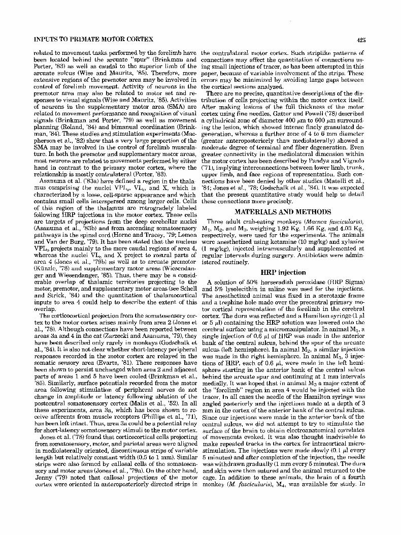
Fig. 1. A. Photomicrograph of a sagittal section of the left hemisphere in animal MI showing the HRP injection site. The section has been stained with diaminobenzidine and counterstained with thionine. Calibration bar
= 1 mm. B. Photomicrograph of a TMB-stained coronal section of the right thalamus in animal M, showing labeled thalamocortical cells. Calihration bar = 20 pm.

Fig. 2. A. Photomicrograph of a TMB-stained sagittal section of the left hemisphere in animal MI showing a part of the precentral area of the cortex and labeled corticocortical cells. B. Enlargement of the marked rectangle in A to show TMB-stained corticoeortical cells in detail. Calibration bars = 200 pm for A, 20 pm for B.

INPUTS TO PRIMATE MOTOR CORTEX
TABLE 1. Numbers of Ipsilateral Corticocortical Cells Counted
Cytoarchitectonic Animal Animal area M, M?
Area 4 Lam I11 Lam V
Area 6 Lam I11 (lateral) Lam V
SMA Lam I11 Lam V
Area 3a Lam I11 Lam V
Area 3b Lam I11 Lam V
Area 1 Lam I11 Lam V
Area 2 Lam I11 Lam V
Area 5 Lam 111 Lam V
Area 7 Lam I11 Lam V
SII Lam 111 Lam V
Area 24 Lam I11 Lam V
Others Total Lam 111
(laminae) Lam V
2,995 (47.2)" 45 1,516 (24.9) 82 Not counted3
543 (8.45) 1 93 (1.4) 0 206 (3.2) 1 539 (8.4) 4 170 (2.7) 1 110 (1.7) 0 123 (1.9) 0 Not counted3
8 (0.1) 6,303 (98) 134 (2)
1,738 (44.5) 217 1,163 (31.2) 207 244 (6.2) 29 318 (7.5) 13 10 (0.2) 0 157 (3.6) 3 193 (4.6) 10 38 (0.9) 2 3 (0.1) 1 31 (0.7) 0 12 (0.3) 0 2 (0.1) 3,909 (89) 483 (11)
Animal M?l
191 (2.0) 13 908 (11.1) 213 2,019 (26.2) 621 75 (0.9) 12 102 (1.0) 3 632 (6.5) 29 1,418 (15.4) 134 1,581 (16.6) 95 998 (11.2) 132 382 (5.0) 126 253 (4.0) 155 17 (0.2) 8,576 (84.8) 1,533 (15.2)
TOTAL 6,437 4,392 10,109 - 'In animal Ma, every sixth section of the series was examined for numerical analysis; in animals MI and Mz, every third section of the series was thus examined. 'Numbers in parentheses indicate percentages.
counts of cells in the SMA and area 24 were not considered reliable. animal M I , some of the medial sections of the left hemisphere were lost; hence, the
criteria, it was possible to avoid including stained dendritic and axonal profiles, blood vessels, or other artifacts in the count.
Using these criteria, it was possible to avoid including stained dendritic and axonal profiles, blood vessels, or other artifacts in the count.
In animals M1 and Ma, all the TMB-stained sections were analyzed quantitatively. Thus, approximately one-third of all cells projecting to the injection site were counted and plotted on the projection drawings. In animal M3, every alternate section in the TMB-stained series was analyzed. Thus, approximately one-sixth of all cells projecting to the injection site were counted and plotted on the projection drawings. Those TMB-stained (alternate) sections of animal MB, which were not analyzed numerically, were carefully studied to ensure that there were no remarkable differences in the topography and density of labeling. Therefore, in all the animals, regions of the cortex between adjacent TMB- stained sections that were not studied for labeling measured 160 pm in width. The labeled cells in the brain of animal M4 were not counted. They were studied for comparison whenever there was any doubt in the topography of label- ing in animal Ms.
Identification of cortical cytoarchitectonic areas and thalamic nuclei
DAB-stained sections counterstained with thionine were used to delineate cortical cytoarchitectonic areas and tha- lamic nuclei. The criteria of Mountcastle and Powell ('59) and Jones et al. ('78) were used to distinguish between the areas comprising the somatic sensory cortex and its bound- ary with areas 5 and 7. Area 3a was taken as that region of the floor of the central sulcus that had an attenuated lam- ina N and that was encroached upon by large pyramidal cells anteriorly in the hand area (Jones and Porter, '80). The posterior boundary of area 8 was taken to be the middle
429
of the anterior bank of the arcuate sulcus (Walker, '40). It was very difficult to distinguish between areas 5 and 7. For most of its length, the floor of the intraparietal sulcus was taken as the boundary between areas 5 and 7 (Mountcastle et al., '75). The second somatosensory area was demarcated using the descriptions and maps of Jones and Burton ('76). Thalamic nuclei were outlined using the descriptions of 01- szewski ('52) and Asanuma et al. ('83a).
RESULTS Extent of the injection sites in area 4
In TMB-stained sections, at the site of the tracer admin- istration, two regions of dark staining can be discerned an inner zone where the deposition of the reaction product is very dense and individual axons or perikarya cannot be identified, surrounded by a second region where the reac- tion product is less dense and where individuaI Iabeled axons and perikarya (both pyramidal and nonpyramidal) can be detected (Mesulam, '82). The former is the injection site or area of HRP uptake. It was impossible to count labeled cells in the second region-therefore, quantitation of labeling in this region has not been done. Beyond the zone of deposition of reaction product, labeled cells were all pyramidal in shape and have been counted.
In animals MI and Ma, the injection sites involved the full depth of the cortex. However, the maximum spread of the injection sites occurred in the middle layers of the cortex. In animal M1 the injection site was confined to area 4 and located mainly in the upper half of the anterior bank of the central sulcus. It extended to involve the anterior lip of the central sulcus at the level of the spur of the arcuate sulcus (see Fig. 4). The injection site in animal M2 was similar to that in animal MI (see Fig. 6). In animal Ms, the injection site was more extensive; it involved the full thick- ness of the cortex and spread into the underlying white matter. It also involved the full depth of the anterior bank of the central sulcus (area 4) from behind the inferior limb of the arcuate sulcus laterally to the superior precentral sulcus medially. Behind the spur of the arcuate sulcus and the adjacent part of the inferior limb of the sulcus, the reaction product filled the precentral gyrus, thus involving the part of area 6 in that region. Posteriorly it extended for some distance into area 3a (see Fig. 8).
Numbers of cells labeled in the cortex and thalamus The absolute number of cells labeled in such a study
would depend on the volume, concentration, and nature of the tracer injected as well as the sensitivity of the histo- chemical technique. This study aims to describe the distri- bution of the cells that were labeled and their relative numbers in various cortical cytoarchitectonic areas and thalamic nuclei. These descriptions are equally affected by the above parameters in any one animal.
Table 1 shows the number of cortical neurons that were counted in the hemisphere that was injected with the tracer. In both animals M1 and Mz, nearly half of all labeled corticocortical neurons in the ipsilateral hemisphere were located in area 4 itself, about one-fourth were located in postarcuate area 6, about one-fifth in the somatic sensory area I, and the rest in the supplementary motor area, the second somatosensory area, and posterior parietal area. Within the first somatosensory area of animals MI and M2, the majority of the labeled neurons were situated in areas 3a and 2, a smaller number in area 1, and very few in area 3b. In the injected hemisphere of animal M3, very few

430 S. GHOSH, C. BRINKMAN, AND R. PORTER
TABLE 2. Numbers of Callosal Cells Counted
Cytoarchitectonic Animal Animal Animal area M, M9 MQ1
Area 4 Lam 111 116 (82.8)’ 5 3,805 (59.5)
Area 6 Lam I11 22 (14.8) 2 1,990 (32.5)
SMA Lam I11 - - 209 (5.6) Lam V -
Area 24 Lam I11 - - 61 (1.8) Lam V - -
Area 3a Lam111 3 (1.8) - 16 (0.2) Lam V 0
Area 3h Lam I11 - - 5 (0.1) Lam V - -
Area 1 Lam I11 - - 2 (0.1) Lam V - -
Area 2 Lam I11 1 (0.6) - 21 (0.3) Lam V 0
Total Lam I11 142 - 6,114 (82.2) (laminae) Lam V 27 - 1,325 (17.8)
Lam V 24 0 622
(lateral) Lam V 3 0 428
- 204
71
0
0
0
0
-
-
TOTAL 169 7 7,439
’In animal Mn, every sixth section of the series was examined for numerical analysis; in animals MI and Mz, every third section o f the series was thus examined. ‘Numbers in parentheses indicate percentages.
TABLE 3. Numbers of Thalamocortical Cells Counted
Thalamic Animal Animal Animal nuclei Mi Mz
VPL” 579 (66.31’ 756 (61.9) 1,774 (31.9) VLC 162 (18.6) 243 (19.9) 682 (12.3)
88 (10) 86 (7) 433 (7.8) - - 432 (7.8)
V L O
X VApc - - 449 (8.1) VPL, 15 (1.7) 9 (0.7) 61 (1.1) VLm - 4 (0.3) -
VPM l ( O . 1 ) 12 (1.0) 141 (2.5) CM 25 (2.8) 26 (2.1) 380 (6.9) CL l ( O . 1 ) 83 (6.8) 688 (12.4)
- - 283 (5.1) Pen Cdc - - 45 (0.8)
- - 13 (0.2) Grcn R LD l(0.l) HYP MD Total 873 1,221 5,549
1 - - - -
- - 129 (2.3) - 2 (0.2) 38 (0.7)
‘In animal MQ, every sixth section of the series was examined for numerical analysis; in animals M, nd Mz, every third section was thus examined. 2Numhers in parentheses indicate percentages.
neurons were labeled in area 4 (2%) and area 3a (0.9%). In the postarcuate area 6 of this animal, labeled neurons were fewer than expected (11.1%). Instead, a higher proportion of neurons were labeled in the supplementary motor area (26.2%) and posterior parietal areas (27.8%). In all three animals, most labeled corticocortical neurons were located in lamina 111, and only a small percentage of labeled neu- rons were found in lamina V.
Table 2 enumerates the number of neurons counted in the contralateral hemisphere of animals MI, Mz, and M3. It is clear that few callosal neurons were labeled in the first two aqimals, MI and Mz. In contrast, a large number of such neurons were labeled in animal Ms, almost exclusively in area 4 (59.5%), in postarcuate area 6 (32.5%), and in the supplementary motor area (5.6%). Most of the labeled cal- losal neurons were located in lamina 111 of the cortex.
The number of labeled thalamocortical neurons counted in the three animals are detailed in Table 3. The distribu-
tion of these cells in animals M1 and M2 was similar: about two-thirds of the labeled cells located in nucleus V P b , about one-fifth in nucleus VLc; about one-tenth in nucleus VLo; and the rest in the intralaminar nuclei, CM and CL. The labeled thalamocortical cells of animal M3 were also mainly located in nucleus VPLo, VLc, and VLo, but in this animal they accounted for only half of the labeled neurons. Neurons were now labeled in the nuclei VApc and nucleus X (7.8% and 8 .B%, respectively) and a greater proportion of neurons (about one-fourth) were seen in the intralaminar nuclei CM, CL, and Pcn. A few neurons were also labeled in the hypothalamus and in nucleus MD.
Distribution of labeled corticocortical neurons Figures 3 and 4 show the distribution of labeled neurons
in the injected hemisphere of animal MI. Within area 4, labeled cells were found in large numbers around the injec- tion site both in the precentral gyrus as well as in the anterior bank of the central sulcus and extended as far medially as the superior precentral gyrus. This labeling was not uniformly dense anteroposteriorly or mediolater- ally. Labeling was not seen further medially in area 4, in the convexity of the hemisphere or on the medial surface. Laterally, labeling was sparse in the motor cortex in front of the lateral end of the central sulcus. Labeled neurons in postarcuate area 6 were seen in the posterior bank and posterior lip of the spur and inferior limb of the arcuate sulcus as well as in the floor and posterior lip of the superior limb of the arcuate sulcus. The posterior bank of the supe- rior limb of the arcuate sulcus was found to be devoid of labeled neurons, as was the floor of the inferior limb of the arcuate sulcus. In cytoarchitectonic areas 3a, 1, and 2, la- beled neurons were found immediately behind the injection site as well as farther medially, in a mediolateral zone spanning about 10 mm. This zone was more or less aligned posterior to a similar zone of labeled cells seen in area 4. Labeling was sparse in areas 3b and 1. Labeling in areas 5 and 7 was confined to the anterior and posterior banks of the lateral third of the intraparietal sulcus and was contin- uous at the floor of the sulcus.
Figures 5 and 6 detail’ the labeling of corticocortical cells seen in the injected hemisphere of animal Ma. As in ani- mal MI there was extensive labeling within area 4 in the region at the level of the spur and the superior and inferior limbs of the arcuate sulcus. Large parts of area 4, viz., the medial surface of the hemisphere, in the medial part of the convexity, and the region in front of the lateral end of the central sulcus were devoid of label. Labeled neurons in postarcuate area 6 were seen in the posterior bank and posterior lip of the inferior limb of the arcuate sulcus. Unlike the findings in the previous animal (MI), labeled
‘Although both Figures 4B and 6B show an area of “unfolded” cortex in the vicinity of the injection site, the perspectives in these two figure differ. This is because the “unfolding” of the cortex in animal M1 has been done from parasagittal sections with the floor of the central sulcus as the reference point from one section to the next. (Thus, the cortex has been unfolded by stretching it rostro- caudally.) On the other hand, the cortex of animal M2 (and Ms) has been “unfolded” from coronal sections with the upper lip of the lateral sulcus a the reference point from one section to the next (i.e., the cortex has been “unfolded” by stretching it mediola- terrally). Perspective obtained in Figure 6A (as well as 8A and 9A), where the distribution of neurons is shown on the surface of the “folded” cortex, is distorted by the fact that labeling in the banks and lips of the sulci overlap in the vicinity of the sulci.
INPUTS TO PRIMATE MOTOR CORTEX 431
A 4
2
IP
5
7
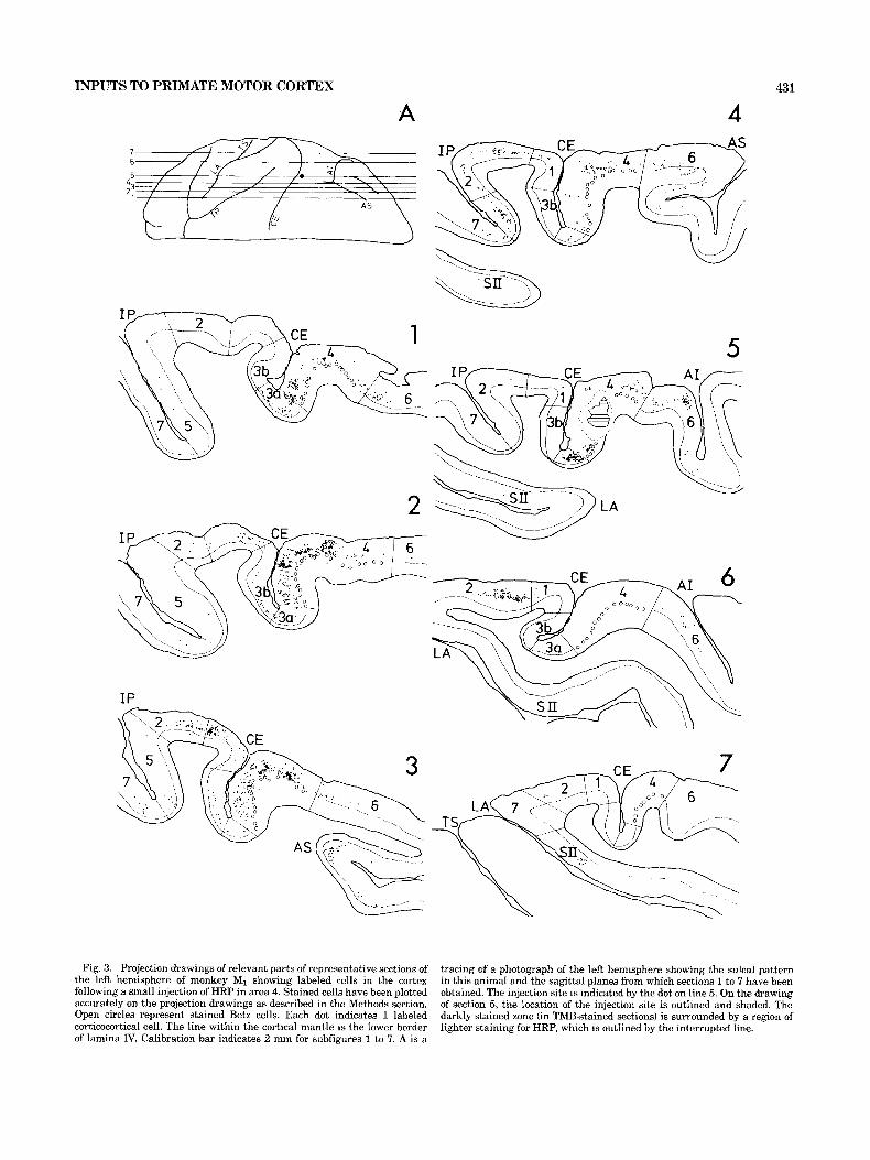
Fig. 3. Projection drawings of relevant parts of representative sections of the left hemisphere of monkey MI showing labeled cells in the cortex following a small injection of HRP in area 4. Stained cells have been plotted accurately on the projection drawings as described in the Methods section. Open circles represent stained Betz cells. Each dot indicates 1 labeled corticocortical cell. The line within the cortical mantle is the Iower border of lamina IV. Calibration bar indicates 2 mm for subfigures 1 to 7. A is a
tracing of a photograph of the left hemisphere showing the sulcal pattern in this animal and the sagittal planes from which sections 1 to 7 have been obtained. The injection site is indicated by the dot on line 5. On the drawing of section 5, the location of the injection site is outlined and shaded. The darkly stained zone (in TMB-stained sections) is surrounded by a region of lighter staining for HRP, which is outIined by the interrupted line.

43 2
A
S. GHOSH, C. BRINKMAN, AND R. PORTER
B
AS . . .
. I . . . . . . . . . .
1 .
. . J:
rd +a. w w w 0 0 0
6
. . . . .
1 : . . I .
. . . . .
. .
I .
. .
ii w
4 3a' 3b 2 ' 517
Fig. 4. A. Tracing of a photograph of the superior view of the left hemi- sphere of animal MI showing that part of the cortex that was unfolded for presentation in B. B. Surface reconstruction from sagittal sections of the left hemisphere of animal MI showing the distribution of labeled cells following a small injection of HRP in area 4. The cortex has been unfolded and the anterior and posterior lips and floors of the central sulcus, arcuate sulcus, and intraparietal sulcus marked with thick lines. The cytoarchitec-
tonic boundaries have been delineated by thin lines. Each dot represents from 1 to 5 TMB-stained corticocortical cells. The extent of the injection site (darkly stained by TMB) is outlined by a continuous line and is surrounded by the lightly stained zone outlined by the interrupted line. The vertical and horizontal calibration bars each represent 2 mm in the mediolateral and anteroposterior planes, respectively.

INPUTS TO PRIMATE MOTOR CORTEX
7 6 5 4 3 2 1
1
433
Fig. 5. Projection drawings of relevant parts of representative coronal sections of the right hemi- sphere of animal Mz showing labeled corticocortical cells. Stained cells have been plotted accurately as dots on the drawings. The upper border of lamina 11, the lower border of lamina IV, and the cytoarchitectonic boundaries have been marked within the cortical mantle. The extent of the injection site is outlined in subfigures 3 and 4. Calibration bar = 2 mm.
434 S. GHOSH, C. BRINKMAN, AND R. PORTER
W
6
INPUTS TO PRIMATE MOTOR CORTEX 435
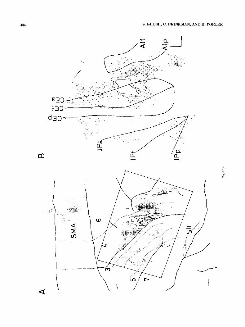
neurons were absent in the floor and posterior lip of the superior limb of the arcuate sulcus (Fig. 6A). Labeling in cytoarchitectonic areas 3a, 1, and 2 formed a patchy zone aligned (mediolaterally) posterior to the labeling in the mo- tor cortex. The posterior bank of the central sulcus was more or less devoid of labeled cells. Labeling in area 5 was located in the anterior bank of the intraparietal sulcus, whereas labeling in area 7 was located in the posterior bank of that sulcus and in the inferior parietal lobule. On the medial surface, labeled cells were seen in the rostral part of area 6 (rostral part of SMA) as well as in the adjacent part of area 24 in the floor of the cingulate sulcus.
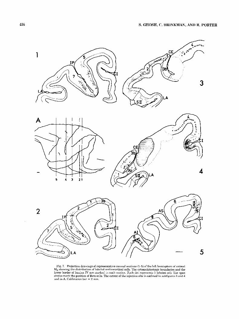
Figures 7 and 8 show the location of labeled neurons in the injected hemisphere of animal M3. Labeled neurons in area 4 were few and distributed sparsely in the medial part of the convexity and medial surface of the hemisphere as well as in the lateral end of the motor cortex, in the same areas that were devoid of label in animals MI and M2. Those regions of area 4 that had showed large numbers of labeled cells in the previous two animals (MI and Mz) were completely filled with HRP reaction product in this animal (M3). In postarcuate area 6 of animal M3, labeled neurons were located on the floor, posterior banks, and posterior lips of the spur and the superior and inferior limbs of the ar- cuate sulcus. A part of area 6 immediately behind the spur and the adjacent inferior limb of the arcuate sulcus was filled with HRP reaction product and, there re, did not
animal. Labeling in the somatic sensory and posterior pa- rietal areas was more extensive. Labeled neurons in the parietal lobe formed a continuous zone of labeling in the lateral half of the postcentral gyrus, in the anterior and posterior banks of the intraparietal sulcus, and in the infe- rior parietal lobule, and extended farther laterally to the upper lip of the lateral sulcus. The labeling then continued onto the upper bank of the lateral sulcus into the second somatosensory area. On the medial surface of the hemi- sphere, labeling was seen throughout the rostrocaudal ex- tent of area 6 (supplementary motor area).
The distribution of neurons in the right hemisphere (con- tralateral to the injection site) of animal M3 is seen in Figure 9. Large numbers of callosal neurons were labeled in the precentral gyrus behind the spur and inferior limb of the arcuate sulcus involving areas 4 and 6. The concen- tration of cells farther medially in areas 4 and 6, behind the superior limb of the arcuate sulcus, was less dense. Labeling in area 6 extended into the posterior banks of the spur and the superior and inferior limbs of the arcuate sulcus. The anterior bank of the central sulcus was devoid of label except at its lateral end. Labeling in the medial surface of the hemisphere was confined to the rostral half of area 6 and adjacent part of area 24. The somatic sensory and posterior parietal areas were devoid of labeled neurons,
reveal any labeling. Area 8 was devoid of 2 abel in this
Fig. 6. A. Tracing of a photograph of the right hemisphere isulci-thick lines) in animal MP. The injection site (thick line in area 41, retrogradely labeled cells, and cytoarchitectonic boundaries (thin lines) have been mapped on the surface view of cortex from the coronal sections. Each dot represents from 1 to 5 labeled cells. A part of the cortex (marked by the rectangle) has been “unfolded” in B. Calibration bar = 2 mm. B. Surface reconstruction of the “unfolded cortex” from coronal sections of the right hemisphere of
except for a few in front of the tip of the intraparietal sulcus.
Distribution of labeled thalamocortical cells Figure 10 shows the distribution, in parasagittal sections,
of thalamocortical neurons labeled in animal MI. In the sections F and G, where the concentration of labeling is densest, the labeled neurons occupy the whole rostrocaudal and dorsoventral extent of nucleus VPL, and extend into the nuclei VL, and VL,. In more lateral and medial sections (D, E, and H), labeled neurons are restricted to the posterior parts of nucleus VPL, and in sections B and C to the ventral part of that nucleus. Therefore, most of the labeled tha- lamic neurons in VPL, form a mediolaterally restricted slab with blurred borders posteriorly.
Figure 11 shows the distribution, in coronal sections, of thalamocortical neurons labeled in animal M2. The medi- olateral restriction of the band of labeled neurons is clear anteriorly (in sections A to I), but becomes less clear poste- riorly where the number of neurons labeled also diminishes (in sections K to 0). In most sections the involvement of the entire dorsoventral extent of VPL, is evident. This band of labeling is not uniformly dense anteroposteriorly (only 9 neurons labeled in section C).
Figure 12 shows some of the labeled neurons in coronal sections of the thalamus of monkey M3. The widening of the band of labeled neurons in nuclei VPL, and VLc is evident (sections B and C) and also the rostral and rostro- medial extension of this band into nuclei VA, VL,, and X (sections A and B).
DISCUSSION Detailed studies of the representation of movements of
the limbs in the motor cortex have been made using cortical stimulation (surface and intracortical) and recording of cor- tical neuronal activity in conscious behaving animals. These studies agree that the representations of movements of the head, forelimb, trunk, and hindlimb occupy separate zones in the motor cortex (Phillips and Porter, ’77). Within the forelimb area of the motor cortex, populations of neurons related to the movements of individual joints and the activ- ity of individual muscles and motor units may be widely dispersed over territories several square millimeters in area, which overlap extensively. Results of most studies (Anderson et al., ’75; Kwan et al., ’78; Sessle and Wiesen- danger, ’82; and Woolsey et al., ’52) confrm that motor cortical neurons related to the movements of fingers are concentrated in the anterior bank of the central sulcus, whereas those related to movements at the wrist, elbow, and shoulders are concentrated in more rostral areas of the motor cortex in the precentral gyrus. However, the overlap of zones containing these populations of neurons is such that movements related to proximal joints may also be represented in the anterior bank of the central sulcus (Kwan et al., ’78; Sessle and Wiesendanger, ’82).
In animal MI, the injection was small, restricted to area 4 in the anterior bank and the rostral lip of the central sulcus and situated in the lateral part of the forelimb representa- tion. Following such an injection, large numbers of neurons were labeled in area 4 itself, restricted mostly to the fore- limb remesentation of the motor cortex. It is not uossible to
animal Mz. The floors and lips of relevant sulci have Ceen marked with thick lines. The thin lines show the injection site-an inner zone of dark staining (uninterrupted line) surrounded by an outer zone of lighter stain- ing (interrrupted line). Each dot represents from 1 to 5 labeled corticocorti-
to what extent labeling in area 4 spread to the “face” representation laterally and to the “trunk” repre- sentation medially. However, large areas Of the motor COr-
cal cells. Calibration bars = 2 mm. tex in the medial part of the convexity, the medial surface
436 S. GHOSH, C. BRINKMAN, AND R. PORTER
1
A I I ' l l
5 4 3 2 1
3
Fig. 7. Projection drawings of representative coronal sections (1-5) of the left hemisphere of animal Ms showing the distribution of labeled corticocortical cells. The cytoarchitectonic boundaries and the lower border of lamina IV are marked in each section. Each dot represents 1 labeled cell. The open circles mark the position of Betz cells. The extent of the injection site is outlined in subfigures 3 and 4 and in A. Calibration bar = 2 mm.

A B
13
Fig.
8.
A.
Tra
cing
of
a ph
otog
raph
of
the
left
hem
isph
ere
(sul
ci, t
hick
lin
es)
in a
nim
al M
s. T
he i
njec
tion
site
(th
ick
line
in
area
41,
labe
led
cells
(d
ots)
, and
cyt
oarc
hite
cton
ic b
ound
arie
s (t
hin
lines
) ha
ve b
een
map
ped
on
the
surf
ace
view
of
the
cort
ex f
rom
cor
onal
sec
tions
. E
ach
dot
repr
esen
ts
from
1 to
5 la
bele
d co
rtic
ocor
tical
cel
ls. T
he i
njec
tion
site
can
be
seen
to
encr
oach
int
o ar
eas
6 an
d 3a
. T
he p
art
of t
he c
orte
x “u
nfol
ded“
in
B i
s m
arke
d by
a r
ecta
ngle
. C
alib
rati
on b
ar =
2 m
m. B
. Sur
face
reco
nstr
uctio
n
of t
he u
nfol
ded
cort
ex fr
om c
oron
al s
ectio
ns o
f the
left
hem
isph
ere
in a
nim
al
M:+
The
lips
and
floo
rs o
f rel
evan
t su
lci h
ave
been
mar
ked
wit
h th
ick
lines
. T
he th
in l
ines
dem
arca
te t
he in
ject
ion
site
-an
inne
r zo
ne o
f da
rk s
tain
ing
unin
terr
upte
d li
ne) a
nd a
n o
uter
zon
e of
lig
hter
sta
inin
g (i
nter
rupt
ed l
ine)
. E
ach
dot
repr
esen
ts f
rom
1 to
5 l
abel
ed c
ortic
ocor
tical
cel
ls.
Cal
ibra
tion
ba
rs =
2 m
m.

A B
\: ..
.
.
$:
. .
.
..
.
.
. -.
..
. -
. ..
..
Fig.
9.
A. T
raci
ng o
f a
phot
ogra
ph o
f th
e ri
ght
hem
isph
ere
(sul
ci, t
hick
rec
tang
le h
as b
een
unfo
lded
in
B.
B.
Surf
ace
reco
nstr
uctio
n of
unf
olde
d lin
es)
in a
nim
al M
3. L
abel
ed c
allo
sal
cells
(do
ts)
and
cyto
arch
itect
onic
cor
tex
from
cor
onal
sec
tions
of
the
righ
t he
mis
pher
e of
ani
mal
Ms.
The
bo
unda
ries
(th
in li
nes)
hav
e be
en m
appe
d on
the
surf
ace
view
of t
he c
orte
x fl
oors
and
lips
of
rele
vant
sul
ci a
re s
how
n. E
ach
dot r
epre
sent
s fr
om 1
to 5
fr
om c
oron
al s
ectio
ns,
Eac
h do
t re
pres
ents
1 to
5 l
abel
ed c
ortic
ocor
tical
la
bele
d co
rtic
ocor
tical
cel
ls. C
alib
rati
on b
ars
= 2
mm
. ce
lls.
Cal
ibra
tion
bar
=
2 m
m.
The
par
t of
the
cor
tex
mar
ked
by t
he

INPUTS TO PRIMATE MOTOR CORTEX 439
A
(55)
C (771
E
129)
F
(281 I
G (1401
H
(671
Fig. 10. Projection drawings of sagittal sections of the thalamus showing most TMB-stained thala- mocortical cells that were counted in animal MI. Each dot represents 1 labeled cell. The number of labeled cells in each section is shown in parentheses. Calibration bar = 2 mm.

440 S. GHOSH, C. BRINKMAN, AND R. PORTER
A
(L7)
B (107)
D
(110)
E
(75)
F (1 23)
G (861
K
(65)
L (36)
1.45)
VPLC
N
( 5 9 )
0
( 6 L )
Fig. 11. Projection drawings of coronal sections of the thalamus showing all TMB-stained thalamo- cortical cells that were counted in animal Mz. The thalamic nuclear boundaries have been drawn using the criteria described by Olszewski (‘52) and Asanuma et al. (‘83). Each dot represents 1 labeled thalamocortical cell. The number of labeled cells in each section is shown in parentheses. Calibration bar = 2 mm.

INPUTS TO PRIMATE MOTOR CORTEX
A
(6321
B
(6301
C (8931
441
Fig. 12. Projection drawings of coronal sections of the thalamus showing some TMB-stained thala- mocortical cells that were counted in animal MS. Each dot represents 1 labeled cell. The number of labeled cells in each section is shown in parentheses. Calibration bar = 2 mm.

442 S. GHOSH, C. BRINKMAN, AND R. PORTER
of the hemisphere, and the lateral end of the central sulcus, representing movements of the hindlimb, trunk, and face, were devoid of labeled cells. Outside the motor cortex, the most prominent labeling occurred in postarcuate area 6 (in the precentral gyrus as well as in the floor and posterior bank of the arcuate sulcus). In the somatic sensory area I, labeled neurons were found mainly in areas 3a and 2, to a smaller extent in area 1, and least in area 3b. These areas of SI that showed labeled cells were immediately posterior to the strip of labeling in MI and represent the forelimb regions of SI (Nelson, Sur, Felleman, and Kas, ’80). Label- ing in the posterior parietal region was restricted to the banks of the lateral third of the intraparietal sulcus. Label- ing in the contralateral hemisphere was very sparse.
It is quite clear that the distribution of labeled neurons in animal Ma, although essentially similar to that in MI, differs slightly. As in animal M1, large numbers of neurons are labeled in the forelimb region of MI and SI, and labeling in the opposite hemisphere is small. However, in animal Mz, there is lack of labeling in the floor and posterior lip of the superior limb of the arcuate sulcus, and labeling is present in area 7 in the inferior parietal lobule. Significant involvement by the tracer of the face area of the motor cortex is precluded by the presence of very little labeling in nucleus VPM of the thalamus (10 out of 1,221 thalamocor- tical neurons labeled; Jones, ’81). On the medial part of the hemisphere, labeling is seen only in the rostral half of SMA.
The injection site in animal M3, as expected, was much larger than in animals M1 and Mz. It involved the full depth of the anterior bank of the central sulcus throughout the mediolateral extent of the forelimb representation. From the number of cells labeled in nucleus VPM (141 out of 5,549 thalamocortical neurons labeled), it could be deduced that some amount of tracer spread to the “face” area of the motor cortex. Similarly, reaction product spread forward into area 6 behind the arcuate spur and backward into area 3a. The significant findings in the cortex of this animal were:
1. Only a small number of neurons were labeled in area 4.
2. A large number of neurons were labeled in the somato- sensory and posterior parietal areas, which formed a contin- uous zone involving the forelimb and face representations of SI (Nelson et al., ’80).
3. A large number of neurons were labeled in the supple- mentary motor area involving most of its rostrocaudal extent.
4. A large number of neurons were labeled in the contra- lateral hemisphere, in the motor, premotor, and supplemen- tary motor areas. However, in the contralateral hemisphere, labeling was absent throughout the anterior bank of the central sulcus except a t its lateral end; this labeling could have been due to involvement by the tracer of the “face” or “thumb” representation (Jenny, ’79). It was clear that most of the motor cortical representation of the forelimb in the anterior bank of the central sulcus was devoid of callosal connections.
Labeled neurons in the thalamus comprised 11.7%, 21.7%, and 24% of all labeled neurons counted in animals MI, M2, and M3, respectively. In animals MI and Mz, which had restricted injections of tracer in area 4, the thalamocortical neurons Oocated mainly in nucleus VPL, and adjacent parts of VL, and VL,) formed a continuous band of labeling,
restricted mediolaterally (about half a millimeter wide) and occupying the full dorsoventral and anteroposterior extent of nucleus VPL,. However, this band was not uniformly dense or wide and its borders were blurred posteriorly. Similar bands have been described regarding thalamocort- ical neurons projecting to both the sensory and the motor cortex of monkeys (Jones et al., ’79b). In animal M3, the aggregation of labeled thalamocortical neurons formed a wider continuous band in nuclei VPL,, VL,, and VL,, which extended into nuclei VA and X. The involvement of nuclei VA and X was due to the involvement by the tracer of rostral parts of area 4 and adjacent parts of area 6 (Jones et al., ’79b; Kunzle, ’78). Labeling in the thalamus of animal M3 was also marked by a larger proportion of labeled neu- rons in the intralaminar nuclei (24.4% of labeled thalamo- cortical neurons). This could be explained by the fact that neurons of the intralaminar nuclei project diffusely to the cortex of the frontal lobe and may be better labeled after a larger injection (Jones, ’75).
Electrical mapping of projections to the spinal cord from the motor cortex, using surface and intracortical stimula- tion, has shown extensive overlap of cortical areas that influence individual muscles and motor neurons (Phillips and Porter, ’77). A reason for this could be an extensive interconnection and interpIay between output elements and modules (functional units) within the cortex. This study shows that within the forelimb representation of the motor cortex there are, indeed, profuse connections. When a small injection of HRP is made in a restricted part of the “fore- limb’ area of the motor cortex, large numbers of cells are labeled both anteriorly and medially, filling up the rest of the “forelimb” representation. However, very few connec- tions derive from areas farther medially (trunk and hind- limb areas) or laterally (face areas). Even when the injection site involves most of the mediolateral and anteroposterior dimensions of the “forelimb” motor area as in animal M3, labeled cells in the other “body” regions (trunk, hindlimb, and face) of the motor cortex are sparse. It is not known how the interconnections within the forelimb region of the motor cortex influence the projection neurons of this area. These connections represent horizontal connectivity be- tween input-output modules. Although these connections are extensive, they may also be specific; they may intercon- nect cells in different locations with the same or related functional specifications even though separated by consid- erable distances. Such connections have also been described in the visual cortex (Gilbert, ’85) and somatosensory cortex (Shanks et al. ’85). It is not known what proportion of these stained cells in area 4 have been labeled through axonal collaterals. Pyramidal cells in lamina I11 of the visual cor- tex have been shown to have collaterals that spread up to 2 mm away tangentially from the cell body (Gilbert, ’85). Similar spread of collaterals have been described for pyra- midal tract neurons in layer V of the motor cortex (Landry et al., ’80).
Projections to the motor cortex from the cytoarchitectonic areas 1 and 2 of SI have been described before (Jones et al., 1978), but these authors did not find any connections be- tween areas 3a and the motor cortex. In our study, in both animals MI and Mz, there was a consistent projection from area 3a to area 4. The topography of this projection from area 3a matched that of the projections from areas 1 and 2. Thus, it was only the “forelimb” regions of these areas of SI that showed retrograde labeling following injection of
INPUTS TO PRIMATE MOTOR CORTEX 443
HRP limited to a part of the “forelimb” region of MI. In animal M3, which received a more extensive injection of HRP, only the “forelimb” and “face” regions of SI showed retrogradely labeled cells. Few neurons were labeled in area 3a of this animal since the reaction product spread to this area and could have masked any cells that were la- beled there.
Neurons in the precentral motor area respond a t short latency to natural stimulation of the limbs, mainly joint movement and muscle palpation (Lemon and Porter, ’76). Precentral neurons, whose discharge can be related to movement about a particular joint of the contralateral fore- limb, usually respond to passive movements about the same joint (Lemon et al., ’76)-a proportion of such neurons do, however, respond to such movements from adjacent joints of that forelimb. These neurons are rarely influenced by natural stimulation of the ipsilateral forelimb, the trunk, or either hindlimb. In correlation with these findings, our anatomical studies also show that mainly the “forelimb” region of the ipsilateral somatosensory cortex projects to the “forelimb” region of the motor cortex.
Of areas outside the motor cortex, the largest proportion of labeled cells was seen in the postarcuate premotor area (area 6) of the same hemisphere (in all animals) and of the opposite hemisphere (in animal M3). Fewer cells than ex- pected were labeled in the left premotor area of animal MB, presumably, because of the spread of the injection site into area 6 in this animal. Following injections of HRP in the “forelimb” regions of the primary motor cortex, cells were labeled in large numbers in the floor, posterior bank, and posterior lip of the spur and the inferior limb of the arcuate sulcus, both rostral and rostrolateral to the injection site. A similar pattern of projection from the premotor area 6 to the primary motor area (posteromedially directed) has also been described by Godschalk et al. (’84). In the same loca- tion in area 6, Brinkman and Porter (‘83) found neurons related to forelimb movements. Fewer cells were labeled in the floor and posterior lip of the superior limb of the arcuate sulcus. Neurons related to forelimb movement have also been located here Wise and Mauritz, ’85). This region has been shown to project to the “hindlimb” region of MI by Muakkassa and Strick (‘79) as well as to premotor regions (area 6) behind the spur and the inferior limb of the arcuate sulcus (Pandya and Vignolo, ’71; Matelli et al., ’84). This implies less strict segregation of forelimb-hindlimb topog- raphy within the postarcuate premotor area than in the primary motor cortex.
Using intracortical microstimuIation to map the SMA, Macpherson et al. (‘82) found that proximal motor effects in the forelimb were the predominant responses. Distal joint movements were found less commonly and more rostrally than proximal joint movements; the two effects overlapped extensively. Hindlimb movements were poorly localized and were obtained from caudal regions of the SMA, intermin- gled with forelimb motor effects. Although these motor effects of SMA may not be entirely through its projection to the motor cortex, our description of this projection com- plements the findings from the stimulation studies. A small injection of HRP in the anterior bank of the central sulcus labels a small number of neurons through the rostral half of SMA. A larger injection site in the whole “forelimb” region labels a relatively greater number of neurons in the whole rostrocaudal extent of SMA (see Muakkassa and Strick, ’79). The unexpectedly large number of labeled cells
in the SMA of animal M3 may also imply a diffuse projec- tion from the SMA to MI.
In our study, injection of HRP limited to the anterior bank of the central sulcus labeled a few cells in the poste- rior parietal cortex (area 5 and 7b). However, when the injection was extended more anteriorly (as well as medi- ally) to include rostral parts of area 4, a much higher proportion of these cells was labeled. Neurons of area 7b have been shown to project mainly to the premotor area 6 behind the inferior limb of the arcuate sulcus (Godschalk et al., ’841, but our study shows that these neurons also project to area 4 (in greater numbers to rostral than caudal parts of area 4). Neurons of areas 5 and 7 have been related to both passive and active movements of the limb, manipula- tive movements of the hand, visual fixation and tracking, and visual coordination of hand movements (Mountcastle et al., ’75).
Cells in the thalamic nuclei VPL,, VL,, and VL, are labeled following an injection of HRP restricted to the cau- dal region of the motor cortex. However, when the injection includes motor cortical areas anterior to it (which also pro- ject to the more posterior motor areas), labeling extends into nuclei VA and X. Now a smaller proportion of cells is labeled in nucleus VPL,. Thus, inputs from both the deep cerebellar nuclei (Asanuma et al., ’83b) and basal ganglia (Kim et al., ’76) are focused on a small region of the motor cortex and adjacent premotor region, through the thalamus.
Somatosensory inputs to the motor cortex arise from the postcentral cytoarchitectonic areas 1, 2, and 3a, as well as from the thalamus. There is an interesting similarity be- tween the intrinsic connections within area 4 and its con- nections with the somatic sensory cortex, and there is considerable convergence of projections from the somato- sensory to the motor cortex.
In summary, this study provides evidence that the pre- motor and supplementary premotor areas, which are found to be so closely related to movement execution, planning, and coordination, in response to both external and internal cues, dominate quantitatively the inputs to the precentral motor cortex. Although the number of thalamocortical neu- rons retrogradely labeled from the motor cortex are fewer than labeled corticocortical cells, they form links through which extensive projections of the lateral cerebellum, basal ganglia, and ascending spinothalamic tracts could influ- ence a small region of motor and adjacent premotor area, areas that are themselves profusely interconnected. How- ever, more information regarding the fine anatomy of these projections to the motor cortex, and how these inputs inter- act to influence the output elements of the motor cortex, is required for better understanding of the control of move- ment execution by the motor cortex.
ACKNOWLEDGMENTS We thank Dr. S.J. Redman, Dr R. Martin-Body, and Dr.
R.E.W. Fyffe for their comments on the manuscript; Shirley Ramsay for typing it; Terrina Thompson, G. Rodda, and K. Collins for excellent technical assistance; and the Pho- tography Department of the JCSMR for help with illustra- tions. S.G. holds a PhD scholarship within the Australian National University.
LITERATURE CITED Anderson, P., P.J. Hagan, C.G. Phillips, and T.P.S. Powell (1975) Mapping
by microstimulation of projections on from Area 4 to motor units of the
444 S. GHOSH, C. BRINKMAN, AND R. PORTER
baboon’s hand. Proc. Roy. SOC. B. 188: 31-60. Asanuma, C., W.T. Thach, and E.G. Jones (1983a) Cytoarchitectonic deline-
ation of the ventral lateral thalamic region in the monkey. Brain Res. Rev. 5: 219-235.
Asanuma, C., W.T. Thach, and E.G. Jones (198313) Distribution of cerebellar terminations and their relation to other afferent terminations in the ventral lateral thalamic region of the monkey. Brain Res. Rev. 5: 237- 265.
Brinkman, C. (1984) Supplementary motor area of the monkey’s cerebral cortex: Short and long term deficits after unilateral ablation and the effect of subsequent callosal section. J. Neurosci. 4: 918-929.
Brinkman, C., and R. Porter (1979) Supplementary motor area in the mon- key: Activity of neurons during performance of a learned motor task. J. Neurophysiol. 42: 681-709.
Brinkman, C., and R. Porter (1983) Supplementary motor area and premotor area of monkey cerebral cortex: Functional organization and activities of single neurons during performance of a learned movement. In J.E. Desmedt (ed): Motor Control Mechanisms in Health and Disease. New York: Raven Press, pp. 393-420.
Brinkman, J., J.G. Colebatch, R. Porter, and D.H York (1985) Responses of precentral cells during cooling of post-central cortex in conscious mon- keys. J. Physiol. 368: 611-625.
Evarts, E.V. (1981) Role of motor cortex in voluntary movement in primates. In J.M. Brookhart and V.B. Mountcastle (eds): Handbook of Physiology. Sect 1 Neurophysiology. Bethesda, M D American Physiological Society, pp. 1083-1120.
Gatter, K.C., and T.P.S. Powell (1978) The intrinsic connections of the cortex of area 4 of the monkey. Brain 101: 513-541.
Gilbert. C.D. (1985) Horizontal inteeration in the neocortex. Trends Neu-
Brain Res. 36: 445-462. Lemon, R.N., J.A. Hanby, and R. Porter (1976) Relationship between the
activity of precentral neurons during active and passive movements in conscious monkeys. Roc. Roy. SOC. B. 194: 341-373.
Macpherson, J.M., C. Marangoz, T.S. Miles, and M. Wiesendanger (1982) Microstimulation of the supplementary motor area (SMA) in the awake monkey. Exp. Brain Res. 45: 410-416.
Malis, L.I., K.H. Pribram, and L. Kruger (1952) Action potentials in motor cortex evoked by peripheral nerve stimulation. J. Neurophysiol. 16: 161- 167.
Matelli, M., R. Camarda, M. Glickstein, and G. Rizzalotti (1984) Intercon- nections within the postarcuate cortex (area 6) of the macaque monkey. Brain Res. 310: 388-392.
Matsumura, M., and K. Kubota (1979) Cortical projections to hand-arm motor area from post-arcuate area in macaque monkeys: A histological study of retrograde transport of horseradish peroxidase. Neurosci. Lett. 11: 241-246.
Mesulam, M.M. (1982) Principles of horseradish peroxidase neurohisto- chemistry and their applications for tracing neural pathways-axonal transport, enzyme histochemistry and light microscopic analysis. In M.M. Mesulam (ed): Tracing Neural Pathways with HRP. New York: John Wiley & Sons, pp. 3-135.
Mountcastle, V.B., and T.P.S. Powell (1959) Cytoarchitecture of the postcen- tral gyrus of Macaca mulatta. John Hopkins Hosp. Bull. 105: 108-132.
Mountcastle, V.B., J.C. Lynch, A. Georgopoulos, H. Sakata, and C. Acuna (1975) Posterior parietal association cortex of the monkey: Command function for operations within extrapersonal spaace. J. Neurophysiol. 38: 872-908.
Muakkassa, K., and P. Strick (1979) Frontal lobe inputs to primate motor I
rosci. 8: 160-165. Godschalk, M., R.N. Lemon, H.G.J.M. Kuypers, and H.K. Ronday (1984)
Cortical afferents and efferents of monkey post-arcuate area: An ana- tomical and electrophysiological study. Exp. Brain Res. 56; 410-424.
Horne, M.K., and D.J. Tracey (1979) The afferents and projections of the ventroDosterolatera1 thalamus in the monkev. EXD. Brain Res. 36: 129-
cortex: Evidence for four somatotopically organized premotor areas. Brain 177r 176-182.
Nelson, R.J., M. Sur, D.J. Felleman, and J.H. Kaas (1980) Representations of the body surface in postcentral parietal cortex of Mucacu fasciculuris. J.
Olszewski, J. (1952) The Thalamus of the Macacu mulutta. Basel: S. Karger. 611-643.
I .
141. Jenny A.B. (1979) Commissural projections of the cortical hand motor area
in monkeys. J. Comp. Neurol. 188: 137-146. Jones, E.G. (1975) Possible determinants of the degree of retrograde label-
ling with horseradish peroxidase. Brain Res. 85: 249-253. Jones, E.G. (1981) Functional subdivision and synaptic organisation of the
mammalian thalamus. In R. Porter (ed): International Review of Physi- ology, Neurophysiology IV. Baltimore: University Park Press, pp. 173- 245.
Jones, E.G., and H. Burton (1976) Areal differences in the laminar distri- bution of thalamic afferents in cortical fields of the insular, parietal and temporal regions of primates. J. Comp. Neurol. 168: 197-248.
Jones, E.G., and R. Porter (1980) What is area 3a? Brain Res. Rev. 2: 1-43. Jones, E.G., J.D. Coulter, and S.H.C. Hendry (1978) Intracortical connectiv-
ity of architectonic fields in the somatic sensory, motor and parietal cortex of monkeys. J. Comp. Neurol. 181: 291-348.
Jones, E.G., J.D. Coulter, and S.P. Wise (1979a) Commissural columns in the sensory motor cortex of monkeys. J. Comp. Neurol. 188; 113-136.
Jones, E.G., S.P. Wise, and J.D. Coulter (1979b) Differential thalamic rela- tionships of sensory, motor and parietal cortical fields in monkeys. J. Comp. Neurol. 183: 833-881.
Kim, R., K. Nakano, A. Jayaraman, and M.B. Carpenter (1976) Projections of the globus pallidus and adjacent structures: An autoradiographic study in the monkey. J. Comp. Neurol. 169: 263-290.
Kunzle, H. (1978) An autoradiographic analysis of the afferent connections from premotor an adjacent prefrontal regions (areas 6 and 9) in Mucuca fascicularis. Brain Behav. Evol. 15: 185-234.
Kwan, H.C, W.A. MacKay, J.T. Murphy, and Y.C. Wong (1978) Spatial organization of precentral cortex in awake primates. I1 Motor outputs. J. Neurophysiol. 41; 1120-1131.
Landry, P., A. Labelle, and M. Deschenes (1980) Intracortical distribution of axonal collaterals of pyramidal tract cells in the cat motor cortex. Brain. Res. 191: 327-336.
Lane, J.K. (1978) A protocol for horseradish peroxidase (HRP) histochemis- try as practised in the laboratory of E.G. Jones. Short Course in Neu- roanatomical Techniques. St. Louis: Society for Neuroscience.
Lemon, R.N., and R. Porter (1976) Afferent input to movement related precentral neurons in conscious monkeys. Proc. Roy. Soc. B. 194: 313- 339.
Lemon, R.N., and J. Van der Burg (1979) Short latency peripheral inputs to thalamic neurons projecting to the motor cortex in the monkey. Exp.
Pandya, D.N., and L.A. Vignolo (1971) Intra and interhemispheric projec- tions of the precentral, premotor and arcuate areas in the rhesus mon- key. Brain Res. 26; 217-233.
Phillips, C.G., and R. Porter (1977) Corticospinal Neurons-Their Role in Movement. London: Academic Press.
Phillips, C.G., T.P.S. Powell, and M. Wiesendanger (1971) Projection from low threshold muscle afferents of hand and forearm to area 3a of ba- boon’s cortex. J. Physiol. 217: 419-446.
Porter, R. (1983) Neuronal activities in primary motor area and premotor regions. Exp. Brain Res. Suppl. 7: 23-29.
Roland, P.E. (1984) Metabolic measurements of the working frontal cortex in man. Trends Neurosci. 7: 430435.
Schell, G.R., and P.L. Strick (1984) The origin of thalamic inputs to the arcuate premotor and supplementary motor areas. J. Neurosci. 4: 539- 560.
Sessle, B.J., and M. Wiesendanger (1982) Structural and functional defini- tion of the motor cortex in the monkey (Macaca fasciculuris). J. Physiol. 323; 245-265.
Shanks, M.F., R.C.A. Pearson, and T.P.S. Powell (1985) The ipsilateral corticocortical connections between the cytoarchitectonic subdivisions of the primary somatic sensory cortex in the monkey. Brain Res. Rev. 9: 67-88.
Walker, E.A. (1940) A cyto-architectural study of the prefrontal area of the macaque monkey. J. Comp. Neurol. 73; 59-86.
Wiesendanger, M. (1981) Organisation of secondary motor areas of cerebral cortex. In J.M. Brookhart and V.B. Mountcastle (eds): Handbook of Physiology, Sect. 1 Neurophysiology. Bethesda, MD: American Physio- logical Society, pp. 1121-1147.
Wiesendanger, R., and M. Wiesendanger (1985) The thalamic connections with medial area 6 (supplementary motor cortex) in the monkey (Mu- cum fasciculuris). Exp. Brain Res. 59: 91-104.
Wise, S.P., and K.H. Mauritz (1985) Set-related neuronal activity in the premotor cortex of rhesus monkeys: effects of changes in motor set. Roc. Roy. Soc. B 223: 331-354.
Woolsey, C.N., P.H. Settlage, D.R. Meyer, W. Sencer, T.P. Hamuy, andA.M. Travis (1952) Patterns of localization in precentral and “supplemen- tary” motor areas and their relation to the concept of a premotor area. Res. Publ. Ass. Res. Nerv. Ment. Dis. 30: 238-264.
Zarzecki, P., and H. Asanuma (1979). Proprioceptive influences in somato- sensory and motor cortex. Prog. Brain Res. 40: 113-119.