use of the Øie-tozer model in understanding mechanisms and...
TRANSCRIPT

DMD #32458
1
Use of the Øie-Tozer Model in Understanding
Mechanisms and Determinants of Drug Distribution
Nigel J. Waters, Franco Lombardo
Metabolism & Pharmacokinetics Group, Novartis Institutes for Biomedical Research,
250 Massachusetts Ave, Cambridge MA 02139 USA
DMD Fast Forward. Published on April 7, 2010 as doi:10.1124/dmd.110.032458
Copyright 2010 by the American Society for Pharmacology and Experimental Therapeutics.
This article has not been copyedited and formatted. The final version may differ from this version.DMD Fast Forward. Published on April 7, 2010 as DOI: 10.1124/dmd.110.032458
at ASPE
T Journals on A
pril 1, 2020dm
d.aspetjournals.orgD
ownloaded from

DMD #32458
2
TITLE RUNNING HEAD Analysis of the Øie-Tozer Model of Drug Distribution
CORRESPONDING AUTHOR Franco Lombardo, Metabolism & Pharmacokinetics Group, Novartis
Institutes for Biomedical Research, 250 Massachusetts Ave, Cambridge MA 02139 USA
[email protected]; +1-617-871-4003
NUMBER OF TEXT PAGES 32
NUMBER OF TABLES 2
NUMBER OF FIGURES 4
NUMBER OF REFERENCES 57
NUMBER OF WORDS IN ABSTRACT 250
NUMBER OF WORDS IN INTRODUCTION 864
NUMBER OF WORDS IN DISCUSSION 1619
ABBREVIATIONS: fup, fraction unbound in plasma; fut, fraction unbound in tissue; GMFE, geometric
mean fold error; RE/I, extravascular-to-intravascular binding protein ratio; VD, volume of distribution;
VDss, volume of distribution at steady-state.
This article has not been copyedited and formatted. The final version may differ from this version.DMD Fast Forward. Published on April 7, 2010 as DOI: 10.1124/dmd.110.032458
at ASPE
T Journals on A
pril 1, 2020dm
d.aspetjournals.orgD
ownloaded from

DMD #32458
3
ABSTRACT
Volume of distribution (VD) is a key pharmacokinetic property that together with clearance determines
the half-life or residence time of drug in the body. It is commonly expressed as steady state volume of
distribution VDss with a physiological basis for its understanding developed by Øie and Tozer (1979).
The Øie-Tozer equation uses terms for plasma protein binding (fup), tissue binding (fut), the
extravascular-to-intravascular ratio of albumin as well as constants for the volumes of plasma,
extracellular fluid and tissue. We explored this model using a dataset of 553 drugs for which VDss and
plasma protein binding were available in human. Eighteen percent of cases (102 compounds) did not
obey the Øie-Tozer model, with the rearranged equation giving an aberrant fut (fut < 0 or fut > 1), in
particular for compounds with VDss < 0.6 L.kg-1 and fup > 0.1. Further analysis of this group of
compounds revealed patterns in physicochemical attributes with a high proportion exemplified by logP
less than zero (i.e. very hydrophilic), polar surface area > 150 Å2, and a difference between logP and
logD > 2.5. In addition there was a high representation of certain drug classes including anti-infectives
as well as neuromuscular blockers and contrast agents. The majority of compounds were also found to
have literature evidence implicating active transport processes in their disposition. This analysis
provides some important insights for pharmacokinetic optimization in this particular chemical space, as
well as in the application of the Øie-Tozer model for predicting volume of distribution in human.
This article has not been copyedited and formatted. The final version may differ from this version.DMD Fast Forward. Published on April 7, 2010 as DOI: 10.1124/dmd.110.032458
at ASPE
T Journals on A
pril 1, 2020dm
d.aspetjournals.orgD
ownloaded from

DMD #32458
4
INTRODUCTION
Volume of distribution is a key pharmacokinetic parameter relating the systemic concentration of
drug to the amount in the body. It is generally considered a theoretical rather than physical term, which
can be expressed in various forms including VD central, VD at steady-state and VD terminal (Gibaldi
et al., 1969). VD central (VDc) represents the initial dilution volume of the drug, calculated as dose
divided by the initial plasma concentration (C0 extrapolated) following an intravenous dose, and is
usually small as equilibration into tissues has yet to occur. VD terminal (VDz or VDβ) is calculated as
clearance divided by the terminal phase rate constant and represents the stage when distribution is
complete with redistribution from tissues to plasma being predominant. As such it is heavily dependent
on the terminal phase rate constant and characterization of this phase can prove problematic as the
limits of bioanalytical quantification are reached. VD steady-state (VDss) can be thought of as a ‘time-
averaged’ volume lying somewhere between VDc and VDz, and when tissue concentrations have
reached a maximum. This VD parameter is calculated as the product of dose and area under the first
moment curve divided by the square of the area under the curve and is generally considered most useful
in assessing potential dosing regimens and expected accumulation in multiple dosing scenarios.
The determinants of volume of distribution tend to include tissue affinity driven by lipophilic and
electrostatic interactions with membrane phospholipids as well as pH partition mechanisms into
organelles such as lysosomes (Smith, 1997; Van de Waterbeemd et al., 2001; Lombardo et al., 2002;
Lombardo et al., 2004; Obach et al., 2008). Plasma protein binding is also important, and it is generally
driven by lipophilicity, plus anionic characteristics due to an electrostatic interaction with a basic
residue in the most abundant plasma protein, albumin. Another contributing factor which has received
much attention recently is the role of active transport processes in the volume of distribution (Grover
and Benet, 2009; Shugarts and Benet, 2009). By analyzing literature data on pharmacokinetic
interactions at the transporter level, Grover and Benet (2009) were able to show that the greatest impact
This article has not been copyedited and formatted. The final version may differ from this version.DMD Fast Forward. Published on April 7, 2010 as DOI: 10.1124/dmd.110.032458
at ASPE
T Journals on A
pril 1, 2020dm
d.aspetjournals.orgD
ownloaded from

DMD #32458
5
of transporters on VD was once distribution equilibrium had occurred i.e. VDss and VDz. In addition,
uptake and efflux interactions at the liver generally decreased VD whilst efflux interactions at the
kidney generally increased VD.
Transport of a xenobiotic against its concentration gradient, utilizing ATP hydrolysis or facilitated by
an opposing endogenous concentration gradient, is an important process in drug disposition. The
proteins responsible are expressed in many tissues including but not limited to intestine, liver, kidney
and brain. This has become, over the years, an area of major focus and a multitude of active
transporters implicated in drug transport have been identified, cloned and recombinantly expressed (Xia
et al., 2008). They typically fall into 2 categories: uptake (from luminal/vascular to tissue) e.g. organic
anion transporting polypeptide (OATP); and efflux (from tissue to luminal/vascular) e.g. multidrug
resistance protein 1 (MDR1) or P-glycoprotein. The most well-known and widely studied transporter is
P-gp (MDR1) identified as playing a key role in limiting oral absorption and CNS penetration as well
as mediating biliary excretion of substrates, all as a result of high expression at the gut wall, blood brain
barrier and hepatocyte sinusoidal/bile canaliculi membranes respectively.
In 1979, Øie and Tozer (Oie and Tozer, 1979) proposed the model for volume of distribution at
steady state described by plasma and tissue drug binding, building on the work of Gillette (Gillette,
1976) by including a term for the extravascular-intravascular ratio of non-specific drug binding sites or
amount of binding protein. The primary model assumption is that steady-state is reached via purely
passive diffusion phenomena and does not account for active transport of drug against concentration
gradients.
More recently, this model was shown to be useful in predicting human VDss, from animal data, by
rearrangement of the equation to describe tissue free fraction. Tissue binding is generally considered to
be consistent across species as this is typically driven by hydrophobic and electrostatic interactions with
common cellular constituents such as membrane phospholipids. By using VDss and plasma free fraction
in preclinical species to generate a ‘species-independent’ tissue free fraction, this figure could be used
This article has not been copyedited and formatted. The final version may differ from this version.DMD Fast Forward. Published on April 7, 2010 as DOI: 10.1124/dmd.110.032458
at ASPE
T Journals on A
pril 1, 2020dm
d.aspetjournals.orgD
ownloaded from
DMD #32458
6
together with human plasma free fraction in the original form of the model to calculate human VDss
(Lombardo et al., 2002; Lombardo et al., 2004). The aim of the present work was to explore the Øie-
Tozer model further using a dataset of VDss and plasma protein binding values in human on 553
compounds. A reasonable proportion of the dataset (18%) did not obey the model giving rise to
aberrant fut values either less than zero or greater than 1. The sensitivity of the model to the exact value
of extravascular-intravascular binding protein ratio was also demonstrated. Moreover, on closer
analysis, the violating compounds revealed trends in physicochemical properties and therapeutic class,
with the majority of them having literature data supporting their action as substrates of various active
transporters. Finally, we show that for these types of compounds, the application of the Øie-Tozer
equation could lead to erroneous predictions if aberrant fut values are not observed in animals, and then
used for human VDss prediction.
This article has not been copyedited and formatted. The final version may differ from this version.DMD Fast Forward. Published on April 7, 2010 as DOI: 10.1124/dmd.110.032458
at ASPE
T Journals on A
pril 1, 2020dm
d.aspetjournals.orgD
ownloaded from

DMD #32458
7
METHODS
This analysis utilized a database of human intravenous pharmacokinetic parameters recently
published (Obach et al., 2008). Within this 670 compound dataset, there were 553 compounds for
which human plasma protein binding data were available. Data on these 553 compounds formed the
basis for this work.
The physiological model for volume of distribution at steady state (VDss) as defined by Øie and Tozer
(1979) is as follows:
ut
upRIEPEPupIEPss f
fVRVVVfRVVD +−++= )/()1( //
where fup is the fraction unbound in plasma, fut is the fraction unbound in tissues, RE/I is the
extravascular:intravascular ratio of binding proteins (usually 1.4 for albumin). VP, VE and VR refer to
the volumes of plasma, extracellular fluid and remainder fluid with values of 0.0436, 0.151 and 0.38
L.kg-1 respectively in human. This equation was rearranged to express fut in terms of VDss and fup as
follows:
[ ] [ ]PIEupEupPss
upRut V)Rf()V(fVVD
fVf
/1−−−−=
The sensitivity of the RE/I index on generating an aberrant fut value for 102 compounds was assessed
by varying this parameter from 0.1 to 2.5 in 0.1 unit increments with all volume terms (VP, VE, VR)
held constant.
This article has not been copyedited and formatted. The final version may differ from this version.DMD Fast Forward. Published on April 7, 2010 as DOI: 10.1124/dmd.110.032458
at ASPE
T Journals on A
pril 1, 2020dm
d.aspetjournals.orgD
ownloaded from

DMD #32458
8
Physicochemical descriptors were calculated using Volsurf+ (version 1.0, Molecular Discovery Ltd.,
UK) and included lipophilicity parameters (logP, logD7.4) as well as polar surface area (PSA) and
molecular weight. Literature searches for compound related information pertaining to substrates of
active transport processes were undertaken using SciFinder 2007.
This article has not been copyedited and formatted. The final version may differ from this version.DMD Fast Forward. Published on April 7, 2010 as DOI: 10.1124/dmd.110.032458
at ASPE
T Journals on A
pril 1, 2020dm
d.aspetjournals.orgD
ownloaded from
DMD #32458
9
RESULTS
The rearranged Øie-Tozer equation expressing fut in terms of VDss and fup gave aberrant results for
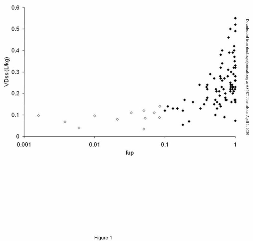
102 of 553 compounds, where fut was less than zero or greater than 1. The 102 outlier compounds all
had VDss of less than 0.6 L.kg-1, and as illustrated in Figure 1, fall into 2 classes with respect to their
plasma protein binding; 12 compounds were highly plasma protein bound with fup < 0.1 and VDss less
than 0.2 L.kg-1, whilst the remaining 90 compounds had fup > 0.1 with a large proportion showing no
binding to plasma proteins (fup = 1).
Based on this finding, simulations were performed across a range of VDss and fup values as shown in
Figure 2. From this analysis it is clear that for high VDss (1 and 3 L.kg-1) there are no model violation
occurrences across the range of fup. However, in the two low VDss situations (0.3 and 0.5 L.kg-1) there
are 2 separate threshold values of fup which lead to aberrant fut values (fup > 0.3 and > 0.85 respectively).
In the case of very low VDss (0.1 L.kg-1), generally accepted as the distributional volume of albumin,
the model fails across the entire range of fup.
In light of the number of model violations (102 out of 553) the sensitivity of the extravascular-to-
intravascular ratio of drug binding protein (RE/I) parameter was explored. Typically this is set at 1.4
based on the distribution of albumin. For the 102 outlier compounds, simulations were performed
varying RE/I from 0.1 (very low extravascular distribution of plasma proteins) to 2.5 (extremely high
extravascular distribution of plasma proteins) with 2 separate classes based on extent of plasma protein
binding (Figure 3). Incremental increases in RE/I from 1.4 to 2.5 had no impact for either group on the
proportion of compounds generating fut values between 0 and 1. However, a change was observed when
RE/I was lowered, with low fup (< 0.1) compounds showing a much higher sensitivity to changes in this
parameter. For example, if RE/I is lowered to unity, a substantial number of highly bound compounds
(40%) generate fut values that fall within the acceptable range whilst there is a negligible change for the
low binding compounds.
This article has not been copyedited and formatted. The final version may differ from this version.DMD Fast Forward. Published on April 7, 2010 as DOI: 10.1124/dmd.110.032458
at ASPE
T Journals on A
pril 1, 2020dm
d.aspetjournals.orgD
ownloaded from
DMD #32458
10
The physicochemical properties of the 90 outlier compounds, where VDss is less than 0.6 L.kg-1 and
fup is greater than 0.1, were investigated and are summarized in Figure 4. Trends were observed with
lipophilicity expressed as clogP, polar surface area (PSA), molecular weight and extent of ionization
(expressed as the difference between clogP and clogD) relative to the whole dataset of 553 compounds.
From these plots it is clear the majority of the aberrant compounds have very low lipophilicity with
clogP typically less than zero; 46% of the compounds with clogP less than zero give rise to an aberrant
fut. High polarity is also an important contributing factor with 50% of the compounds with PSA > 150
Å2 also leading to Øie-Tozer model violations. In addition, there is a trend with the degree of ionization
where 34% of highly ionized compounds (defined as a difference between logP and logD greater than
2.5 log units) produce aberrant fut values. The trend with MW is less clear although there are a greater
proportion of outlier compounds with high MW in excess of 400.
Possible explanations for the behavior of the 90 outlier compounds with low VDss and high fup were
investigated further. Table 1 shows a considerable number of these compounds have been shown to be
substrates for various human active transport proteins.
Furthermore, in order to determine the predictive accuracy of the approach on the 102 violating
compounds, calculations of human VDss were made using measured fup and 3 hypothetical fut values
within the normal range (0.1, 0.5 and 0.9), as might normally be obtained from the rearranged Øie-
Tozer equation with preclinical data. The summary statistics are displayed in Table 2.
This article has not been copyedited and formatted. The final version may differ from this version.DMD Fast Forward. Published on April 7, 2010 as DOI: 10.1124/dmd.110.032458
at ASPE
T Journals on A
pril 1, 2020dm
d.aspetjournals.orgD
ownloaded from

DMD #32458
11
DISCUSSION
The analysis presented has demonstrated a number of outlying scenarios for the Øie-Tozer model
when VDss is low (< 0.6 L.kg-1). Twelve of the compounds giving rise to aberrant fut values have fup <
0.1, likely due to variation at the upper end of the plasma protein binding range, where accurate
experimental determination of very low free fractions can become analytically challenging. In addition,
the model is not applicable for compounds with VDss of 0.1 L.kg-1 or lower: as fup tends to zero, VDss
approximates to 0.105 L.kg-1. There is some sensitivity to the exact value of RE/I used for highly plasma
bound compounds as might be expected; altering the extravascular-to-intravascular distributional ratio
of binding protein has a more significant impact on the fut as the distribution of these compounds is
primarily driven by affinity for plasma proteins such as albumin. In cases of high plasma protein
binding, an RE/I value of 1.4 may not be appropriate and so the model should be applied with caution.
Alternatively, information on the particular plasma proteins involved in drug binding may allow the RE/I
to be tailored to compound-specific predictive application.
The remaining 90 compounds were explored further with respect to physicochemical attributes and
possible pharmacokinetic explanations for the model violations. The Øie-Tozer model has a number of
assumptions which can aid understanding in cases where aberrant fut values are obtained. The
assumptions are: (1) drug distribution is driven by non-specific binding between blood and tissue, (2)
rapid equilibration occurs between blood and tissue, (3) drug distributes uniformly within each organ or
tissue, (4) there is no contribution from active transport processes, and (5) distributional processes are
non-saturating. In this regard, we have shown a substantial number of the 90 violating compounds have
literature evidence supporting their action as substrates of human active transport proteins (Table 1).
The transporters cited are typically of an efflux nature, limiting distribution into those tissues
expressing the transport protein and consequently contributing toward limiting VDss to the volume of
This article has not been copyedited and formatted. The final version may differ from this version.DMD Fast Forward. Published on April 7, 2010 as DOI: 10.1124/dmd.110.032458
at ASPE
T Journals on A
pril 1, 2020dm
d.aspetjournals.orgD
ownloaded from
DMD #32458
12
extracellular fluid (0.6 L.kg-1) or lower. This is not to say that all active transport substrates violate the
Øie-Tozer model e.g. the HIV protease inhibitors and angiotensin receptor antagonists are known to be
actively transported and obey the Øie-Tozer model. In addition, it is not the case that low VDss is the
sole driver for failures of the equation. Of the 216 compounds with VDss < 0.6 L.kg-1, 114 obey the
model whilst 102 compounds do not. Furthermore, it is not possible to substantiate the involvement of
active transport as the exclusive determinant for whether or not model violations will be observed.
There are obviously many other considerations including affinity for the transport protein, the
magnitude of local, unbound concentrations, and dose size. These complicating factors make it difficult
to assess the sensitivity of the model in this respect. For example, some compounds may give rise to
back-calculated fut values that are realistic (i.e. between 0 and 1) but which remain inaccurate due to
one or more model assumptions e.g. many of the statins (OATP1B1 substrates) are low VDss, high fup
compounds which in principle obey the model. Nevertheless, in cases of low VDss and high plasma free
fraction, this analysis implicates the involvement of active transporters limiting tissue partitioning.
It has been observed that tissue-to-plasma partition ratios are elevated in certain tissues in mdr1a
knockout mice relative to wildtype, albeit not for every compound studied and in all tissues (Lee et al.,
2009). An analysis of published reports evidencing changes in distribution volume, in animals and
human, mediated by drug-drug interactions, genetic polymorphism or gene knockout, showed some
similar trends; uptake interactions at the liver tended to cause a decrease in VDss. However, the efflux
interactions at the liver did not trend in the opposing direction which could be a consequence of
assessing interactions in a tissue generally considered part of the central compartment (Grover and
Benet, 2009). This work highlights the potential complexity of drug distribution; a paradigm shift from
simple passive diffusion phenomena to more intricate, active mechanisms including efflux, uptake and
intracellular sequestration. In addition, Dobson et al. (Dobson and Kell, 2008) concluded that active
transport processes are fundamental in determining drug disposition being likely more common than
This article has not been copyedited and formatted. The final version may differ from this version.DMD Fast Forward. Published on April 7, 2010 as DOI: 10.1124/dmd.110.032458
at ASPE
T Journals on A
pril 1, 2020dm
d.aspetjournals.orgD
ownloaded from

DMD #32458
13
usually assumed, citing the mounting evidence in the literature on specific drug examples as well as
observations that drugs can concentrate in specific tissues.
It is clear that a large proportion of the 90 compounds are beta-lactam antibiotics. Interestingly, this
class of compounds was intentionally excluded from the Rule-of-5 analysis since these natural products
(and close derivatives) are highly likely to have ‘evolved’ transport protein interactions (Lipinski et al.,
1997; Ganesan, 2008). In addition, there are examples in the literature of this class of compounds
accumulating in specific tissues by various mechanisms, a further model assumption violation. The
beta-lactam antibiotics have been shown to accumulate in the choroid plexus of rat via an active
carrier-mediated transport process (Nohjoh et al., 1989). The aminoglycosides listed in Table 1,
including amikacin, gentamicin, isepamicin, kanamycin, netilmicin and tobramycin, have been shown
to selectively accumulate in renal cortex leading to renal injury. This selective accumulation is thought
to be mediated by megalin, a large endocytic receptor abundantly expressed in renal proximal tubules
(Tod et al., 2000; Nagai, 2006). Furthermore, a similar mechanism by which these compounds
accumulate in sensory hair cells leading to ototoxicity has also been identified (Hashino et al., 1997).
Following receptor-mediated endocytosis, the compounds are transported by vesicular trafficking into
lysosomes. Accumulation of drug leads to lysosomal disruption and rupture, with subsequent hair cell
degeneration. Compare this active mechanism of lysosomal and mitochondrial trapping to that
generally regarded for basic drugs where a passive, pH partition mechanism is implicated (Okumura et
al., 1989; Daniel and Wojcikowski, 1997). Contrast agents are also well represented including a
number of iodinated compounds such as iohexol as well as the gadolinium-containing compound,
gadoversetamide. These agents are typically limited to the intravascular and extracellular fluid spaces
and are renally cleared by glomerular filtration, in line with their intended clinical imaging applications.
However, cytochemistry studies with the diagnostic indicator dye, fluorescein, have demonstrated
active transport-driven renal accumulation in mitochondria (Masereeuw et al., 1994).
This article has not been copyedited and formatted. The final version may differ from this version.DMD Fast Forward. Published on April 7, 2010 as DOI: 10.1124/dmd.110.032458
at ASPE
T Journals on A
pril 1, 2020dm
d.aspetjournals.orgD
ownloaded from

DMD #32458
14
As well as model assumptions concerning no active transport processes and uniform tissue
distribution there is also a requirement that distributional characteristics are non-saturating. This could
explain cases such as cefazolin where plasma protein binding has been shown to be non-linear in rat
(Tsuji et al., 1983). The free fraction in rat serum at 10, 100 and 200 μg.mL-1 was measured as 11, 20
and 41 % respectively. This marked change in plasma protein binding would likely shift the
distributional behavior in tissues.
The Øie-Tozer model has been effectively applied to the prediction of human VDss for basic and
neutral compounds (Obach et al., 1997; Lombardo et al., 2002; Lombardo et al., 2004). In this
approach the rearranged Øie-Tozer equation is used together with VDss and fup in preclinical species in
order to calculate fut. This value is considered species-independent since tissue binding tends to be
determined by the extent of interaction with phospholipid membranes. This fut together with
experimental human fup measurements can be put into the standard form of the Øie-Tozer model to
generate VDss for human (Obach et al., 1997). In order to determine the predictive accuracy of the
approach on the 102 violating compounds, calculations of VDss were made using measured fup and 3
hypothetical fut values within the normal range (0.1, 0.5 and 0.9), as might normally be obtained from
the rearranged Øie-Tozer equation with preclinical data. From the summary statistics displayed in Table
2, it is clear that when apparently normal fut values from preclinical species are used, large errors in
prediction can be observed; assuming passive diffusion-mediated distribution leads, in the vast majority
of cases, to over-prediction of human VDss. Even when the more likely scenario of low tissue binding is
applied (fut 0.9), the percentage of predictions with less than 2-fold error is lower than 60%, with a
maximum error of 9-fold in this test set. The errors observed could be further exacerbated by the high
proportion of actively transported drugs within the 102 compound set, especially given the known
species differences in transporter expression and activity.
This article has not been copyedited and formatted. The final version may differ from this version.DMD Fast Forward. Published on April 7, 2010 as DOI: 10.1124/dmd.110.032458
at ASPE
T Journals on A
pril 1, 2020dm
d.aspetjournals.orgD
ownloaded from

DMD #32458
15
Multiple linear regression approaches have also been applied to the calculation of fut using 2
experimentally determined physicochemical properties, ElogD and fraction ionized at pH 7.4
(Lombardo et al., 2002; Lombardo et al., 2004). With the exception of the borderline example,
metronidazole, where a slightly different plasma free fraction was reported, there are no violating
compounds present in these original reports. However, in recent work where the ElogD approach was
modified to an HPLC-IAM measurement, a number of acidic compounds were utilized and, despite
some of them giving rise to aberrant fut values (log fut > 0), they were included in the subsequent model
building (Sui et al., 2009). The present investigation highlights the importance of careful application of
the Øie-Tozer model and the need to be aware of the potential for aberrant fut values. In the same
manner, and as shown in this work, the generation of an aberrant fut parameter from preclinical data,
although not an all-encompassing diagnostic, can give some useful insights into potential disposition
properties of novel chemical entities, implicating active transport, selective tissue accumulation or non-
linear, non-uniform distributional behavior. The judicious application of the Øie-Tozer model to
predictions of human VDss for novel compounds is also noteworthy, in cases where the
physicochemical property profile or drug class overlaps with that demonstrated in this analysis. Further
work in the field of drug transporters will help elucidate the nature of drug distributional behavior and
provide further insights to aid drug design.
This article has not been copyedited and formatted. The final version may differ from this version.DMD Fast Forward. Published on April 7, 2010 as DOI: 10.1124/dmd.110.032458
at ASPE
T Journals on A
pril 1, 2020dm
d.aspetjournals.orgD
ownloaded from

DMD #32458
16
REFERENCES
Apiwattanakul N, Sekine T, Chairoungdua A, Kanai Y, Nakajima N, Sophasan S and Endou H
(1999) Transport properties of nonsteroidal anti-inflammatory drugs by organic anion transporter 1
expressed in Xenopus laevis oocytes. Mol. Pharmacol. FIELD Full Journal Title:Molecular
Pharmacology 55:847-854.
Banerjee SK, Jagannath C, Hunter RL and Dasgupta A (2000) Bioavailability of tobramycin
after oral delivery in FVB mice using CRL-1605 copolymer, an inhibitor of P-glycoprotein. Life Sci.
FIELD Full Journal Title:Life Sciences 67:2011-2016.
Bretschneider B, Brandsch M and Neubert R (1999) Intestinal transport of b-lactam antibiotics:
analysis of the affinity at the H+/peptide symporter (PEPT1), the uptake into Caco-2 cell monolayers
and the transepithelial flux. Pharm. Res. FIELD Full Journal Title:Pharmaceutical Research 16:55-61.
Ci L, Kusuhara H, Adachi M, Schuetz JD, Takeuchi K and Sugiyama Y (2007) Involvement of
MRP4 (ABCC4) in the luminal efflux of ceftizoxime and cefazolin in the kidney. Mol. Pharmacol.
FIELD Full Journal Title:Molecular Pharmacology 71:1591-1597.
Climax J, Lenehan TJ, Lambe R, Kenny M, Caffrey E and Darragh A (1986) Interaction of
antimicrobial agents with human peripheral blood leukocytes: uptake and intracellular localization of
certain sulfonamides and trimethoprims. J. Antimicrob. Chemother. FIELD Full Journal Title:Journal
of Antimicrobial Chemotherapy 17:489-498.
Daniel WA and Wojcikowski J (1997) Contribution of lysosomal trapping to the total tissue
uptake of psychotropic drugs. Pharmacol. Toxicol. (Copenhagen) FIELD Full Journal
Title:Pharmacology & Toxicology (Copenhagen) 80:62-68.
This article has not been copyedited and formatted. The final version may differ from this version.DMD Fast Forward. Published on April 7, 2010 as DOI: 10.1124/dmd.110.032458
at ASPE
T Journals on A
pril 1, 2020dm
d.aspetjournals.orgD
ownloaded from

DMD #32458
17
Dobson PD and Kell DB (2008) Carrier-mediated cellular uptake of pharmaceutical drugs: an
exception or the rule? Nat. Rev. Drug Discovery FIELD Full Journal Title:Nature Reviews Drug
Discovery 7:205-220.
Dogruel M, Gibbs JE and Thomas SA (2003) Hydroxyurea transport across the blood-brain and
blood-cerebrospinal fluid barriers of the guinea-pig. J. Neurochem. FIELD Full Journal Title:Journal
of Neurochemistry 87:76-84.
Ganapathy ME, Huang W, Rajan DP, Carter AL, Sugawara M, Iseki K, Leibach FH and
Ganapathy V (2000) b-Lactam antibiotics as substrates for OCTN2, an organic cation/carnitine
transporter. J. Biol. Chem. FIELD Full Journal Title:Journal of Biological Chemistry 275:1699-1707.
Ganapathy ME, Prasad PD, Mackenzie B, Ganapathy V and Leibach FH (1997) Interaction of
anionic cephalosporins with the intestinal and renal peptide transporters PEPT 1 and PEPT 2. Biochim.
Biophys. Acta, Biomembr. FIELD Full Journal Title:Biochimica et Biophysica Acta, Biomembranes
1324:296-308.
Ganesan A (2008) The impact of natural products upon modern drug discovery. Curr. Opin.
Chem. Biol. FIELD Full Journal Title:Current Opinion in Chemical Biology 12:306-317.
Gibaldi M, Nagashima R and Levy G (1969) Relation between drug concentration in plasma or
serum and amount of drug in the body. J. Pharm. Sci. FIELD Full Journal Title:Journal of
Pharmaceutical Sciences 58:193-197.
Gillette JR (1976) Overview of factors affecting drug interactions. Ann. N. Y. Acad. Sci. FIELD
Full Journal Title:Annals of the New York Academy of Sciences 281:136-150.
Grover A and Benet LZ (2009) Effects of drug transporters on volume of distribution. AAPS J.
FIELD Full Journal Title:AAPS Journal 11:250-261.
Hashino E, Shero M and Salvi RJ (1997) Lysosomal targeting and accumulation of
aminoglycoside antibiotics in sensory hair cells. Brain Res. FIELD Full Journal Title:Brain Research
777:75-85.
This article has not been copyedited and formatted. The final version may differ from this version.DMD Fast Forward. Published on April 7, 2010 as DOI: 10.1124/dmd.110.032458
at ASPE
T Journals on A
pril 1, 2020dm
d.aspetjournals.orgD
ownloaded from

DMD #32458
18
Hawkins BT, Ocheltree SM, Norwood KM and Egleton RD (2007) Decreased blood-brain
barrier permeability to fluorescein in streptozotocin-treated rats. Neurosci. Lett. FIELD Full Journal
Title:Neuroscience Letters 411:1-5.
Hill G, Cihlar T, Oo C, Ho ES, Prior K, Wiltshire H, Barrett J, Liu B and Ward P (2002) The
anti-influenza drug oseltamivir exhibits low potential to induce pharmacokinetic drug interactions via
renal secretion-correlation of in vivo and in vitro studies. Drug Metab. Dispos. FIELD Full Journal
Title:Drug Metabolism and Disposition 30:13-19.
Imaoka T, Kusuhara H, Adachi M, Schuetz JD, Takeuchi K and Sugiyama Y (2007) Functional
involvement of multidrug resistance-associated protein 4 (MRP4/ABCC4) in the renal elimination of
the antiviral drugs adefovir and tenofovir. Mol. Pharmacol. FIELD Full Journal Title:Molecular
Pharmacology 71:619-627.
Kido Y, Tamai I, Okamoto M, Suzuki F and Tsuji A (2000) Functional clarification of MCT1-
mediated transport of monocarboxylic acids at the blood-brain barrier using in vitro cultured cells and
in vivo BUI studies. Pharm. Res. FIELD Full Journal Title:Pharmaceutical Research 17:55-62.
Larrayoz IM, Casado FJ, Pastor-Anglada M and Lostao MP (2004) Electrophysiological
Characterization of the Human Na+/Nucleoside Cotransporter 1 (hCNT1) and Role of Adenosine on
hCNT1 Function. J. Biol. Chem. FIELD Full Journal Title:Journal of Biological Chemistry 279:8999-
9007.
Lee EJD, Lean CB and Limenta LMG (2009) Role of membrane transporters in the safety
profile of drugs. Expert Opin. Drug Metab. Toxicol. FIELD Full Journal Title:Expert Opinion on Drug
Metabolism & Toxicology 5:1369-1383.
Li M, Anderson GD, Phillips BR, Kong W, Shen DD and Wang J (2006) Interactions of
amoxicillin and cefaclor with human renal organic anion and peptide transporters. Drug Metab. Dispos.
FIELD Full Journal Title:Drug Metabolism and Disposition 34:547-555.
This article has not been copyedited and formatted. The final version may differ from this version.DMD Fast Forward. Published on April 7, 2010 as DOI: 10.1124/dmd.110.032458
at ASPE
T Journals on A
pril 1, 2020dm
d.aspetjournals.orgD
ownloaded from
DMD #32458
19
Lipinski CA, Lombardo F, Dominy BW and Feeney PJ (1997) Experimental and computational
approaches to estimate solubility and permeability in drug discovery and development settings. Adv.
Drug Delivery Rev. FIELD Full Journal Title:Advanced Drug Delivery Reviews 23:3-25.
Liu L, Cui Y, Chung AY, Shitara Y, Sugiyama Y, Keppler D and Pang KS (2006) Vectorial
transport of enalapril by Oatp1a1/Mrp2 and OATP1B1 and OATP1B3/MRP2 in rat and human livers.
J. Pharmacol. Exp. Ther. FIELD Full Journal Title:Journal of Pharmacology and Experimental
Therapeutics 318:395-402.
Lombardo F, Obach RS, Shalaeva MY and Gao F (2002) Prediction of Volume of Distribution
Values in Humans for Neutral and Basic Drugs Using Physicochemical Measurements and Plasma
Protein Binding Data. J. Med. Chem. FIELD Full Journal Title:Journal of Medicinal Chemistry
45:2867-2876.
Lombardo F, Obach RS, Shalaeva MY and Gao F (2004) Prediction of human volume of
distribution values for neutral and basic drugs. 2. Extended data set and leave-class-out statistics. J.
Med. Chem. FIELD Full Journal Title:Journal of Medicinal Chemistry 47:1242-1250.
Masereeuw R, van den Bergh EJ, Bindels RJM and Russel FgM (1994) Characterization of
fluorescein transport in isolated proximal tubular cells of the rat: evidence for mitochondrial
accumulation. J. Pharmacol. Exp. Ther. FIELD Full Journal Title:Journal of Pharmacology and
Experimental Therapeutics 269:1261-1267.
Morimoto K, Nakakariya M, Shirasaka Y, Kakinuma C, Fujita T, Tamai I and Ogihara T (2008)
Oseltamivir (Tamiflu) efflux transport at the blood-brain barrier via P-glycoprotein. Drug Metab.
Dispos. FIELD Full Journal Title:Drug Metabolism and Disposition 36:6-9.
Murakami Y, Kohyama N, Kobayashi Y, Ohbayashi M, Ohtani H, Sawada Y and Yamamoto T
(2005) Functional characterization of human monocarboxylate transporter 6 (SLC16A5). Drug Metab.
Dispos. FIELD Full Journal Title:Drug Metabolism and Disposition 33:1845-1851.
This article has not been copyedited and formatted. The final version may differ from this version.DMD Fast Forward. Published on April 7, 2010 as DOI: 10.1124/dmd.110.032458
at ASPE
T Journals on A
pril 1, 2020dm
d.aspetjournals.orgD
ownloaded from

DMD #32458
20
Nagai J (2006) Molecular mechanisms underlying renal accumulation of aminoglycoside
antibiotics and mechanism-based approach for developing nonnephrotoxic aminoglycoside therapy.
Yakugaku Zasshi FIELD Full Journal Title:Yakugaku Zasshi 126:327-335.
Nohjoh T, Suzuki H, Sawada Y, Sugiyama Y, Iga T and Hanano M (1989) Transport of
cefodizime, a novel third generation cephalosporin antibiotic, in isolated rat choroid plexus. J.
Pharmacol. Exp. Ther. FIELD Full Journal Title:Journal of Pharmacology and Experimental
Therapeutics 250:324-328.
Obach RS, Baxter JG, Liston TE, Silber BM, Jones BC, Macintyre F, Rance DJ and Wastall P
(1997) The prediction of human pharmacokinetic parameters from preclinical and in vitro metabolism
data. J. Pharmacol. Exp. Ther. FIELD Full Journal Title:Journal of Pharmacology and Experimental
Therapeutics 283:46-58.
Obach RS, Lombardo F and Waters NJ (2008) Trend analysis of a database of intravenous
pharmacokinetic parameters in humans for 670 drug compounds. Drug Metab. Dispos. FIELD Full
Journal Title:Drug Metabolism and Disposition 36:1385-1405.
Ogihara T, Kano T, Wagatsuma T, Wada S, Yabuuchi H, Enomoto S, Morimoto K, Shirasaka
Y, Kobayashi S and Tamai I (2009) Oseltamivir (Tamiflu) is a substrate of peptide transporter 1. Drug
Metab. Dispos. FIELD Full Journal Title:Drug Metabolism and Disposition 37:1676-1681.
Oie S and Tozer TN (1979) Effect of altered plasma protein binding on apparent volume of
distribution. J Pharm Sci FIELD Full Journal Title:Journal of pharmaceutical sciences 68:1203-1205.
Okumura K, Yoshida H, Kamiya A and Hori R (1989) Submitochondrial distribution of basic
drugs in the isolated perfused lung. Chem. Pharm. Bull. FIELD Full Journal Title:Chemical &
Pharmaceutical Bulletin 37:1109-1111.
Rizwan AN and Burckhardt G (2007) Organic Anion Transporters of the SLC22 Family:
Biopharmaceutical, Physiological, and Pathological Roles. Pharm. Res. FIELD Full Journal
Title:Pharmaceutical Research 24:450-470.
This article has not been copyedited and formatted. The final version may differ from this version.DMD Fast Forward. Published on April 7, 2010 as DOI: 10.1124/dmd.110.032458
at ASPE
T Journals on A
pril 1, 2020dm
d.aspetjournals.orgD
ownloaded from
DMD #32458
21
Safaei R (2006) Role of copper transporters in the uptake and efflux of platinum containing
drugs. Cancer Lett. (Amsterdam, Neth.) FIELD Full Journal Title:Cancer Letters (Amsterdam,
Netherlands) 234:34-39.
Shibayama T, Sugiyama D, Kamiyama E, Tokui T, Hirota T and Ikeda T (2007)
Characterization of CS-023 (RO4908463), a novel parenteral carbapenem antibiotic, and meropenem
as substrates of human renal transporters. Drug Metab. Pharmacokinet. FIELD Full Journal
Title:Drug Metabolism and Pharmacokinetics 22:41-47.
Shugarts S and Benet LZ (2009) The Role of Transporters in the Pharmacokinetics of Orally
Administered Drugs. Pharm. Res. FIELD Full Journal Title:Pharmaceutical Research 26:2039-2054.
Smith DA (1997) Physicochemical properties in drug metabolism and pharmacokinetics.
Comput.-Assisted Lead Find. Optim., [Eur. Symp. Quant. Struct.-Act. Relat.], 11th FIELD Full
Journal Title:Computer-Assisted Lead Finding and Optimization: Current Tools for Medicinal
Chemistry, [European Symposium on Quantitative Structure-Activity Relationships], 11th, Lausanne,
Sept. 1-6, 1996:267-276.
Sui X, Sun J, Li H, Wang Y, Liu J, Liu X, Zhang W, Chen L and He Z (2009) Prediction of
volume of distribution values in human using immobilized artificial membrane partitioning
coefficients, the fraction of compound ionized and plasma protein binding data. Eur. J. Med. Chem.
FIELD Full Journal Title:European Journal of Medicinal Chemistry 44:4455-4460.
Takeda M, Babu E, Narikawa S and Endou H (2002) Interaction of human organic anion
transporters with various cephalosporin antibiotics. Eur. J. Pharmacol. FIELD Full Journal
Title:European Journal of Pharmacology 438:137-142.
Takeda M, Narikawa S, Hosoyamada M, Cha SH, Sekine T and Endou H (2001)
Characterization of organic anion transport inhibitors using cells stably expressing human organic
anion transporters. Eur. J. Pharmacol. FIELD Full Journal Title:European Journal of Pharmacology
419:113-120.
This article has not been copyedited and formatted. The final version may differ from this version.DMD Fast Forward. Published on April 7, 2010 as DOI: 10.1124/dmd.110.032458
at ASPE
T Journals on A
pril 1, 2020dm
d.aspetjournals.orgD
ownloaded from

DMD #32458
22
Tamai I, Sai Y, Ono A, Kido Y, Yabuuchi H, Takanaga H, Satoh E, Ogihara T, Amano O, Izeki
S and Tsuji A (1999) Immunohistochemical and functional characterization of pH-dependent intestinal
absorption of weak organic acids by the monocarboxylic acid transporter MCT1. J. Pharm. Pharmacol.
FIELD Full Journal Title:Journal of Pharmacy and Pharmacology 51:1113-1121.
Tanihara Y, Masuda S, Sato T, Katsura T, Ogawa O and Inui K-i (2007) Substrate specificity of
MATE1 and MATE2-K, human multidrug and toxin extrusions/H+-organic cation antiporters.
Biochem. Pharmacol. FIELD Full Journal Title:Biochemical Pharmacology 74:359-371.
Tod M, Padoin C and Petitjean O (2000) Clinical pharmacokinetics and pharmacodynamics of
isepamicin. Clin. Pharmacokinet. FIELD Full Journal Title:Clinical Pharmacokinetics 38:205-223.
Tsuji A, Yoshikawa T, Nishide K, Minami H, Kimura M, Nakashima E, Terasaki T, Miyamoto
E, Nightingale CH and Yamana T (1983) Physiologically based pharmacokinetic model for b-lactam
antibiotics. I: Tissue distribution and elimination in rats. J. Pharm. Sci. FIELD Full Journal
Title:Journal of Pharmaceutical Sciences 72:1239-1252.
Tsukaguchi H, Shayakul C, Berger UV, Mackenzie B, Devidas S, Guggino WB, Van Hoek AN
and Hediger MA (1998) Molecular characterization of a broad selectivity neutral solute channel. J.
Biol. Chem. FIELD Full Journal Title:Journal of Biological Chemistry 273:24737-24743.
Uchida Y, Kamiie J, Ohtsuki S and Terasaki T (2007) Multichannel Liquid Chromatography-
Tandem Mass Spectrometry Cocktail Method for Comprehensive Substrate Characterization of
Multidrug Resistance-Associated Protein 4 Transporter. Pharm. Res. FIELD Full Journal
Title:Pharmaceutical Research 24:2281-2296.
Uchino H, Tamai I, Yamashita K, Minemoto Y, Sai Y, Yabuuchi H, Miyamoto K-i, Takeda E
and Tsuji A (2000) p-Aminohippuric Acid Transport at Renal Apical Membrane Mediated by Human
Inorganic Phosphate Transporter NPT1. Biochem. Biophys. Res. Commun. FIELD Full Journal
Title:Biochemical and Biophysical Research Communications 270:254-259.
This article has not been copyedited and formatted. The final version may differ from this version.DMD Fast Forward. Published on April 7, 2010 as DOI: 10.1124/dmd.110.032458
at ASPE
T Journals on A
pril 1, 2020dm
d.aspetjournals.orgD
ownloaded from

DMD #32458
23
Uwai Y, Ida H, Tsuji Y, Katsura T and Inui K-i (2007) Renal Transport of Adefovir, Cidofovir,
and Tenofovir by SLC22A Family Members (hOAT1, hOAT3, and hOCT2). Pharm. Res. FIELD Full
Journal Title:Pharmaceutical Research 24:811-815.
Van de Waterbeemd H, Smith DA and Jones BC (2001) Lipophilicity in PK design: methyl,
ethyl, futile. J. Comput.-Aided Mol. Des. FIELD Full Journal Title:Journal of Computer-Aided
Molecular Design 15:273-286.
Varma MVS, Feng B, Obach RS, Troutman MD, Chupka J, Miller HR and El-Kattan A (2009)
Physicochemical Determinants of Human Renal Clearance. J. Med. Chem. FIELD Full Journal
Title:Journal of Medicinal Chemistry 52:4844-4852.
Xia CQ, Yang JJ and Balani SK (2008) Drug transporters in drug disposition, drug interactions,
and drug resistance. Drug Metab. Drug Des. Dev. FIELD Full Journal Title:Drug Metabolism in Drug
Design and Development:137-203.
Yabuuchi H, Tamai I, Morita K, Kouda T, Miyamoto K-I, Takeda E and Tsuji A (1998) Hepatic
sinusoidal membrane transport of anionic drugs mediated by anion transporter Npt1. J. Pharmacol.
Exp. Ther. FIELD Full Journal Title:Journal of Pharmacology and Experimental Therapeutics
286:1391-1396.
Yuan H, Feng B, Yu Y, Chupka J, Zheng JY, Heath TG and Bond BR (2009) Renal organic
anion transporter-mediated drug-drug interaction between gemcabene and quinapril. J. Pharmacol.
Exp. Ther. FIELD Full Journal Title:Journal of Pharmacology and Experimental Therapeutics
330:191-197.
This article has not been copyedited and formatted. The final version may differ from this version.DMD Fast Forward. Published on April 7, 2010 as DOI: 10.1124/dmd.110.032458
at ASPE
T Journals on A
pril 1, 2020dm
d.aspetjournals.orgD
ownloaded from

DMD #32458
24
FIGURE LEGENDS
Figure 1. Plot of volume of distribution at steady state against log plasma free fraction for the 102
compounds which gave rise to an aberrant tissue free fraction using the rearranged Øie-Tozer equation.
Plasma free fraction less than 0.1 (open circles) and greater than 0.1 (closed circles).
Figure 2. Simulations of the Øie-Tozer model for hypothetical compounds with VDss of 0.1, 0.3, 0.5,
1 and 3 L.kg-1 with varying fup, illustrating model violations and the scenarios where fut becomes
aberrant (grey shading).
Figure 3. Sensitivity analysis of the plasma protein extravascular-to-intravascular ratio (RE/I) for
compounds with high (fup > 0.1, closed symbols) and low (fup < 0.1, open symbols) plasma free fraction.
All compound dependent variables and physiological terms remained constant whilst RE/I was varied
from 0.1 to 2.5.
Figure 4. Trends in physicochemical properties observed between compounds with fut within normal
range (0< fut <1; black) and compounds with aberrant fut (closed circle data-points in Figure 1. i.e. 0 >
fut or fut >1; hashed). A, clogP; B, polar surface area (in Å2); C, molecular weight; D, extent of
ionization described by the difference between clogP and clogD at pH 7. Numbers above each bar
represent % of compounds with aberrant fut within each range.
This article has not been copyedited and formatted. The final version may differ from this version.DMD Fast Forward. Published on April 7, 2010 as DOI: 10.1124/dmd.110.032458
at ASPE
T Journals on A
pril 1, 2020dm
d.aspetjournals.orgD
ownloaded from

DMD #32458
25
Table 1. Compounds which do not obey the Øie-Tozer model together with designated therapeutic
activity and literature evidence for interaction with active transporters.
Drug Therapeutic Class Transporter/s or
active mechanism
implicated 1
Reference
Acetylsalicylic acid NSAID OAT1 (Apiwattanakul et al.,
1999)
Adefovir anti-viral MRP4, OAT1, OAT3,
Secretory renal CL
(Imaoka et al., 2007;
Uwai et al., 2007;
Varma et al., 2009)
Amikacin anti-bacterial Megalin (Tod et al., 2000;
Nagai, 2006)
Aminohexanoic acid, 6- hematologic agent
Amoxicillin anti-bacterial PEPT1, PEPT2, OAT1,
secretory renal CL
(Hill et al., 2002; Li et
al., 2006; Varma et al.,
2009)
Ampicillin anti-bacterial MRP4, NPT1 (mouse),
PEPT1
(Yabuuchi et al., 1998;
Uchida et al., 2007;
Dobson and Kell, 2008)
Atracurium neuromuscular blocker
Azlocillin anti-bacterial Secretory renal CL (Varma et al., 2009)
Aztreonam anti-bacterial Secretory renal CL (Varma et al., 2009)
This article has not been copyedited and formatted. The final version may differ from this version.DMD Fast Forward. Published on April 7, 2010 as DOI: 10.1124/dmd.110.032458
at ASPE
T Journals on A
pril 1, 2020dm
d.aspetjournals.orgD
ownloaded from

DMD #32458
26
Biapenem anti-infective
Busulphan immunosuppressant/anti
-neoplastic
Carbenicillin anti-bacterial Secretory renal CL (Varma et al., 2009)
Carboplatin anti-neoplastic ATP7A, ATP7B (Safaei, 2006)
Carumonam anti-bacterial
Cefadroxil anti-bacterial PEPT1, OAT1, OAT3,
OAT4, secretory renal
CL
(Bretschneider et al.,
1999; Takeda et al.,
2002; Varma et al.,
2009)
Cefamandole anti-bacterial OATs, secretory renal
CL
(Rizwan and
Burckhardt, 2007;
Varma et al., 2009)
Cefatrizine anti-bacterial Secretory renal CL (Varma et al., 2009)
Cefazolin anti-bacterial OAT1, OAT3, OAT4,
MRP4, secretory renal
CL
(Takeda et al., 2002; Ci
et al., 2007; Varma et
al., 2009)
Cefepime anti-bacterial OCTN2 (Ganapathy et al.,
2000)
Cefetamet anti-bacterial
Cefixime anti-bacterial PEPT1, PEPT2, NPT1 (Ganapathy et al.,
This article has not been copyedited and formatted. The final version may differ from this version.DMD Fast Forward. Published on April 7, 2010 as DOI: 10.1124/dmd.110.032458
at ASPE
T Journals on A
pril 1, 2020dm
d.aspetjournals.orgD
ownloaded from

DMD #32458
27
(mouse) 1997; Yabuuchi et al.,
1998)
Cefmetazole anti-bacterial MRP4 (Uchida et al., 2007)
Cefodizime anti-bacterial Unidentified
transporter in isolated
rat choroid plexus
(Nohjoh et al., 1989)
Ceforanide anti-bacterial
Cefotaxime anti-bacterial OAT1, OAT3, OAT4,
secretory renal CL
(Takeda et al., 2002;
Varma et al., 2009)
Cefotetan anti-bacterial
Cefoxitin anti-bacterial
Cefpirome anti-bacterial
Cefprozil anti-bacterial
Ceftazidime anti-bacterial MRP4 (Uchida et al., 2007)
Ceftizoxime anti-bacterial MRP4, NPT1 (mouse) (Yabuuchi et al., 1998;
Ci et al., 2007)
Ceftobiprole anti-bacterial
Cefuroxime anti-bacterial Secretory renal CL (Varma et al., 2009)
Cephalexin anti-bacterial MATE1, secretory
renal CL
(Varma et al., 2009)
Tanahara et al., 2007;
This article has not been copyedited and formatted. The final version may differ from this version.DMD Fast Forward. Published on April 7, 2010 as DOI: 10.1124/dmd.110.032458
at ASPE
T Journals on A
pril 1, 2020dm
d.aspetjournals.orgD
ownloaded from

DMD #32458
28
Cephaloridine anti-bacterial OAT1, OAT3, OAT4,
OCTN2, NPT1 (mouse)
(Yabuuchi et al., 1998;
Ganapathy et al., 2000;
Takeda et al., 2002)
Cephalothin anti-bacterial OAT1, OAT3, OAT4,
NPT1 (mouse)
(Yabuuchi et al., 1998;
Takeda et al., 2002)
Cephapirin anti-bacterial
Cephradine anti-bacterial MATE1, OAT1, NPT1
(mouse)
(Yabuuchi et al., 1998;
Rizwan and
Burckhardt, 2007;
Tanihara et al., 2007)
Cidofovir anti-viral MRP4, OAT1, OAT3,
secretory renal CL
(Imaoka et al., 2007;
Uwai et al., 2007;
Varma et al., 2009)
Cilastatin protease inhibitor OAT3 (Takeda et al., 2001)
Cisatracurium neuromuscular blocker
Clavulanic Acid anti-bacterial
Dalfopristin anti-bacterial
Dexrazoxane immunosuppressant/anti
-neoplastic
Diatrizoic acid
(Amidotrizoate)
contrast agent
This article has not been copyedited and formatted. The final version may differ from this version.DMD Fast Forward. Published on April 7, 2010 as DOI: 10.1124/dmd.110.032458
at ASPE
T Journals on A
pril 1, 2020dm
d.aspetjournals.orgD
ownloaded from

DMD #32458
29
Doxacurium neuromuscular blocker
Doxifluridine immunosuppressant/anti
-neoplastic
CNT1, secretory renal
CL
(Larrayoz et al., 2004;
Varma et al., 2009)
Enalaprilat anti-hypertensive MRP2, secretory renal
CL
(Liu et al., 2006;
Varma et al., 2009)
Eptifibatide hematologic agent
Ertapenem anti-bacterial
Fluorescein contrast agent OAT3, MRP2, active
transport-driven
accumulation in
mitochondria
(Masereeuw et al.,
1994; Hawkins et al.,
2007)
Fluorouracil, 5- immunosuppressant/anti
-neoplastic
AQP9, OAT2 (Tsukaguchi et al.,
1998; Rizwan and
Burckhardt, 2007)
Foscarnet anti-viral MCT1 (rat), NPT1
(mouse)
(Yabuuchi et al., 1998;
Tamai et al., 1999;
Kido et al., 2000)
Fosfomycin anti-bacterial
Gadoversetamide contrast agent
Gentamicin anti-bacterial Megalin (Tod et al., 2000;
Nagai, 2006)
This article has not been copyedited and formatted. The final version may differ from this version.DMD Fast Forward. Published on April 7, 2010 as DOI: 10.1124/dmd.110.032458
at ASPE
T Journals on A
pril 1, 2020dm
d.aspetjournals.orgD
ownloaded from

DMD #32458
30
Hydroxyurea anti-neoplastic OATP2, MDR1 (Dogruel et al., 2003)
Imipenem anti-bacterial
Iohexol contrast agent
Iopamidol contrast agent
Iopromide contrast agent
Iothalamic acid
(Iothalamate)
contrast agent
Isepamicin anti-bacterial Megalin
(Tod et al., 2000;
Nagai, 2006)
Kanamycin anti-bacterial Megalin (Tod et al., 2000;
Nagai, 2006)
Lamifiban hematologic agent
Melagatran hematologic agent
Meropenem anti-bacterial OAT1, OAT3,
secretory renal CL
(Shibayama et al.,
2007; Varma et al.,
2009)
Metocurine neuromuscular blocker
Metrizoate contrast agent Secretory renal CL (Varma et al., 2009)
Metronidazole anti-protozoal/anti-
bacterial
This article has not been copyedited and formatted. The final version may differ from this version.DMD Fast Forward. Published on April 7, 2010 as DOI: 10.1124/dmd.110.032458
at ASPE
T Journals on A
pril 1, 2020dm
d.aspetjournals.orgD
ownloaded from

DMD #32458
31
Mezlocillin anti-bacterial Secretory renal CL (Varma et al., 2009)
Miglitol hypoglycemic agent
Mivacurium (cis/cis) neuromuscular blocker
Moxalactam anti-bacterial
Netilmicin anti-bacterial Megalin (Tod et al., 2000;
Nagai, 2006)
Oseltamivir acid anti-viral Secretory renal CL,
MDR1, PEPT1
(Morimoto et al., 2008;
Ogihara et al., 2009;
Varma et al., 2009)
Penicillin G anti-bacterial MRP4, OATPs,
PEPT1, PEPT2, NPT1,
OAT1, OAT3,
secretory renal CL
(Uchino et al., 2000;
Varma et al., 2009)
Piperacillin anti-bacterial MRP4, secretory renal
CL
(Uchida et al., 2007;
Varma et al., 2009)
Pipecuronium neuromuscular blocker
Probenecid uricosuric agent/anti-
bacterial adjunct
OAT1, MCT6 (Murakami et al., 2005;
Rizwan and
Burckhardt, 2007)
Quinaprilat anti-hypertensive OAT3, secretory renal
CL
(Varma et al., 2009;
Yuan et al., 2009)
Rocuronium neuromuscular blocker OATP1A2 (Dobson and Kell,
This article has not been copyedited and formatted. The final version may differ from this version.DMD Fast Forward. Published on April 7, 2010 as DOI: 10.1124/dmd.110.032458
at ASPE
T Journals on A
pril 1, 2020dm
d.aspetjournals.orgD
ownloaded from

DMD #32458
32
2008)
Streptomycin anti-bacterial
Sulbactam anti-bacterial MRP4, secretory renal
CL
(Uchida et al., 2007;
Varma et al., 2009)
Sulbenicillin anti-bacterial Secretory renal CL (Varma et al., 2009)
Sulfadiazine anti-protozoal/anti-
bacterial
Active uptake in
leukocytes
(Climax et al., 1986)
Ticarcillin anti-bacterial Secretory renal CL (Varma et al., 2009)
Tobramycin anti-bacterial MDR1, megalin (Banerjee et al., 2000;
Tod et al., 2000; Nagai,
2006)
Tomopenem anti-bacterial
Zanamivir anti-viral
1 Compounds classified as exhibiting net secretory renal CL based on threshold defined by Varma et
al. (2009) where CLr > 1.2•fup•GFR.
This article has not been copyedited and formatted. The final version may differ from this version.DMD Fast Forward. Published on April 7, 2010 as DOI: 10.1124/dmd.110.032458
at ASPE
T Journals on A
pril 1, 2020dm
d.aspetjournals.orgD
ownloaded from

DMD #32458
33
Table 2. Summary statistics on the performance of the Øie-Tozer model on the 102 outlier
compounds applying various hypothetical estimates of fut (0.1, 0.5 and 0.9) as may usually be obtained
from preclinical species.
Statistic fut 0.1 fut 0.5 fut 0.9
GMFE 9.7 2.7 1.9
% within 2-fold 3 46 57
Maximum fold error 55 13 9
% > 2-fold over-predicted 97 50 43
% > 2-fold under-predicted 0 4 0
This article has not been copyedited and formatted. The final version may differ from this version.DMD Fast Forward. Published on April 7, 2010 as DOI: 10.1124/dmd.110.032458
at ASPE
T Journals on A
pril 1, 2020dm
d.aspetjournals.orgD
ownloaded from
This article has not been copyedited and formatted. The final version may differ from this version.DMD Fast Forward. Published on April 7, 2010 as DOI: 10.1124/dmd.110.032458
at ASPE
T Journals on A
pril 1, 2020dm
d.aspetjournals.orgD
ownloaded from

This article has not been copyedited and formatted. The final version may differ from this version.DMD Fast Forward. Published on April 7, 2010 as DOI: 10.1124/dmd.110.032458
at ASPE
T Journals on A
pril 1, 2020dm
d.aspetjournals.orgD
ownloaded from

This article has not been copyedited and formatted. The final version may differ from this version.DMD Fast Forward. Published on April 7, 2010 as DOI: 10.1124/dmd.110.032458
at ASPE
T Journals on A
pril 1, 2020dm
d.aspetjournals.orgD
ownloaded from

This article has not been copyedited and formatted. The final version may differ from this version.DMD Fast Forward. Published on April 7, 2010 as DOI: 10.1124/dmd.110.032458
at ASPE
T Journals on A
pril 1, 2020dm
d.aspetjournals.orgD
ownloaded from