tudent teffen au tudent number university of cape town … · university of cape town ... the...
TRANSCRIPT

Univers
ity of
Cap
e Tow
n
Reasonability of gastro-oesophageal reflux study
requests (contrast swallows and milk scans) for the
detection of gastro-oesophageal reflux disease at
Red Cross War Memorial Children’s Hospital
– a retrospective analysis
by
STUDENT: STEFFEN BAU
STUDENT NUMBER: BXXSTE001
SUBMITTED TO THE UNIVERSITY OF CAPE TOWN
In fulfilment of the requirements of the degree
Master of Medicine (MMed) in Paediatrics
FCPaeds – Part 3
Faculty of Health Sciences
UNIVERSITY OF CAPE TOWN
Date of first submission: Monday, 14 April 2014
Date of final revision: Saturday, 12 September 2015
Supervisor: E Goddard, Paediatric Gastroenterology, University Cape
Town
Co-supervisors: A Brink, Nuclear Medicine, University Cape Town
E Banderker, Paediatric Radiology, University Cape Town

The copyright of this thesis vests in the author. No quotation from it or information derived from it is to be published without full acknowledgement of the source. The thesis is to be used for private study or non-commercial research purposes only.
Published by the University of Cape Town (UCT) in terms of the non-exclusive license granted to UCT by the author.
Univers
ity of
Cap
e Tow
n

1
Declaration
I, Steffen Bau, hereby declare that the work on which this thesis is based is my original work (except where acknowledgements indicate otherwise) and that neither the whole work nor any part of it has been, is being, or is to be submitted for another degree in this or any other university.
I empower the university to reproduce, for the purpose of research, either the whole or any portion of the contents in any manner whatsoever.
Signature: …………………………………
Date: …………12 September 2015..………….

2
Table of contents
CONTENTS PAGE
Title page
Declaration of author ……..………………………………………….. 1
Table of contents .…………………………………………………….. 2 - 4
Glossary ………………………………………………………………... 5
Abstract …………………………………………………………………. 6 - 7
Protocol …………………………………………………………………. 8 - 15
Research question ……………………………………………... 8
Background …………………………….……………………….. 8 - 10
Aims ……………………………………………………………… 10
Method …………………………………………………………... 10 - 12
Statistical analysis ……………………………………………… 12 - 13
Results …………………………………………………………… 13
Ethical considerations …………………………………………. 13 - 14
Conflicts of interest ……………………………………………... 14
References ………………………………………………………. 14 - 15
Structured literature review…………………………………………… 16 - 31
Objectives of literature review ………………………………… 16
Literature search strategy ……………………………………... 16 - 18
Inclusion and exclusion criteria ……………………………….. 18
Summary and interpretation …………………………………... 18 - 27
Literature …………...............................……………..... 18 - 23
Investigation techniques ………………………………. 23 - 25
Timing of investigation ………….……………………… 26
Treatment trial …………………………………………… 26 - 27
References ............................................................................ 27 - 31

3
CONTENTS PAGE
Manuscript ……………………………………………………………… 32 - 56
Background ……………………………………………………... 32 - 35
Aim ……………………………………………………………….. 35
Method …………………………………………………………... 35 - 43
Inclusion and exclusion criteria ……………………….. 36 - 37
Methodology of GORD investigations …..……………. 37 - 38
Reasonability of the study request .…….…………….. 39 - 40
Additional definition of criteria...................................... 40 - 41
Investigation timing …………………………………….. 41
Correct modality of study used …………….………….. 42
Analysis ………………………………………………….. 42 - 43
Results …………………………………………………………… 43 - 49
Reasonability of study request 46
Appropriateness of request timing 47
Choice of correct investigation modality 47
Preceding treatment trial 47 - 48
Comparison between modality used and findings 48 - 49
Comparison between findings and indication 49
Discussion ………………………………………………….……. 49- 55
Limitations of this study ............................................... 53 - 54
Identification of gaps and need for further research ... 54 - 55
Conclusion ………………………………………………………. 55
Conflict of interest ………………………………………………. 55
Acknowledgements …………………………………………….. 55 - 56
References ………………………………………………………. 56 - 58

4
CONTENTS PAGE
Tables and Graphs…………………………………………………….. 59 - 70
Table 1 …………………………………………………………... 59
Table 2 …………………………………………………………... 60
Table 3 …………………………………………………………... 61 - 62
Table 4 …………………………………………………………... 63
Table 5 …………………………………………………………... 64
Table 6 …………………………………………………………... 65
Table 7 …………………………………………………………… 66
Table 8 ................................................................................... 67
Graph 1 .....………………………………………………………. 68
Graph 2 ................................................................................. 69
Graph 3 ................................................................................. 70

5
Glossary
ABBREVIATION DEFINITION
ALTE Acute or apparent life threatening event
BAL Bronco-alveolar lavage
CLD Chronic lung disease
CMV Cytomegalovirus
ENT Ear-Nose-Throat / otolaryngology
ESPGHAN European Society for Paediatric Gastroenterology,
Hepatology and Nutrition
FTT Failure to thrive
GIT Gastro-intestinal tract
GOR Gastro-oesophageal reflux
GORD Gastro-oesophageal reflux disease
GSH Groote Schuur Hospital
HIV Human immunodeficiency virus
IQR Inter quartile range 25-75% of distribution
LLAM Lipid-laden alveolar macrophages
LOS Lower oesophageal sphincter
NASPGHAN North American Society for Pediatric Gastroenterology,
Hepatology and Nutrition
PAXIM Picture archiving and communication system - radiology
image and report review system used at RCWMCH
PEG Percutaneous endoscopic gastrostomy
pH-metry Ambulatory 24 hour oesophageal pH monitoring
PPI Proton pump inhibitor
RSV Respiratory syncytial virus
RTHC Road-to-health-chart, the child’s record booklet for
immunisations, measurements and health treatments
RCWMCH Red Cross War Memorial Children’s Hospital
WHO World Health Organization

6
Abstract
Poor weight gain, recurrent vomiting and fussiness, chronic cough and
recurrent chest infections are among the wide variety of signs that are often
attributed to gastro-oesophageal reflux disease (GORD). The difficulty lies in
distinguishing between physiological gastro-oesophageal reflux (GOR) and
GORD and none of the tests available can, alone, give conclusive evidence
for the latter. Clinicians are often at a loss which investigation to request in
order to assess for GOR and assist in a diagnosis of GORD. Our hypothesis
was that GORD investigations at Red Cross War Memorial Children’s
Hospital (RCWMCH) are requested without considering the appropriate
modality required and without clear indications for suspecting GORD. This
was supported by practical experience and a short preliminary review of
request forms. In South Africa no specific guidelines exist regarding the
diagnosis of GORD and there is a poor understanding of available tests and
their role in aiding the diagnosis. Thus many unnecessary tests are
requested.
To review how appropriate the requests for GORD investigations were we
analysed all requests made to the departments of nuclear medicine and
radiology at RCWMCH for the purpose of GORD investigation between
January and April 2011. This analysis was based on a review of the folders
and the data of tests performed on all included patients. The two
examination modalities involved were gastro-oesophageal radionuclide
scintigrams (commonly known as milk scans) and contrast swallows. The
specific points assessed were reasonability of the request, appropriate timing
of the investigation, use of the correct modality for the question investigated
and lastly evaluation of prior treatment with antacids.
We found that most of the studies performed were requested on appropriate
grounds and that the timing of the majority of the investigations was
reasonable.

7
We however showed that close to one fifth of patients investigated had the
incorrect choice of modality. For most of these, contrast swallows were
requested where a milk scan would have been the appropriate modality of
investigation. Furthermore we showed that close to a third of the enrolled
patients had both modalities requested at some point in time, when the
underlying question could, in many cases, have been answered by one
investigative modality alone.
We also confirmed the superiority of the milk scan in diagnosing GOR over
the contrast swallow in a sub-group that had both investigations performed.
We showed that structural abnormalities were found mainly in patients where
this was suspected on contrast swallow, indicating that it is not useful to
perform a contrast swallow for structural abnormalities if history is not
suggestive. Aspiration was detected in our study patients most frequently
with contrast swallow in keeping with medical literature.
In view of the radiation exposure, requests should not be made when they
are not necessary. Without clinical guidelines too many patients undergo
GORD studies without the explicit need to do so. Therefore, this paper
suggests a guideline for GORD investigation at RCWMCH. This guideline
includes the use of contrast swallow for investigation of dysphagia or
odynophagia and for symptoms in keeping with aspiration or incoordinate
swallowing. For GORD investigation, a milk scan should be requested. Prior
to investigation with milk scan we propose a two to four weeks trial of
antacids. We do not recommend a trial of hypoallergenic diet as first line prior
to investigation in keeping with current literature.

8
Protocol
Primary Investigator: Steffen Bau
Student number: BXXSTE001
Supervisor: Liz Goddard
Co-supervisors: Anita Brink, Ebrahim Banderker
Cape Town, 23/10/2012 revised version 11/11/2013
Research question: Reasonability of gastro-oesophageal reflux study requests (contrast swallow
and milk scans) for the detection of gastro-oesophageal reflux disease at
Red Cross War Memorial Children’s Hospital - a retrospective analysis
Protocol:
Background: Clinicians, especially in the field of paediatrics, are often faced with the
question whether or not symptoms of failure to thrive, recurrent chest
infections or even seizure-like spasmodic episodes - known as Sandifer
syndrome - in a child may be due to underlying gastro-oesophageal reflux
disease (GORD). Gastro-oesophageal reflux (subsequently referred to as
reflux) is a condition in which the stomach content leaks back into the
oesophagus, bypassing or overcoming the natural barriers for the
unidirectional passage of food and liquid from oral to aboral. In very young
children this is a frequent condition even if they are well and poses no danger
to normal development. The difficulty is to distinguish between normal reflux
and GORD, where the regurgitation of stomach content creates actual health
problems. Severe GORD can result in failure to thrive, frequent coughing
episodes, especially at night and after feeds and to aspiration of gastric
content with subsequent respiratory tract disease. In small children apnoea
presents as a potentially serious consequence of GORD. Furthermore it can

9
lead to oesophagitis with increased risk of vomiting, bleeding and
subsequent development of strictures.
There are several tools available to support the diagnosis of GORD and to
assess its severity. The two most commonly used tests at Red Cross War
Memorial Children’s Hospital (RCWMCH) are gastro-oesophageal
radionucleotide scintigrams, also known as milk scans, and contrast
swallows. These tests are often combined in order to make a diagnosis,
although each of them measures a different aspect of the pathology and in
many cases only one of the aspects is of interest. Simplified, the milk scan
shows the function of the oesophagus in swallowing and quantifies the
refluxed content and the number of reflux episodes, whereas the contrast
swallow shows the structure and possible abnormalities of the oesophagus
as well as aspiration on swallowing. In most cases, the question of which
modality should be used can be established by taking a thorough history of
the child’s complaints and reviewing the child’s medical records. Quite often
though both tests are requested without distinction and without clear
indications for suspecting GORD. In view of radiological exposure to the
child a clear indication for these investigations is obligatory. It is also worth
noting that both modalities are not 100 percent accurate due to the
intermittent nature of reflux and there will be false negative results if no reflux
episode occurred during the test. This is particularly important for contrast
swallows as the observed time frame is only three minutes compared to 30
minutes for milk scans, thus substantially increasing the likelihood of missing
a reflux episode. Therefore the conventional modality for detection of GORD
is the milk scan. There is no universally accepted gold standard for the
detection of reflux in children. Ambulatory 24 hour oesophageal pH
monitoring (pH-metry) is often referred to as gold standard in adults, but it
has been reported as not reproducible in infants as it only detects acid reflux
and misses non-acid reflux common in infants.
The timing of both types of studies also deserves due consideration.
Children examined during an acute exacerbation of chest infection produce
high negative intra-thoracic pressures, thus increasing a potential for reflux.
The differentiation between normal reflux and GORD becomes even more

10
difficult in this situation. Also, uncooperative children that cry a lot during
examination will increase their propensity for reflux due to raised intra-
abdominal pressures. Studies should therefore be scheduled preferably in
times of reasonable well being of the child.
The departments of nuclear medicine (milk scan) and paediatric radiology
(contrast swallow) receive frequent requests for reflux studies involving their
respective modality of investigation. Nuclear medicine has had about 420
reflux studies over the past year and paediatric radiology has had about 510
requests in the same period.
Aims: This analysis aims firstly, to review how many of these studies were
requested on reasonable grounds (as defined under Method), and secondly,
to correlate the results of each study with the indication(s) for its request.
A third aim of the study is to interpret if the timing of the investigation was
appropriate.
Furthermore in the literature and guidelines of a number of other countries it
has been suggested to give a patient a trial of antacids for an extended
period of time prior to investigation. There is no South African guideline for
the investigation of GORD. However this paper reviews if in the absence of
such a recommendation the patients received such a treatment trial or if such
a trial would have been feasible.
Method: A folder review and review of the respective results and reports on the
PAXIM computer system as well as the nuclear medicine database will be
conducted to obtain patient history and findings in each case.
The indications that will be considered “reasonable” for investigations are:
• recurrent chest infections – i.e. pneumonia, bronchiolitis (at least more
than two episodes requiring hospital admissions in the past year);

11
• failure to thrive not otherwise explained, defined as crossing of
centiles for at least two plots over a three months period or longer, or
inadequate weight gain in the neonatal period (average weight gain of
less than 15 gram per kilogram per day for term infants after first week
of life, for gestational age 33-37 weeks after two weeks and for less
than 33 week gestational age after four weeks of life);
• Referral for > 50% lipid-laden macrophages on broncho-alveolar
lavage suggesting GORD with aspiration
• not otherwise explained persistent coughing or wheezing episodes
(more than three weeks, no recent RSV or pertussis infection at the
beginning of coughing / wheezing episode), usually after feeding and
at night time;
• referral from ENT after upper gastrointestinal scope suggestive of
mucosal changes due to GORD (including erosions, hyperaemia and
“cobblestoning” suggestive of reflux laryngitis);
• persistent possetting (effortless vomiting) episodes with no other
medical cause found (at least three weeks duration);
• acute life threatening events likely due to aspiration secondary to
GORD, involving either apnoea, cyanosis or choking episodes;
• symptoms in keeping with Sandifer syndrome, such as
spasticity/dystonia in connection with normal neurological
examination, e.g. arching of the back while conscious, often related to
feeding times;
• difficulty swallowing (dysphagia) or painful swallowing (odynophagia)
• workup for percutaneous endoscopic gastrostomy (PEG) in presence
of neurological deficit or cerebral palsy;
• other:
o unexplained anaemia with or without evidence of recurrent
vomiting / possetting;
o recurrent episodes of non-cardiac chest pain.
Regarding the timing of the investigation: An examination during time of
acute illness suggestive of increased respiratory drive (pneumonia,
bronchiolitis, tachypnoea of other reasons) or increased intra-abdominal

12
pressures (distended abdomen, frequent coughing episodes) will be
regarded as inappropriately timed.
This retrospective analysis will include all applications to the nuclear
medicine department and the paediatric radiology department of RCWMCH
for either reflux study - milk scan or contrast swallow - in order to diagnose
GORD from 01 January 2011 until 30 April 2011.
Studies will be excluded:
• if the contrast swallow was done for any other reason than reflux.
This refers in particular to requests for a “modified contrast swallow”,
which by its nature is commonly used to assess swallowing and
aspiration rather than reflux;
• if technical reasons made the result invalid and no repeat study was
done in the specified time interval of this research period, e.g.
bypassing of the stomach on instillation of radio-labelled milk via naso-
gastric tube (too deeply inserted tube) or vomiting of contrast medium.
Results of the nuclear medicine investigation usually state if reflux is present
and how severe it is, and displays the value of the trans-oesophageal transit
time. It describes the gastric emptying phase and indicates if pulmonary
aspiration has been present. This particular study will look only at the
diagnosis and severity of reflux. Other reported milk scan findings will not be
the focus of this study. Regarding the contrast swallow the diagnoses of
reflux as well as aspiration and structural abnormalities that go along with
GORD, like hiatus hernia or oesophagitis, will be regarded as positive
outcomes. It is important to look at this extended spectrum of outcomes for
contrast swallows due to the fact that, as previously mentioned, a contrast
swallow is not warranted simply for the diagnosis of GORD, but rather for
more complex associated pathophysiological states.
Statistical analysis: For the data analysis the assistance of a statistician will be sought. The
analysis is planned to make use of SPSS 20 statistics software as provided
by the University of Cape Town Information and Communication Technology

13
Services (ICTS), as the primary investigator has previously worked with this
software. In collaboration with the statistician the used software might
however be changed if it is beneficial for the analysis of the data.
Results: As a result of this analysis a new request form for reflux diagnosis will be
created. The form will ask directly for known signs and symptoms of GORD,
as well as findings of this analysis. The referring clinician will be required to
tick boxes for each item found in the referred patient. If none of the boxes
can be ticked, the referral will be regarded as unreasonable and the
requested reflux study will not be performed, unless the reasoning was
discussed and agreed to by a senior staff member of paediatric radiology or
nuclear medicine respectively. The current request form is attached hereto.
Following completion of the present MMed dissertation, a second follow-up
analysis may be conducted in order to assess the changes in request
practice after implementation of the altered request form and staff education.
A comparison of the total number of requests and the number of reasonable
requests, as well as an evaluation of the findings may be done between the
two analyses. It is hoped that the implementation of a new form and staff
education will prevent many unnecessary investigations for reflux, and thus
prevent radiological exposure to children that do not have sufficient grounds
for the suspicion of a diagnosis of GORD.
Ethical considerations: This study is a retrospective folder review of cases that have been assessed
for suspected reflux disease using the modalities of milk scans and/or
contrast swallows at RCCWMH in the designated time frame. There will be
no other involvement of patients for this study. No names or identities will be
mentioned in any document. All data will be handled in a completely
confidential manner.
This study will have no implication on the work of the involved departments of
nuclear medicine or paediatric radiology.

14
The only financial implication will be the printing costs of the final request
form for the investigation of reflux studies as a result of this study upon
approval by the departments of nuclear medicine and paediatric radiology. It
does not however form part of the costs of the research itself.
Conflicts of interest: The primary investigator declares no conflicting interests or financial gains
related to the proposed study.
References: Davies, F. & Gupta, R. 2002. Apparent life threatening events in infants
presenting to an emergency department. Emergency Medicine Journal :
EMJ. 19(1):11-16.
Fontana, G.A. & Pistolesi, M. 2003. Cough. 3: chronic cough and gastro-
oesophageal reflux. Thorax. 58(12):1092-1095.
Indrio, F., Riezzo, G., Raimondi, F., Cavallo, L. & Francavilla, R. 2009.
Regurgitation in healthy and non healthy infants. Italian Journal of
Pediatrics. 35(1):39-7288-35-39. DOI:10.1186/1824-7288-35-39;
10.1186/1824-7288-35-39.
Jones, A.B. 2001. Gastroesophageal reflux in infants and children. When to
reassure and when to go further. Canadian Family Physician Medecin de
Famille Canadien. 47:2045-50, 2053.
Mann, M.D. & Wynchank, S. 1994. Chapter 55 - Esophageal function
(transport and motility). In Nuclear Medicine in Clinical Diagnosis and
Treatment. I.P.C.(.P.C. Murray & P.J. Ell, Eds. 2nd ed. Edinburgh:
Edinburgh : Livingstone. 377.
Macfadyen, U.M., Hendry, G.M.A., Simpson, H. 1983. Gastro-oesophageal
reflux in near-miss sudden infant death syndrome or suspected recurrent
aspiration. Archives of Disease in Childhood. 58:87-91.
McGarvey, L.P. 2004. Cough . 6: Which investigations are most useful in the
diagnosis of chronic cough? Thorax. 59(4):342-346.
Moraes-Filho, J.P., Navarro-Rodriguez, T., Barbuti, R., Eisig, J., Chinzon, D.,
Bernardo, W. & Brazilian Gerd Consensus Group 2010. Guidelines for

15
the diagnosis and management of gastroesophageal reflux disease: an
evidence-based consensus. Arquivos de Gastroenterologia. 47(1):99-
115.
Paton, J.Y., Nanayakkhara, C.S. & Simpson, H. 1988. Vomiting and gastro-
oesophageal reflux. Archives of disease in childhood. 63(7):837-838.
Simpson, H. & Hampton, F. 1991. Gastro-oesophageal reflux and the lung.
Archives of Disease in Childhood. 66:277-283.
Vaezi, M.F. 2005. Atypical manifestations of gastroesophageal reflux
disease. MedGenMed : Medscape General Medicine. 7(4):25.

16
Structured Literature Review
Objectives of literature review:
This study is examining the reasonability of GORD investigations at
RCWMCH. The two modalities commonly used at this institution are milk
scans and contrast swallows. The literature search had to identify the
pertinent papers regarding the indications for GORD studies and the
appropriateness of the investigation. It had to address in particular both of
the investigation modalities mentioned above. Furthermore, oesophageal
24-hour pH monitoring is commonly cited in the literature as a major GORD
investigation technique and therefore this literature review also had to cover
some of the most important recent articles about this diagnostic tool.
Reasonability of GORD investigations requires a clear definition. Criteria for
our definition of reasonability had to be reviewed according to the definitions
current in the literature.
Treatment trial with a proton pump inhibitor (PPI) prior to investigation will
also be reviewed, as most of the relevant guidelines suggest treatment prior
to GORD investigation.
Literature search strategy:
The first literature search was performed using Pubmed on 24 March 2012
for the term “milk scan” and for the combination of terms “barium swallow”,
“gastro-oesophageal reflux” or “gastro-esophageal reflux” and “diagnosis”.
The term “paediatric” or “pediatric” was not included into the search as it
would have made the search criterion too restrictive with too few relevant
articles found.
Further searches were performed with different variants of the terms “milk
scan”, “scintigraphy”, “barium swallow”, “contrast swallow”, “GORD” or

17
“GERD”, “reflux”, “gastro-oesophageal” or “gastro-esophageal” and
“diagnosis” using the RefWorks search as well as Google scholar. The
articles found were examined by title and abstract, followed by scanning of
the article text.
In addition, a few of articles were suggested by the co-supervisors of this
paper and these were included into the search result.
The search for “milk scan” resulted in 21 articles found and the search for the
combined terms including “barium swallow” found 127 articles. These
papers were graded as:
• Grade 1 - relevant (diagnosis of GORD is main topic of study)
• Grade 2 - moderately relevant (diagnosis of GORD is at least part of
the paper) and
• Grade 3 - not relevant.
For Grades 1 and 2 a further classification was added as to determine
whether the paper was using milk scan (A), barium swallow (B) or both
modalities (AB).
Also a literature search for relevant definition criteria of reasonability was
made using Pubmed, RefWorks and Google scholar. These were most
commonly specified as the name of the criterion, such as “failure to thrive”, in
combination with the term “definition”. The results were then reviewed by
title, followed by abstract review where appropriate and lastly by scanning of
the article’s full text.
The full text of the relevant articles was retrieved by UCT library electronic
database and the outsource link connected to the study where accessible. In
some cases an article could be found via Google scholar or Internet search
using Google search. In a small number of cases a full text was not available
and the judgement whether the article would be useful had to be made on
the abstract only. None of these studies were considered of vital importance
after review of the abstract, and a pursuit of the full text paper was
discontinued.

18
Lastly some articles were reviewed from the reference list of the previously
found articles and added to the reference list for this analysis.
Inclusion and exclusion criteria:
Out of all papers found, only classification grades 1 and 2 were primarily
recognised as possible articles of interest for GORD investigation. The
decision of relevance was based on the full text review and its importance for
this research. For the definitions of the reasonability criteria any full text
review that included a definition of the item in question was considered
relevant.
Case reports and case series, correspondence letters in journals and articles
without available full text were excluded from the literature review. Also
articles older than ten years were removed, unless they brought an essential
viewpoint that had not been reiterated in newer articles or they included
significant first descriptions of central points and concepts.
The remaining articles were mostly clinical reviews and literature reviews on
the subject of GORD or GORD investigation, including a number of national
or international guidelines. In addition they were comprised of a few cohort
studies, an editorial and a prospective study. Many articles focused on the
surgical approaches of GORD and only if these articles went beyond the
surgical description they were considered for input to this study.
Important findings or discussion points in each article were identified for this
paper. A list was prepared with each point and reference to the article the
point originated in. This list was then used in the write-up of the paper.
Summary and interpretation
Literature:
GOR is a physiological process. It is defined as a passive movement of
gastric content from the stomach into the oesophagus it may or may not be

19
accompanied with regurgitation or vomiting. GORD is present when it
causes severe symptoms and complications. (Vandenplas et al., 2009)
The presence and or absence of severe symptoms and complications
distinguish between GOR and GORD. The criteria used to establish if an
investigation is reasonable are based on the known complications of GORD.
Complications of GORD are broadly defined into two groups; intra-
oesophageal complications for example oesophagitis and extra-oesophageal
complications such as pulmonary complications and acute/apparent life
threatening events (ALTE). (Sherman et al 2009)
Oesophageal complications include:
• Excessive regurgitation,
o Infants with recurrent vomiting and poor weight gain,
o Infants with unexplained crying and or distressed behaviour,
o Children older than 18 months with chronic regurgitation or
vomiting,
• Feeding refusal/anorexia,
• Unexplained crying,
• Choking, gagging and coughing during feeding,
• Abdominal pain or heartburn in older children,
• Reflux oesophagitis,
• Structural changes of the oesophagus due to GORD including
o Oesophageal stricture,
o Barrett’s oesophagus and
o Adenocarcinoma.
Extra-oesophageal syndromes with a definite association with GORD:
• Sandifer syndrome and
• Dental syndrome.
Extra-oesophageal syndromes with a possible association with GORD:
• Reactive airway disease,
• Recurrent pneumonia,

20
• Upper airway symptoms, such as chronic cough and cobble stone
appearance of the larynx and
• Infants with apnoea or ALTE.
(Sherman et al., 2009; Vandenplas et al., 2009)
Unfortunately no clear cut criteria for when to investigate exist in the
literature.
Oesophageal complications:
Vomiting is a common feature in many diseases including most notably acute
gastroenteritis, gastro intestinal tract (GIT) obstruction, raised intracranial
pressure from different causes, drug treatment side effects especially
cytotoxic agents but also many others and substance intoxications. Most of
these do not produce chronic vomiting past three weeks duration without
other identifiable signs and symptoms. Other causes of chronic vomiting
such as chronic gastritis and the exclusion diagnosis of cyclical psychogenic
vomiting, however, can be indistinguishable from a diagnosis of GORD and
require investigation including GORD studies. (Rudolph et al., 2001) There
is no consensus definition of what constitutes persistent or chronic vomiting.
However an isolated symptom of vomiting in infants or toddlers without any
other health problems would not qualify for GORD investigation. (Jones,
2001) These patients are commonly known as “happy spitters”. (Rudolph et
al., 2001)
Failure to thrive (FTT) not otherwise explained is defined as crossing of two
main centiles (5%, 10%, 25%, 50%, 75%, 90%, 95%) for at least two plots
over a three months period or longer. (Olsen et al., 2007). Isolated FTT did
not qualify for GORD investigation, as it is a common feature of many
different diseases and underlying conditions (Al Nofal & Schwenk, 2013).
The difficulty in defining FTT is that no general accepted definition exists.
The term FTT implies negative growth deviation irrespective of the underlying
cause and attempts have been made to define it in many different ways most
of which are based solely on anthropometry (Olsen, 2006). Olsen et al.
(2007) defined seven different criteria for FTT and showed that in a large

21
cohort study of affluent countries all of these criteria correlate poorly with
each other. (Olsen et al., 2007)
Excessive crying is according to Heine et al. (1995) unlikely to be related to
GORD if no vomiting is present.
Extra-oesophageal syndromes with a definite association with GORD:
Sandifer syndrome is an uncommon presentation of GORD. The abnormal
neuro-behavioural symptoms in keeping with this syndrome include arching
of the back or other extensor spasms while conscious, tonic gaze or head
movements, torticollis and dystonic posturing (Kabakus & Kurt, 2006). These
symptoms are often related to feeding times with increased crying or
irritability.
Extra-oesophageal syndromes with a possible association with GORD:
According to Vaezi the strongest association of GORD with any pulmonary
condition exists with asthma, although a cause-and-effect relationship is
difficult to establish. (Vaezi, 2005) The Brazilian guidelines for diagnosis and
management of GORD by Moraes-Filho et al. recommend treatment with
PPIs supported by investigation for a GOR component in asthmatics with a
suggestive history for GORD. They rate this recommendation as Grades A
and B. A successful trial of PPIs may result in improvement in asthma
symptoms, frequency and quality of life. (Moraes-Filho et al., 2010)
Concerning the wheezing no guidelines regarding timing for wheeze
investigation exists, although Brand et al. are of the opinion that
investigations for wheezing are only justified with persistent symptoms from
birth, abnormally severe airway obstruction or incomplete resolution. They
also report that beneficial effects from demonstrating and treating GORD for
wheeze have not been demonstrated. (Brand et al., 2008)

22
GORD may cause interstitial lung disease, recurrent pneumonia and
subsequently chronic lung disease (CLD) and GORD investigation may be
indicated in such circumstances. (Vandenplas et al., 2009). The criteria for
what comprises recurrent chest infections are problematic in the absence of
a general consensus. Respiratory infections are very common in children
and to define what just repeated infections are in a normal child and what are
recurrent chest infections secondary to underlying pathology is a difficult
task. It has been proposed by an Italian Immunology Workgroup in 1988 to
consider any child with more than six respiratory infections (upper and lower)
as well as three or more lower respiratory infections per year as recurrent
chest infections. (Jesenak et al., 2011). Jesenak et al. state that persistent
or recurrent pneumonias indicate more severe pathology.
Another difficult to define term is chronic cough. The literature suggests three
weeks as a definition criterion (Olsen, 2006; Vaezi, 2005; Irwin et al., 1993;
Curley et al., 1988) but elsewhere eight weeks are regarded as a defining
criterion in view of the many respiratory infections in early childhood.
(McGarvey, 2004; Fontana & Pistolesi, 2003) Cough associated with
postural changes or after food intake is most commonly implicated as GORD
related and night time symptoms are also commonly reported. However the
latter are not necessarily GORD related. (Fontana & Pistolesi, 2003) Some
infectious agents are known to produce protracted respiratory symptoms.
(deJongste & Shields, 2003) This further complicates the value of the
chronic cough criterion for the diagnosis of GORD.
Davies & Gupta found in an analysis of ALTE that apart from GORD other
causes included infectious (pertussis, urinary tract infection), neurological
(seizures, brain tumour), cardiac- (atrial tachycardia, persistent ductus
arteriosus) and drug-related (opioid exposure) apnoeas as well as a number
of cases where no diagnosis could be reached. (Davies & Gupta, 2002)
However GORD is commonly implicated and investigation for GOR and
aspiration in this situation is mandatory if the history is suggestive. (Davies &
Gupta, 2002)

23
Diagnosis of GORD suggested on other special investigations:
Lipid-laden alveolar macrophages (LLAM) on bronco-alveolar lavage (BAL)
or tracheal aspirates if intubated, may suggest GORD with aspiration. LLAM
are considered suggestive for chronic aspiration. However the finding of
LLAM is not specific for pulmonary aspiration as they can also be frequently
seen during episodes of chest infections and even in the normal population.
(Kitz et al., 2012; Furuya et al., 2007) Published cut-off values are often
given as a LLAM index and vary widely from 67 to 200. (Furuya et al., 2007)
Other authors use a percentage of oil red O stained LLAM to total number of
macrophages as a cut-off point. Basset-Léobon et al. suggested a level of
6% as a cut-off point to discriminate between normal and pathological
conditions. (Basset-Leobon et al., 2010)
Patients are occasionally referred for GORD investigation from the
otolaryngology department (ENT) after a pharyngeal/laryngeal endoscopy
showing mucosal changes suggestive of GORD. There is no consensus
about what constitutes signs and symptoms of such reflux laryngitis. Even
the diagnosis of reflux laryngitis is not universally recognized in the ENT
community. (Karkos et al., 2007) However frequently stated findings
suggestive of reflux laryngitis on endoscopy include erythema, oedema and
erosions of the arytenoids, posterior glottis, larynx and vocal cords,
hypertrophy of the posterior commissure also known as “cobblestoning” and
formation of granulation tissue. (Khan et al., 2006) The presence of
inflammation or chronic changes of the laryngeal structures in reflux laryngitis
is not necessarily suggestive of GORD, nor indicative of its severity if
present. (Khan et al., 2006) Nevertheless it indicates the possible exposure
of these structures to acid and/or pepsin and therefore warrants investigation
for GORD.
Investigation techniques:
There is no defined gold standard of GORD investigation in children. Some
authors regard ambulatory 24-hour oesophageal pH monitoring (pH-metry)
as the standard technique to diagnose GORD even in children and the

24
available guidelines of most countries refer to it as the appropriate modality.
The investigation with pH-metry is currently still the favoured examination
modality worldwide due to a reported specificity of 100% but with a sensitivity
of only 60-80% and despite its limitations in the paediatric population. (Vaezi,
2005; Vandenplas et al., 2009) Regarding this cited specificity, it has been
well described that the severity of pathologic acid reflux on pH-metry does
not consistently correlate with symptom severity or demonstrable
complications in infants. (Vandenplas et al., 2009). The European and North
American Society for Paediatric Gastroenterology, Hepatology and Nutrition
(NASPGHAN/ESPGHAN) guidelines consider pH-metry a useful tool to rule
out GORD and for evaluation of antacid therapy. Sensitivity and specificity of
this method are however not well established. (Vandenplas et al., 2009)
Vaezi sets the sensitivity for 24-hour pH monitoring at 70-80% with a false
negative rate of up to 50%. (Vaezi, 2005) Vandenplas et al. (2009) also
comment on combined pH and impedance monitoring as another method,
but indicate that there is at present insufficient data to evaluate whether this
method correlates more strongly to symptom severity and GORD
complications than isolated pH-metry. (Vandenplas et al., 2009)
The milk scan as an alternative option for GORD diagnosis is limited to
measurement of postprandial GOR. It therefore will miss non-feed related
GOR episodes that can be observed with pH-metry. It quantifies GOR
independently of the oesophageal and gastric pH and at RCWMCH also
comments on trans-oesophageal transit time pattern and gastric emptying.
(Warrington & Charron, 2007; Vandenplas et al., 2009) Another advantage
of the milk scan as compared to pH-metry studies is the possibility to
comment on aspiration and to measure its amount. A negative finding
however does not rule out intermittent aspiration episodes. A significant
disadvantage of the use of milk scans is the lack of standardized techniques
and age-specific norms. Sensitivity and specificity are reported as 15% to
59% and 83% to 100%, respectively, when compared with 24-hour
oesophageal pH monitoring. (Vandenplas et al., 2009) Most studies
compare the milk scan findings directly to findings to pH-metry. Vandenplas
et al. (1992) compared both modalities in the same patients simultaneously.
25
This study showed that there were significantly more GOR episodes found by
milk scan compared to pH-metry, but highlighted the fact that the two
techniques explore the GORD phenomenon differently. (Vandenplas et al.,
1992) To date there is no data correlating the results of the milk scan to
symptom severity or demonstrable complications. According to Seibert even
one GOR episode found during a one hour milk scan test will be sufficient for
a 24 hour pH-metry result to be positive. (Paton et al., 1988) At RCWMCH
we use a time interval of 30 minutes instead of the one hour investigation in
keeping with the findings of Wynchank. (Wynchank, 1988). Because of the
lack of standardisation, the NASPGHAN/ESPGHAN guidelines do not
recommend nuclear scintigraphy for routine evaluation of paediatric patients
with suspected GORD. (Vandenplas et al., 2009)
Contrast swallows are neither sensitive nor specific for the diagnosis of
GORD with high negative results due to the short observation period and
high false-positive results secondary to over-interpretation of physiological
GOR. There is no place for upper GIT series in GORD investigation. It is
however a useful adjunct to diagnose anatomical abnormalities that might
present with symptoms suggestive of GORD. (Vandenplas et al., 2009)
Other options for GORD investigation are oesophageal endoscopy including
biopsy and oesophageal manometry. The former is an important
investigation to identify or rule out other causes of oesophagitis once GORD
has been disproven with other modalities, whereas the latter is useful in
diagnosing achalasia or motility disorders of the oesophagus mimicking
GORD. Both are not first line investigations for GORD. (Vandenplas et al.,
2009)
With the emergence of very small probes for combined pH and impedance
monitoring this newer technology might replace the use of the pH-metry/milk
scan for the purpose of GORD investigation. However, the combined pH and
impedance monitoring still has to be evaluated against the same criteria of
symptom severity and GORD complications in children as the other two
techniques mentioned above.
26
Timing of investigation:
Coughing and increased abdominal pressure are associated with GOR. Alvin
describes the pathogenesis of GOR as lower oesophageal sphincter
dysfunction involving the lower oesophageal sphincter (LOS) itself as well as
the crural diaphragm and the phreno-oesophageal ligament. He mentions
the transdiaphragmatic pressure difference as a contributing factor and
describes how cough leads to its increase by deep inspiration prior to the
cough and by abdominal pressure increase in the cough phase. He ascribes
other potential mechanisms such as transient LOS relaxation or swallow
induced LOS relaxation in concert with transdiaphragmatic pressure increase
to the GOR development in chronic cough. (Alvin, 2003)]
Treatment trial:
No guidelines exist in the South African context regarding the investigation
for GORD. Other countries have suggested a trial of antacids prior to
investigation for GORD (Moraes-Filho et al., 2010) as it is known that the
introduction of antacids, and in particular the use of PPIs, is one of the most
common and most effective treatment options for GORD. (Wang et al., 2013)
In refractory GORD, eosinophilic oesophagitis needs to be considered and
therefore other guidelines suggest a trial with a hypoallergenic diet in infancy.
(Vandenplas et al., 2009) However a recent guideline from ESPGHAN
recommends an initial trial for eight weeks of PPIs in histologically confirmed
children with eosinophilic oesophagitis prior to a trial/treatment with
hypoallergenic diet. (Papadopoulou et al., 2014) Many invasive
investigations can be prevented if the symptoms abate or recede on
introduction of antacids or on a course of a hypoallergenic diet. (Sherman et
al., 2009) Most indications for investigation allow for a preceding trial of
antacids of at least two to four weeks duration as suggested by some of the
existing guidelines. (Vandenplas et al., 2009) Such a trial could be debatable
in cases where a clear indication of the presence and severity of GORD is
needed, or where a timely diagnosis would be helpful to avert possible
severe consequences, as in investigation of ALTE. For the investigation of
ALTE, Davies & Gupta suggest delayed diagnosis for GORD as the finding of

27
GOR may be misleading with GOR being rather coexistent than causative.
(Davies & Gupta, 2002)
A number of publications present different flow charts and approaches to the
investigation and treatment of GORD. Vandenplas et al. (2009) give several
different flow-charts for investigation of diverse presentations of GORD.
(Vandenplas et al., 2009) These are approaches to:
• an infant with recurrent regurgitation and vomiting,
• an infant with recurrent regurgitation and weight loss,
• the older child with heartburn and
• a child with asthma that may be worsened by GORD.
Beattie (2001) and Indrio et al. (2009) each present a stepwise approach for
the management of GORD beginning with explanation and reassurance,
feeding and positioning adjustments, avoidance of known food exacerbators
and reduction in overweight if present. This is followed by antacids and
PPIs, possibly prokinetics and surgery. These flow charts were reviewed and
utilised for the development of the flow charts presented in this research
manuscript.
References:
Al Nofal, A. & Schwenk, W.F. 2013. Growth failure in children: a symptom or
a disease? Nutrition in Clinical Practice: Official Publication of the
American Society for Parenteral and Enteral Nutrition. 28(6):651-658.
DOI:10.1177/0884533613506015; 10.1177/0884533613506015.
Alvin, J. 2003. Cough and gastro-oesophageal reflux. In Cough: Causes,
Mechanisms and Therapy. K.F. Chung, J.G. Widdicombe & H.A.
Boushey, Eds. Edition 1 ed. Oxford: Wiley-Blackwell. 97-106.
Basset-Leobon, C., Lacoste-Collin, L., Aziza, J., Bes, J.C., Jozan, S. &
Courtade-Saidi, M. 2010. Cut-off values and significance of Oil Red O-
positive cells in bronchoalveolar lavage fluid. Cytopathology : Official
Journal of the British Society for Clinical Cytology. 21(4):245-250.

28
DOI:10.1111/j.1365-2303.2009.00677.x; 10.1111/j.1365-
2303.2009.00677.x.
Beattie, R.M. 2001. Diagnosis and management of gastro-oesophageal
reflux. Current Paediatrics. 11:269-275.
Brand, P.L., Baraldi, E., Bisgaard, H., Boner, A.L., Castro-Rodriguez, J.A.,
Custovic, A., de Blic, J., de Jongste, J.C. et al. 2008. Definition,
assessment and treatment of wheezing disorders in preschool children:
an evidence-based approach. The European Respiratory Journal.
32(4):1096-1110. DOI:10.1183/09031936.00002108;
10.1183/09031936.00002108.
Curley, F.J., Irwin, R.S., Pratter, M.R., Stivers, D.H., Doern, G.V., Vernaglia,
P.A., Larkin, A.B. & Baker, S.P. 1988. Cough and the common cold. The
American Review of Respiratory Disease. 138(2):305-311.
DOI:10.1164/ajrccm/138.2.305.
Davies, F. & Gupta, R. 2002. Apparent life threatening events in infants
presenting to an emergency department. Emergency Medicine Journal :
EMJ. 19(1):11-16.
deJongste, J.C. & Shields, M.D. 2003, Cough 2: Chronic cough in children.
Thorax. 58:998-1003.
Fontana, G.A. & Pistolesi, M. 2003. Cough. 3: chronic cough and gastro-
oesophageal reflux. Thorax. 58(12):1092-1095.
Furuya, M.E., Moreno-Cordova, V., Ramirez-Figueroa, J.L., Vargas, M.H.,
Ramon-Garcia, G. & Ramirez-San Juan, D.H. 2007. Cutoff value of lipid-
laden alveolar macrophages for diagnosing aspiration in infants and
children. Pediatric Pulmonology. 42(5):452-457. DOI:10.1002/
ppul.20593.
Heine, R.G., Jaquiery, A., Lubitz, L., Cameron, D.J.S., Catto-Smith, A.G.
1995. Role of gastro-oesophageal reflux in infant irritability. Archives of
Disease in Childhood. 73:121-125.
Indrio, F., Riezzo, G., Raimondi, F., Cavallo, L. & Francavilla, R. 2009.
Regurgitation in healthy and non healthy infants. Italian Journal of
Pediatrics. 35(1):39-7288-35-39. DOI:10.1186/1824-7288-35-39;
10.1186/1824-7288-35-39.

29
Irwin, R.S., French, C.L., Curley, F.J., Zawacki, J.K. & Bennett, F.M. 1993.
Chronic cough due to gastroesophageal reflux. Clinical, diagnostic, and
pathogenetic aspects. Chest. 104(5):1511-1517.
Jesenak, M., Ciljakova, M., Rennerova, Z., Babusikova, E. & Banovcin, P.
2011. Recurrent Respiratory Infections in Children – Definition,
Diagnostic Approach, Treatment and Prevention. In Bronchitis. I. Martin-
Loeches, Ed. 1st ed.119. Available:
http://www.intechopen.com/books/bronchitis/recurrent-respiratory-
infections-in-children-definition-diagnostic-approach-treatment-and-
prevention.
Jones, A.B. 2001. Gastroesophageal reflux in infants and children. When to
reassure and when to go further. Canadian Family Physician Medecin de
Famille Canadien. 47:2045-50, 2053.
Kabakus, N. & Kurt, A. 2006. Sandifer Syndrome: a continuing problem of
misdiagnosis. Pediatrics International : Official Journal of the Japan
Pediatric Society. 48(6):622-625. DOI:10.1111/j.1442-
200X.2006.02280.x.
Karkos, P.D., Benton, J., Leong, S.C., Karkanevatos, A., Badran, K.,
Srinivasan, V.R., Temple, R.H. & Issing, W.J. 2007. Trends in
laryngopharyngeal reflux: a British ENT survey. European Archives of
Oto-Rhino-Laryngology : Official Journal of the European Federation of
Oto-Rhino-Laryngological Societies (EUFOS) : Affiliated with the German
Society for Oto-Rhino-Laryngology - Head and Neck Surgery.
264(5):513-517. DOI:10.1007/s00405-006-0222-8.
Khan, A.M., Hashmi, S.R., Elahi, F., Tariq, M. & Ingrams, D.R. 2006.
Laryngopharyngeal reflux: A literature review. The Surgeon : Journal of
the Royal Colleges of Surgeons of Edinburgh and Ireland. 4(4):221-225.
Kitz, R., Boehles, H.J., Rosewich, M. & Rose, M.A. 2012. Lipid-Laden
Alveolar Macrophages and pH Monitoring in Gastroesophageal Reflux-
Related Respiratory Symptoms. Pulmonary Medicine. 2012:673637.
DOI:10.1155/2012/673637; 10.1155/2012/673637.
McGarvey, L.P. 2004. Cough . 6: Which investigations are most useful in the
diagnosis of chronic cough? Thorax. 59(4):342-346.

30
Moraes-Filho, J.P., Navarro-Rodriguez, T., Barbuti, R., Eisig, J., Chinzon, D.,
Bernardo, W. & Brazilian Gerd Consensus Group 2010. Guidelines for
the diagnosis and management of gastroesophageal reflux disease: an
evidence-based consensus. Arquivos De Gastroenterologia. 47(1):99-
115.
Olsen, E.M. 2006. Failure to thrive: still a problem of definition. Clinical
Pediatrics. 45(1):1-6.
Olsen, E.M., Petersen, J., Skovgaard, A.M., Weile, B., Jorgensen, T. &
Wright, C.M. 2007. Failure to thrive: the prevalence and concurrence of
anthropometric criteria in a general infant population. Archives of Disease
in Childhood. 92(2):109-114. DOI:10.1136/adc.2005.080333.
Papadopoulou, A., Koletzko, S., Heuschkel, R., Dias, J.A., Allen, K.J., Murch,
S.H., Chong, S., Gottrand, F. et al. 2014. Management guidelines of
eosinophilic esophagitis in childhood. Journal of Pediatric
Gastroenterology and Nutrition. 58(1):107-118.
DOI:10.1097/MPG.0b013e3182a80be1;
10.1097/MPG.0b013e3182a80be1.
Paton, J.Y., Nanayakkhara, C.S. & Simpson, H. 1988. Vomiting and gastro-
oesophageal reflux. Archives of Disease in Childhood. 63(7):837-838.
Rudolph, C.D., Mazur, L.J., Liptak, G.S., Baker, R.D., Boyle, J.T., Colletti,
R.B., Gerson, W.T., Werlin, S.L. 2001. Guidelines for evaluation and
treatment of gastroesophageal reflux in infants and children:
recommendations of the North American Society for Pediatric
Gastroenterology and Nutrition. Journal of Pediatric Gastroenterology
and Nutrition. 32(2):S1-31.
Sherman, P.M., Hassall, E., Fagundes-Neto, U., Gold, B.D., Kato, S.,
Koletzko, S., Orenstein, S., Rudolph, C. et al. 2009. A global, evidence-
based consensus on the definition of gastroesophageal reflux disease in
the pediatric population. The American Journal of Gastroenterology.
104(5):1278-95; quiz 1296. DOI:10.1038/ajg.2009.129; 10.1038/
ajg.2009.129.
Vaezi, M.F. 2005. Atypical manifestations of gastroesophageal reflux
disease. MedGenMed : Medscape General Medicine. 7(4):25.
31
Vandenplas, Y., Derde, M.P. & Piepsz, A. 1992. Evaluation of reflux
episodes during simultaneous esophageal pH monitoring and
gastroesophageal reflux scintigraphy in children. Journal of Pediatric
Gastroenterology and Nutrition. 14(3):256-260.
Vandenplas, Y., Rudolph, C.D., Di Lorenzo, C., Hassall, E., Liptak, G.,
Mazur, L., Sondheimer, J., Staiano, A. et al. 2009. Pediatric
gastroesophageal reflux clinical practice guidelines: joint
recommendations of the North American Society for Pediatric
Gastroenterology, Hepatology, and Nutrition (NASPGHAN) and the
European Society for Pediatric Gastroenterology, Hepatology, and
Nutrition (ESPGHAN). Journal of Pediatric Gastroenterology and
Nutrition. 49(4):498-547. DOI:10.1097/MPG.0b013e3181b7f563;
10.1097/MPG.0b013e3181b7f563.
Wang, Y.K., Hsu, W.H., Wang, S.S., Lu, C.Y., Kuo, F.C., Su, Y.C., Yang,
S.F., Chen, C.Y. et al. 2013. Current pharmacological management of
gastroesophageal reflux disease. Gastroenterology Research and
Practice. 2013:983653. DOI:10.1155/2013/983653;
10.1155/2013/983653.
Warrington, J.C. & Charron, M. 2007. Pediatric gastrointestinal nuclear
medicine. Seminars in Nuclear Medicine. 37(4):269-285.
DOI:10.1053/j.semnuclmed.2007.02.005.
Wynchank, S. 1988. Aspects of Paediatric Gastro Oesophageal Scintigraphy.
Thesis (M.D. Paediatrics and Child Health) - University of Cape Town.

32
Manuscript
Reasonability of gastro-oesophageal reflux study requests (contrast swallows and milk scans) for the detection of gastro-oesophageal reflux disease at
Red Cross War Memorial Children’s Hospital – a retrospective analysis
Background:
Clinicians in the field of paediatrics are often faced with the question whether
or not certain signs and symptoms in children may be due to underlying
GORD. These include, but are not limited to, failure to thrive, recurrent
regurgitation and vomiting, fussiness and irritability, recurrent chest
infections, coughing and wheezing, anaemia, chest pain and even seizure-
like spasmodic episodes - known as Sandifer syndrome. (Rudolph et al.,
2001)
GOR is a condition in which the stomach content flows effortlessly back into
the oesophagus, bypassing or overcoming the natural barriers for the
unidirectional passage of food and liquid from oral to aboral. In young
children this is a frequent condition that occurs several times a day lasting up
to three minutes each (Vandenplas et al., 2009) and poses no danger to
normal development. This physiological GOR usually resolves by the age of
18 months. (Indrio et al., 2009; Jones, 2001) The difficulty is to distinguish
between physiological GOR and GORD, where the regurgitation of stomach
content creates actual health problems. Severe GORD can result in failure
to thrive, epigastric or retrosternal pain, frequent coughing episodes and in
aspiration of gastric content with subsequent respiratory tract disease.
(Beattie, 2001) In neonates and young infants apnoea presents as a
potentially serious consequence of GORD. (Davies & Gupta, 2002)
Furthermore it can lead to oesophagitis with increased risk of vomiting,
bleeding and subsequent development of strictures and/or formation of the
premalignant Barrett oesophagus later in life. (Rudolph et al., 2001)

33
There are several investigations available to support or exclude the diagnosis
of GORD and to assess its severity. The two most commonly used tests at
RCWMCH in connection with GORD symptoms are milk scans and contrast
swallows. The milk scan at RCWMCH quantifies GORD based on height,
volume and duration of GOR activity. The milk scan also describes
oesophageal transit patterns, calculates gastric emptying volume and is able
to detect pulmonary aspiration. The contrast swallow shows the structure
and possible abnormalities of the oesophagus and stomach as well as
aspiration on swallowing. (Indrio et al., 2009) In most cases the question of
which modality should be used can be established by taking a thorough
history of the child’s complaints and reviewing the child’s medical records.
(Indrio et al., 2009) Anecdotal experience from practice at RCWMCH and a
short preliminary review of a number of request forms indicated however that
quite often both tests are requested without distinction and without clear
indications for suspecting GORD. In view of radiation exposure to the child a
clear indication for these investigations is mandatory. For instance an
investigation for GOR when it is apparent and without any evidence of
potential GORD complications is not warranted. (Jones, 2001) GORD
investigation is a common request at RCWMCH.
In the context of the correct choice of the investigation for GORD it is worth
noting that neither modality is hundred percent sensitive nor specific.
(Vandenplas et al., 2009) This is due to the intermittent nature of GOR and
negative results occur frequently if no GOR episode occurred during the test
or false positive results during episodes of non-pathological GOR during the
study. (Vandenplas et al., 2009) Contrast swallows are in this regard
particularly prone to show negative results, as the observed time frame at
RCWMCH is only three minutes compared to 30 minutes for milk scans.
Therefore, the conventional means for detecting GOR at our institution is the
milk scan. There is no universally accepted gold standard for the detection
of GORD in children. In adults pH-metry is often referred to as the gold
standard, but it is not reproducible in infants and it only detects acid reflux
and misses non-acid reflux which is common in infants. (Indrio et al., 2009;
Warrington & Charron, 2007; Mann & Wynchank, 1994) Newer techniques
34
using combined pH and impedance monitoring can detect acid reflux as well
as non-acid reflux, but they are costly and there is insufficient data in children
regarding their correlation with GORD symptoms. (Vandenplas et al., 2009)
The timing of milk scans and contrast swallows also deserves due
consideration. Children examined during an acute exacerbation of airway
disease or infection produce increased negative intra-thoracic pressures,
thus increasing a potential for GOR. (Alvin, 2003) The differentiation
between physiological GOR and GORD becomes even more difficult in this
situation. In addition, children that cry a lot during examination will increase
their propensity for GOR due to raised intra-abdominal pressures, as will any
other cause of abdominal distension or increased intra-abdominal pressures.
(Alvin, 2003) Studies should therefore be preferably scheduled at times of
reasonable wellbeing of the child.
A number of countries have published specific guidelines for detection and
treatment of GORD as referred to by Moraes-Filho et al. (Moraes-Filho et al.,
2010) South Africa has Standard Treatment Guidelines for GORD_(The
National Department of Health, 2006), however no consensus statement for
evaluation and investigation of GORD. The NASPGHAN/ESPGHAN
guidelines recommend a two-week trial of a hypoallergenic diet and/or acid
suppression prior to investigation for regurgitation and irritability once other
causes of vomiting have been excluded. (Vandenplas et al., 2009) Such a
recommendation does not exist in the South African context. Regarding the
use of extensively hydrolysed or hypoallergenic formula for infants with
recurrent vomiting and poor weight gain a recent ESPGHAN guideline for
investigation and treatment of eosinophilic oesophagitis (Papadopoulou et
al., 2014) recommends a trial of antacid treatment for confirmed disease.
This is due to the fact that some of those patients respond to antacids.
Therefore a consideration to start antacid treatment as initial treatment of
choice before further investigation or dietary changes would be the preferred
option. Vaezi (2005) states that in atypical manifestations of GORD empiric
treatment should be regarded as the “gold standard” for diagnosis, given the
poor specificity of diagnostic testing. He also advocates the bi-daily use of

35
antacids for at least three months prior to investigation in some conditions
like asthma. (Vaezi, 2005)
Aim:
The primary aim of the study was to identify:
1. How many studies were requested on reasonable grounds (as defined
under Method),
2. Whether the timing of the requested investigation was appropriate and
3. If the choice of investigation modality was correct for each respective
indication.
Secondary aims:
1. Evaluation of the number of patients who had a treatment trial prior to
investigation,
2. Direct comparison between both modalities in the subset of patients
who had both investigation modalities performed and
3. Correlation of the results of each study with the indication for its
request.
Lastly the intent of this paper was to provide, according to the findings, an
updated request form to streamline investigations for GORD and to allow the
user to tick off indications for the request with background information.
Method:
In 2011 nuclear medicine performed 426 milk scans and radiology 543
contrast swallows. A specified a time frame of four months between
01 January and 30 April 2011 was our review period. A post-hoc power
analysis was performed involving 111 experimental subjects and 77 control
subjects. The data indicated that the failure rate among controls is 0.3. True
failure rates of 0.12 or 0.51 could be detected in exposed subjects with
probability (power) 0.8. The Type I error probability associated with this test
of the null hypothesis that the failure rates for experimental and control
36
subjects are equal is 0.05 (p-value). The minimum difference of -18% or
+21% given the pragmatic sample size was exceeded for the category of
modality used. The other main categories in this paper would have required
a much larger sample size to produce a significant difference, but were
judged not to be of clinical value for the aim of this paper.
The details of patients that received milk scans during the study period were
provided from the electronic nuclear medicine database. The patients who
had contrast swallows during the same period were identified from the
radiology booking register.
Inclusion and exclusion criteria:
All milk scans and all contrast swallows or contrast meals performed for the
express purpose of GORD investigation during the time interval were
included into the review. Contrast investigations labelled as modified
contrast swallows and for clear non-reflux indication were excluded.
Requests for contrast investigations that had insufficient information to
decide if they were performed for GORD diagnosis were not excluded until
further information was obtained by folder review.
In conjunction with the folder review of the selected patients, an assessment
of the respective results and reports on the radiology image and report
review system (PAXIM) as well as the nuclear medicine database was
conducted to obtain the provided history and findings in each case. With this
increased information another judgement was applied to each case if this
truly was a study for the purpose of GORD investigation. This decision was
based on request criteria and/or case history and not on the outcome of the
tests performed. Thus even if GOR was detected in a study performed for
other purposes this study was excluded.
A study was excluded if technical reasons made the result invalid and no
repeat study was done in the specified time interval of this research period.
These technical reasons included bypassing of the stomach on instillation of

37
radio-labelled milk or contrast via nasogastric tube (too deeply inserted tube)
or vomiting of radio-labelled milk or contrast medium onto the chest with
impact on the interpretation of the study.
Methodology of GORD investigations:
All milk scans were performed on the same Philips Axis Dual Head camera
(previously known as Picker and then Marconi) using a low energy high
resolution (LEHR) collimator (Picker International Inc., Cleveland, Ohio,
USA). In most cases a transit study was done before the GOR search. For
the transit study the child was given 5ml labelled feed (expressed breast
milk/ formula milk or apple juice). The feed was labelled with 99mTc Tin
Colloid using the suggested dose for GORD studies on the EANM dosage
card (version 1.5.2008). (Jacobs et al., 2005) The transit studies were
performed with the detector upright and the child’s back against the camera
so that the oesophagus was viewed from the left posterior oblique position.
The image was acquired as a dynamic study, 0.5 seconds per frame, for 120
frames using an image matrix of 128 x128. After the transit study the child
was given the rest of his/her milk feed before the GOR search. If a child was
on nasogastric feeds the labelled feed was given by the nasogastric tube and
the tube was removed before the GOR search.
The GOR search with the child supine on the camera was recorded as a
posterior dynamic sequence of 5 seconds a frame for 30 minutes
(360 frames) and an image matrix of 64 x 64. Three static images were
recorded, one immediately before the GOR search, this was a short
(60 second) acquisition to check if aspiration occurred during the feed. The
second static image was recorded immediately after the GOR search and a
third 120 minutes after the feed. The purpose of the second and third static
image was to calculate the gastric emptying and to detect aspiration. All the
static images were recorded with a 256 x 256 matrix and the duration of the
second and third static image was 300 seconds. The milk scan results were
reported by two experienced observers.

38
There is no standardized grading system for GOR severity on milk scan
images. At RCWMCH GOR findings are reported according to the
frequency, duration, height and volume of GOR episodes and the presence
or absence of pulmonary aspiration. Different cut-off points are used for
each of these items divided in three age categories. (Table 1)
At RCWMCH contrast swallows, commonly referred to in the literature as
“barium swallow”, use a low-osmolar water-soluble contrast medium, as the
use of barium is precluded by the risk of post-aspiration pneumonitis and
fibrosis. Therefore the term contrast swallow is a more accurate description
of this investigation technique.
The water-soluble contrast in a concentration of 150 - 300 mg/ml was
administered in aliquots via bottle or failing that via syringe in smaller
children, and via cup with straw in older children. During swallowing the oral
phase was evaluated for the mechanism of swallowing (deglutition),
nasopharyngeal backflow and laryngeal entry. This was followed by a review
of the oesophageal motility and its structural integrity. Lastly gastric and
duodenal c-loop anatomy were considered. After enough contrast had been
given to create gastric distension, the patient was intermittently observed for
three minutes in the supine position for the presence of GOR. This
concluded the investigation. A radiologist fellow in conjunction with an
experienced radiologist reported all contrast swallow results.
In contrast to the milk scan there is an easy guideline to the grading of GOR
for contrast swallows. GOR is classified as gross if it reaches the level of the
mouth and severe if it reaches above the level of the thoracic inlet. If the
maximum height extends above the level of the carina (middle to upper third
of the thoracic oesophagus) it is described as moderate and if only the distal
third of the oesophagus is reached it is mild GOR. (Jeffery, Rahilly & Read,
1983)

39
Reasonability of the study request:
The indications that were considered “reasonable” for investigations are:
1. Recurrent chest infections, defined as any infections of the lower
airways (pneumonia, laryngo-tracheo-bronchitis or bronchiolitis) that
necessitated at least three or more hospital admissions/presentations
in the past year,
2. FTT not otherwise explained was defined as crossing of two main
centiles (5%, 10%, 25%, 50%, 75%, 90%, 95%) for at least two plots
over a three months period or longer,
3. Persistent coughing or wheezing not otherwise explained were
classified as episodes lasting for more than three weeks without
preceding common infections. The infections considered were
mycobacterium tuberculosis, respiratory syncytial virus (RSV),
rhinovirus A, adenovirus, influenza A, B and C virus, measles virus
causing pneumonia and Bordatella pertussis,
4. Persistent episodes of possetting (effortless vomiting) or vomiting (the
forceful expulsion of gastric contents from the stomach) with no other
medical causal explanation and present for at least a three week
interval,
5. ALTE if they were likely due to aspiration secondary to GORD,
6. Symptoms and signs in keeping with Sandifer syndrome,
7. Workup for bronchiectasis or CLD of unknown origin or progressive
nature,
8. LLAM on BAL of more than 10%,
9. Endoscopic findings suggestive for reflux laryngitis,
10. Difficult to control asthma as judged by an allergy specialist,
11. Workup for percutaneous endoscopic gastrostomy (PEG) insertion in
a symptomatic patient,
12. Dysphagia/odynophagia,
13. Unexplained normocytic anaemia,
14. Non-cardiac chest pain in older children (heartburn),
15. Previously diagnosed GORD with change in symptoms or failing
expected change in symptoms requiring a new investigation and/or

40
16. Additional investigation for GORD by means of milk scan for
suspected GOR finding on contrast swallow.
For both modalities failure to thrive and persistent possetting on their own did
not qualify for GORD investigation. In connection with any other criterion or
combined they were considered additional evidence for the requirement of
GORD investigation.
Studies qualified as exceptional for investigation if none of the above
reasonability criteria were fulfilled but the patient had sufficient clinical
features to warrant further investigation. The decision about this exceptional
qualification was made by a senior consultant of the respective modality.
Additional definition of criteria:
The definition of failure to thrive was based on centiles rather than z-scores
as nearly all reviewed folders were still using growth charts predating the
newer 2006 World Health Organization (WHO) charts. We did not use the
definition of weight-for-height as in the retrospective review height usually
was not a frequently available variable.
For persistent wheezing we chose to apply the same time interval as for
persistent cough.
ALTE that were likely due to aspiration secondary to GORD was considered
when the ALTE involved apnoea, cyanosis or choking episodes without
external causative factors such as obstructing food boluses.
Dysphagia or odynophagia can both be signs of reflux oesophagitis and/or
stricture formation as a consequence of GORD. However these signs are
encountered in other conditions as well. A diagnostic approach includes the
ruling out GORD and its consequences.
41
The workup for PEG insertion generally includes a contrast swallow to
exclude anatomical abnormalities and, at RCWMCH, a milk scan to
determine the severity of GOR in a symptomatic patient.
Workup for bronchiectasis or CLD of unknown origin or progressive nature
was considered a reasonable request as GORD is a valid possibility in cases
where the history and other investigations give little information about the
aetiology of the bronchiectasis or CLD of the patient.
LLAM indicate aspiration or GORD with aspiration, but are not specific for
GORD. In discussion with our laboratory (National Health Laboratory
Service) we accepted a LLAM of more than 10% as high enough to warrant
GORD investigation.
Investigation timing:
Any GOR examination performed during time of acute illness with increased
respiratory drive or increased intra-abdominal pressures was regarded as
inappropriately timed. If the underlying condition was chronic then the timing
was only considered appropriate if the patient was examined in the best
possible state at the time of investigation. Conditions with increased
respiratory drive included lower airway disease (such as pneumonia or
bronchiolitis) or tachypnoea of other reasons (e.g. acidaemia) and
furthermore significant obstruction of the upper or lower airways resulting in
visible signs and symptoms of respiratory distress. As conditions with
increased intra-abdominal pressures were regarded any surgical and non-
surgical condition producing a significantly distended abdomen causing
diaphragmatic splinting and/or difficulty in breathing. Frequent coughing
episodes at the time of investigation were also considered inappropriately
timed for both reasons of increased respiratory drive and raised intra-
abdominal pressures as described by Alvin. (Alvin, 2003)

42
Correct modality of study used:
Each request for GORD investigation was reviewed regarding the indications
on the request form and clinical presentation as described in the patient’s
notes. It was then evaluated according to the listed reasonability criteria for
the respective modality.
A milk scan investigation for GORD was considered the correct modality if it
fulfilled any of the reasonability criteria except for dysphagia/odynophagia, as
this may suggest a structural abnormality rather than GORD.
A contrast swallow investigation was considered the correct investigation
modality if it fulfilled at least one of the following criteria:
1. Persistent coughing or wheezing episodes if present since birth or
related to food intake,
2. FTT,
3. Persistent episodes of vomiting related to food intake with no other
medical causal explanation and present for at least a three week
interval,
4. Dysphagia/odynophagia,
5. ALTE if they were likely due to aspiration,
6. Workup for PEG insertion,
7. Workup for bronchiectasis or CLD of unknown origin or progressive
nature, especially if clinically suggestive of tracheo-oesophageal
fistula or aspiration and/or
8. LLAM on BAL of more than 10%.
Analysis:
Some of the subjects received more than one GORD investigation either with
the same or with the alternative modality in the specified time interval. For
our analysis, the difference in numbers of studies performed to numbers of
patients enrolled provided the problem of partially dependent variables. We
analysed the data for the number of studies where appropriate and resorted
to analysis of the number of patients where the former would have caused a

43
confounding error. To determine which of the studies performed for the
same patient should be analysed, we scrutinized the question asked and the
main modality useful for answering this question. We then eliminated the
less important study or studies for that particular patient for the analysis per
patient. We specified in the result section for each result if it was analysed
per number of studies or per number of patients.
The availability of data from milk scans and contrast swallows in the same
patient provided us with an opportunity to compare the results of both
modalities directly. We therefore performed a sub-analysis of these specific
patients.
For our data analysis we accepted according to general convention a p-value
of less than 0.05 as statistically significant. All continuous variables were
analysed using the Shapiro-Wilk test to examine deviations from the normal
distribution. The distribution of all the datasets was skewed (p<0.001).
Therefore all the statistical analyses employed non-parametric methods
(Mann-Whitney U rank sum test) and were summarized using the median
and interquartile range (IQR). All categorical data were analysed by means
of descriptive statistics, comparing proportions. Either the Fisher’s exact test
or Pearson’s Chi-square test were used where appropriate.
The analysis of all data was performed using the Stata/IC software,
version 11.2 for Mac, in consultation with the biostatistics department of the
University of Cape Town.
Results:
In the specified four month time frame 125 milk scans for GORD
investigation and 148 contrast swallows for definite or presumed GORD
investigation were performed. In total a number of 273 studies were
performed between the two imaging departments.

44
In seven instances the information gained from the folder and request form
was inadequate to provide judgement on the nature of the request as a
GORD diagnostic test and these were therefore excluded. Furthermore
17 GORD studies requested by other institutions than RCWMCH and Groote
Schuur Hospital (GSH) were excluded, as the yield of background
information was insufficient to perform a judgement on the research
questions for these investigations. In addition 57 patients were referred for
contrast studies for reasons other than GORD investigation and were
subsequently excluded. (Table 2) A total 192 studies where the primary aim
of investigation was GORD was therefore available for analysis after the
initial folder and request form review. The breakdown of the rationale and
numbers for exclusion is given in Graph 1.
None of the accepted GORD studies were primarily excluded on technical
reasons. In four cases neither the report nor the actual study was found on
any system and these studies were therefore excluded from analysis. A total
of 188 studies, 111 milk scans and 77 contrast swallows, performed on
161 patients were left for review.
Despite the acceptance of 188 studies for 161 patients as GORD
investigation, not all of them provided enough detail to judge all six proposed
research questions adequately. Some studies/patients had to be removed
from the analysis of a particular aim. Subsequently the total numbers for
each aim differ slightly from the total number of accepted studies:
The reasonability was judged on 187 investigations. For one milk scan that
had been evaluated as a true GORD request a judgement on reasonability
could not be performed due to insufficient background information on folder
review. This study was excluded from the analysis of reasonability.
The clinical notes for five studies provided insufficient data to comment on
the correct timing leaving a remainder of 183 studies for analysis.

45
For the judgement on the correct choice of investigation modality all studies
supplied adequate information.
Regarding the use of an antacid trial prior to investigation 155 patients out of
the total 161 were providing sufficient information. For one study there was
insufficient information to assess if a trial of antacids had been performed
prior to its request and was therefore excluded from this analysis. A further
five patients had non-quantifiable descriptions of treatment duration so that
an appropriate judgement could not be carried out. These were also
excluded.
For the comparison of modality used and GOR found three contrast swallows
did not comment on GOR and the study could not be found on the system for
review. They were subsequently excluded leaving 74 contrast swallows for
analysis.
Two patients had insufficient information for comparison between
investigation findings and indication of request. One of these was requested
for presumed persistent GORD but the contrast swallow result did not
comment on reflux and the study could not be found on the system to review
the result. Both patients were removed from analysis. Thus 159 patients
were available for review.
Of all 161 patients 26 patients had more than one study performed in the
specified time interval. (16.1% of all patients) To avoid confounding error in
the analysis most of the following results had to be analysed by the number
of patients rather than the number of studies performed. For the analysis per
patient we eliminated 27 studies, one study each for 25 patients and two
studies for the remaining patient. In total 18 contrast swallows and nine milk
scans were removed for observations analysed by number of patients. One
of the removed milk scans was a repeat of a previous milk scan. Therefore
25 patients were investigated with both modalities within the research period
(Table 3).

46
A further 23 patients had the other modality of investigation requested at
some stage outside of the study period. One of these was never performed
and one was cancelled after the contrast swallow showed significant GOR.
Therefore a total of 48 patients (29.8%) had received or were booked for
investigations with both modalities at some point. Three of these patients
underwent both investigations for the workup for PEG insertion, as it the
current practice at RCWMCH. The subgroup of 25 patients that had both
study types performed within the study period were used in comparison
analysis.
In the observed studies per patient, there was a significant age difference
between the modalities (z=-3.87; p<0.001*). For milk scans the age ranged
from less than one month to nine years and nine months, the median age of
patients was five months (IQR 13 months). In comparison the age for
contrast swallows extended from one month to twelve years and nine
months, median of 17.5 months (IQR 56 months). The median age of all
study participants was eight months. (IQR 24 months) There was no
difference in gender in the patients allocated to the two modalities
(Chi-square=0.43; degrees of freedom 1; p=0.51).
Reasonability of study request:
According to the reasonability criteria set out in the method section 16 out of
110 milk scans (14.4%) were inappropriately requested. However five of the
inappropriate requests would have been granted after review by a nuclear
physician despite the non-fulfilment of entry criteria, on a basis of exceptional
qualification. Likewise 13 out of 77 contrast swallows (16.9%) were
inappropriately requested with five of these qualifying as exceptional. This
leaves eleven milk scans versus eight contrast swallows, in total 19 requests
that were inappropriately requested. No significance was shown on Chi-
square testing. (Table 4)
* Mann-Whitney U test

47
Appropriateness of request timing:
The majority of the 188 investigations were correctly timed with seven out of
107 milk scans (6.5%) and three out of 76 contrast swallows (3.9%). No
statistical significance was shown. (Table 4)
Choice of correct investigation modality:
There was a significant difference between both modalities with regard to the
choice of the correct study type for the indication on the request form and/or
history. (Table 4) After exclusion of the 19 inappropriately requested studies
on reasonability grounds 100 milk scans and 69 contrast swallows remained
for analysis of correct modality choice. In total 32 of 169 studies (18.9%)
were requested using the incorrect modality. Contrast swallows were
incorrectly chosen in 27 of 69 studies (39.1%) compared to only five
incorrectly chosen milk scans (5%) out of a total of 100 milk scans
performed. The number of incorrectly requested contrast swallows analysed
by number of studies was significantly higher than the number of incorrectly
requested milk scans. (Chi-square=30.98, degree of freedom 1, p<0.001)
Preceding treatment trial:
An antacid trial prior to investigation was performed in 24 out of 155 patients.
(15.5%), all of which used proton pump inhibitors (PPIs) as the antacid of
choice. There was no significant difference in the two modalities between
the numbers of performed versus not performed antacid trials.
(Chi-square=0.14, degree of freedom 1, p=0.70) (Table 4)
Of the 24 patients with PPIs given prior to the investigation the antacid trial
duration ranged from three weeks to five years and two months with a
median of three months. (IQR 7 months)
Of the same 24 patients with PPI trial, 20 (83.3%) had a degree of GOR
diagnosed compared to 90 patients out of 130 that did not have an antacid

48
trial (69.2%). For one patient the contrast swallow did not comment on GOR.
There was no statistical difference between these two groups.
(Chi-square=1.97, degree of freedom 1, p=0.16)
Comparison between modality used and findings:
A significant difference was demonstrated between the modality used and
GOR found during the study. Ninety-four out of 111 milk scans (84.7%) had
a degree of GOR compared to 38 out of 74 contrast swallows (51.4%).
(Chi-square=24.13; degree of freedom 1; p<0.001) The severity grading in
both modalities can be reviewed in Table 5. The severity categories between
milk scan and contrast swallow did not differ significantly. (p=0.13*)
The difference in the presence of GOR between milk scans and contrast
swallows in the sub-group of 25 patients that had both investigations done
during the specified time interval reached significance. (p=0.025*) Milk scans
observed a degree of GOR in 22 of 25 cases (88%) whereas contrast
swallow showed GOR in 14 of 25 cases (56%). (Table 6) The severity
spectrum finding was similar in both modalities. (p=0.12*) In eight cases the
milk scan and contrast swallow showed the same result. However, in six
cases the contrast swallow showed more severe GOR than the milk scan
while in the remaining eleven cases the milk scan showed more severe GOR
than the contrast swallow. (Table 6)
In this sub-group of patients where both investigations were employed,
aspiration was detected in eight patients. One patient had aspiration on both
modalities, the other seven only on contrast swallow. The distribution
between the modalities reached significance. (p=0.049*) In this group
structural abnormalities were found on contrast swallow in four cases
(Table 7).
In addition to the results of the eight patients with aspiration noted in the
subgroup who had both investigations a further four studies out of the total
* Fisher’s exact test

49
188 studies reported aspiration. One of these was shown on milk scan and
was confirmed with a contrast swallow outside of the study period, and the
other three were findings on contrast swallow only. The analysis of all the
studies showed a significant difference in detection of aspiration between
contrast swallows and milk scans. (p=0.004*)
In addition to the four structural abnormalities described in the subgroup
analysis four more were described in the primary study group of 188 studies.
The eight structural abnormalities are described in detail in Table 7. One milk
scan suggested a possible hiatus hernia but this was not confirmed on a
subsequently performed contrast swallow outside the study period.
Comparison between findings and indication:
The indications for the request of studies per patient were summarized in
seven categories. The suspicion or follow-up of GORD constituted the most
common indication followed by apnoea, a combination of GORD and
aspiration, other anatomical issues related to GORD, other functional issues
related to GORD, suspicion or review of aspiration alone and lastly workup
for Nissen’s fundoplication and/or PEG. (Table 8) The analysis of GOR
found by indication showed no significance. (p=0.139*) The sub-group
analysis for the 25 patients with both investigations also showed no
significance for GOR found by indication. (p=0.144*)
Discussion:
About a third of the patients (29.8%) enrolled in the study had GORD
investigations performed with both modalities at some point or at least
requests made for both modalities. Of all patients included in this review,
16.1% (26 out of 161) had two or more investigations in the four months
study period. This complicated the analysis in terms of double counting and
partially dependent variables. Where appropriate we analysed the data per
studies performed and where double counting or partially dependent
* Fisher’s exact test
50
variables would have been a problem we calculated the statistics per patient
as described under the method section. In the results section of this paper
each result specifies whether it refers to analysis of the number of studies or
the number of patients. However, the performance of both test modalities in
the same patient provided us with the opportunity to compare the results of
both tests against each other.
Out of the 187 reviewed for reasonability of request 29 (15.5%) were
requested inappropriately according to the criteria set out. Ten would still
have been performed after discussion with a senior staff member, dropping
the total inappropriate request number to 19 (10.2%). These exceptions
would have been granted on the basis of suggestive GOR history despite
non-fulfilment of any of the individual criteria. These 19 inappropriately
requested GORD tests proved to be statistically insignificant. (Table 4)
Most studies performed in the assessed time interval were appropriately
timed. Ten of 183 studies (5.5%) did not fulfil the timing criterion and this
was statistically insignificant. (Table 4) .
Even though the total numbers of inappropriately requested milk scans and
contrast swallows were relatively low (10.2% and 5.5% respectively) one
would ideally aim to have the lowest possible number of inappropriately
requested investigations to reduce radiation dose to patients.
There was a significantly higher number of inaccurately requested contrast
swallows compared to milk scans. A total of 32 out of 169 studies (18.9%)
were an inappropriate choice of modality. We presume this to be a
consequence of clinicians not fully understanding the use of available GORD
investigation modalities. This highlights the need for an investigation protocol
or guideline for GORD and related conditions.
There is no national guideline regarding a treatment trial of antacids and
therefore only 15.5% of all patients received a trial prior to investigation.
However this analysis showed that 90 of the 130 patients without a preceding
51
trial had a positive GOR result (69.2%). It is likely that some of these would
have responded to a trial of antacids and therefore the radiation exposure for
testing could have been avoided.
There was no significant difference between the indication for the request
(request category) and whether GOR was found or not. (Table 8)
Not surprisingly a significant difference between the modality used and the
finding of GOR was demonstrated. The milk scan showed a positive result
for GOR more often than the contrast swallow (84.7% vs. 51.4%
respectively), although the proportion of severity of GOR for each modality
was similar. This is in keeping with the limitation of contrast swallows in
diagnosing GOR. An additional explanation is that the patients examined by
milk scan were significantly younger than the patients examined by contrast
swallow. (five months versus 17.5 months) As GOR is more frequently
encountered in the younger infant the milk scan is more likely to find patients
with GOR. However it needs to be pointed out that these GOR findings on
either modality could represent either true GOR or false positive results.
Furthermore the non-detection could be explained by sampling error, which
is by comparison larger in contrast swallows due to the ten times shorter
observation period. As currently a gold standard for the paediatric population
is not available this study cannot distinguish between true positive and false
positive GOR results and missed detection by false negative test.
Using the data of the patients that had both investigations performed in the
observed four month time interval, there was no difference between the
severities of GOR found with either modality. A review of the individual
patients indicates that in 76% of patients the milk scan showed more severe
results or the same severity compared to the contrast swallow. According to
our data the contrast swallow tends to show a greater severity than the milk
scan where there is nasopharyngeal backflow or a very irritable child on
examination, or where the intake of radioactive-labelled feed for the milk
scan was insufficient due to poor intake or large volume vomitus. (Table 6)

52
Milkscans are requested to evaluate for possible aspiration. The detection of
aspiration in our study population was significantly more frequent using a
contrast swallow. This finding was significant despite the small number of
patients in the group that had both examinations performed in the set time
frame. This is in keeping with the literature as according to the
NASPGHAN/ESPGHAN guidelines reported sensitivity for microaspiration on
milk scan is low. (Vandenplas et al., 2009)
Of all the patients in the study population with identified structural
abnormalities, all but one had a history suggestive of structural abnormalities
and were correctly investigated by contrast swallow. In one case a
suggestive history was not present and the appropriate investigative modality
would have been a milk scan. However, a contrast swallow was performed
in this patient, which incidentally showed an aberrant subclavian artery.
None of the extended definition criteria on its own showed any significance
between the modalities of milk scan and contrast swallow apart from the “not
otherwise explained persistent cough or wheeze episodes” with a
predominance of such reported indications in the milk scans (p=0.029*).
Some of the indications were reported in very small numbers therefore
possibly missing detection of significance due to sample size. This included
five referrals from ENT that all received a milk scan (two for inflammation and
three for oedema) and six referrals for Sandifer syndrome that all also
received milk scans. A few other investigations were also present only in
small numbers but equally distributed between the modalities. These were
referrals for LLAM with three each per modality, referrals for
dysphagia/odynophagia with four referrals for milk scan versus three for
contrast swallow and a total of six investigations for PEG investigation with
three in each modality. Another two cases were referred for epigastric pains
with one in each modality. No cases were investigated during the study
period for unexplained anaemia
* Fisher’s exact test

53
The intention of this paper was to provide an updated request form to
streamline investigations for GORD. However, during the course of the work
on this paper, RCWMCH changed its request mode from a paper-based to
an online-based request format for all its investigations. This new request
system does not currently allow for further modification of a specific request
in its drop-down menu. Therefore we were unable to pursue the idea of a
new request form. We have, instead, taken a different approach and
suggested a possible guideline for the investigation of GORD at RCWMCH
using flow diagrams addressing the three most common indications for
GORD investigation, possetting/vomiting, chronic cough and chronic chest
infection. (Graph 2 and 3) These flow diagrams also contain some features
such as patient and parent education and life-style changes that have not
been discussed in this paper, but find suggestion elsewhere in the
referenced literature.
Alternative techniques such as pH-metry or pH-impedance monitoring find no
mention in our proposed guideline as they are not readily available at our
institution. Endoscopy and manometry are also excluded from this guideline
as they are not primary GORD investigations and are only performed for
special indications.
Limitations of this study:
This was a retrospective review of clinical folders and some of the
information needed was not available for analysis.
The reasonability criteria had some limitations. Examples included recurrent
chest infections, FTT and chronic cough or wheeze:
Recurrent chest infections are a common complication of GORD and the
definition used in this paper includes only lower respiratory infections
requiring hospitalisation or documentation of an outpatient hospital review.
This is problematic as not every chest infection might require hospitalisation
or presentation to a hospital. In our context many mild or early chest
infections are dealt with at primary level institutions. However if these

54
presentations were recorded in the hospital notes, the criterion would still
have been fulfilled. In many cases presentations to primary level institutions
were not recorded or poorly documented. Some referrals only stated
“multiple” chest infections without further quantification. These referrals with
uncertain terms were removed from analysis of the particular criterion.
The criterion of FTT is also problematic in a retrospective folder review, as
patients often don’t have enough measurements charted to scrutinize the
trend appropriately. The availability of accurately filled road-to-health-charts
(RTHC) would make such an assessment easier, but only rarely have copies
of this document been found during the folder review. A weight-for-height
approach was not possible as too few folders had the height noted.
Chronic cough and wheeze was defined as a cough/wheeze of at least three
weeks duration with the addition of ruling out bacteria and viruses that are
known to cause protracted episodes of coughing or wheezing. This poses
the question of how to deal with patients that did not have investigations for
these infectious agents at the beginning of their symptoms or for whom these
investigations showed false negative results. A number of the patients with
prolonged wheeze or cough will have had one of these infections but not
have been investigated initially. They would therefore have been enrolled
into the study. Some patients who were excluded on the grounds of positive
microbiological cultures may in fact still have GORD. Also if any of the
patients with these initial infections have developed chronic lung disease
they could potentially qualify for GORD analysis on the investigation for
chronic lung disease criterion.
The diagnosis of GORD is a difficult one to make as a degree of GOR is
normal and no highly specific and/or sensitive tests exist to distinguish
GORD from physiological GOR.
Identification of gaps and need for further research
To date there is no data correlating the results of the milk scan to symptom
severity or demonstrable complications. This remains to be investigated.
If the results of such a research project correlate better with symptom

55
severity than pH-metry, the milk scan age specific normal values used in that
study could be validated at other centres.
Conclusion
The detection of GORD is a difficult task due to the many different ways in
which it can present and due to the limitations of the techniques currently
used to assess it.
This study showed that the majority of GORD investigations in the examined
period were appropriately requested and also appropriately timed. However
this study also supports our assumption that a considerable number of
studies are unnecessarily performed. The numbers of patients referred
inappropriately for a contrast swallow were significantly higher than
inappropriate milk scan referrals.
In keeping with some of the international guidelines, we recommend that a
course of antacids be prescribed prior to GORD investigation and that the
duration of such an empiric treatment could range from two to four weeks.
We do not recommend the use of hypoallergenic formula as initial empiric
therapy.
Conflict of interest:
The author of this paper has no conflicting interests to declare.
Acknowledgements:
I am grateful to my supervisor and co-supervisors for being available
whenever I had questions regarding the design, analysis and write-up of this
paper. Furthermore, I want to give special thanks to my co-supervisors for
providing me with background information and understanding of the
technologies involved and for help in reviewing results that were not available
in the folder and not reported on the computerized review systems. I also

56
want to express thanks to Henri Carrera (biostatistician) for being available to
answer my questions on the analysis of the data presented in this paper.
Lastly I want to thank Dr Nura Afshani for revising the paper a few times and
providing suggestions to improve the language and the flow of reading.
As referencing software we used RefWorks 2.0 Web Based Referencing
Manager Software.
References
Alvin, J. 2003. Cough and gastro-oesophageal reflux. In Cough: Causes,
Mechanisms and Therapy. K.F. Chung, J.G. Widdicombe & H.A.
Boushey, Eds. Edition 1 ed. Oxford: Wiley-Blackwell. 97-106.
Beattie, R.M. 2001. Diagnosis and management of gastro-oesophageal
reflux. Current Paediatrics. (11):269.
Davies, F. & Gupta, R. 2002. Apparent life threatening events in infants
presenting to an emergency department. Emergency Medicine Journal :
EMJ. 19(1):11-16.
Indrio, F., Riezzo, G., Raimondi, F., Cavallo, L. & Francavilla, R. 2009.
Regurgitation in healthy and non healthy infants. Italian Journal of
Pediatrics. 35(1):39-7288-35-39. DOI:10.1186/1824-7288-35-39;
10.1186/1824-7288-35-39.
Jacobs, F., Thierens, H., Piepsz, A., Bacher, K., Van de Wiele, C., Ham, H.,
Dierckx, R.A. & European Association of Nuclear Medicine 2005.
Optimised tracer-dependent dosage cards to obtain weight-independent
effective doses. European Journal of Nuclear Medicine and Molecular
Imaging. 32(5):581-588. DOI:10.1007/s00259-004-1708-5.
Jeffery, H.E., Rahilly, P. & Read, D.J. 1983. Multiple causes of asphyxia in
infants at high risk for sudden infant death. Archives of Disease in
Childhood. 58(2):92-100.
Jones, A.B. 2001. Gastroesophageal reflux in infants and children. When to
reassure and when to go further. Canadian Family Physician Medecin
De Famille Canadien. 47:2045-50, 2053.
Mann, M.D. & Wynchank, S. 1994. Chapter 55 - Esophageal function
(transport and motility). In Nuclear medicine in clinical diagnosis and

57
treatment. I.P.C.(.P.C. Murray & P.J. Ell, Eds. 2nd ed. Edinburgh:
Edinburgh : Livingstone. 377.
Moraes-Filho, J.P., Navarro-Rodriguez, T., Barbuti, R., Eisig, J., Chinzon, D.,
Bernardo, W. & Brazilian Gerd Consensus Group 2010. Guidelines for
the diagnosis and management of gastroesophageal reflux disease: an
evidence-based consensus. Arquivos De Gastroenterologia. 47(1):99-
115.
Papadopoulou, A., Koletzko, S., Heuschkel, R., Dias, J.A., Allen, K.J., Murch,
S.H., Chong, S., Gottrand, F. et al. 2014. Management guidelines of
eosinophilic esophagitis in childhood. Journal of Pediatric
Gastroenterology and Nutrition. 58(1):107-118.
DOI:10.1097/MPG.0b013e3182a80be1;
10.1097/MPG.0b013e3182a80be1.
Rudolph, C.D., Mazur, L.J., Liptak, G.S., Baker, R.D., Boyle, J.T., Colletti,
R.B., Gerson, W.T., Werlin, S.L. et al. 2001. Guidelines for evaluation
and treatment of gastroesophageal reflux in infants and children:
recommendations of the North American Society for Pediatric
Gastroenterology and Nutrition. Journal of Pediatric Gastroenterology
and Nutrition. 32 Suppl 2:S1-31.
Standard Treatment Guidelines and Essential Drug List for South Africa -
Hospital Level Paediatrics. 2006. 2nd Edition ed. Pretoria, South Africa:
The National Department of Health.
Vaezi, M.F. 2005. Atypical manifestations of gastroesophageal reflux
disease. MedGenMed : Medscape General Medicine. 7(4):25.
Vandenplas, Y., Rudolph, C.D., Di Lorenzo, C., Hassall, E., Liptak, G.,
Mazur, L., Sondheimer, J., Staiano, A. et al. 2009. Pediatric
gastroesophageal reflux clinical practice guidelines: joint
recommendations of the North American Society for Pediatric
Gastroenterology, Hepatology, and Nutrition (NASPGHAN) and the
European Society for Pediatric Gastroenterology, Hepatology, and
Nutrition (ESPGHAN). Journal of Pediatric Gastroenterology and
Nutrition. 49(4):498-547. DOI:10.1097/MPG.0b013e3181b7f563;
10.1097/MPG.0b013e3181b7f563.
58
Warrington, J.C. & Charron, M. 2007. Pediatric gastrointestinal nuclear
medicine. Seminars in Nuclear Medicine. 37(4):269-285.
DOI:10.1053/j.semnuclmed.2007.02.005.
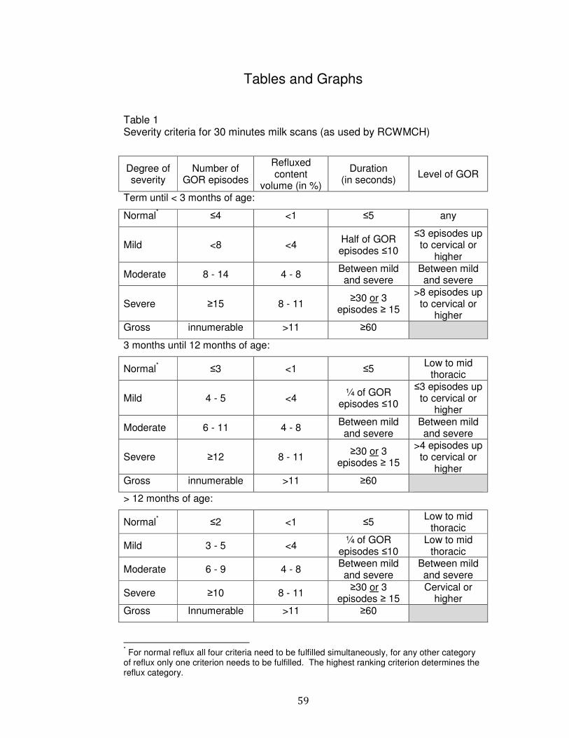
59
Tables and Graphs
Table 1 Severity criteria for 30 minutes milk scans (as used by RCWMCH)
Degree of severity
Number of GOR episodes
Refluxed content
volume (in %)
Duration (in seconds)
Level of GOR
Term until < 3 months of age:
Normal* ≤4 <1 ≤5 any
Mild <8 <4 Half of GOR episodes ≤10
≤3 episodes up to cervical or
higher
Moderate 8 - 14 4 - 8 Between mild and severe
Between mild and severe
Severe ≥15 8 - 11 ≥30 or 3
episodes ≥ 15
>8 episodes up to cervical or
higher Gross innumerable >11 ≥60
3 months until 12 months of age:
Normal*
≤3 <1 ≤5 Low to mid
thoracic
Mild 4 - 5 <4 ¼ of GOR
episodes ≤10
≤3 episodes up to cervical or
higher
Moderate 6 - 11 4 - 8 Between mild and severe
Between mild and severe
Severe ≥12 8 - 11 ≥30 or 3
episodes ≥ 15
>4 episodes up to cervical or
higher Gross innumerable >11 ≥60
> 12 months of age:
Normal* ≤2 <1 ≤5 Low to mid
thoracic
Mild 3 - 5 <4 ¼ of GOR
episodes ≤10 Low to mid
thoracic
Moderate 6 - 9 4 - 8 Between mild and severe
Between mild and severe
Severe ≥10 8 - 11 ≥30 or 3
episodes ≥ 15 Cervical or
higher Gross Innumerable >11 ≥60
* For normal reflux all four criteria need to be fulfilled simultaneously, for any other category of reflux only one criterion needs to be fulfilled. The highest ranking criterion determines the reflux category.

60
Table 2 Excluded patients overview (Indications other than GOR) Indication for procedure performed other than GOR Number of cases excluded
Suspected volvulus (9) or malrotation (5) 14
Post-surgical complications tracheo-oesophageal fistula repaired (4)
*
presumed gastro-enteric fistula (2) repositioning of PEG (2) persistent vomiting post choledochal cyst removal (1) persistent vomiting post Nissen’s (1) persistent vomiting post gastroschisis repair (1) review of bowel lengthening procedure (1) recurrence of hiatus hernia (1)
13
Suspected isolated aspiration (5) or performed modified contrast study (3) 8
Presumed or followed-up oesophageal strictures 7
Presumed tracheo-oesophageal fistula* 5
Other Oesophageal foreign body for perforation review (3) Extended workup for bronchial compression (2) Follow through for gastric outlet obstruction (1) or short bowel (1) Contrast enema for Hirschsprung’s disease (1) chronic constipation and abdominal pain (1) cleft lip and palate (1)
10
Total excluded 57
* One patient had an initial confirmatory scan for trachea-oesophageal fistula and a repeat scan post surgical repair

61
Table 3 Excluded studies per patient with more than one investigation in four months study period Patient Presentation* Reason for exclusion* Study excluded† Study remained*
1 Infant with metabolic encephalopathy for PEG workup
Deciding criterion for addition of Nissen’s depends on evidence of GOR as appropriately assessed by milk scan
Initial & repeated CS after presumed aspiration episode (2 studies)
MS
2 Pulmonary tuberculosis with bronchial compression, possible aspiration, requested by pulmonology
Repeated scan after new LRTI episode, both scans showed identical result
Second MS Initial MS
3-5 Presumed GOR (3 patients) GOR appropriately assessed by MS CS MS
6-11 Vomiting and cyanosis / apnoea after feeds (5 patients)
GOR as appropriately assessed by MS CS MS
12 Presumed GOR confirmed on MS, hold-up at GOJ‡ required CS follow-up
GOR as appropriately assessed by MS CS MS
13 Uncontrolled asthma, possible GOR? GOR as appropriately assessed by MS CS MS
14 Heartburn unsuccessful on treatment GOR as appropriately assessed by MS CS MS 15 PEG workup for known GORD without
improvement on treatment GOR as appropriately assessed by MS CS MS
16 PEG workup for cerebral palsy patient on long-term naso-gastric tube feeds
Deciding criterion for addition of Nissen’s depends on evidence of GOR as appropriately assessed by milk scan
CS MS
17 Toddler with FTT, food allergy and food refusal GOR as appropriately assessed by MS CS MS
18 ENT referral for glottic oedema GOR as appropriately assessed by MS CS MS
† CS = contrast swallow, MS = milk scan
‡ GOJ = gastro-oesophageal junction
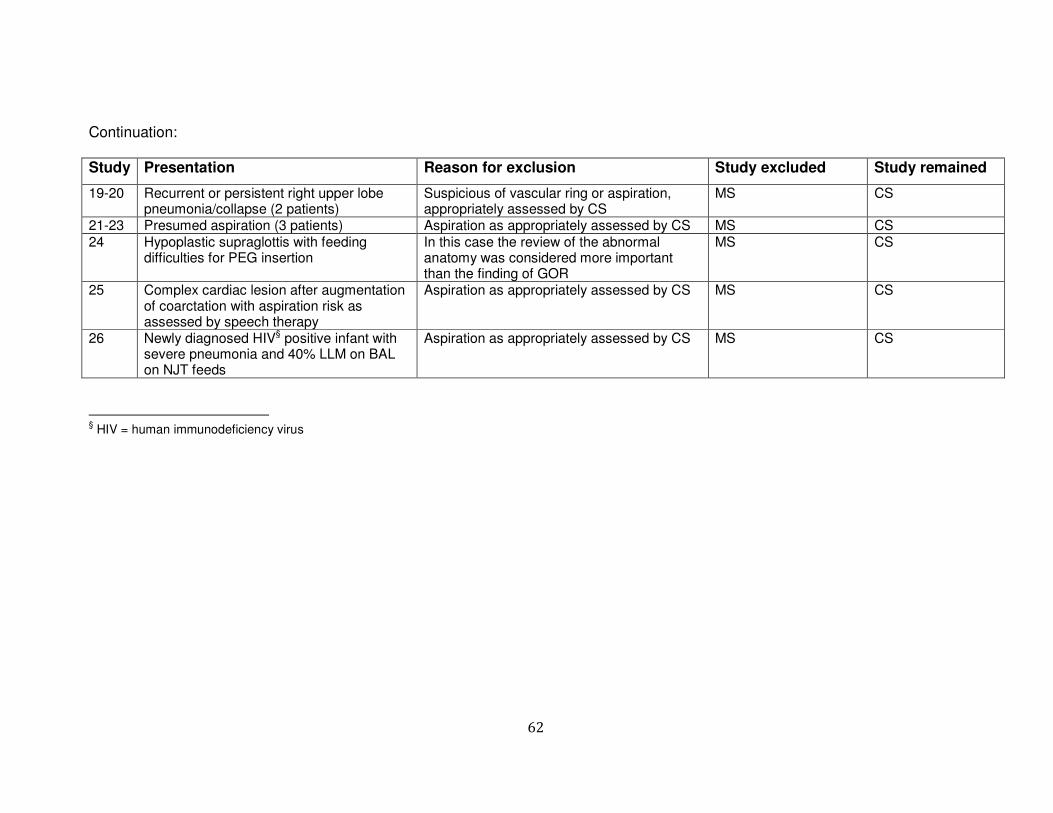
62
Continuation:
Study Presentation Reason for exclusion Study excluded Study remained
19-20 Recurrent or persistent right upper lobe pneumonia/collapse (2 patients)
Suspicious of vascular ring or aspiration, appropriately assessed by CS
MS CS
21-23 Presumed aspiration (3 patients) Aspiration as appropriately assessed by CS MS CS
24 Hypoplastic supraglottis with feeding difficulties for PEG insertion
In this case the review of the abnormal anatomy was considered more important than the finding of GOR
MS CS
25 Complex cardiac lesion after augmentation of coarctation with aspiration risk as assessed by speech therapy
Aspiration as appropriately assessed by CS MS CS
26 Newly diagnosed HIV§ positive infant with severe pneumonia and 40% LLM on BAL on NJT feeds
Aspiration as appropriately assessed by CS MS CS
§ HIV = human immunodeficiency virus

63
Table 4 Table of main results
Appropriate study request* Appropriate timing* Appropriate
modality*
Appropriate modality* (after removal of
inappropriate study requests)
Trial of antacids†
No Yes Exc.
‡ Total No Yes Total No Yes Total No Yes Total No Yes Total
Milk scan frequency 11 94 5 110 7 100 107 5 106 111 5 95 100 82 16 98
percent 57.9 59.5 50.0 58.8 70 57.8 58.5 13.5 70.2 59 15.6 69.3 59.2 62.6 66.7 63.2
Contrast
swallow
frequency 8 64 5 77 3 73 76 32 45 77 27 42 69 49 8 57
percent 42.1 40.5 50.0 41.0 30 42.2 41.5 86.5 29.8 41 84.4 30.7 40.8 37.4 33.3 36.8
Total frequency 19 158 10 187 10 173 183 37 151 188 32 137 169 131 24 155
percent 100 100 100 100 100 100 100 100 100 100 100 100 100 100 100 100
Analysis§ Chi2=0.36 (2); p=0.84 Fisher p=0.53 Chi2=39.49 (1);
p<0.001
Chi2=30.98 (1);
p<0.001
Chi2=0.14 (1);
p=0.70
*analysis done on number of studies performed
†analysis done on number of patients for studies performed
‡Exc. = exception
§Analysis using Chi-square (degrees of freedom) or Fisher’s exact test where appropriate

64
Table 5 GOR severity found by study type per studies performed
No GOR Severity of GOR Normal Mild Moderate Severe Gross Total
Stu
dy typ
e Milk scan frequency 17 22 22 25 24 1 111
percentage 32.1 84.6 78.6 69.4 61.5 33.3 60.0 Contrast swallow frequency 36 4 6 11 15 2 74 percentage 67.9 15.4 21.4 30.6 38.5 66.7 40.0 Total frequency 53 26 28 36 39 3 185 percentage 100 100 100 100 100 100 100

65
Table 6 Comparison of GOR found per modality in subgroup with both examinations
Milk scan found GOR
Contrast swallow found GOR
Number of patients
Details / comment
Yes* No 9 Two of the contrast swallows did not assess for GOR, one of these found an aberrant subclavian artery
No Yes† 1 Contrast swallow noted very irritable child during test, a month later this patient proved to have eosinophilic oesophagitis on gastroscopy with biopsy
No No 2 No GOR Normal Normal 1 Both normal range GOR
Mild Moderate 1 Mild Severe 2 First patient: multiple vomits on milk scan, contrast swallow demonstrated
nasopharyngeal backflow; second patient: swallowed very little milk on milk scan, contrast swallow demonstrated nasopharyngeal backflow
Moderate Moderate 1 Both moderate GOR Moderate Severe 2 First patient: balanced translocation 3p/8q, drinking reluctantly on contrast
swallow, application of contrast via NGT; second patient: contrast swallow demonstrated nasopharyngeal backflow
Severe Minimal 1 Severe Moderate 1
Severe Severe 4 Both severe
* Severe (4), mild (3) and normal reflux (2) † Moderate

66
Table 7 Structural abnormalities with background information and contrast swallow result
No History Finding Expected
1* Fetal alcohol syndrome with congenital cytomegalovirus (CMV) disease and oesophageal strictures, recurrent dilatations
Smooth short segment stricture in distal oesophagus and small hiatus hernia
known
2 Oesophageal atresia with trachea-oesophageal fistula repaired in infancy
Small focal irregularity at anastomosis site, no hold-up demonstrated
known
3 Previous Nissen’s fundoplication with loss of appetite and weight and retrosternal pain
Para-oesophageal hernia suspected
4 VACTERL syndrome, previous redo-Nissen’s fundoplication, persistent vomiting
Several stenoses suspected
5* Hoarseness & glottic oedema on flexible endoscopy Swollen glottis suspected
6* Oesophageal candidiasis (HIV Stage 4) with dysphagia Dilated distal oesophagus suspected
7* Dysphagia & coughing on liquids with hold-up on Milk scan (later diagnosed as Cornelia-de-Lange syndrome)
Aberrant subclavian artery not causing mass effect on oesophagus, small sliding hiatus hernia
suspected
8 Three months old with one month history of vomiting, no other complaints
Aberrant right subclavian artery with minimal pressure effect
incidental
* Patients with both investigation modalities in research interval (refer to subgroup analysis)

67
Table 8 Comparison of indication and results by number of patients
No GOR
Severity of GOR
Normal Mild Moderate Severe Gross Total
Indic
atio
n a
s p
er
re
qu
est
form
GORD* 30 11 15 15 14 1 86
Aspiration† 2 2 1 1 3 1 10
GORD plus aspiration 2 3 1 4 3 0 13
Apnoea 1 4 3 4 5 0 17
For PEG/Nissen’s 2 1 0 1 3 1 8
Other functional‡ 2 2 2 1 2 0 9
Other anatomical§ 7 1 3 3 2 0 16
Total 46 24 25 29 32 3 159
* Split-up: previously diagnosed GORD (16), previously diagnosed GORD with presumed persistence (8) and presumed GORD (62)
† Split-up: previously diagnosed aspiration (1) and presumed aspiration (9), on folder review these studies qualified as reflux investigation, but request was for
aspiration
‡ Split-up: reflux versus asthma (4), Sandifer syndrome (3) and abdominal pain with vomiting (2)
§ Split-up: known stricture (1), presumed stricture (2), presumed tracheo-oesophageal fistula (1), presumed vascular ring (1), presumed hiatus hernia (1),
bronchiectasis or chronic lung disease workup (4) and dysphagia or odynophagia (6)

68
Graph 1 Excluded patients diagram
* For analysis purposes by number of patients the investigation with the most weight regarding the raised question was retained and the other(s) were removed. (Table 3) The remaining study commonly was the milk scan as the symptoms described and question asked were mostly related to investigating for presumed GORD.
Technical aspect: No results 4
Reflux investigations included 188
Number of multiple studies 27
performed per patient
Number of patients included 161*
Reflux investigations remained 192
Insufficient data for judgment 7
Requested by other institutions 17
Indications other than reflux 57
Primarily included reflux studies 273

69
Graph 2 Suggested flow diagram for the investigation of GORD
(persistent vomiting)

70
Graph 3 Suggested flow diagram for the investigation of GORD
(chronic cough and chest infections)