topic 11.2 muscles and movement. 11.2.1 role of bones, ligaments, muscles, tendons and nerves in...
TRANSCRIPT

Topic 11.2 Muscles and Movement

11.2.1 Role of bones, ligaments, muscles, tendons and nerves in human movement
• JOINT aka articulation or arthrosis– point where 2 or more bones contact one another– arthrology = study of joints– rheumatology = branch of medicine dealing with joint
diseases and conditions– kinesiology = study of movement of the body– joints provide mobility– most joints include bones, ligaments, muscles,
tendons and nerves

BONES • Organs contain several different tissues• Functions:
– framework for support– protect soft tissues and organs– act as levers for movement– form blood cells in the marrow– storage of minerals, especially Ca and phosphorous– adults have 206 bones– Be able to identify: clavicle, ribs, humerus, ulna, radius,
carpals, metacarpals, phalanges, femur, tibia, fibula, tarsals and metatarsals
Muscles and Tendons• cords of dense connective tissue• attach muscles to bones• bones act like levers and magnify muscle
contraction force• muscles provide force by shortening the length
of their fibers or cells• antagonistic muscle pairs since only provide
movement by shortening• biceps bends arm, triceps straightens it

Ligaments and Nerves
• ligaments are tough band-like structures that strengthen joints
• connect bone to bone• provide stability• proprioceptors = sensory nerve endings
which monitor positions of joint parts• nerves help prevent overextension of the
joint
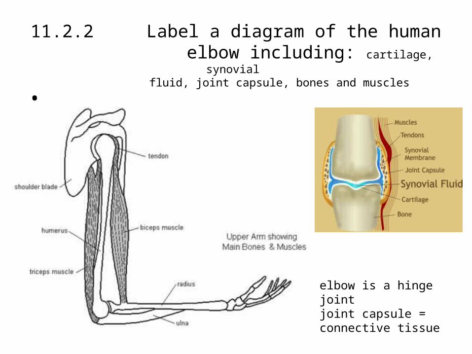
11.2.2 Label a diagram of the human elbow including: cartilage, synovial
fluid, joint capsule, bones and muscles•
elbow is a hinge jointjoint capsule = connective tissue

11.2.3 Outline the function of structures in elbow joint
Joint Part Function
cartilage reduces friction and absorbs compression (shock absorber)
synovial fluid lubricates to reduce friction; provides nutrients to cells of cartilage
joint capsule surround joint; encloses synovial cavity; unites connecting bones
tendons attach muscle to bone
ligaments attach bone to bone
biceps muscle contract to cause flexion (bending) of the arm
triceps muscle contracts to cause extension (straightening) of the arm
humerus acts as a lever that allows anchorage of elbow muscles
radius acts as lever for biceps muscle
ulna acts as lever for triceps muscle

11.2.4 Compare movement of hip joint and knee joint
flex, extend, abduction, adduction, rotation
knee hip
synovial joints
move the leg
required for
walking
socket and ballhinge
moves in one
axis
flex, extend, a little
rotation
multiaxial
flex, extend,
abduction, adduction,
rotation

• HIP MOVEMENTS:–flex and extend = < and > angle
between connecting bones–abduction and adduction = move
away and toward body midline–combination of these = windmill
effect
Muscle Types•skeletal or striated•cardiac or heart muscle•smooth or non-striated
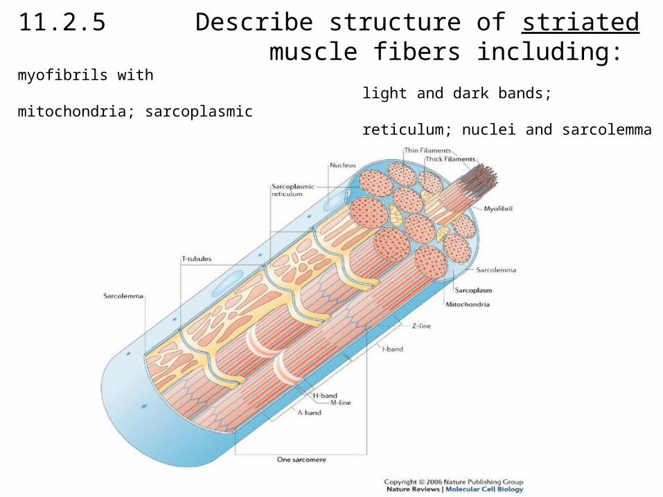
11.2.5 Describe structure of striated muscle fibers including: myofibrils with light and dark bands; mitochondria; sarcoplasmic reticulum; nuclei and sarcolemma

• sarcoplasm = cytoplasm of a muscle cell• sacroplasmic reticulum = internal membrane
which stores and releases calcium ions (Ca2+) to trigger a contraction
• myofibrils = thin fibers that cause a striated (striped) pattern of light and dark bands; contain 2 types of myofilament, myosin and actin (protein-like substances)
• many mitochondria = provide energy• sarcomere = the functional unit of the muscle• sarcolemma- plasma membrane of muscle cell

11.2.6 Draw and label a diagram of a sarcomere including: Z lines; actin filaments; myosin filaments with heads and light and dark bands
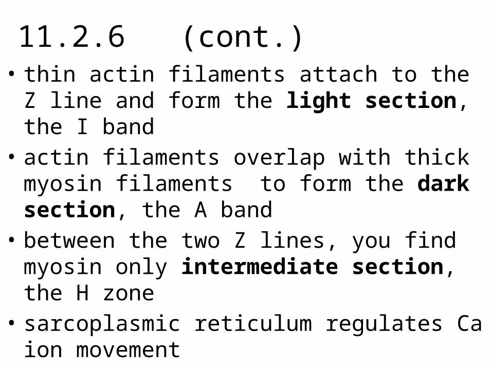
11.2.6 (cont.)• thin actin filaments attach to the Z line and form the
light section, the I band• actin filaments overlap with thick myosin filaments
to form the dark section, the A band• between the two Z lines, you find myosin only
intermediate section, the H zone• sarcoplasmic reticulum regulates Ca ion movement• Ca2+ concentration determines ATPase activity;
ATPase is an enzyme which hydrolyzes ATP to release its E

11.2.7 Explain how skeletal muscle contracts• Sliding filament theory = actin myofilaments slide
over myosin myofilaments• myofilaments do not shorten – when actin slides
over myosin the sarcomere shortens• Sliding Filament Theory:
1. motor neuron carries impulse to neuromuscular junction
2. neurotransmitter (acetylcholine) released into synapse between axon terminal and sarcolemma of muscle fiber
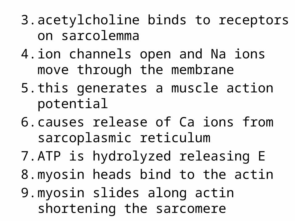
3. acetylcholine binds to receptors on sarcolemma4. ion channels open and Na ions move through
the membrane5. this generates a muscle action potential6. causes release of Ca ions from sarcoplasmic
reticulum7. ATP is hydrolyzed releasing E8. myosin heads bind to the actin9. myosin slides along actin shortening the
sarcomere

11.2.8 Analyze electron micrographs to find the state of contraction of muscle fibersZ line between Z lines = a sarcomere
I band H zone
A band