the skeletal system - linn–benton community...
TRANSCRIPT

10/1/2016
1
The Skeletal System
Skull
Thoracic cage(ribs andsternum)
(a) Anterior view
Facial bonesCranium
Sacrum
Vertebralcolumn
ClavicleScapulaSternumRibHumerusVertebraRadiusUlnaCarpals
PhalangesMetacarpalsFemurPatella
TibiaFibula
TarsalsMetatarsalsPhalanges
Functions1. Support
� For the body and soft organs
2. Protection� For brain, spinal cord and vital organs
3. Movement� Levers for muscle action
4. Mineral reservoir� Calcium and phosphorus
5. Hematopoiesis� Marrow cavities
Bone
� Osseous tissue� Hydroxyapatite (85%)
� Calcium carbonate (10%)
� Inorganic minerals� Magnesium, sodium, fluoride
� Organic material� Collagen
� Chondroitin sulfate
Figure 6.3c
(c)
Yellow
bone marrow
Endosteum
Compact bone
Periosteum
Perforating
(Sharpey’s) fibers
Nutrient
arteries
Bone consists of
multiple tissues
A single bone may be
considered an organ
Figure 6.3a-b
Proximal
epiphysis
(b)
(a)
Epiphyseal
line
Articular
cartilage
Periosteum
Spongy bone
Compact bone
Medullary
cavity (lined
by endosteum)
Compact bone
Diaphysis
Distal
epiphysis
Bones are organs comprised
of more than just osseous tissue

10/1/2016
2
Figure 6.2
Bone ClassificationGeneral Bone Features
�Long bone anatomy (Humerus)� Diaphysis
� Epiphysis
� Metaphysis� Epiphyseal growth plate or line
� Articular cartilage
� Periosteum
� Medullary cavity
� Endosteum
Figure 6.3a-b
Proximal
epiphysis
(b)
(a)
Epiphyseal
line
Articular
cartilage
Periosteum
Spongy bone
Compact bone
Medullary
cavity (lined
by endosteum)
Compact bone
Diaphysis
Distal
epiphysis
Anatomy of a long bone
Figure 6.3c
(c)
Yellow
bone marrow
Endosteum
Compact bone
Periosteum
Perforating
(Sharpey’s) fibers
Nutrient
arteries
Outer layer of periosteum =
dense irregular fibrous sheath
A Closer Look
� Osteoblasts (OB)
� Osteoclasts (OC)
� Periosteum (PO)
� Sharpey’s Fibers (SF)
Proximal
epiphysis
(b)
(a)
Epiphyseal
line
Articular
cartilage
Periosteum
Spongy bone
Compact bone
Medullary
cavity (lined
by endosteum)
Compact bone
Diaphysis
Distal
epiphysis
Epiphyseal line is a remnant
of the epiphyseal plate
Metaphysis

10/1/2016
3
Epiphyseal (Growth) PlateBone Histology
� Components� Cells
� Fibers
� Ground substance
Bone Histology
� Cells� Osteogenic (osteoprogenitor) cells
� Stem cells in periosteum and endosteum → osteoblasts
� Osteoblasts� Bone forming cells
Figure 6.4a-b
(a) Osteogenic cell (b) Osteoblast
Stem cell Matrix-synthesizing
cell responsible
for bone growth
Bone Histology
� Cells� Osteocytes
� Mature bone cells
� Maintain bone matrix
Bone Histology
� Cells� Osteoclasts
� Break down (resorb) bone matrix
� Related to macrophages

10/1/2016
4
Figure 6.4c-d
(c) Osteocyte
Mature bone cell
that maintains the
bone matrix
(d) Osteoclast
Bone-resorbing cell
Bone Histology� Primary bone types
� Compact
� Spongy
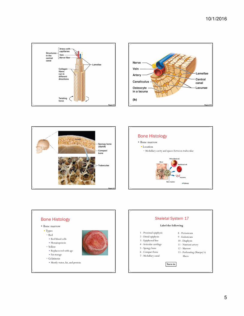
Figure 6.5
Compact
bone
Trabeculae
Spongy bone
(diploë)
Compact bone
covers all spongy bone and the shafts
of long bones
Figure 6.3a-b
Proximal
epiphysis
(b)
(a)
Epiphyseal
line
Articular
cartilage
Periosteum
Spongy bone
Compact bone
Medullary
cavity (lined
by endosteum)
Compact bone
Diaphysis
Distal
epiphysis
Figure 6.7a-c
Endosteum lining bony canals
and covering trabeculae
Perforating
(Volkmann’s) canal
Perforating (Sharpey’s) fibers
Periosteal blood vesselPeriosteum
Lacuna (with
osteocyte)
(a)
(b) (c)
Lacunae
Lamellae
Nerve
Vein
Artery
Canaliculi
Osteocyte
in a lacuna
Circumferential
lamellae
Osteon
(Haversian system)
Central
(Haversian) canal
Central
canal
Interstitial lamellae
Lamellae
Compact
bone
Spongy bone
10/1/2016
5
Figure 6.6
Structures
in thecentral
canal
Artery with
capillaries
Vein
Nerve fiber
Lamellae
Collagen
fibersrun in
different
directions
Twisting
force
Figure 6.3b
(b)
Lacunae
Lamellae
Nerve
Vein
Artery
Canaliculus
Osteocyte
in a lacuna
Central
canal
Figure 6.5
Compact
bone
Trabeculae
Spongy bone
(diploë)
Bone Histology
� Bone marrow� Location
� Medullary cavity and spaces between trabeculae
Bone Histology
� Bone marrow� Types� Red
� Red blood cells
� Hematopoiesis
� Yellow� Replaces red with age
� Fat storage
� Gelatinous� Mostly water, fat, and protein
Skeletal System 17
1 - Proximal epiphysis
2 - Distal epiphysis
3 - Epiphyseal line
4 - Articular cartilage
5 - Spongy bone
6 - Compact bone
7 - Medullary canal
8 - Periosteum
9 - Endosteum
10 - Diaphysis
11 - Nutrient artery
12 - Marrow
13 - Perforating (Sharpey’s)
fibers
Label the following
Turn in
10/1/2016
6
Bone Formation & Maintenance
� Ossification (osteogenesis)� Stages
� Bone formation� Begins in the 2nd month of development
� Postnatal bone growth� Until early adulthood
� Bone remodeling and repair� Lifelong
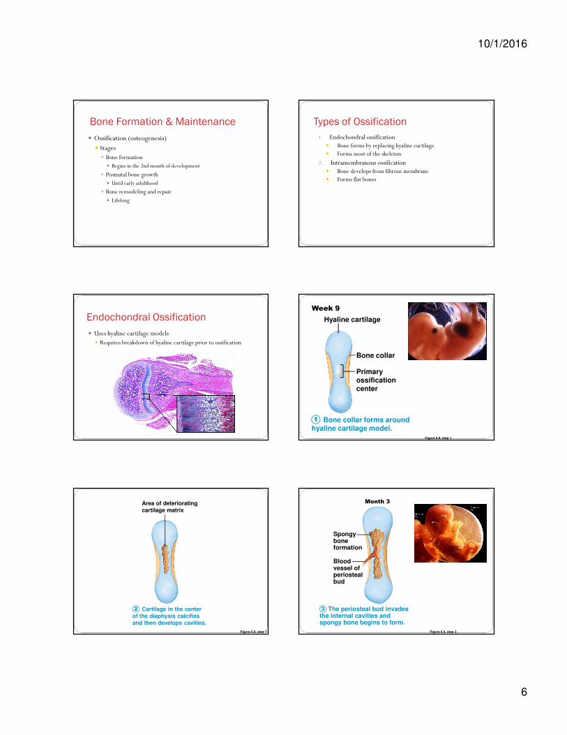
Types of Ossification
1. Endochondral ossification� Bone forms by replacing hyaline cartilage� Forms most of the skeleton
2. Intramembranous ossification� Bone develops from fibrous membrane� Forms flat bones
Endochondral Ossification
� Uses hyaline cartilage models � Requires breakdown of hyaline cartilage prior to ossification
Figure 6.9, step 1
Bone collar forms aroundhyaline cartilage model.
1
Hyaline cartilage
Week 9
Bone collar
Primaryossificationcenter
Figure 6.9, step 2
Cartilage in the center
of the diaphysis calcifiesand then develops cavities.
2
Area of deteriorating
cartilage matrix
Figure 6.9, step 3
The periosteal bud invadesthe internal cavities andspongy bone begins to form.
3
Spongyboneformation
Bloodvessel ofperiostealbud
Month 3

10/1/2016
7
Figure 6.9, step 4
The diaphysis elongates and a medullary cavity formsas ossification continues. Secondary ossification centersappear in the epiphyses in preparation for stage 5.
4
Epiphysealblood vessel
Secondaryossificationcenter
Medullarycavity
Birth
Figure 6.9, step 5
The epiphyses ossify. When completed, hyaline cartilage
remains only in the epiphyseal plates and articular cartilages.
5
Epiphyseal plate
cartilage
Articular cartilage
Childhood to adolescence
Spongy bone
Figure 6.9
Bone collar
forms around
hyaline cartilage
model.
Cartilage in the
center of the
diaphysis calcifies
and then develops
cavities.
The periosteal
bud inavades the
internal cavities
and spongy bone
begins to form.
The diaphysis elongates
and a medullary cavity
forms as ossification
continues. Secondary
ossification centers appear
in the epiphyses in
preparation for stage 5.
The epiphyses
ossify. When
completed, hyaline
cartilage remains only
in the epiphyseal
plates and articular
cartilages.
Hyaline
cartilage
Area of
deteriorating
cartilage matrix
Epiphyseal
blood vessel
Spongy
bone
formation
Epiphyseal
plate
cartilage
Secondary
ossification
center
Blood
vessel of
periosteal
bud
Medullary
cavity
Articular
cartilage
Childhood to
adolescence
BirthWeek 9 Month 3
Spongy
bone
Bone
collarPrimary
ossification
center
1 2 3 4 5
Figure 6.10
Calcified cartilage
spicule
Osseous tissue
(bone) coveringcartilage spicules
Resting zone
Osteoblast depositing
bone matrix
Proliferation zone
Cartilage cells undergo mitosis.
Hypertrophic zone
Older cartilage cells enlarge.
Ossification zone
New bone formation is occurring.
Calcification zone
Matrix becomes calcified; cartilage cells die; matrix
begins deteriorating.
1
2
3
4
Epiphysealgrowth plates
continue to produce
cartilaginouselongation as long
as bones are
increasing in length
Intramembranous Ossification
� Forms flat bones� Skull roof, lower jaw, clavicles
� Uses a fibrous membrane model formed from mesenchymal cells
Figure 6.8, (1 of 4)
Mesenchymal
cell
Collagen
fiberOssification
center
Osteoid
Osteoblast
Ossification centers appear in the fibrous
connective tissue membrane.
• Selected centrally located mesenchymal cells clusterand differentiate into osteoblasts, forming anossification center.
1

10/1/2016
8
Figure 6.8, (2 of 4)
Osteoid
Osteocyte
Newly calcified
bone matrix
Osteoblast
Bone matrix (osteoid) is secreted within the
fibrous membrane and calcifies.
• Osteoblasts begin to secrete osteoid, which is calcified
within a few days.• Trapped osteoblasts become osteocytes.
2
Figure 6.8, (3 of 4)
Mesenchyme
condensingto form theperiosteum
Blood vessel
Trabeculae of
woven bone
Woven bone and periosteum form.
• Accumulating osteoid is laid down between embryonic
blood vessels in a random manner. The result is a network(instead of lamellae) of trabeculae called woven bone.
• Vascularized mesenchyme condenses on the external faceof the woven bone and becomes the periosteum.
3
Figure 6.8, (4 of 4)
Fibrous
periosteum
Osteoblast
Plate of
compact bone
Diploë (spongy
bone) cavitiescontain red
marrow
Lamellar bone replaces woven bone, just deep to
the periosteum. Red marrow appears.
• Trabeculae just deep to the periosteum thicken, and are later
replaced with mature lamellar bone, forming compact boneplates.
• Spongy bone (diploë), consisting of distinct trabeculae, per-sists internally and its vascular tissue becomes red marrow.
4
Bone Maintenance
� Bone is dynamic throughout human lifespan
� Remodeling has several functions� Replacement and repair
� Release of calcium
� Response to stress (modification of density)
Bone Remodeling� Deposit
� Where bone is injured or added strength is required
� Osteoblasts
� Resorption� Releases minerals from
bone
� Osteoclasts
Control of Remodeling
� Hormones� Growth Hormone
� Calcitonin
� Parathyroid Hormone (PTH)
� Sex hormones
10/1/2016
9
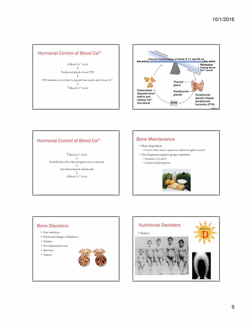
Hormonal Control of Blood Ca2+
↓ Blood Ca2+ levels
↓
Parathyroid glands release PTH
↓
PTH stimulates osteoclasts to degrade bone matrix and release Ca2+
↓
↑ Blood Ca2+ levels
Figure 6.12
Osteoclasts
degrade bonematrix and
release Ca2+
into blood.
Parathyroid
glands
Thyroid
gland
Parathyroid
glands releaseparathyroid
hormone (PTH).
StimulusFalling blood
Ca2+ levels
PTH
Calcium homeostasis of blood: 9–11 mg/100 mlBALANCEBALANCE
Hormonal Control of Blood Ca2+
↑ Blood Ca2+ levels↓
Parafollicular cells of thyroid gland release calcitonin↓
Osteoblasts deposit calcium salts ↓
↓ Blood Ca2+ levels
Bone Maintenance
� Bone deposition� Occurs where bone is injured or added strength is needed
� Development requires proper nutrition � Vitamins C, D, and A
� Calcium and phosphorus
Bone Disorders
� Poor nutrition
� Hormonal changes/imbalances
� Trauma
� Developmental errors
� Infection
� Tumors
Nutritional Disorders
� Rickets

10/1/2016
10
Nutritional Disorders
� Osteomalacia
Hormonal Disorders
� Osteoporosis� Loss of bone mass
� Bone resorption ˃ deposit� Vertebral bodies and neck of femur
Figure 6.16
Hormonal Disorders
� Osteoporosis� Risk factors
� Lack of estrogen� Low calcium or vitamin D� Petite body form� Immobility� Low levels of TSH� Diabetes mellitus
Hormonal Disorders
� Osteoporosis� Treatment and prevention
� Calcium, vitamin D, and fluoride supplements
� ↑Weight bearing exercise throughout life
� Hormone (estrogen) replacement therapy
� Controversial because of increased cardiovascular disease, cancer
� Drugs to increase bone mineral density
� Fosamax, selective estrogen receptor modulators (SERMs), statins (don’t work)
Infections� Osteomyelitis
� Inflammation of bone due to infection
(Not normal)

10/1/2016
11
Bone Fractures� Incomplete
� Does not cross entire bone
� Complete� Bone is broken into two pieces
� Comminuted� Three or more pieces
� Displaced� Bone ends don’t line up
� Open versus closed
Bone Fractures� Transverse
� Spiral
� Impacted
� Depressed
� Greenstick
Fractures
� Pathological� Secondary to coexisting disease
� Osteoporosis
� Cancer
� Malnutrition
� Cushing’s syndrome
� Osteogenesis imperfecta
Fracture Repair� Local periosteum and surrounding blood vessels are torn
� Inflammation� Open vs. closed
� Swelling� Hemorrhage (bleeding)
� Blood clots → fracture hematoma
Fracture Repair1. Fracture hematoma forms within 6-8 hrs
� Torn blood vessels hemorrhage� Clot (hematoma) forms
� Swollen, painful and inflamed
Figure 6.15, step 1
A hematoma forms.1
Hematoma
10/1/2016
12
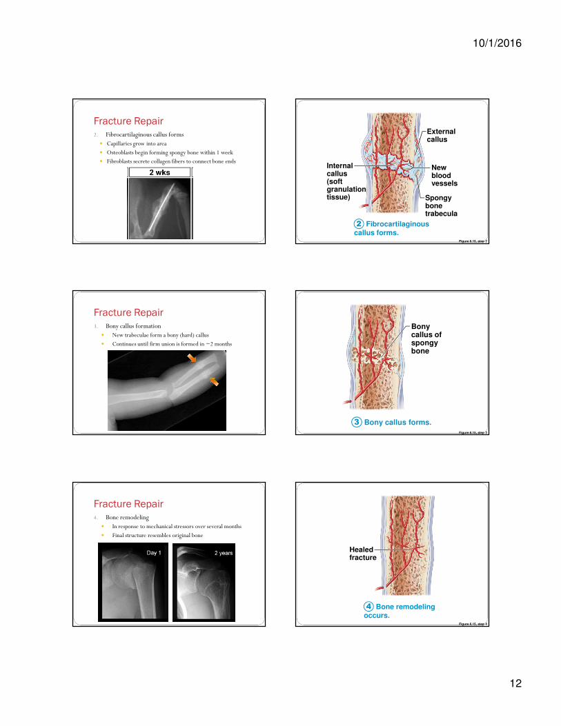
Fracture Repair2. Fibrocartilaginous callus forms
� Capillaries grow into area
� Osteoblasts begin forming spongy bone within 1 week
� Fibroblasts secrete collagen fibers to connect bone ends
Figure 6.15, step 2
Fibrocartilaginouscallus forms.
2
Externalcallus
Newbloodvessels
Spongybonetrabecula
Internalcallus(soft granulationtissue)
Fracture Repair3. Bony callus formation
� New trabeculae form a bony (hard) callus
� Continues until firm union is formed in ~2 months
Figure 6.15, step 3
Bony callus forms.3
Bonycallus ofspongybone
Fracture Repair4. Bone remodeling
� In response to mechanical stressors over several months
� Final structure resembles original bone
Figure 6.15, step 4
Bone remodelingoccurs.
4
Healedfracture
10/1/2016
13
Figure 6.15
Hematoma External
callusBony
callus of
spongy
boneHealed
fractureNew
blood
vessels
Spongy
bone
trabecula
Internal
callus
(fibrous
tissue and
cartilage)
A hematoma forms. Fibrocartilaginous
callus forms.
Bony callus forms. Bone
remodeling
occurs.
1 2 3 4
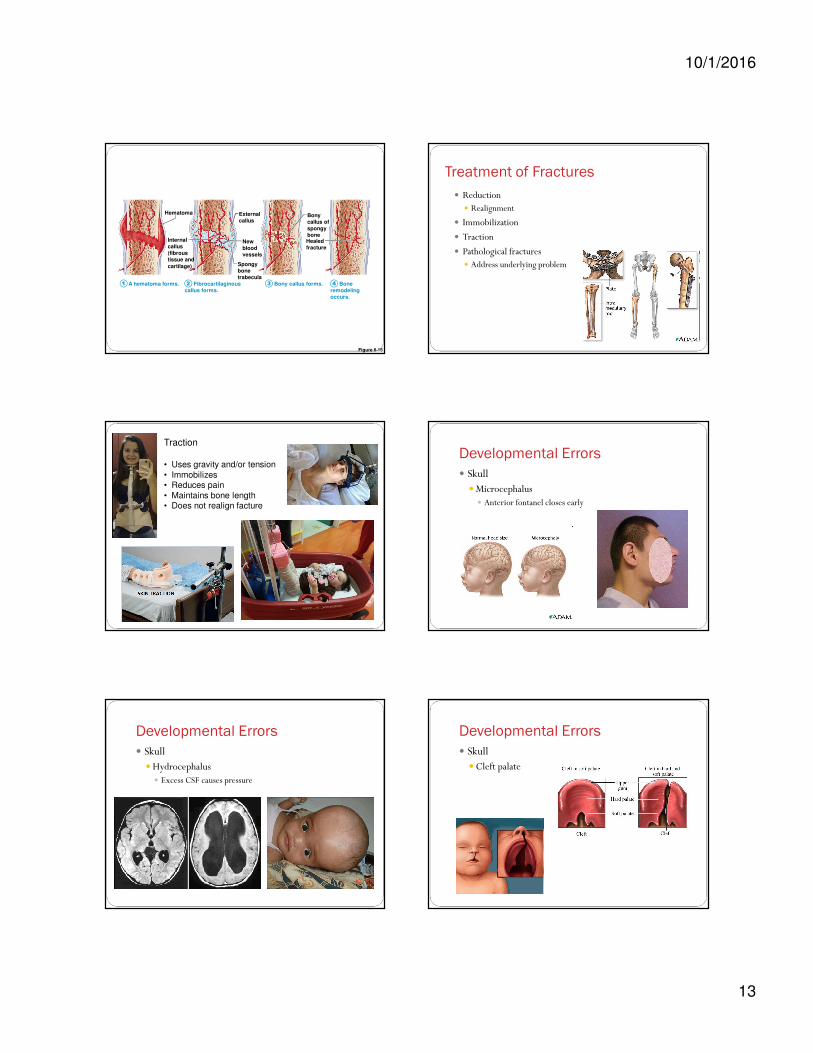
Treatment of Fractures
� Reduction� Realignment
� Immobilization
� Traction
� Pathological fractures� Address underlying problem
Traction
• Uses gravity and/or tension
• Immobilizes• Reduces pain
• Maintains bone length• Does not realign facture
Developmental Errors
� Skull� Microcephalus
� Anterior fontanel closes early
Developmental Errors
� Skull� Hydrocephalus
� Excess CSF causes pressure
Developmental Errors
� Skull� Cleft palate

10/1/2016
14
Figure 7.16
Cervical curvature
(concave)
7 vertebrae, C1–C7
Thoracic
curvature
(convex)
12 vertebrae,T1–T12
Lumbar curvature
(concave)
5 vertebrae, L1–L5
Sacral curvature
(convex)
5 fused vertebrae sacrum
Coccyx
4 fused vertebrae
Anterior view Right lateral view
Spinous
process
Transverse
processes
Intervertebral
discs
Intervertebral
foramen
C1
Disorders
of the
Spinal
Column
Scoliosis
Lordosis Kyphosis
Spina Bifida
Vertebral
defect
Disorders of the Spinal Column� Herniated disc
� Causes and risk factors
� Location
� Treatment

10/1/2016
15
Figure 7.17c
Vertebral spinous process(posterior aspect of vertebra)
Spinal nerve root
Anulus fibrosusof disc
Herniated portionof disc
Nucleuspulposus
of disc
Spinal cord
(c) Superior view of a herniated intervertebral disc
Transverseprocess