submicron calcium phosphate spheres for biomedical
TRANSCRIPT

ACTAUNIVERSITATIS
UPSALIENSISUPPSALA
2016
Digital Comprehensive Summaries of Uppsala Dissertationsfrom the Faculty of Science and Technology 1446
Submicron Calcium PhosphateSpheres for BiomedicalApplications
Synthesis and Use
TAO QIN
ISSN 1651-6214ISBN 978-91-554-9737-8urn:nbn:se:uu:diva-305820

Dissertation presented at Uppsala University to be publicly examined in Å4001,Lägerhyddsvägen 1, Uppsala, Wednesday, 14 December 2016 at 13:00 for the degree ofDoctor of Philosophy. The examination will be conducted in English. Faculty examiner: Dr Jie Huang (University College London (UCL)).
AbstractQin, T. 2016. Submicron Calcium Phosphate Spheres for Biomedical Applications:Synthesis and Use. Digital Comprehensive Summaries of Uppsala Dissertations from theFaculty of Science and Technology 1446. 67 pp. Uppsala: Acta Universitatis Upsaliensis.ISBN 978-91-554-9737-8.
Calcium phosphate spheres as biomaterials have been attracting attention in recent years.Calcium phosphate occurs naturally in bone, and a hollow structure could be advantageous fordrug loading and release. The combination of a calcium phosphate chemistry and a spherical-hollow structure could be an optimal strategy for specific biomaterial applications, e.g., certaindental and drug-delivery applications.
The focus of this thesis is on the synthesis, formation mechanism and applications of hollow,spherical calcium phosphate particles. First, the thesis describes two methods for the synthesisof calcium phosphate (CaP) spherical particles. The first method involves synthesis of hollowcalcium phosphate spherical particles via a supersaturated buffer solution based on a previousstudy. It was utilised to prepare spheres for applications in drug delivery and dentistry. Thesecond method was developed to explain the mechanism of formation of hollow calciumphosphate spheres. It aimed at revealing the particular function of magnesium in the formationof spherical particles. With the use of this modified method, it could be concluded that theonly ions active in the formation of CaP spherical particles are calcium ions, phosphate ionsand magnesium ions. Compared with the thermodynamics of micellisation, a new model, calledthree ions virtual micelle effect, was developed to explain the mechanism of the Mg function.Following this mechanism, a series of spherical particles of other compositions were explored.These spherical particles included strontium phosphate, barium phosphate, calcium fluoride,strontium fluoride and barium fluoride.
In this thesis, CaP spheres were studied for the controlled delivery of active ingredients andas active agent for tooth remineralisation. The first investigated application was to control therelease of vancomycin from Poly(methyl methacrylate) (PMMA) cement via strontium-dopedCaP spheres (SCPS). The results showed that incorporation of CaP spheres into PMMA couldenhance antibiotic release while maintaining the mechanical strength. The second applicationwas to control hydrogen peroxide (HP) release from two bleaching gel, in which CP-loadedCaP spheres were the active ingredient. One gel with low HP concentration was developedas an at-home bleaching gel, and one with high HP concentration was developed as an in-office bleaching gel. The results showed that CaP spheres would give a controlled release ofperoxide and thus have a potential to increase the efficacy of the bleaching. The third applicationwas to investigate the potential for an anti-sensitivity effect of the spheres, as active agentsin toothpaste. We studied the tooth tubules occlusion and the remineralisation effect of CaPspheres. After 7 days of application, the open dentin tubules and surface were fully covered bya newly formed apatite layer, demonstrating the remineralisation potential of the spheres.
Keywords: calcium phosphate, spheres, dentinal hypersensitivity, drug release, toothbleaching
Tao Qin, Department of Engineering Sciences, Applied Materials Sciences, Box 534, UppsalaUniversity, SE-75121 Uppsala, Sweden.
© Tao Qin 2016
ISSN 1651-6214ISBN 978-91-554-9737-8urn:nbn:se:uu:diva-305820 (http://urn.kb.se/resolve?urn=urn:nbn:se:uu:diva-305820)

To my family


List of papers
This thesis is based on the following papers, which are referred to in the text by their Roman numerals.
I. Qin, T., Zhang, P., Wani, IH., Han, YY., Leifer, K., Nikolajeff, F.,
Engqvist, H., A general strategy for synthesis of inorganic spheres via magnesium. Manuscript.
II. Qin, T., López, A., Öhman, C., Engqvist, H., Persson, C., Xia, W.,
(2015) Enhanced drug delivery of antibiotic-loaded acrylic bone cements using calcium phosphate spheres. Journal of Applied Biomaterials & Functional Materials, 13(3):e241-247.
III. Qin, T., Mellgren, T., Jefferies, S., Xia, W., Engqvist, H., A study
for tooth bleaching via carbamide peroxide loaded calcium phosphate spheres. Submitted.
IV. Mellgren, T., Qin, T., Öhman, C., Zhang, YL., Wu, B., Xia, W.,
Engqvist, H., A study on diffusion of hydrogen peroxide through enamel. Submitted.
V. Xia, W., Qin, T., Suska, F., Engqvist, H., (2016) Bioactive spheres:
the way of treating dentin hypersensitivity. ACS Biomaterials Science & Engineering, 2:734−740.
These authors make equal contribution to the work (Paper IV). Reprints were made with permission from the respective publishers.

Author’s Contributions
Paper I Major part of planning, experimental work and writing. Papers II Major part of planning, experimental work and writing. Paper III Major part of planning and writing. Part of experimental work. Part IV Part of planning and experimental work. Major part of writing. Paper V Part of planning, experimental work and writing.

Contents
Background ................................................................................................... 11 Calcium phosphate ................................................................................... 11 Synthesis of CaP spheres .......................................................................... 12 Formation of hollow structure .................................................................. 13 Applications of CaP spheres .................................................................... 14 Drug release from PMMA ........................................................................ 15 Tooth remineralisation ............................................................................. 16 Tooth whitening ....................................................................................... 17 Diffusion of HP ........................................................................................ 18
Aims and objectives ...................................................................................... 19
Materials and methods .................................................................................. 21 Materials ................................................................................................... 21 Synthesis of CaP spheres .......................................................................... 21 Method 1 (Papers II, III, IV and V) .......................................................... 21 Method 2 (Paper I) ................................................................................... 23 Application to antibiotic release from PMMA bone cement (Paper II) ... 25
Loading vancomycin into the spheres ................................................. 25 Preparation of PMMA and PMMA/SCPS specimens ......................... 25 Antibiotic release ................................................................................. 26 Uniaxial Compression Testing ............................................................ 26 Working time ....................................................................................... 26
Tooth bleaching (Paper III) ...................................................................... 27 Tooth sample preparation .................................................................... 27 CP-loaded CaP spheres ........................................................................ 27 Gels for tooth bleaching ....................................................................... 27 HP release from CP-loaded CaP spheres ............................................. 28 Colour evaluation ................................................................................ 28 HP measurement .................................................................................. 28
Diffusivity study (Paper IV) ..................................................................... 29 Tooth sample preparation .................................................................... 29 Preparation of bleaching gels ............................................................... 29 Diffusion experiment ........................................................................... 29
Treating tooth hypersensitivity (Paper V) ................................................ 30 Tooth specimen preparation ................................................................ 30

Toothpaste preparation ........................................................................ 30 Tooth remineralisation ......................................................................... 30
Statistical analysis .................................................................................... 31 Characterisation ........................................................................................ 31
Results and Discussions ................................................................................ 32 Sphere morphology (Papers II, III, IV and V).......................................... 32 The effect of pH, temperature and reaction time on sphere formation ..... 32 The function of magnesium in sphere formation (Paper ) ...................... 35 The function of sodium chloride .............................................................. 41 Proposed formation mechanism (Paper ) ................................................ 43 Modified PMMA (Paper II) ..................................................................... 46 Tooth bleaching (Paper III) ...................................................................... 48 Diffusion of HP (Paper IV) ...................................................................... 49 Occlusion of open tubules (Paper V) ....................................................... 51
Conclusions ................................................................................................... 53
Future perspectives ....................................................................................... 54
Sammanfatting på Svenska ........................................................................... 55
Acknowledgements ....................................................................................... 59
References ..................................................................................................... 61

Abbreviations
ACP Amorphous calcium phosphate α-TCP Alpha-tricalcium phosphate β-TCP Beta-tricalcium phosphate Ba/F Barium-to-fluoride molar ratio Ba/P Barium-to-phosphate molar ratio CP Carbamide peroxide CPP-ACP Casein phosphopeptide-amorphous calcium phosphate CR Critical ratio CT Computed tomography CaP Calcium phosphate Ca/F Calcium-to-fluoride molar ratio Ca/P Calcium-to-phosphate molar ratio Ca-P-Mg Calcium-phosphate-magnesium CS Compressive strength EM Elastic modulus FIB Focused ion beam HA Hydroxyapatite HCPS Hollow calcium phosphate spheres HP Hydrogen peroxide ICP-AES Inductively coupled plasma atomic emission spectroscopy L/P Liquid-to-powder ratio F/Ca Fluoride-to-calcium molar ratio F/Sr Fluoride-to-strontium molar ratio Mg/Ba Magnesium-to-barium molar ratio Mg/Ca Magnesium-to-calcium molar ratio Mg/Ca Magnesium-to-calcium molar ratio Mg/Sr Magnesium-to-strontium molar ratio MgP Magnesium phosphate PBS Phosphate-buffered saline PMMA Poly(methyl methacrylate) RT Room temperature Sr/F Strontium-to-fluoride molar ratio Sr/P Strontium-to-phosphate molar ratio SBF Simulated body fluid SEM Scanning electron microscopy SO4/F Sulphate-to-fluoride molar ratio

SCPS Strontium-doped calcium phosphate spheres TEM Transmission electron microscopy XRD X-ray diffraction

11
Background
Calcium phosphate Bone is a complex tissue with approximately 70wt% mineral, 25wt% organic content and 5wt% water [1]. In 1769, Johan Gottlieb Gahn discovered that the mineral phase of bone was composed of CaP [2]. CaP can be found in several different crystalline phases, including hydroxyapatite, tricalcium phosphate, monetite, brushite and ACP. However, hydroxyapatite and-tricalcium phosphate are the most clinically employed CaP compounds, due to their chemical similarity to the mineral phase of bone [3].
The composition of hydroxyapatite is Ca10(PO4)6(OH)2, which is the inorganic phase of CaP in bone and teeth [4,5]. The composition of tricalcium phosphate is Ca3(PO4)2, and it can be categorised into amorphous tricalcium phosphate, tricalcium phosphate and -tricalcium phosphate. tricalcium phosphate and tricalcium phosphate can be obtained as crystalline phases at high temperature [6,7]. Amorphous tricalcium phosphate can generally be obtained by precipitation methods at low temperature [8]. Amorphous tricalcium phosphate is an unstable phase at room temperature. After annealing at 1100 °C for 1 hour, amorphous tricalcium phosphate has been found to transform into highly crystalline β-tricalcium phosphate [8].
CaPs are generally bioactive [9], biocompatible and osteoconductive [10], which makes them particularly suitable for hard-tissue replacement. CaPs exist as biomaterials in various forms, including cements, granules, putties, scaffolds, implant coatings and multi-scale particles [11–13]. CaP products, bulk as well as injectable CaP cements, have been applied both clinically and commercially as bone fillers [14,15]. Bone scaffolds are three-dimensional, highly porous structures, ideally with an interconnected pore network, which could be used for bone regeneration [16]. At the site of implantation, the CaP scaffold could provide space for cell growth and carry the required load if the mechanical properties are adequate. Biocompatible and bioresorbable CaP scaffolds could match the cell/tissue growth by controlling degradation and resorption rate [17]. Studies have showed that hydroxyapatite coatings could result in more bone contact to the implants and improve the implant fixation [18,19]. Different degradable scaffolds or cements could be

12
achieved by using biphasic or multiphasic compositions such as calcium magnesium phosphate cement [20].
HA granules have been interposed onto the interface of bone and bone cement, giving an enhanced physicochemical bonding of the interfaces [21,22]. Hollow and porous CaP particles have been attracting attention as drug carriers and fillers [23,24]. Since CaP is bioactive, biocompatible and chemically similar to bone, hollow spherical CaP particles could be ideal candidates as drug-delivery systems for orthopaedic applications. Compared to polymers, these spheres could be functional drug reservoirs without negative residual effects.
Magnesium is a well-known dopant for CaP [24,25]. The effect of magnesium on the formation of CaP has been investigated in previous studies [25,26]. Gelatinous CaP precipitates have been achieved by adding sodium hydroxide to the reaction solution at 25 . It was found that the gelatinous CaP had higher solubility as the magnesium concentration in the reaction solution was increased [25]. Heterogeneous nucleation of octacalcium phosphate has been found to take place in neutral aqueous solution when the Mg/Ca ratio is less than or equal to one, while brushite was observed when the Mg/Ca ratio was greater than 1 [26]. It is clear that magnesium has specific effects on the resulting material during synthesis of various types of CaP phases.
Synthesis of CaP spheres The fabrication and current applications of inorganic hollow spheres have been systematically investigated [27]. Fabrication methods have been divided into template-based strategies and template-free strategies. Templates included polymeric templates, inorganic non-metallic templates and metallic templates. Template-based methods have been regarded as more effective and are a common way to synthesise hollow spheres. However, the template residues, energy consumption and complicated processes of template removal are inherent disadvantages. Template-free strategies are likely to become increasingly used in the future [27].
Different synthesis methods of spherical CaP particles have been systematically described in a previous review [13]. There are dozens of methods to synthesise CaP spheres, and they can be grouped by reagents, such as solutions, slurries, pastes and powders. For solutions as reagents, syntheses of CaP spheres are mainly categorised into two strategies. The first strategy is concentrated on different spraying methods, using a dispersion tool [28,29]. Tricalcium phosphate spherical particles have been prepared by applying an electrospraying method [30]: during the synthesis, the reaction solution was sprayed into a controlled electrostatic field. The second strategy is focused on precipitation methods using surfactants and biomolecules

13
[31,32]. These surfactants and biomolecules can direct the nucleation and growth of CaP spheres. Using poly(acrylic acid) (PAA) as a structuring unit, nanoporous CaP spheres have been successfully synthesised [31]. The nucleation and growth of CaP nanocrystals takes place in the PAA matrix and then the PAA phase is removed by a heat treatment [31]. In the presence of dissolved amino acids and dipeptides, CaP hollow spheres can be achieved by self-assembly [32]. It was observed that CaP nanoparticles assembled into hollow spheres within a few seconds [32].
Template removal is a disadvantage of template-based methods, because of template residue and energy consumption. However, these two strategies are still popular in current studies on the synthesis of CaP spherical particles [13].
In recent years, even though methods without surfactants have also been developed [23,24], the drawbacks must be considered. It has been found that porous microspheres of calcium magnesium phosphate can be synthesised using creatine phosphate as the phosphate source [23]. This was achieved using microwave-assisted rapid synthesis without surfactants or templates. However, creatine phosphate is extremely expensive. Hollow CaP spheres have also been synthesised in a supersaturated buffer solution by a hydrothermal method without surfactants or templates [24]. However, it was found that it has to be based on buffer solutions with large amounts of extra solutes such as sodium chloride or potassium chloride. A cheaper method without templates is absolutely required for further applications.
Formation of hollow structure Shape and structure control of inorganic nanostructured crystals is important for a wide range of materials, such as hollow titanium dioxide spheres [33], polymetallic double-walled nanoboxes [34] and colloidal CoO hollow nanocrystals [35]. Minor modifications to the chemical environment might lead to the formation of hollow particles with very different morphology and composition. Controlling the reaction parameters and processes could provide a synthetic route for the production of hollow particles. However, it is only efficient in terms of achieving the desired structure if a full understanding of the formation mechanism is available. At present, self-assembly, Ostwald ripening and Kirkendall effect are well-accepted mechanisms for hollow particles.
Self-assembly is used to describe a phenomenon whereby the formation of a larger functional unit is achieved by spontaneous assembly of smaller components. The driving force for self-assembly is the minimisation of Gibbs free energy. Covalent or non-covalent interactions with capping ligand is also a reason for self-assembly of nanoparticles [36].

14
Experimentally, self-assembly of nanoparticles into hollow spheres has been observed in the process, which only lasted a few seconds [32].
Ostwald ripening is an observed phenomenon in solid solutions or liquid sols, first described by Wilhelm Ostwald in 1896 [37]. The phenomenon describes small crystals or sol particles that dissolve, and redeposit onto larger crystals or sol particles [38]. Ostwald ripening has been applied to the fabrication of hollow inorganic nanostructures (Co3O4, ZnS and TiO2) without templates or surfactants [33,39]. Liu, B. and Zeng, H.C. defined two types of Ostwald ripening: symmetric and asymmetric [39]. They observed that the evolution of solid spheres into symmetric hollow-core spheres took place between 24 to 52 hours via TEM. The evolution of solid spheres into asymmetric core-shell structure was observed via TEM from 12 hours to 24 hours [39].
In 1947, Ernest Kirkendall and Alice Smigelskas discovered the Kirkendall effect. A difference in diffusion rate of different metal atoms led to a motion of the boundary layer between the two metals [40]. The motion was named the Kirkendall effect [41]. The sequential action of galvanic replacement and the Kirkendall effect were employed to explain the mechanism of obtaining polymetallic double-walled nanoboxes [34]. The synthesis of colloidal CoS and CoO hollow nanocrystals was obtained by sulfidation and oxidation of Co nanocrystals, which involves the nanoscale Kirkendall effect [35].
CaP spheres have been synthesised by template-free methods [23,24], but the mechanism of sphere formation has not been well studied.
Applications of CaP spheres Inorganic hollow spheres could be used in a wide array of applications. Inorganic hollow spheres, such as tungsten trioxide, zinc oxide and titania have photocatalytic properties [42–44]. Ba-doped titanium dioxide, platinum and graphene-based hollow spheres have electrocatalytic properties[45–47]. As semiconducting material, hollow spheres could be applied in gas sensing and lithium-ion batteries [48]. The large surface area could significantly benefit the gas diffusion during sensing [49–51]. This large surface area could also enhance the efficiency of absorption of gas, organic molecules and heavy-metal ions [52–54]. In the biomedical field, the hollow structure of inorganic spheres is regarded as an advantage in drug loading and delivery [55–58], as previously mentioned. In particular, magnetic hollow spheres have been the focus for anti-cancer targeted drug delivery [59,60].
A previous review systematically investigated the applications of CaP spherical particles for dentistry and orthopaedics [13]. There are already a few products on the market based on CaP spheres. Curasan has commercialised spherical β-TCP particles (Cerasorb®) for regeneration of

15
bony defects within dentistry [61]. Biomet has commercialised spherical carbonated apatite particles (Calcibon® granules) for orthopaedic applications [62]. In addition, CaP spheres can be used as raw material for CaP cement [63], 3D printing [64], CaP putties [65] and composite material [66]. As raw material for cement, adding spherical tetracalcium phosphate particles was found to have the ability to enhance the injectability of a CaP cement paste [63].
Hollow structures have been a hot topic in studies on controlled release of drugs and genes [67,68]. Combining with its bioactivity, biocompatibility and chemical similarity with bone, CaP hollow spheres have an inherent advantage in hard-tissue repair [69–71], bone regeneration [30,72], cells delivery for tissue engineering [73] and gene delivery for cell transfection [74].
Drug release from PMMA Poly(methyl methacrylate) (PMMA) is a strong and lightweight material, having a good degree of compatibility with human tissue, although it is an inert material, i.e., it has no bioactivity. Antibiotic-loaded PMMA is a conventional strategy to handle infection subsequent to e.g., joint replacement [75], but can also be used for preventive purposes [76–79]. However, in order to maintain high concentrations of antibiotics, large doses need to be loaded into the PMMA cement. Therefore, a concern is that the excessive use of antibiotics could cause bacterial resistance [80]. Improving the release rate of antibiotics from cements is necessary in order to increase the drug efficacy.
Vancomycin is one of the four most commonly used antibiotics (tobramycin, gentamicin, vancomycin and cephalosporins) to treat inflammation [81,82]. Vancomycin has very good properties for its application in cements. For example, vancomycin is thermostable and can endure the heat from the polymerisation of the PMMA cement. Vancomycin is also water-soluble and could diffuse out into the surrounding tissues from cements.
Incorporating hydroxyapatite could enhance osteoblast response to poly(methyl methacrylate) cement [83]. An important increase of the gingival cells grown per cent was observed as the incorporated weight percentage of hydroxyapatite in PMMA [84]. Due to bioactivity of α-TCP, PMMA+α-TCP composites could have a synergic effect and improve osteoblast viability [85].
Porous microspheres of magnesium whitlockite and amorphous calcium magnesium phosphate microspheres have previously been used to control release of doxorubicin for antitumour drug delivery [23]. Cancer cells changed from spindle to spherical, became damaged and died due to the high

16
cytotoxicity of doxorubicin-loaded spheres. It proved that CaP spheres were very promising drug carriers. Meanwhile, one study proved that CaP spheres as ceramic material has osteogenic potential in vitro and in vivo [86]. Therefore, incorporating CaP spheres into PMMA could increase its bioactivity and improve the drug-release profile.
Drug release from non-degradable polymeric matrices is mainly driven by diffusion [87], whereby drugs move from a region of high concentration to a region of low concentration. Specifically, drug migrates from polymer matrices into a release medium in polymeric system [88]. The magnitude is proportional to the concentration gradient. The release rate is influenced by properties of material matrices, release medium and drug compounds [88]. For example, pH and temperature of release medium, solubility of drug and swelling of matrices are the main influencing factors in a polymeric system. The drug-release kinetics are commonly described by empirical mathematical models including the zero-order model, first- order model, Higuchi model and Korsmeyer‒Peppas Model [89].
Tooth remineralisation A human tooth consists of enamel, dentin and pulp. Human enamel is primarily composed of hydroxyapatite, of which the basic unit is enamel rods, averaging 5μm in diameter and up to 3mm in length. Tightly packed, organised hydroxyapatite crystals build up the rod, which is orientated perpendicularly to the underlying dentin [90]. Dentin contains 70wt% hydroxyapatite, 20wt% organic material and 10wt% water, approximately. Dentinal tubules are microscopic channels that extend radially inward from the dentin‒enamel interface to the pulpal side of the dentin. The channels range from 13μm in diameter, increasing as the tubule radiates from the enamel side to the pulpal side [90]. The pulp is the canal for blood vessels and nerves.
Dentin sensitivity generally refers to patients experiencing sharp pain because of an external stimulus, including heat, cold, electric, dehydration and chemical stimuli [91]. However, the internal reason is enamel loss and/or gingival retraction leading to root-surface exposure, which exposes the dentin [92]. Several theories have been used to explain the mechanism behind dentin sensitivity, including the transducer theory, the modulation theory, the gate control and vibration theory and the hydrodynamic theory [93]. Among them, the hydrodynamic theory has been widely accepted [94]. According to this theory, temperature changes or physical osmotic changes cause a movement of the fluids within the dentinal tubules. This movement excites a nerve receptor sensitive to pressure, which transmits the stimulus.
Non-invasive treatments and daily care are employing active ingredients, which could desensitise the nerve and hence relieve the associated pain [95].

17
As desensitiser, potassium nitrite has been proven to reduce nerve excitation and alleviate pain [96]. Moreover, there are no known side effects of potassium nitrite. A study showed that 5% potassium nitrite in toothpaste was not harmful to the tooth surface [97]. Moreover, fluoride has an effect in prevention of dental caries and treating dentinal hypersensitivity [98,99]. This is due to the fact that fluorapatite makes the tooth structure more resistant to additional caries attack [100]. Fluoride included in toothpastes induces fluorapatite formation on the enamel, which is more stable than hydroxyapatite [100].
Tooth remineralisation, reformation of enamel, is a naturally occurring process. During the process, calcium and phosphate ions deposit into cavities in the demineralised enamel. This has been shown to be achieved by a biomimetic method, in which teeth were soaked in simulated body fluid (SBF) [101,102]. CPP-ACP has the potential of remineralising dentin collagen fibrils by a biomimetic method [103]. It has been shown that ACP could rapidly hydrolyse to form apatite, similar to the tooth mineral [104]. A variety of CaPs is usually included in toothpaste formulations for the reformation of enamel [105].
Tooth whitening In recent years, more and more dental patients have shown an interest in bleaching to get a better appearance of their teeth. The general public also has more knowledge about bleaching at present than before. Tooth bleaching is a process of whitening the teeth, in general using CP or HP. CP, also called hydrogen-urea, is a white crystalline compound, which could degrade into HP and urea [106]. Exogenous stain molecules have been shown to be oxidised, resulting in colour loss by dissociation of HP [107]. Bleaching includes two methods: in-office and at-home. In-office bleaching is carried out at a clinic by a dental professional. At-home bleaching is done at home by the individual. At-home bleaching products can either be supplied by a dental professional or available over the counter [107].
However, the side effects of bleaching should not be ignored. In 1995, reports from more than 7,000 dentists claimed that 80% of patients had side effects from bleaching [108]. A previous study showed that bleaching with 10% CP could lead to mild and reversible histological changes in dental pulp [109]. Another study showed that 35% CP could change the enamel surfaces and cause higher roughness [110].
In order to reduce dentinal hypersensitivity, one study added 5% potassium nitrate to a bleaching gel [111]. Potassium ions are supposed to penetrate into the pulp and block synapse transportation [112]. Furthermore, it has been demonstrated that ACP was effective in treating dentinal hypersensitivity [113,114] because ACP could occlude dentin tubules and
18
improve remineralisation. A study proved that casein phosphopeptides (CPP)-ACP could reduce dentinal hypersensitivity due to tooth bleaching [115]. A significant reduction in clinical measures of dentin sensitivity could be achieved by ACP-containing bleaching paste both during and after treatment [116]. A study also revealed that the bleaching effect was not reduced by the combination of tooth whitening agents (HP/CP) with CPP-ACP paste [117].
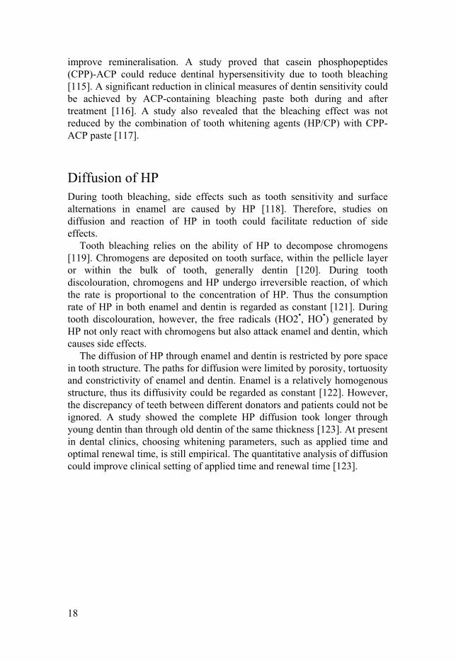
Diffusion of HP During tooth bleaching, side effects such as tooth sensitivity and surface alternations in enamel are caused by HP [118]. Therefore, studies on diffusion and reaction of HP in tooth could facilitate reduction of side effects.
Tooth bleaching relies on the ability of HP to decompose chromogens [119]. Chromogens are deposited on tooth surface, within the pellicle layer or within the bulk of tooth, generally dentin [120]. During tooth discolouration, chromogens and HP undergo irreversible reaction, of which the rate is proportional to the concentration of HP. Thus the consumption rate of HP in both enamel and dentin is regarded as constant [121]. During tooth discolouration, however, the free radicals (HO2•, HO•) generated by HP not only react with chromogens but also attack enamel and dentin, which causes side effects.
The diffusion of HP through enamel and dentin is restricted by pore space in tooth structure. The paths for diffusion were limited by porosity, tortuosity and constrictivity of enamel and dentin. Enamel is a relatively homogenous structure, thus its diffusivity could be regarded as constant [122]. However, the discrepancy of teeth between different donators and patients could not be ignored. A study showed the complete HP diffusion took longer through young dentin than through old dentin of the same thickness [123]. At present in dental clinics, choosing whitening parameters, such as applied time and optimal renewal time, is still empirical. The quantitative analysis of diffusion could improve clinical setting of applied time and renewal time [123].

19
Aims and objectives
The overall objective of the present thesis is to study the synthesis and use of CaP spheres as an inorganic biomaterial. The main focus is on synthesis methods of the spheres, mechanism of sphere formation and applications.
The thesis is focused on the following three aims:
To modify a previous method of synthesis to increase the yield
and thus pave the way for future potential industrial use; To create new understanding of the formation mechanism of CaP
spheres; To investigate the incorporation of CaP spheres into potential
applications, such as PMMA bone cements, toothpastes and bleaching gels.
There are many publications on the mechanism of inorganic and organic sphere formation; however, the mechanism of CaP sphere formation is still unexplored. To achieve this aim, a synthesis method was developed and a model relating formation of spherical particles was created based on experimental data (Paper I). Moreover, its field of application was shown to be extendable to other materials.
The applications of CaP span several areas within medicine; however, applications of CaP spheres synthesised by template-free method have not yet been fully investigated. This thesis will study its applications for controlled drug release and tooth-remineralisation purposes (PaperⅡ, III, IV and V).
The main purpose of Paper II was to modify PMMA cements with CaP spheres while maintaining the mechanical properties. This modification should lead to more efficient antibiotic delivery via the CaP spheres’ hollow structure. Meanwhile, the properties of modified PMMA bone cement including EM, CS, radiopacity and injectability are also investigated.
The objective of Paper III was to investigate bleaching products containing CaP spheres, which could improve tooth remineralisation without affecting the bleaching effect. In the paper, the loading capacity and release behaviour of HP from CP-loaded CaP spheres were studied. The bleaching effect of own-made bleaching products and their ability for tooth remineralisation were also investigated.

20
In Paper IV, the main aim was to investigate HP diffusion of three bleaching gels containing CaP spheres which could improve tooth remineralisation. Steady-state diffusion of HP through enamel discs from three own-made bleaching gels and two commercial gels was studied. An experimental setup was designed to fit a steady-state mathematic model. Under this model, experimental results were utilised to determine the steady-state flux and diffusivity. Moreover, the effect of adding CaP spheres to diffusion of HP was studied.
The study of Paper V aimed to investigate occlusion of tubules and tooth remineralisation via CaP spheres. The aim was to investigate which factors would determine the tooth remineralisation. The application of different times and CaP spheres concentrations were investigated.

21
Materials and methods
Materials Strontium nitrate, potassium chloride, sodium chloride, sodium sulphate, sodium fluoride, sodium phosphate dibasic, potassium phosphate monobasic, magnesium chloride, calcium chloride, CP, urea, vancomycin, 35wt% HP solution and carbopol were purchased from Sigma-Aldrich. All chemicals were received as analytical-grade reagents and no further purification was done before usage.
Surgical Simplex P (Stryker Corporation, Kalamazoo, MI, USA), a commercially available poly(methyl methacrylate) (PMMA) bone cement, was used as the base cement for the study on antibiotic release.
All human teeth samples were collected from patients on the basis of various diagnoses in accordance with routine procedure at Dental Clinic. All the studies involving teeth were carried out according to the guidelines provided by the reginal ethical committee (2016/039). All dentin samples were dried first and then stored in fridge at 0‒4 for further experiments.
Synthesis of CaP spheres In this thesis, two methods of synthesis were investigated. Method 1 was based on a highly concentrated salt solution of sodium chloride and potassium chloride [24]. Method 2 was a modification of Method 1, with no need of sodium chloride and potassium chloride (Paper I).
Method 1 (Papers II, III, IV and V) CaP spheres were synthesised by a hydrothermal method using the following procedure:
1. A clear solution was formed by dissolving sodium chloride, potassium chloride, sodium phosphate dibasic, potassium phosphate monobasic, calcium chloride, magnesium chloride and strontium nitride (optional) in distilled water, one by one. The order of dissolution is critical to avoid immediate precipitation. The

22
concentrations were 8 g/L, 0.2 g/L, 1.15 g/L, 0.2 g/L, 0.1 g/L and 0.1g/L, respectively. The pH value of the obtained solution was 7.4.
2. The obtained clear solution was put into a tightly covered glass bottle and kept in an oven at 80‒100◦C for 1‒24 hours.
3. After the reaction time, the solution was filtered via filter paper (Track-Etch Membrane, Sigma-Aldrich) in a filter device.
4. The precipitates were left on the filter paper, washed with ethanol at least three times and dried at 37◦C before further usage.
Hereinafter, this method with extra sodium chloride and potassium chloride is referred to as Method 1.
The effect of temperature, pH value and reaction time on the formation of CaP spheres was investigated. Temperatures included were 55, 80, 150 and 200 . The pH values investigated were 5, 6, 9 and 10. The reaction times studied were 30 minutes, 2 h, 4 h and 24 h. Precipitation with different ratios of Ca/P and Ca/Mg in the clear solution were also investigated ‒ see Table 1.
Table 1. Molar ratios of Ca/P (0.09 and 4.5) and corresponding different ratios of Ca/Mg in the clear solution investigated in Method 1.
Ca/P Mg/Ca Mg/Ca Mg/Ca Mg/Ca
0.09 0.00 0.55 2.75 16.50
4.50 0.00 2.75 5.50 11.00
Extension to other materials Strontium phosphate spheres were synthesised by Method 1. A clear solution was formed by dissolving sodium chloride, potassium chloride, sodium phosphate dibasic, potassium phosphate monobasic, strontium nitrite and magnesium chloride in distilled water, one by one.
Barium phosphate spheres were synthesised by Method 1. A clear solution was formed by dissolving sodium chloride, potassium chloride, sodium phosphate dibasic, potassium phosphate monobasic, barium chloride and magnesium chloride in distilled water, one by one.
The concentrations of sodium chloride, potassium chloride, sodium phosphate dibasic, potassium phosphate monobasic were the same as in Method 1. The processes following dissolution were also the same. The molar ratios of Sr/P, Ba/P, Mg/Sr and Mg/Ba are shown in Table 2.

23
Table 2. The molar ratios of Sr/P, Mg/Sr (left) and Ba/P, Mg/Ba (right) investigated using Method 1.
Sr/P Mg/Sr Mg/Sr Ba/P Mg/Ba Mg/Ba
0.09 0.00 0.55 0.09 0.00 0.55
Method 2 (Paper I) The synthesis process was carried out according to the following steps:
1. Calcium chloride and magnesium chloride were dissolved in 500 ml
distilled water. 2. Disodium phosphate and monosodium phosphate were dissolved in
500 ml distilled water. 3. A clear solution was formed by rapidly mixing the two solutions at
room temperature. 4. The obtained clear solution was kept in an oven at a range of 80 to
100 at least until the formation of particles. 5. The precipitates were filtered by a filter device and washed with
ethanol three times. 6. The precipitates were stored in a dry place before further usage.
Different molar ratios of Ca/P and Ca/Mg in the clear solution were investigated for the morphology and mechanism study, see Table 3. The concentrations of calcium, phosphate and magnesium ions were in the range of 0.1‒20 mM, 0.1‒20 mM and 0.1‒100 mM, respectively. The clear solution had an initial pH value of 6.0 to 8.0. The process was conducted under static conditions for 1 h to 24 h. Hereinafter, this method is referred to as Method 2.
Table 3. The screening of molar ratios and the resulting morphologies of the particles formed in Method 2. Ca/P denotes molar ratio of Ca/P; a, b, c, d and e denote molar ratios of Mg/Ca: a) 0‒0.34 b) 0.34‒0.55, c) 0.55‒2.73, d) 2.73‒5.5 and e) 5.5 or more.
Ca/P a b c d e
0.0045 CaP debris or particles with various shapes and sizes
CaP spheres
with rough
surfaces
CaP spheres with smooth surfaces
Mixture of CaP
spheres and
MgP micro
particles
Mixture of
MgP particles
and fewer CaP
spheres
As above
0.09 1.00
1.76
2.50 As above As above As above CaP spheres with smooth surfaces
4.50 As above As above As above Fewer CaP spheres 9.00 As above As above As above

24
Extension to other materials (Paper I) Moreover, in this thesis, calcium fluoride, strontium fluoride and barium fluoride spheres were synthesised based on Method 2. M ions, S ions and K ions represent any three ions, which could end up as spheres by Method 2, as shown in Table 4. Control groups: One solution (containing M ions) was mixed with another solution (containing S ions) to form a clear solution at room temperature, as shown in Table 4 (A-1), (B-1), (C-1), (D-1) and (E-1). Standard groups: One solution (containing M ions and K ions) was mixed with another solution (containing S ions) to form a clear solution at room temperature, as shown in Table 4 (A-2), (B-2), (C-2), (D-2) and (E-2). The obtained clear solution had an initial pH value of 6.0 to 8.0. The concentrations of M ions and S ions were in the range of 0.1‒20 mM. The concentrations of K ions were in the range of 0.1-100 mM. The clear solutions were kept in tightly covered glass bottles at 100 for 12 hours.
Calcium chloride, barium chloride, strontium nitrite, sodium fluoride, magnesium chloride and sodium sulphate were used to prepare reaction solutions. The concentrations of sodium ions, potassium ions and chloride ions could be determined by the concentrations of M ions, S ions and K ions, correspondingly. There were no extra salts needed in this clear solution.
Table 4. Ions and their molar ratios in the said clear solution for synthesising spheres by other materials based on Method 2. Groups of (A-1), (B-1), (C-1), (D-1) and (E-1) are the control groups, in which the concentrations of K ions are 0 in the clear solution. Groups of (A-2), (B-2), (C-2), (D-2) and (E-2) are standard groups.
Name of ions M ions S ions K ions Molar ratio of M/S
Molar ratio of K/M
Group A-1 Calcium Fluoride 1.80 0.00
Group A-2 Calcium Fluoride Magnesium 1.80 0.55 Group B-1 Fluoride Calcium 2.61 0.00 Group B-2 Fluoride Calcium Sulphate 2.61 0.49 Group C-1 Strontium Fluoride 0.20 0.00 Group C-2 Strontium Fluoride Magnesium 0.20 1.06 Group D-1 Fluoride Strontium 5.03 0.00 Group D-2 Fluoride Strontium Sulphate 5.03 0.30 Group E-1 Barium Fluoride 0.20 0.00 Group E-2 Barium Fluoride Magnesium 0.20 1.04

25
Application to antibiotic release from PMMA bone cement (Paper II) Loading vancomycin into the spheres Vancomycin (230 mg) was dissolved into 23 ml distilled water. SCPS (570 mg) were soaked into the vancomycin solution, which was stirred for 24 hours. Then, the solution was filtered using filter paper (Track-Etch Membrane, Sigma-Aldrich) in a filter device. The spheres were collected from the filter paper without any washing and dried at room temperature for 24 hours.
The loading capacity of SCPS was calculated by the following equation:
VSCPS = (VSol1-VSol2)/P ×100% (1)
In Equation (1), VSol1 is the amount of drug in the solution before loading, P is the amount of spheres and VSol2 is the amount of drug in the solution after separating the loaded spheres from the solution.
Preparation of PMMA and PMMA/SCPS specimens PMMA and PMMA/SCPS specimens were prepared via hand mixing of the liquid and powder phases. The details about all the groups and the composition of their powder phases can be seen in Table 5. The liquid-to-powder ratio (L/P) was 0.34ml/g for all groups.
The control samples were unmodified PMMA (Simplex). The powder phase of Simplex-10SCPS was made by mixing 90wt% PMMA powder phase and 10wt% SCPS. The powder phase of Simplex-2.5V was made by mixing 97.5wt% PMMA powder phase and 2.5wt% vancomycin. The powder phase of Simplex-10SCPSV was made by mixing 90wt% PMMA powder phase and 10wt% vancomycin- loaded SCPS (SCPSV). The powder phase of Simplex-10SCPSV-2.5V was made by 87.5wt% PMMA powder phase, 10wt% SCPS and 2.5wt% vancomycin.
Following the manufacturer’s instructions, mixing of the powder and the liquid components was carried out in a glass mortar for 30 seconds. The obtained paste was transferred to cylindrical moulds.

26
Table 5. Table 1.Compositions of the powder component of the investigated PMMA and PMMA/SCPS cements loaded with vancomycin.
Wt% PMMA SCPS Vancomycin Vancomycin- loaded PMMA
Final amount of vancomycin in cement
Simplex 100 - - - -
Simplex-10SCPS
90 10 - - -
Simplex-2.5V 97.5 - 2.5 - 2.5Simplex-10SCPSV
90 - - 10 2.77
Simplex-10SCPSV-2.5V
87.5 10 2.5 - 2.5
Antibiotic release Tablet samples were moulded (3.3 mm in height and 6 mm in diameter) and let to set for 24 h in air. Two different PBS solutions, with pH values 4 and 7.4, were used to simulate body fluid.
Simplex-2.5V, Simplex-10SCPSV and Simplex-10SCPSV-2.5V were placed in 3ml PBS in glass bottles. Each group had six samples. For each sampling, 2ml PBS was extracted from the bottle and saved in an Eppendorf tube for further measurement. After extraction, the PBS solution was refilled to 3ml. Time points for sample extraction were 0.5, 3.5, 9.5, 21.5, 45.5, 93.5, 141.5 and 189.5 hours. A UV spectrophotometer (Shimadzu, Kyoto, Japan - 1800) was used to measure the antibiotic concentration at a wavelength of 280 nm.
Uniaxial Compression Testing Eight specimens of each group were moulded (12 mm in height and 6 mm in diameter) and set in air at room temperature. After 1 h, the specimens were removed from the moulds and stored at T = 21 ± 1°C for 24 h before testing. A universal materials testing machine (Shimadzu, Kyoto, Japan) was used with a displacement rate of 20mm/min to determine EM and CS.
Working time A custom-made rig was mounted onto a universal materials testing machine (Shimadzu, Kyoto, Japan). After mixing the powder phase with liquid phase for 30 seconds, the obtained slurry was immediately transferred into a BD 3 ml syringe. Then the syringe was immediately placed on the rig and the test was started. The cement was extruded at a rate of 3 mm/min. The start of

27
mixing was set as time point zero in the analysis of working time for each specimen. The measurement was set to stop when the load reached 200N.
Tooth bleaching (Paper III) Tooth sample preparation Human permanent posterior teeth were collected from a dental clinic under an Institutional Review Board (IRB) exempt protocol. The teeth were free from carious lesions or other obvious defects.
The tooth staining and stain assessment was based on a previous study [124]. The human teeth were soaked in a concentrated tea solution at 4 until a stable external shade of Vita C4 [124] was achieved. The colour changes were measured using a Vita Easyshade® clinical spectrophotometer.
CP-loaded CaP spheres Hollow CaP spheres (0.5 g) and 2.475 g CP were put into 9.5 ml water and stirred under dark conditions at room temperature for 2 hours. The suspension was filtered via filter paper (Track-Etch Membrane, Sigma-Aldrich) in a filter device and was dried under dark conditions for 24 h at room temperature.
A modified method was tested to load more HP into the spheres. The loading temperature was lowered to 9 °C, which was achieved in a water bath cooled by ice. The ratio of HP and urea was 1.2:1. The stirring time was also shortened to 50 min.
Gels for tooth bleaching High concentrated tooth whitening agent, Nupro® White Gold (36% HP) gel was used as control. At-home gel and in-office gel were prepared as per Table 6.
Table 6. The composition of at-home and in-office bleaching gel.
(wt%) CP-loaded CaP spheres
Anhydrous glycerol
Others Estimated whitening agent wt%
At-home gel 50% 50% No 6% HP
In-office gel 50% 50% HP (35wt% HP solution)
20% HP

28
HP release from CP-loaded CaP spheres CP-loaded CaP spheres (0.1g) were put into 10 ml distilled water in a 20 ml-glass bottle. Aliquots were taken at certain time points: 10 min, 30 min, 60 min, 90 min, 150 min and 300 minutes. Each aliquot, 500 l, was extracted from the top clear part of the solution. The measurement was carried out by a titrimetric method via a HP test kit (Hanna instruments, Romania). The aliquots were diluted before measurement since the range of HP concentration of this kit is from 0 to 10 ppm.
Colour evaluation The evaluations of colour values and colour differences were based on CIE L*a*b colour coordinate system, described in a previous study [124].
According to the CIE L*a*b colour coordinate system, L, a and b values represent degree of lightness, greenness or redness and blueness or yellowness, respectively. The measurements of L, a and b values were carried out by Vita Easyshade (Vivadent, Brea, California).
The overall colour difference ∆E was determined by the formula below:
∆ √∆ ∆ ∆ (2) ∆L, ∆a, ∆b are the changes in L, a, b, respectively.
HP measurement The concentration of peroxide was measured using a HP test kit (Hanna instruments, Romania). According to the product instructions, the measurements were divided into three steps: 1) HP slowly reacts with iodide ions and produces iodine, which is blue.
2) The liberated iodine can be reduced back to iodide ions, which is colourless. This could be achieved by being titrated with standard sodium thiosulfate solution.
3) The concentration of HP (CHP) can be calculated by the drops of standard sodium thiosulfate solution.
2 2 2 22 2 2H O H I I H O
6
2 22 2 3 42 2I S O I S O
( ) *HPC ppm Drops A

29
In Equation (3), A is 1 if HP concentration is in range of 2 ppm to 10ppm. A is 0.25 if HP concentration is in range of 0 ppm to 2 ppm.
Diffusivity study (Paper IV) Tooth sample preparation Enamel discs were prepared from bovine central incisors (Tri-State Beef CO, Cincinnati, OH, USA). In dental studies, bovine teeth are the most widely used for substituting human teeth[125].The enamel was cut and polished into circular discs (1 mm thin and 7 mm in diameter). The edge of the disc was first etched with 34% phosphoric acid gel (Patterson Dental, St. Paul, MN, USA) to ensure proper bonding to the resin (Clearfill Majesty Flow, Kuraray America Inc., NY, USA). The resin was applied to fill the void between the disc and the sample holder.
Preparation of bleaching gels Three types of own-made bleaching gels containing CaP spheres were prepared. CP-loaded spheres, anhydrous glycerol and carbopol (added as a thickener) were mixed together to form gel, as shown in Table 7. Extra CP was added into Gel 1.
Table 7. The compositions of the own-made bleaching gels for diffusion study.
CP-loaded spheres CP anhydrous
glycerol carbopol
Gel 1: 0.35g 0.295g 0.35g 0.005g
Gel 2: 0.57g 0.42g 0.01gGel 3: 0.78g 0.21g 0.01g
Additionally, two commercially available whitening gels containing ACP were evaluated: Zoom Day White 9.5% (DayWhite) and Zoom NiteWhite 22% (NiteWhite). According to the products’ instructions for use, DayWhite contains 9.5wt% HP and NiteWhite contains 22wt% CP (Philips Oral Healthcare, Stamford, CT, USA).
Diffusion experiment In Paper IV, a mathematical model was proposed for one-dimension diffusion of HP through enamel membrane with a pseudo-first order and homogeneous reaction. In order to determine diffusivity from experimental results, an appropriate experiment set-up was designed to fit the mathematical model.

30
The device for the diffusion experiment consists of a sink and a sample holder. The sink was filled with deionised water. The enamel disc was mounted on a sample holder, keeping the lower side of the enamel disc in contact with the water in the sink and the upper side in the air.
A total of five groups (Gel 1, Gel 2, Gel 3, NiteWhite and DayWhite) were investigated via sinks, all of which contained 5 ml of deionised water. Gel (0.4 mL) was applied to the surface of the enamel disc not in contact with the water in the sink. The entire set-up was then covered with parafilm (Bemis Company, Inc., Neenah, WI) to inhibit evaporation. In order to reach steady-state, the diffusion set-up was kept for 16 hours at room temperature before taking time points.
Thereafter, the sinks were replaced by new sinks with 5ml deionised water. Aliquots (50 mL) were pipetted from each well every 15 minutes, for up to 2 hours. For each aliquot taken, the well was refilled with the same volume of deionised water.
Treating tooth hypersensitivity (Paper V) Tooth specimen preparation Teeth were cut into 1 mm thick slices using a diamond blade. The cut was placed parallel to the crown surface in order to get perpendicular open tubules. To expose the tubules, the obtained slices were polished with 1200 grit sand paper, and then immersed in 35% phosphoric acid for 30 seconds. After acid etching, acidic residues were eliminated by washing with distilled water.
Toothpaste preparation SCPS powders, water and carboxymethyl cellulose solutions (0.5wt%, CMC) were mixed together to form pastes. SCPS CMC paste (1%) contained 1wt% SCPS. SCPS CMC paste (10%) contained 10wt% SCPS.
Apotek Produktion & Laboratorier (APL, Sweden) prepared the toothpaste containing 10wt% SCPS for this study. Sensodyne repair and protect (B13517001, GSK) was a commercial toothpaste used as a comparison in our study.
Tooth remineralisation The application was carried out according to the following steps: 1. Each tooth slab was brushed with SCPS CMC paste or toothpaste by
hand for 3 minutes.

31
2. In order to remove residues, the slab was washed with tap water for approximately 30 seconds.
3. The slab was placed in a simulated saliva bath and kept in an oven at 37°C for 3 hours.
4. Steps (1-3) were repeated three times a day.
In order to investigate the effect of applied time, the slab was applied with paste for 1 day, 3 days or 7 days.
In order to evaluate the acid resistance of remineralised surfaces, the remineralised teeth slabs were soaked in hydrochloride acid solution (pH 2.5 and 6.0). The soaking periods were 30 seconds and 2 minutes.
Statistical analysis Student t-test was performed for statistical comparison of differences of CS and EM between modified PMMA and unmodified PMMA. One way analysis of variance (ANOVA) was performed for statistical comparison of differences of steady-state flux and diffusivity between Gel 1, Gel 2, Gel 3, NiteWhite and DayWhite. Statistical analysis was carried out in IBM SPSS v19.0 (IBM, Chicago, IL, USA) and the significance level was set to p<0.05.
Characterisation The morphology of the specimens was determined by field emission scanning electron microscopy (SEM, LEO 1550). Before SEM analysis, the specimens were coated with Au/Pd in order to increase conductivity. The composition of single particles was determined by Energy-dispersive X-ray (EDX) analyses.
Cross-sectioning and imaging was performed by FIB/SEM dual beam system (FEI Strata DB235). First, ion beam induced deposition (IBID) technique was used to deposit 2 µm platinum layer on the sample. Then, focused beam of Gallium ions at 1000 pA and 30kV was used to achieve the cross-sections.
The crystallinity of the specimens was determined by X-ray diffraction (Siemens Diffractometer 5000) using Cu Kα radiation (λ = 1.5418 A°). Powder samples were mixed with ethanol and then dropped onto single-crystalline Si wafers, which were substrates for the specimens. The specimens were placed until dry.
The size distribution of spheres was based on counting and calculation from SEM images. The numbers of spheres in different size range divided by total number was the percentage.

32
Results and Discussions
Sphere morphology (Papers II, III, IV and V) The CaP spheres used in Papers II, III, IV and V were synthesised by Method 1. The CaP spheres morphology, XRD pattern, hollow structure and particle size distribution are shown in Figure 1. The precipitates were spheres with hollow structure, see Figure 1 (A) and (B). Particle size ranged from 400nm to 900 nm and more than 80 % of the spheres were in the range of 500 nm to 700 nm, see Figure 1 (C). The composition of the spheres was magnesium-substituted CaP, see Figure 1 (D).
The effect of pH, temperature and reaction time on sphere formation Reaction temperature, reaction time and pH value are the main factors affecting the morphology of the precipitates.
(1) Effect of pH value
The clear solutions obtained with pH 5 and 6 under heat treatment could not precipitate. The solutions kept clear at all the time points and no precipitate was observed.
At pH 7 and 8, the precipitates were spherical particles, see Figure 1. At pH 9 and 10, the precipitates were nanoparticles of various sizes, see Figure 2.
The pH value of the clear solution was critical for the particle morphology. Spherical particles could only be achieved around neutral pH. Acidic environment inhibited the formation of any particles from the solution. Alkaline environment changed the way of nucleation and growth of the particles.

33
Figure 1. Sphere morphology (A-B), size distribution (C) and XRD pattern (D).
34
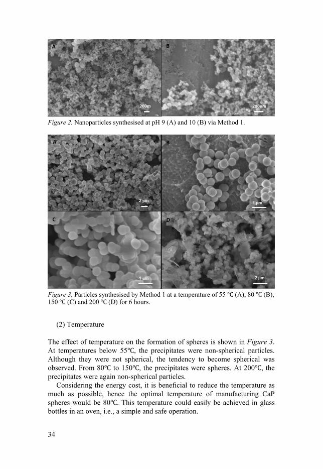
Figure 2. Nanoparticles synthesised at pH 9 (A) and 10 (B) via Method 1.
Figure 3. Particles synthesised by Method 1 at a temperature of 55 (A), 80 (B), 150 (C) and 200 (D) for 6 hours.
(2) Temperature
The effect of temperature on the formation of spheres is shown in Figure 3. At temperatures below 55 , the precipitates were non-spherical particles. Although they were not spherical, the tendency to become spherical was observed. From 80 to 150 , the precipitates were spheres. At 200 , the precipitates were again non-spherical particles.
Considering the energy cost, it is beneficial to reduce the temperature as much as possible, hence the optimal temperature of manufacturing CaP spheres would be 80 . This temperature could easily be achieved in glass bottles in an oven, i.e., a simple and safe operation.

35
Figure 4. Particles synthesised by Method 1 through a reaction time of 30 minutes (A), 2 hours (B), 4 hours (C) and 24 hours (D) at 80 .
(3) Reaction time
The effect of reaction time is shown in Figure 4. With a reaction time of less than 30 minutes, the precipitates were immature and only a few spheres were observed. Well-defined spheres were observed when the reaction time was 2 h, 4 h and 24 h.
The function of magnesium in sphere formation (Paper ) Method 1
First, the molar ratio of Ca/P was 0.09.
Figure 5 shows the morphologies of precipitates synthesised by Method 1 at molar ratio of Ca/P=0.09 and varied molar ratio of Mg/Ca. At a Mg/Ca molar ratio of 0, CaP spheres with a needle-like shell was obtained. At a Mg/Ca of 0.55, CaP spheres with smooth shell was obtained. At a Mg/Ca of 2.75, CaP spheres were observed. At a Mg/Ca of 16.5, the precipitates were porous magnesium phosphate microspheres with a flake-like shell.

36
Figure 5. Phosphate-based particles synthesised by Method 1. Molar ratio of Ca/P kept at 0.09, the molar ratio of Mg/Ca: (A) 0, (B) 0.55, (C) 2.75, (D) 16.5.
Figure 6. CaP synthesised by Method 1. The molar ratio of Ca/P was kept at 4.5, the molar ratio of Mg/Ca: (A) 0, (B) 2.75, (C) 5.5, (D) 11.
In a second case, the molar ratio of Ca/P was 4.5. Figure 6 shows the morphologies of the precipitates synthesised by
Method 1 at a molar ratio of Ca/P=4.5 and varied molar ratio of Mg/Ca. At a

37
Mg/Ca molar ratio of 0, porous CaP spheres with a flake-like shell were obtained. At a Mg/Ca of 2.75, CaP spheres with a needle-like shell were obtained. At a Mg/Ca of 5.5, CaP spheres were observed. At a Mg/Ca of 11, magnesium phosphate particles were smooth spheres.
Method 2
XRD patterns in Paper I indicated the debris synthesised at any ratio of Ca/P with very low Mg/Ca (0.3) were HA. Figure 9 shows the XRD patterns of the spherical particles synthesised by Method 2. At ratios of Mg/Ca less than 2, the particles were magnesium-substituted CaP, whereas at ratios of Mg/Ca greater than 2, the particles tended to be amorphous.
EDX data in Paper I indicated spheres below 1200 nm were magnesium-substituted CaP. The composition of particles above 5 m was magnesium phosphate.
In both Method 1 and Method 2, the key was to achieve a clear solution at room temperature before starting the hydrothermal reaction. In Method 1, a clear solution could be achieved because of a large proportion of sodium chloride and potassium chloride. Conversely, there is no sodium chloride or potassium chloride needed in Method 2, which was a substantial modification to Method 1. The clear solution of Method 2 could be achieved only if a certain procedure was followed. The two solutions with calcium ions and phosphate ions had to be prepared separately at first and thereafter mixed. If directly dissolved one by one in a solution, precipitates were observed immediately at room temperature. The precipitates were CaP without a certain shape. Moreover, the concentration of calcium ions should be increased as the concentration of phosphate ions is decreased, and vice versa. Take total volume 1 litre as an example. When the weight of calcium chloride was 0.1g, the weight of disodium phosphate was 1.15g and that of monosodium phosphate was 0.2g. When the weight of calcium chloride was 0.5g, the weight of disodium phosphate was 0.115g and that of monosodium phosphate was 0.02g.
For the first case, a molar ratio of Ca/P was 0.09. Figure 7 shows the morphologies of precipitates synthesised by Method 2 at molar ratio of Ca/P=0.09 and varied molar ratio of Mg/Ca. At a Mg/Ca molar ratio of 0, CaP precipitates were observed as debris, as seen in Figure 7 (A). At a Mg/Ca molar ratio of 0.55, CaP particles were observed as spheres with a smooth shell, as seen in Figure 7 (B). At Mg/Ca molar ratio of 2.75, the particles were observed as mixture of spherical CaP particles and spherical magnesium phosphate particles, as seen in Figure 7 (C). At Mg/Ca molar ratio of 5.5, magnesium phosphate particles were observed as porous spheres made of a flake-like shell, as seen in Figure 7 (B).

38
Second, the morphologies of precipitates synthesised at a Ca/P ratio of 1 and varied Mg/Ca ratio showed similar trends (Paper I). At low Mg/Ca molar ratios (less than 0.18), the precipitates were observed as CaP debris. At higher Mg/Ca molar ratios (higher than 5.4), the precipitates were observed as magnesium phosphate rhombi along with fewer and fewer CaP spheres. At medium Mg/Ca molar ratios (0.34medium2.75), the precipitates were observed as CaP spheres.
Figure 7. Particles synthesised by Method 2. The molar ratio of Ca/P was kept at 0.09 and varied molar ratios of Mg/Ca: (A) 0, (B) 0.55, (C) 2.75, (D) 5.5 at 100
Third, the molar ratio of Ca/P was 4.5.
Figure 8 shows the morphologies of precipitates synthesised by Method 2 at molar ratio of Ca/P=4.5 and varied molar ratio of Mg/Ca. At Mg/Ca molar ratio of 0.11, CaP particles were observed as debris, as seen in Figure 8 (A). At Mg/Ca molar ratio of 0.45, CaP particles were observed as porous spheres made of needle-like shell, as seen in Figure 8 (B). At Mg/Ca molar ratio of 1.1, CaP particles were observed as spheres with flake-like shell, as seen in Figure 8 (C). At Mg/Ca molar ratio of 3.3, CaP particles were observed as spheres with perfectly smooth surfaces, as seen in Figure 8 (D). At Mg/Ca molar ratio of 6.6, smooth spheres with defect on the shell were observed, as seen in Figure 8 (E). At Mg/Ca molar ratio of 13.2, no precipitates were observed. The hollow structure of CaP spheres was comfirmed by FIB/SEM dual system, as seen in Figure 8 (F).

39
Figure 8. Particles synthesised by Method 2. The molar ratio of Ca/P was kept at 4.5 and varied molar ratios of Mg/Ca: (A) 0.11, (B) 0.45, (C) 1.1, (D) 3.3 and (E) 6.6 at 100 .(F) Cross-section of CaP spheres observed from FIB/SEM.

40
Figure 9. XRD patterns of CaP spherical particles synthesised by Method 2 at molar ratios of Ca/P= (a) 0.09 (b) 0.16 (c)0.45 (d)1(e)2.5(f)4.5(g)4.5(h)4.5, the molar ratio of Mg/Ca: (a) 0.55, (b) 0.5, (c) 2 (d) 3.5 (e) 2, (f) 1.2, (g) 3.3, (h) 6.6.
Based on the results of Method 2 in Paper I, it could be concluded that only calcium ions, phosphate ions and magnesium ions were necessary to form CaP spherical particles. There was no need of sodium ions, chloride ions and potassium ions in the clear solution of Method 2. This will save 8 g of sodium chloride and 0.2 g of potassium chloride for each 0.2 gram spheres produced. Furthermore, CaP spherical hollow particles formed at any ratio of Ca/P, under proper ratios of Mg/Ca, pH and temperature.
Except pH and temperature, the determining factor was the ratio of Mg/Ca. In the case of a deficiency in magnesium (Mg/Ca0.34), CaP scattered debris were obtained. In presence of sufficient magnesium (0.34Mg/Ca2.73), CaP spherical particles were obtained. In presence of abundant magnesium (Mg/Ca2.73) and low Ca/P (Ca/P1), magnesium phosphate particles along with a few CaP spheres were obtained. Moreover, CaP spheres were observed fewer and fewer as the Mg/Ca increased. In the presence of abundant magnesium and high Ca/P (Mg/Ca2.73 and Ca/P=4.5), CaP spherical particles were obtained. The crystallinity of spheres could also be controlled by adjusting the ratios of Ca/P and Mg/Ca. This method is a modification in synthesising CaP spheres as compared with the previous study[24,126], such as cost reduction.

41
The function of sodium chloride Method 1 Without magnesium in the said clear solution, the shapes of the precipitates were non-spherical particles, see Figure 10 (A), (C) and (E). The CaP particles had a needle-like shell and were in multiple sizes. The strontium phosphate precipitates were particles with multiple shapes, including smaller spheres, hexagons and bigger spheres. The barium phosphate particles were self-assembled structures made of rods. However, in presence of magnesium, spherical particles of CaP, strontium phosphate and barium phosphate were obtained, see Figure 10 (B), (D) and (F).
Figure 10. Particles synthesised by Method 1. (A) Ca/P=0.09, Mg/Ca=0, (B) Ca/P=0.09, Mg/Ca=0.55, (C) Sr/P=0.09, Mg/Sr=0, (D) Sr/P=0.09, Mg/Sr=0.55, (E) Ba/P=0.09, Mg/Ba=0, (F) Ba/P=0.09, Mg/Ba=0.55 at 100

42
Method 2 Without magnesium in the solution, the shape of the particles was not spherical, see Figure 11 (A), (C) and (E). The obtained CaP particles were debris. The strontium phosphate precipitates were particles with multiple shapes, including self-assembled structures made of needles and spheres. The barium phosphate particles were stars with six angles. With magnesium, however, precipitates of CaP, strontium phosphate and barium phosphate were all spheres, Figure 11(B), (D) and (F).
Figure 11. Particles synthesised by Method 2. (A) Ca/P=0.09, Mg/Ca=0, (B) Ca/P=0.09, Mg/Ca=0.55, (C) Sr/P=0.09, Mg/Sr=0, (D) Sr/P=0.09, Mg/Sr=0.55, (E) Ba/P=0.09, Mg/Ba=0, (F) Ba/P=0.09, Mg/Ba=0.55 at 100 .
Method 1 and Method 2 were not only employed to synthesise CaP spheres, but also strontium and barium phosphate spheres. The spherical shapes of particles were easily achieved by controlling the amount of magnesium.
43
Comparing Method 2 with Method 1, the function of sodium ions, potassium ions and chloride ions in the formation of particles could be concluded. Firstly, they helped keep the solution clear during and after solution preparation. Secondly, they could alter the way of nucleation. In the absence of magnesium, the shape of particles was observed as different between Method 1 and Method 2. However, this influence will be overridden by the presence of sufficient magnesium ions in the clear solution for both methods.
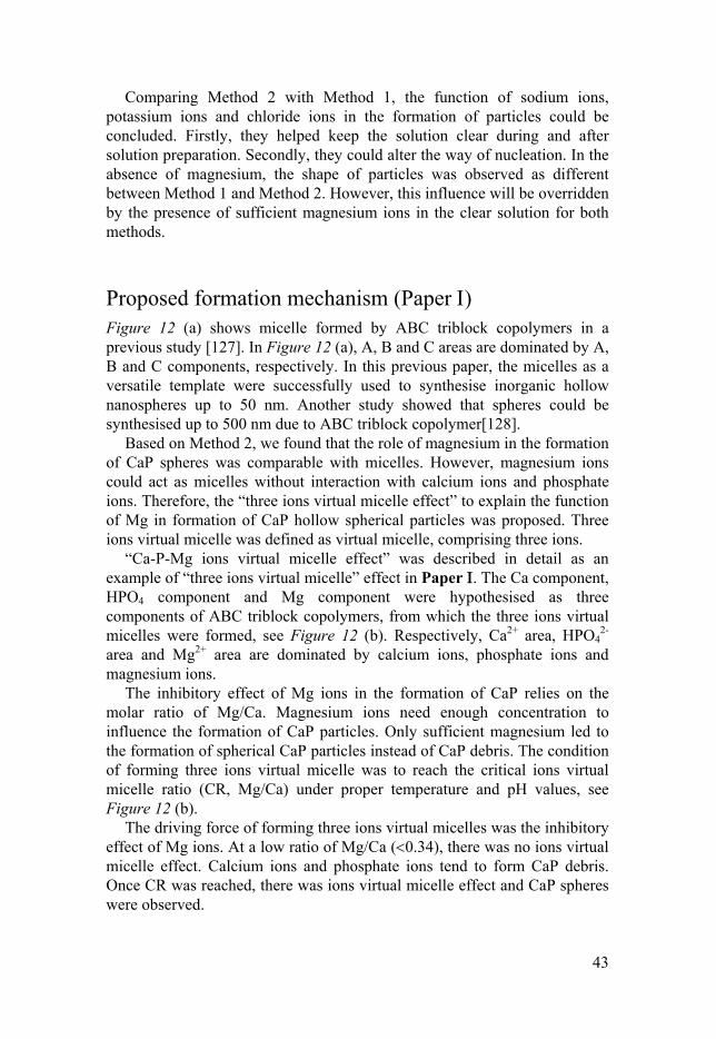
Proposed formation mechanism (Paper ) Figure 12 (a) shows micelle formed by ABC triblock copolymers in a previous study [127]. In Figure 12 (a), A, B and C areas are dominated by A, B and C components, respectively. In this previous paper, the micelles as a versatile template were successfully used to synthesise inorganic hollow nanospheres up to 50 nm. Another study showed that spheres could be synthesised up to 500 nm due to ABC triblock copolymer[128].
Based on Method 2, we found that the role of magnesium in the formation of CaP spheres was comparable with micelles. However, magnesium ions could act as micelles without interaction with calcium ions and phosphate ions. Therefore, the “three ions virtual micelle effect” to explain the function of Mg in formation of CaP hollow spherical particles was proposed. Three ions virtual micelle was defined as virtual micelle, comprising three ions.
“Ca-P-Mg ions virtual micelle effect” was described in detail as an example of “three ions virtual micelle” effect in Paper . The Ca component, HPO4 component and Mg component were hypothesised as three components of ABC triblock copolymers, from which the three ions virtual micelles were formed, see Figure 12 (b). Respectively, Ca2+ area, HPO4
2- area and Mg2+ area are dominated by calcium ions, phosphate ions and magnesium ions.
The inhibitory effect of Mg ions in the formation of CaP relies on the molar ratio of Mg/Ca. Magnesium ions need enough concentration to influence the formation of CaP particles. Only sufficient magnesium led to the formation of spherical CaP particles instead of CaP debris. The condition of forming three ions virtual micelle was to reach the critical ions virtual micelle ratio (CR, Mg/Ca) under proper temperature and pH values, see Figure 12 (b).
The driving force of forming three ions virtual micelles was the inhibitory effect of Mg ions. At a low ratio of Mg/Ca (0.34), there was no ions virtual micelle effect. Calcium ions and phosphate ions tend to form CaP debris. Once CR was reached, there was ions virtual micelle effect and CaP spheres were observed.

44
Figure 12. Schematic drawing for explaining the mechanism described in Paper I. (a) Micelle effect in the reference[127], (b) Ca-P-Mg ions virtual micelle effect due to Ca ions, HPO4 ions and Mg ions interactions. In this figure, Ca2+ area, HPO4
2- area and Mg2+ area are dominated by calcium ions, phosphate ions and magnesium ions, respectively.

45
Figure 13. SEM images of particles synthesised from control groups and standard groups based on Method 2. (A-1) control with Ca/F=1.8, Mg/Ca=0. (A-2) standard with Ca/F=1.8, Mg/Ca=0.55. (B-1) control with F/Ca=2.61, SO4/F=0 (B-2) standard with F/Ca=2.61, SO4/F=0.49. (C-1) control with Sr/F=0.2, Mg/Sr=0. (C-2) standard with Sr/F=0.2, Mg/Sr=1.06. (D-1) control with F/Sr=5.03, SO4/F=0. (D-2) standard with F/Sr=5.03, SO4/F=0.3. (E-1) control with Ba/F=0.2, Mg/Ba=0. (E-2) standard with Ba/F=0.2, Mg/Ba=1.04 .

46
Three ions virtual micelle effect could be potentially applied to explain the function of magnesium on the formation of strontium phosphate and barium phosphate spheres, see Figure 11. It could also be potentially applied to fluoride-based materials, see Figure 13.
In the absence of magnesium, precipitates of calcium fluoride, strontium fluoride and barium fluoride were non-spherical particles, see Figure 13 (A-1), (C-1) and (E-1). In the presence of magnesium, however, precipitates of calcium fluoride, strontium fluoride and barium fluoride were spheres, see Figure 13 (A-2), (C-2) and (E-2).
In the absence of sulphate, precipitates of calcium fluoride and strontium fluoride were non-spherical particles, see Figure 13 (B-1) and (D-1). In the presence of sulphate, precipitates of calcium fluoride and strontium fluoride were spheres, see Figure 13 (B-2) and (D-2).
Modified PMMA (Paper II) The weight percentage of drug loaded in SCPS was 27.7%. Simplex-2.5V, Simplex-10SCPSV and Simplex-10SCPSV-2.5V contained 2.5wt%, 2.77wt% and 2.5wt%, respectively, see Table 5.
Figure 14 (A) and (B) show that Simplex-10SCPSV-2.5V released the largest amounts of vancomycin, followed by Simplex-2.5V and Simplex-10SCPSV. In this study, the release profiles of antibiotics of all groups were similar to observations in previous studies [77,129]. The vancomycin had an initial burst release in the first few hours and the major part of the release was finished within 24 h for all the groups. After 24 h, there was a continued low steady release over the entire time period measured.
Figure 14. Cumulative release of vancomycin in PBS, (A) pH=4 (B) pH=7.4.
The cumulative vancomycin release in the PBS medium with a lower pH (pH=4) was higher than that with a higher pH value (pH=7.4). This was probably due to the fact that an acidic environment degraded the PMMA and made it more susceptible to water penetration. Adding SCPS increased the

47
vancomycin release by increasing the physical release possibilities via the spheres in the cement. Drug-loaded SCPS decreased the release compared to the release from regular PMMA because the loaded vancomycin was trapped in the SCPS and was more difficult to release. Paper II showed vancomycin release from Simplex-2.5V, Simplex-10SCPSV and Simplex-10SCPSV-2.5V were Fickian diffusion.
The total amounts of vancomycin released from Simplex-2.5V, Simplex-10SCPSV and Simplex-10SCPSV-2.5V after 1 week were 9%, 6% and 2%, respectively, of the total amount of antibiotic incorporated in the PMMA cements at a pH of 7.4. Compared to some previous studies [130,131], 9% was a high amount to be released from a PMMA cement. A previous study showed that vancomycin-loaded PMMA cement irradiated with ultrasound (US) and microbubble-mediated US could release up to 3.5% [130]. Regarding the size of the PMMA beads, small beads (10 mm in diameter), medium beads (15 mm in diameter) and large beads (20 mm in diameter) were found to release 7.2%, 4.3% and 3.1% of the total amounts of vancomycin, respectively [131].
EM and CS of PMMA and PMMA/SCPS are shown in Figure 15. There was no significant difference between PMMA and PMMA/SCPS. However, the radiopacity and working time significantly increased. In Paper II, the results showed that the radiopacity of PMMA/SCPS was double that of PMMA. The working time of PMMA/SCPS was also 50 seconds longer than that of pure PMMA.
Figure 15. EM and CS of PMMA and PMMA/SCPS.

48
Tooth bleaching (Paper III) The release study of HP was carried out on two types of samples, CP-loaded CaP spheres and modified CP-loaded CaP spheres, see Figure 16. The CP-loaded CaP spheres gave a burst release with a large amount HP released during the first 30 minutes. The release continued with a gradual release till 100 minutes. HP (4.3%) was detected from release.
Modified CP-loaded CaP spheres were loaded at a lower temperature and with a certain HP/Urea ratio. The modified CP-loaded CaP spheres showed a controlled release of HP. The release lasted 150 minutes and almost 10wt% HP was released, which was twice the amount released using the unmodified method (4.3wt% HP).
The paste made of unmodified CP-loaded CaP spheres gave a burst but gradual release in first 50 minutes. In Paper III, the results also showed most degradation of the rhodamine B occurred during the first 50 minutes.
The bleaching effect of control, at-home gel and in-office gel are shown in Table 8. After 45 minutes of bleaching, mean ∆E of control gel was highest, followed by in-office gel and at-home gel. Unsurprisingly, the control with 36wt% HP led to highest colour change ∆E (24.83) because HP was the active bleaching agent. The mean ∆E of the in-office gel and at-home gel was 16.56 and 11.89, respectively. The digital images of original and bleached teeth (Paper III) showed that both in-office gel and at-home gel made the teeth significantly whiter. The in-office gel resulted in more colour changes due to higher HP concentration compared to at-home gel. Therefore, we could conclude that the CaP spheres could maintain the bleaching effect. A previous study has also proved that ACP did not affect the bleaching effect [117].
Surprisingly, the colour change continued to increase after the gel was washed away from the teeth. This could be caused by spheres adhering to the surface. The adhering spheres could continue to release peroxide for up to 300 minutes. In Paper III, new apatite was observed on the surface of the enamel after 3 days of treatment with the at-home gel.

49
Figure 16. HP release profiles from unmodified CP-loaded CaP spheres (A) and modified CP-loaded CaP spheres (B): (A) spheres, CP and water were mixed by stirring for 2 hours at room temperature, (B) spheres, CP and water mixed by stirring for 2 hours at 9 .
Table 8. Mean (± standard deviation) colour changes of teeth after bleaching. Control is a commercial gel with 36% Hydrogen peroxide. At-home gel was CP mixed with CP-loaded CaP spheres and glycerol. In-office gel was CP-loaded CaP spheres mixed with 35% HP.
∆L ∆a ∆b ∆E
Control with 45 min bleaching 22.30 -10.90 -0.70 24.83
45 min bleaching by in-office gel 11.43±3.32 -8.40±2.34 -8.55±2.91 16.56±5.00 15 min after washing by in-office gel
14.58±3.26 -7.07±2.42 -3.97±3.07 16.68±5.09
24 h after washing by at-home gel 20.05±3.63 -5.78±2.39 0.20±1.66 20.87±4.65 45 min bleaching by at-home gel 9.08±3.42 -5.74±1.63 -5.10±3.24 11.89±4.48 2 h after washing by at-home gel 8.94±2.81 -6.46±1.72 -6.26±3.01 12.68±4,47
Diffusion of HP (Paper IV) In Paper IV, the HP concentrations of all five bleaching products (three own-made and two commercial) were measured. The HP concentrations of Gel 1, Gel 2, Gel 3, NiteWhite and DayWhite were 7.1wt%, 2.9wt%, 4.1wt%, 6.2wt% and 11.3wt%, respectively.

50
Figure 17 shows the steady-state flux of HP through the enamel discs. DayWhite showed the highest HP flux, followed by Gel 1. Gel 2, NiteWhite and Gel 3 gave the lowest flux. There was very significant difference of steady-state flux between DayWhite gel and other four gels (p0.01). The steady-state flux of Gel 1 was significantly larger than Gel 3 (p 0.03). Higher concentration of HP caused a higher flux through enamel discs. The studies of HP penetrating enamel and dentin also showed that higher HP concentrations resulted in higher flux [132,133].
No significant difference in the diffusivity of HP was found between Gel 1 and all other gels (p0.06), see Figure 18. However, significant difference was observed between NiteWhite and DayWhite (p0.01). Under steady-state condition, the membrane properties [134] and diffusate properties [135] and temperature [122]determine diffusivity of the diffusate. In this study, HP was the only diffusate and temperature was room temperature. The reason for the significant differences between NiteWhite and DayWhite should be due to the discrepancy between the mathematical model and experimental set-up. After all, the experimental set-up could not reach ideal conditions, while the mathematical model is ideal.
Paper IV showed that it was the diffusion-limited process during HP penetrating through enamel discs. The dominant process was the consumption reaction of HP. Moreover, no effect of CaP spheres on diffusion was observed in this study. Therefore, CaP spheres are suitable as a remineralisation agent in the bleaching products.
Figure 17. The steady-state flux of HP from all bleaching gels (Gel 1, Gel 2, Gel 3, NiteWhite and DayWhite) through the enamel discs. For each gel, n=5.
0
2
4
6
8
10
12
14
16
Gel1 Gel2 Gel3 NiteWhite DayWhite
Stea
dy-
stat
e fl
ux
(ng/
cm^
2/S
)
Steady-state flux
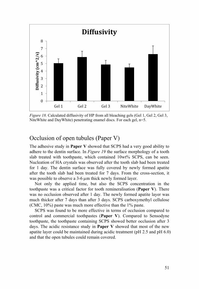
51
Figure 18. Calculated diffusivity of HP from all bleaching gels (Gel 1, Gel 2, Gel 3, NiteWhite and DayWhite) penetrating enamel discs. For each gel, n=5.
Occlusion of open tubules (Paper V) The adhesive study in Paper V showed that SCPS had a very good ability to adhere to the dentin surface. In Figure 19 the surface morphology of a tooth slab treated with toothpaste, which contained 10wt% SCPS, can be seen. Nucleation of HA crystals was observed after the tooth slab had been treated for 1 day. The dentin surface was fully covered by newly formed apatite after the tooth slab had been treated for 7 days. From the cross-section, it was possible to observe a 3-6 m thick newly formed layer.
Not only the applied time, but also the SCPS concentration in the toothpaste was a critical factor for tooth remineralisation (Paper V). There was no occlusion observed after 1 day. The newly formed apatite layer was much thicker after 7 days than after 3 days. SCPS carboxymethyl cellulose (CMC, 10%) paste was much more effective than the 1% paste.
SCPS was found to be more effective in terms of occlusion compared to control and commercial toothpastes (Paper V). Compared to Sensodyne toothpaste, the toothpaste containing SCPS showed better occlusion after 3 days. The acidic resistance study in Paper V showed that most of the new apatite layer could be maintained during acidic treatment (pH 2.5 and pH 6.0) and that the open tubules could remain covered.
0
1
2
3
4
5
6
7
8
Gel1 Gel2 Gel3 NiteWhite DayWhite
Dif
fusi
vity
(cm
^2
/s)
Diffusivity

52
Figure 19. Mineralisation on dentin surfaces induced by TP containing 10wt% SCPS after brushing for 1 day (A, B) and 7 days (C, D), (E) interface between dentin and mineralised surface of a tooth slab treated for 7 days.

53
Conclusions
The work presented in this thesis demonstrates that:
1) CaP spheres could successfully be synthesised by the modified method without large amounts of sodium chloride or potassium chloride. CaP spheres could be formed with a suitable amount of magnesium at any molar ratio of Ca/P. This modification will benefit its industrial application. This modified method could also be applied to synthesize other inorganic spheres, such as calcium fluoride, strontium phosphate, strontium fluoride, barium phosphate and barium fluoride. Furthermore, a proposed model, called three ions virtual micelle effect, could explain the formation of hollow CaP spherical particles without the use of templates. 2) The incorporation of strontium-doped CaP spheres into PMMA cement can facilitate vancomycin release. However, the release rate decreases when the vancomycin is pre-loaded into the strontium-doped CaP spheres. The mechanical properties of PMMA under compression are not significantly changed by the incorporation of strontium-doped CaP spheres. CaP spheres could be an additive to currently used PMMA cements, where enhanced drug delivery is desired. 3) The bleaching gels containing CaP spheres achieve good bleaching effects. Incorporation of CaP spheres does not affect the bleaching negatively. Moreover, CaP spheres could improve the controlled release of HP. CaP spheres also improve tooth remineralisation and maintain the enamel structure. CaP spheres could be an alternative ingredient in bleaching products. 4) The diffusion experiments show that higher concentrations of peroxide cause a higher steady-state flux through the enamel. CaP spheres do not influence the diffusion of hydrogen peroxide through enamel. Our bleaching gels containing low HP concentration could be a good alternative to current bleaching products. 5) Tooth remineralisation via a toothpaste containing strontium-doped CaP spheres was tested for occlusion of exposed dental tubules. The results show that dentin surfaces could be completely covered by newly formed apatite layer after 7 days of treatment. A toothpaste containing strontium-doped CaP spheres could improve tooth remineralisation.

54
Future perspectives
The work in this thesis presents only limited explanations of the three ions virtual micelle effect and a few applications of CaP spheres. Many topics still need to be investigated, and a selection is listed below.
Mechanism study of sphere formation is a hot topic. The current mechanisms, such as self-assembly, Ostwald ripening and Kirkendall effect, have their own limitations. No exception, the three ions virtual micelle effect is also limited. Therefore, further studies about this effect should be carried out.
The controlled release of vancomycin and HP via CaP spheres should be investigated further. Through modification, the release could be better controlled. The loading capacity of the spheres could be further modified. We have seen that the spheres’ loading capacity of HP was increased by the modified method.
Based on quantitative analysis, the clinical parameters should be recommended, such as applied time, renewal time during tooth bleaching. Even though bovine teeth are the most used substitute for human teeth in dental studies, there are still inconsistent outcomes. Therefore, dental research with human teeth will be better for determining clinical parameters.
The real saliva is best medium for remineralisation study in vitro. Tooth remineralisation effect of CaP spheres should be further studied for long term in saliva. The force of brushing is a key factor influencing the remineralisation. Therefore, researches investigating different forces should be performed.
More applications of CaP spheres should be investigated. CaP spheres could be applied as raw material for CaP cement and 3D printing, CaP putties. CaP spheres could also be applied as delivery carrier of more drugs, cells and genes.

55
Sammanfatting på Svenska
Under senare år har sfärer av kalciumfosfat (CaP) blivit uppmärksammad som biomterial. Kalciumfosfater är ett material som med en ihålig struktur kan vara gynnsam för laddning och frisättning av läkemedel eller tillsatts i produkter för tandvård, t.ex. tandkräm, där remineralisering av tandvävnad är viktigt.
Det finns många sätt att framställa sfärer av kalciumfosfat. Metoderna kan indelas i grupper efter vilka typer reagens som används: uppslamning, lösning, pasta och pulver. Används en lösning som reagens, indelas denna metod vidare i två huvudkategorier. Den ena huvudkategorin innebär olika sätt att spreja med ett spridningsverktyg. I en tidigare svensk studie användes electrospray i framställandet av sfäriska partiklar av trikalciumfosfat. Den andra huvudkategorin sker utfällning av sfärer av CaP vid användandet av ytaktiva ämnen och biomolekyler. Surfaktanter och biomolekyler kan styra själva kärnbildningen och tillväxten av CaP sfärer. I närvaron av upplösta aminosyror och dipeptider kan ihåliga CaP sfärer (HCPS) bildas. I aktuella studier är dessa två metoder fortfarande populära vid syntes av sfäriska CaP partiklar. Ett borttagande av mallen är en nackdel med en mallbaserad metod, men i framtiden kommer det vara mer ekonomisk och vanligt att använda mallfria metoder. Under de senare åren, har det varit några genombrott inom syntes av sfäriska CaP partiklar. CaP sfärer kan åstadkommas utan surfaktanter och biomolekyler, men då istället med en specifik fosfatkälla eller en övermättad buffertlösning. Även om det finns metoder att bilda ihålliga sfäriska partiklar av CaP utan närvaro av mall, så måste nackdelarna som begränsar industrialiseringen av detta uppmärksammas och lösas.
En kontroll av formen på oorganiska nanopartiklars kristallstruktur är mycket användbart i en rad tillämpningar. I en kemisk miljö kan en mindre modifiering ge ihåliga nanopartiklar med helt olika morfologi och komposition. Om parametrar och processen kunde kontrolleras under reaktionen, skulle det öppna upp för nya syntesvägar inom produktion av ihåliga partiklar. Syntesvägen är däremot bara effektiv om det föreligger en total förståelse av mekanismen. Självsammansättning används i beskrivningen av fenomen när stora funktionella enheter bildas vid spontan självsammansättning av mindre komponenter. Den drivande kraften bakom denna process är en strävan att minimera Gibbs fria energi. Det finns två modeller i litteraturen för hur partiklar kan bildas ur en lösning ”Ostwald
56
repening” och Kirkendaleffekten. Där Kirkendaleffekten beskriver rörelsen gränsskiktet mellan två metaller, som orsakas av skillnader i diffusionshastigheten hos metaljon.”Ostwald repening” beskriver hur små kristaller som löses och återförs på en större kristall. Processer där en inhomogen struktur ändras över tid kallas ”Ostwald repening”. Vid tillverkning av ihåliga oorganiska nanostrukturer (Co3O4, ZnS och TiO2) har ”Ostwald repening” tillämpats utan använding av mallar eller surfaktanter. Ingen dessa modeller kan beskriva bildandet av ihåliga CaP sfärer från lösningar.
I den här avhandlingen är det syntes, mekanism och tillämpning av CaP som ligger i fokus. Avhandlingen beskriver först och främst två syntesmetoder för att framställa CaP sfäriska partiklar. Den första metoden, en syntes av CaP sfäriska partiklar via en övermättad buffertlösning, är baserad på en tidigare studie. Den används i tillverkningen av sfäriska partiklar för tillämpningar i avhandlingen. I den här metoden, används en stor mängd extra salt som natriumklorid eller kaliumklorid. Den andra metoden kom till med syftet att förklara mekanismen för hur ihåliga CaP sfärer bildas. Metoden kunde avslöja att magnesium medverkar i bildandet av sfäriska partiklar oavsett Ca/P kvot. Bara aktiva joner av kalcium, fosfat och magnesium är med i bildandet av sfäriska partiklar, vilket kan bekräftas av denna förenklade metod. En ny modell kallad tre jonmicellernas effekt, en jämförelse med termodynamiken när misceller bildas, blev framtagen med syftet att förklara magnesiumets funktion. Efter denna mekanism upptäcktes en serie sfäriska partiklar med andra kompositioner. Dessa sfäriska partiklar inkluderade strontiumfosfat, bariumfosfat, kalciumfluorid, strontiumfluorid, och bariumfluorid.
En lokal infektion nära ett implantat kan orsaka alvarliga problem hos patienten. Antibiotika i akryl(PMMA-baserad) cement är ett vanligt sätt att administrera antibiotika med syftet att förebygga och behandla intill höftledsinplantat. I nuläget är ändå inte egenskaperna optimala vid frisättning av läkemedel från PMMA cement. En initiell dumpning påföljd av en långsam frisättning innebär att en onödigt stor mängd antibiotika behöver tillsättas cementet, vilket ökar risken för antibiotika resistensutveckling. Huvudsyftet med den här studien är att öka läkemedelsfrisättning från PMMA cement, utan att påverka de mekaniska egenskaperna. CaP sfärer laddade med strontium (SCPS) inkorporerades i PMMA cement för att förbättra antibiotika frisättningen. Vid undersökningen av strontium och antibiotika (vancomycin) -frisättning användes induktivt kopplade plasma-atomemissionspektroskopi och UV-spektrofotometri. Det visade sig att inkorporeringen av SCPS i PMMA, kunde förbättra frisättningen av antibiotika, och avge strontiumjoner till omgivningen. Inkorporeringen av SCPS ökade både röntgenopaciteten och förlängde bearbetningstiden för cementet. Tryckhållfastheten och styvheteten blev inte påverkad. Våra resultat visar att SPCS/PMMA

57
cementet har en förbättrad frissättning av antibiotika, avger strontiumjoner, och upprätthåller mekaniska egenskaper. Det här indikerar att SCPC kan vara ett bra val av tillsättningsämne vid kontrollerad frisättning av läkemedel från PMMA cement.
Den andra tillämpningen av HCPS är en kontrollerad frisättning av hydrogenperoxid(HP) för säkrare tandblekningsprodukter. I många tandblekningsprodukter är karbamidperoxid (CP) den aktiva ingrediensen. I det här arbetet testades två geler med CP laddade HCPS. Den ena gelen med en låg CP (dvs låg HP) koncentration som exempelvis i en ”hemmablekningsgel”, medan den andra var med en hög koncentration liknande i en ”professionell blekningsgel”. Respektive gels blekningseffekt på tänder blev undersökt med avseende på tandblekningsförmåga över tid. För att kunna undersöka den möjliga remineraliseringseffekten vid användning av HCPS, blev blekta tänder förvarade i Dulbecco fosfatbuffert lösning (PBS). Efter tre dagar i PBS kunde remineralisering observeras på den sidan där HCPS fastnat på tandens yta. HCPS laddad med CP förlänger frisättningen av HP och ger bättre blekningseffekt. HCPS visade sig vara effektiv i att öka vithetsgraden och öka remineraliseringsförmågan efter blekningsprocessen. Resultaten visar att HCPS kan ge en kontrollerad frisättning av HP utan att påverka blekningseffekten. De testade blekningsgelerna som innehåller HCPS kan vara ett alternativ till nuvarande använda blekningsmetoder, framför allt för att öka säkerheten genom att en minskning av peroxidhalt kan vara möjlig.
Det kommer kontinuerligt nya tandblekningsprodukter med olika formuleringar på marknaden. Bedömningen av produkterna är för det mesta empiriska i brist på kvantitativa analyser av mängden väteperoxid som går genom emaljen. Till exempel, patienter fokuserar bara på blekningseffekten, medan bieffekter blir helt ignorerade, eftersom det saknas kvantitativ data eller kunnskap om bieffekterna. En beräkning av diffusionsflödet av väteperoxid genom emaljen är en metod som kan ge värdefull information om själva mekanismen bakom tandblekning i tänder. Avsikten med den här studien är att undersöka om tillsättningen av CaP mikrosfärer ändrar väteperoxids diffusionsegenskaper genom emaljen. I undersökningen användes tre testgeler och två produkter från marknaden. En experimentell metod som anpassar en matematisk modell användes under olika externa betingelser, för att bestämma de fysiska parametrarna av väteperoxidtransporten in i tanden. Resultaten visar att högre koncentration av väteperoxid i produkten i slutändan ger ett högre flöde genom emaljen, oavsett mängd CaP mikrosfärer närvarande.
I den tredje tillämpningen undersöktes sfärernas som additiv till en tandkräm. Genom tillsatsen av strontiumladdade CaP sfärer (SCPS) var målet att påvisa ocklusion samt remineralisering av öppna tandhalsar i bänktester. Efter sju dagars behandling, var dentintubuli täckt med ett nybildat apatittlager. Resultatet visade även att SCPS kunde tränga ner i

58
tandtubuli under tandborstning samt fastna på dentinets yta. Efter tre dagars borstning, täcktes dentinets yta helt av det nybildade apatittlagret. Det nybildade lagret kan observeras även efter att provet har blivit dränkt i en sur lösning med pH 2.5 i två minuter. Den nya tandkrämen som innehåller SCPS, visade en bättre effekt på ocklusion och vidare remineraliseringsförmåga, jämfört med andra tandkrämer i marknaden.
Den här avhandlingen har presenterat en begränsad förklarning på de tre jonmicellernas effekt, samt några möjliga tillämpningar. Därför borde flera nya studier göras.
1) Innan de tre jonmicellernas effekt accepteras av forskare inom forskningsfältet, behövs det en stor insats av en modifiering av de tre joncellernas effekt.
2) Kontrollerad frisättning av vancomycin och peroxid via CaP sfärer bör undersökas vidare. Genom ytterligare optimering kan frisättningen kontrolleras bättre.
3) Fler tillämpningar med CaP sfärer bör undersökas. CaP sfärer kan användas inte bara som råmaterial i kalciumfosfatcement, i 3D printer, i polymercement, men också vid läkemedelsformulering som bärare för flera typer läkemedelssubstanser, gener och celler.

59
Acknowledgements
I would like to express my gratitude at the end of my PhD study in Material in Medicine group (MiM), Uppsala, Sweden.
I would like to express my great gratitude to my main supervisor, Håkan Engqvist. First, thanks for your professional guidance and efforts, sincerely. I always could imagine there are always at least ten manuscripts waiting for your comments. Second, I am very appreciative that I could always believe in you and see hope from you. It was not easy during my PhD study. Third, thanks for your patience during my supervision. What I had thought about manuscript submission, defence and writing was incorrect in most cases. Fourth, thanks for the opportunity for me to do my PhD in MiM group. Academically and in life, I have seen a broad world here. Your wisdom of connecting academy with industry, leading a group and starting companies is very impressive. I believe these will benefit me in my future career.
I would like to express many thanks to my co-supervisor, Fredrik Nikolajeff, for his support and help. Also, I would like to express my thanks to other co-authors: Caroline Öhman, Wei Xia, Cecilia Persson, Klaus Leifer,Alejandro López, Steven Jefferies, Peng Zhang, Ishtiaq Hassan Wani, Yuanyuan Han, Yulong Zhang, Ben Wu, Torbjörn Mellgren, Yixiao Cai, Felicia Suska and so on. Without your hard efforts and valuable comments, I could not have finished this work.
Thanks to all friends and colleagues, Jannica Heinrichs, Gemma Mestres Beà, Hu Li, Elin Larsson, Dongqi Bi, Céline Robo, Charlotte Skjöldebrand, Sara Gallinetti, Susanne Bredenberg, Luimar Correa Filho, Lee Fowler, Le Fu, Malin Eriksson, Oscar Janson, Denis Pavel Andreea, Cuiyan Li and Xiao Huang, Thomas Joffre, Bang Le, Susanne Lewin, Luo Jun and Jiaojiao, Marjam Ott, Johan Persson, Jinbao Zhang, Li Yang, Xiaowen Li, Chengjuan Liu, Yi Ren, Johanna and Erik Unosson, Ling Xie, Kai Hua, Jingtao Chu, Martin Röjås, Amina Vazda, Johnny Eriksson, Liyang Shi, Lei Zhang, Yu Zhang, Changguan Xu, Bing Cai, Ernesto Vargas Catalan, Pierre Piron, David Wenner, Fengping Yang, Cui Li, Han Li, Dan Wu and so on. Thanks to all other members from Microscopy group, Tribology group and Nanotechnology group. There are too many names, so I am sorry that I could not mention everyone here. Thanks for all your help and great time in Uppsala.
Thanks to Jinxing and Yingying, Song and Shu, Dou Du, Wen Huang, Mingzhi and Yurong, Shiuli and Michael, for the delicious food and
60
organising great parties. Thanks to Emil and Strömbom family, for fun times and hospitality. Thanks to Jonatan Bagge, for being the best and kindest computer guy ever. Thanks to Ingrid Ajaxon, for always being helpful. Thanks to Petra Olander for your nice talks and your care. Thanks to Huizhang Guo and Ruitao Wen, for your time and effort. Thanks to Xiaohan Zhang, for the good food and accommodation.
Thanks to all nice friends, classmates, neigbours, teachers and relatives in China, for your kindness and careness. Thanks to my dear Marianne and all family members, for your support, happy times and lovely home. Thanks to my mum, great gratitude for her motherhood and heroic efforts during my study in middle school, high school and college. She is the main reason I was able to go to college and study further. Thanks to my brother, sister-in-law and nephew ‒ I very much appreciate you taking good care of our parents when I am always far away from home.

61
References
1. Rho JY, Kuhn-Spearing L, Zioupos P. Mechanical properties and the hierarchical structure of bone. Med. Eng. Phys. Elsevier Sci Ltd; 1998;20:92–102.
2. Dorozhkin S V. A detailed history of calcium orthophosphates from 1770s till 1950. Mater. Sci. Eng. C. 2013. p. 3085–110.
3. Tadashi Kokubo. Bioceramics and their clinical applications. 2008. 4. Kay MI, Young RA, Posner AS. Crystal Structure of Hydroxyapatite. Nature.
1964;204:1050–2. 5. Elliott JC. The crystallographic structure of dental enamel and related apatites.
Departm eit Dent. Histol. Pathol. University of London; 1964. 6. Bohner M. Calcium Phosphate Emulsions: Possible Applications. Key Eng.
Mater. 2001;192–195:765–8. 7. Tampieri A, Celotti G, Szontagh F, Landi E. Sintering and characterization of HA
and TCP bioceramics with control of their strength and phase purity. J. Mater. Sci. Mater. Med. Chapman & Hall Ltd; 1997;8:29–37.
8. Nasiri-Tabrizi B, Fahami A. Mechanochemical synthesis and structural characterization of nano-sized amorphous tricalcium phosphate. Ceram. Int. 2013;39:8657–66.
9. Cao W, Hench LL. Bioactive materials. Ceram. Int. 1996;22:493–507. 10. Yuan H, Groot Klaas De. Calcium Phosphate Biomaterial: An Overview. Learn.
from Nat. How to Des. Implant. Biomater. 2004;171:37–57. 11. Bohner M. Resorbable biomaterials as bone graft substitutes. Mater. Today.
2010. p. 24–30. 12. da Silva RV, Bertran CA, Kawachi EY, Camilli JA. Repair of cranial bone
defects with calcium phosphate ceramic implant or autogenous bone graft. J. Craniofac. Surg. 2007;18:281–6.
13. Bohner M, Tadier S, Garderen N van, Gasparo A de, Döbelin N, Baroud G. Synthesis of spherical calcium phosphate particles for dental and orthopedic applications. Biomatter. 2013;3:e25103:1-14.
14. Daculsi G, Passuti N, Martin S, Deudon C, Legeros RZ, Raher S. Macroporous calcium phosphate ceramic for long bone surgery in humans and dogs. Clinical and histological study. J. Biomed. Mater. Res. 1990;24:379–96.
15. Ooms EM, Wolke JGC, Van de Heuvel MT, Jeschke B, Jansen JA. Histological evaluation of the bone response to calcium phosphate cement implanted in cortical bone. Biomaterials. 2003;24:989–1000.
16. Hutmacher DW. Scaffolds in tissue engineering bone and cartilage. Biomaterials. 2000;21:2529–43.
17. Bohner M, Galea L, Doebelin N. Calcium phosphate bone graft substitutes: Failures and hopes. J. Eur. Ceram. Soc. 2012;32:2663–71.
18. Dhert WJA, Klein CPAT, Jansen JA, Van der Velde EA, Vriesde RC, Rozing PM, et al. A histological and histomorphometrical investigation of fluorapatite, magnesiumwhitlockite, and hydroxylapatite plasma-sprayed coatings in goats. J. Biomed. Mater. Res. 1993;27:127–38.

62
19. Søballe K, Hansen ES, Brockstedt-Rasmussen H, Bünger C. Hydroxyapatite Coating Converts Fibrous Tissue to bone Around Loaded Implants. J Bone Jt. Surg. 1993;75:270–8.
20. Wu F, Wei J, Guo H, Chen F, Hong H, Liu C. Self-setting bioactive calcium-magnesium phosphate cement with high strength and degradability for bone regeneration. Acta Biomater. 2008;4:1873–84.
21. Tanaka C, Shikata J, Ikenaga M, Takahashi M. Acetabular reconstruction using a Kerboull-type acetabular reinforcement device and hydroxyapatite granules: A 3- to 8-year follow-up study. J. Arthroplasty. 2003;18:719–25.
22. Oonishi H, Kadoya Y, Iwaki H, Kin N. Total hip arthroplasty with a modified cementing technique using hydroxyapatite granules. J. Arthroplasty. 2001;16:784–9.
23. Qi C, Zhu Y-J, Chen F, Wu J. Porous microspheres of magnesium whitlockite and amorphous calcium magnesium phosphate: microwave-assisted rapid synthesis using creatine phosphate, and application in drug delivery. J Mater Chem B. 2015;3:7775–86.
24. Xia W, Grandfield K, Schwenke A, Engqvist H. Synthesis and release of trace elements from hollow and porous hydroxyapatite spheres. Nanotechnology. 2011;22:305–610.
25. Bouble M, Marier J., Rose D. Effect of magnesium on formation of calcium phosphate precipitats. Arch. Biochem. biophys. 1962;96:629–36.
26. Cheng P, Grabher, JJLeGeros R. Effects of magnesium on calcium phosphate formation. Magnesium. 1988;7:123–32.
27. Hu J, Chen M, Fang X, Wu L. Fabrication and application of inorganic hollow spheres. Chem. Soc. Rev. Chem. Soc. Rev. 2011;40:5472–91.
28. Jiao Y, Lu Y-P, Xiao G-Y, Xu W-H, Zhu R-F. Preparation and characterization of hollow hydroxyapatite microspheres by the centrifugal spray drying method. Powder Technol. 2012;217:581–4.
29. Wang A, Lu Y, Zhu R, Li S, Ma X. Effect of process parameters on the performance of spray dried hydroxyapatite microspheres. Powder Technol. 2009;191:1–6.
30. Eltohamy M, Shin US, Won J-E, Kim J-J, Kim H-W. Electrosprayed tricalcium phosphate spherical microcups and antibiotic drug delivery. Mater Lett. 2011;65:2043–6.
31. Wang P, Yook S-W, Jun S-H, Li Y-L, Kim M, Kim H-E, et al. Synthesis of nanoporous calcium phosphate spheres using poly(acrylic acid) as a structuring unit. Mater Lett. 2009;63:1207–1209.
32. Hagmeyer D, Ganesan K, Ruesing J, Schunk D, Mayer C, Dey A, et al. Self-assembly of calcium phosphate nanoparticles into hollow spheres induced by dissolved amino acids. J. Mater. Chem. 2011;21:9219–25.
33. Li J, Zeng HC. Hollowing Sn-Doped TiO2 Nanospheres via Ostwald Ripening. J. Am. Chem. Soc. 2007;129:15839–47.
34. González1 E, Arbiol J, Puntes VF. Carving at the Nanoscale: Sequential Galvanic Exchange and Kirkendall Growth at Room Temperature. Science. 2011;334:1377–80.
35. Wang W, Dahl M, Yin Y. Hollow Nanocrystals through the Nanoscale Kirkendall Effect. Chem. Mater. 2013;25:1179–89.
36. Böker A, He J, Emrick T, Russell TP. Self-assembly of nanoparticles at interfaces. Soft Matter. 2007;3:1231–1248.
37. Holleman A, Wiberg E. Lehrbuch der Anorganischen Chemie. J. Chem. Inf. Model. 1995.

63
38. Gold V. Compendium of Chemical Terminology. Int. Union Pure Appl. Chem. 1997.
39. Liu B, Zeng HC. Symmetric and Asymmetric Ostwald Ripening in the Fabrication of Homogeneous Core–Shell Semiconductors. Small. 2005;1:566–71.
40. Nakajima H. The Discovery and Acceptance of the Kirkendall Effect. JOM. 1997;49:15–9.
41. Smigelskas AD, Kirkendall EO. Zinc Diffusion in Alpha Brass. Trans Tms-aime. 1947;171:130–42.
42. Chen D, Ye J. Hierarchical WO3 hollow shells: Dendrite, sphere, dumbbell, and their photocatalytic properties. Adv. Funct. Mater. 2008;18:1922–8.
43. Yu J, Yu X. Hydrothermal synthesis and photocatalytic activity of zinc oxide hollow spheres. Environ. Sci. Technol. 2008;42:4902–7.
44. Bala H, Yu Y, Zhang Y. Synthesis and photocatalytic oxidation properties of titania hollow spheres. Mater. Lett. 2008;62:2074–7.
45. Son JE, Chattopadhyay J, Pak D. Electrocatalytic performance of Ba-doped TiO2 hollow spheres in water electrolysis. Int. J. Hydrogen Energy. 2010;35:420–7.
46. Liang HP, Zhang HM, Hu JS, Guo YG, Wan LJ, Bai CL. Pt hollow nanospheres: Facile synthesis and enhanced electrocatalysts. Angew. Chemie - Int. Ed. 2004;43:1540–3.
47. Wu L, Feng H, Liu M, Zhang K, Li J. Graphene-based hollow spheres as efficient electrocatalysts for oxygen reduction. Nanoscale. 2013;5:10839–43.
48. Wu Z, Yu K, Zhangand S, Xie Y. Hematite hollow spheres with a mesoporous shell: Controlled synthesis and applications in gas sensor and lithium ion batteries. J. Phys. Chem. C. 2008;112:11307–13.
49. Jun Z, Shurong W, Mijuan X, Yan W, Huijuan X, Shoumin Z, et al. Polypyrrole-coated sno2 hollow spheres and their application for ammonia sensor. J. Phys. Chem. C. 2009;113:1662–5.
50. Gyger F, H??bner M, Feldmann C, Barsan N, Weimar U. Nanoscale SnO2 hollow spheres and their application as a gas-sensing material. Chem. Mater. 2010;22:4821–7.
51. Zhang J, Wang S, Wang Y, Xu M, Xia H, Zhang S, et al. ZnO hollow spheres: Preparation, characterization, and gas sensing properties. Sensors Actuators, B Chem. 2009;139:411–7.
52. Wang S, Shen H, Fan S, Zhao Y, Ma X, Gong J. CaO-based meshed hollow spheres for CO2 capture. Chem. Eng. Sci. Elsevier Ltd; 2015;135:532–9.
53. Zhuang Y, Yang Y, Xiang G, Wang X. Magnesium silicate hollow nanostructures as highly efficient absorbents for toxic metal ions. J. Phys. Chem. C. 2009;113:10441–5.
54. Syoufian A, Satriya OH, Nakashima K. Photocatalytic activity of titania hollow spheres: Photodecomposition of methylene blue as a target molecule. Catal. Commun. 2007;8:755–9.
55. Xu Z, Cao Y, Li C, Ma P, Zhai X, Huang S, et al. Urchin-like GdPO4 and GdPO4:Eu3+ hollow spheres – hydrothermal synthesis, luminescence and drug-delivery properties. J. Mater. Chem. 2011;21:3686.
56. Zhang X, Zhang W, Yang Z, Zhang Z. Nanostructured hollow spheres of hydroxyapatite: Preparation and potential application in drug delivery. Front. Chem. Sci. Eng. 2012;6:246–52.
57. Yang G, Gai S, Qu F, Yang P. SiO2@YBO3:Eu3+ hollow mesoporous spheres for drug delivery vehicle. ACS Appl. Mater. Interfaces. 2013;5:5788–96.

64
58. Zhu Y, Ikoma T, Hanagata N, Kaskel S. Rattle-type Fe3O4@SiO2 hollow mesoporous spheres as carriers for drug delivery. Small. 2010;6:471–8.
59. Zhu Y, Fang Y, Kaskel S. Folate-conjugated Fe3O4@SiO2 hollow mesoporous spheres for targeted anticancer drug delivery. J. Phys. Chem. C. 2010;114:16382–8.
60. Zhu Y, Fang Y, Kaskel S. Folate-Conjugated Fe 3 O 4 @SiO 2 Hollow Mesoporous Spheres for Targeted Anticancer Drug Delivery. J. Phys. Chem. C. 2010;114:16382–8.
61. Peters F, Reif D. Functional materials for bone regeneration from beta-tricalcium phosphate. Materwiss. Werksttech. 2004;35:203–7.
62. Tas AC. Preparation of porous apatite granules from calcium phosphate cement. J. Mater. Sci. Mater. Med. 2008;19:2231–9.
63. Kunio I. Effects of spherical tetracalcium phosphate on injectability and basic properties of apatitic cement. Key Eng Mater. 2003;240-2:369-72.
64. Suwanprateeb J, Sanngam R, Panyathanmaporn T. Influence of raw powder preparation routes on properties of hydroxyapatite fabricated by 3D printing technique. Mater. Sci. Eng., C. 2010;30:610–7.
65. Matsuno T, Hashimoto Y, Adachi S, Omata K, Yoshitaka Y, Ozeki Y, et al. Preparation of injectable 3D-formed beta-tricalcium phosphate bead/alginate composite for bone tissue engineering. Dent. Mater. J. 2008;27:827–34.
66. Hattori T, Lwadate Y. Hydrothermal preparation of calcium hydroxyapatite powders. J Am Ceram Soc. 1990;73:1803–5.
67. Wu H, Zhang S, Zhang J, Liu G, Shi J, Zhang L, et al. A Hollow-Core, Magnetic, and Mesoporous Double- Shell Nanostructure: In Situ Decomposition/Reduction Synthesis, Bioimaging, and Drug-Delivery Properties. Adv. Funct. Mater. 2011;21:1850–62.
68. Chen Y, Chu C, Zhou Y, Ru Y, Chen H, Chen F, et al. Reversible Pore-Structure Evolution in Hollow Silica Nanocapsules: Large Pores for siRNA Delivery and Nanoparticle Collecting. Small. 2011;7:2935–44.
69. Walsh LJ. Contemporary technologies for remineralization therapies: A review. Int. Dent. SA. 2009;11:7–15.
70. Cochrane N, Saranathan S, Cai F, Cross K, Reynolds E. Enamel Subsurface Lesion Remineralisation with Casein Phosphopeptide Stabilised Solutions of Calcium, Phosphate and Fluoride. Caries Res. 2008;42:88–97.
71. Cochrane N, Cai F, Huq N, Burrow M, Reynolds E. New Approaches to Enhanced Remineralization of Tooth Enamel. J Dent Res. 2010;89:1187–97.
72. Verron E, Khairoun I, Guicheux J, Bouler J-M. Calcium phosphate biomaterials as bone drug delivery systems: a review. Drug Discov Today. 2010;15:547–52.
73. Hashimoto Y, Adachi S, Matsuno T, Omata K. Effect of an injectable 3D scaffold for osteoblast differentiation depends on bead size. Biomed Mater Eng. 2009;19:391–400.
74. CHEUNG HS, HUBBARD TRDW. Calcium phosphate particle induction of metalloproteinase and mitogenesis: effect of particle sizes. Osteoarthr. Cartil. 1997;5:145–51.
75. Cui Q, William M. Mihalko, Shields JS, Ries M, Saleh KJ. Antibiotic-Impregnated Cement Spacers for the Treatment of Infection Associated with Total Hip or Knee Arthroplasty. J. Bone Jt. Surg. 2007;89:871–82.
76. Nowinski RJ, Gillespie RJ, Shishani Y, Cohen B, Walch G, Gobezie R. Antibiotic-loaded bone cement reduces deep infection rates for primary reverse total shoulder arthroplasty: a retrospective, cohort study of 501 shoulders. J Shoulder Elb. Surg. 2012;21:324–8.

65
77. Miola M, Alessandro B, Valsania M, Bianco C, Fucale G, Verné E. Antibiotic-loaded acrylic bone cements: An in vitro study on the release mechanism and its efficacy. Mater. Sci. Eng., C. 2013;33:3025–3032.
78. Nandi SK. Local antibiotic delivery systems for the treatment of osteomyelitis – A review. Mater. Sci. Eng., C. 2009;29:2478–2485.
79. Wininger DA, Fass RJ. Antibiotic-Impregnated Cement and Beads for Orthopedic Infections. Antimicrob. Agents Chemother. 1996;40(12):2675–2679.
80. John E. Antimicrobial Resistance in Hospital Organisms and Its Relation to Antibiotic Use. Clin. Infect. Dis. 1983;5:1033–48.
81. Joseph TN, Chen AL, Di Cesare PE. Use of antibiotic-impregnated cement in total joint arthroplasty. J. Am. Acad. Orthop. Surg. 2003;11:38–47.
82. Koo KH, Yang JW, Cho SH, Song HR, Park HB, Ha YC, et al. Impregnation of vancomycin, gentamicin, and cefotaxime in a cement spacer for two-stage cementless reconstruction in infected total hip arthroplasty. J. Arthroplasty. 2001;16:882–92.
83. Moursi AM, Winnard A V., Winnard PL, Lannutti JJ, Seghi RR. Enhanced osteoblast response to a polymethylmethacrylate-hydroxyapatite composite. Biomaterials. 2002;23:133–44.
84. Tihan TG, Ionita MD, Popescu RG, Iordachescu D. Effect of hydrophilic-hydrophobic balance on biocompatibility of poly(methyl methacrylate) (PMMA)-hydroxyapatite (HA) composites. Mater. Chem. Phys. 2009. p. 265–9.
85. Fini M, Giavaresi G, Nicoli Aldini N, Torricelli P, Botter R, Beruto D, et al. A bone substitute composed of polymethylmethacrylate and tricalcium phosphate: Results in terms of osteoblast function and bone tissue formation. Biomaterials. 2002;23:4523–31.
86. Hulsart-Billström G, Xia W, Pankotai E, Weszl M, Carlsson E, Forster-Horvath C, et al. Osteogenic potential of Sr-doped calcium phosphate hollow spheres in vitro and in vivo. J. Biomed. Mater. Res. - Part A. 2013;101 A:2322–31.
87. Arifin DY, Lee LY, Wang C-H. Mathematical modeling and simulation of drug release from microspheres: Implications to drug delivery systems. Adv. Drug Deliv. Rev. 2006;58:1274–325.
88. Fu Y, Kao WJW. Drug release kinetics and transport mechanisms of non-degradable and degradable polymeric delivery systems. Expert Opin. Drug Deliv. 2010;7:429–44.
89. Shaikh HK, Kshirsagar R V., Patil SG. Mathematical and Kinetic Models for Drug Release Characterization: A Review. World J. Pharm. Pharm. Sci. 2015;4:324–38.
90. Nanci A. Ten Cate’s oral histology, 8th edition. BDJ. Elsevier Mosby; 2012;213:194–194.
91. Pader M. Oral hygiene products and practice. New Yorker: Dekker; 1988. 92. Brannstrom M, Lindén L, Johnson G. Movement of dentinal and pulpal fluid
caused by clinical procedures. J Dent Res. 1968;47:679–82. 93. Berman LH. Dentinal sensation and hypersensitivity. A review of mechanisms
and treatment alternatives. J. Periodontol. 1985;56:216–22. 94. Brännström M, Aström A. The hydrodynamics of the dentine; its possible
relationship to dentinal pain. Int dent J. 1972;22:219–27. 95. Walters P. Dentinal hypersensitivity: A review. J Contemp Dent Pr. 2005;6:107–
17.

66
96. Poulsen S, Errboe M, Hovgaard O, Worthington HW. Potassium nitrate toothpaste for dentine hypersensitivity. Cochrane Database Syst. Rev. 2001;CD001476.
97. Tarbet WJ, Buckner A, Stark MM, Fratarcangelo PA, Augsburger R. The pulpal effects of brushing with a 5 percent potassium nitrate paste used for desensitization. Oral Surgery, Oral Med. Oral Pathol. 1981;51:600–2.
98. DePaola PF, Soparkar PM, Triol C, Volpe AR, Garcia L, Duffy J, et al. The relative anticaries effectiveness of sodium monofluorophosphate and sodium fluoride as contained in currently available dentifrice formulations. Am. J. Dent. 1993;6 Spec No: S7-12.
99. Thrash WJ, Dodds MW, Jones DL. The effect of stannous fluoride on dentinal hypersensitivity. Int. Dent. J. 1994;44:107–18.
100. Vieira a., Hancock R, Dumitriu M, Schwartz M, Limeback H, Grynpas M. How Does Fluoride Affect Dentin Microhardness and Mineralization? J. Dent. Res. 2005;84:951–7.
101. Xu A-W, Ma Y, Cölfen H. Biomimetic mineralization. J. Mater. Chem. 2006;17:415–49.
102. Niu LN, Zhang W, Pashley DH, Breschi L, Mao J, Chen JH, et al. Biomimetic remineralization of dentin. Dent. Mater. 2014;30:77–96.
103. Cao Y, Mei ML, Xu J, Lo ECM, Li Q, Chu CH. Biomimetic mineralisation of phosphorylated dentine by CPP-ACP. J Dent. 2003;4I:818–25.
104. Tung MS, Eichmiller FC. Amorphous calcium phosphates for tooth mineralization. Compend. Contin. Educ. Dent. 2004. p. 9–13.
105. Tschoppe P, Zandim DL, Martus P, Kielbassa AM. Enamel and dentine remineralization by nano-hydroxyapatite toothpastes. J. Dent. 2011;39:430–7.
106. Merchant V, Livingston M, Pashley D. Dentin permeation: Comparison of diffusion with filtration. J Dent Res 1977;561161– 4. 1977;56:1161–4.
107. Joiner A. The bleaching of teeth: A review of the literature. J. Dent. 2006. p. 412–9.
108. Christensen G, Christensen R. Home use bleaching study 1995. CRA Newsl. 1995;19:1–9.
109. Fugaro J, Nordahl I, Fugaro O. Pulp reaction to vital bleaching. Oper Dent. 2004;29:363–8.
110. Cavalli V, Arrais C, Giannini M, Ambrosano G. High-concentrated carbamide peroxide bleaching agents’ effects on enamel surface. J Oral Rehabil. 2004;31:155–9.
111. Leonard RH, Smith LR, Garland GE, Caplan DJ. Desensitizing agent efficacy during whitening in an at-risk population. J Esthet. Restor Dent. 2004;16:49–56.
112. Cummins D. Dentin hypersensitivity: from diagnosis to a breakthrough therapy for everyday sensitivity relief. J Clin Dent. 2009;20:1–9.
113. Tung MS, Eichmiller F. Dental Applications of Amorphous Calcium Phosphates. J. Clin. Dent. 1999;10:1–6.
114. Yates R, Owens J, Jackson R, Newcotnbe R, Addy M. A split-mouth placebo-controlled study to determine the effect of amorphous calcium phosphate in the treatment of dentine hypersensitivity. J Clin Periodontol. 1998;25:687–92.
115. Strassler H. Tooth whiting-now and in the future: Part 2. Contemp Esthet Restor Pr. 2004;8:50–5.
116. Giniger M, Macdonald J, Ziemba S, Felix H. The clinical performance of professionally dispensed bleaching gel with added amorphous calcium phosphate. J Am Dent Assoc. 2005;136:383–92.

67
117. Vasconcelos A, Cunha A, Borges B, Machado C, dos Santos A. Tooth whitening with hydrogen/carbamide peroxides in association with a CPP-ACP paste at different proportions. Aust Dent J. 2012;57:213–9.
118. Tredwin CJ, Naik S, Lewis NJ, Scully C. Hydrogen peroxide tooth-whitening (bleaching) products: review of adverse effects and safety issues. Br. Dent. J. 2006;200:371–6.
119. Bor Shiunn L, Shih Hao H, Yu Chih C, Yu Shan C, Chung Yuan M, Chun Pin L. Development of in vitro tooth staining model and usage of catalysts to elevate the effectiveness of tooth bleaching. Dent Mater. 2008;24:57–66.
120. Addy M, Moran J. Mechanisms of Stain Formation on Teeth, in Particular Associated with Metal Ions and Antiseptics. Adv. Dent. Res. 1995;9:450–6.
121. Satish S. Modeling Peroxide Release Dynamics and Pulp Penetration during Tooth-Bleaching Procedures. 2010.
122. Petersen BK, Wu BM. Quantitative analysis of the diffusion of hydrogen peroxide through teeth. 2012.
123. Camps J, de Franceschi H, Idir F, Roland C, About I. Time-Course Diffusion of Hydrogen Peroxide Through Human Dentin: Clinical Significance for Young Tooth Internal Bleaching. J. Endod. 2007;33:455–9.
124. Sulieman M, Addy M, Rees JS. Development and evaluation of a method in vitro to study the effectiveness of tooth bleaching. J. Dent. 2003;31:415–22.
125. Yassen GH, Platt J a., Hara AT. Bovine teeth as substitute for human teeth in dental research: a review of literature. J. Oral Sci. 2011. p. 273–82.
126. Xia W, Engqvist H. Ions substituted calcium phosphate particles. 2012. p. US No. 2012/0134919 A1.
127. Sasidharan M, Nakashima K. Core-shell-corona polymeric micelles as a versatile template for synthesis of inorganic hollow nanospheres. Acc. Chem. Res. 2014;47:157–67.
128. Lin S, Zhu W, He X, Xing Y, Liang L, Chen T, et al. Multicompartmental hollow micelles formed by linear ABC triblock copolymers in aqueous medium. J. Phys. Chem. B. American Chemical Society; 2013;117:2586–93.
129. van de Belt H, Neut D, Uges D, Schenk W, van Horn J, van der Mei H, et al. Surface roughness, porosity and wettability of gentamicin-loaded bone cements and their antibiotic release. Biomaterials. 2000;21:1981–7.
130. Lin T, Cai XZ, Shi MM, Ying ZM, Hu B, Zhou CH, et al. In vitro and in vivo evaluation of vancomycin-loaded PMMA cement in combination with ultrasound and microbubbles-mediated ultrasound. Biomed Res. Int. Hindawi Publishing Corporation; 2015;2015.
131. Shinsako K, Okui Y, Matsuda Y, Kunimasa J, Otsuka M. Effects of bead size and polymerization in PMMA bone cement on vancomycin release. Biomed. Mater. Eng. 2008;18:377–85.
132. Benetti AR, Valera MC, Mancini MNG, Miranda CB, Balducci I. In vitro penetration of bleaching agents into the pulp chamber. Int. Endod. J. 2004;37:120–4.
133. Gokay O, Tuncbilek M, Ertan R, Gökay O, Tunçbilek M. Penetration of the pulp chamber by carbamide peroxide bleaching agents on teeth restored with a composite resin. J. Oral Rehabil. 2000;27:428–31.
134. Van Brakel J, Heertjes PM. Analysis of diffusion in macroporous media in terms of a porosity, a tortuosity and a constrictivity factor. Int. J. Heat Mass Transf. 1974;17:1093–103.
135. Hattab FN. Diffusion of fluorides in human dental enamel in vitro. Arch. Oral Biol. 1986;31:811–4.
Acta Universitatis UpsaliensisDigital Comprehensive Summaries of Uppsala Dissertationsfrom the Faculty of Science and Technology 1446
Editor: The Dean of the Faculty of Science and Technology
A doctoral dissertation from the Faculty of Science andTechnology, Uppsala University, is usually a summary of anumber of papers. A few copies of the complete dissertationare kept at major Swedish research libraries, while thesummary alone is distributed internationally throughthe series Digital Comprehensive Summaries of UppsalaDissertations from the Faculty of Science and Technology.(Prior to January, 2005, the series was published under thetitle “Comprehensive Summaries of Uppsala Dissertationsfrom the Faculty of Science and Technology”.)
Distribution: publications.uu.seurn:nbn:se:uu:diva-305820
ACTAUNIVERSITATIS
UPSALIENSISUPPSALA
2016