sleep loss alters synaptic and intrinsic neuronal properties in mouse prefrontal cortex
TRANSCRIPT

B R A I N R E S E A R C H 1 4 2 0 ( 2 0 1 1 ) 1 – 7
Ava i l ab l e on l i ne a t www.sc i enced i r ec t . com
www.e l sev i e r . com/ loca te /b ra i n res
Research Report
Sleep loss alters synaptic and intrinsic neuronal properties inmouse prefrontal cortex
Bradley D. Winters, Yanhua H. Huang, Yan Dong, James M. Krueger⁎
Program in Neuroscience, Washington State University, Pullman, WA 99164-6520, USA
A R T I C L E I N F O
⁎ Corresponding author at: WWAMI Medical E1495, USA. Fax: +1 509 358 7627.
E-mail addresses: [email protected].(Y. Dong), [email protected] (J.M. Kru
Abbreviations: SD, sleep deprivation; PFC, pcurrent; mIPSC, miniature inhibitory postsynNREM, non-rapid eye movement; TTX, artifmethyl-4-isoxazolepropionic acid; AHP, after
0006-8993/$ – see front matter © 2011 Elseviedoi:10.1016/j.brainres.2011.08.078
A B S T R A C T
Article history:Accepted 31 August 2011Available online 7 September 2011
Despite sleep-loss-induced cognitive deficits, little is known about the cellular adaptationsthat occur with sleep loss. We used brain slices obtained from mice that were sleep de-prived for 8 h to examine the electrophysiological effects of sleep deprivation (SD). Weemployed a modified pedestal (flowerpot) over water method for SD that eliminatedrapid eye movement sleep and greatly reduced non-rapid eye movement sleep. In layerV/VI pyramidal cells of the medial prefrontal cortex, miniature excitatory post synapticcurrent amplitude was slightly reduced, miniature inhibitory post synaptic currents wereunaffected, and intrinsic membrane excitability was increased after SD.
© 2011 Elsevier B.V. All rights reserved.
Keywords:Sleep deprivationPrefrontal cortexMembrane excitabilityEPSCIPSCSynapticFlowerpot
1. Introduction
The prefrontal cortex (PFC) mediates cognitive responses(Goldman-Rakic, 1995; Miller and Cohen, 2001; Robbins, 2005)that are sensitive to disruption by sleep loss. Following sleepdeprivation (SD), memory, planning, decision-making, andother PFC-mediated cognitive abilities are impaired (Chuah,2006; Thomas et al., 2000; Wu et al., 2006). Accompanyingthese post-SD behavioral alterations, the PFC exhibits largerincreases in slow-wave (0.5–4.0 Hz) activity during sleep thanother brain regions (Finelli et al., 2000; Mauzur et al., 2002),suggesting the PFC is disproportionately affected by SD
ducation Program, Health
edu (B.D. Winters), yhuaneger).refrontal cortex; mPFC, maptic current; EMG, elecroicial cerebral spinal fluid,hyperpolarization
r B.V. All rights reserved.
(Steriade, 2006). Indeed, the generation of high amplitudeelectroencephalogram (EEG) slow-waves is initiated in thePFC and then extends to other cortical areas (Massimini,2004).
A great deal is known about impairment of PFC function bysleep loss in humans (Killgore, 2010); however less is knownabout how these functions are affected in rodents as highercognition is more difficult to measure in these animals. None-theless, memory is clearly impaired by sleep loss in tasks thatare hippocampus dependent, but also critically involve themedial PFC (mPFC) for consolidation (Marshall and Born,2007; Nieuwenhuis and Takashima, 2011). In rats, 4 h of flow-
Sciences Building, Room 280M, PO Box 1495, Spokane, WA 99210-
[email protected] (Y.H. Huang), [email protected]
edial prefrontal cortex; mEPSC, miniature excitatory postsynapticmyogram; EEG, electroencephalogram; REM, rapid eye movement;tetrodotoxin; AP, action potential; AMPA, α-amino-3-hydroxy-5-

2 B R A I N R E S E A R C H 1 4 2 0 ( 2 0 1 1 ) 1 – 7
erpot SD is sufficient to impair performance in the Morriswater maze (Le Marec et al., 2001; Smith and Rose, 1996). Inmice, 6 h of SD by the gentle handling method impairs objectrecognition memory (Palchykova et al., 2006). Also in mice,5 h of gentle handling SD impairs consolidation of fear condi-tioning (Graves, 2003).
Despite evident effects on PFC function, it remains poorlyunderstood how sleep loss affects basic properties of PFC neu-rons. To begin to address this knowledge gap, we examinedthe impact of SD on synaptic input and membrane excitabili-ty. The integration of these two parameters determines theoverall functional output of PFC neurons (Hille, 2001).
We focused on pyramidal neurons located in layers V andVI of the mPFC. These neurons are particularly importantfor planning, attention, working memory, and other cognitiveresponses, because they coordinate and direct output toother cortical regions as well as the dorsal and ventral stria-tum (Berendse et al., 1992; Groenewegen and Uylings, 2000;Groenewegen et al., 1990; Sesack et al., 1989).
We show herein that following SD, miniature excitatorypostsynaptic currents (mEPSCs) and miniature inhibitorypostsynaptic currents (mIPSCs) were either slightly altered orunchanged. Along with these synaptic effects, SD increasedthe intrinsic membrane excitability of mPFC neurons. Takentogether, these SD-induced synaptic and membrane alter-ations suggest that following SD, mPFC neurons may exhibithigher activity levels. The relevancy of increased PFC pyrami-dal cell membrane excitability to function remains to be de-termined; it may represent a compensatory mechanism bywhich the brain attempts to maintain a functional output or,conversely, it may be a cellular manifestation of the dysfunc-tions associated with sleep loss.
2. Results
2.1. Use of the pedestal method for acute sleep deprivation
To determine the effectiveness of the pedestal method forshort-term SD, we made EEG recordings of mice subjected topedestal SD for 8 h (n=8). Baseline recordings for 24 h weremade, then 24 h recordings were made from the same ani-mals beginning at the start of the SD period (Fig. 1). Asexpected, pedestal SD eliminated rapid eye movement (REM)
Fig. 1 – Sleep deprivation and methodology. A, photo showing aillustrating the light dark cycle and SD period. C, EEG traces showpatterns. D, mouse brain atlas plate (modified with permission finfralimbic (IL) and prelimbic (PrL) regions of the PFC relative to t
sleep (2-factor ANOVA with repeated measures, significantmain effect of treatment F1,7=14.55, p=0.007, Fig. 2B). The an-imals exhibited only 46% of baseline non-rapid eyemovement(NREM) sleep during the SD period (Fig. 2A) and displayed amoderate rebound in both REM and NREM sleep (2-factorANOVA with repeated measures, significant main effect oftreatment F1,7=23.32, p=0.002, Fig. 2A) during the first 5 hafter SD (Figs. 2A,B). We did not observe REM sleep reboundin excess of loss as occurs after some stressors (Bodosi et al.,2000). After SD, the mice exhibited a substantial reboundover baseline in NREM sleep delta power (2-factor ANOVAwith repeated measures, significant interaction effect oftreatment*time F23,161=3.45, p<0.001, Fig. 2C). This effectwas limited to the first 3 h after SD. Analysis of the NREMpower spectra for the first 1 h after SD revealed that the in-crease in power was mainly in the 0.5 to 3 Hz range (2-factorANOVA with repeated measures, significant interaction ef-fect of treatment*time F39,273=2.28, p=0.001, Fig. 2D).
Since mice used for patch clamp recordings would not beequipped with EEG tethers, a group of animals were discon-nected from their tethers during SD and reconnected to recordtheir recovery period. These animals (n=4, data not shown)exhibited similar rebounds in sleep time and EEG deltapower as animals that remained tethered. Video recordingsduring pedestal SD were made of untethered animals (n=6,data not shown). Using these recordings we observed thatthe animals remained quite active for 8 h, often switchingbetween pedestals, and frequently fed on the provided foodpellet.
2.2. Synaptic effects of SD
The effects of SD on excitatory synaptic efficacy were mea-sured by examining mEPSCs (control: n=15 cells, 5 animals;SD: n=15 cells, 4 animals). Although the average mEPSC am-plitude was lower in the SD group, it was not significantly dif-ferent (p=0.16, control vs. SD, t-test, Figs. 3A,C). However, thecumulative frequency distribution of thesemEPSC amplitudesafter SD was significantly shifted toward lower amplitudes(p<0.001, Kolmogorav–Smirnov test, Fig. 3D). The averagemEPSC frequency was not affected by SD (p>0.05, t-test,Figs. 3A,B).
The effects of SD on inhibitory synapses were assessed bymeasuring mIPSCs in another set of animals (control: n=24
n EEG-equipped mouse in the SD chamber. B, diagraming typical wake (top), NREM sleep (middle), and REM sleep
rom Paxinos and Franklin, 2001) showing the location of thehe corpus callosum (CC) and lateral ventricle (LV).
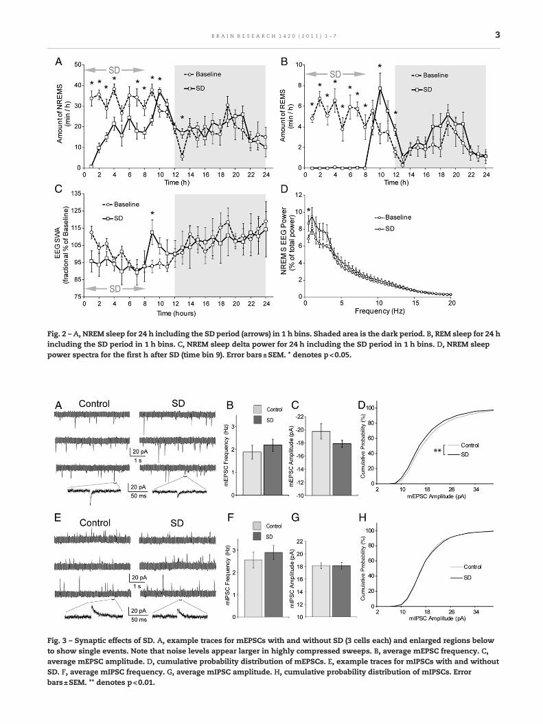
Fig. 2 – A, NREM sleep for 24 h including the SD period (arrows) in 1 h bins. Shaded area is the dark period. B, REM sleep for 24 hincluding the SD period in 1 h bins. C, NREM sleep delta power for 24 h including the SD period in 1 h bins. D, NREM sleeppower spectra for the first h after SD (time bin 9). Error bars±SEM. * denotes p<0.05.
Fig. 3 – Synaptic effects of SD. A, example traces for mEPSCs with and without SD (3 cells each) and enlarged regions belowto show single events. Note that noise levels appear larger in highly compressed sweeps. B, average mEPSC frequency. C,average mEPSC amplitude. D, cumulative probability distribution of mEPSCs. E, example traces for mIPSCs with and withoutSD. F, average mIPSC frequency. G, average mIPSC amplitude. H, cumulative probability distribution of mIPSCs. Errorbars±SEM. ** denotes p<0.01.
3B R A I N R E S E A R C H 1 4 2 0 ( 2 0 1 1 ) 1 – 7

Table 1 – Intrinsic property averages±SEM for control andSD group cells.
Parameter Control (n=22) SD (n=27)
Break-in RMP (mV) −69.71±1.76 −69.26±1.05Ri at −100 pA (MΩ) 223.79±7.41 238.40±8.89Ri at +25 pA (MΩ) 329.77±16.33 323.76±16.59Latency 1st spike (ms) 406.89±25.30 433.36±19.39Rheobase (pA) 101.14±5.06 99.07±5.08AP threshold (mV) −33.50±0.93 −35.14±0.61AP amplitude (mV) 70.70±1.27 69.75±1.56AP halfwidth (ms) 0.98±0.03 0.98±0.03
4 B R A I N R E S E A R C H 1 4 2 0 ( 2 0 1 1 ) 1 – 7
cells, 4 animals; SD: n=20 cells, 4 animals). Neither the averagefrequency nor average amplitude was different (p>0.05, controlvs. SD, t-test, Figs. 3E,F,G). The cumulative frequency distribu-tions of the mIPSCs amplitudes were also not significantlydifferent between the SD and control groups (p=0.25, Kol-mogorav–Smirnov test, Fig. 3H).
2.3. Membrane effects of SD
The intrinsic membrane excitability of mPFC neurons wasassessed by measuring and comparing the evoked action po-tential (AP) firing in control and SD-treated mice (control:n=22 cells, 7 animals; SD: n=27 cells, 6 animals). The evokedAP firing (injected current steps from75 to 225 pA fromamem-brane potential near −70 mV) was significantly higher in theSD-treated mice than in control mice (two-factor ANOVA,main effect of treatment F(1,329)=8.16, p=0.005, Figs. 4A,B),suggesting that SD increases the intrinsic excitability ofdeep layer mPFC neurons. To attempt to gain insight intothe ionic basis underlying SD-induced upregulation of mem-brane excitability, we compared averaged AP afterhyperpo-larizations (AHPs) between control and SD groups (data notshown) and found no differences. We performed an analysisof the sag ratio in response to hyperpolarizing currents (datanot shown) and found there was no effect of SD. We alsomeasured and compared the general membrane propertiesof mPFC neurons between control and SD-treated mice. Nosignificant alterations were detected in any membrane pa-rameters that we examined (Table 1), but the modest de-crease in AP threshold (p=0.13, t-test) and increase in inputresistance (p=0.27 at −100 pA, t-test) we observed would con-tribute increased excitability. The lack of significant regula-tion of any single parameter measured suggests that theobserved effects of SD on membrane excitability may not bemediated by a single type, but by a combination of moderate-ly modulated ionic conductances.
3. Discussion
The flowerpot or pedestal over water method of SD has tradi-tionally been used with rodents for long periods of REM sleepdeprivation (Davis et al., 2006; McDermott et al., 2003). Themethod is effective because the animal will fall from the
Fig. 4 – Intrinsic membrane effects of SD. A, action potential firing(top) and SD. B, average number of action potentials for control a
pedestal due to muscle atonia upon entering REM sleep, andthus be awakened. The pedestal method was indeed very ef-fective at eliminating REM sleep. Additionally, we found thatthis method substantially reduced NREM sleep as well duringrelatively short (8 h) sessions in mice (Fig. 2A). Short-termpedestal SD should be considered a rather mild method ofSD because it allows the animals to obtain some NREMsleep; nonetheless, a clear NREM sleep and delta power aswell as REM sleep rebound were evident (Fig. 2), suggestingthat sleep need or homeostatic drive for sleep was substan-tially generated. The pedestal method has the advantageof requiring less involvement of the experimenter; thus, ismore objective and potentially less stressful, although wedid not directly investigate stress levels.
After SD, there was amodest but significant decrease in theamplitude of mEPSCs impinging on deep-layer mPFC neurons(Fig. 3D), suggesting a post-synaptic decrease in α-amino-3-hydroxy-5-methyl-4-isoxazolepropionic acid (AMPA) recep-tors (Turrigiano et al., 1998). This contrastswith a recent reportin which an increase in both the amplitude and frequencyof mEPSCs was observed in neurons within layer II/III of thefrontal association cortex from rats and mice after 4 h SD bygentle handling (Liu et al., 2010). The SD protocol used in thiscase was especially mild since these animals were not dis-turbed for the first 2 h after light onset when NREM sleep ismost intense. These seemingly discrepant results may reflectthat SD differentially regulates discrete brain regions ratherthan simply exerting across-the-board effects on the brain(Crick and Mitchison, 1983; Krueger and Obal, 1993; Tononi
example traces at 150 and 225 pA injected current for controlnd SD. Error bars±SEM, ** denotes p<0.01.

5B R A I N R E S E A R C H 1 4 2 0 ( 2 0 1 1 ) 1 – 7
and Cirelli, 2006). To extend this notion, the layers of the cerebralcortex are thought to represent discrete functional networks(Grossberg, 2006). In the PFC, layers V and VI are thought tointegrate and organize output, whereas layers II and III arethought to mainly be involved in processing (Berendse et al.,1992; Groenewegen and Uylings, 2000; Groenewegen et al., 1990;Sesack et al., 1989). Thus, the differential effects of SDondifferentcortical regions and layers may contribute to distinct aspects ofSD-induced behavioral alterations.
Recently it was reported that burst firing patterns, reminis-cent of those occurring during NREM sleep, induced by currentinjections into somatosensory layer V pyramidal cells resultedin depression of AMPA receptor-mediated currents (Lantéet al., 2011). By inference, reducing NREM sleep would inhibitnormal removal of AMPA receptors leading to their accumula-tion. If that is the case, although not experimentally demon-strated, Lanté et al. results would be inconsistent with ourcurrent result that sleep restriction depressed mEPSC ampli-tudes. Further, Lanté et al. did not examine whether overallAMPA-mediated currents were larger with higher sleep pres-sure at the end of the dark period.
Evoked AP firing rates were higher following SD, suggestingan adaptation toward higher intrinsic membrane excitability(Fig. 4). This result is consistent with a recent report showinga similar increase in excitability of deep-layer PFC pyramidalcells from rats following 4 h of gentle handling SD (Yanet al., 2011). Our work is further consistent with Yan et al.(2011) in that the fast AHP and sag ratio in response to hyper-polarizing current were not affected by SD. However, ouranalysis of the later AHP did not reveal the decrease in AHPassociated with SD found by Yan and colleagues. Possiblereasons for this discrepancy include species differences andthe SD protocol used. Perhaps the amount of NREM sleep ac-quired by the animals in the present study is sufficient tomask or reduce AHP differences below detection levels.Results seemingly similar to our finding of increased excit-ability with SD were observed in layers II, III and V (pooled to-gether) of the barrel cortex of intact rats, in which theaverage unit spontaneous AP firing frequency is increasedfollowing SD (Vyazovskiy et al., 2009). However, this observedeffect in the barrel cortex could also be mediated by SD-induced synaptic alteration such as an increase in excitatorysynaptic input as has been suggested for other corticalregions (Liu et al., 2010).
Our observed effect of SD on PFC neurons in layer V/VIis opposite to that observed in subcortical neurons, suchas rat hippocampal CA1 neurons, in which SD induces sub-stantial downregulation of intrinsic membrane excitability(McDermott, 2005; McDermott et al., 2003; Yang et al., 2010).These authors used a multiple platform method of SD similarto the method used here; however, the SD period was for 72 h.Tartar and colleagues also reported a decrease in rat hippo-campal excitability with 24 h of sleep fragmentation (Tartaret al., 2010). Despite differences in SD duration and methodol-ogy, these observations in contrast with our observation ofincreased cortical excitability demonstrate that differentbrain sites respond to sleep loss in different ways. This infer-ence is consistent with the interpretation that local site-specific activity-dependent sleep loss-associated cellularchanges occur (Krueger et al., 2008).
Our findings at the synaptic and membrane levels wouldhave opposite effects on integrated output. This impliesthat in this system the effects of SD may strive to counter-act each other functionally to some degree. Nonetheless, ashift of synapse–membrane interaction toward higher ex-citability may adjust the gain of the input/output of PFCneurons as they perform cognitive tasks. For example, dur-ing evaluating or selecting tasks, previously sub-thresholdand thus “silent” excitatory inputs that otherwise cannotactivate PFC neurons may become capable of exciting PFCneurons, thus decreasing the signal-to-noise ratio. Clearly,a detailed understanding of the cellular basis of sleep-mediated regulation of PFC-based cognitive responses de-mands more extensive, in particular, in vivo studies. TheSD-induced effects on basic neuronal properties character-ized here in the PFC do provide initial cellular groundworkfor more sophisticated ex vivo and in vivo manipulations infuture studies.
4. Experimental procedures
4.1. Experimental animals
Male C57BL/6 mice 5 to 11 weeks old were used in this study(Simonsen Labs). Mice were maintained at room temperature(22±2 °C), on a 12:12-h light–dark cycle (lights on 00:00, lightsoff 12:00), and individually housed with constant access tofood and water. All animal procedures were approved by theWashington State University Institutional Animal Care andUse Committee.
4.2. Sleep recordings
For surgeries, mice were anesthetized with intraperitonealketamine and xylazine (87 and 13 mg/kg, respectively). Astainless-steel wire elecromyogram (EMG) electrode wasinserted into the nuchal neck muscle and three gold-platedwire EEG electrodes (Plastics One, Roanoke, VA) wereinstalled through the skull over the parietal and frontal corti-ces, as previously described (Krueger et al., 1993; Szentirmaiet al., 2009). Electrode leads were gathered into a plastic sock-et (Plastics One) and fixed to the skull with dental cement(Patterson Dental Supply, Saint Paul, MN). Animals were con-nected to the amplifiers via lightweight cables and commuta-tors (Plastics One). For habituation, the cables were connectedto the mice for one week prior to experimental recordings.
Signals were amplified using Grass model 7P511 amplifiers(Grass-Telefactor, West Warwick, RI). The EEG was filteredbelow 0.1 Hz and above 100 Hz. The EMG was filtered below30 Hz and above 3 kHz. All signals were digitized at 128 Hzand collected using Sleep Analysis software (Biosoft Studios,Hershey, PA) and visually scored for sleep state in 10 s epochsusing Sleep Sign for Animal software (Kissei Comtec, Nagano,Japan) as previously described (Kapas et al., 2008; Kruegeret al., 1993). Briefly, REM sleep is typically identified by a lossof muscle tone and a very regular low amplitude EEG with alarge theta (6–9 Hz) frequency component. NREM sleep EEGtypically has high amplitude and regular delta (0.5–5 Hz) fre-quency oscillations. Wakefulness produces a more irregular

6 B R A I N R E S E A R C H 1 4 2 0 ( 2 0 1 1 ) 1 – 7
and generally low amplitude EEG, often accompanied by mus-cle activity (Fig. 1C).
4.3. Sleep deprivation
A modified pedestal or flowerpot method was used for SD.Mice were deprived of sleep for the 8 h following light onset(00:00–08:00, Fig. 1B). During the SD procedure, animals wereplaced on pedestals in a 50 cm deep plastic tub containingwater 1.5 cm deep. The water was kept warm (30 °C) usingelectric heating pads placed under the tub. The three plasticpedestals protruding from the bottom of the tub were 2 cm di-ameter, 6.5 cm high, and separated by 7.5 cm (Fig. 1A). Having3 pedestals that the animal could easily move between servedto limit immobility. A standard food pellet was suspendedfrom a thread near one of the pedestals. Animals were habit-uated to the pedestals and the environment for 2 h at leastone day prior to the experimental day.
4.4. Patch clamp recordings
Mice for these experiments were sacrificed immediately fol-lowing the SD period. Control mice remained in their homecages without disturbance and were killed at the same timeof day as SD animals. Control mice were killed within 5 minof arousal. Animals were deeply anesthetized with isoflurane,decapitated, and the brain quickly removed. Coronal slices in-cluding the mPFC (Paxinos and Franklin, 2001) (Fig. 1D) wereprepared as previously described (Huang et al., 2009; Ishikawaet al., 2009). Briefly, slices were cut on a Leica V1200 vibratingmicrotome 250–300 μm thick in an ice-cold oxygenated cut-ting solution (in mM: 135 NMDG, 1 KCl, 1.2 KH2PO4, 1.5MgCl2, 0.5 CaCl2, 20 choline bicarbonate, 10 glucose, 295–305 mOsm, equilibrated with 95%O2/5% CO2), then submergedin 36 °C oxygenated standard artificial cerebral spinal fluid(ACSF, in mM:126 NaCl, 1.6 KCl, 1.2 NaH2PO4, 1.2 MgCl2, 2.5CaCl2, 18 NaHCO3, 11 glucose, 285–295 mOsm, equilibratedwith 95% O2/5% CO2) for 30 min, then stored at room temper-ature with constant oxygenation until being transferred tothe recording chamber. During recordings, slices were con-stantly perfused with oxygenated ACSF at 30 °C via an inlinesolution heater (Harvard Apparatus, Holliston, MA). Record-ings were selectively made in layer V/VI of the infralimbic re-gion of the mPFC (Fig. 1D). Pyramidal cells were preferentiallytargeted based on soma shape, large size, and dendritic polar-ity, if visible, using differential interference contrast micros-copy (Olympus, Center Valley, PA). Whole-cell patch-clamprecordings were made using a Multiclamp 700B amplifier andDigidata 1440A digitizer (Molecular Devices, Sunnyvale, CA)through borosilicate glass electrodes (3–5 MΩ) and recordedat 20 kHz using Clampex 10.2 software (Molecular Devices,Sunnyvale, CA). Only stable cells with access resistances<25 MΩ were included in data analyses.
Voltage-clamp recordings were used to measure mEPSCsand mIPSCs in separate sets of animals. For these recordings,pipettes were filled with cesium-based solution (in mM: 117cesium methanesulfonic acid, 20 HEPES, 0.4 EGTA, 2.8 NaCl, 5TEA-Cl, 2.5 MgATP, 0.25 Na3GTP,1 QX-314 [Sigma-Aldrich], pH7.2–7.4; 285–295 mOsm). For mEPSCs, membrane potentialwas held at −65 mV and perfusion was switched to ACSF
containing tetrodotoxin (TTX, 1 μM, Tocris, Ellisville, MO) andpicrotoxin (100 μM, Sigma-Aldrich). For mIPSCs, membranepotential was held at 0 mV and perfusion was switched toACSF containing TTX (1 μM), D-APV (50 μM, Tocris), andNBQX (2 μM, Tocris). Cells were allowed to stabilize after start-ing toxin-containing solutions for at least 10 min. Due to possi-ble effects of action potential blockade on synaptic properties(Turrigiano, 2008), all mEPSC and mIPSC recordings weremade within 30 min after TTX was introduced to the slice.For miniature event analysis, 200 sequential events fromeach cell were individually identified visually using Clampfit10.2 software (Molecular Devices, Sunnyvale, CA). Eventswere at least 10 pA, and the baseline amplitude was adjustedfor each event to mid signal at the initiation time of the event.
In another set of animals, APs were recorded in current-clamp mode with “resting” membrane potentials normalizedto −70 mV. For these recordings, pipettes were filled withpotassium-based solution (in mM: 130 K-methanesulfate, 10KCl, 10 HEPES, 0.4 EGTA, 2.0 MgCl2, 2.5 MgATP, 0.25 Na3GTP,pH 7.2–7.4; 285–295 mOsm). At least 10 min after initiation ofthe whole-cell mode, 600 ms current steps were injectedfrom −200 pA to 225 pA in 25 pA increments with an inter-pulse interval of 10 s. Only cells with stable AP numberswere included in data analysis. Three runs were averaged foreach cell. The majority of recorded cells (>90%) were regularspiking (steady increase from a low number of AP with in-creasing current); only these cells were used for final analysis.
4.5. Statistics
For quantification of sleep time and power, 2-factor ANOVAwithrepeatedmeasureswasused to assessmajor effects of treatment.The multiple imputation method was used for missing values inpower data time bins with no NREM sleep. Where ANOVA indi-cated a significant effect, paired Student's t-tests were used forpost hocanalysis to assessdifferences between treatments at indi-vidual time points. For quantification of patch clamp recordings,cell based statistic were used; however, we also examined thisdata after reducing variability by averaging into animal basedbins. There were no differences in outcomes of significance. Thenon-parametric Kolmogorav–Smirnov testwasused to assess cu-mulative frequency distributions of post synaptic currents. Thestatistical software SPSS (Somer, NY) was used for analysis. Prob-ability less than 0.05 was considered significant.
Acknowledgments
This work was supported by NIH DA023206 (YD), NS025378 (JK)and NS031453 (JK) and the Humboldt Foundation (YD).
R E F E R E N C E S
Berendse, H.W., Galis-de Graaf, Y., Groenewegen, H.J., 1992.Topographical organization and relationship with ventralstriatal compartments of prefrontal corticostriatal projectionsin the rat. J. Comp. Neurol. 316, 314–347.
Bodosi, B., Obal Jr., F., Gardi, J., Komlodi, J., Fang, J., Krueger, J.M.,2000. An ether stressor increases REM sleep in rats: possible

7B R A I N R E S E A R C H 1 4 2 0 ( 2 0 1 1 ) 1 – 7
role of prolactin. Am. J. Physiol. Regul. Integr. Comp. Physiol.279, R1590–R1598.
Chuah, Y.M.L., 2006. The neural basis of interindividual variabilityin inhibitory efficiency after sleep deprivation. J. Neurosci. 26,7156–7162.
Crick, F., Mitchison, G., 1983. The function of dream sleep. Nature304, 111–114.
Davis, C.J., Meighan, P.C., Taishi, P., Krueger, J.M., Harding, J.W.,Wright, J.W., 2006. REM sleep deprivation attenuatesactin-binding protein cortactin: a link between sleep andhippocampal plasticity. Neurosci. Lett. 400, 191–196.
Finelli, L.A., Baumann, H., Borbely, A.A., Achermann, P., 2000. Dualelectroencephalogram markers of human sleep homeostasis:correlation between theta activity in waking and slow-waveactivity in sleep. Neuroscience 101, 523–529.
Goldman-Rakic, P.S., 1995. Cellular basis of working memory.Neuron 14, 477–485.
Graves, L.A., 2003. Sleep deprivation selectively impairs memoryconsolidation for contextual fear conditioning. Learn. Mem. 10,168–176.
Groenewegen, H.J., Berendse, H.W., Wolters, J.G., Lohman, A.H.,1990. The anatomical relationship of the prefrontal cortex withthe striatopallidal system, the thalamus and the amygdala:evidence for a parallel organization. Prog. Brain Res. 85, 95–116(discussion 116–118).
Groenewegen, H.J., Uylings, H.B., 2000. The prefrontal cortex andthe integration of sensory, limbic and autonomic information.Prog. Brain Res. 126, 3–28.
Grossberg, S., 2006. In: Cisek, P., Drew, T., Kalaska, J. (Eds.),Towards a Unified Theory of Neocortex: Laminar CorticalCircuits for Vision and Cognition. Elsevier, Amsterdam.
Hille, B., 2001. Ion Channels of Excitable Membranes. SinauerAssociates.
Huang, Y.H., Lin, Y., Mu, P., Lee, B.R., Brown, T.E., Wayman, G.,Marie, H., Liu, W., Yan, Z., Sorg, B.A., 2009. In vivo cocaineexperience generates silent synapses. Neuron 63, 40–47.
Ishikawa,M.,Mu, P., Moyer, J.T.,Wolf, J.A., Quock, R.M., Davies, N.M.,Hu, X.T., Schluter, O.M., Dong, Y., 2009. Homeostaticsynapse-driven membrane plasticity in nucleus accumbensneurons. J. Neurosci. 29, 5820–5831.
Kapas, L., Bohnet, S.G., TTraynor, T.R., Majde, J.A., Szentirmai, E.,Magrath, P., Taishi, P., Krueger, J.M., 2008. Spontaneous andinfluenza virus-induced sleep are altered in TNF-double-receptordeficient mice. J. Appl. Physiol. 105, 1187–1198.
Killgore, W.D., 2010. Effects of sleep deprivation on cognition.Prog. Brain Res. 185, 105–129.
Krueger, J.M., Kapas, L., Kimura, M., Opp, M.R., 1993. Somnogeniccytokines: methods and overview. Meth. Neurosci. 17, 111–129.
Krueger, J.M., Obal, F., 1993. A neuronal group theory of sleepfunction. J. Sleep Res. 2, 63–69.
Krueger, J.M., Rector, D.M., Roy, S., Van Dongen, H.P.A., Belenky,G., Panksepp, J., 2008. Sleep as a fundamental property ofneuronal assemblies. Nat. Rev. Neurosci. 9, 910–919.
Lanté, F., Toledo-Salas, J.C., Ondrejcak, T., Rowan, M.J., Ulrich, D.,2011. Removal of synaptic Ca2+-permeable AMPA receptorsduring sleep. J. Neurosci. 31, 3953–3961.
Le Marec, N., Beaulieu, I., Godbout, R., 2001. Four hours ofparadoxical sleep deprivation impairs alternationperformance in a water maze in the rat. Brain Cogn. 46,195–197.
Liu, Z.W., Faraguna, U., Cirelli, C., Tononi, G., Gao, X.B., 2010. Directevidence for wake-related increases and sleep-relateddecreases in synaptic strength in rodent cortex. J. Neurosci. 30,8671–8675.
Marshall, L., Born, J., 2007. The contribution of sleep tohippocampus-dependent memory consolidation. TrendsCogn. Sci. 11, 442–450.
Massimini, M., 2004. The sleep slow oscillation as a travelingwave. J. Neurosci. 24, 6862–6870.
Mauzur, A., Pace-Schott, E.F., Hobson, A.J., 2002. The prefrontalcortex in sleep. Trends Cogn. Sci. 11, 475–481.
McDermott, C.M., 2005. Sleep deprivation-induced alterations inexcitatory synaptic transmission in the CA1 region of the rathippocampus. J. Physiol. 570, 553–565.
McDermott, C.M., LaHoste, G.J., Chen, C., Musto, A., Bazan, N.G.,Magee, J.C., 2003. Sleep deprivation causes behavioral,synaptic, and membrane excitability alterations inhippocampal neurons. J. Neurosci. 23, 9687–9695.
Miller, E., Cohen, J.D., 2001. An integrative theory of prefrontalcortex function. Annu. Rev. Neurosci. 24, 167–202.
Nieuwenhuis, I.L.C., Takashima, A., 2011. The role of theventromedial prefrontal cortex in memory consolidation.Behav. Brain Res. 218, 325–334.
Palchykova, S., Winskysommerer, R., Meerlo, P., Durr, R., Tobler, I.,2006. Sleep deprivation impairs object recognition in mice.Neurobiol. Learn. Mem. 85, 263–271.
Paxinos, G., Franklin, K.B.J., 2001. The Mouse Brain in StereotaxicCoordinates. Academic Press, San Diego.
Robbins, T.W., 2005. Chemistry of the mind: neurochemicalmodulation of prefrontal cortical function. J. Comp. Neurol.493, 140–146.
Sesack, S.R., Deutch, A.Y., Roth, R.H., Bunney, B.S., 1989.Topographical organization of the efferent projections of themedial prefrontal cortex in the rat: an anterogradetract-tracing study with Phaseolus vulgaris leucoagglutinin.J. Comp. Neurol. 290, 213–242.
Smith, C., Rose, G.M., 1996. Evidence for a paradoxical sleepwindow for place learning in the Morris water maze. Physiol.Behav. 59, 93–97.
Steriade, M., 2006. Grouping of brain rhythms in corticothalamicsystems. Neuroscience 137, 1087–1106.
Szentirmai, E., Kapas, L., Sun, Y., Smith, R.G., Krueger, J.M., 2009.The preproghrelin gene is required for the normal integrationof thermoregulation and sleep in mice. Proc. Natl. Acad. Sci.106, 14069–14074.
Tartar, J.L., McKenna, J.T., Ward, C.P., McCarley, R.W., Strecker, R.E.,Brown, R.E., 2010. Sleep fragmentation reduceshippocampal CA1 pyramidal cell excitability and response toadenosine. Neurosci. Lett. 469, 1–5.
Thomas, M., Sing, H., Belenky, G., Holcomb, H., Mayberg, H.,Dannals, R., Wagner, H., Thorne, D., Popp, K., Rowland, L.,Welsh, A., Balwinski, S., Redmond, D., 2000. Neural basis ofcognitive performance impairments during sleepiness. I.Effects of 24 h of sleep deprivation on waking human reginalbrain activity. J. Sleep Res. 9, 335–352.
Tononi, G., Cirelli, C., 2006. Sleep function and synaptichomeostasis. Sleep Med. Rev. 10, 49–62.
Turrigiano, G.G., 2008. The self-tuning neuron: synaptic scaling ofexcitatory synapses. Cell 135, 422–435.
Turrigiano, G.G., Leslie, K.R., Desai, N.S., Rutherford, L.C., Nelson,S.B., 1998. Activity-dependent scaling of quantal amplitude inneocortical neurons. Nature 391, 892–896.
Vyazovskiy, V.V., Olcese, U., Lazimy, Y.M., Faraguna, U., Esser, S.K.,Williams, J.C., Cirelli, C., Tononi, G., 2009. Cortical firing and sleephomeostasis. Neuron 63, 865–878.
Wu, J.C., Gillin, J.C., Buchsbaum, M.S., Chen, P., Keator, D.B.,Khosla Wu, N., Darnall, L.A., Fallon, J.H., Bunney, W.E., 2006.Frontal lobe metabolic decreases with sleep deprivation nottotally reversed by recovery sleep. Neuropsychopharmacology31, 2783–2792.
Yan, J., Li, J.-C., Xie, M.-L., Zhang, D., Qi, A.-P., Hu, B., Huang, W.,Xia, J.-X., Hu, Z.-A., 2011. Short-term sleep deprivationincreases intrinsic excitability of prefrontal cortical neurons.Brain Res. 1401, 52–58.
Yang, R.-H., Wang, W.-T., Hou, X.-H., Hu, S.-J., Chen, J.-Y., 2010.Ionic mechanisms of the effects of sleep deprivation onexcitability in hippocampal pyramidal neurons. Brain Res.1343, 135–142.