performance of patients with ventromedial prefrontal, dorsolateral prefrontal, and non-frontal
TRANSCRIPT

University of IowaIowa Research Online
Theses and Dissertations
Fall 2010
Performance of patients with ventromedialprefrontal, dorsolateral prefrontal, and non-frontallesions on the Delis-Kaplan Executive FunctionSystemEkaterina KeiferUniversity of Iowa
Copyright 2010 Ekaterina Keifer
This dissertation is available at Iowa Research Online: https://ir.uiowa.edu/etd/830
Follow this and additional works at: https://ir.uiowa.edu/etd
Part of the Educational Psychology Commons
Recommended CitationKeifer, Ekaterina. "Performance of patients with ventromedial prefrontal, dorsolateral prefrontal, and non-frontal lesions on the Delis-Kaplan Executive Function System." PhD (Doctor of Philosophy) thesis, University of Iowa, 2010.https://doi.org/10.17077/etd.zzkcikx2.

PERFORMANCE OF PATIENTS WITH VENTROMEDIAL PREFRONTAL,
DORSOLATERAL PREFRONTAL, AND NON-FRONTAL LESIONS ON THE
DELIS-KAPLAN EXECUTIVE FUNCTION SYSTEM
by
Ekaterina Keifer
An Abstract
Of a thesis submitted in partial fulfillment of the requirements for the
Doctor of Philosophy degree in Psychological and Quantitative Foundations
in the Graduate College of The University of Iowa
December 2010
Thesis Supervisors: Professor Elizabeth Altmaier Professor Daniel Tranel

1
ABSTRACT
Executive functioning is a multidimensional concept encompassing higher-order
adaptive abilities, such as judgment, decision-making, self-monitoring, planning, and
emotional regulation. Disruption in executive functioning often results in devastating
impairments in vitally-important areas of life, such as one’s ability to hold employment
and maintain social relationships.
Executive functions have been associated primarily with the prefrontal cortex.
However, the nature and degree of the association between frontal lobe damage and
performance on executive functioning tests remains controversial. Research suggests that
the association may vary based on the specific location of damage within the prefrontal
cortex, as well as the used measure of executive functioning. Few investigations have
systematically addressed these variables. The current study employed the lesion method
to investigate the relationship between performance on a battery of executive functioning
tests and damage to specific regions of the prefrontal cortex. Three groups of participants
with lesions in one of the locations of interest [ventromedial prefrontal (VMPC, n = 14),
dorsolateral prefrontal (DLPC, n = 14), and non-frontal (n = 18)] were administered the
Delis-Kaplan Executive Function System (D-KEFS, 2001), a comprehensive battery of
executive functioning tests. Results revealed no statistically-significant differences
between group performances on the D-KEFS primary measures. However, a qualitative
analysis of the results revealed several meaningful group differences. It appears that
some relationship exists between frontal lobe damage, particularly in the DLPC, and
decreased performance on several executive functioning tests but further research

2
overcoming the methodological limitations of most existing literature on this topic is
needed to clearly resolve this issue.
Abstract Approved: ________________________________________________ Thesis Supervisor ________________________________________________ Title and Department ________________________________________________ Date ________________________________________________ Thesis Supervisor ________________________________________________ Title and Department ________________________________________________ Date

PERFORMANCE OF PATIENTS WITH VENTROMEDIAL PREFRONTAL,
DORSOLATERAL PREFRONTAL, AND NON-FRONTAL LESIONS ON THE
DELIS-KAPLAN EXECUTIVE FUNCTION SYSTEM
by
Ekaterina Keifer
A thesis submitted in partial fulfillment of the requirements for the
Doctor of Philosophy degree in Psychological and Quantitative Foundations
in the Graduate College of The University of Iowa
December 2010
Thesis Supervisors: Professor Elizabeth Altmaier Professor Daniel Tranel

Copyright by
EKATERINA KEIFER
2010
All Rights Reserved

Graduate College The University of Iowa
Iowa City, Iowa
CERTIFICATE OF APPROVAL ___________________________
PH.D. THESIS ____________
This is to certify that the Ph.D. thesis of
Ekaterina Keifer has been approved by the Examining Committee for the thesis requirement for the Doctor of Philosophy degree in Psychological and Quantitative Foundations at the December 2010 graduation. Thesis Committee: _____________________________________________________ Elizabeth Altmaier, Thesis Supervisor _____________________________________________________ Daniel Tranel, Thesis Supervisor _____________________________________________________ David Moser _____________________________________________________ Timothy Ansley _____________________________________________________ Sam Cochran III

ii
To my Grandfather Nikolai Vasilyevich Levenkov

iii
ACKNOWLEDGMENTS
I would like to thank Dr. Tranel for allowing me to use the Department of
Neurology Patient Registry for data collection and providing support and guidance during
the process. I could not have completed the project without this help. I would also like
to thank Dr. Altmaier for her continuous guidance not only with the content of the
dissertation but with multiple practical considerations and requirements. Thank you to
Ruth Henson and Keary Saul for scheduling participants for the study. I would also like
to thank Tasha Feuerbach who assisted in data collection. And, finally, I would like to
thank individuals who graciously agreed to participate in two hours of often challenging
testing.
iv
TABLE OF CONTENTS
LIST OF TABLES ............................................................................................................. vi
LIST OF FIGURES .......................................................................................................... vii
LIST OF SYMBOLS AND ABBREVIATIONS ............................................................ viii
CHAPTER ONE INTRODUCTION ...........................................................................1
Definition of Executive Functions………………………………...1 Executive Functions and the Prefrontal Cortex…………………...4 Examination of Brain-Behavior Relationships………………......11 Measurement of Executive Functions……………………………12 CHAPTER TWO PRELIMINARY INFORMATION AND LITERATURE ............... REVIEW ........................................................................................17 Wisconsin Card Sorting Test .........................................................17 Trail Making Test, Parts A and B ..................................................17 Stroop Color-Word Test ................................................................18 Fluency Tests .................................................................................18 Tower Tests ....................................................................................19 The Delis-Kaplan Executive Function System (D-KEFS) ............19 Description of the D-KEFS Subtests .............................................22 Literature Review Organization .....................................................26 Conclusions ....................................................................................51 Predictions......................................................................................51 Alternate Outcomes……………………………………………...55
CHAPTER THREE METHODS ....................................................................................57
Procedures ......................................................................................57 Power Analysis ..............................................................................57 Participants .....................................................................................58 Lesion Classification ......................................................................59 Instruments……………………………………………………….62 D-KEFS Standardization and Norms…………………………….65 D-KEFS Reliability and Validity………………………………...66 D-KEFS Trail Making Test ...............................................67 D-KEFS Verbal Fluency Test ............................................68 D-KEFS Design Fluency Test ...........................................68 D-KEFS Color-Word Interference Test .............................69 D-KEFS Sorting Test. ........................................................69 D-KEFS Twenty Questions Test ......................................70 D-KEFS Word Context Test……………………………..71

v
D-KEFS Tower Test……………………………………..71 D-KEFS Proverb Test……………………………………72 CHAPTER FOUR RESULTS ......................................................................................74
Descriptive Statistics ......................................................................74 D-KEFS Performances...................................................................75 Comparison of Participants with VMPC, DLPC, and Non-Frontal Lesions................................................................78 Proportion of Impaired Performances in Each Group ...................79 Proportion of Performances = or < the Sixteenth Percentile .........80 Lesion Laterality in Patients with DLPC Lesions………………..83 CHAPTER FIVE DISCUSSION ................................................................................85
Limitations and Directions for Future Research ..........................106 Implications for Clinical Practice ................................................110 REFERENCES ................................................................................................................111

vi
LIST OF TABLES
Table 1. D-KEFS Variables and Units of Measurement .........................................52
Table 2. Performance Predictions ...........................................................................56
Table 3. Lesion Etiologies ......................................................................................62
Table 4. Demographic characteristics, lesion chronicity, FSIQ, and BDI-II scores ……………………………………………………….74
Table 5. D-KEFS performances in Scaled Scores………………………………...75
Table 6. FSIQ-adjusted group means and standard errors…..…………………….77 Table 7. ANCOVA comparing group D-KEFS performances with FSIQ as a covariate………………….………………………………………….79 Table 8. Number and percentage of participants performing at or below the
Scaled Score (SS) of five (fifth percentile)……….………………………80 Table 9. Number and percentage of participants performing at or below SS of seven (sixteenth percentile)………………………………………………82

vii
LIST OF FIGURES
Figure 1. Illustration of DLPC and VMPC Regions ...............................................60
Figure 2. D-KEFS performances in Scaled Scores ...................................................76

viii
LIST OF SYMBOLS AND ABBREVIATIONS
n Statistical notation for sample size
η2 Partial eta-squared: the proportion of total variability attributable to a particular
factor
< Less than
= Equal to
SS Scaled score
SD Standard deviation

1
CHAPTER ONE
INTRODUCTION
Definition of Executive Functions
Abilities that comprise executive functioning, such as decision-making, judgment,
social reasoning and others, have been a subject of interest and debate for centuries. It
seems that the most basic reason for this interest is the desire to understand why people
think and behave as they do. The complexity of this question is demonstrated by the fact
that scientists in the present are not only still trying to answer it but are still trying to
define the question. There remains wide variability in the definition of executive
functions as well as theories underlying their mechanisms.
Generally, agreement exists that executive functions are distinct higher-order
cognitive abilities that are adaptive in nature. Examples of such functions include
judgment, motivation, working memory, initiation and discontinuation of behavior,
cognitive flexibility, decision-making, planning, and personality/affective functioning.
These functions are distinct because they cannot be subsumed under any other mental
functions. In fact, executive functions rely on a variety of lower-order, concrete
functions, such as perception and memory. In a recent literature review of executive
functioning measurement, Alvarez and Emory (2006) defined executive functions as
“higher-level cognitive functions involved in the control and regulation of “lower-level”
cognitive processes and goal-directed, future-oriented behavior” (p. 17). While it is
suggested that executive functions contribute to lower-level functions by imposing
higher-order analysis and organization, basic cognitive functions often remain

2
remarkably unimpaired in the presence of severe impairment in some aspect of executive
functioning (Tranel et al., 1994).
In their chapter addressing the development of the concept of executive
functioning, Tranel et al. (1994) provided an overview of the descriptions for the term as
well as their own definition based on the overview. They noted that most of the early
knowledge about executive functions came from observations of people and animals who
had sustained an injury to the frontal lobes. These early accounts highlighted changes in
personality, failure to hold information in mind (currently defined as working memory),
perseveration, and the loss of an “abstract attitude”, which included abstract reasoning,
initiative, and the ability to adapt to new situations.
More recently, scientists have tried to systematically organize knowledge
available on executive functioning into overarching categories of skills. For example,
Lezak et al. (2004) identified four main components: volition, planning, purposive action,
and effective performance. Each of these overarching tasks consists of smaller units
necessary to effectively perform the tasks (Lezak et al., 2004). Among other scientists,
Fuster (1989) elaborated on the definition by including concepts of prospective memory
and failure to inhibit interference. In addition, Fuster (1989) highlighted affective and
personality functioning as an integral feature of executive functioning. He described two
types of common disturbance in this domain: the “apathetic syndrome” exemplified by a
lack of initiative and self-awareness, as well as affective blunting and motor slowing; and
the “euphoric syndrome” characterized by excessive activity, disinhibition, socially
inappropriate behavior, and elevated mood.
3
Tranel at al. (1994) identified four major components that seemed to be most
frequently described and agreed upon in the literature. These components include
planning, decision-making, judgment, and self-perception. Each component subsumes
multiple supporting elements. For example, planning includes being able to identify
appropriate, realistic, and timely courses of action that extend into the future. Decision-
making consists of the ability to accurately appraise the courses of action and their
consequences and to choose the most advantageous one. Tranel et al. (1994) highlight
the distinct nature of social decision-making, which requires decisions to be made “on the
spot”, based on a quick “reading” of the situation in terms of both its manifest and
implied meaning” (p. 130). Judgment subsumes the ability to make accurate assessments
of the situation and underlies the functions of planning and decision-making. Finally,
self-perception is a broad concept encompassing the ability for self-monitoring, self-
correction, and the use of external cues to guide behavior. This concept also includes the
broad notion of personality, which describes the general tendencies and characteristics of
a person by which he or she is known. Personality change can vary in its expression but
is a common feature of executive dysfunction. At the end of the review, Tranel et al.
(1994) noted that while the term “executive functions” is broad and encompasses loosely
connected functions, it has proven to be useful for conceptual and communicative
purposes.
In summary, executive functions are higher-order adaptive functions, relatively
distinct from lower-order cognitive processes. As Chan et al. (2008) pointed out in a
recent review, executive functions can be divided into two general categories: a “cold”
category, examples of which include the ability to sustain attention, inhibit irrelevant

4
information, problem-solve, and multitask; and a “hot” category, which involves more
emotionally- and socially-based abilities, such as social behavior, emotional regulation,
level of motivation and energy, as well as decision-making relying on personal
preferences and desires.
Executive functions often operate on an automatic level outside the immediate
awareness. They come to the forefront when dysfunction occurs and the automatic
mechanism allowing for effective execution of these functions fails. As multiple case
examples have shown, deficits in executive functioning can profoundly affect all areas of
life, including interpersonal, occupational, financial, and others. It becomes apparent that
intact executive functioning is essential for successful performance of simple everyday
tasks, such as making a meal and running errands, as well as complex planning into the
future and maintaining meaningful relationships with others. The vital importance of
intact executive functioning explains continuing scientific efforts to further understand
the concept despite the challenges presented by its complexity.
Executive Functions and the Prefrontal Cortex
Prior to expanding the discussion about the association between executive
functions and the frontal lobes, it is important to briefly discuss the anatomical structure
and subdivision of the frontal lobes. The frontal lobes constitute nearly one-third of the
brain. The most posterior section of the frontal lobes, the precentral gyrus directly in
front of the central sulcus, is the primary motor cortex responsible for basic motor
functioning. Anterior to the primary motor cortex are the premotor area and the
supplementary motor area. As a general functional description, these three areas are
associated with learning, planning and precise execution of movement. Given that these

5
areas have bidirectional connections to the prefrontal cortex and lesions here have been
observed to disrupt motor learning and execution of movements, Tranel et al. (1994)
concluded that “the premotor area is involved in executive functions” (p. 134) but its
exact role still remains to be investigated. The area in front of the premotor and
supplementary motor cortices encompassing the frontal pole is the prefrontal cortex. As
Stuss and Benson (1984) noted, this area can be subdivided according to a variety of
principles, including cytoarchitectural layers, connections with other brain areas, and
vascular distributions. However, the most common way of dividing the prefrontal lobes
is by location, resulting in three major areas: superior mesial, dorsolateral, and orbital (or
ventromedial) (Damasio & Anderson, 2003).
Stuss and Benson (1984) asserted that “the importance of the frontal lobes derives
from rich connections, both afferent and efferent, with almost all other areas of the
central nervous system” (p. 4). The following paragraphs will provide brief descriptions
of the anatomical structure and functions of the three main subdivisions of the prefrontal
cortex.
The prefrontal superior mesial region is closely related in function to the
previously mentioned supplementary motor area. In fact it is sometimes considered to be
a rostral extention of the supplementary motor area (Afifi & Bergman, 2005). This
region is sometimes referred to as the cingulate or limbic cortex due to the underlying
cingulate gyrus (Lezak et al., 2004). The superior mesial region mediates initiation of
behavior based on drives and emotional states. Individuals with lesions in this area often
experience a diminution or loss of emotional reactions, which results in low drive states
and lack of motivation needed to initiate behavior. With severe damage, even life-

6
sustaining drives for food or drink may be lost. The primary physiological mechanism of
such impairment is considered an interruption of the connection between the superior
mesial region with the diencephalon (Lezak et al., 2004).
The dorsolateral prefrontal cortex (DLPC) has reciprocal connections with
multiple cortical and subcortical brain regions, including the parietal association areas,
superior temporal lobe, the cingulate cortex, basal ganglia, thalamus, and superior
colliculus (Kolb and Wishaw, 2003). It is associated with “cold” executive functions
which are targeted by many laboratory-based neuropsychological tests. One of the more
prominent of these functions is “working memory” or the ability to hold information “on-
line” in order to effectively process and organize it. Many abilities (e.g., planning,
reasoning, problem-solving) rely on intact working memory. Thus, an impairment in
working memory is likely to impact a variety of other functions important for adaptive
functioning (Damasio et al., in press).
The prefrontal cortex, and primarily the dorsolateral regions, appear to play an
important role in the allocation and modulation of attention. Research has consistently
shown that damage to this region results in diminished attention to novel stimuli and
increased vulnerability to distraction and interference (Stuss & Benson, 1984). Recent
research shows that the right prefrontal cortex may be crucial for the ability to sustain
attention (Lezak et al., 2004). Another attentional deficit frequent in patients with
dorsolateral prefrontal damage is difficulty shifting attention between stimuli or concepts,
which results in cognitive inflexibility and perseveration. Perseverative behavior which
consists of abnormal repetition of the same behavior or idea/concept is a common
manifestation of frontal lobe damage (Damasio et al., in press). Lezak et al. (2004) refer

7
to functions like working memory and attentional shifting as supramodal or present in a
variety of modalities and participating in practically all mental activities. Thus, it is not
surprising that people with lesions primarily in the dorsolateral prefrontal region may
exhibit deficits in many cognitive domains including memory. However, an anterograde
memory impairment following prefrontal damage does not result from dysfunction of the
memory systems in the brain. Rather, it results from a deficit in executive functions
which regulate the effective encoding and retrieval of information. Thus, people with
dorsolateral prefrontal lesions may fail to organize new information resulting in
inefficient encoding and retrieval strategies (Lezak et al., 2004). Prospective memory
impairment or “remembering to remember” causes some of the most serious practical
problems patient with prefrontal damage face. Their employment, rehabilitation, and
daily self-care may be jeopardized because of their failure to remember to go to work,
attend appointments, make meals, and bathe.
The ventromedial prefrontal cortex (VMPC) receives input from all sensory
modalities, including olfaction, vision, audition, taste, and somatosensation. It also has
strong bidirectional connections with the amygdala and the hippocampus via the uncinate
fasciculus. This region projects to the hypothalamus, likely influencing changes that
occur in the autonomic nervous system during emotional responses (Kolb & Wishaw,
2003). Projections from the hypothalamus have been more difficult to establish but some
evidence exists for the presence of such projections (Damasio et al., 2010). Other
projections arising from the orbital region are to the claustrum, subthalamic regions, and
the mesencephalon.

8
The VMPC is associated with “hot” executive functions involving decision-
making, judgment, and conduct based on emotional and social variables. Since
emotional and social components permeate almost all human activities, damage to the
orbital region can result in devastating consequences in multiple domains of life.
Interestingly, people with ventromedial damage often have nearly intact psychometric
intelligence and perform in the normal range on neuropsychological tests of executive
function, phenomenon likely due to the preservation of the dorsolateral prefrontal cortex.
The consequences of ventromedial damage are readily seen in real-life circumstances and
present themselves as difficulties in decision-making, maintenance of relationships with
others, and affective lability, among others.
The concept of executive functioning has historically become strongly connected
to the frontal lobes, and more specifically, to the prefrontal cortex. As Tranel et al.
(1994) noted, “It is virtually impossible to find a discussion of prefrontal lobe lesions that
does not make reference to disturbances of executive functions, and, in parallel fashion,
there is rarely a discussion of disturbances of executive functions that does not make
reference to dysfunction of the prefrontal brain regions” (p. 126). In fact, the terms
“frontal” dysfunction and “executive” dysfunction are often used synonymously.
However, Damasio and Anderson (2003) warn against such interchangeable use of the
anatomical and functional terms because it suggests a one-to-one correspondence and
does not do justice to the complexity of the relationship.
The association between the frontal lobes and executive functions has been
primarily established based on observations of patients with frontal lobe lesions. One of
the most famous of these patients was Phineas Gage who sustained an injury to the

9
orbitofrontal cortex when a railroad tamping iron pierced his head. Much of the account
of Phineas Gage’s recovery after the injury comes from Dr. Harlow (1868), Gage’s
treating doctor. According to this account, the most remarkable consequence of the
injury was a profound personality change: from a serious, hard-working, and respected
member of the community, Phineas Gage turned into an irresponsible, childish, and
inappropriate man, incapable of sustaining a job or relationships with family and friends.
The magnitude of this change is expressed in Dr. Harlow’s famous quote: “In this regard
his mind was radically changed, so decidedly that his friends and acquaintances said he
was “no longer Gage” (p.340).
The case of Phineas Gage and other patients with prefrontal lesions have shown
that the prefrontal cortices play a critical role in executive functioning but establishing a
relationship between the functional and anatomical components is complicated by several
factors. First of all, executive functions rely on lower-order functions, so it is logical to
suppose that a deficit in the lower-order functions, on which executive functions rely,
would result in a deficit in executive functions. However, as Tranel et al. (1994) pointed
out, this is rarely the case. Therefore, the reliance of the executive functions on lower-
order functions is not straightforward and involves mechanisms that are not well-
understood at this point. Second, lesion and functional imaging studies generally find
that measurements of components of executive functions (e.g., working memory,
inhibition) are sensitive but not specific to damage to the prefrontal lobes (Alvarez &
Emory, 2006). Imaging studies, for example, demonstrate activation of distributed
networks in the brain during the performance of laboratory-based executive functioning
tests. Alvarez and Emory (2006) noted that this pattern of activation is not surprising
10
given the complex nature of executive functioning and the extensive connections between
the prefrontal lobes and other cortical and subcortical regions. Another complicating
factor in the relationship between the frontal lobes and executive functions is the
variability of outcomes resulting from frontal lobe damage.
Damasio and Anderson (2003) argue against the frequently used notion of the
“frontal lobe syndrome” because the frontal lobes encompass a large area of the brain and
“the locus of a lesion within the frontal lobe is a crucial factor in the profile of the frontal
lobe signs” (p. 409). In 1994, Tranel et al. noted the limited information available on the
functions of specific regions within the prefrontal cortex and asserted that “the trend in
recent years of breaking down into subcomponents both the cognitive operations
subsumed by executive functions… and the neuroanatomical regions to which executive
functions are closely connected… is a very definite step in the right direction” (p. 145).
While considerable research efforts have been directed at determining the association
between specific regions of the prefrontal cortex and specific cognitive functions, this
area remains a subject of debate and needs further scientific clarification. One of the
difficulties associated with research on this topic is a practical one as it is challenging to
find participants with damage limited to a particular area of the brain. Therefore, studies
usually have a small number of participants with varying degrees of damage to the
regions of interest. Consideration of lesion etiology, laterality effects, and overall
intellectual functioning creates further complications in this type of research.
As the literature review will demonstrate, research investigating executive
functioning deficits following injury to specific regions of the prefrontal cortex has
produced variable and sometimes contradictory findings. While the assignment of “hot”

11
executive functions to the VMPC and “cold” executive functions to the DLPC is a useful
general guideline, research shows that it may be a serious over-simplification of the
brain-behavior relationship. Further investigation of executive functioning deficits
following brain injury to specific regions of the prefrontal cortex, as well as to non-
frontal regions, is necessary to clarify the existing findings.
Examination of Brain-Behavior Relationships
Two primary methods are used in the examination of brain-behavior relationships:
the lesion method and the functional neuroimaging method. The lesion method uses
participants who have sustained an injury to the brain due to a variety of possible causes
(e.g., stroke, tumor removal, ruptured aneurism). These lesions are carefully
characterized through structural imaging and neuropsychological profiles are obtained
from the patients. The lesion method can be used in single-subject, multiple single case
studies approach, or in group studies. The lesion method helps to determine brain
functioning without the injured part, which allows inferences about functions that
required the injured part.
The second method of studying cognitive functions is functional neuroimaging.
This method takes advantage of the technological imaging advances allowing for
visualization of brain activity as it occurs. The most commonly used tools include
functional magnetic resonance imaging (fMRI), which measures the brain’s
hemodynamic response to task demands, and computerized tomography (CT) imaging,
which uses radioactive ligands to track their usage in the brain during specific cognitive
tasks. The subtraction technique is used to compute the difference between brain activity
during the control condition and the target condition. One of the advantages of the

12
functional imaging method is its ability to show changes in correlates of cognitive
activities (e.g. hemodynamic response, glucose metabolism) as they happen in different
regions of the brain. While the method allows to determine which areas of the brain are
involved in a particular task, it does not allow to determine which areas are essential for
the task performance.
Measurement of Executive Functions
Both the lesion and the functional neuroimaging methods require participants to
engage in cognitive tasks of interest. In the case of executive functioning, these tasks
attempt to engage components of executive processes. Examples of such components
include inhibition, cognitive flexibility, working memory, initiation and persistence of
behavior, among others. For example, the function of decision-making consists of
multiple smaller components that are more amenable to measurement in the laboratory,
such as the ability to think abstractly, to inhibit irrelevant information, to hold multiple
pieces of information in mind while working with them, to generate appropriate
alternatives, and others. Thus, one task may engage several higher- and lower-order
cognitive functions, complicating the interpretation of defective performance.
In the discussion of executive functioning measurement, it is important to
highlight a distinction between adaptive intelligence and psychometric intelligence.
Adaptive intelligence refers to the capacity to effectively function in the real world while
psychometric intelligence refers to cognitive skills measured in a laboratory setting. The
relationship between the two concepts is complex as clinical experience and research
have shown that high psychometric intelligence and low adaptive intelligence can coexist
in one person (Eslinger & Damasio, 1985). Therefore, such a person could possess the

13
intellectual potential for successful adaptive functioning but be unable to actualize that
potential in a real-world setting. At the same time, people with lower psychometric
intelligence can function quite adaptively in the real world. Executive functions
generally represent one’s adaptive capacities that are necessary for survival, well-being,
and self-directed life style. However, what is often measured on neuropsychological
testing are the psychometric functions, thus often resulting in a dissociation between real-
life functioning and neuropsychological performance.
An example of the dissociation between psychometric intelligence and real-life
functioning is the case of E.V.R. described by Eslinger and Damasio (1985). At the age
of 35, E.V.R. had undergone a surgery to remove a massive orbitofrontal meningioma,
which resulted in bilateral damage mostly to the ventromedial area, primarily on the
right. Prior to the development of the tumor and its surgical treatment, E.V.R. was an
exemplary employee, father, church member, and a role model for many friends and
family members. After the surgery, however, E.V.R. was unable to return to work as an
accountant or to keep any other job due to unreliable attendance and poor performance.
He demonstrated lapses in judgment and decision-making which resulted in bankruptcy
and eventual financial dependence on other people. His marriage ended soon after the
surgery, and a second marriage, begun within a month after the first divorce, lasted for
two years, demonstrating difficulty in sustaining social relationships following. E.V.R.’s
case clearly demonstrates a dissociation between psychometric and adaptive intelligence
as E.V.R. performed well on most neuropsychological measures of intelligence and
personality, including executive functioning tests, but was unable to effectively function
in the real world.

14
As the previous case example illustrates, one of the greatest challenges in
studying executive functioning is systematically simulating the complex, adaptive, and
real-life operation of executive functions in the laboratory. Initiation, self-direction, and
structuring of behavior in an ambiguous and distracting environment are key elements of
real-life executive functioning. In the laboratory, these functions do not get an
opportunity to be expressed due to the highly structured nature of most
neuropsychological evaluations where the examiner usually provides clear instructions,
starting and ending points, and limited response options. This disconnection between
real-life and laboratory-based application of executive functions explains the fact that
many patients perform normally on executive function tests while having an obvious
functional impairment. Therefore, laboratory-based tests may fail to reflect the real-life
problems a person may be experiencing, presenting a significant lack of ecological
validity.
Other sources of difficulty in the measurement of executive functioning are the
multifactorial nature of the tests and the reliance on summary indices for obtaining
results. For example, the verbal fluency test requires the ability to initiate and sustain a
mental search, to monitor one’s search according to the task rules and criteria, to inhibit
incorrect responses, and to switch from one item/letter to the next. However, often the
only measure on which the formal result is based is the number of words produced.
Looking at the distribution of word production over the allotted time and at the type of
errors could potentially provide valuable information about the nature of the difficulties
the patient may be having (e.g., if the person produced most words within the first fifteen
seconds of the task, he or she may be experiencing difficulty with persistence) but this

15
information is often overlooked. Therefore, traditional executive functioning tests often
fail to isolate important components of the tests that reflect different aspects of executive
functions.
It is clear that many of the commonly used neuropsychological methods for
assessing executive functioning have disadvantages. However, clinical
neuropsychologists rely on these tools, as well as behavioral observations, to make
inferences about patients’ cognitive abilities in real-world situations. They also rely on
cognitive patterns known to be associated with particular brain injuries and diseases. The
current scientific findings regarding executive functioning and its association with
damage to certain areas of the brain remain somewhat variable and contradictory. As the
previous section illustrated and as will be seen in the following literature review, the
relationship between the current measures of executive functioning and neuroanatomical
locations necessary for normal performance has not been well-established and requires
further study.
The current study used the lesion method to test a set of specific predictions
regarding lesion location and executive functions, as measured by a relatively new
instrument, the Delis-Kaplan Executive Function System (D-KEFS; Delis, Kaplan, &
Kramer, 2001). The D-KEFS consists of nine subtests assessing different areas of “cold”
executive functioning. Some of the subtests are original and others were derived from
existing tests. Two of the D-KEFS’ greatest advantages are: it allows a more detailed
analysis of each performance by breaking the tests and scoring methods into smaller
components, and it allows direct comparisons between subtests as they were normed on
the same standardization sample. Three groups of patients with lesions in different parts

16
of the brain (ventromedial prefrontal, dorsolateral prefrontal, and non-frontal) were
administered the D-KEFS, after which their performances were analyzed to detect
associations between locations of brain damage and executive test performance. It was
hoped that the investigation would add much needed information about the effects of
ventromedial prefrontal, dorsolateral prefrontal, and non-frontal lesions on executive
functioning, as measured by a new comprehensive battery of executive tests.

17
CHAPTER TWO
PRELIMINARY INFORMATION AND LITERATURE REVIEW
Prior to reviewing the relevant literature, it is important to provide basic
information about the most commonly-used executive functioning tests. Many of the D-
KEFS subtests are based on these existing tasks and they will be frequently mentioned in
the literature review. The following section will provide a brief overview of the most
commonly-used tasks designed to evaluate components of executive functioning.
Wisconsin Card Sorting Test (WCST)
The test requires participants to sort cards according to a particular principle (e.g.,
color, shape) which has to be identified by the participant through response feedback
(“correct” or “incorrect”). Once the principle has been identified, the participant is
supposed to apply the principle to ten subsequent sorts, after which the principle changes
and the participant attempts to identify and apply a new principle. Results are provided
in terms of the number of sorting categories completed, the number of perseverative
responses, and the number of failures to maintain a cognitive set, among others. WCST
is used to assess abstract reasoning and concept formation, ability to problem-solve based
on external feedback and changing contingencies, as well as mental set shifting.
Trail Making Test (TMT), Parts A and B
TMT-A (Army Individual Test Battery, 1944) is a measure of attention, visual
search, and psychomotor speed. It requires the participant to quickly connect consecutive
numbers randomly distributed on a page (e.g., 1, 2, 3). TMT-B involves an added set-
shifting and working memory component where the participant is asked to alternate
between consecutive numbers and letters (e.g., 1-A, 2-B, 3-C). Results consist of the

18
times it takes to complete the tasks and provide a measure of sustained attention, visual
search, psychomotor speed, set shifting, and working memory.
Stroop Color-Word Test
The Stroop Color-Word Test presents participants with three conditions: 1.
Speeded reading of names of colors printed in black; 2. Speeded naming of colors
presented as lines or dots; 3. Speeded naming of ink colors printed as names of colors
incongruent with the color of the ink (e.g., word red written in green ink). The results are
obtained from the number of correctly named items within a set time limit. The third
condition of the test measures participants’ ability to inhibit a pre-potent response
(reading).
Fluency Tests
There are two commonly used variations of fluency tests: verbal and design.
Controlled Oral Word Association Test (COWAT) is a phonemic verbal fluency test that
requires test takers to produce as many words starting with a particular letter as they can.
The test includes three trials, each with a different letter and a 60-second time limit, as
well as rules prohibiting the inclusion of proper nouns and the same word with a different
ending (e.g., big, bigger, biggest). Another version of the verbal fluency test is semantic
fluency where participants are asked to name as many examples of a particular category
(e.g., fruits and vegetables) as they can in 60 seconds. The design fluency test is a non-
verbal variant of the oral fluency test and requires participants to draw as many different
designs as they can under a time limit. Participants are presented a page with squares
containing identically-arranged dots. They have to create different designs in each square
by connecting dots with straight lines. Fluency tests measure one’s ability to efficiently

19
and quickly perform a mental search and produce non-perseverative items that comply
with environmentally imposed limitations.
Tower Tests
The Tower of Hanoi test and its variant the Tower of London Test require test
takers to build a target tower out of several pieces, starting from a predetermined position
and using as few moves as possible, while following several rules. The Tower Tests are
meant to measure planning ability as the participants have to plan their moves to build the
target tower in the most efficient way.
The Delis-Kaplan Executive Function System
The D-KEFS (D-KEFS; Delis, Kaplan, & Kramer, 2001) served as the main
neuropsychological instrument in the current project. It is a relatively new
comprehensive battery for assessing executive functions and their components. The
norming sample consisted of 1,700 children and adults, matched demographically with
the U.S. population and spanning in age from eight to 89. The battery consists of nine
subtests, some of which are new tests and others are modified version of existing
instruments. The D-KEFS includes subtests that are either primarily verbal or primarily
non-verbal, and facilitates comparison between test performances due to a common
norming sample. An examiner can choose to administer the entire instrument, one or
several subtests, or even a component of a subtest. In order to account for practice effects
during re-evaluation, the D-KEFS has alternative forms for three subtests found to be
most susceptible to practice effects: the Sorting Test, the Twenty Questions Test, and the
Verbal Fluency Test.
20
The D-KEFS was chosen as the primary cognitive assessment tool because it has
several unique features that may give it an advantage over traditionally used measures of
executive functions. As previously mentioned, tests measuring executive functions rely
on many lower-order fundamental abilities. Therefore, if a participant performs poorly
on a particular test, the reason for the poor performance may be unclear unless lower-
order skills are taken into account. Most traditional executive functioning measures
provide a single index of performance (e.g., time to completion or number of completed
items), which may miss important aspects of performance. The D-KEFS, in contrast,
allows for the isolation and standardized analysis of fundamental components likely
affecting test performance (e.g., motor speed on the Trail Making Test). The ability to
isolate lower-level functions on several subtests makes the D-KEFS an efficient
instrument, especially since many D-KEFS subtests take the same time to administer as
their traditional counterparts.
Another unique feature of the D-KEFS is its integration of cognitive switching,
capture stimuli, and increased processing demands into multiple subtests in order to
increase sensitivity to mild executive deficits. Cognitive switching is considered the
hallmark of executive functioning and consists of shifting attention between several
different stimuli in an effective and timely manner. An example of the integration of
cognitive switching into the D-KEFS is the addition of a fourth condition to the Color-
Word Interference subtest. It requires participants to switch between naming the
dissonant color of the ink and reading the word. Capture stimuli are environmental
conditions that pull the participant to revert to habitual, stimulus-bound responding. The
ability to disengage from environmentally-driven responses in favor of novel adaptive

21
responses is a key component of executive functioning. An example of the integration of
capture stimuli can be seen in the D-KEFS Trail Making Test, where subsequent numbers
(e.g., 3 and 4) are placed near each other, pulling the test taker to automatically connect
them instead of following the directions of the test to alternate between numbers and
letters (e.g., 3-C-4-D). The addition of the cognitive switching and capture stimuli
components increases processing demands of the tasks. Other examples of features that
increase processing demands include the requirement to identify more categories on the
D-KEFS Sorting Test than on traditional measures, such as the Wisconsin Card Sorting
Test, as well as the provision of a larger area for visual-motor scanning (an unfolding
paper) than on the commonly used Trail Making Test.
Finally, the D-KEFS manual (2001) claims that the battery is suitable for
administration to individuals with very mild or severe brain damage due to the wide
range of task difficulty. High ceiling effects were achieved through previously described
methods of increasing processing demands, which makes the test suitable for use with
highly intelligent participants. On the other hand, easy items were included to increase
the range of difficulty and, thus, lower the floor effects. In addition, a card summarizing
task instructions can be displayed for the examinee to refer to throughout the task. This
was done for the purpose of reducing the effects of somewhat complicated task directions
which are common with executive tests, and to minimize the effects of memory
impairments on performance.

22
Description of the D-KEFS Subtests
D-KEFS Trail Making Test
The test was derived from earlier versions of the task (Trail Making Test, Parts A
and B), consisting of two conditions: number sequencing and number-letter switching.
The D-KEFS Trail Making Test added three more conditions to the traditional test in
order to isolate basic components of the task. Therefore, the D-KEFS Trail Making Tests
includes five conditions, with the main executive task being the Number-Letter Switching
where participants quickly draw lines alternating between numbers and letters (e.g., 1-A-
2-B-3-C, etc.). The other four conditions assess visual scanning, motor speed, number
sequencing, and letter sequencing. The main purpose of the Trail Making Test is to
assess cognitive flexibility.
D-KEFS Verbal Fluency Test
Earlier versions of letter (e.g., COWAT) and category fluency tests served as
templates for this test. The D-KEFS Verbal Fluency Test has three conditions: Letter
Fluency, Category Fluency, and Category Switching. In the Letter Fluency condition,
participants are asked to say as many words that start with a particular letter (e.g., F, A,
S) as possible over three trials of 60 seconds. The Category Fluency condition requires
participants to say as many words belonging to a particular semantic category (e.g.,
animals, tools) as they can in two trials of 60 seconds. In the final condition, participants
are asked to switch between words belonging to two different semantic categories in one
trial of 60 seconds (e.g., fruits and furniture). The test requires one to exercise mental
flexibility, avoid perseverative responses, and engage in an effective and quick mental
search.

23
D-KEFS Design Fluency Test
This test is a nonverbal variant of the verbal fluency test. It is based on earlier
versions of the task, such as Design Fluency (Jones-Gotman & Milner, 1977) and others.
The D-KEFS Design Fluency Test consists of three conditions. In the first condition
(Filled Dots), the examinee is presented with a page that has rows of boxes with five
asymmetrically-placed filled dots in each box. The goal is to create as many designs as
possible by connecting the dots with four lines. In the second condition (Empty Dots),
the boxes have ten dots, five filled and five empty. The examinee is asked to make
designs using four lines by connecting only the empty dots. In the final condition
(Switching), same boxes as in the Empty Dots condition are presented and the
examinee’s task is to make designs, using four lines, alternating between filled and empty
dots. Each condition has a time limit of 60 seconds. The first condition assesses design
fluency, the second condition – design fluency and inhibition, and the third condition –
design fluency, cognitive flexibility, and inhibition.
D-KEFS Color-Word Interference Test
The test was adapted from the original and frequently used Stroop Color-Word
Interference Test. The main differences between the Stroop Test and the D-KEFS Color-
Word Interference Test are the lack of the 45 second time limit and the addition of a
fourth condition which requires both inhibition and cognitive switching. The test consists
of four conditions: Naming of color patches; Reading of color names printed in black ink;
Naming ink colors of words depicting color names that are incongruent with the ink
color; Switching between naming ink colors and reading incongruent words. The test

24
measures the ability to inhibit a more automatic response (reading) in favor of a novel
conflicting response.
D-KEFS Sorting Test
This test was also adapted from the previous versions of the task, with the
California Card Sorting Test (Delis et al., 1992) being the most recent one. It differs
from the frequently used Wisconsin Card Sorting Test in several ways: First, it uses both
verbal and perceptual stimuli allowing the test taker to sort according to verbal and
nonverbal strategies. Second, it has sixteen sorting principles while the WCST has only
three. Third, it allows for spontaneous sorting initiated by the examinee and structured
sorting initiated by the examiner. In addition, the task provides multiple process
measures for in depth analysis of performance. The D-KEFS Sorting Task has two
conditions: Free Sorting and Sort Recognition. In the Free Sorting condition, participants
spontaneously sort six cards with verbal and nonverbal stimuli into as many categories as
possible (maximum of eight, three verbal and five nonverbal) and describe their sorting
principles. In the Sort Recognition condition, the examiner sorts the same cards
according to the eight possible principles while the examinee is asked to describe the
sorting rules used by the examiner. The descriptions are evaluated for correctness and
quality to assess the reasoning processes.
D-KEFS Twenty Questions Test
The task originated from a popular game and was subsequently adapted into a
neuropsychological measure in the 1960’s. In the D-KEFS version of the test, examinees
are presented with a page depicting 30 common objects. The objects are subsumed under
categories (e.g., living things) and subcategories (e.g., animals). The goal is to identify

25
the target object by asking as few yes/no questions as possible. Therefore, the most
effective strategy is to ask questions that eliminate a large number of objects at once (e.g.,
“Is it a living thing?”) as opposed to concrete questions targeting one object (e.g., “Is it a
fish?”). The task requires the examinees to identify the categories and use them to come
up with the most effective and efficient yes/no questions. It also allows to assess the
participant’s initial level of abstraction by calculating how many objects were eliminated
with the first question of each trial.
D-KEFS Word Context Test
The test was adapted from an earlier version created by Edith Kaplan in the
1940’s. It requires participants to deduce the meaning of an unfamiliar word based on
five clue sentences that provide some information about the meaning. The clue sentences
are presented one at a time and contain progressively more detailed information. The
task is to correctly guess the meaning of the word using as few clue sentences as possible.
Executive abilities tapped by this verbally-based task include reasoning skills, mental
flexibility, and the ability to integrate multiple pieces of information to form a
hypothesis.
D-KEFS Tower Test
The test is a modified version of the popular existing tower tasks, such as the
Tower of Hanoi and the Tower of London. The modifications were made in order to
improve the psychometric properties of the task. For example, floor and ceiling effects
were minimized by including nine items ranging in difficulty from easy to more difficult.
The test materials include five discs of different sizes and three vertical rods. The
examiner places two to five discs on the rods in a particular starting position and displays

26
a picture of the target condition. The examinee’s task is to reach the target condition by
moving disks as few times as possible. The number of moves necessary to reach the
target condition varies from one to 26. The examinee has to follow two rules: moving
only one disk at a time and never placing a larger disk on top of a smaller disk. The
Tower test examines spatial planning, problem-solving, inhibition of impulsive and
perseverative responses, and the ability to learn and follow environmental rules.
D-KEFS Proverb Test
The first version of the proverb test was developed in the 1950’s as a measure of
verbal abstraction, and the most recent version of it is the California Proverb Test. The
D-KEFS Proverb Test is a modification of the existing instruments and consists of eight
common and uncommon proverbs presented in two conditions: Free Inquiry and Multiple
Choice. In the Free Inquiry condition, participants are asked to orally interpret the
presented proverbs, one at a time, starting with the more familiar ones. The interpretation
is rated based on accuracy and level of abstraction. In the Multiple Choice condition, the
same eight proverbs are presented with four multiple choice interpretations including a
correct abstract, correct concrete, incorrect but phonemically similar, and incorrect
unrelated. The examinee is asked to choose the best option. As previously mentioned,
this test assesses verbal abstraction.
Literature Review Organization
The literature review will provide a summary of the current research on each of
the D-KEFS subtests, as well as tests from which the subtests were derived or ones
measuring similar constructs. The reason for the inclusion of measures related to the D-
KEFS subtests in the review is that the D-KEFS is a relatively new instrument and has

27
not yet accumulated as extensive of a literature as some of the older measures. The
review is organized in sections corresponding to the D-KEFS subtests. Each section will
review evidence from lesion and functional imaging studies pertaining to the localization
of executive functions using the D-KEFS and related measures.
D-KEFS Trail Making Test
Three studies using the D-KEFS Trail Making Test in patients with frontal lobe
damage were found. Yochim et al. (2007) examined set shifting in twelve patients (eight
men and four women) with focal lesions to the lateral prefrontal cortex (five right and
seven left). They found significant differences in completion time between the frontal
lesion patients and healthy comparisons on three of the five conditions: Motor Speed,
Letter Sequencing, and Number-Letter Switching. The difference between frontal
patients and controls on the Number-Letter Switching condition remained significant
after controlling for the effects of motor speed and letter sequencing, indicating that set-
shifting in particular presented a challenge for these patients. In addition, patients with
frontal lobe lesions made significantly more set-shifting and sequencing errors on the
Number-Letter Switching condition than controls.
McDonald et al. (2005) used the D-KEFS Trail Making Test to study set-shifting
in patients with frontal or temporal lobe epilepsy. Twenty three patients with frontal lobe
epilepsy participated in the study, fifteen of whom had identifiable structural lesions
(seven right, seven left and one bilateral). All twenty temporal lobe epilepsy patients had
evidence of mesial temporal sclerosis. Results showed that the frontal epilepsy group
was significantly slower on the Number-Letter Switching condition than the temporal
epilepsy group and healthy controls. Using nonparametric methods due to positively

28
skewed distribution, the analysis of set-loss errors showed that the frontal epilepsy group
committed more set-loss errors than the temporal epilepsy group and healthy controls.
The final study (Cato et al., 2004) was a case study of a man (C.D.) with a
bilateral ventromedial prefrontal lesion (mostly on the left). C.D. sustained the brain
injury at the age of 26 as a result of a military motor vehicle accident when a metal rail
crushed his forehead. While his psychometric intelligence remained normal after the
injury (Wechsler Adult Intelligence Scale-III [WAIS-III]: Full Scale IQ [FSIQ] = 113,
Verbal IQ [VIQ] = 119, Performance IQ [PIQ] = 103, Processing Speed Index =111), he
experienced a significant decline in the psychosocial/occupational realms, being unable
to keep consistent employment or to sustain social relationships. This pattern of
impairment is consistent with previous reports of patients with damage to the
ventromedial prefrontal cortex (e.g., Phineas Gage and E.V.R.). While these
psychosocial/personality changes are evident in real life, they are often undetected by
common neuropsychological measures. The authors of the case study undertook a
careful neuropsychological evaluation of C.D.’s cognitive functioning, which included
several subtests from the D-KEFS. The authors found that if only the traditional index of
time to completion was used to evaluate C.D.’s performance on the Number-Letter
Switching condition, the result would fall in the above average range (84th percentile).
However, he committed five errors during this condition: three set-loss errors (connecting
two numbers or two letters instead of alternating between numbers and letters) and two
sequencing errors (connecting the wrong number or letter while preserving the
alternation). Since the D-KEFS allows for a statistical analysis of error rates, C.D.’s
cumulative error rate was at the sixteenth percentile (fifth percentile for set-loss errors

29
and nineteenth - for sequencing errors). The authors concluded that C.D’s performance
on the set-shifting condition was indicative of a significant speed-accuracy tradeoff
where the task was performed quickly but inaccurately.
Other Trail Making Tests: Lesion and Functional Imaging Studies
Since the Trail Making Test involves speeded visual scanning, as well as
cognitive shifting, it is considered to be sensitive to brain damage in general (Stuss et al.,
2001). However, Part B (letter-number switching) draws on skills that are considered
executive functions, such as set shifting and activity monitoring. Therefore, considerable
research has been devoted to investigate the role of the frontal lobes in the Trail Making
Test, Part B, performance. For example, Tranel et al. (1994) described seven patients
with focal frontal lobe lesions, six of whom performed normally on TMT Parts A and B.
The authors noted that six of the patients had real-life executive impairments (e.g.,
inability to return to work, impairment in decision-making, lack of initiative) but “the
TMT provided limited opportunity for expression of the higher level defects so evident in
these patients’ daily behavior” (p. 137). On the other hand, Stuss et al. (2001) found that
patients with frontal lesions were generally slower on the TMT than patients with non-
frontal lesions and healthy controls. Moreover, all of the patients who committed more
than one error on TMT, Part B, had frontal lesions.
Regarding lateralization effects and more specific localization in the brain, Stuss
et al. (2001) found that patients with left frontal lesions were the slowest on the task, and
that patients with dorsolateral frontal lesions committed the most mistakes on TMT, Part
B. Patients with inferior frontal lesions (ventromedial) were the least impaired on both
parts of the test. Gouveia et al. (2007) confirmed Stuss et al.’s (2001) conclusions, as

30
their patients with left frontal lesions performed significantly slower and committed
significantly more errors than patients with right frontal lesions and healthy controls.
Davidson et al. (2008), on the other hand, found that patients with right frontal lesions
were significantly slower and made more errors than normal controls. Within the right-
frontal damage group, no regional specificity was noted. A study by Zlatowska et al.
(2007) challenged Stuss et al.’s (2001) findings about the ventromedial prefrontal region
lesions resulting in no/minimal impairment on TMT. They found that patients with right-
and left-sided resections of the gyrus rectus (ventromedial) were significantly slower on
the test compared to frontal lesion patients without gyrus rectus resections and healthy
controls.
The majority of functional neuroimaging studies of the TMT have employed a
verbal variant of the test. For example, Moll et al. (2002) used fMRI to explore brain
activation during verbal TMT in seven healthy participants. Results revealed significant
activations primarily in the left hemisphere and, more specifically, in the left dorsolateral
prefrontal cortex, supplementary motor/cingulate sulcus, inferior frontal sulcus, middle
frontal gyrus, and intraparietal sulcus. Zakzanis et al. (2005) employed a new method of
TMT functional imaging called “virtual stylus”, which approximates the paper-and-pencil
task better than the verbal version of the test. The study revealed primary activations in
the left dorsolateral and medial frontal areas during TMT, Part B, compared to TMT, Part
A, providing further evidence for the involvement of dorsolateral and medial frontal
regions, especially on the left, during TMT performance.

31
D-KEFS Verbal Fluency Test
Two studies used the D-KEFS Verbal Fluency Test. Baldo et al. (2001)
investigated the performance of eleven patients with focal frontal lobe lesions on the D-
KEFS Verbal and Design Fluency Tests. They found that patients with left frontal
lesions were significantly more impaired on the verbal fluency task than patients with
right-sided lesions and healthy controls. However, patients with frontal lobe lesions were
not disproportionately impaired on the Switching condition compared to healthy controls.
Error rates were too low in both groups to merit statistical analysis.
Cato et al. (2004) administered the D-KEFS Verbal Fluency Test to a patient
(C.D.) with a bilateral (mostly left) ventromedial lesion. C.D. performed in the average
range on all conditions of the test (Letter Fluency, Category Fluency, and Switching).
However, his performance on the Category Fluency condition was significantly lower
than on the Letter Fluency, with the contrast between the two falling in the ninth
percentile.
Other Verbal Fluency Tests: Lesion and Functional Imaging Studies
Verbal fluency tasks are a common tool for assessing executive and frontal lobe
functioning. Consequently, there has been a large number of studies examining
performance on such tasks in patients with frontal and non-frontal lesions. As with other
executive functioning tests, some controversy exists about the sensitivity and specificity
of verbal fluency tasks to frontal lobe damage. Starting with findings by Benton (1968)
and Perret (1974), poor phonemic fluency performance has been associated with frontal
lesions, and, more specifically, with left frontal and bifrontal lesions. However, a verbal
fluency impairment may also result from right frontal damage (Davidson et al., 2008).

32
Davidson et al. (2008) did not find that patients with dorsolateral prefrontal damage were
more impaired on a verbal fluency task than patients with damage to other prefrontal
areas. The finding that phonemic fluency tasks are sensitive to frontal lobe damage has
been supported by multiple studies (Baldo & Shimamura, 1998; Baddeley et al., 1997;
Gouveia et al., 2007; Shamay-Tsoory et al., 2004). However, other studies have shown
poor phonemic fluency performance in patients with non-frontal lesions (e.g. Loring,
Meador, & Lee, 1994), calling into question the test’s specificity to frontal lobe damage.
There have been two meta-analyses of verbal fluency tasks in patients with focal
lesions (Henry & Crawford, 2004; Alvarez & Emory, 2006). Both reviews came to the
conclusion that phonemic fluency tasks are sensitive to frontal lobe damage. Moreover,
both noted that the task is not only more sensitive to frontal dysfunction than non-frontal,
but it is generally more sensitive to left-sided damage. Therefore, the largest impairment
on the task is most likely to follow left sided lesions in general, and, more commonly, left
frontal lesions.
Henry and Crawford (2004) also provided a systematic review of findings
regarding semantic fluency. They found that semantic fluency was as sensitive to frontal
lobe damage as phonemic fluency. However, the authors also concluded that semantic
fluency tasks are more sensitive to left temporal lobe damage than to left frontal damage
because patients with temporal lobe damage were more impaired on the task (close to
significant) and performed more poorly on it than on the phonemic fluency task.
Of note is a study by Baddeley et al. (1997) which investigated the sensitivity of
three tasks, including phonemic fluency, to dysexecutive patterns of real-life behavior in
patients with frontal lobe lesions. The sample included 24 patients with frontal lobe

33
lesions, twelve of whom were identified as “dysexecutive” and twelve – as “non-
dysexecutive”. Both verbal fluency and WCST were unable to differentiate between the
two groups. The only measure that differentiated the two groups was a Dual-Task
paradigm where cognitive demands were increased by asking patients to perform two
tasks simultaneously. This study is of interest because several of the D-KEFS subtests
have added cognitive demands and thus have the potential to detect differences between
patients with real-life impairments and without. Preliminary support for this potential is
provided in the Cato et al. (2004) case study where only tasks with increased processing
demands detected cognitive deficits in a patient with ventromedial prefrontal damage.
Ravknilde et al. (2002) examined brain activation during a phonemic fluency task
using positron emission tomography (PET). They found increased activation in multiple
frontal and non-frontal regions. However, the primary activations were in the left
supplementary motor cortex, the dorsolateral prefrontal cortex, left and right inferior
frontal cortex, left and mid-anterior cingulate gyrus, as well as left orbitofrontal cortex.
These findings are consistent with a meta-analysis of functional imaging studies of
phonemic fluency performed by Alvarez and Emory (2006). They also pointed out that
the phonemic fluency task is consistently associated with increased activation in frontal
areas, such as the left dorsolateral prefrontal cortex, left inferior frontal gyrus, and
anterior cingulate. However, they also noted that other areas of the brain are also
activated during the task, which is not surprising given the complex nature of the task and
its reliance on more basic and distributed functions.

34
D-KEFS Design Fluency Test
Four articles using the D-KEFS Design Fluency Test to evaluate its neural
correlates were found. Baldo et al. (2001) reported the performances of eleven patients
(four females and seven males) with focal frontal lobe lesions (six left and five right) on
the D-KEFS Design Fluency Test. The results showed that, overall, frontal lesion
patients were impaired on the design fluency task compared to normal controls, and,
contrary to the authors’ expectations, there was no significant difference between patients
with right frontal or left frontal lesions. Although frontal lesion patients committed more
errors numerically on the Switching condition, their error rate was not disproportionally
higher than that of normal controls.
Cato et al. (2004) described the performance of a patient with a bilateral (mostly
left) ventromedial lesion on the D-KEFS Design Fluency Test. The patient performed
well on the first two conditions of the test which do not heavily rely on set shifting.
However, he was moderately-severely impaired on the Switching condition, mostly due
to constructing incorrect designs (Scaled Score = three) and committing set-loss errors
(Scaled Score = seven). Interestingly, his number of attempted designs was average,
demonstrating again the speed/accuracy tradeoff seen in the D-KEFS Trail Making Test
(Number-Letter Switching condition).
Another study examined the relationship between set shifting, as measured by the
D-KEFS Design Fluency Test, and lobar volumes, as measured by magnetic resonance
imaging (MRI), in 101 subjects (36 healthy controls, sixteen patients with probable
Alzheimer’s Disease, 30 patients with Frontotemporal Dementia, and nineteen patients
with Semantic Dementia) (Kramer et al., 2007). Results indicated that only left and right

35
frontal lobe volumes were significantly correlated with the ability to shift sets. The
results remained significant after controlling for performance on the Mini Mental State
Examination (MMSE) and working memory, assessed by the Digit Span Backward
condition of the WAIS-III.
The final study (McDonald et al., 2005) investigated design fluency performance
of patients with frontal-lobe epilepsy (FLE), temporal-lobe epilepsy (TLE), and normal
controls. In general, they found that participants with FLE performed significantly more
poorly than the two other groups only in the Switching condition of the D-KEFS Design
Fluency subtest. When considering the laterality of the effect, the authors found that only
participants with left-sided lesions visible on neuroimaging, generated significantly fewer
designs than controls in the Switching condition.
Other Design Fluency Tasks: Lesion and Functional Imaging Studies
Limited research investigating design fluency after frontal lobe damage exists.
Jones-Gotman and Milner (1977) found that patients with right frontal damage had the
most severe deficits in design fluency compared to patients with left frontal and right
temporal damage. Boone et al. (1999) investigated design fluency performance in eleven
patients with primarily right-sided frontotemporal dementia (FTD) with eleven patients
with left-sided FTD. They found that patients with right FTD performed significantly
more poorly than patients with left FTD on the design fluency task, supporting the right-
hemisphere dominance for this task. A regional cerebral blood flow study (Elfgren &
Risberg, 1998) found significant increase in blood flow in both frontal lobes during a
design fluency task.

36
D-KEFS Color-Word Interference Test
Cato et al. (2004) utilized the D-KEFS Color-Word Interference Test to assess
inhibition and mental flexibility in a previously mentioned patient (C.D.) with a bilateral
ventromedial lesion. They found that C.D. performed normally on all four conditions of
the test if performance was quantified according to the traditional measure – time to
completion. However, on the fourth condition, which combined inhibition and cognitive
switching, C.D. committed eleven errors (eight uncorrected and three self-corrected),
putting him at the first percentile in his age group. Interestingly, error rate was normal
for the condition which included only inhibition, demonstrating that a more demanding
task was necessary to detect a cognitive deficit.
McDonald et al. (2005) also used the D-KEFS Color-Word Interference Test to
study response inhibition and set shifting in patients with frontal and temporal epilepsy.
There were 23 patients with frontal lobe epilepsy (thirteen females and nine males),
fifteen of whom had lesions confirmed by structural imaging (seven right, seven left, and
one bilateral). The temporal lobe epilepsy group consisted of 20 participants with mesial
temporal sclerosis. The researchers found that patients with frontal lobe epilepsy were
significantly slower on all four conditions of the test than healthy controls, but they did
not differ significantly from patients with temporal lobe epilepsy. When the effects of
lesion side were considered, the left frontal epilepsy group performed significantly worse
than right frontal, left temporal, and right temporal patient groups. Interestingly, in the
Inhibition/Switching condition, the left frontal and left temporal groups were both
significantly impaired compared to right-sided groups.
Other Color-Word Interference Tests: Lesion and Functional Imaging Studies

37
Considerably more research has been done on the Stroop Color-Word Interference
Test than on its D-KEFS counterpart. However, results have not been consistent with
regard to sensitivity to frontal lobe damage and localization of functions recruited by the
task (e.g. inhibition). In 1994, Tranel et al. pointed out that, starting with early research
by Perret in 1974, the interference effect was associated with left frontal damage, but
concluded that evidence about the relationship between the Stroop Test and frontal lobes
was not conclusive at that time. In a recent meta-analysis, Alvarez and Emory (2006)
concluded that the Stroop test was not highly sensitive to frontal lobe damage, and
significantly less sensitive than the WCST and phonemic fluency tests.
Research has also been in disagreement regarding laterality effects. For example,
Vendrell et al. (1995) found that patients with right lateral prefrontal lesions performed
more poorly than patients with other prefrontal lesions and healthy controls. This study
was notable for separating reaction time and errors, which are usually proportional in the
standard administration (errors are corrected during the task, thus increasing the total
reaction time). The authors concluded that reaction time and errors must be considered
separately, as their results showed that increased errors but not reaction time was clearly
associated with right lateral prefrontal lesions. Other studies have also implicated right
hemisphere importance in the Stroop effect but in a different location. Zlatowska et al.
(2007), for example, found that patients with right gyrus rectus resection in the medial
orbitofrontal cortex performed more poorly on the test than patients with left gyrus rectus
resection, patients without gyrus rectus resection, and healthy controls. Aron et al.
(2004) reviewed functional imaging and animal and human lesion studies in their attempt
to localize the function of inhibition. They concluded that the right inferior frontal cortex

38
is the most crucial brain structure supporting inhibition. This conclusion coincides with
Vendrell et al. (1995) finding, implicating right ventrolateral prefrontal cortex in
inhibition.
However, several studies have supported the left-sided localization of the Stroop
effect. Stuss et al. (2001) found the patients with left frontal lesions performed slower on
all three conditions of the test than patients with right frontal and non-frontal lesions, and
healthy controls. Alexander et al. (2007) administered a Stroop-like test to 42 patients
with frontal lesions. They found different regions in the frontal cortex performing
different inhibitory functions. Lesions in the left ventrolateral region resulted in a
significantly greater number of incorrect responses to distractor stimuli, while lesions to
the right superior medial region (anterior cingulate, supplementary motor area, pre-
supplementary motor area, and the dorsolateral area) resulted in slower reaction time and
decreased number of correct responses to target stimuli. In a meta-analysis of the Stroop
Test, Alvarez and Emory (2006) concluded that performance on the test tends to be
consistently impaired after medial and lateral frontal lesions. It appears that the question
about lateralization remains open at this time.
Functional imaging studies consistently show strong activation of the anterior
cingulate during the Stroop Test (Ravkilde et al., 2002; Alvarez & Emory, 2006). More
specifically, in a PET activation study, Ravkilde et al. (2002) found primary activations
in the left anterior cingulate gyrus and left supplementary motor cortex, while activation
of the prefrontal cortex was much weaker. Other regions consistently activated during
the task include the middle frontal gyrus (e.g., Leung et al., 2000), motor areas, and other
distributed non-frontal areas (Alvarez & Emory, 2006).

39
D-KEFS Sorting Test
Only one lesion study using the D-KEFS Sorting Test was found (Cato et al.,
2004). It is a detailed case study examining the neuropsychological performance of a
man (C.D.) with a bilateral (mostly left) ventromedial prefrontal lesion. C.D’s ability to
sort cards and describe sorting principles was normal in the Free Sort and Sort
Recognition conditions. However, in the Sort Recognition condition, C.D. produced a
significantly elevated number of incorrect sorting descriptions, which resulted in a
deficient performance on that part of the test. The authors noted that C.D.’s mistakes
often consisted of describing a grouping principle for only two cards in a three-card
category, which, in their opinion, likely reflected a loss of set.
Other Sorting Tasks: Lesion and Functional Imaging Studies
The D-KEFS Sorting Test was derived from the California Card Sorting Test
(CCST) (Delis et al., 1989), and, therefore, the two tests are similar. The Spontaneous
Sorting and Structured Sorting conditions of the CCST are similar to the Free Sort and
Sort Recognition conditions of the D-KEFS. The CCST has a third condition, Cued
Sorting, where the participant is asked to sort cards according to abstract or concrete
clues about sorting rules provided by the examiner. Delis et al. (1992) examined the
performance of patients with frontal lobe damage and amnestic patients on the CCST.
For the frontal lobe patients, their sample consisted of four men and four women with
confirmed lesions in the frontal lobes. Five patients had unilateral lesions (two left and
three right) and three-bilateral. The frontal lobe patients’ performance was significantly
worse than that of amnestic patients on all three conditions of the test. They were
impaired in generating correct sorts, describing sorting rules, identifying sorting rules
40
used by the examiner, and using clues about sorting rules to perform accurate sorts. The
only index on which frontal lobe patients performed normally was the number of
attempted sorts.
Dimitrov et al. (1999) used the CCST to evaluate concept formation and concept
shifting in seventeen patients with frontal lobe lesions and eight patients with Parkinson’s
disease. All patients with frontal lobe lesions were male, six of whom had bilateral
lesions and ten-unilateral (two left and eight right). Similarly to Delis et al. (1992), they
found that the frontal lesion patients performed significantly more poorly than patients
with Parkinson’s disease on all three conditions of the California Card Sorting Test
(CCST): Spontaneous Sorting, Structured Sorting, and Cued Sorting. More specifically,
during the Spontaneous Sorting condition, frontal lesion patients attempted significantly
fewer sorts, particularly sorts based on spatial features. Significantly fewer of the
attempted sorts and rule descriptions were correct, and the number of perseverative errors
was significantly higher. In the Structured Sorting condition, participants with frontal
lesions produced significantly fewer verbal rule explanations, while on the Cued Sorting
condition, they were less able to benefit from abstract cues than normal controls. No
effects of lateralization were found but authors cautioned against making firm
conclusions on this topic since their left-sided lesion group consisted of only two people.
The Wisconsin Card Sorting Test (WCST) is the most common sorting test used
by clinical neuropsychologists to assess executive functioning (Alvarez & Emory, 2006).
Contradictory evidence exists about whether it is sensitive and specific to frontal lobe
damage. Some studies found that the test does not differentiate between patients with
frontal and non-frontal lesions (Anderson et al., 1991; Shamay-Tsoory et al., 2004) while

41
others found significant differences in performance between the two groups (Stuss et al.,
2001; Goldstein et al., 2004). In a recent meta-analysis, Alvarez and Emory (2006)
concluded that the WCST was sensitive but not specific to frontal lobe damage.
However, based on effect size calculations, the test proved more sensitive to frontal lobe
damage than verbal fluency or Stroop Color-Word Interference Test. Tranel et al. (1994)
concluded that the number of perseverative errors was the most consistent detector of
frontal lobe dysfunction. This conclusion was supported by a meta-analysis by Demakis
(2003) who found that both perseverative errors (but not non-perseverative errors) and
the number of completed categories were reliable indicators of frontal lobe damage.
Barcelo (2002), however, found that patients with left lateral prefrontal lesions
committed significantly more random and perseverative errors than healthy controls.
Regarding lateralization effects, Demakis (2003) concluded that the side of the
lesion did not matter in WCST performance, although there have been studies showing
significantly more perseverative errors and fewer completed categories in patients with
left frontal lesions (Goldstein et al., 2004), as well as studies showing no lateralization
effects (Gouveia et al., 2007). Davidson et al. (2008) investigated WCST performance in
20 patients with right frontal lesions. They found only a marginally significant difference
compared to normal comparisons. As far as lesion location effects, Demakis (2003)
found that lesions in the dorsolateral prefrontal cortex were most strongly associated with
poor performance. This conclusion is consistent with the majority of lesion studies
investigating the issue. For example, Stuss et al. (2000) found that right dorsolateral, left
dorsolateral, and superior medial frontal lesion groups achieved significantly fewer
categories than patients with non-frontal lesions and healthy controls. The three groups

42
also made significantly more perseverative errors than healthy controls. Patients with
inferior medial lesions were least impaired in their performance.
Functional imaging studies have generally supported the involvement of the
frontal lobes in the WCST performance. Specifically, several studies found activation in
the dorsolateral prefrontal area (Kawasaki et al., 1993; Parellada et al., 1998) and in the
ventromedial prefrontal cortex (e.g. Mentzel et al., 1998). However, as Alvarez and
Emory (2006) pointed out, the task draws on multiple levels of skills and, therefore,
creates distributed activations throughout the brain.
D-KEFS Twenty Questions Test
One study using the D-KEFS Twenty Questions Test was found (Baldo et al.,
2004). It included twelve participants (four females and eight males) with focal lesions
to the prefrontal cortex (six left, five right, and one bilateral). Results revealed that
frontal lobe patients asked significantly more questions to identify the target object than
healthy controls. In fact, five of the twelve patients exceeded the limit of 20 questions on
one or more of the four trials. The poor performance was not due to perseveration or set-
loss errors but to the tendency of patients to ask concrete questions, thus eliminating only
one option at a time. Due to small sample sizes, statistical analysis to evaluate laterality
effects was not performed, but, numerically, the left lesion group on average asked more
single-item questions than the right lesion group. No other associations between
performance and lesion location or size were found. Authors noted that poor
performance on the Twenty Questions Test was not related to impairments in verbal
fluency, learning, or retrieval. However, it was strongly correlated with performance on
the D-KEFS Sorting Test. The authors concluded that the prefrontal cortex is important

43
for “on-line organization and conceptualization of category exemplars in concept-
formation tasks” (p. 407).
The study by Cato et al. (2004) also used the D-KEFS Twenty Questions Test in
their evaluation of a patient with a bilateral ventromedial lesion. Although the authors
did not specifically comment on the patient’s performance on this test, an included table
indicated that his overall performance was average.
Other Measures Similar to the Twenty Questions Test: Lesion and Functional Imaging
Studies
Upton and Thompson (1999) used the Twenty Questions Test in patients with
frontal and temporal epilepsy to determine whether the test is related to frontal lobe
functioning. The Twenty Questions Test served as a model for the D-KEFS Twenty
Questions Test and, therefore, the two tests are very similar. However, in the present
study, an open version of the test was used, which means that the participant was not
presented with response options, as in the D-KEFS test, but is verbally given a category
(e.g. animals) to which the target object belongs. Participants included 88 patients with
frontal lobe epilepsy (42 left frontal, 32 right frontal, and fourteen bifrontal), 73 of whom
had a lesion identifiable by MRI. The frontal group was further divided into smaller
groups (dorsolateral, mesial, orbitofrontal, motor/premotor, and extensive). Fifty seven
patients had temporal lobe epilepsy (31 left and 26 right) and 28 healthy participants
served as controls. Results indicated that the bifrontal and left frontal groups asked
significantly more questions to obtain the correct answer than the right frontal and
healthy control groups. In addition, the orbitofrontal group was significantly impaired on
the first guess measure, meaning that these patients asked significantly fewer questions

44
than other groups before making their first guess. The authors concluded such a finding
to be consistent with literature implicating impulsivity as one of the main features of
orbitofrontal damage. It is important to note that conclusions about localization should
be made cautiously when they are based on patients with seizures because other areas of
the brain may be damaged by the spreading nature of the seizures.
D-KEFS Word Context Test
One study that used the D-KEFS Word Context Test to study inferential ability in
patients with focal frontal lobe lesions was found. Keil et al. (2005) administered the test
to twelve patients (eight male and four female) with prefrontal lesions (seven with left
hemisphere lesions and five – with right). Lesions were located in the lateral prefrontal
cortex for ten patients and in the ventral prefrontal region for the remaining two. Frontal
lobe patients compared to healthy controls demonstrated an impairment in inferring the
meaning of made-up words within the context of one sentence, as well as multiple
sentences. The authors concluded that patients with frontal lobe lesions had significant
difficulty with updating and integrating information. Patients with both right- and left-
sided lesions were significantly impaired on the test compared to controls. However,
effect sizes revealed a more significant impairment with left-sided damage. The study
demonstrated the importance of the prefrontal cortex, especially on the left, for inferential
language processing, although it did not allow conclusions about the specificity of the test
for frontal damage due to the lack of a non-frontal control group.
Other Word-Context Tests: Lesion and Functional Imaging Studies
Research on the Word Context test is limited. The only study related to the
localization of semantic context processing used magnetic evoked responses with healthy

45
subjects (Shtyrov, & Friedemann, 2007). The strongest responses were found in the left
superior temporal lobe and the left inferior frontal lobe, with the superior temporal
response preceding the frontal by several milliseconds.
D-KEFS Tower Test
Cato et al. (2004) administered the D-KEFS Tower Test to a previously
mentioned patient (C.D) with a bilateral ventromedial lesion (mostly left). The authors
did not specifically discuss C.D.’s performance on this test but his overall achievement
score was in the 25th percentile (Scaled Score = eight). While the score is in the low-
average/average range, it is slightly lower than expected, given C.D’s FSIQ (81st
percentile) and PIQ (58th percentile).
In a recent article, Yochim et. al. (2008), administered the D-KEFS Tower Test to
twelve patients (nine men and three women) with focal lateral prefrontal lesions (eight
left hemisphere lesions, and four – right) and twelve healthy controls matched for age,
education, and socioeconomic background. Results showed that patients with PFC
lesions completed significantly fewer towers and obtained a significantly lower total
achievement score than healthy controls. In addition, PFC patients had a significantly
higher time-per-move ratio and more rule violations. In fact, ten of the twelve patients
had two or more rule violations. Since the number of rule violations was moderately
correlated with the size of the lesion, the authors excluded two participants with the
largest lesions from the analysis and obtained the same significant results. The authors
also calculated the sensitivity and specificity of the D-KEFS Tower Test in the
identification of frontal lobe lesions. They found a total achievement score of fourteen or
below and the completion of seven or fewer towers both resulted in 75% sensitivity and

46
83% specificity. Two or more rule violations resulted in 83% sensitivity and 100%
specificity, leading authors to support previous findings regarding the importance of the
frontal lobes for error monitoring and inhibition. Interestingly, the first-move time or the
move accuracy ratio did not differ between patients and control participants.
Other Tower Tests: Lesion and Functional Imaging Studies
The Tower of Hanoi and its variant the Tower of London tests are frequently used
to assess planning abilities. While the two tasks are similar, Goel and Grafman (1995)
pointed out that the Tower of Hanoi involves a goal-subgoal conflict while the Tower of
London does not. The goal-subgoal conflict describes a situation where the test-taker has
to make a counterintuitive move that seemingly takes him or her away from the goal
design but is necessary for most effective completion of the task. Goel and Grafman
(1995) found that patients with prefrontal lesions were significantly impaired (determined
by the number of moves to complete the task) on the Tower of Hanoi task compared to
healthy controls. However, after further analysis of the moves, it became clear that
patients were impaired not due to a different or defective planning strategy but due to
failure to resolve the goal-subgoal conflict. The authors attributed this failure to the
inability of patients with frontal lobe lesions to inhibit a prepotent response in favor of a
novel contradictory response. Similarly, Morris et al. (1997) found that patients with left
frontal lesions made significantly more moves to reach the goal state because of the
failure to resolve the goal-subgoal conflict than patients with right frontal and non-frontal
lesions, as well as healthy controls. The results of this study also showed that the right
temporal group performed significantly worse than the healthy control group but this
finding was explained by a spatial memory deficit in the right temporal group.

47
Morris et al.’s (1997) finding about most significant impairments in patients with
left frontal lesions coincides with several other reports. Tranel et al. (1994) summarized
two such studies - Shallice (1982) and Glosser and Goodglass (1990). While the latter
study did not find significant differences in performance between patients with right vs.
left and frontal vs. nonfrontal lesions, the left frontal lesion group’s performance was
significantly worse than the control group.
Regarding more specific lesion localization related to performance on the two
Tower tests, Morris et al. (1997) found no differences between patients with dorsolateral,
orbitofrontal, or dorsolateral+orbitofrontal lesions. The authors warned, however, that
this analysis was not conclusive because of small sample sizes in each group. Owen et al.
(1990) also did not find a significant effect of site or side of prefrontal lesions on the
Tower of London performance in twelve patients with inferior and frontal pole lesions,
five patients with posterior lateral lesions, and one patient with a medial lesion.
However, they did find that patients with frontal lobe lesions made significantly more
moves to reach the goal states than healthy comparisons. In addition, while participants
with frontal lobe lesions had a normal first move time (time before the first move is
made), they took significantly longer to make subsequent moves, resulting in slower
overall performance (Owen et al., 1990).
Unlike the above-mentioned lesion studies, functional imaging studies strongly
point to the dorsolateral prefrontal cortex, especially on the left, as an active area during
the Tower task performance. Morris et al. (1993) used single-photon emission
computerized tomography (SPECT) to study brain activations associated with the Tower
of London Test (computerized version). They found significant activations in the left

48
dorsolateral prefrontal cortex and the superior prefrontal area. Moreover, participants
who took more time to plan the execution of the task and who used the fewest moves to
achieve the goal tower exhibited significantly higher activation in the left prefrontal
cortex compared to participants who did not. Baker et al. (1996) conducted a similar
study using PET and found increased activations in the DLPC, bilateral rostrolateral
prefrontal cortex (PFC), and the juncture between the anterior insula and inferior frontal
gyrus, as well as strong activations in the parietal lobes. Baker concluded that both
dorsolateral and rostrolateral prefrontal cortices were important for the task, with the
DLPC responsible for working memory and the rostrolateral PFC-for selection and
evaluation of responses. Since the activation in rostrolateral PFC was bilateral, the
authors hypothesized that a unilateral lesion in this area may not result in impaired
performance on the task. Unterrainer and Owen (2006) conducted a meta-analysis of
lesion and functional imaging studies using the Tower of London task and came to the
conclusion that the mid-dorsolateral prefrontal cortex plays the most important role in the
performance of this task, although other cortical and subcortical regions are also
involved.
D-KEFS Proverb Test
Only one study using the D-KEFS Proverb test with participants with brain
damage was found (McDonald et al., 2007). It compared proverb interpretation abilities
in 22 patients with frontal lobe epilepsy (FLE), 20 – with temporal lobe epilepsy (TLE),
and 23 healthy controls. Fifteen of the FLE subjects had identifiable structural lesions
(seven right, seven left, and one bilateral). Results indicated that patients with FLE were
significantly more impaired than healthy controls in the Free Inquiry condition, while

49
patients with TLE did not significantly differ from controls. However, the difference
between FLE and TLE groups approached significance but failed to reach it in the Free
Inquiry condition. Patients with FLE performed more poorly than controls but not
patients with TLE on the Multiple Choice condition of the test, indicating that they not
only had difficulties generating abstract interpretations but also failed to choose abstract
interpretations from multiple choices. As a result of further analysis, the authors
concluded that participants with left FLE showed the most impairment in proverb
interpretation ability.
Other Tests of Figurative Expression: Lesion and Functional Imaging Studies
In a recent literature review of research investigating the neuroanatomical
correlates of non-literal language processing, Thoma and Daum (2006) pointed out that
this function has been primarily associated with the right hemisphere, an association
based on a number of lesion studies with participants with unilateral brain damage (e.g.,
Burgess & Chiarello, 1996; MacKenzie et al., 2005). In these studies, participants with
right-sided damage were impaired in figurative language understanding, as evidenced by
providing or choosing literal interpretations. However, not all lesion studies have found
this pattern. In fact, several studies (e.g., Papagno et al., 2004) reported similar
impairments in figurative language understanding in participants with left-sided and
right-sided lesions. Oliveri et al. (2004) investigated the effects of repetitive transcranial
magnetic stimulation (rTMS) when applied over the right temporal or left temporal lobes.
They found non-literal language interpretation to be more impaired with left rTMS than
right rTMS, implying that the left hemisphere may play an important role in this language
function. Thoma and Daum (2006) concluded that “the evidence available from lesion

50
studies is inconsistent, suggesting that damage to both hemispheres may affect the
comprehension of figurative language” (p. 1187). Notably, the above-mentioned lesion
studies investigated primarily the laterality of non-literal language comprehension,
without commenting on whether lesion to a particular lobe results in a greater
impairment. However, unlike other tests of executive function, the Proverb test seems to
have the weakest relationship to the frontal lobes, according to the lesion studies.
Neuroimaging studies have also attempted to understand the processes and
anatomical structures involved in non-literal language comprehension. In 1994, using
PET, Bottini et al. found that judging the plausibility of literal and metaphorical
sentences activated a wide network of brain regions mainly in the left hemisphere with
strong activations in the prefrontal cortex, middle and inferior frontal gyri, the basal
frontal cortex, the temporal pole, the parietal cortex and the precuneus. The role of the
left inferior frontal gyrus was further supported by other functional imaging studies. For
example, Rapp et al. (2004) used fMRI to investigate the neural correlates of metaphor
processing and found that the left inferior frontal gyrus, as well as inferior temporal and
posterior middle/inferior temporal gyri, were activated when healthy subjects read
metaphors and literal sentences. Similar results were reported by Stringaris et al. (2006)
and Eviatar and Just (2006). Using the divided visual field technique, Schmidt et al.
(2007) found that “the right hemisphere is preferentially involved in the processing of
metaphors with distant semantic relationships found in unfamiliar metaphors, while the
left hemisphere processes the close semantic relationship in familiar metaphors” (p. 140).

51
Conclusions
As a result of the literature review, two general conclusions can be made. First,
the literature review generally appears to support the idea that there is an association
between the frontal lobes and executive functions. However, the sensitivity and
specificity of executive functioning tests to frontal lobe damage remain contradictory.
Second, investigations of executive functioning after dorsolateral prefrontal and
ventromedial prefrontal damage are few and have produced variable results. The current
study aimed to provide further clarification of the latter issue by investigating executive
functioning (as measured by the D-KEFS) after damage to two specific regions of the
prefrontal cortex – dorsolateral and ventromedial.
Predictions
Table 1 presents the D-KEFS variables considered in this investigation, as well as
their units of measurement.
At the risk of oversimplification in order to facilitate the execution of the project,
the following paragraphs will outline a set of specific predictions regarding each of the
D-KEFS subtests and possible neuroanatomical correlates. A summary of the predictions
may also be found in Table 2. While words such as “normal” and “impaired” are used,
they are meant to illustrate an estimate of the relationship between scores of the three
groups based on the literature review. Regarding units of measurement, raw scores were
converted to age-corrected scaled scores using normative data presented in the D-KEFS
manual. Scaled scores (mean = 10, SD = 3) were used for several reasons. First of all,
they allowed to take into account the effect of age on test performances. Secondly, they
provided a common scale of measurement for each D-KEFS subtest. And thirdly, using
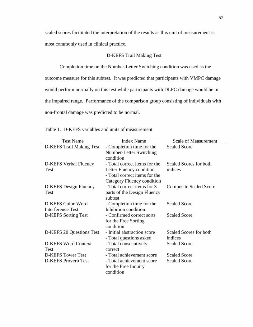
52
scaled scores facilitated the interpretation of the results as this unit of measurement is
most commonly used in clinical practice.
D-KEFS Trail Making Test
Completion time on the Number-Letter Switching condition was used as the
outcome measure for this subtest. It was predicted that participants with VMPC damage
would perform normally on this test while participants with DLPC damage would be in
the impaired range. Performance of the comparison group consisting of individuals with
non-frontal damage was predicted to be normal.
Table 1. D-KEFS variables and units of measurement
Test Name Index Name Scale of Measurement D-KEFS Trail Making Test - Completion time for the
Number-Letter Switching condition
Scaled Score
D-KEFS Verbal Fluency Test
- Total correct items for the Letter Fluency condition - Total correct items for the Category Fluency condition
Scaled Scores for both indices
D-KEFS Design Fluency Test
- Total correct items for 3 parts of the Design Fluency subtest
Composite Scaled Score
D-KEFS Color-Word Interference Test
- Completion time for the Inhibition condition
Scaled Score
D-KEFS Sorting Test - Confirmed correct sorts for the Free Sorting condition
Scaled Score
D-KEFS 20 Questions Test - Initial abstraction score - Total questions asked
Scaled Scores for both indices
D-KEFS Word Context Test
- Total consecutively correct
Scaled Score
D-KEFS Tower Test - Total achievement score Scaled Score D-KEFS Proverb Test - Total achievement score
for the Free Inquiry condition
Scaled Score

53
D-KEFS Verbal Fluency Test
The outcome measures for this subtest included the total correct responses for the
Letter Fluency condition and the total correct responses for the Category Fluency
condition. Participants with VMPC lesions were predicted to perform normally on both
measures while participants with DLPC were predicted to have impaired performance.
More specifically, participants with DLPC lesions on the left side would be more
impaired than ones with DLPC lesions on the right. Regarding the performance of the
non-frontal comparison group, normal performance was predicted except for a possible
impairment in semantic fluency in participants with left temporal lesions.
D-KEFS Design Fluency Test
The composite score for all three conditions of the test (Filled Dots, Empty Dots,
and Switching) was used as the outcome measure for this subtest. It was predicted that
participants with VMPC damage would perform normally while participants with DLPC
lesions would have impaired performance. Regarding laterality effects, participants with
right DLPC lesions were predicted to be more severely impaired than participants with
left DLPC lesions. Performance of the non-frontal comparison group was predicted to be
normal.
D-KEFS Color-Word Interference Test
The outcome measure used for this subtest was the completion time for the
Inhibition condition. Performance of participants with VMPC damage was predicted to
be normal unless there was damage to the anterior cingulate, in which case performance
would be impaired. Impaired performance was predicted for the group with DLPC
damage, while the non-frontal comparison group was predicted to perform normally.

54
D-KEFS Sorting Test
Confirmed number of correct sorts in the Free Sorting condition was used as the
outcome measure. Normal performance was predicted for participants with VMPC
damage and for the non-frontal comparison group. Impaired performance was predicted
for the group with DLPC damage.
D-KEFS Twenty Questions Test
The outcome measures were the total number of questions asked and the initial
abstraction score (the degree of abstraction of the first question asked was determined by
the number of items it eliminated). It was predicted that participants with VMPC lesions
would perform normally on the measure of total number of questions asked, while the
prediction regarding performance on the initial abstraction measure remained open due to
the lack of research on this topic. For patients with DLPC damage, predictions included
impaired performance on the total number of questions asked but also remain
undetermined for the initial abstraction score. The comparison group was predicted to
perform normally on both measures.
D-KEFS Word Context Test
The outcome measure was the total number of consecutively correct items.
Participants with VMPC lesions were predicted to perform normally while participants
with DLPC lesions were predicted to perform in the impaired range. Performance of the
non-frontal comparison group was predicted to be normal.
D-KEFS Tower Test
The outcome measure for the D-KEFS Tower Test was the Total Achievement
score. For participants with VMPC damage, normal performance was expected. For

55
participants with DLPC damage, impaired performance was expected. Normal
performance was expected for participants with non-frontal damage, unless the damage is
in the right temporal region, which may result in impaired performance.
D-KEFS Proverb Test
The total achievement score for the Free Inquiry condition was used as the
outcome measure. Participants with VMPC lesions and with non-frontal lesions were
expected to perform normally while participants with DLPC lesions were expected to
have impaired performance.
Alternate Outcomes
At this point, it is important to discuss alternate outcomes of the study and their
consequences. One of the alternate outcomes is finding no significant differences
between participants with DLPC and VMPC damage. This scenario may call research
findings showing differences in executive functioning as a result of VMPC and DLPC
damage into question. Given the fact that there is significant evidence for functional
differences between the VMPC and the DLPC, finding no differences in this study may
prompt a careful examination of the D-KEFS as a sensitive instrument for assessing
executive functioning.

56
Table 2. Performance Predictions
Trails V. Fluency D. Fluency C-W Sorting 20 Quest. Word Cont. Tower Proverb
Measures Completion time on the
Number-Letter Switching condition
Total correct responses for Letter Fluency and Category
Fluency
Total correct designs
Completion time for the Inhibition condition
Number of confirmed
correct sorts in the Free
Sorting condition
Initial abstraction
score and the total number of questions
asked
Total consecutively correct items
Total Achievement
Score
Total achievement score for the Free Inquiry
condition
VMPC Predictions
Normal performance
Normal performance
Normal performance
Normal performance
unless damage to the
anterior cingulate, in which case –
impaired performance
Normal performance
Undetermined performance on the initial abstraction
score; normal performance on the total number of questions
asked
Normal performance
Normal performance
Normal performance
DLPC Predictions
Impaired performance
Impaired performance;
left-sided lesions will
result in greater
impairment than right-
sided lesions
Impaired performance; right-sided lesions will
result in greater
impairment than left-
sided lesions
Impaired performance
Impaired performance
Impaired performance on the total number of questions
asked; initial abstraction prediction
remains open
Impaired performance
Impaired performance
Impaired performance
Non-Frontal
Predictions
Normal performance
Normal performance; left temporal lesions may
result in impairment in
semantic fluency
Normal performance
Normal performance
Normal performance
Normal performance
Normal performance
Normal performance; right temporal lesions may
result in impaired
performance
Normal performance

57
CHAPTER THREE
METHODS
Procedures
Participants who met the inclusion criteria described in the following paragraphs
were contacted by the research assistant in the Cognitive Neuroscience program. Those
who agreed to participate in the project were invited to come to the Neurology
Department at the University of Iowa Hospitals and Clinics. The University of Iowa
Institutional Review Board approved the study under a larger existing Patient Registry
research project. Informed consent was reviewed and signed by the participants in the
context of the existing overarching project, which included the D-KEFS administration.
The main investigator administered the D-KEFS to the consented participants. In four
cases, the battery was administered by a trained research assistant. The battery took
approximately 90-120 minutes to administer.
Power Analysis
A power analysis was attempted to determine the optimal number of participants
in each group. Previous studies investigating performance of participants with brain
lesions on the D-KEFS were used to estimate performances in the current study.
Unfortunately, the number of studies using the D-KEFS in patients with brain lesions is
limited, and one of the investigations was a case study. Another complicating factor was
that the sample size estimates varied based on the subtest of the D-KEFS. For example,
based on previous literature, the average scaled score of the DLPC lesion group on the
Twenty Questions subtest was five, eleven for the VMPC group, and ten for the non-
frontal lesion group (none of the studies used patients with non-frontal lesions, so this

58
number was based on the prediction of average performance estimated from studies using
similar measures). Based on these estimates, only nine people per group would be
needed to reach power of 80 percent. On the other hand, based on previous D-KEFS
literature, the DLPC group was predicted to achieve a scaled score of 8.75 on the Tower
Test, the VMPC group – a scaled score of eight (based on a case study), and the non-
frontal group – a scaled score of ten. Based on these estimates, even twenty people per
group would not be enough to reach adequate power.
Based on the general predictions about group performances on all subtests of the
D-KEFS, if one estimates a scaled score of six as the mean performance of the DLPC
group, a scaled score of nine as the mean performance of the VMPC group, and a scaled
score of ten as the mean performance of the non-frontal group, the study would require
seventeen people per group in order to reach power of 80 percent. Based on this power
analysis, the goal number of participants for each group was seventeen.
Participants
The final study included 46 participants identified through the Patient Registry of
the Division of Behavioral Neurology and Cognitive Neuroscience in the Department of
Neurology at the University of Iowa Hospitals and Clinics (patient information is
presented in chapter four). Inclusion criteria for the entire sample were: 1. Focal lesions
predominantly limited to one target area (ventromedial prefrontal, dorsolateral prefrontal,
or non-frontal). Exclusion criteria included: 1. Presence of aphasia; 2. Lobectomy
(surgical removal of a part of a particular lobe implicated in seizure onset) as the cause of
the lesion; 3. Location of the lesion in the anterior temporal lobe to exclude participants
with damage to subcortical structures involved in emotional regulation and memory
59
(amygdala and the hippocampus). At the beginning of the study, the author compiled a
list of patients suitable for the investigation. This procedure was based on previous lists
used in similar studies, file review, including most recent neuropsychological exam, and
neuroimaging data. Dr. Daniel Tranel, Director of the Cognitive Neuroscience program
at the University of Iowa, assisted in lesion classification. The study did not include a
group of patients with superior mesial lesions due to an inadequate number of such
patients in the Patient Registry.
Lesion Classification
All patients included in the Patient Registry have stable focal lesions, well
characterized through structural imaging. The neuroanatomical lesion analysis was
performed on the basis of MRI for the majority of patients, and on the basis of CT for
patients with conditions precluding MRI, such as metal clipping. The imaging material
was obtained at least three months after the lesion onset and was mapped according to
standard procedures of the University of Iowa Human Neuroanatomy and Neuroimaging
Laboratory using Brainvox (Damasio & Frank, 1992; Frank, Damasio, & Grabowski,
1997). Participants in the current study were classified into one of the research groups
based on neuroanatomical analysis. The VMPC was defined as the region encompassing
the medial orbital area including the gyrus rectus and the medial portion of the orbital
gyri, and the lower medial prefrontal area. The DLPC was defined as the region on the
top and the sides of the dorsal prefrontal cortex. Figure 1 presents an illustration of
regions identified as DLPC and VMPC. Finally, non-frontal lesions were defined as any
lesions outside the frontal lobes consistent with the inclusion and exclusion criteria listed
in the previous section.

60
Figure 1. Illustration of DLPC and VMPC regions
All patients in the Patient Registry were carefully screened by a team consisting
of a board-certified neurologist and a board-certified neuropsychologist to exclude
individuals with a history of mental retardation, learning disability, psychiatric disorder,
substance abuse, or a neurological condition other than the one resulting in the lesion.
These criteria remain in place throughout the patients’ participation in the Registry, so
that patients who do not continue to meet the criteria are eliminated. This is done
through regular monitoring of their cognition and psychiatric health, as well as through

61
repeated structural imaging. All participants were in the chronic stage of recovery and
were neuropsychologically stable.
The Patient Registry includes data on participants’ intellectual status (e.g. WAIS-
III scores), as well as psychological and emotional functioning (e.g. Beck Depression
Inventory-II [BDI-II], Beck Anxiety Inventory [BAI], and the Minnesota Multiphasic
Personality Inventrory-2 [MMPI-2]). The investigation considered the effects of general
intellectual level [Full Scale IQ (FSIQ) from Wechsler Adult Intelligence Scale – III
(WAIS-III)] and level of depression (BDI-II) to ensure the specificity of the findings. As
a general rule, FSIQ scores used in the study were obtained within two years prior to
participation in the current project. Regarding BDI-II scores, existing scores were used
if they were obtained within the last six months of the current investigation. If this
condition was not met, BDI-II was administered during the same visit as the D-KEFS.
Fifty one patients with specified lesions were tested during the data collection
process. Five were excluded for the following reasons: aphasia interfering with test
performance, suspected dementia, lesions with developmental onset (in two participants);
and anterior temporal lobectomy with amygdala damage. The decision to exclude
participants with developmental lesions was made in light of research indicating a
differential developmental course and neuropsychological performance in patients with
child and adult lesion onset (Anderson et al., 2006).
Among participants with VMPC lesions, six had bilateral lesions, three – right-
sided lesions, and five – left-sided lesions. Among participants with DLPC lesions, two
had bilateral lesions, seven – right-sided lesions, and five – left-sided lesions. Six
participants had damage located primarily in the DLPC but extending slightly to a non-

62
frontal region. As for participants with non-frontal lesions, one had a bilateral lesion, six
– right-sided lesions, and eleven left-sided lesions. Table 3 presents lesion etiologies for
each group.
Table 3: Lesion etiologies among three groups
VMPC (n=14)
DLPC (n=14)
Non-Frontal (n=18)
CVA 1 8 14
Aneurysm 8 2 0
Meningioma 3 3 3
TBI 1 0 1
AVM 1 1 0
Note: CVA = Cerebrovascular accident (ischemic or hemorrhagic); TBI = Traumatic
brain injury with focal neuroimaging findings; AVM = Arteriovenous malformation;
Meningioma = resection (surgical removal) of benign brain tumor
Instruments
The main instrument used in the project was the Delis-Kaplan Executive Function
System (D-KEFS; Delis, Kaplan, & Kramer, 2001). It is a battery of tests allowing for a
comprehensive and detailed examination of executive functioning. The battery consists
of nine subtests derived from existing measures. Detailed descriptions of the subtests can
be found in chapter two.

63
In addition to the D-KEFS, two other executive functioning batteries were
considered as possible primary measures for the investigation: the Cambridge
Neuropsychological Testing Automated Battery (CANTAB) and the Behavioral
Assessment of the Dysexecutive Syndrome (BADS). The following paragraphs will
explain the advantages and disadvantages of these measures as well as the reasons for
choosing the D-KEFS instead of the CANTAB or the BADS.
The CANTAB is a computerized battery assessing several areas of cognition,
including memory, attention, and executive functioning. The latest version consists of 22
subtests, five of which are subsumed under executive function, working memory, and
planning category (Intra/Extradimensional Shift, One Touch Stockings of Cambridge,
Stockings of Cambridge, Spatial Span, and Spatial Working Memory). Four additional
tasks assess decision-making and response control sets based on emotional factors. The
CANTAB has several positive features, such as the ease of administration and scoring
(performed by the computer), its non-verbal nature presumably creating a language- and
culture-independent test, and the possibility of selecting specific tasks of interest.
However, it also has several disadvantages. The non-verbal nature of the tests does not
allow for the expression of verbal abilities and precludes the administration of verbally-
based executive tasks (e.g. verbal fluency). The CANTAB consists of several tasks
adapted from studies with animals and presented in a format which makes comparisons to
more traditional neuropsychological tests extremely challenging. In addition, questions
about the adequacy of the CANTAB reliability have been raised (Lowe & Rabbit, 1998).
One of the most important reasons for choosing the D-KEFS instead of the
CANTAB as the main measure for the study is the fact that the CANTAB is largely a

64
research tool with unexplored clinical applications. While its research application is
valuable, the goal of this study is to provide information about brain-behavior
relationships that would be immediately applicable not only for scientific purposes but
also for clinical ones. The computerized nature of the CANTAB and the practical
considerations of its use (e.g. cost and training) will likely continue to deter
neuropsychologists from using this instrument for clinical purposes. In 2000, Camara et
al. reported that only three percent of psychologists used computers for test
administration. While this percentage has likely increased by the present time, non-
computerized administration still remains the primary testing method. The D-KEFS, on
the other hand, is much more applicable in clinical settings and, in fact, it is already being
used in clinical practice. It provides an assessment of both verbal and non-verbal aspects
of a wide range of executive functions. It can be easily compared to existing measures
of executive functions, which facilitates the validation and evaluation of the D-KEFS as a
neuropsychological tool. Based on the presented information, the D-KEFS seemed as a
more appropriate measure to meet the goals of the current project.
The BADS is a battery of tests aimed to detect symptoms of the dysexecutive
syndrome predictive of real-world impairments. The battery consists of six tests
designed to resemble real-life tasks to increase the ecological validity of the battery and a
questionnaire to be filled out by the patient and another person familiar with the patient.
The tests include: Rule Shift Cards Test, Action Program Test, Key Search Test,
Temporal Judgment Test, Zoo Map Test, Modified Six Elements Test, and the
Dysexecutive Questionnaire. The battery yields one standardized score characterizing
the level of executive dysfunction. While the BADS is an important and promising
65
measure, it has limitations pointed out by several test reviewers. For example, D’Amato
and Haynes (Mental Measurement Yearbook, 14th ed., 2001) in two separate reviews
concluded that the items on some of the BADS tests, particularly the Temporal Judgment
Test, were designed for the British population and would not be culturally appropriate for
American test-takers. Since the composite score for the BADS is based on all subtest
scores, the Temporal Judgment Test would have to be excluded or modified, which may
limit the interpretations based on the composite score. D’Amato also pointed out that the
authors of the BADS provided little guidance for interpreting scores on individual tests.
In addition, the BADS has a relatively small norming sample of 216 individuals divided
into four large age bands. While the D-KEFS represents a more traditional measure of
executive functions, it had a large U.S. - based norming sample and is more culturally-
appropriate for American test-takers, which made it better-suited for this study. The
following section will focus on the psychometric properties of the D-KEFS.
D-KEFS Standardization and Norms
The standardization sample consisted of 1,750 healthy adults and children, ages
8-89. The sample was representative of the 2000 U.S. Census population on the
dimensions of age, sex, race/ethnicity, years of education, and geographic region. The
sample was divided into sixteen age groups, where each year from 8-15 represented a
single age group, 16-19 year old – another group, and the remaining groups spanned nine
years (e.g. 20-29, 30-39, etc.). Sample size for in each age band ranged from 75 to 175
people. Each age group consisted of approximately equal number of males and females,
except for the older groups where there were more females than males (consistent with
the 2000 U.S. Census). For each age group, the proportions of African/American,

66
Hispanic, White, and other racial/ethnic groups were matched to the 2000 U.S. Census.
The entire standardization sample was divided into five groups based on years of
education (according to the 2000 U.S. Census): less than or equal to 8 years, 9-11 years,
12 years, 13-15 years, and 16 or more years. Parental educational achievement level was
used for children and adolescents of 8-19 years of age. Finally, individuals from four
regions of the United States (specified by the U.S. Census data) participated in the
standardization of the D-KEFS (Northeast, North Central, South, and West). For a
detailed description of the sample, please refer to the Standardization Study chapter of the
D-KEFS Technical Manual (Delis, Kaplan, & Kramer, 2001).
For all of the performance indices used in this study, raw scores were normally
distributed and converted into age-corrected scaled scores with a mean of ten and
standard deviation of three.
D-KEFS Reliability and Validity
The D-KEFS Technical Manual has been criticized by some researchers for not
providing sufficient evidence about validity (Schmidt, 2003). In 2004, the test creators
published a response to this criticism explaining that the manual only has the most recent
validity information but the vast majority of the validity data was presented through
articles in peer-reviewed journals. They provided examples of studies that have used the
D-KEFS to investigate executive functioning with different populations, including
patients with frontal lobe lesions (Baldo et al., 2001; Cato et al., 2004; Delis et al., 1992),
frontotemporal dementia (Kramer et al., 2007; Carey et al., 2008), schizophrenia (Beatty
et al., 1994), mild cognitive impairment, Parkinson’s disease (Bondi et al., 1993; Beatty
et al., 1990), multiple sclerosis (Beatty et al., 1995), attention deficit disorder (Donnelly

67
et al., 2001), and fetal alcohol syndrome (Mattson et al., 1999), among others. The
authors also explained the lack of factor-analytic studies for most D-KEFS subtests. In
their opinion, a factor-analytic validation approach has serious limitations when used
with process-oriented tests. As a final remark, the authors commented on the moderate
reliability coefficients for many subtests of the D-KEFS by pointing out that many
executive functioning tests, such as the Wisconsin Card Sorting Test, also have moderate
reliability coefficients, likely due to the fact that executive functioning tasks are highly
complex and recruit multiple cognitive processes, creating greater potential for variable
performance. A more recent investigation of the reliability of D-KEFS contrast measures
(Crawford et al., 2008) found low reliabilities and suggested avoiding the use of the
measures in clinical decision-making.
The reliability and validity data presented in the D-KEFS Technical Manual are
briefly described below.
D-KEFS Trail Making Test
Internal consistency values ranged from moderate to high for each age group (.57-
81) for the Composite Letter Sequencing+Number Sequencing conditions. Test-retest
reliability for all D-KEFS subtests was evaluated with a diverse sample of 101
individuals. The average time between test administrations was 25 days. Most
correlations for all conditions were in the moderate range, with highest correlations for
the motor speed condition. Notably, for the Time to Completion in the Switching
condition (variable of interest in this study), test-retest reliability for individuals ages 20-
49 was .36, and for individuals ages 50-89, it was .55. For Standard Error of

68
Measurement values for each age group, please refer to Table 2.3 in the D-KEFS
Technical Manual (p. 22).
Since the D-KEFS Trail Making Test was derived from the original Trail Making
Test (TMT), the authors have relied on previous TMT validity evidence to support the
validity of the D-KEFS Trail Making Test. The only validity variables presented in the
manual are the intercorrelations between the conditions of the test for three age groups
(8-19, 20-49, and 50-89). The three sequencing trials of the D-KEFS Trail Making Test
(Letter Sequencing, Number Sequencing, and Number-Letter Switching) were
moderately positively correlated with each other for all age groups. The Visual Scanning
and Motor Speed conditions had low correlations with the three sequencing conditions.
D-KEFS Verbal Fluency Test
Internal consistency values for all three conditions of the test were in the
moderate-high range, with the highest coefficients in the Letter Fluency Condition (.68-
.90). Test-retest reliability was moderate-high for the Letter and Category conditions, but
was slightly lower for the Category Switching condition. For the Letter Fluency Total
Correct score, test-retest reliability of the 20-49 years old group was .76, and .88 for the
50-89 years old group. Standard Error of Measurement values for the 90% and 95%
confidence interval are presented in Table 2.6 in the D-KEFS Technical Manual (p. 25).
Similarly to the D-KEFS Trail Making Test, validity evidence for the D-KEFS
Verbal Fluency Test was based on the existing tasks of this nature (e.g. COWA).
Moderate positive intercorrelations were found between the three test conditions (Letter
Fluency, Category Fluency, and Category Switching) for the three previously mentioned
age groups.

69
D-KEFS Design Fluency Test
Internal consistency reliability was not investigated for this test due to item
interdependence. Test-retest reliability was generally moderate. For example, for the
Correct Designs Filled Dots Only condition, test-retest reliability was .62 for the 20-49
years old group, and .43 for the 50-89 group. Table 2.8 of the D-KEFS Technical
Manual presents the Standard Error of Measurement values for the three conditions of the
test (p. 27).
The existing Design Fluency tests provided validity evidence for the D-KEFS
Design Fluency Test. The three conditions of the test (Filled Dots, Empty Dots, and
Switching) were moderately positively correlated with each other, with a stronger
association between the two non-switching conditions.
D-KEFS Color-Word Interference Test
Internal consistency of the composite Color Naming + Word Reading completion
times score was moderate-high in all age groups (.62-.86). Test-retest reliability
coefficients were also in the moderate-high range. For the Inhibition condition, test-retest
reliability coefficient was .86 for the 20-49 age group, and .56 for the 50-89 age group.
Standard Error of Measurement values are presented in Table 2.11 of the D-KEFS
Technical Manual (p. 30).
Validity evidence for the D-KEFS Color-Word Interference Test is based on
existing interference tests, such as the Stroop Color-Word Interference Test.
Intercorrelations between the four conditions of the test were in the moderate range for all
three age groups.

70
D-KEFS Sorting Test
The internal consistency values are in the moderate to high range. For the Free
Sorting Confirmed Sorts condition, internal consistency reliability ranged from .55-.86.
Test-retest reliability was moderate for all conditions (.51 for the 20-49 age group and .62
for the 50-89 age group for the Free Sorting Confirmed Sorts condition). Standard Error
of Measurement information can be found in Table 2.14 of the D-KEFS Technical
Manual (p. 33).
As the authors mention in the technical manual, the D-KEFS Sorting Test was
derived from the California Card Sorting Test (CCST) and research has shown that it
measures somewhat different factors of executive function than the Wisconsin Card
Sorting Test (WCST). Construct validity of the D-KEFS Sorting Test was provided in
the manual via a review of studies that used the test or its predecessor CCST. Delis et al.
(1992) and Dimitrov et al. (1999) have shown that patients with frontal lobe lesions
perform more poorly on the test than patients with non-frontal damage or normal
controls. In addition, multiple studies of populations with high rates of executive
dysfunction (e.g. Korsakoff’s syndrome, Parkinson’s disease, schizophrenia)
demonstrated that these populations performed deficiently on the sorting test.
Intercorrelations between the D-KEFS Sorting Test measures were generally
robust and positive. Part-whole correlations showed that the two card sets contributed
roughly equally to the total score.
D-KEFS Twenty Questions Test
Due to some item interdependence, split-half reliabilities were used to determine
internal consistency. The Initial Abstraction score had moderate-high correlations (.72-

71
.87), while the Total Weighted Achievement score had moderate-low correlations (.10-
.55), likely attributable to item interdependence. Test-retest reliability values were
moderate-low. For example, for the Initial Abstraction score, test-retest reliability for the
20-49 age group was .24, and .42 for the 50-89 age group.
Validity for the D-KEFS Twenty Questions Test was mainly based on the
previous versions of the test. Intercorrelations between the Total Questions Asked
measure and the Total Weighted Achievement Score were positive and high, while the
Initial Abstraction Score was weakly correlated with the Total Weighted Achievement
Score.
D-KEFS Word Context Test
Split-half reliabilities were calculated for this test, resulting in moderate
correlations for most age groups (.47-.74). Test-retest reliability was good for both adult
age groups (.73 for the 20-49 age group, and .78 for the 50-89 age group). Standard
Error of Measurement values can be found in Table 2.20 of the D-KEFS Technical
Manual (p. 39).
Validity evidence for the D-KEFS Word Context Test was based on existing
Word Context tests. Moderate intercorrelations were found between the total first trial
consistently correct measure and the repeated incorrect responses score. Slightly lower
intercorrelations were found between the total first trial consistently correct measure and
the consistently correct ratio.
D-KEFS Tower Test
Split-half reliability ranged from moderate-high for all age groups (.43-.78). Test-
retest reliability was moderate (.41 for the 20-49 age group, and .38 for the 50-89 age

72
group). Standard Error of Measurement values are reported in Table 2.23 in the D-KEFS
Technical Manual (p. 42).
The D-KEFS Tower Test validity relied on previously gathered evidence for
existing tower tests (e.g. Tower of London and Tower of Hanoi). The time per move
measure weakly positively correlated with the total achievement score and negatively
with the move accuracy ratio, suggesting that people who make responses quickly arrive
at a higher achievement score but make more errors. The first-move time was not
correlated with other measures on the test. This was not unexpected, according to the
authors, since higher or lower first-move response times reflect different cognitive
problems (impulsivity versus lack of initiation).
D-KEFS Proverb Test
Split-half correlations indicated that internal consistency was good for all age
groups (.68-.81). Test-retest reliability was moderate in the 20-49 age group (.66) and
high in the 50-89 age group (.81). Standard Error of Measurement is reported in Table
2.26 in the D-KEFS Technical Manual (p. 45).
Accuracy and Abstraction scores were highly intercorrelated while other
measures were moderately correlated. Correlations between the overall achievement
scores on the different D-KEFS subtests were low and positive, indicating that the
subtests measure separate aspects of executive functioning. Process/Efficiency scores,
response initiation indicators, and processing speed indices were positively but weakly
correlated across the subtests.
Mean correlations of the D-KEFS Sorting Test and the California Verbal
Learning Test-Second Edition Short Delay Free Recall Total Correct and Long Delay

73
Free Recall Total Correct (CVLT-II; Delis, Kaplan, Kramer, & Ober, 2000) were -.13
and -.22 respectively, indicating that the D-KEFS Sorting Test measures a different set of
cognitive abilities (executive functioning) than the CVLT-II. A pilot study (n=23)
examining the relationship between the WCST and the D-KEFS subtests found moderate
correlations between the number of completed categories on the WCST and several D-
KEFS measures (e.g., correct sorts and sort recognition on the D-KEFS Sorting Test,
category switching on the D-KEFS Verbal Fluency Test, and empty/filled dot switching
on the D-KEFS Design Fluency Test). The number of perseverative errors on the WCST
also positively correlated, although at a slightly lower level than the number of completed
categories, with several measures of the D-KEFS.

74
CHAPTER FOUR
RESULTS
Descriptive Statistics
Participants’ demographic information, chronicity (time since lesion onset in years), WAIS-III Full Scale IQ (FSIQ), and BDI-II scores are presented in Table 4.
Table 4. Demographic characteristics, lesion chronicity, FSIQ, and BDI-II scores
VMPC (n=14)
DLPC (n=14)
Non-Frontal (n=18)
Sex 9 Male 5 Female
11 Male 3 Female
7 Male 11 Female
Age (SD) 60.43(6.85) 58.21(9.71) 60.83(9.98)
Education (SD) 13.79(2.8) 13.57(2.14) 15.00 (3.79)
Handedness* 12R;1L;1Mix 12R;1L;1Mix 15R; 3Mix
Chronicity 15.64 (9.48) 9.79 (9.23) 10.56 (8.36)
FSIQ 109.93(18.97) 97.07(13.07) 107.78(15.73)
BDI-II 7.36(6.64) 10.14(9.68) 7.39(6.20)
* R=Right-handed; L=Left-handed; Mix=Mixed-handed Note: Lesion chronicity refers to time (in years) since lesion onset.
The VMPC, DLPC, and non-frontal groups did not significantly differ with regard
to age [F (2,43) = .36, p = .70], education [F (2,43) = 1.03, p = .37], chronicity [F (2,43)
=1.81, p = .18], FSIQ [F (2,43) = 2.63, p = .08], and BDI-II scores [F (2,43) = .66, p =
.53]. Since the results of the preliminary analysis showed that age, education, lesion
chronicity, and BDI-II scores were not significantly different among the groups, these
variables were excluded from the following analyses. FSIQ was retained as a covariate

75
to ensure the specificity of D-KEFS performances to executive functioning. All analyses
were performed using SPSS for Windows (17th edition) using alpha of .01 to account for
multiple comparisons.
D-KEFS Performances
D-KEFS performances of participants with VMPC, DLPC, and non-frontal
lesions are presented in table 5 and figure 2.
Table 5. D-KEFS performances in Scaled Scores
VMPC (n=14)
DLPC (n=14)
Non-Frontal (n=18)
Trails Letter Number Switching
9.93(4.10) 8.50(3.6) 10.00(3.03)
Letter Fluency
10.14(4.59) 7.71(2.6) 9.22(3.93)
Category Fluency
10.36(4.62) 7.00(2.88) 9.83(2.68)
Design Fluency Composite
11.00(3.62) 9.29(2.20) 11.94(3.12)
Color-Word Inhibition
9.57(4.03) 7.43(3.63) 10.39(2.20)
Confirmed Correct Sorts
10.07(3.32) 9.00(1.84) 11.61(2.83)
20 Questions Initial Abstraction
11.21(3.42) 9.64(1.55) 11.67(4.22)
20 Questions Total Questions Asked
10.36(2.79) 10.43(2.59) 11.11(3.16)
Word Context Total Consecutively Correct
10.50(3.74) 9.14(3.35) 10.06(2.18)
Tower Total Achievement
12.29(3.8) 10.43(3.25) 10.89(2.70)
Proverb Free Inquiry Total Achievement
11.43(2.68) 10.07(3.87) 11.50(2.75)
Note: Mean = 10, SD = 3

76
Figure 2. D-KEFS performances in Scaled Scores
Note: Y-axis = D-KEFS Scaled Scores (Mean = 10, SD = 3). 1 = D-KEFS Trail Making Test Letter-Number Sequencing; 2 = D-KEFS Letter Fluency Total Correct; 3 = D-KEFS Category Fluency Total Correct; 4 = D-KEFS Design Fluency Composite Score; 5 = D-KEFS Color-Word Interference Test Inhibition condition; 6 = D-KEFS Sorting Test; 7 = D-KEFS Twenty Questions Initial Abstraction Score; 8 = D-KEFS Twenty Questions Total Questions Asked; 9 = D-KEFS Word Context Total Consecutively Correct; 10 = D-KEFS Tower Total Achievement Score; 11 = D-KEFS Proverb Total Achievement Score in the Free Inquiry condition
It is notable that all group mean scaled scores in the VMPC and non-frontal lesion
groups were above a scaled score of nine (37th percentile). However, mean scaled scores
for the DLPC group were below a scaled score of nine on four tests (Trails Letter
Number Switching, Letter Fluency, Category Fluency, and Color-Word Inhibition). In
general, on virtually every measure, performances of participants with DLPC lesions
were lower than performances of the other two groups. Furthermore, several group
differences approached or exceeded one standard deviation.
5
6
7
8
9
10
11
12
13
1 2 3 4 5 6 7 8 9 10 11
VMPC
DLPC
Non-Frontal

77
Performances adjusted for the covariate (FSIQ) are presented in table 6.
Interestingly, adjusting group means for FSIQ diminishes group differences, suggesting
that FSIQ may explain some of the group differences seen when scores are not adjusted
for FSIQ. However, some notable differences remained. For example, based on a
qualitative examination, the DLPC lesion group performed consistently worse than the
other two groups on the Category Fluency test regardless of whether scaled scores were
FSIQ-corrected or not.
Table 6. FSIQ-adjusted mean scaled scores (mean = 10, SD = 3) and standard errors
VMPC (n=14)
DLPC (n=14)
Non-Frontal (n=18)
Trails Letter Number Sequencing
9.39(.84) 9.41(.86) 9.71(.74)
Letter Fluency
9.62(.93) 8.60(.95) 8.94(.81)
Category Fluency
10.00(.88) 7.61(.91) 9.64(.77)
Design Fluency Composite
10.49(.69) 10.16(.71) 11.67(.60)
Color-Word Inhibition
9.16(.82) 8.13(.84) 10.16(.71)
Confirmed Correct Sorts
9.69(.66) 9.65(.68) 11.40(.58)
20 Questions Initial Abstraction
10.61(.77) 10.58(.79) 11.37(.67)
20 Questions Total Questions Asked
10.10(.76) 10.86(.78) 10.97(.66)
Word Context Total Consecutively Correct
9.87(.61) 10.23(.62) 9.71(.53)
Tower Total Achievement
12.00(.85) 10.92(.87) 10.73(.74)
Proverb Free Inquiry Total Achievement
10.87(.68) 11.02(.70) 11.19(.59)

78
Comparison of Participants with VMPC, DLPC, and Non-Frontal Lesions
Statistical analyses comparing D-KEFS performances of the VMPC, DLPC, and
non-frontal lesion groups were performed using analysis of covariance (ANCOVA) with
FSIQ as a covariate. Alpha level was kept at .01 to account for multiple comparisons.
Analyses revealed no statistically significant differences in group performances on the D-
KEFS measures included in this investigation. Results are displayed in Table 7.
As previously mentioned, based on a qualitative inspection of the results, some
group differences appeared to be meaningfully different even though the differences did
not reach statistical significance. To illustrate the strength of the effect and to aide in the
interpretation of the results, Cohen’s d effect sizes were calculated for the differences
between groups with the lowest and highest performances on several measures. For
example, for the D-KEFS Category Fluency subtest, the greatest difference was observed
between performances of the VMPC and DLPC groups. Cohen’s d for that difference
was .87. For the Color-Word Inhibition measure, Cohen’s d of the difference between
the DLPC and non-frontal lesion groups was 1.02. Similarly, Cohen’s d of the difference
between the DLPC and the non-frontal groups on the D-KEFS Sorting test was 1.07. All
three of the Cohen’s d values reflect large effect sizes (Cohen, 1992) and warrant further
consideration even though statistical significance was not reached.
Statistical analyses showed that the covariate (FSIQ) was significant for almost all
comparisons with the exception of Category Fluency, Twenty Questions Total Questions
Asked, and Tower Test Total Achievement Score. This indicates that for the majority of
the measures, FSIQ accounted for a significant amount of variance in group
performances.

79
Table 7. ANCOVA comparing D-KEFS group performances with FSIQ as a covariate
F (2,42) P Part. η2 Covariate
F (FSIQ) Covariate
P Trails Letter Number Switching
.052 .949 .002 14.59 .000
Letter Fluency .303 .740 .014 11.27 .002
Category Fluency 2.011 .147 .087 5.809 .020
Design Fluency Composite
1.543 .226 .068 19.938 .000
Color-Word Inhibition 1.676 .199 .074 9.254 .004
Confirmed Correct Sorts 2.695 .079 .114 11.877 .001
20 Questions Initial Abstraction
.372 .692 .017 18.382 .000
20 Questions Total Questions Asked
.418 .661 .020 4.106 .049
Word Context Total Consecutively Correct
.196 .823 .009 39.544 .000
Tower Total Achievement .697 .504 .032 4.103 .049
Proverb Free Inquiry Total Achievement
.067 .936 .003 24.465 .000
Proportion of Impaired Performances in Each Group
Another way to present the data is to consider the proportion of individuals in
each group who performed in the mildly impaired range or lower (Scaled Score = or <
five; fifth percentile or lower). Table 8 shows the results of this analysis. For example,
on the D-KEFS Trail Making Test, 21 percent of individuals with frontal lesions obtained
a score at or below the fifth percentile (fourteen percent for the VMPC group and 29

80
percent for the DLPC group), while only six percent of individuals with non-frontal
lesions were at or below the fifth percentile.
Table 8. Number and percentage of participants performing at or below scaled score of 5
(5th percentile)
Frontal (n = 28)
VMPC (n = 14)
DLPC (n = 14)
Non-Frontal (n = 18)
Trails Letter Number Switching
6 (21%) 2 (14%) 4 (29%) 1 (6%)
Letter Fluency 5 (18%) 2 (14%) 3 (21%) 3 (17%)
Category Fluency
5 (18%) 2 (14%) 3 (21%) 1 (6%)
Design Fluency
2 (7%) 1 (7%) 1 (7%) 1 (6%)
Color-Word Inhibition
5 (18%) 2 (14%) 3 (21%) 0
Confirmed Correct Sorts
0 0 0 0
20 Questions Init. Abstract.
0 0 0 1 (6%)
20 Questions Tot. Questions
2 (7%) 1 (7%) 1 (7%) 1 (6%)
Word Context Total Correct
4 (14%) 2 (14%) 2 (14%) 1 (6%)
Tower Total Achievement
2 (7%) 1 (7%) 1 (7%) 1 (6%)
Proverb Free Inquiry Total
2 (7%) 0 2 (14%) 1 (6%)

81
Qualitatively, a higher percentage of participants with frontal lesions performed at
or below the fifth percentile on the following D-KEFS subtests, compared to participants
with non-frontal lesions: Trails Letter Number Sequencing, Category Fluency, Color-
Word Inhibition, and Word Context Total Consecutively Correct. A higher percentage of
participants with DLPC lesions performed in the mildly impaired range (fifth percentile)
or lower on Trails Letter Number Sequencing, Letter Fluency, Category Fluency, Color-
Word Inhibition, and Proverb Free Inquiry compared to participants with VMPC lesions.
Proportion of Performances at or below the Sixteenth Percentile
Results of a supplementary analysis considering percentage of participants in each
group whose scores were at or below a Scaled Score of seven (sixteenth percentile) are
presented in Table 9.
Interestingly, consideration of scores at or below the sixteenth percentile presents
a more complicated picture. On six of the eleven subtests, the DLPC group had a higher
percentage of participants with scores in the low average range or lower. Notably, on the
D-KEFS Category Fluency subtest, more than half (57 percent) of participants with
DLPC lesions performed at or below the sixteenth percentile compared to 29 and 22
percent in the VMPC and non-frontal lesion groups, respectively. Similarly,
performances were at or below the low average range for 50 percent of participants with
DLPC lesions on the D-KEFS Color-Word Inhibition measure compared to 21 percent in
the VMPC group and eleven percent in the non-frontal group. It also becomes clear that
while few participants in the VMPC group performed below the fifth percentile, a much
higher proportion had scores between the fifth and sixteenth percentile. For example, it is
notable that the VMPC lesion group had the highest percentage of participants (36

82
percent) who performed at or below the low average range on the D-KEFS Sorting Test
out of all three groups.
Table 9. Number and percentage of participants performing at or below SS of 7 (16th
percentile)
Frontal (n = 28)
VMPC (n = 14)
DLPC (n = 14)
Non-Frontal (n = 18)
Trails Letter Number Sequencing
6 (21%) 2 (14%) 4 (29%) 3 (17%)
Letter Fluency 9 (32%) 4 (29%) 5 (36%) 7 (39%)
Category Fluency
12 (43%) 4 (29%) 8 (57%) 4 (22%)
Design Fluency
4 (14%) 1 (7%) 3 (21%) 1 (6%)
Color-Word Inhibition
10 (36%) 3 (21%) 7 (50%) 2 (11%)
Confirmed Correct Sorts
8 (29%) 5 (36%) 3 (21%) 1 (6%)
20 Questions Init. Abstract.
3 (11%) 2 (14%) 1 (7%) 3 (17%)
20 Questions Tot. Questions
5 (18%) 3 (21%) 2 (14%) 1 (6%)
Word Context Total Correct
8 (29%) 4 (29%) 4 (29%) 3 (17%)
Tower Total Achievement
3 (11%) 1 (7%) 3 (21%) 1 (6%)
Proverb Free Inquiry Total Achievement
5 (18%) 2 (14%) 3 (21%) 3 (17%)
83
Similarly, the VMPC lesion group had a greater percentage of performances at or
below the sixteenth percentile on the Initial Abstraction Score and the Total Number of
Questions Asked of the D-KEFS Twenty Questions Test compared to participants with
DLPC lesions. The analysis also revealed that the non-frontal lesion group had the
highest percentage of participants performing at or below the low average range on the
D-KEFS Letter Fluency and Twenty Questions Initial Abstraction compared to the two
frontal lesion groups.
When all participants with frontal lobe lesions were combined in one group, the
larger frontal group had higher percentages of performances at or below the sixteenth
percentile on eight of the eleven measures compared to the non-frontal group (Trails
Letter Number Sequencing, Category Fluency, Design Fluency, Color-Word Inhibition,
Confirmed Correct Sorts, Twenty Questions Total Number of Questions Asked, Word
Context, and Tower).
Lesion Laterality in Patients with DLPC Lesions
It was predicted that participants with DLPC lesions in the left hemisphere would
perform worse on the Letter Fluency subtest of the D-KEFS than participants with DLPC
lesions in the right hemisphere. Conversely, participants with right-sided lesions were
predicted to perform worse on the Design Fluency subtest than participants with left-
sided lesions. To investigate this hypothesis, participants with DLPC lesions were
divided into two groups based on lesion laterality. Two participants had bilateral lesions
and were excluded from the analysis. Seven of the remaining participants had right -
sided lesions and five - left-sided lesions. A quantitative analysis was not conducted due
to the small number of participants in each group. Qualitatively, prediction about worse

84
performance of participants with left DLPC lesions on the D-KEFS Letter Fluency test
compared to participants with right DLPC lesions, was confirmed [Left DLPC Mean SS
= 6.8 (SD = 2.4); Right DLPC Mean SS = 8.4 (SD = 3.3)]. However, performances of
participants with right- and left-sided lesions on the D-KEFS Design Fluency subtest
were similar [Left DLPC Mean SS = 9.2 (SD = 2.6); Right DLPC Mean SS = 9.3 (SD =
2.3)].

85
CHAPTER FIVE
DISCUSSION
In neuropsychology literature and practice, executive dysfunction has often been
associated with frontal lobe damage. However, a careful review of available research
indicates that the relationship between frontal lobes and executive functions remains
controversial. In fact, the former president of the Division 40 (Clinical
Neuropsychology) of the American Psychological Association Carl Dodrill (1997)
described the assumed connection between frontal lobe damage and executive
dysfunction (as measured by defective performance on common neuropsychological
measures) as one of the popular “myths” in neuropsychology because multiple
investigations and meta-analyses have failed to show a specific connection.
It appears that in the discussion on the nature and degree of association between
frontal lobe damage and executive dysfunction, it is important to define what is meant by
the term “executive dysfunction”. One definition of “executive dysfunction” may
represent difficulties seen in real-life functioning (e.g., poor judgment, decision making,
multitasking). However, a definition that seems to be most commonly referred to in
discussions on “executive dysfunction” is impaired performance on laboratory tests of
executive functioning. Dodrill (1997) is not necessarily challenging the relationship
between frontal lobe damage and executive dysfunction but the relationship between
frontal lobe damage and executive dysfunction as measured by widely used
neuropsychological tests.
A complicating factor in interpreting research investigating the association
between executive functioning and frontal lobes is the fact that particular regions of the

86
frontal lobes are often not differentiated. Instead, all participants with frontal lobe lesions
are combined in one group. Valuable information may be lost as a result of using this
method since research suggests that lesions in different regions of the frontal lobes may
result in different clinical presentations and neuropsychological performances (Tranel et
al., 1994).
According to one organizational system (Chan et al., 2008), executive functions
can be divided into “cold” (e.g., cognitive set shifting, inhibition of a prepotent response,
strategy-generation, and problem-solving) and “hot” (e.g., decision-making based on
social/emotional variables). Generally, the “cold” executive functions have been
associated with the dorsolateral prefrontal cortex while the “hot” functions – with
ventromedial prefrontal cortex. In neuropsychological assessment, executive functions
are most often measured using instruments that attempt to isolate and target “cold”
executive skills. The measurement of the “hot” functions, however, remains elusive.
This is partially due to the nature of controlled laboratory testing that does not allow for
the expression of deficits in “hot” executive functions, as well as the limited number of
widely available measures targeting these functions. Few studies directly comparing
neuropsychological functioning in people with VMPC and DLPC lesions have been
published. Therefore, is it possible that Carl Dodrill’s general conclusion about the lack
of scientific evidence for the association between frontal lobe damage and executive
functioning deficits (as measured by commonly used tests) is too general? Is it possible
that the association varies based on the region of damage within the frontal lobes, as well
as the instrument?

87
The main purposes of this investigation were: 1. To compare the performances of
patients with DLPC, VMPC, and non-frontal lesions on a relatively new battery of
executive functioning tests (D-KEFS, 2001); 2. To determine the proportion of
participants in each group who performed in the impaired range. Dividing participants
with frontal lobe damage into two groups based on lesion location allows to clarify if
damage to specific areas of the frontal lobes results in lower performances on tests of
executive functioning. Using the D-KEFS battery of commonly used tests allows to
address the question of whether damage to specific regions of the frontal lobes (VMPC
and DLPC) and non-frontal regions results in greater impairment on particular tests.
While the D-KEFS provides multiple indices for each of its nine subtests, only
selected primary indices were used for the purpose of this study. Based on the literature
review, the DLPC group was expected to be the most impaired overall, while the VMPC
and non-frontal groups were expected to be the least impaired on most measures.
Participants in each group did not significantly differ with regard to age,
education, lesion chronicity, FSIQ, or BDI-II scores. Main analyses controlled for the
effect of FSIQ to ensure specificity to executive functioning. Overall, comparisons of
group mean scaled scores for the eleven measures did not reach statistical significance.
However, on virtually every measure, the mean score of the DLPC group was lower than
the scores of the VMPC and non-frontal lesion groups. Moreover, several of the
differences appeared meaningful and resulted in large effect sizes. Specifically, the
largest group differences were observed on the D-KEFS Category Fluency Test, D-KEFS
Color-Word Interference Test (Inhibition condition), the D-KEFS Letter Fluency Test,
and the D-KEFS Sorting Test (Number of Confirmed Correct Sorts in the Free Sorting
88
condition). On three subtests (Category Fluency, Color-Word Inhibition, and Letter
Fluency) the mean group scaled scores of the DLPC group were below a scaled score of
eight (25th percentile). Conversely, no performances of the VMPC and non-frontal lesion
groups were below a scaled score of nine (37th percentile).
The second goal of the study was to determine the percentage of participants in
each group with scores in the impaired range (fifth percentile or below). This analysis
revealed that on five of the subtests, a greater percentage of participants with DLPC
lesions performed in the impaired range (Trails Letter Number Switching, Letter Fluency,
Category Fluency, Color-Word Inhibition, and Proverb Free Inquiry) than participants
with VMPC and non-frontal lesions. The percentage of participants performing in the
impaired range on Design Fluency, Twenty Questions Total Questions Asked, and the
Tower Test was similar in all three groups. The non-frontal lesion group had the highest
percentage of impaired performances on the Twenty Questions Initial Abstraction Score.
There were no impaired performances on the Sorting test in any of the groups.
A supplementary analysis investigating the percentage of participants in each
group who performed at or below the sixteenth percentile (scaled score of seven)
revealed a more complex picture. On five subtests, the DLPC group had the highest
percentage of performances at or below the scaled score of seven (Trails Letter Number
Sequencing, Category Fluency, Design Fluency, Color-Word Inhibition, and the Tower
Test). Notably, 57 percent of participants with DLPC lesions performed at or below the
sixteenth percentile on the Category Fluency subtest compared to 29 percent in the
VMPC group and 22 – in the non-frontal group. In addition, 50 percent of participants
with DLPC lesions performed at or below the sixteenth percentile on the Color-Word

89
Inhibition subtest compared to 21 percent in the VMPC group and eleven - in the DLPC
group. The highest percentage of participants with VMPC lesions performed at or below
the sixteenth percentile on the Sorting subtest and on the Twenty Questions subtest (Total
number of questions asked). Interestingly, Baldo et al. (2004) found that performance of
patients with frontal lobe lesions on the D-KEFS Twenty Questions Task was most
strongly correlated with their performance on the D-KEFS Sorting Test. The current
study also shows that there may be an association between patients’ performances on the
two measures, particularly for patients with VMPC lesions. Baldo et al. (2004)
hypothesized that the both tests require to categorize the presented material, which may
indicate that patients with frontal lesions have difficulty in this domain. The current
study tentatively suggests that this may be particularly true for patients with VMPC
lesions. Given the lack of scientific findings identifying measures sensitive to VMPC
damage, this finding may be significant and warrants further investigation.
A qualitative analysis revealed that participants with right-sided DLPC lesions
performed better on the D-KEFS Letter Fluency test than participants with left-sided
DLPC lesions, which was consistent with predictions based on the literature review.
Performances of participants with left- and right-sided DLPC lesions were nearly
identical on the D-KEFS Design Fluency test. These findings are consistent with Baldo
et al.’s (2001) results in a sample of eleven patients with focal frontal lobe lesions. They
found that on the D-KEFS Letter Fluency test, participants with left frontal lesions
performed significantly worse than participants with right frontal lesions. Similarly,
meta-analyses investigating performances of patients with frontal lobe lesions on other
letter fluency tasks (Henry & Crawford, 2004; Alvarez & Emory, 2006) found that the
90
test was generally sensitive to frontal lobe damage but especially in the left frontal lobe.
Baldo et al. (2001) found that lesion laterality did not impact performance on the D-
KEFS Design Fluency test, which stands in contrast to earlier findings illustrating greater
impairment with right frontal damage (Jones Gotman & Milner, 1977). The current
study tentatively supports Baldo et al.’s (2001) finding regarding similar performances of
patients with right- and left-sided lesions on the D-KEFS Design Fluency test. To
summarize, current results support literature showing the importance of laterality on letter
fluency tasks but not on design fluency tasks.
The following paragraphs will present the results of the investigation separately
for each variable along with a discussion regarding the findings in relation to existing
literature.
D-KEFS Trail Making Test
There was no statistically significant difference between performances of the
VMPC, DLPC, and non-frontal lesion groups on the completion time for the Letter-
Number Sequencing condition of the D-KEFS Trail Making Test. Qualitatively, it is
worth mentioning that the mean of the DLPC lesion group was half a standard deviation
lower than the mean of the Non-Frontal lesion group, while the VMPC and the Non-
Frontal lesion group means were very similar. When considering the percentage of
participants in each group who performed below the fifth percentile, the DLPC group had
the highest percentage (four people, 29 percent) compared to the Non-Frontal group (one
person, six percent) and the VMPC group (two people, fourteen percent). To summarize,
while group differences were not statistically significant, qualitatively the DLPC group
was slower than the VMPC and Non-Frontal groups, and had the highest percentage of

91
performances below the fifth percentile. Similarly, the DLPC lesion group had the
highest percentage of participants who performed at or below the sixteenth percentile
compared to the VMPC and non-frontal lesion groups.
Previous literature on D-KEFS Trail Making Test performances in patients with
brain lesions seems to indicate that patients with lateral prefrontal cortex (PFC) lesions
perform slower on the D-KEFS Letter-Number Switching condition than healthy
comparisons (Yochim et al., 2007). Yochim et al. (2007) also found that patients with
frontal lobe damage had a significantly higher number of errors in the D-KEFS Letter-
Number Switching condition. While the current investigation did not find statistically
significant differences between patients with VMPC, DLPC, and non-frontal lesions,
there was a trend for worse performances in the DLPC group, which tentatively supports
Yochim et al.’s (2007) conclusions regarding compromised performance in the lateral
PFC group. This is also consistent with Stuss et al. (2001) finding regarding
significantly impaired performance of the DLPC lesion group compared to the VMPC
lesion group and non-frontal comparisons on the Trail Making Test, part B. Functional
neuroimaging research using the Trail Making Test also implicates the DLPC,
particularly on the left, and the medial frontal region as areas highly involved in the task
performance (Moll et al., 2002; Zakzanis et al., 2005).
In a case study involving a patient with a VMPC lesion, Cato et al. (2004) found
that the patient’s performance was above average for the time to completion of the D-
KEFS Letter-Number Switching condition but his error rate was low average (sixteenth
percentile). Stuss et al. (2001) also found that patients with VMPC lesions performed
normally on the Trail Making Test, Part B. The current findings support results from the

92
Cato et al. (2004) and Stuss et al. (2001) studies illustrating normal completion time for
the Letter-Number Switching condition in patients with VMPC damage. However, an
investigation by Zlatowka et al. (2007) found that patients with gyrus rectus resection
were slower on the Trail Making Test, Part B, than patients with other frontal lobe lesions
and healthy comparisons. The current study did not consider error rates, but research
suggests that error rates may be a promising area of future research as patients with
frontal lobe lesions consistently have a greater number of errors than patients with non-
frontal lesions and healthy comparisons (Yochim et al., 2007; Cato et al., 2004; Stuss et
al., 2001).
D-KEFS Letter Fluency
There were no statistically significant differences in performances of the VMPC,
DLPC, and non-frontal lesion groups on the D-KEFS Letter Fluency condition.
However, qualitatively, group performance of the DLPC group was almost one standard
deviation below performance of the VMPC group and half a standard deviation below
performance of the non-frontal group. The DLPC group also had the largest percentage
of participants who performed below the fifth percentile (three people, 21 percent),
although this difference does not appear particularly significant compared to the non-
frontal group (three people, seventeen percent). The VMPC group had the lowest
percentage of impaired performances (fourteen percent). Interestingly, when percentage
of participants who performed at or below the sixteenth percentile was considered, the
non-frontal group actually had the highest percentage of such performances (39 percent).
Therefore, the DLPC group had a meaningfully lower mean group scaled score compared
to the other two groups but the non-frontal group had the highest percentage of

93
participants who performed at or below the sixteenth percentile. The VMPC group
qualitatively actually performed slightly better on the task with regard to the group mean
and percent of participants with impaired performances than the non-frontal group.
A case study by Cato et al. (2004) showed that a patient with a VMPC lesion
performed in the average range on D-KEFS Letter Fluency. The current investigation is
consisted with Cato et al.’s results showing normal performance of patients with VMPC
damage on the D-KEFS Letter Fluency condition. While the current investigation is
statistically consistent with Davidson et al.’s (2008) finding that patients with DLPC
lesions did not perform significantly different from patients with other PFC lesions on a
phonemic fluency task, a qualitative examination of the current data points to a definite
trend of DLPC patients performing worse than VMPC patients on the D-KEFS Letter
Fluency task.
In general, the majority of research using phonemic fluency tasks seems to
indicate that these tasks are more sensitive to frontal lobe damage than to non-frontal lobe
damage, unless the non-frontal damage is located in the left anterior temporal lobe
(Henry & Crawford, 2004; Alvarez & Emory, 2006). The current study suggests a
valuable clarification to existing research by showing that phonemic fluency tasks may be
particularly sensitive to DLPC damage rather than frontal lobe damage in general.
D-KEFS Category Fluency
Performances of participants with VMPC, DLPC, and non-frontal lesions were
not statistically different. However, a qualitative examination of the results showed that
the mean group performance of the DLPC group was more than one standard deviation
below the VMPC group and nearly one standard deviation below the non-frontal group.

94
The DLPC group also had the highest percentage of participants who performed at or
below the fifth percentile (three people, 21 percent) compared to the VMPC group (two
people, fourteen percent) and the non-frontal group (one person, six percent). Notably,
57 percent of participants with DLPC lesions performed at or below the sixteenth
percentile. Therefore, based on the qualitative analysis, performance of the DLPC group
was substantially worse than performances of the VMPC and non-frontal groups.
Current results illustrating solidly average performance in the VMPC group
(based on the group mean scaled score) are consistent with results of a case study by Cato
et al. (2004) involving a patient with a VMPC lesion. A meta-analysis by Henry and
Crawford (2004) concluded that semantic fluency tasks were as sensitive to frontal lobe
damage as phonemic fluency tasks, which is consistent with results of the current
investigation. However, they also concluded that such tasks were sensitive to left-sided
damage in general (e.g., left anterior temporal lesions), not only in the frontal lobes. As
with phonemic fluency, results of the current investigation tentatively provide a
clarification of the previous conclusion regarding the sensitivity of semantic fluency tasks
to frontal lobe damage by specifying that such tasks may be more sensitive to DLPC
damage. In fact, it is possible that the previous conclusion of sensitivity of semantic and
phonemic fluency tasks to frontal lobe damage may be accounted for by damage to the
DLPC rather than the VMPC. Patients with anterior temporal lesions often have seizure
disorders and amygdala damage and were excluded from the present study. Therefore,
previous findings regarding sensitivity of semantic fluency tasks to left anterior temporal
lobe damage could not be considered. However, current results show that patients with

95
non-frontal lesions outside the anterior temporal region generally performed within the
average range.
D-KEFS Design Fluency: Composite
The current investigation showed no significant differences in performances of
participants with DLPC, VMPC, and non-frontal lesions. While all group means were
solidly in the average range, group mean scores for participants with DLPC lesions were
close to one standard deviation below the non-frontal group and half a standard deviation
below the VMPC group. Percentage of participants whose scores were at or below the
fifth percentile was similar across groups (seven percent for the VMPC and DLPC
groups, six percent for the non-frontal group). Twenty one percent of participants with
DLPC lesions had scores at or below the sixteenth percentile compared to seven percent
in the VMPC lesion group and six percent – the non-frontal lesion group. Baldo et al.
(2001) found that patients with frontal lobe lesions performed significantly worse on the
D-KEFS Design Fluency test than healthy comparisons. The current investigation
showed that participants with frontal (DLPC and VMPC) lesions generally performed in
the average range but, relative to each other, the DLPC group tended to perform slightly
worse than the non-frontal and VMPC groups.
D-KEFS Color-Word Interference Test: Inhibition Condition
No significant differences emerged between performances of participants with
VMPC, DLPC, and non-frontal lesions on the Inhibition condition of the D-KEFS Color-
Word Interference Test. However, a qualitative analysis showed that the DLPC group
performance was the lowest of the three groups (nearly one standard deviation below the
non-frontal group and two thirds of a standard deviation below the VMPC group). The

96
DLPC lesion group had the highest percentage of participants with impaired
performances (three people, 21 percent), while the VMPC group had fourteen percent
(two people) and none of the participants in the non-frontal group performed in the
impaired range. Fifty percent of participants with DLPC lesions performed at or below
the sixteenth percentile compared to 21 percent of participants with VMPC lesions and
eleven percent – with non-frontal lesions.
The majority of lesion and functional imaging research seems to conclude that
regions within the frontal lobes most strongly associated with inhibition tasks similar to
the D-KEFS Color-Word Interference Test include the anterior cingulate gyrus (Ravkilde
et al., 2002; Alvarez & Emory, 2006), and right inferior or ventrolateral PFC (Aron et al.,
2004; Vendrell et al., 1995), although research showing left-sided localization of the
interference effect also exists (Stuss et al., 2001). In a meta-analysis, Alvarez and Emory
(2006) concluded that the Stroop Color-Word Interference test is less sensitive to frontal
lobe damage than phonemic fluency tasks or the Wisconsin Card Sorting Test. They also
noted, however, that impaired performance on the task is most frequently associated with
medial and lateral frontal damage. Interestingly, McDonald et al. (2005) found that
performances of participants with frontal lobe epilepsy were not significantly different
from those of participants with temporal lobe epilepsy. While performance differences
between VMPC, DLPC, and non-frontal groups were not statistically significant in the
current investigation, the DLPC group appears to have the lowest mean performance
relative to non-frontal and VMPC groups and the highest percentage of participants
performing at or below the fifth and the sixteenth percentiles, which provides consistent
evidence of lower performance on the task associated with DLPC damage. This finding

97
appears to be consistent with some previous research pointing to the association between
lateral frontal damage and lower performance. The current investigation shows that
damage to the VMPC alone is less likely to result in impaired performance than damage
to the DLPC.
D-KEFS Sorting Test: Confirmed Correct Sorts in the Free Sorting Condition
There were no statistically significant differences between participants with
VMPC, DLPC, and non-frontal lesions on the number of confirmed correct sorts in the
Free Sorting Condition of the D-KEFS. Descriptively, group mean for the DLPC lesion
group was close to one standard deviation below the mean of the non-frontal lesion group
and one third of a standard deviation below the mean of the VMPC lesion group.
Interestingly, no participants in any group performed in the impaired range (at or below
the fifth percentile). While the group mean scaled score for the VMPC group was solidly
average, it was half of a standard deviation below the mean of the non-frontal group.
Interestingly, the VMPC group had the highest percentage of participants who performed
at or below the sixteenth percentile (36 percent) compared to 21 percent in the DLPC
group and six percent in the non-frontal group. In summary, while all mean group scores
were in the average range, patients with non-frontal lesions had the highest group mean
followed by patients with VMPC lesions, while patients with DLPC lesions had the
lowest group mean. However, when considering percentages of participants performing
at or below the sixteenth percentile, the VMPC group had the highest percentage of the
three groups.
Only one published study used the D-KEFS Sorting Test (Cato et al., 2004) to
describe performance of a patient with a VMPC lesion. They reported that the patient

98
achieved an average number of confirmed corrects sorts in the Free Sorting Condition of
the D-KEFS. It is difficult to establish whether the current findings support Cato et al.’s
(2004) findings because if only mean scaled scores are considered, the VMPC group did
not show any indication of difficulties. However, the high percentage of participants
performing at or below the sixteenth percentile in the VMPC group suggests possible
difficulty on this measure.
Comparison between the D-KEFS Sorting Test and the WCST has to be done
cautiously given that the correlation between the number of completed categories on the
WCST and the number of confirmed corrects sorts in the free sorting condition of the D-
KEFS Sorting Test is moderate (.59) and only 16-36 percent of variance is shared among
the instruments (D-KEFS Technical Manual, 2001). D-KEFS’ authors concluded that
the D-KEFS Sorting Test and the WCST measure some overlapping components of
executive functioning but also contribute unique variance in the assessment of other
aspects of executive functioning. Research findings comparing performances of
participants with frontal and non-frontal lesions on the WCST are varied with some
studies finding no significant differences (Anderson et al., 1991; Shamay-Tsoory et al.,
2004) while others finding that frontal lesion participants perform significantly worse
than non-frontal lesion participants (Stuss et al., 2001; Goldstein et al., 2004). In a meta-
analysis, Alvarez and Emory (2006) concluded that the WCST was sensitive but not
specific to frontal lobe damage. They found the WCST to be more sensitive to frontal
lobe damage than phonemic fluency or the Stroop Color-Word Interference Test. There
is consistent evidence that damage to the DLPC regions is most strongly associated with
poor performance on the WCST (Stuss et al., 2000; Demakis et al., 2003). In the current

99
study, participants with DLPC lesions had the lowest group mean score (although still
solidly average), tentatively supporting the association between damage to the DLPC and
lower performance on sorting tasks. However, the VMPC lesion group had the highest
percentage of performances at or below the sixteenth percentile. One possible reason for
this is that the D-KEFS Sorting Test is more sensitive to VMPC damage than the WCST.
The D-KEFS Sorting test requires patients to identify a greater number of categories from
a greater number of stimuli, which may give it an advantage over the WCST that has only
three categories. This is an interesting question that deserves further investigation.
D-KEFS Twenty Questions Test: Initial Abstraction Score and Total Number of
Questions Asked
There were no statistically significant differences between performances of the
VMPC, DLPC, and non-frontal lesion groups on the D-KEFS Twenty Questions Test
Initial Abstraction Score and Total Questions Asked. Qualitatively, for the Initial
Abstraction Score the DLPC group mean score was two thirds of a standard deviation
below the non-frontal group score and half of a standard deviation below the VMPC
group’s score, while the VMPC and the non-frontal groups had similar performances.
None of the participants in the two frontal lesion groups had scores at or below the fifth
percentile, while the non-frontal group had one impaired score (six percent). When
performances at or below the sixteen percentile were considered, the non-frontal group
had the highest percentage of such performances (seventeen percent), the VMPC had the
second highest (fourteen percent), while the DLPC group had the lowest percentage
(seven percent). For the Total Number of Questions Asked index, performances of the
VMPC and DLPC groups were very similar with regard to mean group scores, while the
100
non-frontal group had a slightly higher mean score. The percentage of impaired
performances was similar in all three groups (one person in each group). However, the
VMPC group had the highest percentage of performances at or below the sixteenth
percentile (21 percent) compared to fourteen percent in the DLPC lesion group and six
percent in the non-frontal lesion group. Therefore, if only scaled scores are considered,
the DLPC group had the lowest score while the VMPC and non-frontal groups were
similar. For the Total Number of Questions Asked, the VMPC and the DLPC group
mean scaled scores were similar with the non-frontal group performing slightly better.
However, if percentage of performances at or below the sixteenth percentile is
considered, the VMPC group had the highest percentage of such performances.
There have been no previous lesion studies commenting on the D-KEFS Initial
Abstraction index. However, Baldo et al. (2004) found that patients with frontal lobe
lesions asked significantly more questions than healthy comparisons on the D-KEFS
Twenty Questions Test, primarily due to asking concrete questions that eliminated few
options at a time. In that study, performance on the D-KEFS Twenty Questions Test was
strongly correlated with performance on the D-KEFS Sorting Test, leading the authors to
conclude that the frontal regions were important for categorical organization and
reasoning. Baldo et al. (2004) did not specify lesion locations within the frontal lobes. In
the context of Baldo et al.’s (2004) findings regarding a strong correlation between
performance on the D-KEFS Twenty Questions test and the D-KEFS Sorting test, it is
likely not a coincidence that the VMPC lesion group had the highest percentage of
participants performing at or below the sixteenth percentile on both of the measures (and
only those two measures). The D-KEFS Twenty Questions test and the D-KEFS Sorting

101
test are similar in that both present patients with a large array of stimuli that need to be
organized into meaningful categories. Further research is needed to investigate whether
patients with VMPC lesions are more likely to have difficulty on similar tasks.
D-KEFS Word Context Test: Total Consecutively Correct
No statistically significant differences were found in performances of the VMPC,
DLPC, and non-frontal lesion groups on the D-KEFS Word Context Total Consecutively
Correct index. Qualitatively, all group means were in the average range with the DLPC
group mean nearly half of a standard deviation below the VMPC group mean but
performances of the VMPC and non-frontal lesion groups were very similar. Two
participants in each the VMPC and DLPC group (fourteen percent) and one participant in
the non-frontal group (six percent) performed in the impaired range. The VMPC and
DLPC lesion groups had the same number of participants who performed at or below the
sixteenth percentile (29 percent) while the percentage was seventeen percent for the non-
frontal group.
Previous research on the D-KEFS Word Context Test and similar measures is
limited. Keil et al. (2005) found that participants with frontal lobe lesions (primarily
lateral frontal) performed significantly worse of the D-KEFS Word Context Test than
healthy comparisons. Participants with left lateral frontal lesions were the most impaired.
In a functional neuroimaging study, Shtyrov and Friedemann (2007) found the strongest
activations in the left superior temporal and left inferior frontal regions when performing
a word context task. Therefore, there seems to be agreement in both studies that the
inferior lateral frontal region on the left is implicated in performance on tasks of meaning
interpretation based on context. It is difficult to say whether the current study supports

102
these findings since group differences were rather small. However, it appears that the D-
KEFS Word Context Test may not be very effective at differentiating VMPC, DLPC, and
non-frontal lesions.
D-KEFS Tower Test: Total Achievement Score
Performances of the VMPC, DLPC, and non-frontal groups were not significantly
different. Notably, the VMPC group mean score was the highest among the three groups
followed by the non-frontal group, and the DLPC group. The difference between the
VMPC and DLPC group was almost two thirds of a standard deviation while
performances of the DLPC and non-frontal groups were similar. Each group had one
participant who performed in the impaired range, which constituted seven percent in the
VMPC and DLPC groups and six percent in the non-frontal group. The DLPC group had
the highest percentage of participants performing at or below the sixteenth percentile (21
percent) while the percentage was similar in the VMPC and non-frontal groups.
The current finding of intact performance in the VMPC group coincides with Cato
et al. (2004) case study illustrating normal performance in a patient with a VMPC lesion.
Yochim et al. (2008) found that patients with lateral prefrontal lesions attained a
significantly lower D-KEFS Tower Total Achievement Score compared to healthy
comparisons. This finding is consistent with considerable functional neuroimaging
literature implicating the DLPC regions, particularly on the left, as playing an important
role in performance on various Tower Tests (Unterrainer & Owen, 2006; Morris et al.,
1993; Baker et al., 1996). However, lesion studies have not consistently supported this
conclusion. For example, Morris et al. (1997) found no differences in performances of
participants with DLPC, orbitofrontal (OFC), and DLPC plus OFC lesions on the Tower
103
of Hanoi Test. Regarding specificity to frontal lobe damage, Glosser and Goodglass
(1990) found no differences in performance of participants with frontal and non-frontal
lesions on the Tower of Hanoi. The current study supports Glosser and Goodglass’
(1990) conclusion regarding similar performances of participants with frontal and non-
frontal lesions on another tower task, the D-KEFS Tower Test Total Achievement Score.
Furthermore, it also supports conclusions regarding the lack of difference in performance
between groups with lesions in different regions of the PFC (VMPC and DLPC). While
neuroimaging studies point to involvement of the DLPC, particularly on the left, in tower
task performance, the current study did not strongly support the finding that damage to
the DLPC is associated with impaired performance. In a neuroimaging study, Baker et
al. (1996) reported bilateral activation in rostrolateral PFC while performing a tower task,
which led them to conclude that unilateral damage may not result in impairment. The
majority of participants with DLPC lesions had unilateral lesions, which may partially
explain normal performances in this group.
D-KEFS Proverb Test: Total Achievement Score in the Free Inquiry Condition
The difference between performances of the VMPC, DLPC, and non-frontal
lesion groups on the D-KEFS Proverb Test Total Achievement Score in the Free Inquiry
Condition was not statistically significant. Qualitatively, the performances of the non-
frontal and VMPC groups were similar, while performance of the DLPC group was
slightly lower (almost half of a standard deviation below scores of the VMPC and non-
frontal groups) but still solidly average. Two participants in the DLPC group performed
in the impaired range (fourteen percent) compared to none in the VMPC group and one in
the non-frontal group (six percent). The DLPC group also had the highest percentage of

104
participants performing at or below the sixteenth percentile (21 percent) compared to
fourteen percent in the VMPC lesion group and seventeen percent in the non-frontal
lesion group.
The only study that used the D-KEFS Proverb Test in patients with brain lesions
was by McDonald et al. (2007). It examined performances of patients with frontal lobe
epilepsy (FLE), temporal lobe epilepsy (TLE), and healthy comparisons. They found that
performances of patients with FLE and TLE were not significantly different from each
other. However, the FLE group, but not the TLE group, performed significantly worse
than healthy comparisons. Current findings generally support the lack of meaningful
differences between performances of patients with frontal and non-frontal lesions on
proverb interpretation. In general, previous research on the topic of non-literal language
interpretation has focused on laterality effects rather than other localization to a particular
lobe or area (Thoma & Daum, 2006). Two functional neuroimaging studies have
implicated the importance of the left inferior frontal gyrus, as well as the inferior and
middle temporal regions, in non-literal language processing (Bottini et al., 1994; Rapp et
al., 2004). Since damage to the left posterior inferior frontal gyrus is often associated
with aphasia, such patients were excluded from the current study, thus preventing the
exploration of their performance on the D-KEFS Proverb Test. In general, however,
patients with DLPC lesions performed slightly worse, although not statistically. Further
research is warranted in this area.
To broadly summarize the results, there were no statistically significant
differences between participants with VMPC, DLPC, and non-frontal lesions. Given the
relatively small sample size and low power, a qualitative analysis of the results was

105
warranted. As a result, several potentially meaningful observations emerged. First of all,
it appears that the D-KEFS Category Fluency and Color-Word Inhibition subtests most
consistently resulted in lower performances of the DLPC group based on the group mean
scaled scores and percentage of participants with performances below the sixteenth
percentile. An interesting finding was that the highest percentage of participants with
VMPC lesions performed at or below the sixteenth percentile on the D-KEFS Sorting
Test and the Twenty Questions Test (Total number of questions asked), which may
reflect subtle difficulty in categorical reasoning. However, based on a general analysis
of the mean group scaled scores, the DLPC group had meaningfully lower scores than the
VMPC and non-frontal lesion groups on several measures, while performances of the
VMPC and non-frontal lesions groups were generally more similar.
Do the results of the current investigation support Carl Dodrill’s conclusion that
the association between frontal lobe damage and executive dysfunction (as measured by
neuropsychological tests) is one of the most popular myths in the field of
neuropsychology? It appears that this statement may reflect an oversimplification of the
issue. The current investigation suggests that the specification of particular region of
damage within the frontal lobes is important in establishing the degree and nature of the
association between damage to the specific region and performances on tests of executive
functioning. There were meaningful differences in performances of the DLPC and
VMPC groups that become lost if the groups are combined to form one bigger group of
participants with frontal lobe lesions.
Results also illustrate that the degree of association between frontal lobe damage
and lower performances on measures of executive functioning also depends on the type

106
of measure, as well as the criterion used for quantifying group performances (e.g., mean
group scaled scores or percentage of participants performing in the impaired range). For
example, when group mean scaled scores were compared, the VMPC group mean for the
D-KEFS Sorting Test was solidly average, indicating no significant effect of VMPC
lesions on test performance. However, when percentage of performances at or below the
sixteenth percentile was considered, the VMPC group had the highest percentage (36
percent) of the three groups. This point highlights the complexity of defining and
measuring the association between frontal lobe damage and executive dysfunction, as
both terms (frontal lobe damage and executive dysfunction) can be defined in multiple
ways, likely changing the association.
Limitations and Directions for Future Research
Several limitations need to be considered when interpreting results of the current
investigation. The first consideration is the limited sampling of D-KEFS measures used
in this study. One of the D-KEFS’ potential advantages is that it attempts to combine
quantitative and qualitative methods of performance evaluation, thus providing multiple
indices characterizing performances on each subtest of the battery. For example, error
rates and types, contrast measures, and item completion times have the potential to
provide highly valuable information about a patient’s performance. However, the current
investigation concentrated only on selected primary measures, thus excluding potentially
valuable and informative variables. Future research may show that some of these
variables are more sensitive and specific to frontal lobe dysfunction and/or dysfunction
within a particular region of the frontal lobes. The value and clinical utility of these
variables remains to be examined.

107
Another limitation of the current study is a relatively small sample size that may
not have allowed to detect significant group differences. Most lesion studies face this
limitation due to the sparse number of available participants with well-characterized
lesions restricted to a particular brain region. In this regard, the sample size of the current
study is comparable to sample sizes used in other lesion studies. Nevertheless, future
research should strive to increase sample size in lesion investigations to ensure adequate
power.
The current investigation showed that combining individuals with frontal lesions
in one group regardless of the specific lesion location may result in a loss of valuable
information. If participants with lesions in a particular region perform worse than
participants with lesions in a different region, combining their performances may create a
less meaningful average. This observation supports the value of considering lesions in
different areas of the prefrontal cortex separately.
Notably, individuals included in the Patient Registry commonly participate in
research projects. Most of them have been administered common measures of executive
functioning as part of research or clinical care. These measures include primarily the
Trail Making Test (Parts A and B), Controlled Oral Word Association Test (COWAT;
letter fluency), Category Fluency, and the Stroop Color-Word Interference Test. Since
these measures served as a foundation for some of the D-KEFS measures, it is possible
that practice effects played a role in performances on some of the D-KEFS measured
derived from other commonly-used measures of executive functioning.
Other limitations of the study include the lack on consideration of lesion size and
limited consideration of lesion side. Future research projects should strive to address

108
these issues. In the current sample, six participants had lesions that were not completely
confined to the DLPC region but extended slightly to the neighboring non-frontal
regions. While it is difficult to find patients with lesions strictly confined to a particular
area in the brain, such a sample would permit to make conclusions regarding the
consequences of damage to that region with greater certainty. In addition, lesion
etiologies varied among participants, which was a confounding variable considering
findings that neuropsychological functioning may vary based on lesion etiology
(Anderson et. al., 1990).
The fact that all of the mean group scaled scores of the VMPC and non-frontal
groups were above the 37th percentile and all of the mean group scaled scores of the
DLPC group were above the sixteenth percentile was surprising. As a generalization
based on group mean performances, all groups performed within the average-low average
range on all of the measures. While it may be the case that these results accurately reflect
tested abilities, the sensitivity of the D-KEFS to impaired abilities also needs to be
considered. The D-KEFS Technical Manual mentions the integration of increased
ceilings to ensure appropriateness of the battery for high-functioning individuals. Indeed,
the final item on the D-KEFS Tower Test is quite challenging as it requires a minimum of
26 moves to reach the target tower arrangement. It is possible that increasing task
difficulty has also made the D-KEFS, or at least some subtests on the D-KEFS, a test of
aptitude rather than ability. As a result, most individuals achieve scores in the average
range even after completing few items of the test correctly. By lowering floor effects and
raising ceiling effects, the D-KEFS may not have an adequate range of items of medium

109
difficulty that would increase the discrimination power of its measures. Further
examination of this issue is critical to determine the clinical utility of the D-KEFS.
In clinical practice, performances are generally interpreted in the context of the
appropriate normative group, as well as personal characteristics of the individual patient.
Scaled scores presented in the current study represent interpretation based on the
normative group while interpretation based on individual variables was not included.
Individual variables include the patient’s premorbid level of cognitive functioning, which
is usually estimated based on the patient’s educational and occupational history, as well
as performances on measures that tend to remain stable in the face of neurological insult
or disease (e.g., word reading, vocabulary knowledge, general fund of information).
While some variability in neuropsychological scores is normal and expected, the majority
of performances should be generally consistent with the estimated premobid level of
functioning. Therefore, a normatively average score may represent a relative weakness
or even an impairment for an individual whose estimated premorbid cognitive status was
in the superior range. One direction for future research is to analyze the data on an
individual level comparing each participant’s estimated level of premorbid functioning to
his or her performances on the D-KEFS. Such an analysis may reveal a discrepancy
between some participants’ estimated premorbid level of cognitive functioning and their
performances on the D-KEFS, which will help inform a more clinically-relevant
interpretation of the results.
Finally, research investigating the ecological validity of executive functioning
measures is limited (Burgess et al., 2006). Neuropsychologists may hypothesize which
real-life activities may be difficult for individuals who perform defectively on certain

110
tests of executive functioning but more research is needed to investigate real-life
correlates of poor and intact test performances. The majority of lesion studies use
anatomical damage as the independent variable, attempting to find measures sensitive to
damage in the specific area. It would be interesting to use real-life difficulties as the
independent variable and attempt to identify measures sensitive to specific real-life
difficulties.
Implications for Clinical Practice
Findings of this investigation have implications for clinical practice of
neuropsychology. First of all, the current study provides preliminary information about
performances of patients with VMPC, DLPC, and non-frontal lesions on the primary
measures of the D-KEFS. This is valuable because there have been few lesion studies
using the D-KEFS but the instrument is already widely used in clinical practice. In
addition, the study provided information regarding particular subtests of the D-KEFS that
may be more useful in detecting damage to the VMPC and DLPC. More specifically, if
group mean scaled scores are considered, the VMPC and non-frontal groups performed
similarly, while the DLPC group tended to perform meaningfully lower on several of the
measures, particularly the D-KEFS Category Fluency, Letter Fluency, Color-Word
Inhibition, and Trails Letter Number Sequencing.
Consideration of percentages of participants who performed at or below the fifth
and the sixteenth percentiles provided an indication of the frequency with which
participants in each group perform in the specified ranges. This information may be
useful to determine the likelihood of a poor performance being associated with damage to
a particular region of the frontal lobes. A greater percentage of participants with DLPC

111
lesions had lower performances (regardless of which percentile range was considered) on
the D-KEFS Category Fluency, Color-Word Inhibition, and Trails Letter-Number
Sequencing test. Strikingly, fifty seven percent of participants with DLPC lesions
performed at or below the sixteenth percentile on the Category Fluency subtests, and fifty
percent – on the Color-Word Inhibition subtest. Combined with information regarding
mean performances in scaled scores, this information suggests that these three tests may
be most impacted by damage to the DLPC region.
Interestingly, the group with non-frontal lesions had the highest percentage of
participants performing at or below the sixteenth percentile on the D-KEFS Letter
Fluency task. This finding suggests that Category Fluency may actually be more
sensitive to frontal lobe damage, particularly in the DLPC region. This is somewhat
consistent with Henry and Crawford’s (2004) findings showing that semantic fluency is
as sensitive to frontal lobe damage as phonemic fluency. It is important for
neuropsychologists to be aware of the fact that the D-KEFS Category Fluency test may
be equally or more sensitive to DLPC lesions in individuals without aphasia than the D-
KEFS Letter Fluency test. Another interesting finding that needs to be investigated
further is the highest percentage of participants with VMPC lesions performing at or
below the sixteenth percentile on the D-KEFS Sorting Test (number of confirmed correct
sorts in the free sorting condition) and the Total Number of Questions Asked on the
Twenty Questions subtest. Since few measures sensitive to the VMPC damage have been
identified, the finding of increased rate of low average performances on certain indices of
the D-KEFS Sorting Test and the Twenty Questions tests is encouraging.

112
Overall, a general overview of the results indicates that the relationship between
frontal lobe damage and lower performance on the considered measures of the D-KEFS
appears to be stronger, particularly for DLPC lesions, than that of poor performances and
non-frontal damage. However, given the lack of definitive scientific information on this
issue, neuropsychologists need to exercise caution when localizing brain damage based
on measures of executive functioning. While it is often useful for neuropsychologists to
localize brain damage as accurately as possible based on test results, it is also important
to realize the limitations of neuropsychological instruments in their ability to localize
brain damage.
Even if a measure is not found to be sensitive or specific to damage in a particular
brain region, this does not mean that the measure does not provide valuable information
regarding a patient’s cognitive functioning and, possibly, real-life difficulties. In fact, a
description of observed difficulties is often as valuable and useful, particularly to the
patient and his or her family. Most clinical recommendations are based on the
descriptive nature of results, rather than on a specific location of brain damage or
diagnosis.
It is also important to keep in mind that current commonly-used tests of executive
function, including the D-KEFS, sample only a small portion of the complex domain of
executive functioning. Therefore, normal performances may be seen in cases where real-
life difficulties in some area of executive functioning are clearly observable. As an
illustration of this, some of the participants, including E.V.R. mentioned in the
introduction section, had clearly documented difficulties in real-life functioning (e.g.,
decision-making, ability to sustain relationships) but performed in the normal range on

113
the D-KEFS, confirming that such tests may not be very effective in detecting difficulties
associated with “hot” executive functions. This highlights the importance of considering
the limited scope of commonly-used tests of executive functioning and the value of a
careful assessment of patients’ functioning in everyday life.

114
REFERENCES
Afifi , K.A., & Bergman, R.A. (2005). Functional Neuroanatomy: Text and Atlas (2nd ed.). NewYork: McGraw-Hill. Alexander, M.P., Stuss, D.T., Picton, T., Shallice, T., & Gillingham, S. (2007). Regional frontalinjuries cause distinct impairments in cognitive control. Neurology, 68, 1515-1523. Alvarez, J.A., & Emory, E. (2006). Executive function and the frontal lobes: A meta- analytic review. Neuropsychology Review, 16, 17-42. Anderson, S.W., Barrash, J., Bechara, A., & Tranel, D. (2006). Impairments of emotion and real-world complex behavior following childhood- or adult-onset damage to ventromedial prefrontal cortex. Journal of the International Neuropsychological Society, 12, 224-235. Anderson, S.W., Damasio, H., Jones, R.D., & Tranel, D. (1991). Wisconsin Card Sorting Test performance as a measure of frontal lobe damage. Journal of Clinical and Experimental Neuropsychology, 13, 909-922. Anderson, S.W., Damasio, H., & Tranel, D. (1990). Neuropsychological impairments
associated with lesions caused by tumor or stroke. Archives of Neurology, 47, 397-405.
Army Individual Test Battery (1944). Manual of directions and scoring. Washington,
D.C.: War Department, Adjutant General’s Office. Aron, A.R., Robbins, T.W., & Poldrack, R.A. (2004). Inhibition and the right inferior frontal cortex. TRENDS in Cognitive Sciences, 8, 170-177. Baddeley, A., Della Sala, S., Papagno, C., & Spinnler, M. (1997). Dual-task performance in dysexecutive and nondysexecutive patients with a frontal lesion. Neuropsychology, 11, 187-194. Baker, S.C., Rogers, R.D., Owen, A.M., Frith, C.D., Dolan, R.J., Frackowiak, R.S.J., & Robbins,T.W. (1996). Neural systems engaged by planning: A PET study of the Tower of London task. Neuropsychologia, 34, 515-526. Baldo, J.V., Delis, D.C., Wilkins, D.P., & Shimamura, A.P. (2004). Is it bigger than a breadbox?Performance of patients with prefrontal lesions on a new executive function test. Archives of Clinical Neuropsychology, 19, 407-419. Baldo, J.V., & Shimamura, A.P. (1998). Letter and category fluency in patients with frontal lobe lesions. Neuropsychology, 12, 259-267.

115
Baldo, J.V., Shimamura, A.P., & Delis, D.C. (2001). Verbal and design fluency in patients with frontal lobe lesions. Journal of the International Neuropsychological Society, 7, 586-596. Barcelo, F., & Knight, R.T. (2002). Both random and perseverative errors underlie WCST deficits in prefrontal patients. Neuropsychologia, 40, 349-356. Beatty, W.W., Hames, K.a., Blanco, C.R., & Paul, R.H. (1995). Verbal abstraction deficit in multiple sclerosis. Neuropsychology, 9, 198-205. Beatty, W.W., Jocic, Z., Monson, N., & Katzung, V.M. (1994). Problem solving by schizophrenic and schizoaffective patients on the Wisconsin and California card sorting tests. Neuropsychology, 8, 49-54. Benton, A.L. (1968). Differential behavioral effects in frontal lobe disease. Neuropsychologia, 6, 53-60. Bondi, M.W., Kazniak, A.W., Bayles, K.A., & Vance, K.T. (1993). Contributions of frontal system dysfunction to memory and perceptual abilities in Parkinson’s disease. Neuropsychology, 7, 89-102. Boone, K.B., Berman, A., Sherman, D.T., Miller, N., Lee, D., & Stuss, B.L. (1999).
Neuropsychological patterns in right versus left frontotemporal dementia. Journal of the International Neuropsychological Society, 5, 616-622.
Bottini, G., Corcoran, R., Sterzi, R., Paulesu, P., Schenone, P., Scarpa, P., Frackowiak, R.S., & Frith, C.D. (1994). The role of the right hemisphere in the interpretation of figurative aspects of language: A positron emission tomography activation study. Brain, 117, 1241-1253. Burgess, P.W., Alderman, N., Forbes, C., Costello, A., Coates, L.M., Dawson, D. R.,
Anderson, N.D., Gilbert, S., I., Dumontheil, I., & Channon, S. (2006). The case for the development and use of “ecologically valid” measures of executive function in experimental and clinical neuropsychology. Journal of the International Neuropsychological Society, 12, 194-209.
Burgess, C., & Chiarello, C. (1996). Neurocognitive mechamisms underlying metaphor
comprehension and other figurative language. Metaphor Symb. Act., 11, 67-84.
Camara, W.J., Nathan, J.S., & Puente, A.E. (2000). Psychological test usage: Implications in professional psychology. Professional Psychology: Research and Practice, 31, 141-154.

116
Carey, C.L., Woods, S.P., Damon, J., Halabi, C., Dean, D., Delis, D.C., Miller, B.L., & Kramer, J.H. (2008). Discriminant validity and neuroanatomical correlates of rule monitoring in frontotemporal dementia and Alzheimer’s disease. Neuropsychologia, 46, 1081-1087. Cato, M.A., Delis, D.C., Abildskov, T.J., & Bigler, E. (2004). Assessing the elusive cognitive deficits associated with ventromedial prefrontal damage: A case of modern-day Phineas Gage. Journal of International Neuropsychological Society, 10, 453-465. Chan, R.C.K., Shum, D., Toulopoulou, T., Chen, E.Y.H. (2008). Assessment of executive
functions: Review of instruments and identification of critical issues. Archives of Clinical Neuropsychology, 23, 201-216.
Cohen, J. (1992). A power primer. Psychological Bulletin, 112, 155-159.
Crawford, J.R., Sutherland, D., & Carthwaite, P.H. (2008). On the reliability and standard errors of measurement of contrast measures from the D-KEFS. Journal of the International Neuropsychological Society, 14, 1069-1073.
D’Amato & Haynes (2001). [Review of the Behavioral Assessment of the Dysexecutive
Syndrome]. In the fourteenth Mental Measurements Yearbook. Available from http://buros.unl.edu/buros.
Damasio, A.R., Anderson, S.W., & Tranel. D. (in press). The frontal lobes. In K.M. Heilman & E. Valenstein (Eds.), Clinical Neuropsychology, 5th edition. New York: Oxford University Press. Damasio, A.R., & Anderson, S.W. (2003). The frontal lobes. In K.M. Heilman & E. Valenstein (Eds.), Clinical Neuropsychology, 4th edition, (pp. 404-446). New York: Oxford University Press. Damasio, H., & Frank, R. (1992). Three-dimensional in vivo mapping of brain lesions in humans. Archives of Neurology, 49, 137-143. Davidson, P.S.R., Gao, F.Q., Mason, W.P., Winocur, G., & Anderson, N.D. (2008). Verbal fluency, Trail Making, and Wisconsin Card Sorting Test performance following right frontal lobe tumor resection. Journal of Clinical and Experimental Neuropsychology, 30, 18-32. Delis, D.C.., Kaplan, E., & Kramer, J.H. (2001). Delis-Kaplan Executive Function System:Technical Manual. San Antonio, TX: Harcourt Assessment Company. Delis, D.C., Kramer, J.H., Kaplan, E., & Holdnack, J. (2004). Reliability and validity of the Delis-Kaplan Executive Function System: An update. Journal of the International Neuropsychological Society (2004), 10, 301-303.

117
Delis, D.C., Squire, L.R., Bihre, a., & Massman, P. (1992). Componential analysis of problem-solving ability: Performance of patients with frontal lobe damage and amnestic patients on a new sorting task. Neuropsychologia, 30, 683-697. Demakis, G.J. (2003). A meta-analytic review of the sensitivity of the Wisconsin Card Sorting Test to frontal and lateralized frontal brain damage. Neuropsychology, 17, 255-264. Dimitrov, M., Grafman, J., Soares, A.H.R., & Clark, K. (1999). Concept formation and concept shifting in frontal lesion and Parkinson’s disease patients assessed with the California Card Sorting Test. Neuropsychology, 13, 135-143. Dodrill, C. B. (1997). Myths of neuropsychology. Clinical Neuropsychologist, 11(1), 1- 17. Donnelly, J.F., Carte, E., Kramer, J.H., Zupan, B., & Hinshaw, S. (2001). Executive functioning in girls with subtypes of ADHD. Journal of the International Neuropsychological Society, 7, 201. Elfgren, C.I., & Risberg, J. (1998). Lateralized frontal blood flow increases during fluency tasks: Influence of cognitive strategy. Neuropsychologia, 36, 505-512. Eslinger, P.J., & Damasio, A.R. (1985). Severe disturbance in higher cognition after bilateral frontal lobe ablation: Patient EVR. Neurology, 35, 1731-1741. Eviatar, Z., & Just, M.A. (2006). Brain correlates of discourse processing: An fMRI investigation of irony and conventional metaphor comprehension. Neuropsychologia, 44, 2348-2359. Frank, R., Damasio, H., & Grabowski, T.J. (1997). Brainvox: An interactive, multimodal
visualization and analysis system for neuroanatomical imaging. NeuroImage, 5, 13-30.
Fuster, J.M. (1989). The prefrontal cortex: Anatomy, physiology, and neuropsychology of the frontal lobe. New York, NY: Raven Press. Glosser, G., & Goodglass, H. (1990). Disorders in executive control functions among aphasic and other brain-damaged patients. Journal of Clinical and Experimental Neuropsychology, 12, 485-501. Goel, V., & Grafman, J. (1995). Are the frontal lobes implicated in “planning” functions? Interpreting data from the Tower of Hanoi. Neuropsychologia, 33, 623-642. Goldstein, B., Obrzut, J.E., John, C., Ledakis, G., & Armstrong, C.L. (2004). The impact of frontal and non-frontal brain tumor lesions on Wisconsin Card Sorting Test performance. Brain and Cognition, 54, 110-116.

118
Gouveia, P.A.R., Brucki, S.M.D., Malheiros, S.M.F., & Bueno, O.F.A. (2007). Brain and Cognition, 63, 340-246. Harlow, J.M. (1868). Recovery after severe injury to the head. Publications of the
Massachusetts Medical Society, 2, 327-347.
Henry, J.D., & Crawford, J.R. (2004). A meta-analytic review of verbal fluency performance following focal cortical lesions. Neuropsychology, 18, 284-295. Jones-Gotman, M., & Milner, B. (1977). Design fluency: The invention of nonsense drawings after focal cortical lesions. Neuropsychologia, 15, 653-674. Kawasaki, Y, Maedo, M., Urata, K., Higashima, M., Kiba, K., Yamaguchi, N., Matsuda, H., & Hisada, K. (1993). SPECT analysis of regional cerebral blood flow changes in patients with severe schizophrenia during the Wisconsin Card Sorting Test. Schizophrenia Research, 114, 109-116. Keil, K., Baldo, J., Kaplan, E., Kramer, J., & Delis, D.C. (2005). Role of frontal cortex in inferential reasoning: Evidence from the Word Context Test. Journal of International Neuropsychological Society, 11, 426-433. Klouda, G.V. & Cooper, W.E. (1990). Information search following damage to the frontal lobes. Psychological Reports, 67, 411-416. Kolb, B., & Whishaw, I.Q. (2003). Fundamentals of Human Neuropsychology (5th ed.). New York: Worth Publishers. Kramer, J.H., Quitania, L., Dean, D., Neuhaus, J., Rosen, H.J., Halabi, C., Weiner, M.W.,
Magnotta, V.A., Delis, D.C., & Miller, B.L. (2007). Magnetic resonance imaging correlates of set shifting. Journal of the International Neuropsychological Society, 13, 386-392.
Leung, H.C., Skudlarski, P., Gatenby, J.C., Peterson, B.S., & Gore, J.C. (2000). An event-related functional MRI study of the Stroop color-word interference task. Cerebral Cortex, 10, 552-560. Lezak, M.D., Howieson, D.B., & Loring, D.W. (2004). Neuropsychological Assessment. New York, NY: Oxford University Press. Loring, D.W., Meador, K.J., & Lee, G.P. (1994). Effects of temporal lobectomy on generative fluency and other language functions. Archives of Clinical Neuropsychology, 9, 229-238. Lowe, C., & Rabbit, P. (1998). Test/Re-test reliability of the CANTAB and ISPOCD
neuropsychological batteries: Theoretical and practical issues. Neuropsychologia, 36, 915-923.

119
Mackenzie, C., Begg, T., Brady, M., & Lees, K.R. (2005). The effects on verbal communication skills of right hemisphere stroke in middle age. Aphasiology, 11, 929. Mattson, S.N., Goodman, A.M., Caine, C., Delis, D.C., & Riley, E.P. (1999). Executive functioning in children with heavy prenatal alcohol exposure. Alcoholism: Clinical and Experimental Research, 23, 1808-1815. McDonald, C.R., Delis, D.C., Norman, M.A., Tecoma, E.S., & Iraqui, V.J. (2005).
Discriminating patients with frontal-lobe epilepsy and temporal-lobe epilepsy: Utility of a multilevel Design Fluency test. Neuropsychology, 19, 806-813.
McDonald, C.R., Delis, D.C., Norman, M.A., Tecoma, E.S., & Iragui-Madoz, V.J. (2005). Is impairment in set-shifting specific to frontal-lobe dysfunction? Evidence from patients with frontal-lobe or temporal-lobe epilepsy. Journal of the International Neuropsychological Society, 11, 477-481. McDonald, C.R., Delis, D.C., Kramer, J.H., Tecoma, E.S., & Iragui, V.J. (2007). A
componential analysis of proverb interpretation in patients with frontal lobe epilepsy and temporal lobe epilepsy: Relationships with disease-related factors. The Clinical Neuropsychologist, 22, 480-496.
Mentzel, M.J., Gaser, C., Volz, M.P., Rzanny, R., Hager, F., Sauer, M., & Kaiser, W.A. (1998). Cognitive stimulation with Wisconsin Card Sorting Test: Functional MR imaging at 1.5 T. Radiology, 207, 399-404. Moll, J., de Oliveira-Souza, R., Tovar Moll, F., Bramati, I.E., & Andreiuolo, P.A. (2002). The cerebral correlates of set-shifting: An fMRI study of the trail making test. Arq. Neuropsiquiatr., 60, 900-905. Morris, R.G., Ahmed, S., Syed, GM., & Toone, B.K. (1993). Neural correlates of planning ability: Frontal lobe activation during the Tower of London test. Neuropsychologia, 31, 1367-1378. Morris, R.G., Miotto, E.C., Feigenbaum, J.D., Bullock, P., & Polkey, C.E. (1997). The effect of goal-subgoal conflict on planning ability after frontal- and temporal-lobe lesions in humans. Neuropsychologia, 35, 1147-1157. Oliveri, M., Romero, L., & Papagno, C. (2004). Left but not right temporal involvement in opaque idiom comprehension: A repetitive transcranial magnetic stimulation study. Journal of Cognitive Neuroscience, 16, 848-855. Owen, A.M., Downes, J.J., Sahakian, B.J., Polkey, C.E., & Robbins, T.W. (1990). Planning and spatial working memory following frontal lobe lesions in man. Neuropsychologia, 28, 1021-1034.

120
Papagno, C., Tabossi, P., Colombo, M.R., & Zampetti, P. (2004). Idiom comprehension in aphasic patients. Brain and Language, 89, 226-234. Perret, E. (1974). The left frontal lobe of man and the suppression of habitual responses in verbal categorical behavior. Neuropsychologia, 12, 323-330. Rapp, A.M., Leube, D.T., Erb, M., Grodd, W., & Kircher, T.T.J. (2004). Neural correlates of metaphor processing. Cognitive Brain Research, 20, 395-402. Schmidt, M. (2003). Hit or miss? Insight into executive functions. Journal of the International Neuropsychological Society, 9, 962-964. Schmidt, G.L., DeBuse, C.J., & Seger, C.A. (2007). Right hemisphere metaphor processing? Characterizing the lateralization of semantic processes. Brain and Language, 100, 127-141. Shamay-Tsoory, S.G., Tomer, R., Goldsher, D., Berger, B.D., & Aharon-Peretz, J. (2004). Impairment in cognitive and affective empathy in patients with brain lesions: Anatomical and cognitive correlates. Journal of Clinical and Experimental Neuropsychology, 26, 1113-1127. Shtyrov, Y, & Friedemann, P. (2007). Early MEG activation dynamics in the left temporal and interior frontal cortex reflect semantic context integration. Journal of Cognitive Neuroscience, 19, 1633-1642. Stringaris, A.K., Medford, N., Giora, R., Giampietro, V.C., Brammer, M.J., & David, A.S. (2006). How metaphors influence semantic relatedness judgments: The role of the right frontal cortex. Neuroimage, 33, 784-793. Stuss, D.T., & Benson, D.F. (1984). Neuropsychological studies of the frontal lobes.
Psychological Bulletin, 95, 3-28.
Stuss, D.T., Bisschop, S.M., Alexander, M.P., Levine, B., Katz, D., & Izukawa, D. (2001). The Trail Making Test: A study in focal lesion patients. Psychological Assessment, 13, 230-239. Stuss, D.T., Levine, B., Alexander, M.P., Hong, J., Palumbo, C. Hamer, L., Murphy, K.J., & Izukawa, D. (2000). Wisconsin Card Sorting Test performance in patients with focal frontal and posterior brain damage: Effects of lesion location and test structure on separable cognitive processes. Neuropsychologia, 38, 388-402. Szatkowska, I., Szymanska, O., Bojarski, P., & Grabowska, A. (2007). Cognitive inhibition in patients with medial orbitofrontal damage. Experimental Brain Research, 181, 109-115.

121
Thoma, P., & Daum, I. (2006). Neurocognitive mechanisms of figurative language processing –evidence from clinical dysfunctions. Neuroscience and Biobehavioral Reviews, 30, 1182-1205. Tranel, D., Anderson, S.W., & Benton, A. (1994). Development of the concept of “executive function” and its relationship to the frontal lobes. In F. Boller & J. Grafman (Eds.), Handbook of Neuropsychology (pp.125-148). New York, NY: Elsevier Science. Ravnkilde, B., Videbech, P., Rosenberg, R., Gjedde, A., & Gade, A. (2002). Putative tests of frontal lobe function: A PET-study of brain activation during Stroop’s Test and Verbal Fluency. Journal of Clinical and Experimental Neuropsychology, 24, 534-547. Unterrainer, J.M., & Owen, A.M. (2006). Planning and problem-solving: Neuropsychology to functional neuroimaging. Journal of Physiology – Paris, 99, 308-317. Upton, D., & Thompson, P.J. (1999). Twenty Questions Task and frontal lobe dysfunction. Archives of Clinical Neuropsychology, 14, 203-216. Vendrell, R., Junque, C., Pujol, J., Jurado, M.A., Molet, J., & Grafman, J. (1995). The role of prefrontal regions in the Stroop Task. Neuropsychologia, 33, 341-352. Yochim, B., Baldo, J., Kane, K., & Delis, D.C. (2008). D-KEFS Tower Test performance in patients with lateral prefrontal cortex lesions: The importance of error monitoring. Journal of Clinical and Exprimental Neuropsychology, iFirst, 1-6. Yochim, B., Baldo, J., Nelson, A., & Delis, D.C. (2007). D-KEFS Trail Making Test
performance in patients with lateral prefrontal cortex lesions. Journal of International Neuropsychological Society, 13, 704-709.
Zakzanis, K.K., Mraz, R., & Graham, S.J. (2005). An fMRI study of the Trail Making Test. Neuropsychologia, 43, 1878-1886.