synaptic remodeling, synaptic growth and the …teachline.ls.huji.ac.il/72369/kandel 2008...
TRANSCRIPT
W.S. Sossin, J.-C. Lacaille, V.F. Castellucci & S. Belleville (Eds.)
Progress in Brain Research, Vol. 169
ISSN 0079-6123
Copyright r 2008 Elsevier B.V. All rights reserved
CHAPTER 10
Synaptic remodeling, synaptic growth and thestorage of long-term memory in Aplysia
Craig H. Bailey2,3,� and Eric R. Kandel1,2,3
1Howard Hughes Medical Institute, New York, NY 10032, USA2Department of Neuroscience, College of Physicians and Surgeons of Columbia University,
New York State Psychiatric Institute, New York, NY 10032, USA3Kavli Institute for Brain Sciences, 1051 Riverside Drive, New York, NY 10032, USA
Abstract: Synaptic remodeling and synaptic growth accompany various forms of long-term memory.Storage of the long-term memory for sensitization of the gill-withdrawal reflex in Aplysia has beenextensively studied in this respect and is associated with the growth of new synapses by the sensory neuronsonto their postsynaptic target neurons. Recent time-lapse imaging studies of living sensory-to-motorneuron synapses in culture have monitored both functional and structural changes simultaneously so as tofollow remodeling and growth at the same specific synaptic connections continuously over time and toexamine the functional contribution of these learning-related structural changes to the different time-dependent phases of memory storage. Insights provided by these studies suggest the synaptic differentiationand growth induced by learning in the mature nervous system are highly dynamic and often rapidprocesses that can recruit both molecules and mechanisms used for de novo synapse formation duringdevelopment.
Keywords: active zone; apCAM; Aplysia; cell adhesion molecules; learning and memory; long-term memory; long-term sensitization; nascent synapse; presynaptic facilitation; presynaptic terminal;silent synapse; structural changes; synapse formation; synaptic growth; synaptic plasticity; synapticremodeling
Introduction
Studies of simple forms of implicit memory inhigher invertebrates and more complex forms ofexplicit memory in mammals have found that thestorage of long-term memory is represented atthe cellular level by activity-dependent modulation
of both the function and the structure of specificsynaptic connections (Kandel, 2001). Although anumber of molecular components that underlie thefunctional changes associated with memory sto-rage have been characterized, little is known abouthow these are regulated by and coupled to thesignaling pathways that give rise to the synapticstructural changes. This in turn raises two ques-tions central to an understanding of the mecha-nisms that underlie long-term memory: (1) does thealteration in synaptic strength that accompaniesmemory storage result from a structural change in
�Corresponding author. Tel.: +1 (212) 543-5404;
Fax: +1 (212) 543-5797; E-mail: [email protected]
DOI: 10.1016/S0079-6123(07)00010-6 179

pre-existing connections — for example, from theconversion of nonfunctional (silent) synapses tofunctional synapses — from the addition of newlyformed functional synapses, or from perhaps both?(2) how closely do the mechanisms and signalinginteractions that regulate alterations in the struc-ture of the synapse that are induced by learningresemble those that govern de novo synaptogenesisand the fine-tuning of synaptic connections duringdevelopment?
In this chapter, we address these questions byfocusing on molecular and structural studies oflong-term memory storage in Aplysia. We begin byexamining the remodeling and growth of identifiedsensory neuron synapses that accompany long-termsensitization — an elementary form of implicitmemory. We then turn to recent in vitro studies ofthe sensory-to-motor neuron synapse reconstitutedin dissociated cell culture and consider the cellularand molecular events that give rise to theselearning-related structural changes and their func-tional contribution to the different temporal phasesof long-term memory storage. Finally, we outlinesome of the insights that have been provided bythese studies of synaptic remodeling and synapticgrowth in Aplysia and discuss how molecules andmechanisms important for synapse formation dur-ing the development of the nervous system may bereutilized in the adult for the purposes of synapticplasticity and memory storage.
Functional architecture of the synapse
The interaction between neurons is largelyrestricted to specialized cellular sites where onenerve cell comes into close apposition with itstarget cell. This junction is called the synapse,a term introduced by Charles Sherrington in1897. Although the concept of the synapse wasoriginally framed in physiological terms, it wasalso realized that there had to be a stable physicalstructure mediating the function of each synapse.Morphological support for this idea was firstprovided by Sherrington’s contemporary, SantiagoRamon y Cajal, who demonstrated that allsynapses had two conserved elements: a presynap-tic terminal and a postsynaptic target site. Ramon
y Cajal also inferred the existence of a thirdelement, the synaptic cleft, the space betweenthe presynaptic and postsynaptic elements. Collec-tively, these three components of the synapseunderlie the ability of neurons to communicatewith one another, the process of synaptic trans-mission.
Modern studies of the central nervous system(CNS), beginning with those of Palay and Palade,have revealed that chemical synapses are asym-metric sites of cell–cell contact designed for rapidand repetitive signaling between neurons. Thepresynaptic compartment contains a highly spe-cialized and restricted region, known as the activezone, where synaptic vesicles preferentially dockand fuse with the presynaptic membrane. Thefine structure of the active zone consists of anelectron-dense meshwork of cytoskeletal fila-ments and associated proteins at the plasmamembrane, embedded with clusters of synapticvesicles, which is contiguous with the electron-dense postsynaptic density (PSD) of the targetneuron. The cytoskeletal matrix associated withthe active zone (CAZ) contains a large family ofscaffolding and signaling molecules. These arethought to play a fundamental role in the for-mation and organization of sites along the pre-synaptic membrane where neurotransmitter isreleased, maintaining the presynaptic active zonein full alignment with the PSD, and regulating themobilization of synaptic vesicles and the refillingof transmitter release sites in the presynapticcompartment.
Directly apposed to the active zone, andseparated from it by a distance of typically10–50 nm, is a postsynaptic membrane specializa-tion — the PSD — that serves as a molecularapparatus for both the reception and transductionof the chemical information released by thepresynaptic neuron. The PSD consists of a densenetwork of cytoskeletal filaments that extendsacross the synaptic cleft to the presynaptic activezone as well as into the cytoplasm of the post-synaptic compartment where it is also associatedwith a family of scaffolding and regulatoryproteins. This postsynaptic matrix serves to anchorand cluster neurotransmitter receptors, ion chan-nels regulated by the receptors and cell adhesion
180
molecules (CAMs) in the postsynaptic membraneand can recruit a variety of signaling cascadesthat link these structures with the cytoskeleton tocoordinate electrical and more enduring cellularresponses. The close apposition of the pre- andpostsynaptic compartments as well as the precisestructural alignment of the molecular componentsfor transmitter release and transmitter receptionacross the synaptic cleft facilitates the efficacy ofsynaptic transmission.
Until approximately three decades ago chemicalsynapses were thought to convey information inonly one direction — from the presynaptic to thepostsynaptic neuron. It now is clear that synaptictransmission is a bidirectional and self-modifiableform of cell–cell communication (Jessell andKandel, 1993). The bidirectional nature of signal-ing across synapses has been demonstrated inbiophysical studies of synaptic transmission, and italso is evident in the assembly of synapses duringdevelopment and during activity-dependent plas-ticity of synapses in the mature brain. The relativecontributions of both the pre- and postsynapticneuron and their reciprocal signaling interactionsis consistent with the current view of intercellularcommunication that incorporates the biology ofnerve cells and, specifically, signaling in thenervous system, into the broader field of cellbiology. This emerging appreciation provides aconceptual framework for the interpretation oflearning-related changes in the structure of thesynapse.
Synaptic plasticity and memory storage
Modern behavioral and biological studies haverevealed that memory is not a unitary faculty ofthe mind but consists of distinct families of mentalprocesses that can be grouped into at leasttwo general categories, each with its own rules(Polster et al., 1991; Squire and Zola-Morgan,1991). Explicit or declarative memory is theconscious recall of knowledge about people,places and things, and is particularly well deve-loped in the vertebrate brain. Implicit or non-declarative memory is memory for motor andperceptual skills as well as other tasks and is
expressed through performance, without consciousrecall of past experience. Implicit memory inclu-des simple associative forms of memory such asclassical and operant conditioning, and nonasso-ciative forms such as sensitization and habituation.Explicit and implicit memory have been localizedto different neural systems within the brain(Milner, 1985; Polster et al., 1991). Explicitmemory is critically dependent on structures inthe medial temporal lobe of the cerebral cortex,including the hippocampal formation. Implicitmemory is a family of different processes thatare represented in a number of brain systemsincluding the cerebellum, the striatum, the amyg-dala and in the simplest cases, the sensory andmotor pathways recruited during the learningprocess for particular perceptual or motor skills.As a result, implicit memory can be studied ina variety of simple reflex systems, including thoseof higher invertebrates, whereas explicit memorycan best (and perhaps only) be studied inmammals.
Two experimental model systems have beenextensively studied as representative examples ofthese two forms of memory storage: sensitizationin the marine snail Aplysia californica as anexample of implicit memory, and spatial memoryformation in the rodent hippocampus as anexample of explicit memory. In both modelsystems, the elementary events that underliesynaptic plasticity, the ability of neurons tomodulate the strength of their synapses in responseto extra- or intracellular cues, are thought to befundamental for both the fine-tuning of synapticconnections during development, as well as forbehavioral learning and memory storage in theadult organism. Cell biological and molecularstudies in both Aplysia and the hippocampussuggest that activity-dependent modulation ofsynaptic function and synaptic structure is animportant mechanism by which information isencoded, processed and stored within the brain(Kandel, 2001; Bliss et al., 2003).
For both implicit and explicit memory, twogeneral types of storage mechanisms have beendescribed: short-term memory lasting minutes andlong-term memory lasting days, weeks or longer.This temporal distinction in behavior is reflected in
181

specific forms of synaptic plasticity that underlieeach form of behavioral memory as well as specificmolecular requirements for each of these twoforms of synaptic plasticity. The short-term formsinvolve the covalent modifications of pre-existingproteins by a variety of kinases (e.g., PKA andMAPK) and are expressed as alterations in theeffectiveness of pre-existing connections. By con-trast, in addition to the kinases recruited duringthe short-term forms, the long-term forms alsorequire CREB-mediated gene expression, newmRNA and protein synthesis and are oftenassociated with the growth of new synapticconnections (Bailey et al., 1996). For both implicitand explicit memory storage, the synaptic growthis thought to represent the final and self-sustainingcellular change that stabilizes the long-termprocess (Bailey and Kandel, 1993). Moreover,recent studies in Aplysia and mammals haveprovided evidence that activity-dependent remo-deling of pre-existing synapses and the growth ofnew synapses might play an important role in thestorage of information at both the level ofindividual synaptic connections as well as in morecomplex neuronal networks by modulating andperhaps reconfiguring the activity of the neuralnetwork in which this occurs (Bailey et al., 2004;Lamprecht and LeDoux, 2004).
Although each chemical synapse consists of twoprecisely aligned, tightly adherent, highly specia-lized and functionally coupled anatomical compo-nents, most studies of the structural changes thataccompany long-lasting forms of synaptic plasti-city have focused on either the presynaptic orpostsynaptic compartment. For example, learning-related remodeling and growth of the presynapticcompartment have been most extensively studiedin the sensory neurons of the gill-withdrawalreflex of Aplysia following long-term sensitization— a simple form of implicit memory storage.Conversely, activity-dependent structural changesin the postsynaptic compartment have been themajor focus of studies on dendritic spines inthe hippocampus during long-term potentiation(LTP) — a more complex form of explicit memorystorage in mammals. Historically, this dicho-tomy reflects, at least in part, the unique experi-mental advantages of each model system as well as
the underlying hypotheses regarding the mecha-nisms that give rise to these different forms ofplasticity.
The significance of learning-related changes ineither the structure of pre-existing synapses orchanges in the number of synapses must ultimatelybe considered in a functional context, that is,the contribution of a specific structural modifica-tion to the change in effectiveness of that synap-tic connection. When viewed in this light, it isreadily apparent that studies of the structuralchanges associated with synaptic plasticity shouldconsider each synaptic contact in its entirety andrecognize that functionally relevant changes inthe morphology of the presynaptic compartmentare likely to be accompanied by reciprocal chan-ges in the morphology of the postsynapticcompartment. Of the changes in pre-existingsynapses, the most reliable and potentially bestsuited for correlation with changes in synapticeffectiveness are those that involve reorganizationof the active zone and associated cytoskeletalmatrix (CAZ) in the presynaptic compartmentand the PSD in the postsynaptic compartment.Any change in the number, size, continuity orshape of one of these focal and highly specializedregions of the synapse should be reflected by acomparable remodeling in its cognate partner.Similarly, in order to modulate synaptic strength,an increase or decrease in the number of synapsesmust consist of parallel changes in both thepresynaptic transmitter release machinery and thepostsynaptic receptive apparatus. Indeed, altera-tions in the number and structure of both thepresynaptic and postsynaptic compartments havebeen found to accompany long-term sensitizationin Aplysia and LTP in mammals, and an increasingbody of evidence now suggests that coordinatedpre- and postsynaptic mechanisms may underlieeach form of memory storage (Hawkins et al., 2006).
Growth of new sensory neuron synapses and the
persistence of long-term sensitization
The CNS of Aplysia contains only approximately20,000 large and frequently identifiable nervecells, clustered into 9 major ganglia. The ability
182

to identify many of the individual neurons ofthis nervous system and record their activityhas made it possible to define the major compo-nents of the neuronal circuits of specific beha-viors, and to delineate the critical sites andunderlying mechanisms used to store memory-related representations.
The mechanisms contributing to implicit memo-ry storage have been most extensively studiedfor sensitization of the gill-withdrawal reflex inAplysia (Kandel, 2001). Sensitization is an elemen-tary type of nonassociative learning, a form oflearned fear, by which an animal learns about theproperties of a single noxious stimulus. Whena light touch is applied to the siphon of the snail,the snail responds by withdrawing its gill andsiphon. This response is enhanced when the animalis given a noxious, sensitizing stimulus. As withother forms of defensive behaviors, the memoryfor sensitization of the withdrawal reflex is graded,and repeated tail shocks lead to a longer-lastingmemory: a single tail shock produces short-termsensitization that lasts for minutes, whereasrepeated tail shocks given at spaced intervalsproduce long-term sensitization that lasts for upto several weeks (Frost et al., 1985).
The simplicity of the neuronal circuit underlyingthis behavioral modification — including directmonosynaptic connections between identifiedmechanoreceptor sensory neurons and their fol-lower cells (Castellucci et al., 1970) — has allowedreduction of the analysis of the short- and long-term memory for sensitization to the cellular andmolecular level. This monosynaptic sensory tomotor neuron connection, which is thought to beglutamatergic, can be reconstituted in dissociatedcell culture. Despite its simplification, this in vitromodel system reproduces what is observed inthe whole animal during behavioral training.In this simplified culture preparation tail shocksare replaced with brief applications of serotonin(5-HT), a modulatory transmitter normallyreleased by sensitizing stimuli in the intact animal(Montarolo et al., 1986). A single brief applicationof 5-HT produces a short-term change in synapticeffectiveness (short-term facilitation) much as doesa single tail shock, whereas repeated and spacedapplications produce changes in synaptic strength
that can last for more than a week (long-termfacilitation or LTF). A component of the increasein synaptic strength observed during both theshort- and long-term changes is due, in each case,to enhanced release of transmitter by the sensoryneuron onto its follower cells, and is accompaniedby an increase in the excitability of the sensoryneuron attributable to the depression of specificsets of potassium channels (Klein and Kandel,1980; Frost et al., 1985; Hochner et al., 1986;Montarolo et al., 1986; Dale et al., 1987; Scholzand Byrne, 1987).
Despite this superficial similarity, the short-termcellular changes differ fundamentally from thelong-term changes in at least two important ways:(1) the long-term change, but not the short-termchange, requires new protein synthesis (Schwartzet al., 1971; Montarolo et al., 1986; Castellucciet al., 1989) and (2) the long-term but not theshort-term process involves a structural change(Bailey and Chen, 1983, 1988a, b, 1989). Long-term sensitization is associated with the growth ofnew synaptic connections by the sensory neuronsonto their follower cells.
In the early 1980s, studies in Aplysia first beganto explore the morphological basis of the synapticplasticity that might underlie the transition fromshort-term to long-term memory. By combiningselective intracellular labeling techniques with theanalysis of serial thin sections and transmissionelectron microscopy, complete reconstructions ofunequivocally identified sensory neuron synapseswere quantitatively analyzed from both controland behaviorally modified animals. The storage oflong-term memory for sensitization (lasting severalweeks) was accompanied by a family of distinctstructural changes at identified sensory neuronsynapses. These changes reflected a learning-induced remodeling of the functional architectureof presynaptic sensory neuron varicosities at twodifferent levels of synaptic organization: (1)alterations in focal regions of membrane speciali-zation of the synapse that mediate transmitterrelease — the number, size and vesicle complementof sensory neuron active zones were larger insensitized animals than in controls (Bailey andChen, 1983, 1988b) and (2) a growth process thatappeared similar to synaptogenesis during
183
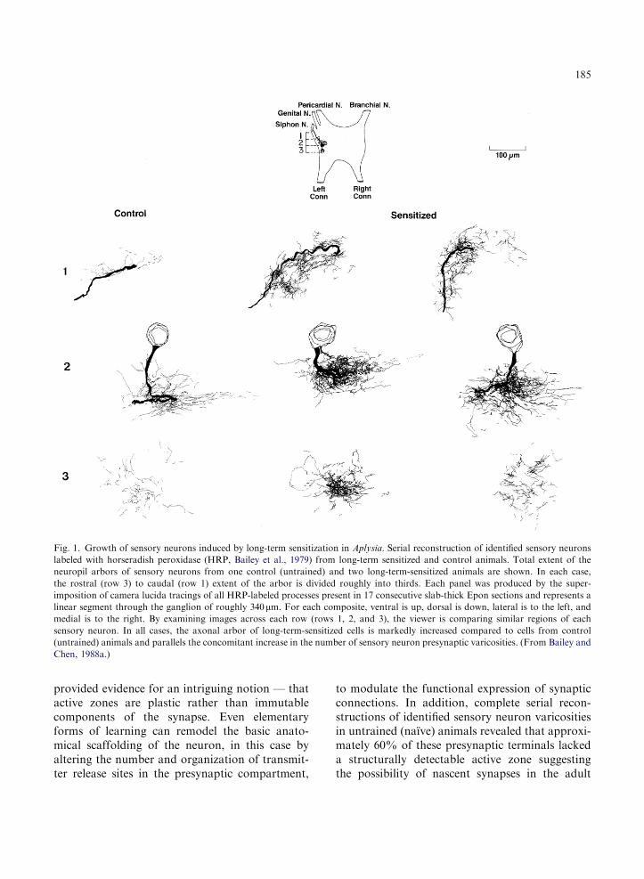
development and led to a pronounced increase inthe total number of presynaptic varicosities persensory neuron (Bailey and Chen, 1988a). Thus,sensory neurons from long-term sensitized animalsexhibited a twofold increase in the numberof synaptic varicosities, as well as an enlargementin the linear extent of each neuron’s axonalarbor when compared to sensory neurons fromuntrained animals (Fig. 1).
To determine which class of structural changesat sensory neuron synapses might contributeto the retention of long-term sensitization, Baileyand Chen (1989) compared the time course foreach morphological change with the behavioralduration of the memory. They found that not allof the structural changes persisted as long asthe memory. The increase in the size and synap-tic vesicle complement of sensory neuron activezones present 24 h following the completion ofbehavioral training returned to control levelswhen tested 1 week later. These data indicatedthat, insofar as this relatively transient modula-tion of active zone size and associated synapticvesicles is one of the structural mechanismsunderlying long-term sensitization, it is associatedwith the initiation and early expression of thelong-term process and not with its persistence.By contrast, the duration of changes in varicosityand active zone number, which persisted un-changed for at least 1 week and were partiallyreversed at the end of 3 weeks, paralleled thebehavioral time course of memory storage indi-cating that only the increase in the number ofsensory neuron synapses contributes to the stablemaintenance of long-term sensitization. Theseresults directly linked a change in synapticstructure to a long-lasting behavioral memoryand suggested that the morphological altera-tions could represent an anatomical substratefor memory consolidation. In addition, the findingthat some components of the learning-inducedchanges in synaptic architecture were transientwhereas others endured suggested that not allof these modifications were regulated synchro-nously. At the structural level, the sensory neuronappears to have multiple mechanisms and para-meters of plasticity available to it. Thus, duringthe later phases of long-term memory storage for
sensitization, although there are more synapses,each individual synapse may recruit all of themechanisms of plasticity that were present beforetraining.
Unlike the extensive anatomical changesobserved at sensory neuron synapses followinglong-term training, the structural correlates ofshort-term memory in Aplysia (lasting minutesto hours rather than days to weeks) are far lesspronounced (Bailey and Chen, 1988c). For exam-ple, the decrease in the strength of the sensoryto motor neuron connection that accompaniesshort-term habituation is not associated withany detectable alterations in either the numberof sensory neuron presynaptic varicosities or thenumber of active zones within the presynapticvaricosities. Nor does it alter the size of activezones or the total number of synaptic vesicleswithin the presynaptic varicosity. Rather, thereis a reduction in the number of vesicles thatare docked at the active zones and thus thereare fewer packets of transmitter ready to bereleased.
Taken together, these initial morphologicalstudies of short- and long-term memory in Aplysia
began to suggest a clear difference in the nature,extent and time course of changes in the functionalarchitecture of the synapse that may underliememories of differing durations. The transientdurations of short-term memories involving cova-lent modifications of pre-existing proteins (pro-teins that turn over slowly) are accompaniedonly by modest structural rearrangements thatappear to be restricted to shifts in the proximityof synaptic vesicle populations contiguous to therelease site. By contrast, the prolonged durationsof long-term memories depend on altered geneexpression and the synthesis of new proteinsand are associated with more substantial andpotentially more enduring structural alterationsthat are reflected by frank changes in both thenumber of synaptic contacts and their active zonemorphology.
These studies also demonstrated, for the firsttime, that learning-induced structural changescould be detected at the level of specific, identifiedsynaptic connections known to be criticallyinvolved in the behavioral modification and
184
provided evidence for an intriguing notion — thatactive zones are plastic rather than immutablecomponents of the synapse. Even elementaryforms of learning can remodel the basic anato-mical scaffolding of the neuron, in this case byaltering the number and organization of transmit-ter release sites in the presynaptic compartment,
to modulate the functional expression of synapticconnections. In addition, complete serial recon-structions of identified sensory neuron varicositiesin untrained (naı̈ve) animals revealed that approxi-mately 60% of these presynaptic terminals lackeda structurally detectable active zone suggestingthe possibility of nascent synapses in the adult
Fig. 1. Growth of sensory neurons induced by long-term sensitization in Aplysia. Serial reconstruction of identified sensory neurons
labeled with horseradish peroxidase (HRP, Bailey et al., 1979) from long-term sensitized and control animals. Total extent of the
neuropil arbors of sensory neurons from one control (untrained) and two long-term-sensitized animals are shown. In each case,
the rostral (row 3) to caudal (row 1) extent of the arbor is divided roughly into thirds. Each panel was produced by the super-
imposition of camera lucida tracings of all HRP-labeled processes present in 17 consecutive slab-thick Epon sections and represents a
linear segment through the ganglion of roughly 340mm. For each composite, ventral is up, dorsal is down, lateral is to the left, and
medial is to the right. By examining images across each row (rows 1, 2, and 3), the viewer is comparing similar regions of each
sensory neuron. In all cases, the axonal arbor of long-term-sensitized cells is markedly increased compared to cells from control
(untrained) animals and parallels the concomitant increase in the number of sensory neuron presynaptic varicosities. (From Bailey and
Chen, 1988a.)
185

brain. The extent to which learning and memorycan convert these immature, and presynapticallysilent synapses into mature and functionally com-petent synaptic connections is discussed below.Finally, these initial studies in Aplysia suggestedthat the growth of new sensory neuron synap-ses may represent the final and perhaps moststable phase of long-term memory storage, andraised the possibility that the stability of thelong-term process might be achieved, at least inpart, because of the relative stability of synapticstructure.
Subsequent studies by Wainwright et al. (2002)have examined the effects of different sensitizationtraining protocols on the structure of sensoryneurons in the pleural ganglion mediating thetail-siphon withdrawal reflex in Aplysia. A 4-daytraining period produced a robust and localizedoutgrowth of both neuritic processes and presy-naptic varicosities in these sensory neuronsobserved 24 h after the end of training. Thesestructural changes were consistent with the pre-vious results of Bailey and Chen (1988a) in siphonsensory neurons. By contrast, 1 day of sensiti-zation training, which can induce both long-termbehavioral sensitization and synaptic facilitation(Frost et al., 1985; Cleary et al., 1998), was notassociated with a comparable outgrowth of tailsensory neurons indicating that storage of thememory for sensitization induced by the 1 day oftraining and the spaced 4 day protocols are likelyto recruit different mechanisms. These investiga-tors have also reported a dissociation of morpho-logical and physiological changes associated withlong-term sensitization of the tail-siphon with-drawal reflex in Aplysia (Wainwright et al., 2004).The behavioral effects of long-term sensitizationtraining were restricted to the trained side of theanimal as were changes in the strength of sensoryto motor connections. By contrast, long-termtraining produced varicosity formation on bothsides of the animal. Interestingly, on the trainedside, this outgrowth was reflected by anincrease in the number of putative contacts withfollower neurons as well as with an increase insynaptic strength and behavioral enhancementsuggesting a causal relationship between thesechanges.
The long-lasting growth of new synaptic con-nections between sensory neurons and theirfollower cells during long-term sensitization canbe reconstituted in sensory–motor neuronco-cultures by five repeated applications of 5-HT(Glanzman et al., 1990; Bailey et al., 1992b) as wellas induced in the intact ganglion by the intracel-lular injection of cAMP, a second messengeractivated by 5-HT (Nazif et al., 1991). In culture,the synaptic growth can be correlated with thelong-term (24–72 h) enhancement in synapticeffectiveness and depends on the presence of anappropriate target cell similar to the synapseformation that occurs during development.
Time-lapse imaging reveals LTF is associated with
presynaptic activation of silent synapses and growth
of new functional synapses
In most model learning systems, the functionalcontribution of the structural changes that accom-pany long-lasting forms of synaptic plasticityremains largely unknown. In particular one wouldlike to know if changes in the number or struc-ture of synaptic connections induced by learningare functionally effective and capable of contri-buting to the storage of long-term memory. Bothtechnical and experimental limitations preventedthe earlier studies in Aplysia discussed abovefrom examining whether the increase in synapticstrength during long-term sensitization resul-ted from the conversion of pre-existing butnonfunctional (silent) synapses to active synap-ses, or from the addition of newly formedfunctional synapses, or perhaps both. To addressthese issues directly, more recent in vitro studiesof the sensory-to-motor neuron synapse in Aplysia
culture have monitored both functional andstructural changes simultaneously so as to followremodeling and growth at the same specificsynaptic varicosities continuously over time andto examine the functional contribution of thesepresynaptic structural changes to the differenttime-dependent phases of LTF.
Toward that end, Kim et al. (2003) combinedtime-lapse confocal imaging of individual presy-naptic varicosities of sensory neurons labeled with
186

three different fluorescent markers: the whole cellmarker Alexa-594, and two presynaptic markerproteins: synaptophysin-eGFP which monitorschanges in the distribution of synaptic vesicleswithin individual varicosities and synapto-
PHluorin (synPH), a monitor of active transmitterrelease sites (Miesenbock et al., 1998). They foundthat repeated pulses of 5-HT induce two tempo-rally, morphologically and molecularly distinctclasses of presynaptic changes: (1) the rapidactivation of silent presynaptic terminals throughthe filling of pre-existing empty varicosities withsynaptic vesicles, which requires translation butnot transcription and (2) the generation of newsynaptic varicosities which occurs more slowly andrequires both transcription and translation. Theenrichment of pre-existing but empty varicositieswith synaptophysin is completed within 3–6 h,parallels intermediate-term facilitation and accountsfor approximately 32% of the newly activatedsynapses evident at 24h. By contrast, the newsensory neuron varicosities, which account for 68%of the newly activated synapses at 24h, do not formuntil 12–18h after exposure to 5 pulses of 5-HT. Therapid activation of silent presynaptic terminalssuggests that in addition to its role in LTF, thismodification of pre-existing synapses may alsocontribute to the intermediate phase of synapticplasticity and memory storage (Ghirardi et al., 1995;Mauelshagen et al., 1996; Sutton et al., 2001) (Fig. 2).
This temporal analysis of the synaptic remodel-ing and growth that underlie the developmentof LTF served to bring the structural changesinto register with the physiological correlates ofthe different phases of long-term synaptic plasti-city. For example, the different time windowsfor the two classes of presynaptic structuralchanges — a rapid enrichment of pre-existingempty varicosities with synaptic vesicle proteinsand a slower generation of new varicosities —appear to be consistent with the onset andduration of intermediate-term and LTF, respec-tively. Since intermediate-term facilitation lastsfor about 3–6 h whereas LTF first appears at10–15 h and lasts for days (Ghirardi et al., 1995;Mauelshagen et al., 1996), these two classes ofpresynaptic changes may represent distinct struc-tural correlates of the two phases of memory
storage (Sutton et al., 2001). This idea is furthersupported by the finding that the 5-HT-inducedclustering of synaptic vesicles into pre-existingempty varicosities is blocked by the inhibition ofde novo protein synthesis, which has previouslybeen shown to attenuate both intermediate-termfacilitation and the corresponding intermediatephase of memory (Ghirardi et al., 1995; Suttonet al., 2001).
To test this hypothesis, Kim et al. (2003)developed a reduced 5-HT protocol to induceselectively facilitation in the intermediate-termtime domain without inducing LTF (see alsoGhirardi et al., 1995). They found that the isolatedintermediate-term facilitation is also accompaniedby the redistribution and clustering of synap-tophysin-eGFP into empty varicosities at 0.5 and3 h similar to what occurs when intermediate- andLTF are recruited together. However, the presynap-tic structural changes induced by the reduced 5-HTprotocol differed from those induced by the long-term protocol in at least two ways. First, therewas no growth of new sensory neuron varicositiesin the isolated intermediate phase. Second, unlikethe filling of pre-existing empty varicosities duringthe intermediate-term phase induced by the long-term protocol, the newly filled varicosities did notpersist for 24h and were unaffected by inhibitorsof protein synthesis suggesting that the structuralremodeling activated by the reduced 5-HT protocolinvolved only a simple rearrangement of pre-existingsynaptic components. This may reflect a fundamen-tal difference in the molecular mechanisms thatunderlie the two 5-HT protocols. Although bothprotocols induce intermediate-term facilitation,the long-term protocol may recruit additionalmolecular events (including the machinery fortranslational activation) required to set up thelong-term phase, perhaps by stabilizing the inter-mediate phase.
To quantitatively assess the contribution ofthese two distinct classes of presynaptic structuralchanges to LTF, Kim et al. (2003) monitored thefunctional state of individual sensory neuronvaricosities in living cells before and 24 h after5 pulses of 5-HT using the activity-sensitive fusionprotein synPH. With synPH, release of labeledsynaptic vesicles yields an increase in fluorescence
187

due to the externalization of pHluorin to a morebasic exterior medium, which returns to basallevels by the re-acidification of synaptic vesiclesfollowing endocytosis in a Ca2+-dependent fas-hion (Sankaranarayanan and Ryan, 2001). Whenexpressed in Aplysia sensory neurons, depolariza-tion by bath application of KCl leads to theevoked exocytotic release of synaptic vesicleswithin individual varicosities as indicated by anincrease in the fluorescence signal of synPH as hasbeen previously reported in cultured hippocampalneurons (Miesenbock et al., 1998).
In this fashion, Kim et al. (2003) were ableto monitor continuously over time individual5-HT-induced structural changes at the samesensory neuron varicosities and to examine directlythe functional contribution of two distinct classesof presynaptic structural changes to the differenttemporal phases of LTF. They found that 24 hafter repeated pulses of 5-HT there was a 59%increase in the total number of synPH-activesensory neuron varicosities. These real time
experiments on living synapses in culture wereremarkably consistent with the earlier electronmicroscopic studies of Bailey and Chen (1983)which had demonstrated that long-term sensitiza-tion in the intact animal is accompanied by anincrease of 65% in the number of sensory neuronvaricosities that contained fully differentiatedpresynaptic active zones (transmitter release sites)when compared to varicosities from untrained(naı̈ve) animals. The enrichment of pre-existingbut empty varicosities accounted for approxi-mately 32% of these newly activated synapses at24 h, whereas newly formed varicosities accountedfor approximately 68%. These results suggestedthat both classes of structural changes — thepresynaptic activation of pre-existing silentsynapses and the growth of new functionalsynapses — appear to contribute to the synapticenhancement that characterizes LTF at 24 h.
Previous studies have shown that specific 5-HTprotocols (Sun and Schacher, 1998; Casadio et al.,1999) or experimental manipulation in Aplysia
Fig. 2. Time course and functional contribution of two distinct presynaptic structural changes associated with intermediate- and
long-term facilitation in Aplysia. Repeated pulses of 5-HT in sensory to motor neuron co-cultures trigger two distinct classes
of presynaptic structural changes: (1) the rapid clustering of synaptic vesicles to pre-existing silent sensory neuron varicosities
(3–6 h) and (2) the slower generation of new sensory neuron synaptic varicosities (12–18h). The resultant newly filled and newly
formed varicosities are functionally competent (capable of evoked transmitter release) and contribute to the synaptic enhance-
ment that underlies LTF. The rapid filling and activation of silent presynaptic terminals at 3 h suggests that, in addition to its role
in LTF, this modification of pre-existing varicosities may also contribute to the intermediate phase of synaptic plasticity. Red triangles
represent transmitter release sites (active zones). (Modified from Kim et al., 2003.) (See Color Plate 10.2 in color plate section.)
188

(Hatada et al., 2000; Udo et al., 2005) can induceLTF at 24 h without the formation of newvaricosities. How might such an increase insynaptic strength persist for 24 h in the absenceof synaptic growth? The results of Kim andassociates suggest that additional modificationsof pre-existing connections including the acti-vation of previously silent synapses, may playa role in the initial phases of synaptic mainte-nance and highlight the fact that there are likelyto be multiple types of structural mechanismsthat can contribute to LTF at 24 h (Schacheret al., 1997; Bailey et al., 2000; Sutton and Carew,2000).
Of the two classes of presynaptic structuralplasticity induced by 5-HT in culture, synapticgrowth appears to contribute more to the synapticenhancement present at 24 h than does the activationof pre-existing silent synapses. It will be of interest tosee if the functional contribution by newly formedsynapses increases with time when the growthprocess is more fully developed and memory storageis likely to be more stable. This would be consistentwith the earlier studies in the intact animal outlinedabove, which have shown that only the increases inthe number of sensory neuron varicosities and activezones persist for several weeks in parallel with thebehavioral duration of the memory, as well as morerecent work in culture which has demonstrated thatsynaptic growth plays a more prominent role in theexpression of the later phases of LTF (Martin et al.,1997b; Casadio et al., 1999).
The activation of silent synapses also seems toplay a major role in LTP in the hippocampus.Although in mammals the term refers to a veryspecific molecular configuration found in synapsesin different regions of the CNS of vertebrates(Malinow et al. 2000; Malinow and Malenka,2002). In this case, the term silent synapse refers toexcitatory glutamatergic synapses whose postsy-naptic membrane contains NMDARs but noAMPARs. The lack of AMPAR-mediated signal-ing renders these synapses inactive, or ‘‘silent’’,under normal conditions. Synaptic stimulationactivates these silent synapses through the inser-tion of AMPARs into the postsynaptic membrane,a phenomenon some times referred to as AMPA-
fication. Calcium/calmodulin-dependent protein
kinase II (CaMKII) plays a critical role in thisprocess. Once this kinase is activated by highfrequency stimulation, it phosphorylates AMPARsor associated proteins, triggering their insertioninto the postsynaptic membrane. The synapse isthen no longer silent and postsynaptic responsesare, by consequence, enhanced.
Remodeling of the presynaptic actin network
for learning-related synaptic growth requires
activation of Cdc42-mediated signaling pathways
Actin is enriched in both the pre- and postsynapticcompartment of neurons (Matus, 2000). Althoughthe activity-dependent modulation of actindynamics at postsynaptic spines has been welldocumented, the extent and role of actin regula-tion in presynaptic terminals is less clear (Colicoset al., 2001; Antonova et al., 2001). Duringdevelopment, reorganization of actin in growthcones has been shown to play an important role inaxonal path finding (Yuan et al., 2003). However,in mature neurons, it has been suggested that thepresynaptic actin network may function more asan intracellular scaffold rather than providing apropulsive force that could contribute directly tothe type of rapid structural remodeling reported forpostsynaptic dendritic spines (Sankaranarayananet al., 2003).
In Aplysia, repeated applications of 5-HT thatlead to LTF induce a slower and more persistentalteration in the dynamics of the presynaptic actinnetwork leading to the remodeling and growth ofsensory neuron synapses. Both the 5-HT-inducedenrichment of synaptic proteins in pre-existingvaricosities and the formation of new and func-tionally competent sensory neuron varicositiesduring LTF involve an activity-dependent rear-rangement of the presynaptic actin cytoskeleton(Udo et al., 2005). These findings in Aplysia areconsistent with previous reports that structuralremodeling of synapses in response to physiologi-cal activity requires the reorganization of actin(Colicos et al., 2001; Huntley et al., 2002) and thatthe inhibition of actin function blocks synapseformation and interferes with long-term synaptic
189

plasticity (Hatada et al., 2000; Krucker et al., 2000;Zhang and Benson, 2001). Furthermore, severalsynaptic proteins such as synapsin can bind tothe presynaptic actin cytoskeleton and participatein synaptic vesicle trafficking (Humeau et al.,2001) that may contribute to the 5-HT-inducedenrichment of pre-existing varicosities observedduring LTF.
One attractive molecular candidate for the5-HT-induced reorganization of the cytoskeletonat sensory neuron presynaptic varicosities is theRho-family of small GTPases, which can modulateactin polymerization in response to extracellularsignals and can be regulated by neuronal activityin vivo (Luo et al., 1996; Hall, 1998). In Aplysia,Udo et al. (2005) found that the application oftoxin B, a general inhibitor of the Rho family,blocks 5-HT-induced LTF, as well as growthof new synapses in sensory–motor neuronco-cultures. Moreover, repeated pulses of 5-HTselectively induce the spatial and temporal regula-tion of the activity of only one of the smallGTPases-Cdc42-at a subset of sensory neuronpresynaptic varicosities. The activation ofApCdc42 induced by 5-HT is dependent on boththe P13K and PLC pathways and, in turn, recruitsthe downstream effectors PAK (p21-Cdc42/Rac-activated kinase) and N-WASP (neuronalWiskott–Aldrich syndrome protein) to regulatethe presynaptic actin network. This initial mole-cular cascade leads to the outgrowth of filopodia,some of which represent morphological precursorsfor the growth of new sensory neuron varicositiesassociated with the storage of LTF.
5-HT-induced internalization of apCAM:
a preliminary and permissive step for initiation
of learning-related synaptic growth
Cell adhesion molecules (CAMs) are cell surfaceglycoproteins that mediate cell-to-cell and cell-to-extracellular matrix adhesions. In the CNS,CAMs are involved in cell migration, neuriteoutgrowth and more recently have been shownto participate in synapse formation during
development (Scheiffele, 2003; Washbourne et al.,2004) as well as various forms of learning-relatedsynaptic plasticity in the adult brain (Martinand Kandel, 1996; Fields and Itoh, 1996; Bensonet al., 2000). Some of the first evidence for a roleof CAMs during learning and memory camefrom studies of an immunoglobulin-relatedCAM in Aplysia, designated apCAM, which ishomologous to NCAM in vertebrates and Fasci-clin II in Drosophila (Mayford et al., 1992).Following the application of 5-HT or cAMP,there is a decrease in the expression of apCAMand this occurs in a transcriptionally dependentmanner. Furthermore, imaging of fluorescentlylabeled MAbs to apCAM indicates that not onlyis there a decrease in the level of expressionbut that even pre-existing protein is lost fromthe surface membrane of the sensory neuronswithin 1 h after the addition of 5-HT. This tran-sient modulation by 5-HT of CAMs, therefore,may represent one of the early molecular stepsrequired for initiating learning-related growthof synaptic connections. Indeed, blocking theexpression of the antigen by MAb causes defasci-culation, a step that appears to precede synapseformation during development in Aplysia (Kellerand Schacher, 1990).
To examine the mechanisms that underlie the5-HT-induced down-regulation of apCAM and,in particular, how these relate to the initiationof synaptic growth, Bailey et al. (1992a) combinedthin-section electron microscopy with immuno-labeling using a gold-conjugated MAb specificto apCAM. They found that a 1 h applicationof 5-HT led to a 50% decrease in the densityof gold-labeled apCAM complexes at the surfacemembrane of the sensory neuron. This down-regulation was particularly prominent at adherentprocesses of the sensory neurons and was achievedby a heterologous, protein synthesis-dependentactivation of the endosomal pathway, leading tointernalization and apparent degradation ofapCAM. As is the case for the down-regulationat the level of expression, the 5-HT-inducedinternalization of apCAM can be simulated bycAMP. Concomitant with the down-regulation ofapCAM, Hu et al. (1993) further demonstrated
190

that, as part of this coordinated program for endo-cytosis, 5-HT and cAMP also induce an increase inthe number of coated pits and coated vesicles in thesensory neurons and an increase in the expressionof the light chain of clathrin (apClathrin). Since theapClathrin light chain contains the importantfunctional domains of both LCa and LCb ofmammalian clathrin thought to be essential for thecoated pit assembly and disassembly, the increasein clathrin may be an important component inthe activation of the endocytic cycle required for theinternalization of apCAM.
The learning-induced internalization of apCAMis thought to have at least two major structuralconsequences: (1) disassembly of homophilicallyassociated fascicles of the sensory neurons (defas-ciculation), a process that may destabilize adhesivecontacts normally inhibiting growth, and (2)endocytic activation that may lead to a redistribu-tion of membrane components to sites wherenew synapses form. Thus, aspects of the initialsteps in the learning-related growth of synapticconnections that is a hallmark of the long-termprocess may eventually be understood in thecontext of a novel and targeted form of receptor-mediated endocytosis.
These initial effects of 5-HT on the remodelingof the surface and internal membrane systems ofsensory neurons in Aplysia bear a striking simila-rity to the morphological changes induced innon-neuronal systems by growth factors, whichsuggests that modulatory transmitters importantfor learning, such as 5-HT, may serve a doublefunction. In addition to producing a transientregulation of the excitability of neurons, withrepeated or prolonged exposure they may alsoproduce an action comparable to that of a growthfactor, which results in a more persistent regula-tion of the architecture of the neuron.
To further define the mechanisms whereby 5-HTleads to apCAM down-regulation, Bailey et al.(1997) used epitope tags to examine the fate ofthe two apCAM isoforms (transmembrane andGPI-linked) and found that only the transmem-brane form (TM-apCAM) is internalized (Fig. 3).This internalization was blocked by overexpres-sion of TM-apCAM with a point mutation in the
two MAPK phosphorylation consensus sites, aswell as by injection of a specific MAPK antagonistinto sensory neurons. These data suggest thatactivation of the MAPK pathway is importantfor the internalization of TM-apCAM and mayrepresent one of the initial and perhaps permissivestages of learning-related synaptic growthin Aplysia. In addition, the differential down-regulation of the GPI-linked and transmembraneforms of apCAM raised the interesting possibilitythat learning-related synaptic growth in the adultis initiated by an activity-dependent recruitment ofspecific isoforms of CAMs, similar to the modula-tion of cell-surface receptors during the fine-tuningof synaptic connections in the developing nervoussystem. One consequence of isoform recruitmentis that it would allow neuronal activity to regu-late the surface expression of each isoform, aprocess that might take on additional functionalsignificance if these surface molecules were dis-tributed differentially along the three-dimensionalextent of the neuron and, thus may provide aregulatory unit capable of acting sequentially atmultiple cytoplasmic and plasma membrane sitesduring the early inductive phases of the long-termprocess.
Han et al. (2004) have examined more closelythe relationship between the 5-HT-induced down-regulation of TM-apCAM and synaptic growth byoverexpressing various HA-epitope tagged recom-binant apCAMs in Aplysia sensory neurons. Theyfound that overexpression of TM-apCAM, butnot the GPI-linked isoform of apCAM, blockedboth LTF as well as the associated increase in thenumber of sensory neuron varicosities. By inter-rupting the adhesive function of apCAM withan anti-HA antibody, this inhibition of LTFinduced by the overexpression of TM-apCAMwas restored. Moreover, LTF could be completelyblocked by overexpression of the cytoplasmic tailportion of apCAM alone. These studies indicatedthat the extracellular domain of TM-apCAM hasan inhibitory function that is neutralized byinternalization to induce LTF and suggested thatthe cytoplasmic domain provides an interactiveplatform for both signal transduction and theinternalization machinery.
191
Nuclear translocation of apCAM-associated
protein (CAMAP) activates presynaptic gene
transcription for induction of LTF
Lee et al. (2007) have examined the 5-HT-inducedsignaling interactions mediated by the cytoplasmicdomain of TM-apCAM and found an additional,and novel role for this CAM in synapse-specificforms of long-lasting plasticity. LTF at the sensoryto motor neuron synapse requires the activationof CREB1 in the nucleus of the sensory neuron(Bartsch et al., 1998). Activated CREB1 inducesthe transcription factor ApC/EBP that in turn actson downstream genes encoding proteins importantfor synaptic growth and the stable maintenanceof LTF (Alberini et al., 1994). As discussed above,an initial step, thought to be permissive, forthe initiation of learning-related growth is theclathrin-mediated internalization and consequentdown-regulation of TM-apCAM.
To examine directly how the internalizationof TM-apCAM is related to the initiation oftranscription, Lee et al. (2007) first looked formolecules that could bind to the cytoplasmic tailof TM-apCAM and cloned an CAMAP byyeast two-hybrid screening. They found that5-HT signaling at the synapse activates PKAwhich in turn phosphorylates CAMAP to inducethe dissociation of CAMAP from apCAM andthat this dissociation is a prerequisite for theinternalization of apCAM. The 5-HT-induceddissociated CAMAP is subsequently translocatedto the nucleus of the sensory neurons. In thenucleus, CAMAP acts as a transcriptional co-activator for CREB1 that is essential for theactivation of ApC/EBP required for the initiationof LTF. Combined, these data suggest thatCAMAP is one of the retrograde signals from thesynapse to the nucleus where it acts as a co-regulator of the presynaptic gene expressionassociated with the induction of LTF in Aplysia.In addition, these findings demonstrate the impor-tance, for learning-related synaptic plasticity, ofsignal propagation into the nucleus from thesurface membrane of activated synaptic sitesmediated by a molecule directly interacting witha cell surface adhesion molecule and suggest anovel presynaptic molecular mechanism to turn
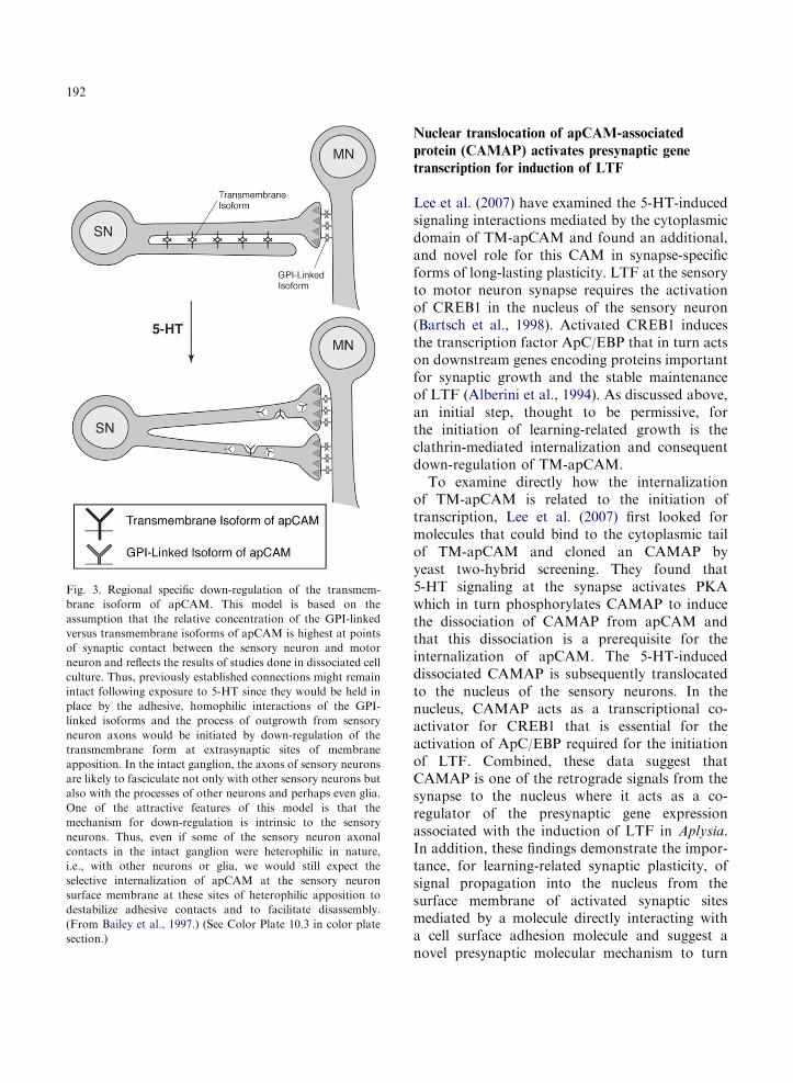
Fig. 3. Regional specific down-regulation of the transmem-
brane isoform of apCAM. This model is based on the
assumption that the relative concentration of the GPI-linked
versus transmembrane isoforms of apCAM is highest at points
of synaptic contact between the sensory neuron and motor
neuron and reflects the results of studies done in dissociated cell
culture. Thus, previously established connections might remain
intact following exposure to 5-HT since they would be held in
place by the adhesive, homophilic interactions of the GPI-
linked isoforms and the process of outgrowth from sensory
neuron axons would be initiated by down-regulation of the
transmembrane form at extrasynaptic sites of membrane
apposition. In the intact ganglion, the axons of sensory neurons
are likely to fasciculate not only with other sensory neurons but
also with the processes of other neurons and perhaps even glia.
One of the attractive features of this model is that the
mechanism for down-regulation is intrinsic to the sensory
neurons. Thus, even if some of the sensory neuron axonal
contacts in the intact ganglion were heterophilic in nature,
i.e., with other neurons or glia, we would still expect the
selective internalization of apCAM at the sensory neuron
surface membrane at these sites of heterophilic apposition to
destabilize adhesive contacts and to facilitate disassembly.
(From Bailey et al., 1997.) (See Color Plate 10.3 in color plate
section.)
192

on the gene transcription required for long-termmemory.
An overall view
Perhaps the most striking finding in the biologyof memory storage is that long-term memoryinvolves transcription in the nucleus and structuralchanges at the synapse. The structural changesassociated with the storage of long-term memorycan be grouped into two general categories:remodeling of pre-existing synapses and growthof new synapses (Greenough and Bailey, 1988;Bailey and Kandel, 1993; Yuste and Bonhoeffer,2001; Bailey et al., 2004; Lamprecht and LeDoux,2004). Despite an increasing body of evidence forchanges in the number or structure of synapticconnections and long-term memory, it has so farproven difficult to follow individual structuralchanges at the same synapse over time and torelate directly this remodeling to physiologicalfunction and memory storage.
Recent studies have found that activity-dependent remodeling of pre-existing synapsesand the growth of new synaptic connections occursin the mammalian CNS (Buchs and Muller, 1996;Engert and Bonhoeffer, 1999; Maletic-Savaticet al., 1999; Toni et al., 1999; Colicos et al.,2001; De Paola et al., 2003). However, in themammalian brain these structural changes aredifficult to study because the effects are oftenmodest. Moreover, the specific role of thisstructural plasticity remains unclear because thefunctional contribution of individual synapses tomemory processes in these more complex neuro-nal networks is not yet well defined (Lamprechtand LeDoux, 2004; Hayashi and Majewska,2005; Segal, 2005). For example, although thegeneration and enlargement of dendritic spineshas been associated with the production of LTPand synaptic activity in organotypic hippocampalslices (Matsuzaki et al., 2004; Nagerl et al., 2004)and acute slices of neonatal animals (Zhou et al.,2004), these structural changes are much moresubtle in the adult brain (Lang et al., 2004). Inadults there is only a modest production of newspines (Zuo et al., 2005), and learning-related
plasticity seems to rely more on subcellularchanges than on anatomical changes. Thus,neuronal activity regulates the transport of poly-somes from dendritic shafts to active spines(Ostroff et al., 2002), as well as the traffickingof neurotransmitter receptors (Malinow andMalenka, 2002).
By contrast, in Aplysia the learning-inducedstructural changes that accompany long-termsensitization in vivo and LTF in vitro are robust,highly reproducible and easy to study and can beshown to be both functionally effective andcapable of contributing to memory storage.Time-lapse imaging studies of the sensory tomotor neuron synapse in culture have revealedthat LTF is accompanied by two temporally andmorphologically distinct classes of presynapticstructural change: the rapid activation of silentpre-existing varicosities by filling with synapticvesicles and the slower growth of new functionalvaricosities. These findings, the first to be made onindividually identified presynaptic varicosities,suggest that the duration of the changes insynaptic effectiveness that accompany memorystorage may be reflected by the differential regula-tion of two fundamentally disparate forms ofpresynaptic compartment: (1) nascent (silent)varicosities that can be rapidly and reversiblyremodeled into active transmitter release sites and(2) mature, more stable and functionally compe-tent varicosities that following long-term trainingmay undergo a process of fission to form newstable synaptic contacts.
Recent live imaging studies in the mammalianCNS have reported a comparable remodelingand differentiation of the presynaptic compartmentin developing synapses (for review, see Ziv andGarner, 2004). For example, the establishment offunctional transmitter release sites in culturedhippocampal neurons can occur in less than 1 h(Ahmari et al., 2000; Friedman et al., 2000). Thisshort delay is similar to the 5-HT-induced enrich-ment and subsequent activation of pre-existingsilent varicosities in Aplysia, which also occursvery rapidly — initial changes in the recruitmentof synaptic vesicle proteins to empty presynapticvaricosities can be detected as early as 30min afterthe completion of 5-HT training.
193

How is this rapid differentiation of nascentpresynaptic compartments achieved? One model ofactive zone assembly suggests proteins that com-prise the cytoskeletal matrix of the active zone(CAZ) are packaged into transport vesicles fordelivery and fusion with the plasma membraneat nascent synaptic contacts (Zhai et al., 2001;Shapira et al., 2003). These transport vesiclescontain multiple molecular components includingPiccolo and Bassoon — important for assemblyand structural maintenance of the active zone — aswell as other CAZ scaffolding molecules impli-cated in synaptic vesicle exocytosis but typicallynot synaptic vesicle proteins. The rapid remodelingand presynaptic activation of nascent sensoryneuron varicosities induced by 5-HT in Aplysia
culture is consistent with this molecular modelfor active zone assembly in developing synapses ofthe mammalian CNS. Moreover, the apparentheterogeneity in the content of these mobile pre-assembled packets — some of which containsynaptic vesicle proteins and the others whichcontain components required for assembly of theactive zone (Friedman et al., 2000; Zhai et al.,2001) could explain why more than half of thesensory neuron varicosities enriched in onlysynaptophysin following 5-HT-induced LTF werenot functional.
The second general class of learning-relatedpresynaptic structural change associated with LTFin Aplysia involves a slower generation of new andfunctionally effective sensory neuron varicosities.How are these new varicosities formed? Time-lapseimaging of individual sensory neuron varicositiesindicates that the 5-HT-induced recruitment ofsynaptic proteins to a pre-existing varicosity leadsdirectly to both an enrichment of these presynapticconstituents as well as to an overall increase in thesize of the varicosity. The presynaptic remodelingand growth is followed by the apparent division orsplitting of a subset of these pre-existing varico-sities (Hatada et al., 2000; Kim et al., 2003; Udoet al., 2005). This physical process may lead tothe budding off of components of the active zoneand associated synaptic vesicle cluster from eachpre-existing presynaptic compartment that couldthen serve as a nucleation site to seed theformation of a new varicosity. Aspects of these
structural transformations of the presynapticcompartment that precede learning-related synap-tic growth in Aplysia have also been reportedat developing synapses in mammals. For exam-ple, imaging studies of the early stages of syna-pse formation have shown that presynaptic sitesformed immediately after initial contact ofaxonal and dendritic processes are highly unstable.Moreover, even apparent mature presynaptic sitesare relatively unstable as occasionally ‘‘orphanrelease sites’’ break off from fully formed boutonsand either migrate to adjacent presynaptic sites orparticipate in the formation of completely newones (Friedman et al.; 2000; Hopf et al., 2002;Krueger et al., 2003).
Taken together, these time-lapse observationsof living synapses indicate that differentiation andgrowth of the presynaptic compartment, eitherinduced by learning in the mature nervous systemor as mechanistic steps during development,are highly dynamic and rapid processes that canrecruit both pre-existing proteins as well as pre-assembled synaptic components. The instabilityof nascent presynaptic compartments during theearly stages of development is characterized bythe dispersion of mobile packets of synapticcomponents and a renewal of their migration oncethe transient pre- and postsynaptic contactsbreakup. As the neurons mature, an increasingproportion of these initial contacts develop intomore stable and fully functional presynapticterminals (Ziv and Garner, 2004). Results in theAplysia sensory to motor neuron culture prepara-tion further indicate that even some of thesemature presynaptic contacts can be selectivelydestabilized during learning and memory leadingto the generation of new and functionally compe-tent synaptic varicosities by processes thatappear similar to those that govern developmentalsynaptogenesis.
Conclusions
Over the past two decades, it has become clear thatsynaptic growth and the formation of new synap-ses are hallmarks of long-term, learning-relatedsynaptic plasticity. The morphological corres-
194
pondence between the studies of long-term sensi-tization in Aplysia and LTP in the mammalianhippocampus indicates that learning resembles aprocess of growth and neuronal differentiationacross a broad segment of the animal kingdomand suggests that learning-related synapse forma-tion may be a highly conserved feature for thestorage of both implicit and explicit forms oflong-term memory. Such changes are likely toreflect the recruitment by environmental stimuliof developmental processes that are latent orinhibited in the fully differentiated neuron. Recentstudies of the sensory to motor neuron connectionin Aplysia have begun to characterize the cellularand molecular events that underlie the structuralchanges induced by learning at the level ofindividual identified synapses. This in turn hassuggested the synaptic remodeling and synapticgrowth that accompany learning and memorystorage in the adult brain may reutilize mecha-nisms important for de novo synapse formationand the fine-tuning of synaptic connections duringdevelopment of the nervous system.
Acknowledgments
Research in this review was supported in part byNational Institutes of Health grant MH37134(to C.H.B.), the Howard Hughes Medical Institute(to E.R.K.), and the Kavli Institute for BrainSciences.
References
Ahmari, S.E., Buchanan, J. and Smith, S.J. (2000) Assembly of
presynaptic active zones from cytoplasmic transport packets.
Nat. Neurosci., 3: 445–451.
Alberini, C.M., Ghirardi, M., Metz, R. and Kandel, E.R.
(1994) C/EBP is an immediate-early gene required for the
consolidation of long-term facilitation in Aplysia. Cell, 76:
1099–1114.
Antonova, I., Arancio, O., Trillat, A.-C., Wang, H.G., Zablow,
L., Udo, H., Kandel, E.R. and Hawkins, R.D. (2001) Rapid
increase in clusters of presynaptic proteins at onset of long-
lasting potentiation. Science, 294: 1547–1550.
Bailey, C.H., Bartsch, D. and Kandel, E.R. (1996) Toward a
molecular definition of long-term memory storage. Proc.
Natl. Acad. Sci. U.S.A., 93: 13445–13452.
Bailey, C.H. and Chen, M. (1983) Morphological basis of long-
term habituation and sensitization in Aplysia. Science, 220:
91–93.
Bailey, C.H. and Chen, M. (1988a) Long-term memory in
Aplysia modulates the total number of varicosities of single
identified sensory neurons. Proc. Natl. Acad. Sci. U.S.A., 85:
2373–2377.
Bailey, C.H. and Chen, M. (1988b) Long-term sensitization in
Aplysia increases the number of presynaptic contacts onto the
identified gill motor neuron L7. Proc. Natl. Acad. Sci.
U.S.A., 85: 9356–9359.
Bailey, C.H. and Chen, M. (1988c) Morphological basis of
short-term habituation in Aplysia. J. Neurosci., 8: 2452–2459.
Bailey, C.H. and Chen, M. (1989) Time course of struc-
tural changes at identified sensory neuron synapses during
long-term sensitization in Aplysia. J. Neurosci., 9: 1774–1780.
Bailey, C.H., Chen, M., Keller, F. and Kandel, E.R. (1992a)
Serotonin-mediated endocytosis of apCAM: an early step
of learning-related synaptic growth in Aplysia. Science, 25:
645–649.
Bailey, C.H., Giustetto, M., Zhu, H., Chen, M. and
Kandel, E.R. (2000) A novel function for serotonin-mediated
short-term facilitation in Aplysia: conversion of a transident
cell-wide homosynaptic Hebbian plasticity into a persistent,
protein synthesis-independent synapse-specific enhancement.
Proc. Natl. Acad. Sci. U.S.A., 97: 11581–11586.
Bailey, C.H., Kaang, B.K., Chen, M., Marin, C., Lim, C.S.,
Casadio, A. and Kandel, E.R. (1997) Mutation in the
phosphorylation sites of MAP kinase blocks learning-related
internalization of apCAM in Aplysia sensory neurons.
Neuron, 18: 913–924.
Bailey, C.H. and Kandel, E.R. (1993) Structural changes
accompanying memory storage. Annu. Rev. Physiol., 55:
397–426.
Bailey, C.H., Kandel, E.R. and Si, K. (2004) The persistence
of long-term memory: a molecular approach to self-sustaining
changes in learning-induced synaptic growth. Neuron, 44:
49–57.
Bailey, C.H., Montarolo, P.G., Chen, M., Kandel, E.R. and
Schacher, S. (1992b) Inhibitors of protein and RNA synthesis
block the structural changes that accompany long-term
heterosynaptic plasticity in the sensory neurons of Aplysia.
Neuron, 9: 749–758.
Bailey, C.H., Thompson, E.B., Castellucci, V.F. and Kandel,
E.R. (1979) Ultrastructure of the synapses of sensory neurons
that mediate the gill-withdrawal reflex in Aplysia. J.
Neurocytol., 8: 415–444.
Bartsch, D., Casadio, A., Karl, K.A., Serodio, P. and
Kandel, E.R. (1998) CREB1 encodes a nuclear activator, a
repressor, and a cytoplasmic modulator that form a regulatory
unit critical for long-term facilitation. Cell, 95: 211–223.
Benson, D.L., Schnapp, L.M., Shapiro, L. and Huntley, G.W.
(2000) Making memories stick: cell adhesive molecules in
synaptic plasticity. Trends Cell Biol., 10: 473–480.
Bliss, T.V., Collingridge, G.L. and Morris, R.G. (2003)
Introduction. Long-term potentiation and structure of the
issue. Philos. Trans. R. Soc. Lond. B Biol. Sci., 358: 607–611.
195

Buchs, P.A. and Muller, D. (1996) Induction of long-
term potentiation is associated with major ultrastructural
changes of activated synapses. Proc. Natl. Acad. Sci. U.S.A.,
93: 8040–8045.
Casadio, A., Martin, K.C., Giustetto, M., Zhu, H., Chen, M.,
Bartsch, D., Bailey, C.H. and Kandel, E.R. (1999) A
transient, neuron-wide form of CREB-mediated long-term
facilitation can be stabilized at specific synapses by local
protein synthesis. Cell, 99: 221–237.
Castellucci, V., Pinsker, H., Kupfermann, I. and Kandel, E.R.
(1970) Neuronal mechanisms of habituation and dishabitua-
tion of the gill-withdrawal reflex in Aplysia. Science, 167:
1745–1748.
Castellucci, V.F., Blumenfeld, H., Goelet, P. and Kandel, E.R.
(1989) Inhibitor of protein synthesis blocks long-term
behavioral sensitization in the isolated gill-withdrawal reflex
of Aplysia. Science, 220: 91–93.
Cleary, L.J., Lee, W.L. and Byrne, J.H. (1998) Cellular
correlates of long-term sensitization in Aplysia. J. Neurosci.,
18: 5988–5998.
Colicos, M.A., Collins, B.E., Sailor, M.J. and Goda, Y. (2001)
Remodeling of synaptic actin induced by photoconductive
stimulation. Cell, 107: 605–616.
Dale, N., Kandel, E.R. and Schacher, S. (1987) Serotonin
produces long-term changes in the excitability of Aplysia
sensory neurons in culture that depend on new protein
synthesis. J. Neurosci., 7: 2232–2238.
De Paola, V., Arber, S. and Caroni, P. (2003) AMPA receptors
regulate dynamic equilibrium of presynaptic terminals in
mature hippocampal networks. Nat. Neurosci., 6: 491–500.
Engert, F. and Bonhoeffer, T. (1999) Dendritic spine changes
associated with hippocampal long-term synaptic plasticity.
Nature, 399: 66–70.
Fields, R.D. and Itoh, K. (1996) Neural cell adhesion molecules
in activity-dependent development and synaptic plasticity.
Trends Neurosci., 19: 473–480.
Friedman, H.V., Bresler, T., Garner, C.C. and Ziv, N.E. (2000)
Assembly of new individual excitatory synapses: time course
and temporal order of synaptic molecule recruitment.
Neuron, 27: 57–69.
Frost, W.N., Castellucci, V.F., Hawkins, R.D. and Kandel,
E.R. (1985) Monosynaptic connections made by the
sensory neurons of the gill- and siphon-withdrawal reflex
in Aplysia participates in the storage of long-term memory
for sensitization. Proc. Natl. Acad. Sci. U.S.A., 82:
8266–8269.
Ghirardi, M., Montarolo, P.G. and Kandel, E.R. (1995) A
novel intermediate stage in the transition between short- and
long-term facilitation in the sensory to motor neuron synapse
of Aplysia. Neuron, 14: 413–420.
Glanzman, D.L., Kandel, E.R. and Schacher, S. (1990) Target-
dependent structural changes accompanying long-term
synaptic facilitation in Aplysia neurons. Science, 249: 779–
802.
Greenough, W.T. and Bailey, C.H. (1988) The anatomy of a
memory: convergence of results across a diversity of tests.
Trends Neurosci., 11: 142–147.
Hall, A. (1998) Rho GTPases and the actin cytoskeleton.
Science, 279: 509–514.
Han, J.H., Lim, Y.S., Kandel, E.R. and Kaang, B.K. (2004)
Role of Aplysia cell adhesion molecules during 5-HT-induced
long-term functional and structural changes. Learn. Mem.,
11: 421–435.
Hatada, Y., Wu, F., Sun, Z.Y., Schacher, S. and Goldberg, D.J.
(2000) Presynaptic morphological changes associated with
long-term synaptic facilitation are triggered by actin poly-
merization at preexisting varicosities. J. Neurosci., 20:
p. RC82.
Hawkins, R.D., Kandel, E.R. and Bailey, C.H. (2006)
Molecular mechanisms of memory storage in Aplysia. Biol.
Bull., 210: 174–191.
Hayashi, Y. and Majewska, A.K. (2005) Dendritic spine
geometry: functional implication and regulation. Neuron,
46: 529–532.
Hochner, B., Klein, M., Schacher, S. and Kandel, E.R. (1986)
Additional components in the cellular mechanisms
of presynaptic facilitation contributes to behavioral dish-
abituation in Aplysia. Proc. Natl. Acad. Sci. U.S.A., 83:
8794–8798.
Hopf, F.W., Walters, J., Mehta, S. and Smith, S.J. (2002)
Stability and plasticity of developing synapses in hippocam-
pal neuronal cultures. J. Neurosci., 22: 775–781.
Hu, Y., Barzilai, A., Chen, M., Bailey, C.H. and Kandel, E.R.
(1993) 5-HT and cAMP induce the formation of coated
pits and vesicles and increase the expression of clathrin light
chain in sensory neurons of Aplysia. Neuron, 10: 921–929.
Humeau, Y., Doussau, F., Vitiello, F., Greengard, P.,
Benfenati, F. and Poulain, B. (2001) Synapsin controls both
reserve and releasable synaptic vesicle pools during neuronal
activity and short-term plasticity in Aplysia. J. Neurosci., 21:
4195–4206.
Huntley, G.W., Benson, D.L. and Colman, D.R. (2002)
Structural remodeling of the synapse in response to
physiological activity. Cell, 108: 1–4.
Jessell, T.M. and Kandel, E.R. (1993) Synaptic transmission:
a bidirectional and self-modifiable form of cell-cell commu-
nication. Cell 72/Neuron 10, 1–30.
Kandel, E.R. (2001) The molecular biology of memory
storage: a dialogue between genes and synapses. Science,
294: 1030–1038.
Keller, Y. and Schacher, S. (1990) Neuron-specific membrane
glycoproteins promoting neurite fasciculation in Aplysia
californica. J. Cell Biol., 111: 2637–2650.
Klein, M. and Kandel, E.R. (1980) Mechanism of calcium
current modulation underlying presynaptic facilitation and
behavioral sensitization in Aplysia. Proc. Natl. Acad. Sci.
U.S.A., 77: 6912–6916.
Kim, J.-H., Udo, H., Li, H.-L., Youn, T.Y., Chen, M., Kandel,
E.R. and Bailey, C.H. (2003) Presynaptic activation of silent
synapses and growth of new synapses contribute to inter-
mediate and long-term facilitation in Aplysia. Neuron, 40:
151–165.
Krucker, T., Siggins, G.R. and Halpain, S. (2000) Dynamic
actin filaments are required for stable long-term potentiation
196

(LTP) in area CA1 of the hippocampus. Proc. Natl. Acad.
Sci. U.S.A., 97: 6856–6861.
Krueger, S.R., Kolar, A. and Fitzsimonds, R.M. (2003) The
presynaptic release apparatus is functional in the absence of
dendritic contact and highly mobile within isolated axons.
Neuron, 40: 945–957.
Lamprecht, R. and LeDoux, J. (2004) Structural plasticity and
memory. Nat. Rev. Neurosci., 5: 45–54.
Lang, C., Barco, A., Zablow, L., Kandel, E.R., Siegelbaum,
S.A. and Zakharenko, S.S. (2004) Transient expansion of
synaptically connected dendritic spines upon induction of
hippocampal long-term potentiation. Proc. Natl. Acad. Sci.
U.S.A., 101: 16665–16670.
Lee, S.-H., Lim, C.-S., Park, H., Lee, J.-A., Han, J.-H., Kim,
H., Cheang, Y.-H., Lee, S.-H., Lee, Y.-S., Ko, H.-G., Jang,
D.-H., Kim, H., Miniaci, M.C., Bartsch, D., Kim, E.,
Bailey, C.H., Kandel, E.R. and Kaang, B.-K. (2007)
Nuclear translocation of CAM-associated protein activates
transcription for long-term facilitation in Aplysia. Cell, 129:
801–812.
Luo, L., Hensch, T.K., Ackerman, L., Barbel, S., Jan, L.Y. and
Jan, Y.N. (1996) Differential effects of the Rac GTPase on
Purkinje cell axons and dendritic trunks and spines. Nature,
379: 837–840.
Maletic-Savatic, M., Maliriow, R. and Svoboda, K. (1999)
Rapid dendritic morphogenesis in CAl hippocampal den-
drites induced by synaptic activity. Science, 283: 1923–1927.
Malinow, R. and Malenka, R.C. (2002) AMPA receptor
trafficking and synaptic plasticity. Annu. Rev. Neurosci.,
25: 103–126.
Malinow, R., Mainen, Z.F. and Hayashi, Y. (2000) LTP
mechanisms: from silence to four-lane traffic. Curr. Opin.
Neurobiol., 10: 352–357.
Martin, E.C., Casadio, A., Zhu, H., Yaping, E., Rose, J.,
Chen, M., Bailey, C.H. and Kandel, E.R. (1997) Synapse-
specific long-term facilitation of Aplysia sensory somatic
synapses: a function for local protein synthesis memory
storage. Cell, 91: 927–938.
Martin, K.C. and Kandel, E.R. (1996) Cell adhesion molecules,
CREB and the formation of new synaptic connections during
development and learning. Neuron, 17: 567–570.
Matsuzaki, M., Honkura, N., Ellis-Davies, G.C.R. and Kasai,
H. (2004) Structural basis of long-term potentiation in single
dendritic spines. Nature, 429: 761–766.
Matus, A. (2000) Actin-based plasticity in dendritic spines.
Science, 290: 754–758.
Mauelshagen, J., Parker, G.R. and Carew, T.J. (1996)
Dynamics of induction and expression of long-term synaptic
facilitation in Aplysia. J. Neurosci., 16: 7099–7108.
Mayford, M., Barzilai, A., Keller, F., Schacher, S. and Kandel,
E.R. (1992) Modulation of an NCAM-related adhesion
molecule with long-term synaptic plasticity in Aplysia.
Science, 256: 638–644.
Miesenbock, G., De Angelis, D.A. and Rothman, J.E.
(1998) Visualizing secretion and synaptic transmission
with pH-sensitive green fluorescent proteins. Nature, 394:
192–195.
Milner, B. (1985) Memory and the human brain. In: Shafto M.
(Ed.), How We Know. Harper and Rowe, San Francisco,
CA; Acad. Sci. U.S.A., 95, 1864–1869.
Montarolo, P.G., Goelet, P., Castellucci, V.F., Morgan, J.,
Kandel, E.R. and Schacher, S. (1986) A critical period for
macromolecular synthesis in long-term heterosynaptic faci-
litation in Aplysia. Science, 234: 1249–1254.
Nagerl, U.V., Eberhorn, N., Cambridge, S.B. and Bonhoeffer,
T. (2004) Bidirectional activity-dependent morphological
plasticity in hippocampal neurons. Neuron, 44: 759–767.
Nazif, F.A., Byrne, J.H. and Cleary, L.J. (1991) cAMP induces
long-term morphological changes in sensory neurons of
Aplysia. Brain Res., 539: 324–327.
Ostroff, L.E., Fiala, J.C., Allwardt, B. and Harris, K.M. (2002)
Polyribosomes redistribute from dendritic shafts into spines
with enlarged synapses during LTP in developing rat
hippocampal slices. Neuron, 35: 535–545.
Polster, M.R., Nadel, L. and Schachter, D.L. (1991) Cognitive
neuroscience: an analysis of memory: a historical perspective.
J. Cogn. Neurosci., 3: 95–116.
Sankaranarayanan, S., Atluri, P.P. and Ryan, T.A. (2003)
Actin has a molecular scaffolding, not propulsive, role in
presynaptic function. Nat. Neurosci., 6: 127–135.
Sankaranarayanan, S. and Ryan, T.A. (2001) Calcium acce-
lerates endocytosis of vSNAREs at hippocampal synapses.
Nat. Neurosci., 4: 129–136.
Schacher, S., Wu., F. and Sun, Z.-Y. (1997) Pathway-specific
synaptic plasticity: activity-dependent enhancement and
suppression of long-term facilitation at converging inputs
on a single target. J. Neurosci., 17: 597–606.
Scheiffele, P. (2003) Cell-cell signaling during synapse forma-
tion in the CNS. Annu. Rev. Neurosci., 26: 485–508.
Scholz, K.P. and Byrne, J.H. (1987) Long-term sensitization in
Aplysia: biophysical correlates in tail sensory neurons.
Science, 235: 685–687.
Schwartz, H., Castellucci, V.F. and Kandel, E.R. (1971)
Functions of identified neurons and synapses in abdominal
ganglion of Aplysia in absence of protein synthesis. J.
Neurophysiol., 34: 9639–9653.
Segal, M. (2005) Dendritic spines and long-term plasticity.
Nat. Rev. Neurosci., 6: 277–284.
Shapira, M., Zhai, R.G., Dresbach, T., Bresler, T., Torres, V.I.,
Gundelfinger, E.D., Ziv, N.E. and Garner, C.C. (2003)
Unitary assembly of presynaptic active zones from piccolo-
bassoon transport vesicles. Neuron, 38: 237–252.
Squire, L.R. and Zola-Morgan, S. (1991) The medial temporal
lobe memory system. Science, 253: 1380–1386.
Sun, Z.Y. and Schacher, S. (1998) Binding of serotonin to
receptors at multiple sites is required for structural plasticity
accompanying long-term facilitation of Aplysia sensorimotor
synapses. J. Neurosci., 18: 3991–4000.
Sutton, M.A. and Carew, T.J. (2000) Parallel molecular
pathways mediated expression of distinct forms of inter-
mediate-term facilitation at tail sensory-motor synapses in
Aplysia. Neuron, 26: 219–231.
Sutton, M.A., Masters, S.E., Bagnall, M.W. and Carew, T.J.
(2001) Molecular mechanisms underlying a unique
197
intermediate phase of memory in Aplysia. Neuron, 31:
143–154.
Toni, N., Buchs, P.A., Nikonenko, I., Bron, C.R. and Muller,
D. (1999) LTP promotes formation of multiple spine
synapses between a single axon terminal and a dendrite.
Nature, 402: 421–425.
Udo, H., Jin, I., Kim, J-H., Li, H-L., Youn, T., Hawkins, R.D.,
Kandel, E.R. and Bailey, C.H. (2005) Serotonin-induced
regulation of the actin network for learning-related synaptic
growth requires CdC42, N-WASP and PAK in Aplysia
sensory neurons. Neuron, 45: 887–901.
Wainwright, M.L., Byrne, J.H. and Cleary, L.J. (2004)
Dissociation of morphological and physiological changes
associated with long-term memory in Aplysia. J. Neurophy-
siol., 92: 2628–2632.
Wainwright, M.L., Zhang, H., Byrne, J.H. and Cleary, L.J.
(2002) Localized neuronal outgrowth induced by long-term
sensitization training in Aplysia. J. Neurosci., 22: 4132–4141.
Washbourne, P., Dityatev, A., Scheiffele, P., Biederer, T.,
Weiner, J.A., Christopherson, K.S. and El-Husseini, A.
(2004) Cell adhesion molecules in synapse formation. J.
Neurosci., 24: 9244–9249.
Yuan, X.B., Jin, M., Xu, X., Song, Y.Q., Wu, C.P., Poo,
M.M. and Duan, S. (2003) Signaling and crosstalk of GTPases
in mediating axon guidance. Nat. Cell Biol., 5: 38–45.
Yuste, R. and Bonhoeffer, T. (2001) Morphological changes in
dendritic spines associated with long-term synaptic plasticity.
Annu. Rev. Neurosci., 24: 1071–1108.
Zhai, R.G., Vardinon-Friedman, H., Cases-Langhoff, C.,
Becker, B., Gundelfinger, E.D., Ziv, N.E. and Garner, C.C.
(2001) Assembling the presynaptic active zone: a characte-
rization of an active one precursor vesicle. Neuron, 29:
131–143.
Zhang, W. and Benson, D.L. (2001) Stages of synapse develop-
ment defined by dependence on F-actin. J. Neurosci., 21:
5169–5181.
Zhou, Q., Homma, K.J. and Poo, M.M. (2004) Shrinkage
of dendritic spines associated with long-term depression of
hippocampal synapses. Neuron, 44: 749–757.
Ziv, N.E. and Garner, C.G. (2004) Cellular mechanisms of
presynaptic assembly. Nat. Rev. Neurosci., 5: 385–399.
Zuo, Y., Lin, A., Chang, P. and Gan, W.B. (2005) Development
of long-term dendritic spine stability in diverse regions of
cerebral cortex. Neuron, 46: 181–189.
198