mediodorsal thalamic afferents to layer iii of the rat...
TRANSCRIPT

Mediodorsal Thalamic Afferents to Layer III of the Rat Prefrontal Cortex: Synaptic Relationships to Subclasses of Interneurons
by
Diana Codruta Rotaru
MD, University of Medicine and Pharmacy (Timisoara, Romania), 1999
Submitted to the Graduate Faculty of
Arts and Sciences in partial fulfillment
of the requirements for the degree of
Master of Science
University of Pittsburgh
2004

UNIVERSITY OF PITTSBURGH
FACULTY OF ARTS AND SCIENCES
This dissertation was presented
by
Diana Codruta Rotaru
It was defended on
July 8, 2004
and approved by
[Dr. Pat Card]
[Dr. Susan R. Sesack]
[Dr. German Barrionuevo] Thesis Advisor
ii

Mediodorsal Thalamic Afferents to the Rat Prefrontal Cortex: Synaptic Relationships to Subclasses of Interneurons
Diana Codruta Rotaru, MSc
University of Pittsburgh, 2004
The mediodorsal nucleus of the thalamus (MD) represents the main subcortical structure that
projects to the prefrontal cortex (PFC) and regulates key aspects of the cognitive functions of this
region. Within the PFC, GABA local circuit neurons shape the activity patterns and hence the
“memory fields” of pyramidal cells. Although the connections between the MD and PFC are
well established, little attention has been given to the functional connections between projecting
fibers from the MD and different subclasses of GABA cells in the PFC. In order to address this
issue in the rat, we examined MD axons labeled by tract-tracing in combination with
immunogold-silver to identify different calcium binding proteins localized within separate
populations of interneurons. Electron micrographic examination of PFC sections from these
animals revealed that MD terminals made primarily asymmetric synapses onto dendritic spines
and less commonly onto dendritic shafts. Most of the dendrites receiving MD synaptic input
were immunoreactive for parvalbumin (ParV), whereas dendrites labeled for calretinin or
calbindin received synapses from MD fibers less frequently. We also observed that some MD
terminals were themselves immunoreactive for calcium binding proteins, again more commonly
for ParV. These results suggest that the MD exerts a dual influence on PFC pyramidal cells:
direct inputs onto spines and an indirect influence mediated via synapses onto each subclass of
interneurons. The preferential input to ParV cells endows MD afferents with a strong indirect
iii

influence on pyramidal cell activity by virtue of ParV cell synapses onto soma, proximal
dendrites and axon initial segments.
iv

TABLE OF CONTENTS PREFACE.................................................................................................................................... viii 1. INTRODUCTION .................................................................................................................. 1 2. MATERIAL AND METHODS.............................................................................................. 8
2.1. Tract tracing procedures ................................................................................................. 8 2.2. Perfusion ......................................................................................................................... 9 2.3. Immunocytochemistry .................................................................................................... 9 2.4. Light and electron microscopy...................................................................................... 12 2.5. Analysis of immunostained tissue ................................................................................ 12
3. RESULTS ............................................................................................................................. 14 3.1. Light Microscopy.......................................................................................................... 14
3.1.1. Tracer injections and electrolytic lesions.............................................................. 14 3.1.2. The projection pattern of the MD within the medial PFC .................................... 15 3.1.3. The distribution of the calcium binding proteins within the medial PFC............. 16
3.2. Electron Microscopy..................................................................................................... 16 3.2.1. General description of the immunolabeled presynaptic and postsynaptic elements within the medial PFC .......................................................................................................... 16 3.2.2. MD terminals within layer III of the medial PFC................................................. 18 3.2.3. Relationship of MD terminals to ParV-ir structures ............................................. 19 3.2.4. Relationship of MD terminals to CalB-ir structures ............................................. 20 3.2.5. Relationship of MD terminals with CalR-ir structures ......................................... 21 3.2.6. The presence of calcium binding proteins in MD terminals................................. 23
4. DISCUSSION....................................................................................................................... 24 4.1. Methodological considerations ..................................................................................... 24
4.1.1. Sensitivity of tract-tracing..................................................................................... 25 4.1.2. Sensitivity of immunocytochemistry .................................................................... 27 4.1.3. Specificity of the antibodies.................................................................................. 28
4.2. MD inputs to layer III of the rat PFC............................................................................ 28 4.2.1. General observations............................................................................................. 28
4.2.1.1. Synapses onto spines..................................................................................... 28 4.2.1.2. Synapses onto dendritic shafts ...................................................................... 29
4.2.2. MD inputs onto local circuit neurons.................................................................... 31 4.2.2.1. ParV-ir structures .......................................................................................... 31 4.2.2.2. CalB-ir structures .......................................................................................... 33 4.2.2.3. CalR-ir structures .......................................................................................... 35
4.3. Specific aspects of the relationship between MD and local circuit interneurons ......... 36 4.4. Functional Considerations ............................................................................................ 38
BIBLIOGRAPHY......................................................................................................................... 49
v

LIST OF TABLES Table 1. Synaptic Relationships Between MD Afferents and Somatodendritic Targets...... 48
vi

LIST OF FIGURES Figure 1. Light micrographic images of coronal sections through the rat MD or PFC. ............... 40 Figure 2. MD axons in the rat prelimbic and infralimbic PFC ..................................................... 41 Figure 3. MD axons directly apposed to ParV-ir dendrites .......................................................... 42 Figure 4. MD axons making axo-dendritic synapses with ParV-ir dendrites ............................... 43 Figure 5. MD axons synapsing onto CalB-ir spines ..................................................................... 44 Figure 6. MD axons in vicinity of CalB-ir structures ................................................................... 45 Figure 7. MD axon synapsing onto a CalB-ir perykaryon............................................................ 46 Figure 8. MD axons in vicinity of CalR-ir structures ................................................................... 47
vii

PREFACE
I would to thank to my committee members, Dr German Barrionuevo, Dr.Susan Sesack
and Dr. Pat Card for guiding my academic development throughout the two years of graduate
school.
I would also want to thank to all the members from Dr. Sesack lab, Dr. Lee Ann Miner,
Dr. Natalia Omelchencko, Dr. Aline Pinto, Erika Holmstrand, Joy Balcita and Neil Medvitz for
giving me the opportunity to work in a great environment. I also want to thank to Dr.
Barrionuvo’s lab members, Dr. Guillermo Burgos, Dr. Eduardo Calixto and Dr. Sven Kroener.
I want to address my special gratitude to my mentors, Dr. German Barrionuevo and Dr.
Susan Sesack for their support and dedication. Their guidance and continuous advice during my
graduate school helped me to define my career goals and to develop as a researcher.
viii
1. INTRODUCTION
Thalamocortical interactions have been considered an important link in understanding
how information is represented and processed in the brain, given that the thalamus constitutes the
main gateway through which the cortex receives specific information about the external world. It
is known that the thalamus is designed for sending to the cortex three different types of
information. First, primary inputs from the peripheral sense organs are transmitted, via the
thalamic principal relay nuclei, to modality specific related cortical regions (Bernardo and
Woolsey, 1987; Chmielowska et al., 1989; Freund et al., 1985; Jensen and Killackey, 1987;
Romanski and LeDoux, 1993). Second, so-called “second order nuclei,” such as the mediodorsal
(MD), lateral posterior, or pulvinar nuclei project to the association cortices of the frontal,
parietal or temporal lobes and have an effect during more complex stages of information
processing (Castro-Alamancos and Connors, 1997; Deschenes et al., 1998; Guillery, 1995;
Guillery and Sherman, 2002). It is known that the first order nuclei maintain their receptive field
properties after their target cortex is destroyed, whereas second order nuclei lose these properties
if the connections with the related cortical areas are interrupted (Bender, 1983; Diamond et al.,
1992). This suggests a more complex role of second order nuclei in the later stages of
information processing. Third, the midline and intralaminar thalamic nuclei receive an extensive
projection from the mesencephalic pontine and medullary reticular formation (Cornwall and
Phillipson, 1988; Hallanger et al., 1987; Newman and Ginsberg, 1994). Because of their diffuse
and widespread pattern of projections into the superficial layers of the cortex (Berendse and
Groenewegen, 1991; Steriade and Glenn, 1982), the intralaminar thalamic nuclei are considered
1

to have an important role in maintaining the awake state and levels of arousal and awareness
(Kinomura et al., 1996; Moruzzi and Magoun, 1995; Van der Werf et al., 2002).
Within this highly organized system, our study is mainly focused on the ultrastructural
relationship between the MD and the prefrontal cortex (PFC). This relationship is of special
interest, because the PFC mediates an important role in cognitive behavior. As suggested by
Warren and Kolb (Warren and Kolb, 1978), these cognitive functions can be further classified as
either class-common behaviors, when they refer to general capacities present in all mammals, or
as species-typical behaviors, if the functions evolved in order to promote survival for that
species. Although there is still great debate about the extent to which the PFC is represented in
rats (Heidbreder and Groenewegen, 2003; Preuss, 1995; Uylings et al., 2003; Uylings and van
Eden, 1990), the fact that there are certain similarities between primates and rodents, including
cytoarchitectonic patterns, functional properties, presence of specific neurotransmitters and
receptors, and embryological development, has made people consider that at least some parts of
the frontal lobe in rats can be considered as PFC (Uylings et al., 2003). Early lesion studies that
involved the rostral part of the frontal lobe in rats produced behavioral manifestations that were
similar to those observed in monkeys with lesions of the dorsolateral and orbitofrontal cortices
(Kolb, 1984; Kolb, 1990). Moreover, different divisions of the rat PFC seem to be implicated in
different aspects of cognitive behavior. In a recent review (Uylings et al., 2003), Uylings argues
for the presence of PFC in rats, based on some of the functional impairments following medial
PFC lesions. For example such lesions induce cognitive deficits belonging to class-common
behaviors like acquisition and retention of working memory tasks such as delayed response
(Kolb, 1974c) or delayed alternation (Divac, 1971; Wikmark et al., 1973), attentional deficits
(Muir et al., 1996), and strategy formation (Kolb et al., 1994). Other studies showed that medial
2

PFC lesions also generate impairment of species-typical behaviors like sequenced behavior for
food hoarding, nest building, or latch opening (Kolb, 1974a; Kolb, 1974b; Kolb and Whishaw,
1981).
Another criterion that has been used to argue for the presence of PFC in the rat is the MD
projection pattern (Uylings and van Eden, 1990). MD represents the main thalamic nucleus that
provides inputs to the PFC. Moreover, the connections between these two areas are reciprocal
and highly topographically organized (Groenewegen, 1988; Krettek and Price, 1977; Kuroda et
al., 1998a). In the late 40s, Broadman’s view that the granular frontal region is unique to
primates was challenged, and a new criterion based on projection of the MD was introduced to
help delineate this area in different species (Rose and Woolsey, 1948). In primates, the medial
(magnocellular) division of the MD targets the orbital cortex, the lateral (parvocellular) division
sends inputs to the dorsolateral region and the frontal eye field receives projections from the
most lateral MD, the paralamellar division (Preuss, 1995). In rats, the medial and central
segments of the MD together are homologous to the medial, magnocellular subnucleus of the
primate MD, given that this medial MD segment in both rats and monkeys receives inputs from
limbic, olfactory and ventral pallidal structures (Groenewegen, 1988) However, the projection
patters of these two subdivisions in rats are rather different in that the medial segment is
reciprocally connected with the infralimbic, prelimbic, ventral anterior cingulate, and dorsal
agranular insular areas, whereas the central segment is interconnected with ventral agranular
insular and lateral orbital areas. The lateral segment of the rat MD has reciprocal connections
with the dorsal part of the cingulate area and to a lesser extent with the ventral orbital area
(Groenewegen, 1988). This rat MD subdivision is not so strongly developed as in primates, but it
does receive similar inputs from brain stem structures. The comparison of the anatomical
3

projection patterns involving PFC in rats and primate, has lead people to consider that the ventral
part of the medial PFC and the orbitofrontal areas of the monkey correspond in rats to the
prelimbic area (Preuss, 1995).
If functional aspects are considered, some authors argue in favor of a dorsolateral-like
PFC in rats. These features would be present in area Fr2 and the anterior cingulate, and the
prelimbic area also seems to be implicated in some dosolateral-like features (Uylings et al.,
2003). With respect to this definition of the PFC in rats, we focused our study on the medial
PFC, specifically the prelimbic and infralimbic subdivisions, being generally accepted that this
PFC area shares at least some of the higher cognitive functions encountered in primates. It is also
known that some of these functions are segregated within the rat medial PFC. The ventral region
encompassing the ventral prelimbic and infralimbic subregions appears to be involved in an
animal’s ability to adapt behavior to new spatial cues and, via connections with autonomic
centers, to integrate the internal physiological state of the animal with environmental cues. The
dorsal region that includes the dorsal anterior cingulate and dorsal prelimbic subregions is
responsible for temporal shifting in behavioral sequences.
The way that the reciprocal interactions between the PFC and MD affect cognitive
behavior is not fully understood. Behavioral studies in rats subjected to MD lesions or
electrophysiological recordings from MD showed that a series of cognitive functions, including
object recognition (Mumby et al., 1993), working memory (Freeman et al., 1996), planning and
prospective coding (Daum and Ackermann, 1994), depended on intact inputs from the MD.
Floresco et al. (Floresco et al., 1999) showed that the interactions between these two structures
are important in the executive processing that is involved when previously acquired trial-unique
information must be used to guide memory-based behavior after a delay. Other similar studies
4

suggest that MD impairments generate behaviors that resemble those observed after PFC lesions
(Hunt and Aggleton, 1998a; Hunt and Aggleton, 1998b). Therefore, specific evidence suggests
the interdependence of the PFC and MD with working memory functions but also with other
cognitive aspects of behavior.
Another important component of cortical circuits that is essential for the normal
functioning of the brain is the inhibitory network created by local circuit neurons. The main
function of interneurons is to shape the receptive fields of pyramidal cells via inhibitory
GABAergic neurotransmission. In somatosensory and visual cortex, interneurons contribute to
orientation and direction tuning of excitatory neurons via lateral inhibition (Eysel, 1992;
Kisvarday et al., 1994; Petersen and Sakmann, 2001; Sillito, 1975). Interneurons also control
both the number of active pyramidal cells and their firing frequency by feedforward and
feedback inhibition (Cobb et al., 1995; Pouille and Scanziani, 2001). Furthermore, in the
neocortex, it has been suggested that the synchronous activity that is associated with different
states like sensory perception, arousal or sleep are achieved and maintained by networks of
inhibitory interneurons that are connected with each other via electrical synapses (Steriade,
1997).
In recent years, several studies have been designed to analyze cortical interneurons and
their relationships with pyramidal cells. There are still controversial opinions about the different
ways of classifying GABA cells, but it is accepted that there are several subtypes of inhibitory
interneurons defined in part on the basis of their content of calcium binding proteins (Kawaguchi
and Kubota, 1997). These differ in their physiological, morphological, and immunohistochemical
characteristics, in ways that appear to correlate with different functions. There is general
acceptance that the parvalbumin (ParV) type of interneurons includes two morphological
5

subtypes: chandelier cells that make synaptic contacts with axonal initial segments of pyramidal
cells and basket cells that make synaptic contacts with soma or proximal dendrites of pyramidal
cells (Kawaguchi and Kondo, 2002). By electrophysiological characterization, ParV neurons
exibit low input resistance and non-adapting repetitive discharges, for which these cells are
referred to as “fast spiking” interneurons (Kawaguchi and Kondo, 2002). Other subtypes include
calbindin (CalB) positive interneurons such as Martinotti cells with ascending axonal arbors to
layer I and calretinin (CalR) positive interneurons like double bouquet cells with descending
axonal arbors (Miettinen et al., 1992). Both of these subtypes can be either burst-spiking or
regular-spiking with adapting firing frequencies (Kawaguchi, 1995; Kawaguchi and Kubota,
1997).
The anatomical relationship between MD and PFC has been studied in some detail during
the past several years. It was well established that there are reciprocal and topographically dense
connections between the MD and the PFC (Groenewegen, 1988; Krettek and Price, 1977).
Kuroda, in an extensive series of electron microscopic studies, described the main recipient of
the MD projecting fibers as being the spines belonging to pyramidal neurons (Kuroda et al.,
1996a; Kuroda et al., 1995a; Kuroda et al., 1993; Kuroda et al., 1995b; Kuroda et al., 1998a) and
the dendrites of GABA local circuit neurons (Kuroda et al., 2004). However, it is still unknown
if any of the GABA cell subtypes receive inputs from the MD.
Therefore, the main question of the present study is whether there are selective synaptic
connections between MD terminals and the different subtypes of interneurons in the PFC
identified by their immunoreactivity to one of the three calcium binding proteins. It is known that
interneurons are connected with each other via electrical synapses and also via chemical
inhibitory synapses (Galarreta and Hestrin, 2001a; Kawaguchi and Kubota, 1997). It is also
6

known that GABA cells receive excitatory inputs that ultimately drive inhibition upon pyramidal
cells (DeFelipe, 1997; Kawaguchi and Kubota, 1997). Hence, it is of great importance to identify
the main sources of activation that project upon these inhibitory cells. Previous anatomical
studies have investigated some of the excitatory sources that contact inhibitory interneurons in
other cortical areas. In barrel cortex, it was shown that GABA cells receive an important input
from the ventroposteromedial (VPM) thalamic nucleus (Keller and White, 1987). Moreover,
another study showed multiple inputs from this thalamic nucleus onto the ParV subtype of
interneurons (Staiger et al., 1996a).
Within the PFC, the main excitatory inputs that drive inhibitory interneurons have not
been fully defined. In the monkey PFC, it was shown that the local axon collaterals of pyramidal
cells make contacts predominantly with ParV interneurons and less often with CalR subtypes
(Melchitzky and Lewis, 2003). In the rat PFC, the local collaterals of pyramidal cells seem to
make contacts only with ParV cells (Sesack et al., 2001).
Given the results of these previous studies in the PFC (Melchitzky and Lewis, 2003;
Sesack et al., 2001) and other cortical areas (Keller and White, 1987; Staiger et al., 1996a), we
hypothesized that the main recipient of MD inputs to local circuit neurons in the PFC would be
the ParV cells.
7

2. MATERIAL AND METHODS
All animal procedures were performed with approval of the Institutional Animal Care and
Use Committee from the University of Pittsburgh and in accordance with the National Institutes
of Health Guide for the Care and Use of Laboratory Animals.
2.1. Tract tracing procedures
For this study, 16 Spraque-Dawley rats were used ranging approximately between 300
and 400 g. The animals were kept in colonies on a 12 hour light/dark cycle and had unlimited
access to water and chow. The rats were anesthetized with a mixture of 34 mg/kg ketamine, 0.1
mg/kg acetopromazine, and 6.8 mg/kg xylazine i.m. and placed in a stereotaxic frame. Eight
animals received tracer injections: four rats received biotinylated dextran amine (BDA; 10,000
molecular weight, Molecular Probes, dissolved as a 10% solution in 10 mM sodium phosphate
buffer), whereas four rats received Phaseolus vulgaris leucoagglutinin (PHA-L). The injection
site coordinates were centered within the medial subdivision of the MD, corresponding to 2.1
and 2.4 mm posterior to Bregma, 0.6 mm lateral to the midline sinus and 5.2 and 5.7 mm
respectively ventral from the skull surface (Paxinos and Watson, 1986). Injections were made
using a glass micropipette with a 50 µm tip diameter. The tracer was iontophoretically delivered
unilaterally by using a positive 5 µA current pulsed 7 seconds on/off for 15 minutes.
In order to label the thalamocortical projections by degeneration, eight other animals
received unilateral electrolytic lesions of the MD using an anodal current of 0.5 mA for 15
seconds passed thorough an Epoxylite-coated stainless steel monopolar electrode exposed for 1.0
8

mm at the tip. The optimal survival time for anterograde degeneration was determined to be three
days based on our prior analysis of projections from the paraventricular nucleus of the thalamus
(Pinto et al., 2003) and our empirical observations of dense degenerating axons from the MD in
the present study at this time point.
2.2. Perfusion
After a survival period adapted for each of the three conditions (5 days for BDA, 10 days
for PHA-L, and 3 days for animals that received electrolytic lesions), the rats were deeply
anesthetized with an i.p. injection of 60 mg/kg sodium pentobarbital. The rats were also treated
with sodium diethyldithiocarbamate (1g/kg, Sigma)(Veznedaroglu and Milner, 1992) as a zinc
chelator to prevent silver binding to endogenous zinc. All rats were perfused through the aorta
with 10 ml of a heparin-saline solution (1,000 U/ml heparin in 0.9% saline) followed by 50 ml of
3.75% acrolein in 2% paraformaldehyde and then 250-300 ml of 2% paraformaldehyde in 0.1M
phosphate buffer (PB). The brains were extracted and postfixed for approximately one hour in
2% paraformaldehyde. Brain blocks containing the PFC or MD were sectioned at 50 µm using a
vibratome and collected in 0.1M PB.
2.3. Immunocytochemistry
After vibratome sectioning, brain slices were incubated for 30 minutes in a solution of
1% sodium borohydride in 0.1M PB followed by rinses in 0.1M PB and 0.1M tris buffered saline
(TBS), pH 7.6. Sections were then incubated for 30 minutes in blocking solution containing 3%
normal goat serum, 1% bovine serum albumin and either high detergent concentration (0.2%
Triton X-100) for light microscopy or low detergent concentration (0.04% Triton) for electron
microscopy. This was followed by overnight incubation in primary antibodies diluted in blocking
9

solution. For rats receiving BDA injections or electrolytic lesions of the MD, alternate sections
though the PFC were incubated in polyclonal antibodies raised in rabbit against different calcium
binding proteins: anti-ParV (diluted 1:1000), anti-CalB (diluted 1:2000) or anti-CalR (diluted
1:1000). For rats receiving PHA-L injections, PFC sections were incubated in both mouse
monoclonal antibodies against calcium binding proteins (all diluted 1:1000) and rabbit anti-
PHA-L antiserum (diluted 1:1000, Accurate). All primary antibodies against calcium binding
proteins were obtained commercially from SWANT (Swiss Antibodies).
Following incubation in primary antibodies, the brain sections were rinsed in TBS and
differentially processed depending on the type of anterograde tracing. The BDA or PHA-L
tracers were visualized in sections containing either PFC or MD by the immunoperoxidase
method. For the rats that received BDA injections, sections were incubated for 2 hours in 1:100
avidin-biotin peroxidase complex (ABC). Peroxidase was visualized by incubation in 0.002%
3,3-diaminobenzidine (DAB) as a chromogen and 0.003% hydrogen peroxide as a cofactor. The
brain sections through the MD and several sections containing the PFC that were previously
incubated in high concentration detergent were used for light microscopic examination and were
directly processed for immunoperoxidase without prior incubation in primary antibodies. After
peroxidase staining, sections for electron microscopy were incubated for 30 minutes in a washing
buffer containing 0.8% bovine serum albumin and 1% fish gelatin in 0.01 M phosphate buffered
saline (PBS). Sections were then incubated overnight in a 1:50 dilution of 1 nm gold-conjugated
goat anti-rabbit antiserum (Amersham). Sections were then rinsed in washing buffers followed
by rinsing in PBS and a 10 minute post-fixation in 2% glutaraldehyde, followed again by rinsing
in PBS. Then tests were made to determine the optimum time for silver enhancement. Sections
10

were rinsed in 0.2M citrate buffer, and silver solution (Amersham) was used to enhance the gold
particles.
For the cases that received electrolytic lesions of the MD, no immunoperoxidase reaction
was necessary, and we therefore followed the same steps previously described, but omitting
those involving ABC incubation and DAB reaction.
For the cases that received injections with PHA-L, we used a different combination of
antibodies. All the steps were similar prior to incubation in primary antibodies. Alternate
sections through the PFC were incubated overnight in a combination of a polyclonal antibody
raised in rabbit against PHA-L (Accurate) and monoclonal antibodies raised in mouse against
calcium binding proteins. The second day, sections were rinsed in PBS, then incubated for 30
minutes in a 1:400 dilution of biotinylated goat anti rabbit IgG (Vector) followed by ABC
incubation and DAB reaction. Sections through the MD and several sections through the PFC
were incubated in blocking solution with high detergent concentration only with antibodies
against PHA-L and were used for light microscopy. Then the sections for electron microscopy
were blocked in washing buffer and incubated overnight in a 1:50 dilution of 1 nm gold
conjugated goat anti-mouse antibodies (Amersham). From this point, all the steps were similar
for all three cases.
All the antibodies used in this study, both the monoclonal and the polyclonal antibodies,
were tested for specificity and potency in previous studies. These tests involved preadsorbtion
controls, Western blot analysis, or radioimmunoassay (Celio, 1986; Celio et al., 1988; Celio and
Heizmann, 1981; Conde et al., 1994; Rogers, 1987; Schwaller et al., 1993; Zimmermann and
Schwaller, 2002). The specificity of some of these antibodies (the polyclonal rabbit anti-ParV
and rabbit anti-CalR) is further supported by the absence of staining when these antibodies were
11

omitted (Sesack et al., 1995a; Sesack et al., 1998). As implied by the manufacturer, SWANT, the
polyclonal rabbit anti-calbindin antibody has a 10% cross-reaction with calretinin in
immunoblots. The implications of this cross-reaction for the present results will be addressed in
the Discussion section.
2.4. Light and electron microscopy
All sections for light microscopy were mounted onto glass slides, dehydrated in ethanol
and xylene and coversliped. All sections for electron microscopy were post-fixed one hour in 2%
osmium tetroxide, dehydrated in increasing concentrations of ethanol and propylene oxide and
incubated overnight in a 1:1 epon:propylene oxide solution. The next day, sections were
embedded in epoxy resin and polymerized at 60oC for at least 18 hours. Ultrathin sections were
cut from the infralimbic or prelimbic divisions of the PFC, collected onto copper-mesh grids,
counterstained with heavy metals, and examined on a Zeiss 902 or a Phillips Morgagni
transmission electron microscope. The PFC was then examined for evidence of MD axon
terminals in synaptic relationship to dendrites immunoreactive (-ir) for ParV, CalB, or CalR.
2.5. Analysis of immunostained tissue
For each animal, one or two sections through the PFC were analyzed for each of the three
calcium binding proteins. The pattern of axon labeling for BDA or PHA-L within the PFC
guided our selection of regions for electron microscopic analysis. For each vibratome section, the
analyzed ultrathin sections were separated by approximately 10-15 other sections in order to
avoid sampling the same axons. We limited our analysis to the level of interface between epon
and tissue to assure for proper penetration of both immunoperoxidase and immunogold-silver
reagents which is generally highest at the superficial regions of the tissue. The analyzed interface
12

area was always restricted to those squares of the copper-mesh grids that contained both tissue
and epon. Usually this boundary was present in several serial sections, ensuring that when these
sections were used to prove the presence of synapses, they were still at a superficial level.
Within the area of examination, all MD terminals labeled by anterograde tracer or degeneration
were counted if they were in the same field (approximately 13.8 µm2 at 20,000x magnification)
with profiles immunoreactive for calcium binding proteins. Every incidence of synapses or direct
appositions without synaptic specializations between MD terminals and labeled or unlabeled
dendritic structures were counted. We differentially classified the MD synapses as being onto
either spines or dendritic shafts. The latter were further classified as being either labeled or
unlabeled. A dendrite was differentiated from a spine by its varicose aspect, presence of one or
more mitochondria, receipt of one or more synaptic inputs, and a larger size compared to the
heads of spines (Chmielowska et al., 1988; Gabbott et al., 1986b; Gabbott and Somogyi, 1986c;
Keller and White, 1986). Spines were recognized by their small size, spine apparatus, cup shape,
and the receipt of asymmetric synapses onto the head. In many cases, only the latter two features
were present in the analyzed sections. All direct appositions of MD terminals onto dendritic
shafts were followed in serial sections where possible to determine whether a synapse was
present. A total of 1,881,097 µm2 of tissue was analyzed within the PFC: 673,668 µm2 for ParV-
ir sections, 681,986 µm2 for CalB-ir sections, and 525,443 µm2 for CalR-ir sections.
13
3. RESULTS
3.1. Light Microscopy
3.1.1. Tracer injections and electrolytic lesions
We used for this study 16 animals, but because our area of interest was the medial PFC,
we focused only on those cases with an injection site that provided labeling of the MD fibers
within this anatomical region (one case for BDA, and three cases for PHA-L, and two cases of
electrolytic lesion). In addition, we will describe only the projections to the medial PFC, even
though the MD projects to more widespread cortical areas. Four animals that received tracer
injections (three cases with BDA and one case with PHA-L) were not included in our analyses
because either the injection site did not include the medial division of the MD or retrograde
transport from the MD was observed (two cases with BDA).
Tracer deposits of either BDA (Fig. 1A) or PHA-L were confined to the MD. In 3 of the
4 cases, the deposit was restricted to the medial segment of the MD. The largest injection site
extended between 2.1 and 2.9 mm caudal to Bregma and the smallest one between 2.3 to 2.5 mm
caudal to Bregma. In the other case, the tracer filled the rostral MD and included both the medial
and lateral divisions. In all these cases, the tracer was anterogradely transported in a highly
topographical manner into different regions of the PFC (Fig. 1C). Conversely, slight lateral
deviations in other injection sites not analyzed here labeled the central MD and failed to produce
appreciable transport to the medial PFC.
14

The two electrolytic lesion cases resulted in ablation of the entire MD in one case and
ablation of the rostral part of the MD in the other case (Fig. 1B). Light microscopic analysis of
the degenerating fibers in the PFC was not performed. However, based on the size of these
lesions, it was presumed that they included the portions projecting to the medial PFC, an
assumption confirmed by electron microscopy. Six cases that received electrolytic lesions were
not included in our analyses because the injection site extended beyond the MD in four cases or
did not include the medial division of MD in two other cases.
3.1.2. The projection pattern of the MD within the medial PFC
The size of the injection site determined the amount of labeled fibers within the PFC. The
projection pattern of the fibers coming from the MD into the PFC (Fig. 1C) was topographically
organized and was in accordance with prior descriptions (Groenewegen, 1988; Krettek and Price,
1977). More rostral tracer injections within the MD (which included both medial and lateral
parts) generated labeled fibers in the dorsal prelimbic area, whereas more caudal injections
(which included the medial MD) generated labeling in medial orbital, infralimbic and ventral
prelimbic divisions. We also observed labeling in lateral regions (agranular insular and lateral
orbital cortices) corresponding to injections that included the central as well as the medial
divisions of the MD.
The densest projection of the MD within the PFC was in layers I and III (Fig. 1C) in
accordance with prior descriptions (Groenewegen, 1988; Krettek and Price, 1977; Kuroda et al.,
1993). Lighter projections were observed in layer V. The axons coming from MD coursed
mostly parallel to the pial surface and were highly branched and varicose (Fig. 1C).
15

Light microscopic observation of the anterogradely transported BDA or PHA-L into the
medial PFC guided the selection of regions for electron microscope examination. These included
the prelimbic and/or infralimbic divisions and were centered on layer III.
3.1.3. The distribution of the calcium binding proteins within the medial PFC
Because the sections for electron microscopy were incubated in low detergent
concentrations, in order to preserve morphological structure, the penetration of antibodies against
different calcium binding proteins was decreased. Moreover, these proteins were localized using
immunogold-silver, which is an order of magnitude less sensitive than immunoperoxidase (Chan
et al., 1990). Therefore, at the light microscopic level, we were not able to detect the entire
dendritic or axonal trees of the three major types of interneurons but only the distribution of their
soma and proximal dendrites (Fig. 1D).
ParV-ir cell bodies were found in all layers but were more highly represented within deep
layer III and layer V. CalB-ir cells were highly represented within the medial PFC, both within a
lightly labeled population of pyramidal-shaped neurons in layers II and III and a more even
distribution of heavily labeled, presumed interneurons in all layers. CalR-ir cells were present in
layers I and III in a density roughly equivalent to the densely stained CalB cells.
3.2. Electron Microscopy
3.2.1. General description of the immunolabeled presynaptic and postsynaptic elements
within the medial PFC
For this study, ultrathin sections through the infralimbic and/or prelimbic cortices were
analyzed at the electron microscopic level. The data sets generated by the analysis of each of
these two divisions were combined as no significant differences were found between them.
16

Examination of these immunostained ultrathin sections between deep layer II and superficial
layer V revealed a large number of MD profiles labeled by two different methods. For
anterograde transport of BDA or PHA-L, MD axons were recognized by the presence of a
flocculent, dark immunoperoxidase reaction product (Fig. 2A,C,D). Neither BDA nor PHA-L
was observed within cell bodies or dendrites, suggesting that they did not undergo retrograde
transport. For the electrolytic lesions that produced anterograde degeneration, MD axons were
easily recognized by their electron-dense appearance, swollen mitochondria, and disrupted
vesicles (Fig. 2B,E). Many degenerating MD terminals and their synaptic targets were
surrounded, at least in part, by glial processes (Fig. 2E). Labeled by either method, the MD
profiles consistently formed asymmetric synapses onto the heads of spines and occasionally onto
dendritic shafts (Fig. 2). For simplicity, we will now refer to axons labeled by tract tracing or
degeneration from the MD and forming synapses within the medial PFC as MD terminals.
The different calcium binding proteins were recognized by black grains that resulted from
silver-enhanced immunogold. The amount of black grains was higher in axon terminals
compared to dendrites and was also dense within soma. We detected a difference in the density
of grains between the three calcium binding proteins. The highest amount of grains was found in
tissue immunolabeled for CalB, whereas the lowest was found in the tissue immunolabeled for
CalR. Moreover, in the PHA-L cases, the density of grains for each of the three calcium binding
proteins was lower. This was most likely due to a lower sensitivity of the primary antibodies
raised in mouse that had to be used in combination with the rabbit primary antibody against
PHA-L
Within each ultrathin section, layer III boundaries were easily recognizable by the
presence of a large number of tightly packed cell bodies in the deep part of layer II and an
17

increased density of myelinated axons in the superficial part of layer V. Layer III itself was
characterized by a more homogeneous distribution of cell bodies and neuropil. Our analysis
focused on layer III, although parts of deep layer II and superficial layer V were probably also
included. It should also be noted that tracer-labeled or degenerating MD axons that exhibited
myelination were observed in lower layer III and upper layer V.
3.2.2. MD terminals within layer III of the medial PFC
The main focus of this study was to assess the synaptic relationships between MD
terminals and the three major types of local circuit neurons within layer III of the medial PFC.
We, therefore paid particular attention to inputs onto dendritic shafts versus dendritic spines,
given that GABA interneurons are only sparsely spiny (Gabbott et al., 1985; Gabbott et al.,
1986b) and establish most of their synaptic contacts onto dendritic shafts, whereas pyramidal
cells establish synaptic contacts of the asymmetric type predominantly onto dendritic spines
(Smiley and Goldman-Rakic, 1993). From this perspective, we observed three types of spatial
associations between MD axons and dendritic structures that have functional implications for this
study. The most common spatial relationship was the presence of MD terminals synapsing onto
dendritic spines within the same field as profiles immunoreactive for calcium binding proteins.
The second type of spatial arrangement was a non-synaptic apposition between MD terminals
and dendritic shafts. These relationships were less frequent and often involved the MD terminals
synapsing onto an adjacent dendritic spine. For quantitative purposes, situations like these were
classified as synapses onto dendritic spines without including them into the “appositions”
category as well. However, MD terminals that were apposed to dendrites without synapsing on
any other structure were counted as appositional contacts. The third type of arrangement
occurred in only a few cases and involved an MD terminal making a synapse onto a dendritic
18

shaft. In some of these cases, the MD terminal formed a second synapse onto a dendritic spine in
the vicinity. Because we wished to identify as many axo-dendritic synapses as possible, the latter
contacts were only counted as synapses onto dendrites. In a few cases, we also detected synaptic
contacts of MD terminals onto cell bodies. For simplicity, we included these together with axo-
dendritic synapses.
A total number of 5737 MD axon profiles were counted in the medial PFC. These
represented axons, axon terminals, and synaptic boutons. The MD boutons varied in size from
small to medium (0.1-1.5 µm), with the large varicosities having one, two, or occasionally three
mitochondria. All MD terminals contained densely packed clear, round vesicles. Of all the MD
axons observed, approximately 2396 (42%) formed synapses onto identifiable structures (Table
1). The great majority (2336; 97.5%) of synaptic MD axons formed asymmetric axo-spinous
synapses (Fig. 2A,B,D,E), and it was presumed that most of these target spines belonged to
pyramidal cells. In many cases, one single MD terminal contacted more than one spine (Fig.
2D,E). Only a small number of synapses (n=60) were found between MD terminals and dendritic
shafts (Fig. 2C). This represents 2.5% of all the synapses observed.
3.2.3. Relationship of MD terminals to ParV-ir structures
Within the infralimbic and prelimbic cortices of sections stained for ParV, we counted
2027 MD terminals, of which 829 (41%) formed synapse. Of these synaptic MD terminals,
809(97.6%) (Table 1) synapsed onto dendritic spines (Fig. 3A-C). We never detected ParV
labeling in spines, although two large structures with spine-like necks were seen to emerge from
ParV-ir dendrites (Fig. 4D).
As previously mentioned, MD terminals established two main types of contacts with
ParV-ir dendrites. The first type is represented by a non-synaptic apposition (Fig. 3A-C). A total
19

of 99 MD terminals apposed to dendritic shafts were counted in this set of experiments, and of
these, 64 (65%) were apposed to ParV-ir dendrites without having any synaptic contacts with
other structures, even when examined in serial sections. In other cases, MD terminals were
apposed to ParV-ir dendrites and also synapsed onto one or more spines (Fig. 3A-C). These were
counted as axo-spinous synapses in the quantitative analysis. Within this tissue, 35 MD terminals
(35%) were apposed to unlabeled dendrites within the PFC.
In 20 cases, MD terminals made axo-dendritic synapses (Fig. 3D, 4). Out of this number,
15 (75%) were synapses onto ParV-ir structures, which included 13 MD terminals contacting
ParV-ir dendritic shafts (Fig. 4A-C) and 2 MD terminals contacting spine-like protrusions that
emerged from ParV-labeled dendrites (Fig. 4D). These structures exhibited extensions of
cytoplasm through thin neck-like attachments, but they contained mitochondria and lacked other
classical characteristics of spines, including the spine apparatus. MD inputs had no obvious
preference for the caliber of ParV-ir dendrites contacted; these were large, medium, or small
caliber. Six MD terminals (25%) made synaptic contacts onto unlabeled dendritic shafts in the
vicinity of ParV-ir structures (Fig. 3D). These probably belonged to other types of interneurons
or to pyramidal cells.
3.2.4. Relationship of MD terminals to CalB-ir structures
The relationship of MD terminals to CalB-ir structures was more complex compared to
what we found in the other two sets of tissue immunostained either for ParV or for CalR. This is
because a large population of pyramidal cells within layer II-III is lightly labeled for CalB in
addition to a smaller population of more heavily-labeled CalB-ir interneurons. Within this tissue,
we counted 1889 MD terminals, of which 806 (43%) formed synapses and 756 (97.5% of the
terminals making synapses) formed asymmetric synapses onto spines (Table 1). Out of this latter
20

number, 30 synapses (4%) were onto CalB-ir spines (Fig. 5A,B) probably belonging to the
dendrites of layer II-III pyramidal cells. We also observed axon terminals immunoreactive for
CalB synapsing onto CalB-ir spines (Fig. 5C). In some cases, the spines receiving synaptic
inputs from MD terminals emerged from CalB-ir dendrites but were not themselves
immunoreactive for CalB (Fig. 5D). This suggests that the extent of MD synaptic input to the
spines of CalB-containing pyramidal cells was probably underestimated.
We also found in this tissue the same pattern of dendritic relationships that was
previously described for ParV-labeled tissue. In this set of CalB labeled sections, 57 MD
terminals were apposed to dendrites without synapsing, and 23 (40%) of these non-synaptic
appositions were with CalB-ir dendrites (Fig. 6A). Many other MD terminals apposed to CalB-ir
dendrites still preferred to make axo-spinous synapses onto spines in the vicinity (Fig. 6A).
A similarly low number of axo-dendritic synapses formed by MD terminals were
observed in tissue labeled for CalB as for ParV. In this tissue, 20 MD terminals formed synapses
onto dendritic shafts or soma (Fig. 6B-D) and of these, 11 (55%) contacted unlabeled dendrites
in the vicinity of CalB-ir structures (Fig. 6B). Six MD terminals synapsed onto CalB-ir dendritic
shafts (Fig. 6C,D) and three additional MD terminals made synapses onto CalB-ir perikarya (Fig.
7) (Table 1).
3.2.5. Relationship of MD terminals with CalR-ir structures
For the last set of tissue, we analyzed the relationship of MD terminals with CalR-ir
dendrites. Within this tissue, 1822 MD terminals were counted of which 764 (42%) formed
synapses. From this sample, 744 MD terminals (97.4%) (Table 1) made axo-spinous synapses
(Fig. 8A, B). MD terminals made the same types of spatial arrangements with dendritic shafts as
was previously described for the other two sets of data. We found a total number of 85
21

appositions with dendritic shafts, of which 16 (18%) involved appositions with CalR-ir dendrites
(Fig. 8A). As was typical, many other MD terminals made axo-spinous synapses while apposed
to a CalR-ir dendrite (Fig. 8A)
Axo-dendritic synapses formed by MD terminals numbered 20 for this set of data. Out of
these, 14 synapses (70%) were onto unlabeled dendrites (Fig. 8C) and only 4 synapses were onto
CalR-ir dendrites (Fig. 8D) (Table 1). In this set of experiments, we also detected 2 MD
terminals making synapses onto CalR-ir perikarya (Fig. 8E).
The relative extent to which MD terminals were found to synapse onto dendrites
immunoreactive for different calcium binding proteins was analyzed for statistical significance.
The three synapses onto CalB-ir soma and two synapses onto CalR-ir perikarya were included in
this analysis, because there was no a priori reason to exclude them. A 2x3 Chi-square analysis
revealed that the pattern of MD inputs to labeled versus unlabeled somatodendritic structures
was significantly different between calcium binding proteins [χ2 (2, n = 60) = 7.2, p < 0.05].
Post-hoc analyses were then conducted by Fisher's exact test, which is more sensitive than
Chi-square for 2x2 comparisons (Matthews, 1996). In addition, p values for comparisons
involving ParV-ir targets were determined using one-tailed tests, because the a priori hypothesis
was that MD terminals would synapse more frequently onto this cell class. The difference in
frequency of MD terminal synapses onto ParV-ir versus CalB-ir structures just reached
significance (p = 0.05), whereas the difference in synaptic frequency onto ParV-ir versus CalR-ir
targets was greater (p = 0.005). There was no significant difference in the extent to which MD
axons synapsed onto somatodendritic structures immunoreactive for CalB versus CalR (p = 0.5,
two-tailed). Repetition of these statistical tests with the exclusion of the MD synapses onto soma
22

produced higher significance values for the main effect and all post-hoc comparisons, except for
the comparison of inputs to CalB-ir versus CalR-ir dendrites, which was still not significant
3.2.6. The presence of calcium binding proteins in MD terminals
In cases in which we used BDA or PHAL to label MD terminals within the PFC, we
detected dual labeling for immunoperoxidase and immunogold-silver in a relatively small
number of cases. The highest number was found in the tissue immunolabeled for ParV, in which
3% of the total number of MD terminals was found to be dually-labeled for ParV and the
anterograde tracer (Fig. 4A). In CalB-labeled tissue, 2% of the fibers were dually-labeled (Fig.
6B), while for CalR labeled tissue, only 1% of the MD terminals were found to be double-
labeled (Fig. 8B).
23

4. DISCUSSION
In this study, we assessed the synaptic relationships between axons coming from the MD
and the three different types of GABA interneurons identified by their immunoreactivity for
either ParV, CalB or CalR, in layer III of the rat medial PFC. Previous electrophysiological
studies have suggested the possibility that MD afferents make synapses onto GABA interneurons
within the PFC (Floresco and Grace, 2003). However, this is the first ultrastructural analysis that
clearly revealed synaptic connections between fibers coming from the MD and the local
inhibitory interneurons within the PFC. We found a preference of MD terminals for the ParV
subclass of interneurons. However, the other two subclasses of cells also received inputs from
the MD, albeit at a lower frequency. Given that each type of interneuron subserves different
functions in information processing, these results suggest an important role of the MD in
controlling the types of information that are processed within the PFC via these connections to
interneurons. It can be expected that the feed forward inhibition transmitted via MD synapses
onto ParV cells will be profound and efficient because these interneurons have themselves a
strong influence on pyramidal neurons via contacts with proximal dendrites, cell bodies and
axonal initial segments.
4.1. Methodological considerations
The reliability of the analysis required for this study depends on the inclusion of a
substantial portion of the thalamocortical pathway via tract-tracing, the sensitivity and specificity
of the immunostaining, and the morphological integrity of the tissue. Hence, the processing
24

protocols for the electron microscopic analysis implicate at least two technical issues that can
affect the study’s outcome and that will be discussed below.
4.1.1. Sensitivity of tract-tracing
In order to obtain labeling of MD terminals within the PFC, we combined two principal
tract-tracing methods: anterograde transport of either BDA or PHA-L and anterograde
degeneration following electrolytic lesions. We chose to combine these two tracer techniques in
order to maximize the advantages linked with either of them, the sensitivity being different for
each method. One important advantage of using anterograde tracers is that they preserve the
normal morphology of MD axons (Veenman and Reiner, 1996; Wouterlood and Groenewegen,
1985). In addition these methods also allow presynaptic double labeling (Veenman et al., 1992)
in order to assess the presence of calcium binding proteins within MD axons. A critical feature
affecting the density of labeled axons that can be produced within the PFC is the capacity of MD
neurons to take up and anterogradely transport tracer along the entire length of the axon. In this
regard, anterograde transport seems to be equally sensitive for both BDA and PHA-L (Novikov,
2001; Reiner et al., 2000; Wouterlood and Jorritsma-Byham, 1993). Nevertheless, between the
two tracers, BDA might be considered more sensitive compared to PHA-L, given that the latter
requires an immunostaining step that limits antibody penetration to the surface of the section
(Reiner et al., 2000). In the present study however, this issue may not have been overly
restrictive, given that immunogold-silver labeling for calcium binding proteins was always
restricted to the tissue surface.
BDA is more likely to undergo transport in the retrograde direction as compared with
PHA-L (Reiner et al., 2000; Veenman et al., 1992). In fact, we did note some retrogradely
labeled cells in the PFC of some animals following BDA injections in the MD. These animals
25

were omitted from this study, and preference was then given to PHA-L, which never produced
retrograde transport from the MD to the PFC. However the PHA-L procedure utilizes a rabbit
primary antibody that necessitated the use of antibodies against calcium binding proteins raised
in another species. In our hands, the mouse monoclonal antibodies produced less dense
immunostaining than the rabbit antibodies against calcium binding proteins used with BDA or
degeneration. However, as discussed below, this reduced sensitivity appeared to affect all three
antibodies equivalently and so should not have biased the results.
Many of the disadvantages of BDA or PHA-L could be overcome by using the
electrolytic lesion technique that allows a more sensitive way of labeling projecting fibers.
Indeed, we achieved a higher number of labeled MD terminals within the PFC by anterograde
degeneration. It was possible to use this technique for rat, because in this species, the
thalamocortical axon terminals degenerate nearly simultaneously, and most of them can be
identified after a 3 day survival period (Pinto et al., 2003; White, 1978). In most other species,
thalamocortical axon terminals degenerate over variable time courses, making it difficult to
assess the actual density of axon terminals (Shanks and Powell, 1981). For the electrolytic lesion
technique, an important factor that has to be controlled is the post-lesion survival time. A too
long survival time will permit extensive glial invasions and ultimate phagocytosis of the
degenerating terminals and often their targets. We did observe some glial ensheathments around
degenerating MD terminals and their targets, but this was not commonly observed for all
degenerating axons. Therefore, for our purposes, the electrolytic lesion technique had several
major advantages: high sensitivity, penetration of the section by a non-immunostaining method,
and use of the more sensitive rabbit antibodies against calcium binding proteins. The principal
disadvantages of this method are that it generates abnormal morphology of the MD axons and
26
does not allow presynaptic double labeling. Therefore the optimum approach to our study was to
combine both tract-tracing and degeneration methods in order to maximize the advantages of
each technique. Indeed, positive results were obtained with both methods for each calcium
binding protein.
4.1.2. Sensitivity of immunocytochemistry
The second important issue regarding technical procedures was the sensitivity of
immunogold-silver staining for the three calcium binding proteins. All the antibodies were
purchased from the same supplier and had the same tests of specificity. Yet, we noticed that
antibodies raised in mouse were less sensitive compared with those raised in rabbit. In addition,
the penetration of all antibodies into dendrites was decreased by the low levels of detergent that
were used in order to maintain morphological integrity. The capacity of each antibody to bind to
its respective antigen might also be affected by the fixation protocol that was chosen to achieve
good ultrastructure. Nevertheless, all the antibodies used were shown to label well the tissue
fixed with acrolein, in a manner that was comparable to what was observed in other studies using
different fixatives (Gabbott et al., 2002; Sesack et al., 1995a; Sesack et al., 1998). Evidence for
limited antibody penetration was particularly noted in the studies of CalB, for which many of the
spines that emerged from CalB-ir dendrites of pyramidal neurons were found to contain no
detectable immuno-reactivity.
Hence, we have to conclude that the values obtained in this study are lower than the real
numbers of inputs from the MD onto different structures in the PFC, and they must be
considered as relative numbers. However, we attempted to minimize these limitations as much as
possible by performing our analysis at the surface of sections that exhibited optimal labeling for
both immunoperoxidase and immunogold-silver. Moreover, there is no evidence to indicate that
27

immunolabeling of any one calcium binding protein was compromised relative to the others.
Hence our comparison of the relative extent of MD synaptic inputs to different local circuit
neurons labeled by this method is still valid.
4.1.3. Specificity of the antibodies
As mentioned in the Material and Methods section, the specificity of the antibodies used
in this study was previously tested and proved to be reliable (Celio, 1986; Celio et al., 1988;
Celio and Heizmann, 1981; Conde et al., 1994; Rogers, 1987; Schwaller et al., 1993; Sesack et
al., 1995a; Sesack et al., 1998; Zimmermann and Schwaller, 2002). Still in the manufacturer
immunoblot control test, it was estimated that the polyclonal rabbit anti-CalB antibody cross-
reacted by 10% with CalR. For our study, the exact extent of cross-reaction is unknown, as it
depends on fixation, the processing protocol, and the ability of the antibody to recognize antigens
in tissue as apposed to blots. Therefore our conclusion that the MD synapses onto the dendrites
of CalB-ir neurons is based primarily on the cases in which we used the monoclonal mouse anti-
CalB that was proved not to cross-react with CalR or other related proteins (Celio et al., 1990).
4.2. MD inputs to layer III of the rat PFC
4.2.1. General observations
4.2.1.1. Synapses onto spines
Our finding that the great majority of inputs (97.5%) coming from MD to layer III of the
PFC make asymmetric synapses onto dendritic spines is in accordance with previous studies of
Kuroda showing that the main recipient of MD fibers are the dendritic spines arising from the
apical dendrites of layer III and V pyramidal cells (Kuroda et al., 1996a; Kuroda et al., 1995a;
28

Kuroda et al., 1995b; Kuroda et al., 1996b). Kuroda also found that a low number of dendritic
shafts also receive MD inputs (Kuroda et al., 1993) and that some of these targets express GABA
(Kuroda et al., 2004). FFPreferential termination of thalamic afferents onto dendritic spines of
pyramidal cells in other cortices has also been shown for cingulate (Vogt et al., 1981),
somatosensory (Hersch and White, 1981a; Hersch and White, 1981b; Hersch and White, 1981c)
or visual cortex (Peters and Saldanha, 1976c). Together, the present and previous studies suggest
that the principal action of the MD is mediated via direct synaptic inputs to pyramidal neurons in
the PFC.
4.2.1.2. Synapses onto dendritic shafts
The main finding of this study is the presence of MD inputs onto each of the three
subpopulations of cells immunoreactive for calcium binding proteins with different preferences
for each subclass. The majority of MD axo-dendritic synapses were found onto ParV-ir
dendrites, whereas lower percentages were found onto CalB-ir and CalR-ir structures,
respectively.
Many previous studies (DeFelipe and Farinas, 1992a; DeFelipe and Jones, 1992b; Peters,
1987; Sesack et al., 1995; Smiley and Goldman-Rakic, 1993) suggest that pyramidal cells
receive the majority of their excitatory inputs onto dendritic spines, whereas the local circuit
neurons, being mostly aspiny cells (Chmielowska et al., 1988; Gabbott et al., 1997; Gabbott and
Somogyi, 1986c; Sesack et al., 1995; Smiley and Goldman-Rakic, 1993), receive inputs onto
dendritic shafts. In the present study, it was assumed that MD terminals contacting dendritic
shafts were innervating local circuit neurons. Where possible, we used criteria to differentiate
between dendrites belonging to pyramidal cells versus interneurons, namely a spiny aspect being
indicative of pyramidal cell dendrites and a varicose morphology, absence of spines, and
29

multiple synaptic inputs being linked with inhibitory interneuron dendrites (Sesack et al., 1995;
Smiley and Goldman-Rakic, 1993). However, these criteria are not absolute in differentiating
pyramidal cells from interneurons. Moreover, it was not always possible to assess all criteria
(e.g. when dendritic shafts were cut in cross-section). It is important to note that Kuroda (Kuroda
et al., 1993) observed in a few cases MD terminals synapsing onto the dendritic shafts of
pyramidal cells retrogradely labeled from the MD. Hence, it must be acknowledged in the
present study that some MD axo-dendritic synapses involved inputs to pyramidal cells. This
caveat applies in particular to MD axon synapses onto CalB-ir and unlabeled dendrites, as ParV
and CalR have not been localized to pyramidal cells. Moreover, we suspect that some of the
CalB-ir dendrites that received MD input did belong to pyramidal cells, given that a large
population of layer II-III pyramidal cells are CalB-ir, and we found inputs onto CalB-ir spines,
dendrites, and soma.
Other anatomical studies of the PFC have described excitatory inputs onto local circuit
neurons. In rats, afferents coming from the amygdala (Bacon et al., 1996), hippocampus
(Gabbott et al., 2002), or local pyramidal cells (Sesack et al., 2001) have been shown to make
contacts with interneurons. Also in the monkey PFC, the local collaterals of pyramidal cells were
also shown to contact interneurons (Melchitzky and Lewis, 2003; Melchitzky et al., 2001).
Where it has been tested, most previous studies suggest a preference for excitatory synapses to
target ParV-ir dendrites, and the results of the current study fit within this scheme. Although
previous studies in somatosensory cortex demonstrated the presence of VPM inputs onto ParV-ir
perikarya (Staiger et al., 1996a), we found synapses only onto CalB and CalR-ir and not onto
ParV-ir perikarya.
30

Finally, it should be noted that MD synapses onto dendritic shafts in layer III does not
imply that the target neurons also have their cell soma in this layer. Hence, our observations of
MD synaptic targets probably include those of cells in other layers whose dendrites cross layer
III boundaries. Moreover, it is possible that the MD innervation to other layers of the PFC might
exhibit target specificity that differs from that observed in layer III.
4.2.2. MD inputs onto local circuit neurons
4.2.2.1. ParV-ir structures
The ParV-ir dendrites received the majority of the MD synapses that occurred onto
dendritic shafts, a finding that is in accordance with prior reports in other cortical regions. In the
rat barrel cortex, ultrastructural studies found that the VPM sends afferents onto GABA
interneurons (Keller and White, 1987) and that ParV-ir interneurons within this cortical area
receive multiple synaptic inputs from the VPM (Staiger et al., 1996a). In vitro electrophysiology
studies that tested the response of inhibitory interneurons in rat barrel cortex after
thalamocortical stimulation also suggested the presence of monosynaptic inputs from the VPM
onto ParV-ir cells (Gibson et al., 1999; Porter et al., 2001). In another in vivo
electrophysiological study, Floresco et al. reported the presence of presumed monosynaptic
excitatory postsynaptic potentials in fast spiking interneurons in the PFC evoked by MD
stimulation (Floresco and Grace, 2003). Fast spiking interneurons are typically associated with
ParV-ir immunoreactivity (Galarreta and Hestrin, 2001a; Gibson et al., 1999; Kawaguchi, 1995).
In the neocortex, ParV labeling has been described only in inhibitory interneurons, and
hence no evidence suggests the presence of this calcium binding protein in pyramidal cells
(Kawaguchi, 1995; Kawaguchi and Kubota, 1997; Lewis and Lund, 1990; Wang et al., 2002).
We also observed multiple synaptic inputs from non-MD axons onto these dendrites when they
31

were sectioned longitudinally, a morphological feature associated with interneurons. We
therefore conclude that the population of ParV-ir dendrites that received MD inputs belonged
exclusively to interneurons. Interestingly, no MD input was found onto ParV cell bodies,
although Staiger found that VPM afferents make synapses onto ParV cell bodies (Staiger et al.,
1996a).
The two well known subclasses of ParV interneurons, chandelier and basket cells
(DeFelipe and Jones, 1992b; Kawaguchi, 1995; Kawaguchi and Kondo, 2002; Kawaguchi and
Kubota, 1997; Lewis and Lund, 1990), have a strong effect upon the output of their postsynaptic
targets via inputs onto axonal initial segments, proximal dendrites or cells bodies of pyramidal
neurons. Therefore the excitatory inputs coming from MD onto ParV cells have the capacity to
transmit a strong feedforward inhibition upon pyramidal neurons. It is likely that an important
role of MD inputs onto the ParV subclass of interneurons is to control the “window of
excitability” of pyramidal cells. In this regard, Floresco et al. (Floresco and Grace, 2003)
demonstrated that the feedforward inhibition mediated by MD into the rat PFC decreased the
amplitude of hippocampal-evoked firing in PFC neurons, suggesting that MD activation of local
circuit neurons ultimately gates pyramidal cell excitability to other inputs.
By our analysis, we are not able to differentiate between the two types of ParV cells,
leaving the possibility that the MD has a preference for one of them. In a recent study, Zhu et al.
(Zhu et al., 2004) suggested that the functional difference between chandelier and basket cells
would be that the formers are involved in controlling the balance of excitatory and inhibitory
inputs by increasing their firing rate dramatically when excitation exceeds inhibition, while the
latter are more prone to processing and coding fast sensory information. These functional aspects
lead to the speculation that excitatory inputs to the PFC from other cortical areas might promote
32

intracortical processing via basket cell inhibition of lateral columns, whereas inputs from the MD
might serve to inhibit task-irrelevant pyramidal neurons via synapses onto chandelier cells.
4.2.2.2. CalB-ir structures
The observation that CalB-ir structures received MD inputs in the present study agrees
with a previous report of the somatosensory cortex showing that some of the inhibitory
interneurons firing action potentials at monosynaptic latencies following thalamocortical
stimulation were immunoreactive for CalB (Porter et al., 2001). Therefore our finding was not
totally unexpected, although the same study also suggested that the main type of interneuron
responding with action potentials after thalamocortical stimulation was the ParV class.
Three main points suggest that the percentage of CalB-ir dendrites that received MD
inputs in our study in fact represented more than one class of cells. The first main issue is that
CalB is expressed in a subpopulation of layer II-III pyramidal cells in addition to interneurons
(Conde et al., 1994; DeFelipe and Jones, 1992b; Gabbott et al., 1997; Hayes and Lewis, 1992).
Consequently, we cannot rule out that some of the CalB-ir dendrites that received MD inputs
belonged to pyramidal cells. The second issue that might have influenced the number of MD
inputs onto CalB-ir structures was the coexpression of CalB with ParV in a subset of inhibitory
cells included in the fast spiking category (del Rio and DeFelipe, 1997; Kubota et al., 1994; van
Brederode et al., 1991). The third issue is that in a low number of cases, CalB is colocalized with
CalR in double bouquet cells (del Rio and DeFelipe, 1997; Kubota et al., 1994). Finally, a
problem that may have caused overrepresentation of CalB immunostaining within our tissue is
the possibility that the polyclonal rabbit anti-CalB antibody cross-reacted with CalR.
Nevertheless, in the tissue labeled with the monoclonal mouse anti-CalB antibody (which shows
no cross-reaction) we still found an MD synapse onto a CalB-ir dendrite that exhibited the
33
characteristic features of an interneuron, i.e. varicose aspect and presence of other synaptic
inputs (Fig. 6D). This suggests that MD inputs do occur onto some CalB local circuit neurons.
Based on these four issues, we believe that probably only a small portion of the CalB-ir dendrites
that received MD inputs in fact belonged to the CalB class of interneurons previously described
(Cauli et al., 1997; Cauli et al., 2000; Kawaguchi and Kondo, 2002; Kawaguchi and Kubota,
1997). Additional studies using a different marker for this population of local circuit neurons are
needed to test this hypothesis.
Within the CalB-ir class of local circuit neurons, two other different morphological
subtypes have been described. The double bouquet cells extend their axonal arbor across several
layers but within a narrow vertical cylinder (DeFelipe and Jones, 1992b; del Rio and DeFelipe,
1997; Kawaguchi and Kubota, 1997), suggesting that they constitute a microcolumnar inhibitory
system (DeFelipe et al., 1990; del Rio and DeFelipe, 1995; del Rio and DeFelipe, 1997). Another
distinctive type of CalB-ir interneurons are the Martinotti cells (Conde et al., 1994; Gabbott et
al., 1997; Gupta et al., 2000; Kawaguchi and Kubota, 1997) that send their axon terminals to
layer I. Compared with ParV cells that contact proximal segments of pyramidal neurons, CalB
local circuit neurons make symmetric axo-dendritic or axo-spinous contacts with more distal
dendrites of pyramidal cells. In this way, they probably have a smaller effect on the firing
probability of pyramidal cells but instead are more likely to influence recurrent excitation onto
more distal compartments of these cells. Therefore MD afferents can have an influence via
inputs onto Martinotti cells upon integration of information coming into layer I of the PFC, i.e.
nonspecific afferents from intralaminar and midline thalamic nuclei and complex information
from other associative areas.
34

4.2.2.3. CalR-ir structures
The CalR-ir dendrites that received MD inputs represented the smallest portion of
synapses onto immunolabeled structures. Previous anatomical studies have shown excitatory
inputs onto CalR-ir cells in different regions of the cortex. In the monkey PFC, it was shown that
the local collaterals of pyramidal cells make preferential contacts onto ParV-ir cells but also a
low percentage of inputs onto CalR-ir dendrites (Melchitzky and Lewis, 2003). In rat
somatosensory cortex, inputs from the VPM were found onto cells labeled for vasoactive
intestinal peptide (Staiger et al., 1996b), which is often colocalized with CalR in interneurons
(Kawaguchi and Kubota, 1997). In the rat hippocampus, an ultrastructural study revealed
excitatory inputs of unknown origin onto CalR-ir dendrites, but with a lower incidence compared
to the excitatory inputs onto ParV-ir dendrites (Gulyas et al., 1999). We also found inputs of the
MD onto CalR-ir cell bodies. This was unexpected, as no previous studies have noted MD
synapses onto perikarya. Similar to the case for ParV, there is no evidence so far to suggest the
presence of CalR within pyramidal cells of the rat neocortex. Hence, we consider it most likely
that the small number of MD inputs to these cells represents synapses onto local circuit neurons.
The class of CalR-ir interneurons has been morphologically associated with double
bouquet cells having a dense axonal arbor that contacts mainly other inhibitory cells in the
vicinity as well as a thinner vertical bundle that sometimes spans more than one layer and mainly
contacts spines of pyramidal cells (Kawaguchi and Kubota, 1997; Tamas et al., 1997; Tamas et
al., 1998). Upon excitatory inputs coming from the MD, these cells can therefore have a dual
effect within the neuronal network: direct inhibition of the distal segments of pyramidal cells,
and an indirect disinhibition by inhibiting other interneurons to allow pyramidal cells to recover
from inhibited state.
35

4.3. Specific aspects of the relationship between MD and local circuit interneurons
In each of the three tissue sections differentially immunolabeled for calcium binding
proteins, we found synapses between MD terminals and unlabeled dendrites. In theory, the
percentage of unlabeled dendrites from one immunolabeled tissue should account for the sum of
percentages from the two other immunolabeled tissues. However, in our study the percentage of
labeled dendrites for each calcium binding protein that received MD input summed to more than
100%. These observations suggest that immunoreactivity for one or more calcium binding
proteins was over-represented in the sample of MD synaptic targets. Given that CalB is
colocalized partially with ParV and to a lesser extent with CalR (del Rio and DeFelipe, 1997;
Kubota et al., 1994), and that CalB-ir occurs within a class of pyramidal cells, we believe that
this group of immunoreactive targets occurred in the sample at a proportion higher than the
actual synaptic input from MD to interneurons expressing only CalB.
Our study shows a preference of MD terminals for the ParV subclass of interneurons in
the rat PFC. It has been estimated that within the medial PFC, the percentages of ParV, CalB and
CalR interneurons represent 5.7%, 3.8% and 4.0%, respectively, of all cortical neurons over all
layers (Gabbott et al., 1997). While the percentage of ParV cells is highest overall, there is also
an increased representation of these cells within layer III compared with CalB and CalR-ir
interneurons (Gabbott et al., 1997). Therefore the preference of the MD for ParV local circuit
neurons may, in fact, result from a higher representation of ParV cells within layer III of the rat
medial PFC. However, in other brain areas, excitatory afferents also show a preference for ParV
compared with CalB or CalR neurons (Gulyas et al., 1999).
An important functional aspect of our study is the small number of MD inputs onto
dendrites compared with the number of MD inputs onto spines. Although the exact number of
36

axodendritic synapses was probably underestimated by the single section analyses, it was still
low. The fact that the input from MD onto the dendrites of interneurons is sparse may be
compensated by other specific features of inhibitory interneurons. For example the activation of
thalamocortical axons produces a threefold larger amplitude EPSP on fast-spiking interneurons
as compared with pyramidal cells (Beierlein and Connors, 2002; Beierlein et al., 2003). Other
studies show that inhibitory interneurons respond at lower thresholds and with shorter latencies
(Simons and Carvell, 1989; Welker et al., 1993) and are metabolically more active than
pyramidal cells (McCasland and Hibbard, 1997; Nie and Wong-Riley, 1995). The fact that some
MD inputs synapse onto cell bodies also can be an important factor in having a more effective
MD influence on local circuit neurons. In these cases, MD inputs occurred only onto CalB and
CalR cell bodies and never onto ParV cell bodies. Hence, we can speculate that the lower
number of MD inputs onto CalB and CalR cells may be compensated in part by the inputs onto
cell bodies.
In a low number of cases, we also found that MD terminals were dually labeled for both
tracer and calcium binding proteins. In the monkey thalamocortical system, it has been shown
that a matrix of CalB-ir neurons extends over wide thalamic areas and provides diffuse inputs to
superficial layers of the cortex. Conversely, a core of ParV-ir neurons restricted only to certain
thalamic nuclei projects to middle layers and provides more specific information to the cortex
(Jones, 2001; Melchitzky et al., 1999). The same level of thalamocotical organization and input
segregation may vary across species. However the presence of calcium binding proteins within
some MD terminals suggests the possibility that a similar type of organization may also be
present in rats. We also found, to a lesser extent, the presence of CalR in MD terminals. Other
thalamic nuclei, such as the paraventricular have been shown to contain a high density of CalR-ir
37

neurons (Bubser et al., 1998a; Bubser and Deutch, 1998b; Bubser et al., 2000). Moreover, the
presence of CalR has been described within the nucleus dense fiber plexus in the accumbus shell,
which is a specific projection area of this nucleus (Bubser et al., 2000). In a light microscopic
investigation, we detected CalR immunolabeling in the most medial part of the medial MD
division and also in the lateral MD (unpublished data). These observations are consistant with
the finding of CalR in some MD axons in the PFC.
4.4. Functional Considerations
The functional aspects of our study could have importance in understanding different
stages in working memory processes. It was previously shown that the MD is implicated in the
normal functioning of different memory processes, both in rats (Floresco et al., 1999; Freeman et
al., 1996; Hunt and Aggleton, 1998a; Hunt and Aggleton, 1998b) and monkeys (Gaffan and
Parker, 2000; Tanibuchi and Goldman-Rakic, 2003). In one study, it was actually found that MD
neurons fired during the delay period of a working memory task (Tanibuchi and Goldman-Rakic
2003). In this regard, it is interesting to note that within monkey PFC, both pyramidal cells and
interneurons possess “memory fields” (Rao et al., 1999; Rao et al., 2000). Hence, it is possible
that MD inputs onto both pyramidal and non-pyramidal neurons contribute to specific stages of
the working memory mechanism. It could be speculated that activity beginning in layer V-VI
pyramidal neurons recruits MD cells (Kuroda et al., 1993) and ultimately drives ParV
interneurons to feed forward inhibition onto the pyramidal cells that are not relevant for the
specific stimuli processed during a task. Furthermore, ParV cells are able to spread inhibition via
electrical synapses, generating a synchronized network of activated inhibitory cells (Galarreta
and Hestrin, 2001a; Galarreta and Hestrin, 2001b). Recovery from the inhibitory state may be
realized by MD inputs onto CalR cells that are able to inhibit other interneurons. The role of MD
38

inputs onto CalB cells is more difficult to hypothesize, but these may have an effect on
“irrelevant” information (i.e. non-task releted) arriving at layer I by hyperpolarizing the distal
dendrites of pyramidal cells. Further anatomical and electrophysiological studies are needed to
test these various hypotheses.
Our data also have significance for pathological aspects of the cerebral cortex.
Dysfunction of the PFC in schizophrenia might be related to abnormalities in afferents from the
MD. Although, the role of thalamus in schizophrenia has been recently questioned (Cullen et al.,
2003; Dorph-Petersen et al., 2004), the fact that several other studies found a reduction in the
volume and number of cells in the MD associated with this disorder implicates the MD in at least
some aspects of schizophrenia pathology (Pakkenberg, 1990; Popken et al., 2000; Volk and
Lewis, 2002; Young et al., 2000). Other studies (Lewis, 2000; Lewis et al., 2001; Woo et al.,
1997) found a reduction in the number of ParV related elements in schizophrenic patients. This
decrease in a specific subclass of local circuit neurons may directly reflect their preferential
receipt of MD synapses in the normal brain. Therefore the direct synaptic relationship between
MD axons and ParV-ir cells described in this study presents a further link in understanding the
functional relationship between different network elements and the pathology of schizophrenia.
39

Figure 1. Light micrographic images of coronal sections through the rat MD or PFC.
Panel A shows an injection of BDA into the dorsomedial MD just lateral to the paraventricular nucleus of the thalamus (PV). Some leakage of tracer is evident in the medial but not the lateral habenula (LHb). In B, a large electrolytic lesion of the MD is evident ventral to the LHb and lateral to the PV. The habenula is also damaged in this case. C illustrates the anterograde transport of BDA to the PFC from the injection site shown in panel A. Labeled axons are visible in all layers but are most dense in layers I and III. In panel D, immunogold-silver labeling for ParV is seen within soma (large arrow) and dendrites (small arrow) in the PFC. Abbreviations: 3V, third ventricle; fm, forceps minor. Scale bar in D represents 500 µm in B, 250 µm in A and C, and 31.25 µm in D.
40

Figure 2. MD axons in the rat prelimbic and infralimbic PFC
Electron micrographs of the rat infralimbic or prelimbic PFC illustrating axon terminals labeled either by immunoperoxidase for BDA or PHA-L anterogradely transported from the MD (MD-t in A, C, and D) or by anterograde degeneration following MD lesions (dMD-t in B and E). With either method, MD-ts make primarily asymmetric synapses (large arrows) onto spines (s) presumably belonging to pyramidal neurons. Some of the spines postsynaptic to MD-ts exhibit a spine apparatus (sa in A and D). In the case of degenerating MD-ts, some postsynaptic spines are partially enveloped by glial processes (asterisks in E). MD-ts less commonly synapse onto dendritic shafts (d in panel C), some of which receive additional asymmetric synapses from unlabeled terminals (t). Scale bar in E represents 0.5 µm in A, B, D, E; scale bar in C represents 0.5 µm in C.
41
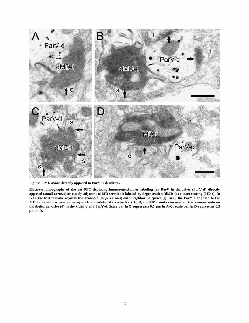
Figure 3. MD axons directly apposed to ParV-ir dendrites
Electron micrographs of the rat PFC depicting immunogold-silver labeling for ParV in dendrites (ParV-d) directly apposed (small arrows) or closely adjacent to MD terminals labeled by degeneration (dMD-t) or tract-tracing (MD-t). In A-C, the MD-ts make asymmetric synapses (large arrows) onto neighboring spines (s). In B, the ParV-d apposed to the MD-t receives asymmetric synapses from unlabeled terminals (t). In D, the MD-t makes an asymmetric synapse onto an unlabeled dendrite (d) in the vicinity of a ParV-d. Scale bar in B represents 0.5 µm in A-C; scale bar in D represents 0.5 µm in D.
42

Figure 4. MD axons making axo-dendritic synapses with ParV-ir dendrites
Electron micrographs of the rat PFC showing immunogold-silver labeling for ParV in dendrites (ParV-d) that receive asymmetric synapses (large arrows) from MD axons labeled either by tract-tracing (MD-t) or anterograde degeneration (dMD-t). In A, the MD-t is dually labeled for BDA and ParV. In C, the ParV-d receives an additional asymmetric synapse from an unlabeled terminal (t). In D, the dMD-t synapses onto a part of the ParV-d that appears spine-like, although its large size and the presence of a mitochondrion (m) are uncharacteristic of pyramidal cell spines. Scale bar in D represents 0.5 µm in A-D.
43

Figure 5. MD axons synapsing onto CalB-ir spines
Electron micrographs of the rat PFC illustrating immunogold-silver labeling for CalB in dendrites (CalB-d), spines (CalB-s) or axon terminals (CalB-t) and their relationship to MD terminals labeled by immunoperoxidase (MD-t) or degeneration (dMD-t). In A and B, the dMD-ts make asymmetric synapses (large arrows) onto CalB-labeled spines. In C, a CalB-s receives an asymmetric synapse from a CalB-t. In D, an MD-t makes an asymmetric synapse onto a spine that is unlabeled but arises from a CalB-d. Scale bar in C represents 0.5 µm in A-C; scale bar in D represents 0.5 µm in D.
44

Figure 6. MD axons in vicinity of CalB-ir structures
Electron micrographs of the rat PFC illustrating immunogold-silver labeling for CalB in dendrites (CalB-d) or perikarya (CalB-p) in relationship to degenerating (dMD-t) or immunoperoxidase-labeled MD terminals (MD-t). In A, a dMD-t is apposed (small arrow) to a CalB-d that receives asymmetric synaptic input (large arrow) from an unlabeled terminal (t). As is typical, the dMD-t synapses onto an adjacent unlabeled spine (s). In B, an MD-t that is dually labeled for BDA and CalB synapses onto an unlabeled dendrite (d) in the vicinity of a CalB-p with a deeply invaginated nucleus (n). In C and D, CalB-ds receive asymmetric synaptic inputs from MD-ts. Scale bar in A represents 0.5 µm in A; scale bar in B represents 0.5 µm in B and D; scale bar in C represents 0.5 µm in C.
45

Figure 7. MD axon synapsing onto a CalB-ir perykaryon
Electron micrograph of the rat PFC illustrating immunogold-silver labeling for CalB in a perikaryon (CalB-p) that receives asymmetric synaptic input (large arrow) from a degenerating MD terminal (dMD-t). The insert shows this synapse at higher magnification. The CalB-p contains a large, only slightly invaginated nucleus (n). Scale bars represent 2 µm at high magnification and 0.5 µm in the insert.
46

Figure 8. MD axons in vicinity of CalR-ir structures
Electron micrographs of the rat PFC illustrating immunogold-silver labeling for CalR in dendrites (CalR-d), axons (CalR-a) or perikarya (CalR-p) and their relationship to MD terminals labeled by degeneration (dMD-t) or tract-tracing (MD-t). In A and B, MD-ts make asymmetric synapses (large arrows) onto spines (s). In A, the dMD-t is in close apposition (small arrow) to a CalR-d. In B, the MD-t is dually labeled by immunoperoxidase for PHA-L and immunogold-silver for CalR. In C, an MD-t synapses onto an unlabeled dendrite (d) in the same field as a CalR-a. In D and E, dMD-ts make asymmetric synapses onto a CalR-d or a CalR-p, respectively. A small part of the nucleus (n) of the CalR-p is visible in E, and the dMD-t makes an additional synapse onto a spine. Scale bar in E represents 0.5 µm in A, D and E; scale bar in C represents 0.5 µm in C.
47

Table 1. Synaptic Relationships Between MD Afferents and Somatodendritic Targets Immunoreactive for Calcium Binding Proteins in the Rat PFC
Tissue labeled for
ParV CalB CalR
Total number of MD axons and terminals observed 2027 1889 1822
Number (%) of MD terminals forming synapses 829 (41%) 806 (43%) 764 (42%)
Number (%) of MD synapses onto spines 809 (97.6%) 786 (97.5%)* 744 (97.4%)
Number (%) of MD synapses onto dendrites or soma 20 (2.4%) 20 (2.5%) 20 (2.6%)
onto labeled dendrites or soma 15 (75%) 9 (45%) 6 (30%)
* includes CalB+ spines
48

BIBLIOGRAPHY Bacon SJ, Headlam AJ, Gabbott PL, Smith AD. 1996. Amygdala input to medial prefrontal
cortex (mPFC) in the rat: a light and electron microscope study. Brain Res 720(1-2):211-219.
Beierlein M, Connors BW. 2002. Short-term dynamics of thalamocortical and intracortical synapses onto layer 6 neurons in neocortex. J Neurophysiol 88(4):1924-1932.
Beierlein M, Gibson J, Connors B. 2003. Two dynamically distinct inhibitory networks in layer 4 of the neocortex. J Neurophysiol 90(5):2987-3000.
Bender D. 1983. Visual activation of neurons in the primate pulvinar depends on cortex but not colliculus. Brain Res 279(1-2):258-261.
Berendse H, Groenewegen H. 1991. Restricted cortical termination fields of the midline and intralaminar thalamic nuclei in the rat. Neuroscience 42(1):73-102.
Bernardo KL, Woolsey TA. 1987. Axonal trajectories between mouse somatosensory thalamus and cortex. J Comp Neurol 258(4):542-564.
Bubser M, de Brabander J, Timmerman W, Feenstra M, Erdtsieck-Ernste E, Rinkens A, van Uum J, Westerink B. 1998a. Disinhibition of the mediodorsal thalamus induces fos-like immunoreactivity in both pyramidal and GABA-containing neurons in the medial prefrontal cortex of rats, but does not affect prefrontal extracellular GABA levels. Synapse 30(2):156-165.
Bubser M, Deutch AY. 1998b. Thalamic paraventricular nucleus neurons collateralize to innervate the prefrontal cortex and nucleus accumbens. Brain Res 787(2):304-310.
Bubser M, Scruggs JL, Young CD, Deutch AY. 2000. The distribution and origin of the calretinin-containing innervation of the nucleus accumbens of the rat. Eur J Neurosci 12(5):1591-1598.
Castro-Alamancos M, Connors B. 1997. Thalamocortical synapses. Prog Neurobiol 51(6). Cauli B, Audinat E, Lambolez B, Angulo MC, Ropert N, Tsuzuki K, Hestrin S, Rossier J. 1997.
Molecular and physiological diversity of cortical nonpyramidal cells. J Neurosci 17(10):3894-3906.
Cauli B, Porter JT, Tsuzuki K, Lambolez B, Rossier J, Quenet B, Audinat E. 2000. Classification of fusiform neocortical interneurons based on unsupervised clustering. Proc Natl Acad Sci U S A 97(11):6144-6149.
Celio MR. 1986. Parvalbumin in most gamma-aminobutyric acid-containing neurons of the rat cerebral cortex. Science 231(4741):995-997.
Celio MR, Baier W, Scharer L, de Viragh PA, Gerday C. 1988. Monoclonal antibodies directed against the calcium binding protein parvalbumin. Cell Calcium 9(2):81-86.
Celio MR, Baier W, Scharer L, Gregersen HJ, de Viragh PA, Norman AW. 1990. Monoclonal antibodies directed against the calcium binding protein Calbindin D-28k. Cell Calcium 11(9):599-602.
Celio MR, Heizmann CW. 1981. Calcium-binding protein parvalbumin as a neuronal marker. Nature 293(5830):300-302.
49

Chan J, Aoki C, Pickel V. 1990. Optimization of differential immunogold-silver and peroxidase labeling with maintenance of ultrastructure in brain sections before plastic embedding. Journal of Neuroscience Methods 33:113-127.
Chmielowska J, Carvell GE, Simons DJ. 1989. Spatial organization of thalamocortical and corticothalamic projection systems in the rat SmI barrel cortex. J Comp Neurol 285(3):325-338.
Chmielowska J, Stewart MG, Bourne RC. 1988. gamma-Aminobutyric acid (GABA) immunoreactivity in mouse and rat first somatosensory (SI) cortex: description and comparison. Brain Res 439(1-2):155-168.
Cobb S, Buhl E, Halasy K, Paulsen O, Somogyi P. 1995. Synchronization of neuronal activity in hippocampus by individual GABAergic interneurons. Nature 378(6552):75-78.
Conde F, Lund JS, Jacobowitz DM, Baimbridge KG, Lewis DA. 1994. Local circuit neurons immunoreactive for calretinin, calbindin D-28k or parvalbumin in monkey prefrontal cortex: distribution and morphology. J Comp Neurol 341(1):95-116.
Cornwall J, Phillipson O. 1988. Afferent projections to the dorsal thalamus of the rat as shown by retrograde lectin transport. II. The midline nuclei. Brain Res Bull 21(2):147-161.
Cullen TJ, Walker MA, Parkinson N, Craven R, Crow TJ, Esiri MM, Harrison PJ. 2003. A postmortem study of the mediodorsal nucleus of the thalamus in schizophrenia. Schizophr Res 60(2-3):157-166.
Daum I, Ackermann H. 1994. Frontal-type memory impairment associated with thalamic damage. Int J Neurosci 77(3-4):187-198.
DeFelipe J. 1997. Types of neurons, synaptic connections and chemical characteristics of cells immunoreactive for calbindin-D28K, parvalbumin and calretinin in the neocortex. J Chem Neuroanat 14(1):1-19.
DeFelipe J, Farinas I. 1992a. The pyramidal neuron of the cerebral cortex: morphological and chemical characteristics of the synaptic inputs. Prog Neurobiol 39(6):563-607.
DeFelipe J, Hendry SH, Hashikawa T, Molinari M, Jones EG. 1990. A microcolumnar structure of monkey cerebral cortex revealed by immunocytochemical studies of double bouquet cell axons. Neuroscience 37(3):655-673.
DeFelipe J, Jones EG. 1992b. High-Resolution Light and Electron Microscopic Immunocytochemistry of Colocalized GABA and Calbindin D-28k in Somata and Double Bouquet Cell Axons of Monkey Somatosensory Cortex. Eur J Neurosci 4(1):46-60.
del Rio MR, DeFelipe J. 1995. A light and electron microscopic study of calbindin D-28k immunoreactive double bouquet cells in the human temporal cortex. Brain Res 690(1):133-140.
del Rio MR, DeFelipe J. 1997. Double bouquet cell axons in the human temporal neocortex: relationship to bundles of myelinated axons and colocalization of calretinin and calbindin D-28k immunoreactivities. J Chem Neuroanat 13(4):243-251.
Deschenes M, Veinante P, Zhang Z. 1998. The organization of corticothalamic projections: reciprocity versus parity. Brain Res Brain Res Rev 28(3):286-308.
Diamond M, Armstrong-James M, Budway M, Ebner F. 1992. Somatic sensory responses in the rostral sector of the posterior group (POm) and in the ventral posterior medial nucleus (VPM) of the rat thalamus: dependence on the barrel field cortex. J Comp Neurol 319(1):66-84.
50

Divac I. 1971. Frontal lobe system and spatial reversal in the rat. Neuropsychologia 9(2):175-183.
Dorph-Petersen KA, Pierri JN, Sun Z, Sampson AR, Lewis DA. 2004. Stereological analysis of the mediodorsal thalamic nucleus in schizophrenia: volume, neuron number, and cell types. J Comp Neurol 472(4):449-462.
Eysel U. 1992. Lateral inhibitory interactions in areas 17 and 18 of the cat visual cortex. Prog Brain Res 90:407-422.
Floresco S, Braaksma D, Phillips A. 1999. Thalamic-cortical-striatal circuitry subserves working memory during delayed responding on a radial arm maze. J Neurosci 19(24):11061-11071.
Floresco S, Grace A. 2003. Gating of hippocampal-evoked activity in prefrontal cortical neurons by inputs from the mediodorsal thalamus and ventral tegmental area. J Neurosci 23(9):3930-3943.
Freeman JJ, Cuppernell C, Flannery K, Gabriel M. 1996. Context-specific multi-site cingulate cortical, limbic thalamic, and hippocampal neuronal activity during concurrent discriminative approach and avoidance training in rabbits. J Neurosci.
Freund TF, Martin KA, Whitteridge D. 1985. Innervation of cat visual areas 17 and 18 by physiologically identified X- and Y- type thalamic afferents. I. Arborization patterns and quantitative distribution of postsynaptic elements. J Comp Neurol 242(2):263-274.
Gabbott P, Headlam A, Busby S. 2002. Morphological evidence that CA1 hippocampal afferents monosynaptically innervate PV-containing neurons and NADPH-diaphorase reactive cells in the medial prefrontal cortex (Areas 25/32) of the rat. Brain Res 946(2):314-322.
Gabbott PL, Dickie BG, Vaid RR, Headlam AJ, Bacon SJ. 1997. Local-circuit neurones in the medial prefrontal cortex (areas 25, 32 and 24b) in the rat: morphology and quantitative distribution. J Comp Neurol 377(4):465-499.
Gabbott PL, Somogyi J, Stewart MG, Hamori J. 1985. GABA-immunoreactive neurons in the rat dorsal lateral geniculate nucleus: light microscopical observations. Brain Res 346(1):171-175.
Gabbott PL, Somogyi J, Stewart MG, Hamori J. 1986b. GABA-immunoreactive neurons in the rat cerebellum: a light and electron microscope study. J Comp Neurol 251(4):474-490.
Gabbott PL, Somogyi P. 1986c. Quantitative distribution of GABA-immunoreactive neurons in the visual cortex (area 17) of the cat. Exp Brain Res 61(2):323-331.
Gaffan D, Parker A. 2000. Mediodorsal thalamic function in scene memory in rhesus monkeys. Brain 123 (Pt 4):816-827.
Galarreta M, Hestrin S. 2001a. Electrical synapses between GABA-releasing interneurons. Nat Rev Neurosci 2(6):425-433.
Galarreta M, Hestrin S. 2001b. Spike transmission and synchrony detection in networks of GABAergic interneurons. Science 292(5525):2295-2299.
Gibson J, Beierlein M, Connors B. 1999. Two networks of electrically coupled inhibitory neurons in neocortex. Nature 402(6757):75-79.
Groenewegen H. 1988. Organization of the afferent connections of the mediodorsal thalamic nucleus in the rat, related to the mediodorsal-prefrontal topography. Neuroscience 24((2)):379-431.
Groenewegen HJ. 1988. Organization of the afferent connections of the mediodorsal thalamic nucleus in the rat, related to the mediodorsal-prefrontal topography. Neuroscience 24(2):379-431.
51

Guillery R. 1995. Anatomical evidence concerning the role of the thalamus in corticocortical communication: a brief review. J Anat 187(Pt 3):583-592.
Guillery R, Sherman S. 2002. Thalamic relay functions and their role in corticocortical communication: generalizations from the visual system. Neuron 33(2):163-175.
Gulyas AI, Megias M, Emri Z, Freund TF. 1999. Total number and ratio of excitatory and inhibitory synapses converging onto single interneurons of different types in the CA1 area of the rat hippocampus. J Neurosci 19(22):10082-10097.
Gupta A, Wang Y, Markram H. 2000. Organizing principles for a diversity of GABAergic interneurons and synapses in the neocortex. Science 287(5451):273-278.
Hallanger A, Levey A, Lee H, Rye D, Wainer B. 1987. The origins of cholinergic and other subcortical afferents to the thalamus in the rat. J Comp Neurol 262(1):105-124.
Hayes T, Lewis D. 1992. Nonphosphorylated neurofilament protein and calbindin immunoreactivity in layer III pyramidal neurons of human neocortex. Cereb Cortex 2(1):56-67.
Heidbreder C, Groenewegen H. 2003. The medial prefrontal cortex in the rat: evidence for a dorso-ventral distinction based upon functional and anatomical characteristics. Neurosci Biobehav Rev 27(6):555-579.
Hersch SM, White EL. 1981a. Quantification of synapses formed with apical dendrites of Golgi-impregnated pyramidal cells: variability in thalamocortical inputs, but consistency in the ratios of asymmetrical to symmetrical synapses. Neuroscience 6(6):1043-1051.
Hersch SM, White EL. 1981b. Thalamocortical synapses involving identified neurons in mouse primary somatosensory cortex: a terminal degeneration and golgi/EM study. J Comp Neurol 195(2):253-263.
Hersch SM, White EL. 1981c. Thalamocortical synapses with corticothalamic projection neurons in mouse SmI cortex: electron microscopic demonstration of a monosynaptic feedback loop. Neurosci Lett 24(3):207-210.
Hunt P, Aggleton J. 1998a. An examination of the spatial working memory deficit following neurotoxic medial dorsal thalamic lesions in rats. Behav Brain Res 97(1-2):129-141.
Hunt P, Aggleton J. 1998b. Neurotoxic lesions of the dorsomedial thalamus impair the acquisition but not the performance of delayed matching to place by rats: a deficit in shifting response rules. J Neurosci 18(23):10045-10052.
Jensen KF, Killackey HP. 1987. Terminal arbors of axons projecting to the somatosensory cortex of the adult rat. I. The normal morphology of specific thalamocortical afferents. J Neurosci 7(11):3529-3543.
Jones EG. 2001. The thalamic matrix and thalamocortical synchrony. Trends Neurosci 24(10):595-601.
Kawaguchi Y. 1995. Physiological subgroups of nonpyramidal cells with specific morphological characteristics in layer II/III of rat frontal cortex. J Neurosci 15(4):2638-2655.
Kawaguchi Y, Kondo S. 2002. Parvalbumin, somatostatin and cholecystokinin as chemical markers for specific GABAergic interneuron types in the rat frontal cortex. J Neurocytol 31(3-5):277-287.
Kawaguchi Y, Kubota Y. 1997. GABAergic cell subtypes and their synaptic connections in rat frontal cortex. Cereb Cortex 7(6):476-486.
Keller A, White E. 1987. Synaptic organization of GABAergic neurons in the mouse SmI cortex. J Comp Neurol 262(1):1-12.
52

Keller A, White EL. 1986. Distribution of glutamic acid decarboxylase-immunoreactive structures in the barrel region of mouse somatosensory cortex. Neurosci Lett 66(3):245-250.
Kinomura S, Larsson J, Gulyas B, Roland P. 1996. Activation by attention of the human reticular formation and thalamic intralaminar nuclei. Science 271(5248):512-515.
Kisvarday Z, Kim D, Eysel U, Bonhoeffer T. 1994. Relationship between lateral inhibitory connections and the topography of the orientation map in cat visual cortex. Eur J Neurosci 6(10):1619-1632.
Kolb B. 1974a. Dissociation of the effects of lesions of the orbital or medial aspect of the prefrontal cortex of the rat with respect to activity. Behav Biol 10(3):329-343.
Kolb B. 1974b. Prefrontal lesions alter eating and hoarding behavior in rats. Physiol Behav 12(3).
Kolb B. 1974c. Some tests of response habituation in rats with discrete lesions to the orbital or medial frontal cortex. Can J Psychol 28(2):260-267.
Kolb B. 1984. Functions of the frontal cortex of the rat: a comparative review. Brain Res 320(1):65-98.
Kolb B. 1990. Animal models for human PFC-related disorders. Prog Brain Res 85:501-519. Kolb B, Buhrmann K, McDonald R, Sutherland R. 1994. Dissociation of the medial prefrontal,
posterior parietal, and posterior temporal cortex for spatial navigation and recognition memory in the rat. Cereb Cortex 4(6):664-680.
Kolb B, Whishaw I. 1981. Neonatal Frontal Lesions in the rat: sparing of learned but not species-typical behavior in the presence of reduced brain weight and cortical thickness. J Comp Physiol Psychol 95(6):863-879.
Krettek J, Price J. 1977. The cortical projections of the mediodorsal nucleus and adjacent thalamic nuclei in the rat. J Comp Neurol 171(2):157-191.
Kubota Y, Hattori R, Yui Y. 1994. Three distinct subpopulations of GABAergic neurons in rat frontal agranular cortex. Brain Res 649(1-2):159-173.
Kuroda M, Murakami K, Igarashi H, Okada A. 1996a. The convergence of axon terminals from the mediodorsal thalamic nucleus and ventral tegmental area on pyramidal cells in layer V of the rat prelimbic cortex. Eur J Neurosci 8(7):1340-1349.
Kuroda M, Murakami K, Kishi K, Price JL. 1995a. Thalamocortical synapses between axons from the mediodorsal thalamic nucleus and pyramidal cells in the prelimbic cortex of the rat. J Comp Neurol 356(1):143-151.
Kuroda M, Murakami K, Oda S, Shinkai M, Kishi K. 1993. Direct synaptic connections between thalamocortical axon terminals from the mediodorsal thalamic nucleus (MD) and corticothalamic neurons to MD in the prefrontal cortex. Brain Res 612(1-2):339-344.
Kuroda M, Murakami K, Shinkai M, Ojima H, Kishi K. 1995b. Electron microscopic evidence that axon terminals from the mediodorsal thalamic nucleus make direct synaptic contacts with callosal cells in the prelimbic cortex of the rat. Brain Res 677(2):348-353.
Kuroda M, Ojima H, Igarashi H, Murakami K, Okada A, Shinkai M. 1996b. Synaptic relationships between axon terminals from the mediodorsal thalamic nucleus and layer III pyramidal cells in the prelimbic cortex of the rat. Brain Res 708(1-2):185-190.
Kuroda M, Yokofujita J, Murakami K. 1998a. An ultrastructural study of the neural circuit between the prefrontal cortex and the mediodorsal nucleus of the thalamus. Prog Neurobiol 54(4):417-458.
53

Kuroda M, Yokofujita J, Oda S, Price JL. 2004. Synaptic relationships between axon terminals from the mediodorsal thalamic nucleus and gamma-aminobutyric acidergic cortical cells in the prelimbic cortex of the rat. J Comp Neurol 477(2):220-234.
Lewis DA. 2000. GABAergic local circuit neurons and prefrontal cortical dysfunction in schizophrenia. Brain Res Brain Res Rev 31(2-3):270-276.
Lewis DA, Cruz DA, Melchitzky DS, Pierri JN. 2001. Lamina-specific deficits in parvalbumin-immunoreactive varicosities in the prefrontal cortex of subjects with schizophrenia: evidence for fewer projections from the thalamus. Am J Psychiatry 158(9):1411-1422.
Lewis DA, Lund JS. 1990. Heterogeneity of chandelier neurons in monkey neocortex: corticotropin-releasing factor- and parvalbumin-immunoreactive populations. J Comp Neurol 293(4):599-615.
Matthews DEaVTF. 1996. Fisher's Tests for 2x2 Contingency Tables. Using and Understanding Medical Statistics.Basel, Karger. p 19-37.
McCasland JS, Hibbard LS. 1997. GABAergic neurons in barrel cortex show strong, whisker-dependent metabolic activation during normal behavior. J Neurosci 17(14):5509-5527.
Melchitzky D, Lewis D. 2003. Pyramidal neuron local axon terminals in monkey prefrontal cortex: differential targeting of subclasses of GABA neurons. Cereb Cortex 13(5):452-460.
Melchitzky DS, Gonzalez-Burgos G, Barrionuevo G, Lewis DA. 2001. Synaptic targets of the intrinsic axon collaterals of supragranular pyramidal neurons in monkey prefrontal cortex. J Comp Neurol 430(2):209-221.
Melchitzky DS, Sesack SR, Lewis DA. 1999. Parvalbumin-immunoreactive axon terminals in macaque monkey and human prefrontal cortex: laminar, regional, and target specificity of type I and type II synapses. J Comp Neurol 408(1):11-22.
Miettinen R, Gulyas A, Baimbridge K, Jacobowitz D, Freund T. 1992. Calretinin is present in non-pyramidal cells of the rat hippocampus--II. Co-existence with other calcium binding proteins and GABA. Neuroscience 48(1):29-43.
Moruzzi G, Magoun H. 1995. Brain stem reticular formation and activation of the EEG. 1949. J Neuropsychiatry Clin Neurosci 7(2):251-267.
Muir J, Everitt B, Robbins T. 1996. The cerebral cortex of the rat and visual attentional function: dissociable effects of mediofrontal, cingulate, anterior dorsolateral, and parietal cortex lesions on a five-choice serial reaction time task. Cereb Cortex 6(3):470-481.
Mumby D, Pinel J, Dastur F. 1993. Mediodorsal thalamic lesions and object recognition in rats. Psychobiology 21:27-36.
Newman D, Ginsberg C. 1994. Brainstem reticular nuclei that project to the thalamus in rats: a retrograde tracer study. Brain Behav Evol 44(1).
Nie F, Wong-Riley MT. 1995. Double labeling of GABA and cytochrome oxidase in the macaque visual cortex: quantitative EM analysis. J Comp Neurol 356(1):115-131.
Novikov LN. 2001. Labeling of central projections of primary afferents in adult rats: a comparison between biotinylated dextran amine, neurobiotin and Phaseolus vulgaris-leucoagglutinin. J Neurosci Methods 112(2):145-154.
Pakkenberg B. 1990. Pronounced reduction of total neuron number in mediodorsal thalamic nucleus and nucleus accumbens in schizophrenics. Arch Gen Psychiatry 47(11):1023-1028.
Paxinos G, Watson C. 1986. The Rat Brain in Stereotaxic Coordinates, Ed 2. New York: Academic.
54
Peters A. 1987. Number of neurons and synapses in primary visual cortex. Cerebral Cortex. New York: Plenium Press. p 267-294.
Peters A, Saldanha J. 1976c. The projection of the lateral geniculate nucleus to area 17 of the rat cerebral cortex. III. layer VI. Brain Res 105(3):533-537.
Petersen C, Sakmann B. 2001. Functionally independent columns of rat somatosensory barrel cortex revealed with voltage-sensitive dye imaging. J Neurosci 21(21):8435-8446.
Pinto A, Jankowski M, Sesack SR. 2003. Projections from the paraventricular nucleus of the thalamus to the rat prefrontal cortex and nucleus accumbens shell: ultrastructural characteristics and spatial relationships with dopamine afferents. J Comp Neurol 459(2):142-155.
Popken GJ, Bunney WE, Jr., Potkin SG, Jones EG. 2000. Subnucleus-specific loss of neurons in medial thalamus of schizophrenics. Proc Natl Acad Sci U S A 97(16):9276-9280.
Porter J, Johnson C, Agmon A. 2001. Diverse types of interneurons generate thalamus-evoked feedforward inhibition in the mouse barrel cortex. J Neurosci 21(8):2699-2710.
Pouille F, Scanziani M. 2001. Enforcement of temporal fidelity in pyramidal cells by somatic feed-forward inhibition. Science 293(5532):1159-1163.
Preuss T. 1995. Do rats have prefrontal cortex? The Rose-Woolsey-Akert program reconsidered. Journal of Cognitive Neuroscience 7:1-24. Rao S, Williams G, Goldman-Rakic P. 1999. Isodirectional tuning of adjacent interneurons and
pyramidal cells during working memory: evidence for microcolumnar organization in PFC. J Neurophysiol 81(4):1903-1916.
Rao S, Williams G, Goldman-Rakic P. 2000. Destruction and creation of spatial tuning by disinhibition: GABA(A) blockade of prefrontal cortical neurons engaged by working memory. J Neurosci 20(1):485-494.
Reiner A, Veenman CL, Medina L, Jiao Y, Del Mar N, Honig MG. 2000. Pathway tracing using biotinylated dextran amines. J Neurosci Methods 103(1):23-37.
Rogers JH. 1987. Calretinin: a gene for a novel calcium-binding protein expressed principally in neurons. J Cell Biol 105(3):1343-1353.
Romanski LM, LeDoux JE. 1993. Organization of rodent auditory cortex: anterograde transport of PHA-L from MGv to temporal neocortex. Cereb Cortex 3(6):499-514.
Rose J, Woolsey C. 1948. The orbitofrontal cortex and its connections with the mediodorsal nucleus in rabbit, sheep and cat. Res Publ Assoc Nerv Ment Dis 27:210-232.
Schwaller B, Buchwald P, Blumcke I, Celio MR, Hunziker W. 1993. Characterization of a polyclonal antiserum against the purified human recombinant calcium binding protein calretinin. Cell Calcium 14(9):639-648.
Sesack S, Medvitz N, Miner L, Ekema G. Pyramidal neuron intrinsic collaterals in the rat prefrontal cortex: synaptic targets and selective input to parvalbumin-containing interneurons; 2001.
Sesack SR, Bressler CN, Lewis DA. 1995a. Ultrastructural associations between dopamine terminals and local circuit neurons in the monkey prefrontal cortex: a study of calretinin-immunoreactive cells. Neurosci Lett 200(1):9-12.
Sesack SR, Hawrylak VA, Melchitzky DS, Lewis DA. 1998. Dopamine innervation of a subclass of local circuit neurons in monkey prefrontal cortex: ultrastructural analysis of tyrosine hydroxylase and parvalbumin immunoreactive structures. Cereb Cortex 8(7):614-622.
55

Sesack SR, Snyder CL, Lewis DA. 1995. Axon terminals immunolabeled for dopamine or tyrosine hydroxylase synapse on GABA-immunoreactive dendrites in rat and monkey cortex. J Comp Neurol 363(2):264-280.
Shanks MF, Powell TP. 1981. An electron microscopic study of the termination of thalamocortical fibres in areas 3b, 1 and 2 of the somatic sensory cortex in the monkey. Brain Res 218(1-2):35-47.
Sillito A. 1975. The contribution of inhibitory mechanisms to the receptive field properties of neurones in the striate cortex of the cat. J Physiol 250(2):305-329.
Simons DJ, Carvell GE. 1989. Thalamocortical response transformation in the rat vibrissa/barrel system. J Neurophysiol 61(2):311-330.
Smiley J, Goldman-Rakic P. 1993. Heterogeneous targets of dopamine synapses in monkey prefrontal cortex demonstrated by serial section electron microscopy: a laminar analysis using the silver-enhanced diaminobenzidine sulfide (SEDS) immunolabeling technique. Cereb Cortex 3(3).
Staiger J, Zilles K, Freund T. 1996a. Distribution of GABAergic elements postsynaptic to ventroposteromedial thalamic projections in layer IV of rat barrel cortex. Eur J Neurosci 8(11):2273-2285.
Staiger J, Zilles K, Freund T. 1996b. Innervation of VIP-immunoreactive neurons by the ventroposteromedial thalamic nucleus in the barrel cortex of the rat. J Comp Neurol 367(2).
Steriade M. 1997. Synchronized activities of coupled oscillators in the cerebral cortex and thalamus at different levels of vigilance. Cereb Cortex 7(6):583-604.
Steriade M, Glenn L. 1982. Neocortical and caudate projections of intralaminar thalamic neurons and their synaptic excitation from midbrain reticular core. J Neurophysiol 48(2):352-371.
Tamas G, Buhl E, Somogyi P. 1997. Massive autaptic self-innervation of GABAergic neurons in cat visual cortex. J Neurosci 17(16).
Tamas G, Somogyi P, Buhl E. 1998. Differentially interconnected networks of GABAergic interneurons in the visual cortex of the cat. J Neurosci 18(11).
Tanibuchi I, Goldman-Rakic PS. 2003. Dissociation of spatial-, object-, and sound-coding neurons in the mediodorsal nucleus of the primate thalamus. J Neurophysiol 89(2):1067-1077.
Uylings H, Groenewegen H, Kolb B. 2003. Do rats have a prefrontal cortex? Behav Brain Res 146(1-2):3-17.
Uylings H, van Eden C. 1990. Qualitative and quantitative comparison of the prefrontal cortex in rat and in primates, including humans. Prog Brain Res 85:31-62.
van Brederode JF, Helliesen MK, Hendrickson AE. 1991. Distribution of the calcium-binding proteins parvalbumin and calbindin-D28k in the sensorimotor cortex of the rat. Neuroscience 44(1):157-171.
Van der Werf Y, Witter M, Groenewegen H. 2002. The intralaminar and midline nuclei of the thalamus. Anatomical and functional evidence for participation in processes of arousal and awareness. Brain Res Brain Res Rev 39(2-3):107-140.
Veenman CL, Reiner A. 1996. Ultrastructural morphology of synapses formed by corticostriatal terminals in the avian striatum. Brain Res 707(1):1-12.
Veenman CL, Reiner A, Honig MG. 1992. Biotinylated dextran amine as an anterograde tracer for single- and double-labeling studies. J Neurosci Methods 41(3):239-254.
56

Veznedaroglu E, Milner T. 1992. Elimination of artifactual labeling of hippocampal mossy fibers seen following pre-embedding immunogold-silver technique by pretreatment with zinc chelator. Microscopy Research and Technique 23:100-101.
Vogt BA, Rosene DL, Peters A. 1981. Synaptic termination of thalamic and callosal afferents in cingulate cortex of the rat. J Comp Neurol 201(2):265-283.
Volk DW, Lewis DA. 2002. Impaired prefrontal inhibition in schizophrenia: relevance for cognitive dysfunction. Physiol Behav 77(4-5):501-505.
Wang Y, Gupta A, Toledo-Rodriguez M, Wu CZ, Markram H. 2002. Anatomical, physiological, molecular and circuit properties of nest basket cells in the developing somatosensory cortex. Cereb Cortex 12(4):395-410.
Warren J, Kolb B. 1978. Generalizations in neuropsychology. S F, editor. New York: Plenium Press.
Welker E, Armstrong-James M, Van der Loos H, Kraftsik R. 1993. The mode of activation of a barrel column: response properties of single units in the somatosensory cortex of the mouse upon whisker deflection. Eur J Neurosci 5(6):691-712.
White EL. 1978. Identified neurons in mouse Sml cortex which are postsynaptic to thalamocortical axon terminals: a combined Golgi-electron microscopic and degeneration study. J Comp Neurol 181(3):627-661.
Wikmark R, Divac I, Weiss R. 1973. Retention of spatial delayed alternation in rats with lesions in the frontal lobes. Implications for a comparative neuropsychology of the prefrontal system. Brain Behav Evol 8(5):329-339.
Woo TU, Miller JL, Lewis DA. 1997. Schizophrenia and the parvalbumin-containing class of cortical local circuit neurons. Am J Psychiatry 154(7):1013-1015.
Wouterlood FG, Groenewegen HJ. 1985. Neuroanatomical tracing by use of Phaseolus vulgaris-leucoagglutinin (PHA-L): electron microscopy of PHA-L-filled neuronal somata, dendrites, axons and axon terminals. Brain Res 326(1):188-191.
Wouterlood FG, Jorritsma-Byham B. 1993. The anterograde neuroanatomical tracer biotinylated dextran-amine: comparison with the tracer Phaseolus vulgaris-leucoagglutinin in preparations for electron microscopy. J Neurosci Methods 48(1-2):75-87.
Young KA, Manaye KF, Liang C, Hicks PB, German DC. 2000. Reduced number of mediodorsal and anterior thalamic neurons in schizophrenia. Biol Psychiatry 47(11):944-953.
Zimmermann L, Schwaller B. 2002. Monoclonal antibodies recognizing epitopes of calretinins: dependence on Ca2+-binding status and differences in antigen accessibility in colon cancer cells. Cell Calcium 31(1):13-25.
57