seating chart (r409)
TRANSCRIPT

2-1 鍾佩倪
2-2 李明輝
陳傳佳 2-3
郭秉翰 2-4
3-1 范芯語
3-2 梁曙臨
王翌馨 3-3
陳佩萱 3-4
4-1 許智程
4-2 林馳耘
劉菉文 4-3
曾理心 4-4
Podium
5-1 苑欣佑
5-2 葉祐丞
方唯蘋 5-3
趙天儀 5-4
Seating chart (R409)
6-1 詹佳恩
6-2 李品賢
季皓勻 6-3
王秉謙 6-4
1-1 林皓添
1-3 孫浚豪
Evany
Vega 1-4
Castro
張峵華 1-5
1 2 3 4
5 6
1-2 李官庭
實驗準備桌
實驗準備桌

Contact information:
GB Lab. Preparation room: 3366-4544
徐馨怡Shin-Yi Shyu
RM 528Life science Bldg.
3366-2497 [email protected]
吳振宇 RM 1128Life science Bldg.
3366-2508 [email protected]
侯正修 RM 738Life science Bldg.
3366-2481 [email protected]
劉上源 RM 1018Life science Bldg.
3366-2528 [email protected]
蔣依恬RM 807Life science Bldg.
3366-2487 [email protected]

Date Title
9/28 Exp.35: Recognition of the woody plants in NTU campus
10/5 Laboratory safety rules; Exp. 1: Introduction of the light microscope
10/12 Exp. 2: Plant cell
10/19 Exp. 5: Roots; Exp. 6: Stems; Exp.7: Leaves
10/26 Exp. 14: Seed germination and -amylase; Exp. 15: Starch phosphorylase
11/2Exp. 18: Photosynthesis – The extraction of plant pigments
Exp. 19: Photosynthesis – Inner structures of C3, C4, and CAM plants
11/9 Exp. 4: Mitosis &Meiosis
11/16 Midterm exam
11/23 Exp. 31: Analysis of artificial grassland community composition in NTU campus
11/30 Exp. 23: Bacteria; Exp. 24, 25: Algae
12/7 Exp. 26: Fungi; Exp. 27: Lichens
12/14 Exp. 32: Hydrophytes
12/21 Exp. 8: Flowers; Exp.9: Fruits; Exp. 10: Seeds and seedlings
12/28 Exp. 20: Measurement of the water potential by the falling-drop method
1/4 Final exam

Laboratory safety rules
Before entering the classroom, please log in with your student ID,
measure your forehead temperature, disinfect your hands with
alcohol, and wear a mask throughout the class.
Eating and drinking are strictly prohibited in the laboratory. You need to
wear laboratory clothes during class, and it is forbidden to wear
slippers or sandals.
This lab class starts at 15:30, please arrive on time. Arrive 30 minutes
late is considered absenteeism. A total of 3 points will be deducted for
a late arrival; a total of 10 points will be deducted for one absenteeism.
Apply for leave according to school regulations. Notify TA about your
leave at least 1 week in advance, and have to make up the class.
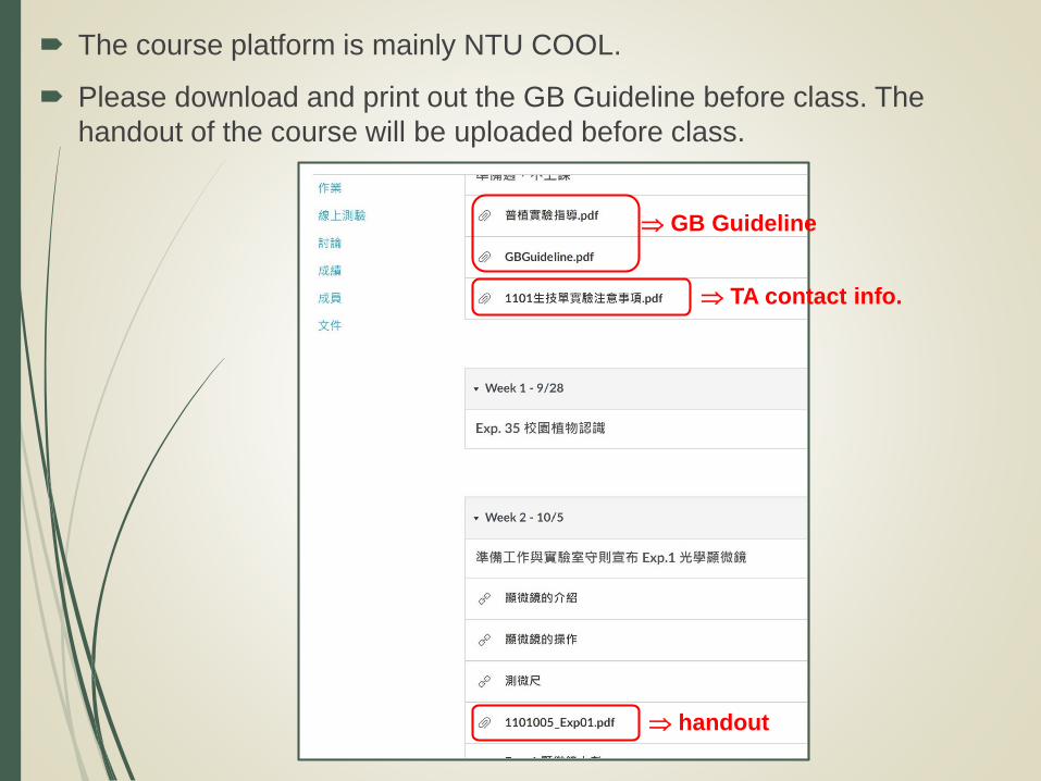
The course platform is mainly NTU COOL.
Please download and print out the GB Guideline before class. The
handout of the course will be uploaded before class.
TA contact info.
GB Guideline
handout

Please visit NTU COOL to watch the video of the course before
the class. The preparatory quiz will be conducted at the beginning
of the class.
Please preview the lesson content of the current week and finish
the diagrams.
The format of the homework is following with the class rules of the
week.10 points will be deducted for late submission within a week,
and 60 points will be calculated for the late submission within one
week.
Score calculation: 50% of the usual score (quizzes, homework,
class performance...), and 50% of the test scores (exams)
Do not leave the classroom by yourself during. If you have other
conditions or needs, please inform the TA yourself.

Experiment report
Format
Title
Introduction: purpose, principle…
Materials & methods
Results
Discussion
Reference
Department level, group, members, writer…
Scientific report:
Faithfully record the experiment process and
results, and make conclusions and analyses
based on the results.

Data presentation
Both figure and table legends should match the width of the table
or graph. Use a numerical digit as the figure number rather than
the full word, e.g. Figure 1.
Figure legends go below the graph and are left-justified.
Table legends go above the body of the table and are left-justified.
Figures 與 Tables © NTU Library, via 臺大圖書館參考服務部落格
Figure legend
Table legend

NATIONAL TAIWAN UNIVERSITY
DATE_________ NAME_________ CORRECTED BY____________
系級_________ 學號_________ 組別_________
(400X)
(400X)
Figure legend (title): Below the drawing
It should state what has been drawn.
The scientific name should be italicized or
underlined.
Drawing: No sketchy lines, only single lines.
No shading, only stippling (dotting).
Include part cell wall of adjacent cells.
Two drawings per page.
Biological drawing example
Labeling: All labels must be neat and intelligible and
should be done in a column at the right-hand side
of the page.
The labels must be aligned.
The label lines must be drawn using a ruler and
should be parallel to one another (never cross).
Others: the proportion, position of the drawing
magnification, scale bar
using H head pencil

Photo: Photos should be trimmed properly
the focus should be clear
appropriate magnification power
Fig. 1 Druses of Begonia sp. (100X)
10 m
Druse
Phloem
Xylem
Protoxylem lacuna
Bundle sheath
Fig. 2 C.S. of stem of Zea mays (400X)
15 m
Photo assignments example
Figure legend (title): Below the photos
It should state what has been shoot.
The scientific name should be italicized or
underlined.
Labeling: All labels must be neat and intelligible and
should be done in a column at the right-hand side
of the page.
The labels must be aligned.
The label lines must be drawn using a ruler and
should be parallel to one another (never cross).
Others: the proportion, position of the photos
magnification, scale bar
using H head pencil


Factors affect image quality
Magnification
Resolving power: the capacity to resolve two points
which are close together.
Human eye:0.1 mm
Light microscope:0.1 m
Electron microscope:0.1 nm
Contrast
Staining
Darkfield and phase contrast microscopy

Microscope Care & Handling
Always carry the microscope with
both hands.
One hand under the base, and the
other on the arm.
Lift the microscope, and don’t push,
pull the microscope as you
transport it from one place to
another.
鏡座(Base)
鏡臂(Arm)

Objective
物鏡
Arm 鏡臂
Coarse & fine
focus adjustment
knobs
粗細調節輪
Base 基座
Eyepiece
目鏡
Stage 載物台
Iris
diaphragm
光圈
Lamp 光源
Tube 鏡筒
Revolving
nosepiece
物鏡轉盤
Condenser
adjustment knob
聚光鏡(集光器)升降調節輪
Illumination
control knob
光源調節輪
顯微鏡的構造

Condenser
聚光鏡
Slide holder
玻片夾
X & Y axial
adjustment
knobs
XY調節鈕Iris diaphragm
光圈
Slide holder
玻片夾
Condenser
adjustment knob
聚光鏡升降調節輪

The diaphragm affects depth of field
the smaller diameter of the diaphragm, the
less light passes through
deeper depth of field, more contrast
lower resolution
for low-power objective
the larger diameter of the diaphragm, the
more light passes through
shallow depth of field, less contrast
higher resolution
for high-power objective

Turn on the light, adjust the light intensity.
Position the condenser as high as it will go, then lower it 1-2mm.
Adjust the aperture diaphragm for appropriate contrast (4X).
Lock the low power (4X) objective lens into place. Put the slide on
the specimen stage, held by the slide clip.
Center the specimen over the circle of light.
Raise the stage to the highest position using the microscope coarse
focus control knob.
Lower the stage slowly by turning the coarse adjustment knob
counter-clockwise until the object is in focus.
Use the fine adjustment to bring the object into sharp focus.
Leave the slide where it focused under low-power and switch to the
next high-power.
ONLY USE THE FINE ADJUSTMENT to focus.
Adjust the aperture diaphragm.
Switch the objective to low-power before removing the slide.
Using the Microscope

Matters needing attention
Always carry the microscope with both hands. One hand
under the base, and the other on the arm.
Rotate the nosepiece to change objectives. When you
change to a higher magnification objective lens, focus only
with the fine focus knob.
Never remove a slide while under high-power objective.
Clean smudges on the microscope glass with a piece of
lens paper. Avoid touching the lenses of the microscope.
Close the cabinet after moving the microscope.
Slides should be on the stage or in the tray.

Put away a microscope
Remove your slide
Turn off the light.
Rack down the stage
Place the 4X objective over the stage
Return the stage mechanism to its central position.
Place the oculars toward the arm.
Wrap the cord correctly
Return the scope to its cabinet

1 ocular micrometer division (μm) =Number of stage micrometer div. × 10μm
Number of ocular micrometer div.
Stage micrometer
Ocular micrometer
10 ×10 μm
3 = = 33.3 μm
10 div. × 10 μm
3 div. × μm
Measurement with the light microscope
Calibration of microscopic ocular micrometer
0 1 2 3 4 5
0 1 2 3 4 5
1mm/100 div. 10μm/1 div.
?

Ocular micrometer
40x 100x 400x
Stage micrometer
1mm/100 div. 10μm/1 div.

Exercise
Calculate the length of 1
ocular micrometer division
of these 3 pictures.
Record your answers
(include unit) in the
“Micrometer exercise” at
NTU COOL.
4X objective
10X objective
40X objective