report of a case a. b. a. karat, c. k. job and s. karatila.ilsl.br/pdfs/v35n1a03.pdfinternationa_l...
TRANSCRIPT
INTER NATIONA_L JO URN AL O F LEI'ROSY Volume 35, Number J Pri lltcd ill U.s.A.
J Erythema Nodosum Leprosum In Borderline Leprosy
Report of a Case l
A. B. A. Karat, C. K. Job and S. Karat2
Since Wade and Rodriguez (1 , 7) introduced the concept of borderline leprosy as a distinct entity in the wide spectl11m of clinical manifestations of leprosy, numerous data have accumulated on the clinical, histologic and immunologic facets of this type of leprosy. Recently Ramanujam and Ramu (5) have given an excellent summary of the literature on this subject.
Among the descriptions of exacerbated phases ( reaction ) of this type of leprosy, we are unable to find any reference to erythema nodosum leprosum (ENL) as one of its manifestations. Wade (6) stated categorically that ENL does not occur in this form of leprosy (2). The published reports of the Panel on Lepra Reactions and the Round Table on Borderline and Indeterminate Leprosy of the VIIIth International Congress of Leprology (3) make no mention of ENL in relation to borderline leprosy.
CASE REPORT
We present here the case repOlt of a patient with borderline leprosy who developed ENL during a phase of exacerbation of the disease.
A.S.R.S. (No. 7891 ) was first seen at the Schieffelin Leprosy Research Sanatorium, Karigiri on 22 November 1965. He gave a history of leprosy of 18 years' duration. The disease apparently began with a hypopigmented, anesthetic patch over the leg and spread gradually to other parts of the body. He had taken treatment at a
' Received for publication 15 August 1966. "1\. B. A. Karat. B.Sc., M.B ., B.S., M.R.C.P.
(Lond .) , M.R.C.P. (Eclin.), C. K. Job, B.Sc., M.D., M.C. Path.; (Mrs.) S. Karat, M.B., B.S. , F.R.C.S. (Ecl in .) ; Schicffelin Leprosy Research Sanatorium, Karigiri, via J{alpacli, South India.
number of places, and for five months prior to his arrival at the sanatorium was on regular medication with DDS under the supervision of another leprosy hospital. Within a few days of starting treatment with DDS, he noted that the existing hypopigmented, Rat lesions quickly became raised, purple, and painful. At about the same time new raised lesions began to appear and he developed pain in the eyes and testis, and considerable swelling of the hands and feet, associated with paresthesia in the limbs. Despite these symptoms he was maintained on DDS.
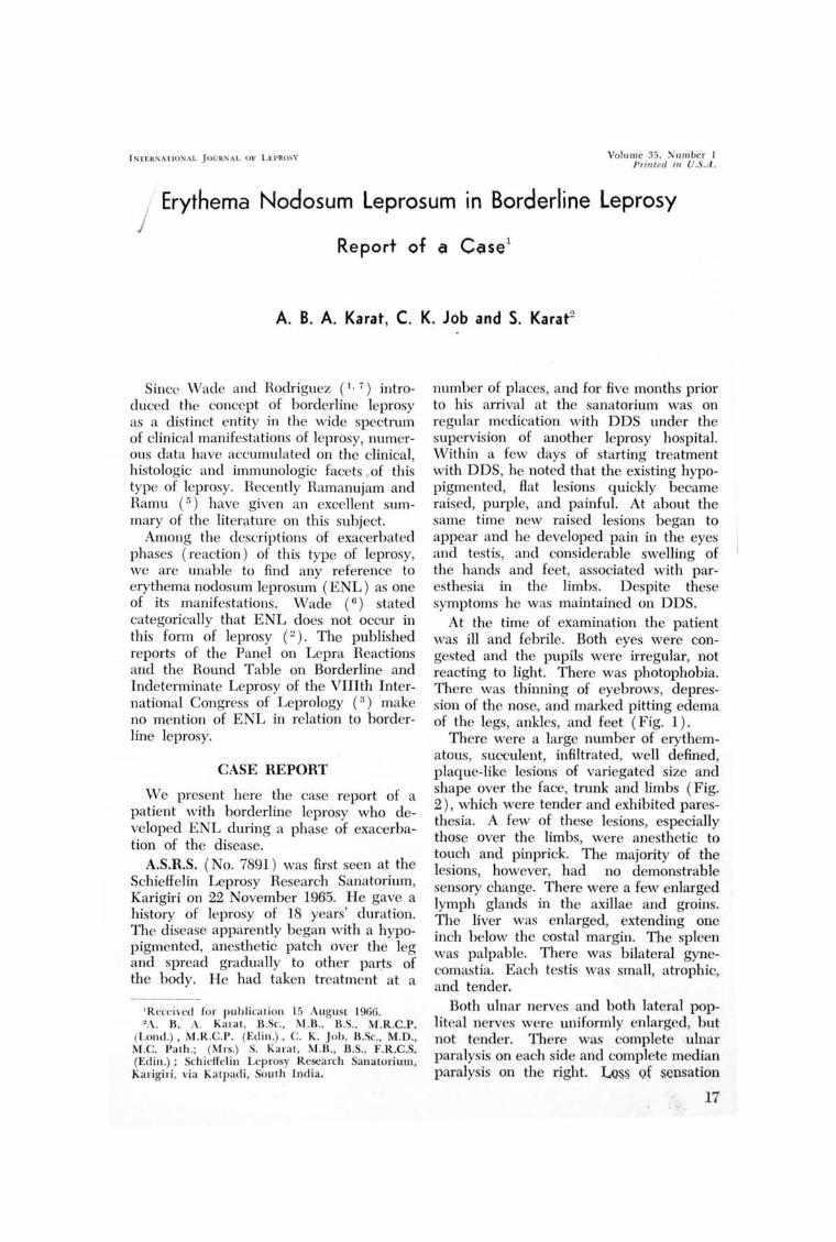
At the time of examination the patient was ill and febrile. Both eyes were congested and the pupils were irregular, not reacting to light. There was photophobia. There was thinning of eyebrows, depression of the nose, and marked pitting edema of the legs, ankles, and feet (Fig. 1).
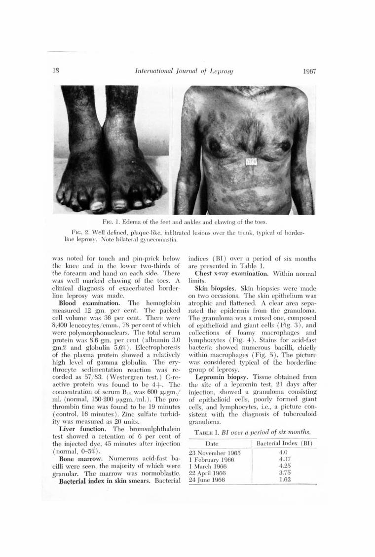
There were a large number of erythematous, succulent, infiltrated, well defined, plaque-like lesions of variegated size and shape over the face, trunk and limbs (Fig. 2), which were tender and exhibited paresthesia. A few of these lesions, especially those over the limbs, were anesthetic to touch and pinprick. The majority of the lesions, however, had no demonstrable sensory change. There were a few enlarged lymph glands in the axillae and groins. The liver was enlarged, extending one inch below the costal margin. The spleen was palpable. There was bilateral gynecomastia. Each testis was small, atrophic, and tender.
Both ulnar nerves and both lateral popliteal nerves were uniformly enlarged, but not tender. There was complete ulnar paralysis on each side and complete median paralysis on the right. L~ Q£ sensation
17
18 Inte1'l1ational Journal of Leprosy 1967
FIG. 1. Edema of the feet and ankles and clawing of the toes.
FIG. 2. Well defin ed , plaque-like, infiltrated lesions over the t1'llnk, typical of borderline leprosy. Note bilateral gynecomastia .
was noted for touch and pin-prick below the knee and in the lower two-thirds of the forearm and hand on each side. There was well marked clawing of the toes. A clinical diagnosis of exacerbated horderline leprosy was made.
Blood examination. The hemoglobin measured 12 gm. per cent. The packed cell volume was 36 per cent. There were 8,400 leucocytes/ cmm., 78 per cent of which were polymorphonuclears. The total serum protein was 8.6 gm. per cent (albumin 3.0 gm.% and globulin 5.6%). Electrophoresis of the plasma protein showed a relatively high level of gamma globulin. The erythrocyte sedimentation reaction was recorded as 57/ 83. ( Westergren tes t. ) C-reactive protein was found to be 4+. The concentration of serum B' 2 was 600 [I.[I.gm.j ml. (normal, 150-200 [I.lJ.gm./ ml. ) . The prothrombin time was found to be 19 minutes (control, 16 minutes) . Zinc sulfate turbidity was measured as 20 units.
Liver function. The bromsulphthalein tes t showed a retention of 6 per cent of the injected dye, 45 minutes after injection ( norma I, 0-5%).
Bone marrow. Numerous acid-fast bacilli were seen, the majority of which were granular. The marrow was normoblastic.
Bacterial index in skin smears. Bacterial
indices (BI ) over a period of six months are presented in Table 1.
Chest x-ray examination. Within normal limits.
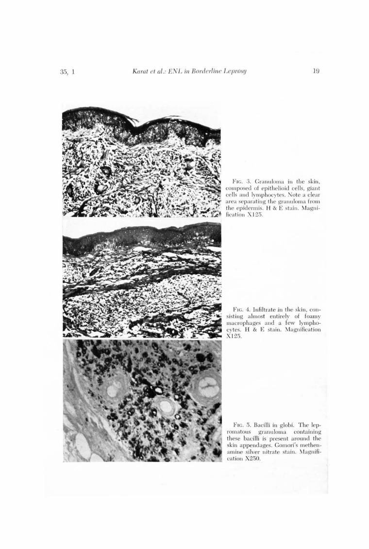
Skin biopsies. Skin biopsies were made on two occasions. The skin epithelium wa~ atrophic and flattened. A clear area separated the epidermis from the granuloma. The granuloma was a mixed one, composed of epithelioid and giant cells (Fig. 3 ), and collections of foamy macrophages and lymphocytes (Fig. 4). Stains for acid-fast bacteria sho"ved numerous bacilli, chiefly within macrophages (Fig. 5). The picture was considered typical of the borderline group of leprosy.
Lepromin biopsy. Tissue obtained from the site of a lepromin test, 21 days after injection, showed a granuloma consisting of epithelioid cells, poorly formed giant cells, and lymphocytes, i.e., a picture consistent with the diagnosis of tuberculoid granuloma.
T ABLE 1. BI over a period of six months.
Date
23 ovember 1965 1 February 1966 1 March 1966 22 April 1966 24 June 1966
Bacterial Index (BI)
4.0 4.37 4.25 3.75 1.62
35, 1 Ku/'{/t et aT.: ENL ill Borderline Leprosy 19
F IG. 3. Granuloma in the skin , composed of epithelioid cells, giant cells and lymphocytes. Note a clea r area separating the granuloma from the epidermis . H & E stain . ~vlagn ifi ca tion X12.5.
FIG. 4. Infiltrate jn the skin , consisting almost entirely of foamy macrophages and a few lymphocytes. H & E stain . ~1agnifica tion X] 2.5.
FIG . .5. Bacilli in globi. The lepromatous granu loma containing these bacilli is present around the skin appendages. Gomori's methenamine silver nitrate stain. ~1agn ifica tion X2.50.
20 International Journal ot Lep'l'osy 1967
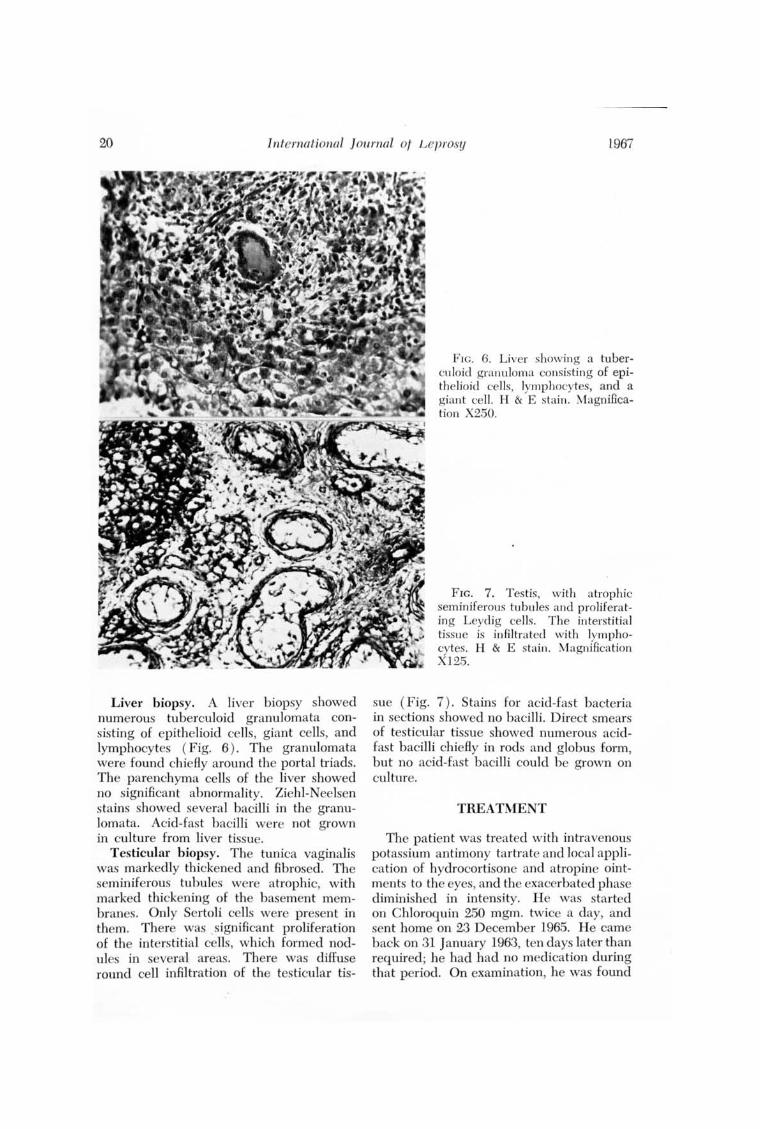
Liver biopsy. A liver biopsy showed numerous tuberculoid granulomata consisting of epithelioid cells, giant cells, and lymphocytes ( Fig. 6 ) . The granulomata were found chiefly around the portal triads. The parenchyma cells of the liver showed no significant abnormality. Ziehl-Neelsen stains showed several bacilli in the granulomata. Acid-fast bacilli were not grown in culture from liver tissue.
Testicular biopsy. The tunica vaginalis was markedly thickened and fibrosed. The seminiferous tubules were atrophic, with marked thickening of the basement membranes. Only Sertoli cells were present in them. There was significant proliferation of the interstitial cells, which formed nodules in several areas. There was diffuse round cell infiltration of th testicular tis-
F I G. 6. Liver showing a tuberculoid granuloma consisting of epithelioid cells, lymphocytes, and a giant cell. H & E stain . Magnification X250.
FIG. 7. Testis, with atrophic seminiferous tubules and proliferating Leydig cells. The interstitial tissue is infiltrated with lymphocytes. H & E stain. Magnification X125.
sue (Fig. 7). Stains for acid-fast bacteria in sections showed no bacilli. Direct smears of testicular tissue showed numerous acidfast bacilli chiefly in rods and globus form, but no acid-fast bacilli could be grown on culture.
TREATMENT
The patient was treated with intravenous potassium antimony tartrate and local application of hydrocortisone and atropine ointments to the eyes, and the exacerbated phase diminished in intensity. H e was started on Chloroquin 250 mgm. twice a day, and sent home on 23 D ecember 1965. H e came b ack on 31 January 1963, ten days later than required ; he had had no medica tion during that period. On examination, he was found
35, 1 Karat et a7.: ENL il1 Borderlil1e Leprosy 21
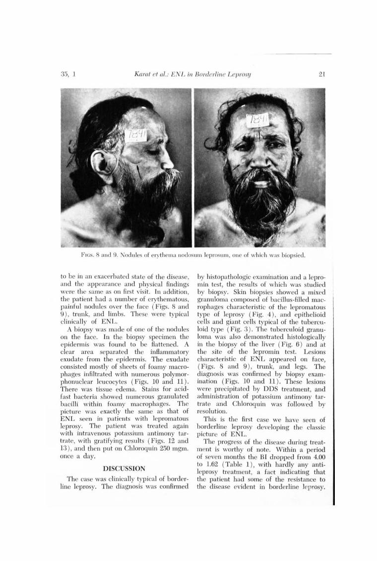
F IGs. 8 and 9. Nod ules of erythema nodosum leprosum , one of which was hiopsied.
to be in an exacerbated state of the disease, and the appearance and physical findings were the same as on first visit. In addition, the patient had a number of erythematous, painful nodules over the face (Figs. 8 and 9 ), trunk, and limbs. These were typical clinically of ENL.
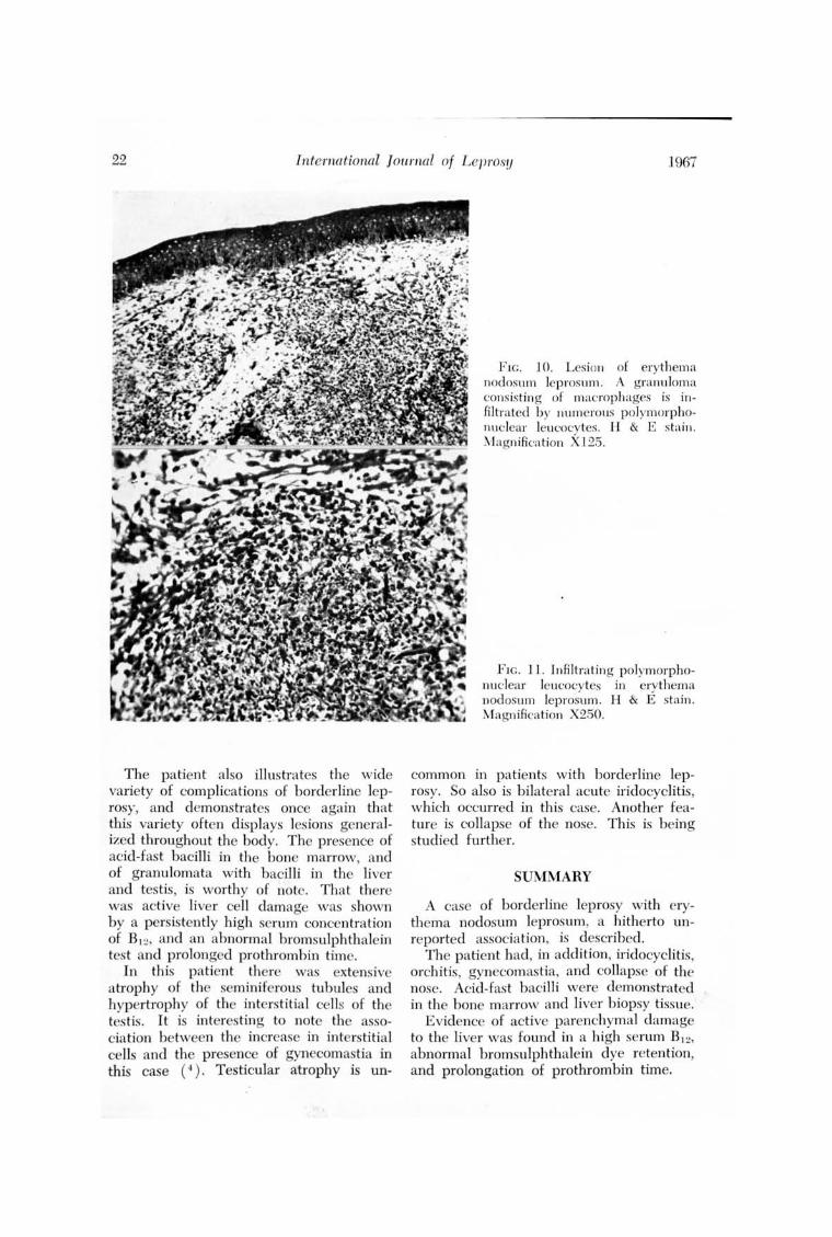
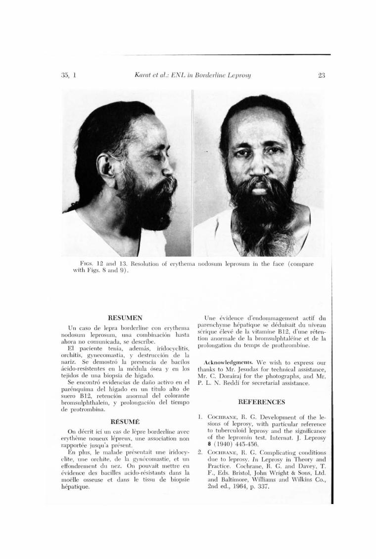
A biopsy was made of one of the nodules on the face. In the biopsy specimen the epidermis was found to be flattened. A clear area separated the inflammatory exudate from the epidermis. The exudate consisted mostly of sheets of foamy macrophages infiltrated with numerous polymorphonuclear leucocytes ( Figs. 10 and 11 ). There was tissue edema. Stains for acidfast bacteria showed numerous granulated bacilli within foamy macrophages. The picture was exactly the same as that of ENL seen in patients with lepromatous leprosy. The patient was treated again with intravenous potass ium antimony tartrate, with gratifying results (Figs. 12 and 13 ), and then put on Chloroquin 250 mgm. once a day.
DISCUSSION
The case was clinically typical of borderline leprosy. The diagnosis was confirmed
by histopathologic examination and a lepromin test, the results of which was studied by biopsy. Skin biopsies showed a mixed granuloma composed of bacillus-filled macrophages characteristic of the lepromatous type of leprosy (Fig. 4 ), and epithelioid cells and giant cells typical of the tuberculoid type (Fig. 3 ). The tuberculoid granuloma was also demonstrated histologically in the biopsy of the liver (Fig. 6 ) and at the site of the lepromin test. Lesions characteristic of ENL appeared on face, (Figs. 8 and 9) , trunk, and legs. The diagnosis was confirmed by biopsy examination (Figs. 10 and 11 ). These lesions were precipitated by DDS treatment, and administration of potassium antimony tartrate and Chloroquin was followed by resolution.
This is the first case we have seen of borderline leprosy developing the classic picture of ENL.
The progress of the disease during treatment is worthy of note. Within a period of seven months the BI dl'Opped from 4.00 to 1.62 (Table 1) , with hardly any antileprosy treatment, a fact indicating that the patient had some of the resistance to the disease evident in borderline Jep osy.
22 International Journal of L eprosy ]967
The patient also illustrates the wide variety of complications of borderline leprosy, and demonsh'ates once again that this variety often displays lesions generalized throughout the body. The presence of acid-fast bacilli in the bone marrow, and of granulomata with bacilli in the liver and tes tis, is worthy of note. That there was active liver cell damage was shown by a persistently high serum concentration of Bl ~' and an abnormal bromsulphthalein test and prolonged prothrombin time.
In this patient there was extensive a trophy of the seminiferous tubules and hypertrophy of the interstitial cells of the testis. It is interes ting to note the association between the increase in interstitial cells and the presence of gynecomastia in this case (4). Testicular atrophy is un-
F 1G. 10. Lesion of erythema nodosum leprosum. A granu loma consisting of macrophages is in filtrated by numerous polymorphonuclear leucocy tes. H & E sta in. Magn ifica tion X125.
F IG . 11. Infiltrating polymorphonuclear leucocytes in erythema nodosum leprosum. H & E stain . Magn ifica tion X250.
common in patients with borderline leprosy. So also is bilateral acute iridocyclitis, which occurred in this case. Another feature is coll apse of the nose. This is being studied further.
SUMMARY
A case of borderline leprosy with erythema nodosum leprosum, a hitherto unreported association , is described.
The patient had, in addition, iridocyclitis, orchitis, gynecomas tia, and collapse of the nose. Acid-fas t bacilli were demonstrated in the bone marrow and liver biopsy tissue.
Evidence of active parenchymal damage to the liver was found in a high serum Bl~ ' abnormal bromsulphthalein dye retention, and prolongation of prothrombin time.
35, 1 Karat el al. : ENL in Borderline Leprosy 23
F1GS. 12 and 13. Resolution of erythema Ilodosu m leprosllm in the face (compare with F igs. 8 and 9).
RESUMEN
Un caso de lepra borderline con erythema nodosllm leprosum , una combinacion hasta ahora no comun icada, se describe.
El paciente tenia, adem<ls, iridocyclitis, orchitis, gynecomastia, y des truccion de la nariz. Se demostro la presencia de bacilos <1cido-resistentes en la medula osea y en los tejidos de una biopsia de hfgado.
Se encon tro evidencias de dano activo en el padmquima del hfgado en un titulo alto de suero B12, retencion anormal del colorante bromslllphthalein, y prolongacion del tiempo de protrombina.
RESUME
On decrit ici UII cas de Icpre borderline avec erytheme noueux lepreux, une association non rapporh~e jusqu'a present.
En plus, Ie malade prcsentait nne iridocyelite, une orchite, de la gynecomastie, et un effondrement du nez. On pouvait mettre en evidence des bacilles acido-resistants dans la moelle osseuse et dans Ie tissu de biopsie hepatique.
Une evidence d 'endommagement actif du parenchyme hepatique se' deduisait du niveau serique eleve de la vitamine B12, d 'une retention anormale de la bromsulphtaleine et de la prolongation elu temps de prothrombine.
Acknowledgments. W e wish to express our thanks to Mr. Jesudas for technical assistance, Mr. C. Dorairaj for the photographs, and Mr. P. L. N . Reddi for secretarial assistance.
REFERENCES
1. COCIIRANE, H. C . Development of the lesions of leprosy, with particular reference to tuberculoid leprosy and the significance of the lepromin tes t. Internat. J . Leprosy 8 (1940) 445-456.
2. COCIIRANE, H. C. Complicating conditions due to leprosy. In Leprosy in Theory and Practice. Cochrane, H. C. and Davey, T . F ., Eds. Bristol, John Wright & Sons, Ltd. and Baltimore, Williams and vVilkins Co., 2nd ed ., 1964, p . 337.
24 International J otlrnal of Leprosy 1967
3. [CONCRESS, RIO DE JANEIRO] Report of the Round Table on Borderline and Indeterminate Leprosy and Report of the Panel on Lepra Reactioil , VlIIth Internat. Congr. Lepro!. , Rio de Janeiro, 1963. Internat. J. Leprosy 31 (1963) 478-482.
4. JOB, C. K. Gynecomastia and leprous orchitis. A preliminary study. Internat. J. Leprosy 29 (1961 ) 423-441. J~AMANUJAM, K. and RAMu, G. A study of
borderli ne leprosy from the clinical, bacteriological and immunological aspects. Leprosy in India 38 ( 1965) 303-311. (Supp!. )
6. WADE, H. W. Hitherto unnoted features of "borderline cases." Internat. J. Leprosy 22 (1954 ) 469-471. (Editorial )
7. WADE, H. W. and RODRICUEZ, J. N. Borderline tuberculoid leprosy. Internat. J. Leprosy 8 (1940 ) 307-332.