clinico-pathological features of erythema nodosum leprosum
TRANSCRIPT

RESEARCH ARTICLE
Clinico-pathological features of erythema
nodosum leprosum: A case-control study at
ALERT hospital, Ethiopia
Edessa Negera1,2*, Stephen L. Walker1, Selfu Girma2, Shimelis N. Doni3, Degafe Tsegaye3,
Saba M. Lambert3, Munir H. Idriss2, Yohanis Tsegay2, Hazel M. Dockrell1,
Abraham Aseffa2, Diana N. Lockwood1
1 London School of Hygiene and Tropical Medicine (LSHTM), London, United Kingdom, 2 Armauer Hansen
Research Institute (AHRI), Addis Ababa, Ethiopia, 3 ALERT Hospital, Addis Ababa, Ethiopia
Abstract
Background
Leprosy reactions are a significant cause of morbidity in leprosy population. Erythema nodo-
sum leprosum (ENL) is an immunological complication affecting approximately 50% of
patients with lepromatous leprosy (LL) and 10% of borderline lepromatous (BL) leprosy.
ENL is associated with clinical features such as skin lesions, neuritis, arthritis, dactylitis, eye
inflammation, osteitis, orchitis, lymphadenitis and nephritis. ENL is treated mainly with corti-
costeroids and corticosteroids are often required for extended periods of time which may
lead to serious adverse effects. High mortality rate and increased morbidity associated with
corticosteroid treatment of ENL has been reported. For improved and evidence-based treat-
ment of ENL, documenting the systems affected by ENL is important. We report here the
clinical features of ENL in a cohort of patients with acute ENL who were recruited for a clin-
ico-pathological study before and after prednisolone treatment.
Materials and methods
A case–control study was performed at ALERT hospital, Ethiopia. Forty-six LL patients with
ENL and 31 non-reactional LL matched controls were enrolled to the study and followed for
28 weeks. Clinical features were systematically documented at three visits (before, during
and after predinsolone treatment of ENL cases) using a specifically designed form. Skin
biopsy samples were obtained from each patient before and after treatment and used for
histopathological investigations to supplement the clinical data.
Results
Pain was the most common symptom reported (98%) by patients with ENL. Eighty percent
of them had reported skin pain and more than 70% had nerve and joint pain at enrolment.
About 40% of the patients developed chronic ENL. Most individuals 95.7% had nodular
skin lesions. Over half of patients with ENL had old nerve function impairment (NFI) while
13% had new NFI at enrolment. Facial and limb oedema were present in 60% patients.
PLOS Neglected Tropical Diseases | https://doi.org/10.1371/journal.pntd.0006011 October 13, 2017 1 / 13
a1111111111
a1111111111
a1111111111
a1111111111
a1111111111
OPENACCESS
Citation: Negera E, Walker SL, Girma S, Doni SN,
Tsegaye D, Lambert SM, et al. (2017) Clinico-
pathological features of erythema nodosum
leprosum: A case-control study at ALERT hospital,
Ethiopia. PLoS Negl Trop Dis 11(10): e0006011.
https://doi.org/10.1371/journal.pntd.0006011
Editor: Gerd Pluschke, Swiss Tropical and Public
Health Institute, SWITZERLAND
Received: March 8, 2017
Accepted: October 4, 2017
Published: October 13, 2017
Copyright: © 2017 Negera et al. This is an open
access article distributed under the terms of the
Creative Commons Attribution License, which
permits unrestricted use, distribution, and
reproduction in any medium, provided the original
author and source are credited.
Data Availability Statement: All relevant data are
within the paper.
Funding: This work was supported by Homes and
Hospital of St Giles, UK for PhD training of EN.
However, the funding did not include publication
fees and any post PhD activities. The funders had
no role in study design, data collection and
analysis, decision to publish, or preparation of the
manuscript.
Competing interests: The authors have declared
that no competing interests exist.

Regarding pathological findings before treatment, dermal neutrophilic infiltration was noted
in 58.8% of patients with ENL compared to 14.3% in LL controls. Only 14.7% patients with
ENL had evidence of vasculitis at enrolment.
Conclusion
In our study, painful nodular skin lesions were present in all ENL patients. Only 58% patients
had dermal polymorphonuclear cell infiltration showing that not all clinically confirmed ENL
cases have neutrophilic infiltration in lesions. Very few patients had histological evidence of
vasculitis. Many patients developed chronic ENL and these patients require inpatient corti-
costeroid treatment for extended periods which challenges the health service facility in
resource poor settings, as well as the patient’s quality of life.
Author summary
Leprosy reactions (Type 1 and 2) are important causes of nerve damage and illness. Ery-
thema Nodosum Leprosum (ENL) also called type 2 reactions is a severe multisystem
immune-mediated complication of borderline and lepromatous leprosy. ENL causes high
morbidity and mortality and usually requires urgent medical attention. ENL can occur
before, during, or after completion of MDT. The diagnosis and treatment of ENL is largely
based on clinical symptoms. However, the clinical symptoms are heterogeneous and may
vary from patient to patient. Although thalidomide is an effective drug for ENL treatment,
it is not available in many leprosy endemic countries including Ethiopia. In spite of its
adverse effects, in many endemic countries corticosteroid is the only available drug for
ENL treatment, usually being used for prolonged periods. Therefore, alternative and effec-
tive drugs are required to reduce the burden of ENL. To establish which drugs will be
effective in the treatment of ENL it is necessary to have a clear picture of the clinical and
histological features of the disease. We systematically documented these features of ENL
and compared them with matched non-reactional LL controls. Thus, the findings will
help to develop better ENL diagnosis and treatment options.
Introduction
Leprosy is a disease caused by Mycobacterium leprae, an intracellular acid-fast bacillus[1]. It
mainly infects the skin and peripheral nerves[2]. The disease manifests with a spectrum of clin-
ical pictures ranging from the localized tuberculoid leprosy (TT) to the generalized leproma-
tous leprosy (LL) types forming the two poles of the five point spectrum [3].
Erythema nodosum leprosum (ENL) is an immune-mediated inflammatory complication
affecting about 50% of patients with lepromatous leprosy (LL) and 10% of borderline leproma-
tous (BL) patients [4–6]. ENL can occur before, during or after successful completion multi-
drug therapy (MDT). The onset of ENL is acute, but it may pass into a chronic phase and can
be recurrent [7].
ENL affects multiple organs and causes systemic illness [8].It is clinically characterized by
the occurrence of crops of tender skin lesions [9]. Histologically, neutrophils are considered
the hall mark of ENL[10]. The histology of ENL lesions shows an intense perivascular infiltrate
Erythema nodosum leprosum
PLOS Neglected Tropical Diseases | https://doi.org/10.1371/journal.pntd.0006011 October 13, 2017 2 / 13

of neutrophils throughout the dermis and subcutis [10]. However, not all clinically confirmed
ENL cases have neutrophilic infiltration in lesions[11].
The underlying immunologic mechanisms of ENL have not been fully understood. The
hypothesis of ENL as an immune-complex mediated disease proposed in the 1960s has yet to
be supported by definitive evidence. Granular deposits of immunoglobulin and complements
in the dermis of ENL lesion has been found by using direct immunofluorescence techniques
which were absent in non-reactional LL lesions [12–14]. However, some investigators have
reported the presence of immunoglobulin and complement deposits in ENL lesions as well as
in LL lesions [15–17].
The contribution of cell-mediated immunity in the pathogenesis of the disease has been
suggested but not supported by definitive evidence[18]. Several studies [19–23] have reported
increased percentage of CD4+ T-cells and reduced CD8+ T-cells with an increased CD4+/
CD8+ ratio in patients with ENL compared to patients with non-reactional lepromatous lep-
rosy. Other studies have however, also reported a reduced CD4+/CD8+ ratio and increased
percentage of CD8+ T-cells in patients with ENL compared to patients with LL [24].
The inflammatory condition of ENL may cause significant morbidity and mortality if it is
not treated on time.[25]. In Ethiopia, patients with ENL are treated with corticosteroids for
several months or years. Many patients require high doses of prednisone to control inflamma-
tion which could lead to complications. A significant proportion of deaths associated with
long-term use of these drugs has been reported [25].
Having awareness of the diverse clinical features of ENL is useful for the accurate diagnosis
and successful management of the disease. However, there are only few prospective studies
describing the clinical features and there relative frequencies in ENL. A cross-sectional inter-
national multicentre study of the clinical features of ENL including 292 patients in 7 countries
has reported that a significant number of patients had extra-cutaneous pathology such as
peripheral oedema, large joint arthritis, lymphadenitis, and orchitis [9].
We set up a case control follow up study to investigate the clinico-pathological features of
ENL. We compared the clinical and histological features in patients with ENL reactions to
matched uncomplicated non-reactional LL patient controls before and after prednisolone
treatment of ENL cases. ENL patients have diverse clinical manifestations. Therefore, prospec-
tive documentation of the clinical manifestations of patients with ENL is useful for accurate
diagnosis of ENL. Unlike previous cross-sectional studies, in the present study we obtained
clinical data and clinical sample (skin biopsy) from cases (ENL) and controls (LL) before, dur-
ing and after treatment. The controls were matched with cases with respect to age, sex and
duration of leprosy diagnosis. Hence, the present findings are more informative and show the
dynamics of clinical features of ENL before and after treatment.
Materials and methods
Ethics statement
Informed written consent for blood and skin biopsies were obtained from patients following
approval of the study by the Institutional Ethical Committee of London School of Hygiene and
Tropical Medicine, UK, (#6391), AHRI/ALERT Ethics review committee, Ethiopia (P032/12)
and the National Research Ethics Review Committee, Ethiopia (#310/450/06).
Study design
A case control study was conducted between December, 2013 and October, 2015 at All Africa
Leprosy and, Tuberculosis Rehabilitation and Training Centre (ALERT) Hospital, Ethiopia.
Erythema nodosum leprosum
PLOS Neglected Tropical Diseases | https://doi.org/10.1371/journal.pntd.0006011 October 13, 2017 3 / 13
This is the main leprosy specialized hospital in Ethiopia. Hence, it is an ideal hospital to obtain
referred leprosy patients from all regions in the country.
Patient recruitment and data collection
Children below 18 years old, adults above 65 years old, pregnant and lactating mothers,
patients with other clinical forms of leprosy (TT, BT, BB, BL and T1R) were excluded from the
study. Forty-six untreated patients with ENL and 31 LL controls were enrolled into the study
and followed for 28 weeks. The controls were age and sex matched with cases (ENL).
ENL was clinically diagnosed when a patient with LL leprosy had painful crops of tender
cutaneous erythematous skin lesions [5]. Lepromatous leprosy was clinically diagnosed when a
patient had widely disseminated nodular lesions with ill-defined borders and BI above 2 [7].
New ENL was defined as the occurrence of ENL for the first time in a patient with LL. The
nature of ENL was defined as acute for a single episode lasting less than 24 weeks while on cor-
ticosteroids treatment, recurrent if a patient experienced a second or subsequent episode of
ENL occurring 28 days or more after stopping treatment for ENL and chronic if occurring for
24 weeks or more during which a patient required ENL treatment either continuously or
where any treatment free period had been 27 days or less [7].
Clinical data were collected using a standard form that had been developed by the Erythema
Nodosum Leprosum International STudy (ENLIST) group. Demographic, clinical and labora-
tory data were recorded including evidence of any nerve function impairment (NFI) using
voluntary muscle and Semmes-Weinstein monofilament sensory testing. Nerve function
impairment (NFI) was defined as clinically detectable impairment of sensory or motor nerve
function. New NFI was defined as NFI present for less than six months[26]. The bacterial
Index (BI) at leprosy diagnosis was obtained for all recruited patients. BI at ENL reaction was
also obtained at enrolment.
Six millimetre skin biopsies were obtained from each ENL case before and on 24th week
after prednisolone treatment of ENL cases. Similarly, 6mm biopsy was obtained during enrol-
ment and on the 24th week of recruitment from matched non-reactional LL controls. Biopsies
were taken from the active erythematous new skin lesions in all patients with ENL and from
nodular LL lesions. Biopsies were obtained from the same area for cases and control. Biopsies
were stored in 10% formalin until processed. Sections were stained with Haematoxylin and
Eosin stain and examined by two histopathologists independently. The pathologists were not
aware of the clinical diagnosis. Bacterial index (BI) was obtained for each patient as a routine
investigation.
When a polymorphonuclear neutrophilic infiltrate on the background of a macrophage
granuloma accompanied by oedema and often with evidence of vasculitis and/or panniculitis
was seen, the sample was classified as ENL. The presence of macrophage and foam cell collec-
tions with numerous bacilli interspersed with sparse number of lymphocytes in histological
sections was defined as LL [27].
Statistical analysis
The anonymised clinical and Histopathology data were entered into an Excel database and
analysed using Stata 14 version 2 and SPSS 23 version 1 Statistical Software. Depending on the
nature of the variable and the normality of the data, either parametric or non-parametric anal-
ysis was used. Categorical variables were analysed by non-parametric methods and normally
distributed numerical variables with parametric methods. Whenever mean is used for compar-
ison, data presentation has followed the form of mean ± standard error of the mean (SE). The
level for statistical significance was set at α = 5% with 95% confidence interval.
Erythema nodosum leprosum
PLOS Neglected Tropical Diseases | https://doi.org/10.1371/journal.pntd.0006011 October 13, 2017 4 / 13

Results
Demographic and clinical characteristic of study subjects
Clinical data were obtained on 77 patients (46 LL patients with ENL reactions and 31 non-
reactional LL patients) at recruitment (Table 1). The male to female ratio was 2:1 with a
median age of 27.5 [range: 18–56] years in patients with ENL and nearly 3:1 with a median age
of 25.0 [range: 18–60] years in patients with non-reactional LL controls. The age range of
females in both groups was relatively narrow (18–35 years) compared to males (18–60). More
than half of the patients with ENL had previously been treated with MDT. Half of the patients
with ENL had acute ENL at the time of enrolment with mean BI 3.9 ±0.205 SE (standard
error). Recurrent ENL cases had the highest mean BI (4.9 ±0.409 SE) at leprosy diagnosis
whereas acute and chronic cases had comparable mean bacterial index (BI) (Table 1).
Pain was the most common symptom reported by patients with ENL. Ninety-eight percent
of the patients with ENL had pain at enrolment. About 80% of the patients with ENL had
reported skin pain and more than 70% had nerve and joint pain during enrolment. Other pain
sites reported include bone, digits, eyes, muscles, lymph nodes and testes (Fig 1).
Fever was reported by 31 (71.7%) patients with ENL. Sixteen (34.8%) patients with ENL
reported depression and 47.8% nasal stuffiness. Other reported symptoms included peripheral
oedema, insomnia, anorexia, weight loss, joint swelling and malaise (Fig 2).
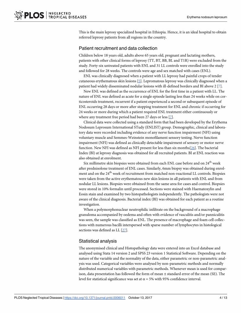
About 96% individuals had nodular cutaneous lesions, about two-third had subcutaneous
nodules and a quarter of patients had scar. While one-third of the patients had ulcerated
Table 1. Demographic and clinical characteristics of study subjects at enrolment.
Variables ENL (n = 46) n (%) LL (n = 31) n (%)
Sex Male 31 (67.4) 23 (74.2)
Female 15 (32.6) 8 (25.8)
Median age in years (range) group 27.5 (18–56) 25.0 (18–60)
Median age in years (range) Male 28 (18–56) 26.0 (18–60)
Median age in years (range) Female 26.7 (18–35) 21.0 (18–30)
MDT status No previous MDT 10 (21.7) 22 (71.0)
Current 9 (19.6) 8 (25.8)
Completed 27 (58.7) 1 (3.2)
HIV status Positive 0 (0.0) 0 (0.0)
Negative 46 (100.0) 31 (100.0)
Duration of current ENL symptom (Episode) Mean ± SE [days] 6.8 ±0.491 (range: 1–15) -
Clinical status at recruitment
ENL type Acute 23 (50.0) -
Recurrent 5 (10.9) -
Chronic 18 (39.1) -
LL type New - 23 (74.2)
Relapse - 5 (16.1)
Defaulter - 3 (9.7)
BI at diagnosis, Mean ± SE (range)
ENL Acute 3.9 ±0.205 (2–6) -
Recurrent 4.9 ±0.409 (4–6) -
Chronic 3.7 ±0.103(3–4) -
LL Untreated (new) 4.1 ±0.259 (2–6)
Relapse 4.2 ±0.330 (4–5)
Defaulter 4.9 ±0.150 (4–5)
https://doi.org/10.1371/journal.pntd.0006011.t001
Erythema nodosum leprosum
PLOS Neglected Tropical Diseases | https://doi.org/10.1371/journal.pntd.0006011 October 13, 2017 5 / 13

lesions, only 4% had necrotic lesions. Eight patients (17.3%) had vesicles, bullae or pustular
lesions (Fig 3).
In most patients with ENL (73.9%), the number of skin lesions recorded at the time of enrol-
ment was between 11 to 50. Few patients had five or less skin lesions. Almost all patients (97.8%)
had skin lesions on the upper limbs. Many patients also had skin lesions on the lower limbs
(95.7%) or on the head and neck (63.0%). Half of the patients reported reduced nerve sensation.
Paraesthesia and hyperaesthesia were reported by 13% and 23.9% of patients respectively (Table 2).
More than half (52.2%) of patients with ENL had old nerve function impairment (NFI)
while 13% had new NFI at the time of enrolment. Facial oedema was reported in 56.5% of the
patients with ENL and nearly half (47.8%) of the patients had oedema on their lower limbs.
Other organs involved in the patients with ENL were small joint arthritis (28.3%), large joint
arthritis (15.2%), conjunctivitis (4.3%), lagophthalmos (2.2%), scleritis (8.7%), lymph node
(15.2%) and dactylitis (2.2%) (Table 2).
Histopathological features of study subjects
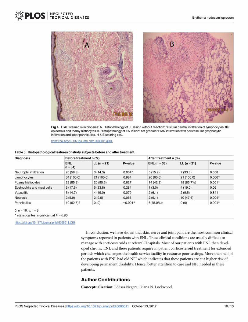
Paraffin- embedded sections of skin biopsy samples from ENL and LL lesions were examined
by a histopathologist (Fig 4). Neutrophils infiltration was noted more ENL lesions (58.9%)
than LL lesions (14.3%) before treatment (P = 0.004). Lymphocytes infiltration was recorded
in all ENL and LL lesions. Foamy histiocytes were more frequently seen in LL lesions (95.3%)
than in ENL lesions (85.3%) although the difference was not statistically significant at enrol-
ment. After 24 weeks treatment of ENL, the percentage of foamy histiocytes was significantly
decreased in ENL cases (42.2%) compared to LL cases (85.7%) (p = 0.001). Panniculitis was
diagnosed in 62.5% of lesions from patients with ENL reactions. After 24 weeks of ENL treat-
ment, neutrophils infiltration was noted in 5 biopsies from patients with ENL reactions, lym-
phocytes infiltration was seen in 20 biopsies of patients with ENL (Table 3).
Discussion
The number of male patients with ENL recruited to the study was twice the number of female
patients and similar to a five-year retrospective data (2008–2013) which showed the number of
Fig 1. Location of pain in the patients with ENL. *value is among 31 males.
https://doi.org/10.1371/journal.pntd.0006011.g001
Erythema nodosum leprosum
PLOS Neglected Tropical Diseases | https://doi.org/10.1371/journal.pntd.0006011 October 13, 2017 6 / 13

male to female ratio to be 1.7:1 [7]. In our study, the median age for male and female patients
with ENL was 28.0 and 26.7 years respectively. Both male and female patients with ENL were
relatively older than the LL patient controls (median age: male = 26 years, female = 21 years).
The slight difference in median age between the two groups could be explained by natural the
course of the disease. Patients usually develop ENL reaction after having either LL or BL clini-
cal forms for some time. Interestingly, the age range of females in both groups was relatively
narrow (18–35 years) compared to males (18–60 years) indicating that either younger females
are more likely to have access to health institutions for various reasons than older females in
low-income countries where health facilities are relatively inadequate[28] or ENL is relatively
common among younger females of child bearing age due to various biological reasons [29–
31].
Our data confirm that a significant proportion of cases had chronic ENL (39%). This
implies that these patients require, in our setting, corticosteroid treatment for extended peri-
ods, often at high doses. . . But high doses of corticosteroids do not always control the inflam-
mation and also pose life-threatening risks for patients [9, 32, 33]. Chronic ENL cases are a
burden to referral hospitals in these resource poor settings. as well as to their communities. A
study in rural India has shown that families with at least one ENL case incur loss of more than
40% of total household income compared to families without ENL case due to out of pocket
expenditure for treatment-seeking (direct cost) and loss of income resulting from reduced pro-
ductivity (earning potential) of household members (indirect cost). This implies that house-
holds affected by ENL face significant economic burden and are at risk of being pushed
further into poverty [34].
Fig 2. Symptoms other than pain in patients with ENL.
https://doi.org/10.1371/journal.pntd.0006011.g002
Erythema nodosum leprosum
PLOS Neglected Tropical Diseases | https://doi.org/10.1371/journal.pntd.0006011 October 13, 2017 7 / 13
In this study, several cutaneous manifestations of ENL were documented highlighting the
heterogeneous nature of ENL clinical manifestation. Pain was was a symptom reported by 98%
of the patients. Most patients had skin pain (80.4%), nerve pain (73.9%), joint pain (71.7%)
and bone pain (69.2%). The most frequent site of pain due to ENL in our study was the skin
which is explained by the fact that 95% of patients with ENL had skin lesions. Our finding is in
agreement with a previous report [7]. Bone pain was reported in two-third of our study
patients which is higher than the previous report [7]. The difference between the two studies is
likely due to the retrospective nature of the previous study which was not reliant on case note
recording unlike the current study.
The nerve function impairment (NFI) was reported in 65% of our study patients, which
was higher than the 51.3% NFI in six countries as reported by Walker et al [35]. Among the
65% of patients reporting NFI, 80% of them had old NFI. This highlights the prevalence of NFI
in patients with ENL the high risk of developing permanent disability. A study by Santos San-
tos, de Mendonca Neto [36], in northern Brazil had identified NFI and leprosy reactions as the
main risk factors associated with the development of disability in leprosy patients. The same
authors reported that NFI was strongly associated with physical disability in children under 15
[37]. In our study, 50% of patients with ENL had WHO disability grade-1 (G1D) while 4.3%
had Grade- 2 disability (G2D). The proportion of grade 2 disability was lower than the national
figure (10.2%) in 2014 [38].
Histopathologically, neutrophil infiltration was noted in 58.8% of patients with ENL com-
pared to 14.3% in LL controls before treatment. This confirms that a neutrophilic infiltration
cannot be used as the sole histological marker for ENL The absence of neutrophil infiltration
has been reported in 36% of ENL skin lesions in Pakistani patients who had classical signs and
symptoms of ENL[11]. Similarly, a cross-sectional study on the histological features of leprosy
Fig 3. Frequency of the different skin lesions in patients with ENL.
https://doi.org/10.1371/journal.pntd.0006011.g003
Erythema nodosum leprosum
PLOS Neglected Tropical Diseases | https://doi.org/10.1371/journal.pntd.0006011 October 13, 2017 8 / 13

reactions in Indian patients by Sarita, Muhammed [39] showed that 43% ENL skin lesions did
not have histological evidence of neutrophil infiltration. Our findings agree with these two
studies. Previous studies by others [40–42], reported finding neutrophil infiltration in all ENL
lesions. The varying reports of neutrophil infiltration in ENL lesions could be attributed to sev-
eral factors. If the definition of ENL includes the presence of neutrophils in the case definition
then all cases will have it, as did Aldhe et al who investigated the presence of cellular neutrophil
infiltration on histologically confirmed ENL cases [42]. Delay between the onset of reaction
and the timing of obtaining the biopsy in those without neutrophilic infiltrate, as dermal
oedema may be missed in older reactional lesions could cause these differences. Discordance
between pathologists and standard operating procedures (SOPs) of slide preparations are also
potential areas that should be further investigated to evaluate their impact on the findings of
neutrophil infiltration in tissue sections. Previous reports suggested vasculitis as part of ENL
reaction commonly seen in Indian patients [43], only 5(14.7%) of our patients had evidence of
vasculitis. Similar observations had been made by Sarita et al and Adhe et al [39, 44].
Inclusion of a large number of patients with ENL and LL controls was one of the strengths
of this study. The other strength of the study had been that clinical data were obtained from
each patient three times unlike the previous cross-sectional studies. A weakness of the study is
that there may have been biased recruitment because of the need to have good follow–up of
patients.
Table 2. Other clinical pictures in patients with ENL at enrolment.
Number of skin lesions number %
<5 3 6.5
6–10 4 8.7
11–20 18 39.1
21–50 16 34.8
>50 5 10.9
Location of skin lesions Head/neck 29 63.0
Trunk 18 39.1
Upper limbs 45 97.8
Lower limbs 44 95.7
Nerve symptoms Reduced Sensation 23 50.0
Paraesthesia 6 13.0
Hyperaesthesia 11 23.9
Weakness 35 76.1
Nerve function impairment (NFI) Old 24 52.2
New 6 13.0
Organs involved in ENL
Oedema Hand 26 56.5
Face 16 34.8
Lower limbs 22 47.8
Dactylitis 1 2.2
Large joint Arthritis 7 15.2
Small joint arthritis 13 28.3
Conjunctivitis 2 4.3
Lagophthalmos 1 2.2
Scleritis 4 8.7
Lymph node 7 15.2
https://doi.org/10.1371/journal.pntd.0006011.t002
Erythema nodosum leprosum
PLOS Neglected Tropical Diseases | https://doi.org/10.1371/journal.pntd.0006011 October 13, 2017 9 / 13
In conclusion, we have shown that skin, nerve and joint pain are the most common clinical
symptoms reported in patients with ENL. These clinical conditions are usually difficult to
manage with corticosteroids at referral Hospitals. Most of our patients with ENL then devel-
oped chronic ENL and these patients require in patient corticosteroid treatment for extended
periods which challenges the health service facility in resource poor settings. More than half of
the patients with ENL had old NFI which indicates that these patients are at a higher risk of
developing permanent disability. Hence, better attention to care and NFI needed in these
patients.
Author Contributions
Conceptualization: Edessa Negera, Diana N. Lockwood.
Fig 4. H &E stained skin biopsies: A. Histopathology of LL lesion without reaction: reticular dermal infiltration of lymphocytes, flat
epidermis and foamy histiocytes.B. Histopathology of EN lesion: flat granular PMN infiltration with perivascular lymphocytic
infiltration and lobar panniculitis. H & E staining x40.
https://doi.org/10.1371/journal.pntd.0006011.g004
Table 3. Histopathological features of study subjects before and after treatment.
Diagnosis Before treatment n (%) After treatment n (%)
ENL
n = 34)
LL (n = 21) P-value ENL (n = 33) LL (n = 21) P-value
Neutrophil infiltration 20 (58.8) 3 (14.3) 0.004* 5 (15.2) 7 (33.3) 0.058
Lymphocytes 34 (100.0) 21 (100.0) 0.984 20 (60.6) 21 (100.0) 0.006*
Foamy histiocytes 29 (85.3) 20 (95.3) 0.627 14 (42.2) 18 (85.7%) 0.001*
Eosinophils and mast cells 6 (17.6) 5 (23.8) 0.284 1 (3.0) 4 (19.0) 0.06
Vasculitis 5 (14.7) 4 (19.0) 0.079 2 (6.1) 2 (9.5) 0.841
Necrosis 2 (5.9) 2 (9.5) 0.068 2 (6.1) 10 (47.6) 0.004*
Panniculitis 10 (62.5)δ 0 (0) <0.001* 6(75.0%)ε 0 (0) 0.001*
δ; n = 16. ε; n = 8.
* statistical test significant at P = 0.05.
https://doi.org/10.1371/journal.pntd.0006011.t003
Erythema nodosum leprosum
PLOS Neglected Tropical Diseases | https://doi.org/10.1371/journal.pntd.0006011 October 13, 2017 10 / 13

Data curation: Edessa Negera, Stephen L. Walker, Selfu Girma, Shimelis N. Doni, Saba M.
Lambert, Munir H. Idriss, Yohanis Tsegay, Hazel M. Dockrell.
Formal analysis: Edessa Negera.
Funding acquisition: Edessa Negera, Diana N. Lockwood.
Investigation: Edessa Negera, Selfu Girma, Shimelis N. Doni, Degafe Tsegaye, Saba M. Lam-
bert, Yohanis Tsegay.
Methodology: Edessa Negera, Stephen L. Walker, Degafe Tsegaye, Munir H. Idriss, Hazel M.
Dockrell, Abraham Aseffa, Diana N. Lockwood.
Project administration: Edessa Negera, Diana N. Lockwood.
Resources: Abraham Aseffa, Diana N. Lockwood.
Software: Edessa Negera.
Supervision: Hazel M. Dockrell, Abraham Aseffa, Diana N. Lockwood.
Validation: Edessa Negera, Shimelis N. Doni, Saba M. Lambert, Munir H. Idriss, Diana N.
Lockwood.
Writing – original draft: Edessa Negera.
Writing – review & editing: Edessa Negera, Stephen L. Walker, Selfu Girma, Degafe Tsegaye,
Hazel M. Dockrell, Abraham Aseffa, Diana N. Lockwood.
References
1. Reich C.V., Leprosy: cause, transmission, and a new theory of pathogenesis. Rev Infect Dis, 1987. 9
(3): p. 590–4. PMID: 3299638
2. Bhat R.M. and Prakash C., Leprosy: An Overview of Pathophysiology. Interdisciplinary Perspectives on
Infectious Diseases, 2012. 2012: p. 6.
3. Ridley D.S. and Jopling W.H., Classification of Leprosy according to immmunity: Five group system.
Inter J lepr other Micobacterial Diseases, 1966. 34(3).
4. Kumar B., Dogra S., and Kaur I., Epidemiological characteristics of leprosy reactions: 15 years experi-
ence from north India. Int J Lepr Other Mycobact Dis, 2004. 72(2): p. 125–33. https://doi.org/10.1489/
1544-581X(2004)072<0125:ECOLRY>2.0.CO;2 PMID: 15301592
5. Pocaterra L., et al., Clinical Course of Erythema Nodosum Leprosum: An 11-year Chohort Study in
Hyderabad, India. Am. J. Trop. Med. Hyg., 2006. 74(5): p. 868–879. PMID: 16687695
6. Van Veen N., et al., Interventions for erythema nodosum leprosum (Review). Cochrane Database of
Systematic Reviews, 2009. 3: p. No.: CD006949.
7. Walker S.L., et al., The Mortality Associated with Erythema Nodosum Leprosum in Ethiopia: A Retro-
spective Hospital-Based Study. PLoS Negl Trop Dis, 2014. 8(3): p. e2690. https://doi.org/10.1371/
journal.pntd.0002690 PMID: 24625394
8. Walker S.L. and Lockwood D.N.J., The clinical and immunological features of leprosy. British Medical
Bulletin, 2006. 77-78(1): p. 103–121.
9. Walker S.L., et al., ENLIST 1: An International Multi-centre Cross-sectional Study of the Clinical Fea-
tures of Erythema Nodosum Leprosum. PLoS Negl Trop Dis, 2015. 9(9): p. e0004065. https://doi.org/
10.1371/journal.pntd.0004065 PMID: 26351858
10. Mabalay M.C., et al., The Histopathology and Histochemistry of Erythema Nodosum Leprosum. Int J
Lepr, 1965. 33: p. 28–49. PMID: 14282354
11. Hussain R., et al., Clinical and histological discrepancies in diagnosis of ENL reactions classified by
assessment of acute phase proteins SAA and CRP. Int J Lepr Other Mycobact Dis., 1995. 63(2): p.
222–230. PMID: 7602217
12. Wemambu S.N.C., et al., Erythema Nodosum Leprosum: A clinical manifestation of the Arthus phenom-
enon. The Lancet, 1969: p. 933–935.
Erythema nodosum leprosum
PLOS Neglected Tropical Diseases | https://doi.org/10.1371/journal.pntd.0006011 October 13, 2017 11 / 13

13. Waters M.F.R., Turk J.L., and Wemambu S.N.C., Mechanisms of Reactions in Leprsoy International
Journal of Leprosy, 1971. 39(2): p. 417–128.
14. Anthony J., Vaidya M.C., and Dasgupta A., Immunoglobulin deposits in Erythema Nodosum Leprosum
(ENL). Hansen. Int., 1978. 3(1): p. 12–17.
15. Rojas-Espinosa O., Mendez-Navarrete I., and Estrada-Parra S., Presence of C1q-reactive immune
complexes in patients with leprosy. Clin Exp Immunol, 1972. 12(2): p. 215–23. PMID: 4630778
16. Bjorvatn B., et al., Immune complexes and complement hypercatabolism in patients with leprosy. Clin.
exp. Immunol., 1976. 26: p. 388–396. PMID: 1009681
17. Wager O., et al., Circulating complexes in leprosy studied by the platelet aggregation test. The platelet
aggregation test and its relation to the Rubino test and other sero-immunological parameters in 135
patients with leprosy. Clin Exp Immunol., 1978. 34(3): p. 326–337. PMID: 369750
18. Rea T.H., et al., Peripheral blood T lymphocyte subsets in leprosy. Int J Lepr Other Mycobact Dis., 1984
52.(3): p. 311–317. PMID: 6332790
19. Modlin R., et al., In situ characterization of T lymphocyte subsets in the reactional states of leprosy.
Clin. exp. Immunol., 1983. 53: p. 17–24. PMID: 6223731
20. Bach M.A., et al., Mechanisms of T-cell Unresponsiveness in Leprosy. Ann. Immunol 1983. 134(75–
84).
21. Wallach D., Cottenot F., and Bach M.A., Imbalances in T Cell Subpopulations in Lepromatous Leprosy.
INTERNATIONAL JOURNAL OF LEPROSY, 1982. 50 (3).
22. Mshana R.N., et al., Immunohistological studies of skin biopsies from patients with lepromatous leprosy.
Journal of Clinical Immunology, 1983. 3(1): p. 22–29 PMID: 6338025
23. Narayanan R., et al., Differences in predominant T cell phenotypes and distribution pattern in reactional
lesions of tuberculoid and lepromatous leprosy. Clin. exp. Immunol 1984. 55(3): p. 623–628. PMID:
6423326
24. Laal S., Bhutani L.K., and nath I., Natural Emergence of Antigen-Reactive T Cells in Lepromatous Lep-
rosy Patients during Erythema Nodosum Leprosum. Infection and Immunity, 1985 p. 887–892. PMID:
2933339
25. Walker S.L., et al., The mortality associated with erythema nodosum leprosum in Ethiopia: a retrospec-
tive hospital-based study. PLoS Negl Trop Dis, 2014. 8(3): p. e2690. https://doi.org/10.1371/journal.
pntd.0002690 PMID: 24625394
26. Lambert S.M., et al., A Randomized Controlled Double Blind Trial of Ciclosporin versus Prednisolone in
the Management of Leprosy Patients with New Type 1 Reaction, in Ethiopia. PLoS Negl Trop Dis, 2016.
10(4): p. e0004502. https://doi.org/10.1371/journal.pntd.0004502 PMID: 27046330
27. Lockwood D.N.J., et al., Comparing the Clinical and Histological Diagnosis of Leprosy and Leprosy
Reactions in the INFIR Cohort of Indian Patients with Multibacillary Leprosy. PLoS Negl Trop Dis, 2012.
6(6): p. e1702. https://doi.org/10.1371/journal.pntd.0001702 PMID: 22745841
28. WHO, Enhanced Global Strategy for Further Reducing the Disease Burden due to Leprosy (Plan
Period: 2011–20. Geneva, Switzerland 2009 1(3): p. 42.
29. Duncan M.E. and Pearson J.M., The association of pregnancy and leprosy—III. Erythema nodosum
leprosum in pregnancy and lactation. Lepr Rev, 1984. 55(2): p. 129–42. PMID: 6748844
30. Lockwood D.N. and Sinha H.H., Pregnancy and leprosy: a comprehensive literature review. Int J Lepr
Other Mycobact Dis, 1999. 67(1): p. 6–12. PMID: 10407623
31. Motta A.C., et al., Leprosy reactions: coinfections as a possible risk factor. Clinics (Sao Paulo), 2012.
67(10): p. 1145–8.
32. Girdhar B.K., Immuno pharmacology of drugs used in leprosy reactions. Indian J Dermatol Venereol
Leprol, 1990. 56(5): p. 354–63.
33. Walker S.L., Waters M.F., and Lockwood D.N., The role of thalidomide in the management of erythema
nodosum leprosum. Lepr Rev, 2007. 78(3): p. 197–215. PMID: 18035771
34. Chandler D.J., et al., Household Costs of Leprosy Reactions (ENL) in Rural India. PLoS Neglected
Tropical Diseases, 2015. 9(1): p. e0003431. https://doi.org/10.1371/journal.pntd.0003431 PMID:
25590638
35. Walker S.L. and Lockwood D.N., Erythema nodosum leprosum research: ENLISTing support. Leprosy
review, 2015. 86(4).
36. Santos V.S., et al., Evaluation of agreement between clinical and histopathological data for classifying
leprosy. International Journal of Infectious Diseases, 2013. 17(3): p. e189–e192. https://doi.org/10.
1016/j.ijid.2012.10.003 PMID: 23158973
37. Santos V.S., et al., Clinical variables associated with disability in leprosy cases in northeast Brazil.
2015. Vol. 9. 2015.
Erythema nodosum leprosum
PLOS Neglected Tropical Diseases | https://doi.org/10.1371/journal.pntd.0006011 October 13, 2017 12 / 13

38. Baye S., Leprosy in Ethiopia: Epidemiological trends from 2000 to 2011. Advances in life scienes and
health 2015. 2(1): p. 31–44.
39. Sarita S., et al., A study on histological features of lepra reactions in patients attending the Dermatology
Department of the Government Medical College, Calicut, Kerala, India. Lepr Rev, 2013. 84(1): p. 51–
64. PMID: 23741882
40. Ridley D.S., Rea T.H., and McAdam K.P., The histology of erythema nodosum leprosum. Variant forms
in New Guineans and other ethnic groups. Lepr Rev, 1981. 52(1): p. 65–78. PMID: 7242219
41. Jayalakshmi P., Ganesapillai T., and Ganesan J., Erythema nodosum leprosum in malaysians. Int. J.
Lepr. Other Mycobact. Dis., 1995. 63(1): p. 109–11. PMID: 7730709
42. Adhe V., Dongre A., and Khopkar U., A retrospective analysis of histopathology of 64 cases of lepra
reactions. Indian J Dermatol, 2012. 57(2): p. 114–7. https://doi.org/10.4103/0019-5154.94278 PMID:
22615507
43. Sehgal V.N., et al., The histopathology of type I (lepra) and type II (ENL) reactions in leprosy. Indian J
Lepr, 1986. 58(2): p. 240–3. PMID: 3805796
44. Adhe V., Dongre A., and Khopkar U., A Retrospective Analysis of Histopathology of 64 Cases of Lepra
Reactions. Indian Journal of Dermatology, 2012. 57(2): p. 114–117. https://doi.org/10.4103/0019-
5154.94278 PMID: 22615507
Erythema nodosum leprosum
PLOS Neglected Tropical Diseases | https://doi.org/10.1371/journal.pntd.0006011 October 13, 2017 13 / 13