erythema nodosum as the first presenting complaint of...
TRANSCRIPT

Hong Kong Journal of Emergency Medicine
Erythema nodosum as the first presenting complaint of asymptomatic
pulmonary tuberculosis
SK Wong, SD Yeung
Correspondence to:Wong Suk Kwan, MBChB
Alice Ho Miu Ling Nethersole Hospital, Accident andEmergency Department, Tai Po, N.T., Hong KongEmail: [email protected]
Yeung Sai Dat, FHKAM(Emergency Medicine)
Tender erythematous swelling on the front of legs, erythema nodosum, is a common symptom that resultin patient seeking medical attention. It has multiple aetiologies. Tuberculosis is a recognised cause. In here,we describe a case of erythema nodosum as the presenting complaint of undiagnosed asymptomaticpulmonary tuberculosis. (Hong Kong j.emerg.med. 2001;8:166-168)
Keywords: chest X-ray, erythema nodosum, panniculitis, pulmonary tuberculosis
Introduction
Erythema nodosum is a panniculitis usually on theshins, with occasional spread to the thighs or arms.1
It is a self-limiting disease characterised by lesionsthat evolve from inflammatory, painful and tender,nonulcerating, nonscarring, cutaneous and subcutaneousred nodules, which are typically multiple and bilateral,becoming bruised in appearance, and finally resolvingcompletely.2 Although it may be idiopathic, it is alsoassociated with a wide spectrum of systemic diseases,infections or drugs. It is important to find out theunderlying conditions.
Case
A 19-year-old female presented to our emergencydepartment with complaint of painful skin rash overboth legs for one month. She also had bilateral legswelling and ankle pain. There was no history oftrauma. She enjoyed good past health. She was asmoker and social drinker. She did not take any
medications, including oral contraceptive pills, beforethe onset of skin rash. She had consulted private doctorswith transient improvement. She did not have cough,haemoptysis or fever. The appetite was good and therewas no weight loss. She did not have abdominal painand the bowel habit was normal. There was no urinarysymptom.
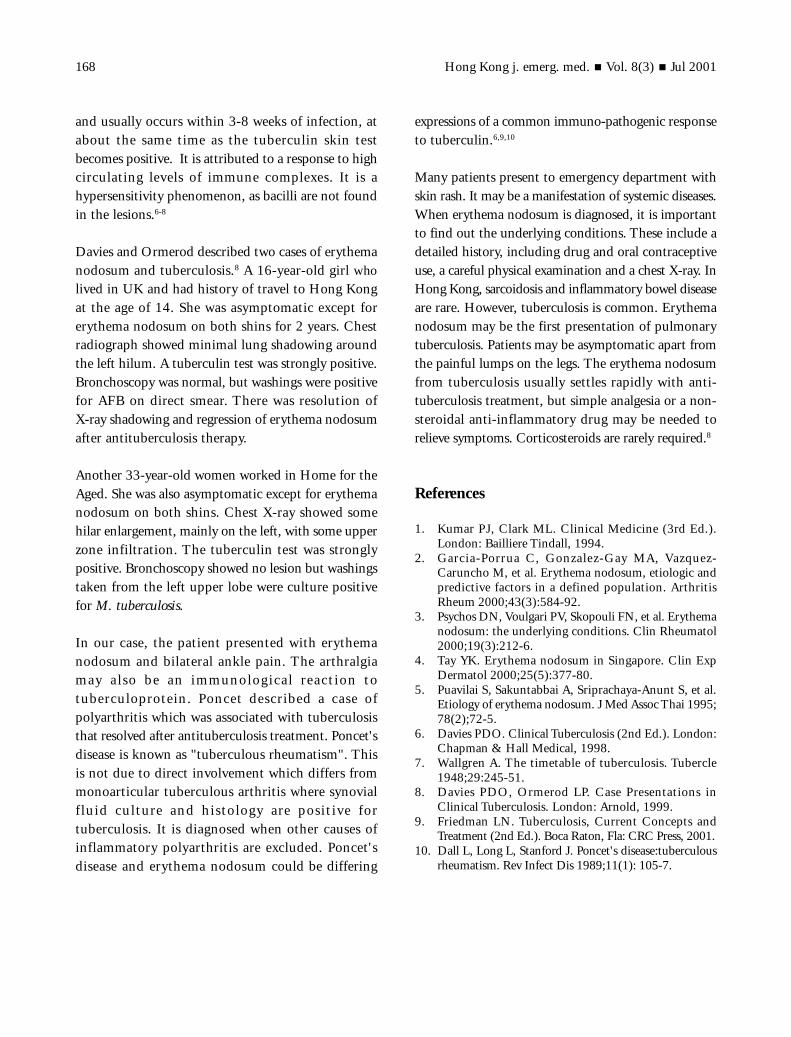
On examinat ion, the genera l condi t ion wassatisfactory. She was afebrile, blood pressure was 109/81 mmHg, pulse 99 per minute. There were multipleerythematous papular-nodular, mild tender skinlesions over both shins, compatible with erythemanodosum. There was mild pitting oedema over bothankles. The range of movement of the ankles wasnormal. There was no enlarged lymph node. Thethroat was clear. The cardiovascular, chest andabdominal examination were unremarkable. Chest X-ray (Figure 1) showed left upper zone hazziness,suggestive of tuberculosis. She was admitted to themedical ward.
During admission, there was no fever, cough orhaemoptysis. Sputum was negative for acid-fast bacilli(AFB) smear. White cell count was normal. ESR was55. Renal and liver function test was normal. Anti-nuclear antibodies titre was 40. Bronchoscopy wasdone and no abnormality was detected. Bronchialaspirate and broncho-alveolar lavage were AFB smearnegative. In view of the radiological evidence of

Wong et al./Erythema nodosum & pulmonary tuberculosis 167
tuberculosis, anti-tuberculosis drugs were started. Shewas then discharged and referred to Chest Clinic forfollow up. AFB culture of sputum, bronchial aspirateand broncho-alveolar lavage was later positive forMycobacterium tuberculosis, which was sensitive toisoniazid, rifamipicin, ethambutol, streptomycin,pyrazinamide and ofloxacin.
Discussion
Erythema nodosum is a panniculitis that producespainful nodules on the shins, and less commonly onthigh and forearm. It is a reaction pattern to infectionand sometimes to drugs. Adult females are principallyaffected. Malaise, fever and arthralgia were common.Histological features suggest that this is an immunologicalreaction. Immune complex deposition within dermalvessels is an important component in the productionof the symptom complex.1
Erythema nodosum is classified as idiopathic orsecondary to other diseases. Some causes of erythemanodosum are listed in Table 1.
The causes of erythema nodosum are differentaccording to the disease pattern of the region. In a
retrospective study of 106 patients (82 females) inSpain by Carlos et al,2 35% was idiopathic. For thesecondary erythema nodosum, 34% was due toinfections (of which 7% was group A Streptococcus βhaemolyt i cus , 5% was tuberculos i s , 13% wasnonstreptococcal upper respiratory tract infection) and22% was due to sarcoidosis. The other causes weredrugs, Sweet's disease, Behcet's disease. Similar resultswere obtained by Psychos et al,3 in which 132erythema nodosum patients (110 females) wereinvestigated in a prospective study in Greece. Thirtyfive percent was idiopathic, while sarcoidosis wasrevealed in 28%, infections in 17.3% and tuberculosisin 1.5%. Other causes were Behcet's syndrome,pregnancy, oral contraceptives.
In Singapore, a retrospective study of 75 patients witherythema nodosum (65 females) found that 60% ofcases were idiopathic. 26% were due to viralrespiratory tract infections and streptococcalpharyngitis, 3% were tuberculosis, and 4% werepregnancy. However, sarcoidosis and inflammatorybowel disease were uncommon.4 This same pattern wasalso revealed in Thailand. Puavilai et al5 studied 100patients (88 females) with erythema nodosum. 72%were idiopathic, 12% had tuberculosis, 7% were dueto drugs, 6% had streptococcal infection and 3% hadBehcet's disease.
Erythema nodosum may be the f i r s t s ign oftuberculosis. It is associated with primary tuberculosis
Table 1. Some causes of erythema nodosum.
Drugs SulphonamidesOral contraceptive pillsPenicillin
Systemic diseases SarcoidosisInflammatory bowel disease
Infection StreptococciTuberculosisLeprosyChlamydiaHistoplasmosisCoccidioidomycosis
Pregnancy
Figure 1. CXR showing pulmonary fuberculosis in apatient with erthyema nodosum.
Hong Kong j. emerg. med. Vol. 8(3) Jul 2001168
and usually occurs within 3-8 weeks of infection, atabout the same time as the tuberculin skin testbecomes positive. It is attributed to a response to highcirculating levels of immune complexes. It is ahypersensitivity phenomenon, as bacilli are not foundin the lesions.6-8
Davies and Ormerod described two cases of erythemanodosum and tuberculosis.8 A 16-year-old girl wholived in UK and had history of travel to Hong Kongat the age of 14. She was asymptomatic except forerythema nodosum on both shins for 2 years. Chestradiograph showed minimal lung shadowing aroundthe left hilum. A tuberculin test was strongly positive.Bronchoscopy was normal, but washings were positivefor AFB on direct smear. There was resolution ofX-ray shadowing and regression of erythema nodosumafter antituberculosis therapy.
Another 33-year-old women worked in Home for theAged. She was also asymptomatic except for erythemanodosum on both shins. Chest X-ray showed somehilar enlargement, mainly on the left, with some upperzone infiltration. The tuberculin test was stronglypositive. Bronchoscopy showed no lesion but washingstaken from the left upper lobe were culture positivefor M. tuberculosis.
In our case, the patient presented with erythemanodosum and bilateral ankle pain. The arthralgiamay a l so be an immuno log i ca l re ac t ion totuberculoprotein. Poncet descr ibed a case ofpolyarthritis which was associated with tuberculosisthat resolved after antituberculosis treatment. Poncet'sdisease is known as "tuberculous rheumatism". Thisis not due to direct involvement which differs frommonoarticular tuberculous arthritis where synovialf lu id cu l ture and h i s to logy a re pos i t ive fortuberculosis. It is diagnosed when other causes ofinflammatory polyarthritis are excluded. Poncet'sdisease and erythema nodosum could be differing
expressions of a common immuno-pathogenic responseto tuberculin.6,9,10
Many patients present to emergency department withskin rash. It may be a manifestation of systemic diseases.When erythema nodosum is diagnosed, it is importantto find out the underlying conditions. These include adetailed history, including drug and oral contraceptiveuse, a careful physical examination and a chest X-ray. InHong Kong, sarcoidosis and inflammatory bowel diseaseare rare. However, tuberculosis is common. Erythemanodosum may be the first presentation of pulmonarytuberculosis. Patients may be asymptomatic apart fromthe painful lumps on the legs. The erythema nodosumfrom tuberculosis usually settles rapidly with anti-tuberculosis treatment, but simple analgesia or a non-steroidal anti-inflammatory drug may be needed torelieve symptoms. Corticosteroids are rarely required.8
References
1. Kumar PJ, Clark ML. Clinical Medicine (3rd Ed.).London: Bailliere Tindall, 1994.
2. Garcia-Porrua C, Gonzalez-Gay MA, Vazquez-Caruncho M, et al. Erythema nodosum, etiologic andpredictive factors in a defined population. ArthritisRheum 2000;43(3):584-92.
3. Psychos DN, Voulgari PV, Skopouli FN, et al. Erythemanodosum: the underlying conditions. Clin Rheumatol2000;19(3):212-6.
4. Tay YK. Erythema nodosum in Singapore. Clin ExpDermatol 2000;25(5):377-80.
5. Puavilai S, Sakuntabbai A, Sriprachaya-Anunt S, et al.Etiology of erythema nodosum. J Med Assoc Thai 1995;78(2);72-5.
6. Davies PDO. Clinical Tuberculosis (2nd Ed.). London:Chapman & Hall Medical, 1998.
7. Wallgren A. The timetable of tuberculosis. Tubercle1948;29:245-51.
8. Davies PDO, Ormerod LP. Case Presentations inClinical Tuberculosis. London: Arnold, 1999.
9. Friedman LN. Tuberculosis, Current Concepts andTreatment (2nd Ed.). Boca Raton, Fla: CRC Press, 2001.
10. Dall L, Long L, Stanford J. Poncet's disease:tuberculousrheumatism. Rev Infect Dis 1989;11(1): 105-7.