physiology and function of the tight...
TRANSCRIPT

Physiology and Function of the Tight Junction
James M. Anderson1 and Christina M. Van Itallie2
1Department of Cell and Molecular Physiology, University of North Carolina at Chapel Hill,6312 MBRB, Chapel Hill, North Carolina 27599-7545
2Department of Medicine, Division of Gastroenterology and Hepatology, University of North Carolina atChapel Hill, Chapel Hill, North Carolina 27599-7545
Correspondence: [email protected]
Understanding of tight junctions has evolved from their historical perception as inert solutebarriers to recognition of their physiological and biochemical complexity. Many proteinsare specifically localized to tight junctions, including cytoplasmic actin-binding proteinsand adhesive transmembrane proteins. Among the latter are claudins, which are criticalbarrier proteins. Current information suggests that the paracellular barrier is most usefullymodeled as having two physiologic components: a system of charge-selective small pores,4 A in radius, and a second pathway created by larger discontinuities in the barrier,lacking charge or size discrimination. The first pathway is influenced by claudin expressionpatterns and the second is likely controlled by different proteins and signals. Recent infor-mation on claudin function and disease-causing mutations have led to a more completeunderstanding of their role in barrier formation, but progress is impeded by lack of highresolution structural information.
Tight junctions form the continuous intercel-lular barrier between epithelial cells, which
is required to separate tissue spaces and regulateselective movement of solutes across the epi-thelium. Although there are now .40 proteins(Schneeberger and Lynch 2004; Yamazaki et al.2008) identified within the tight junction,the claudin family of transmembrane proteins,named from the Latin claudere to close, hasemerged as the most critical for defining tightjunction selectivity. Here, we review evidencethat claudins regulate permselectivity (includ-ing size, electrical resistance, and ionic chargepreference) derived from studies in culturedepithelial cell models and the phenotypes of
knockout mice and human mutants. We high-light the physiologic relevance of selectivitybut only briefly discuss how it might be physio-logically regulated and altered in pathologicsituations. We develop the perspective thatthe barrier is usefully described as havingtwo pathways: first a system of charge-selectiveclaudin-based pores that are 4 A in radius anda second pathway created by larger discontinu-ities in the barrier and that lacks charge andsize discrimination. The two pathways may becontrolled by different proteins and signals.This article focuses on claudins and physiologyand is meant to be read as a companion tothe article in this collection contributed by
Editors: W. James Nelson and Elaine Fuchs
Additional Perspectives on Cell Junctions available at www.cshperspectives.org
Copyright # 2009 Cold Spring Harbor Laboratory Press; all rights reserved; doi: 10.1101/cshperspect.a002584
Cite this article as Cold Spring Harb Perspect Biol 2009;1:a002584
1
on August 11, 2019 - Published by Cold Spring Harbor Laboratory Press http://cshperspectives.cshlp.org/Downloaded from

M. Furuse, which focuses on the molecularstructure, proteins, and cell biology of thetight junction (Furuse 2009). The reader isalso referred to comprehensive reviews onphysiology (Diamond 1978; Powell 1981; VanItallie and Anderson 2006), pathophysiology(Turner 2006; Schmitz et al. 1999; Nusratet al. 2001), regulation (Tsukita et al. 2008;Gonzalez-Mariscal et al. 2008), and molecularcomponents of the junction (Schneebergerand Lynch 2004; Krause et al. 2008; Gonzalez-Mariscal et al. 2003).
EVOLUTION OF IDEAS ABOUTPARACELLULAR TRANSPORT AND THERELEVANCE OF TIGHT JUNCTIONPERMSELECTIVITY
Explicit description of a sealing contact betweenepithelial cells can first be found in the biologicliterature in the latter part of the 19th century(Cereijido and Anderson 2001). Staining ofepithelial tissues, such as the intestine, withvital dyes revealed a distinct region at theapical end of the lateral cell interspaces referredto as the “terminal bar.” This was thought tobe an absolute barrier preventing anythingfrom passing between cells. By the early 20thcentury, studies began to acknowledge thatsome material, for example macrophages andwater, could cross epithelia through the paracel-lular space.
Our current understanding of how materialselectively crosses epithelia originates with thepioneering work of Hans Ussing and associates,beginning in the 1940s. Ussing addressed thequestion of how sodium ions are moved in adirectional fashion across the epithelium offrog skin, resulting in a steady-state electricalpotential across the epithelium (Koefeld-Johnsen and Ussing 1958; Ussing and Zerahn1951). Their first conceptual breakthrough,the so-called “two membrane model,” statedthat the apical and basal membrane surfaceshad different conductance properties, namely,that sodium enters the cell across the apicalmembrane down its concentration gradient(later shown to be through Na channels)but was transported in an energy-dependent
fashion out the basal lateral surface (latershown to be by the NaK-ATPase). Ironically,they chose to study an epithelium with one ofthe electrically tightest junctions in nature,leading them initially to discount the possibilityof ion movements between cells. Subsequentinconsistencies between their model and thedata led to the realization that Cl2 ions mustbe following Naþ in a passive fashion throughthe tight junction (based on electrical circuitmodeling, they called this the shunt pathway)coupled to the electrical gradient generated byactive Naþ transport (Ussing and Windhager1964). This was the beginning of understandingabout how transcellular and paracellular trans-port are physiologically coupled (Fig. 1).
In the 1960s, investigators began to studytransport across other epithelia, such as the gall-bladder, where the paracellular resistance wasmuch lower than in frog skin (reviewed inDiamond 1977). Because the transepithelialelectrical resistance (TER) was so much lowerthan frog skin, there was initial concern thatthese tissues were damaged during experimen-tal preparation and this explained their lowresistance. Gradually, it was accepted thatintact normal epithelia display a wide range ofelectrical resistances (Table 1). Because the cellmembranes are generally of very high resistance
RBL
RA RTJ
Serosa
MucosaTranscellularpathway
Paracellularpathway
RLIS
Figure 1. Electrical circuit model of the series andparacellular resistances across trans- and para-cellular pathways of an epithelial cell monolayer.Transcellular transport is controlled by transportersin the apical and basolateral surfaces. The resistanceof these series elements is typically much higherthan that of the parallel elements of the paracellularpathway. Thus, the overall resistance of an epi-thelium is defined by RTJ, which is defined by thecomposition of claudins in the tight junction (TJ).
J.M. Anderson and C.M. Van Itallie
2 Cite this article as Cold Spring Harb Perspect Biol 2009;1:a002584
on August 11, 2019 - Published by Cold Spring Harbor Laboratory Press http://cshperspectives.cshlp.org/Downloaded from

(Rapical and Rbasolateral), it is the TJ (RTJ) thatdetermines whether the transepithelial resist-ance is high or low (Fig. 1). Theoretically, thelateral intercellular space (Rlis) could contributea resistance in series with the tight junction,but there is little evidence that this is physio-logically significant.
TER differs by several orders of magnitudebetween so-called “tight” and “leaky” epithelia(Table 1). For example, the epithelium of themammalian proximal tubule is only about6 V†cm2 compared with the toad urinarybladder at 300,000 V†cm2 (Powell 1981; VanItallie and Anderson 2006). The relevance ofTER is that epithelia with “tight” tight junctionscan maintain the high electrochemical gradi-ents produced by active transcellular transport.This configuration is used to produce eitherhighly concentrated or diluted secretions,as in the distal nephron, which can produceurine with osmolarity several fold higher orlower than plasma. In contrast, epithelia with“leaky” tight junctions move large amounts ofisosmotic fluids. A good example is providedby the human gastrointestinal tract, whichsecretes and then reabsorbs about 10 L of fluideach day (Boron and Boulpaep 2005). Most of
the gastrointestinal track is of low TER, exceptin the distal colon where steeper electrochemicalgradients are required to reabsorb NaCl andwater and form solid stool.
Study of leaky epithelia revealed anotherinteresting and variable property, namely thattight junctions (TJs) have ionic charge selectiv-ity. Charge selectivity is most relevant in leakytight junctions where higher amounts of ionsflow. Almost all leaky TJs show a preferencefor Naþ over Cl2, and the permeability ratio(expressed as PNa/PCl) ranges from about 10to 0.1 among different epithelia and experi-mental cultured cell models (Table 1). Thisrepresents only a modest ability to discriminatecompared with membrane ion channels. Forcomparison, some amiloride-sensitive Naþ
channels (ENaC) show a 1000-fold preferencefor Naþ over Kþ, an identically charged ion ofsimilar size (Hille 2001). However, even a10-fold cation to anion discrimination has sig-nificant physiologic implications. For example,epithelia that secrete salt and water initiatedby an apical Cl2 channel (such as CFTR in theairway, lacrimal glands, etc.) are found to havea threefold to 10-fold preference for Naþ overCl2. This allows paracellular passage of Naþ
Table 1. Electrical characteristics of some epithelia and endothelia
Tissuesa Species Rcell V � cm2 Rparacellular V � cm2 PNa/PClb
Proximal tubule dog – 6–7 1.4Gallbladder rabbit 229 21 3.3Duodenum rat – 98 –Jejunum rat 67 51 10.0Ileum rabbit 115 100 2.5Distal colon rabbit 730 385 0.6
mouse surface 132 3,200 –crypt 429 – –
Brain endothelium 1,300Gastric fundus Necturus 2,826 10,573 –Urinary bladder rabbit 160,000 300,000 –
Cell lines c
Caco-2 human colon 125–250 – 3.0LLC-PK1 pig prox. tubule 100 – 0.6MDCK dog 60–4000 – 10.0
aAll values can be found in Powell (Powell 1981) or Crone (Crone and Christensen 1981).bPermeability ratio of Naþ versus Cl2. PNa/PCl in free solute is 0.66. Paracellular pathways with ratios above this value are
more permeable for Naþ than Cl2, i.e., cation-selective.cValues for cell lines are the personal observations of Dr. C. Van Itallie. Modified, with permission, from Van Itallie and
Anderson (2004) (# American Thoracic Society).
Physiology and Function of the Tight Junction
Cite this article as Cold Spring Harb Perspect Biol 2009;1:a002584 3
on August 11, 2019 - Published by Cold Spring Harbor Laboratory Press http://cshperspectives.cshlp.org/Downloaded from

to follow secreted Cl2 while relatively discrimi-nating against back diffusion of the Cl2, whichwould defeat the ability to secrete NaCl andwater.
The TJ was first visualized at the ultrastruc-tural level in 1963 by Farquhar and Palade(Farquhar and Palade 1963). They were ablefor the first time to resolve the apical junctioncomplex (terminal bar) into three morphologi-cally discrete contacts from the apical-mostoccluding or TJ (zonula occludens) followedby the cadherin-based adherens junction andthe more basally positioned desmosomes. Gapjunction plaques are also commonly foundwithin the TJ contacts. The TJ zone is formedby a variable number of close cell–cell contactsor “kisses,” which range from a single continu-ous contact (e.g., some endothelial cells) to ahalf dozen ( jejunum) or dozens of contacts inthe most extreme mammalian example(Sertoli cells). Subsequent visualization of thetight junction in freeze fracture EM imagesrevealed that membrane kisses correspond tocontinuous rows of 10-nm transmembraneparticles, which are presumed to contain anoligomerized array of claudins. The physicalbarrier to paracellular diffusion occurs whererows of particles adhere and seal across theintercellular space. The existence of multiplestrands is speculated to provide redundancy tothe barrier. Although there is no experimentalproof for this idea, it seems very reasonable toassume that multiple barriers in series wouldprovide a fail-safe barrier during cell dynamics,such as when cells move relative to each other(Matsuda et al. 2004) or single cells leave theepithelia sheet during apoptosis (Madara et al.1980). In the early 1970s, Philippe Claude pro-posed that TER has a logarithmic dependenceon the number of strands in series, the so-calledClaude hypothesis (Claude 1978). By a quirk ofetymology, the Claude hypothesis is no longeraccepted but the TER is now thought todepend on the profile of different claudinsexpressed.
To summarize, the field of TJs before thediscovery of claudins was well described ata physiologic level. Comparing epithelia:Cell-specific TJ barriers allow the passage of
varying levels of electrical current and non-charged solutes. Based on how paracellularion selectivity varied with extracellular pH,several insightful investigators in the 1970shad already proposed that the junction wascreated by pore-forming proteins with variableside-chain chemistries (Wright and Diamond1968). A major transformation (in fact a resur-rection) of the field occurred with discovery ofthe barrier and pore-forming proteins.
TRANSMEMBRANE TJ PROTEINS AND THECLAUDIN FAMILY
The TJ has a surprisingly complex proteincomposition compared with other cell–celljunctions and is composed of at least 40 differ-ent proteins (Schneeberger and Lynch 2004;Gonzalez-Mariscal et al. 2003; Yamazaki et al.2008). This complexity is a consequence ofits many interrelated roles in cell polarity(Cereijido et al. 1998), signaling (Gonzalez-Mariscal et al. 2008; Van Itallie and Anderson2006), transcriptional regulation, and cell cycle(Balda and Matter 2003; Tsukita et al. 2008)and vesicle trafficking (Yeaman et al. 2004),in addition to creating the paracellular barrier.Excellent reviews of the cytoplasmic plaqueproteins and nonclaudin transmembraneproteins can be found in Gonzalez-Mariscalet al. (2003) and Schneeberger and Lynch(2004). A recent proteomics study suggests thatthere are many more transmembrane pro-teins yet to be characterized (Yamazaki et al.2008). The role of these other transmembraneproteins remains an active area of investiga-tion; however, all current evidence supports acentral role for claudins in defining electricalresistance and permselectivity.
Nonclaudin Transmembrane Proteins
In addition to claudin family members, there arecurrently three other transmembrane proteinslocalized within the adhesive barrier strandsand which in theory might directly influencethe barrier. Occludin (Furuse et al. 1993) andtricellulin (Ikenouchi et al. 2005) are relatedtetraspanning proteins of currently unknown
J.M. Anderson and C.M. Van Itallie
4 Cite this article as Cold Spring Harb Perspect Biol 2009;1:a002584
on August 11, 2019 - Published by Cold Spring Harbor Laboratory Press http://cshperspectives.cshlp.org/Downloaded from

function. Occludin knockdown (KD) cell lines(Yu et al. 2005) and even knockout (KO) micehave no definable barrier defects (Saitou et al.2000). KD cell lines reform TJs more slowlyafter a Ca-switch and have elevated levelsof active GTP-bound RhoA (Yu et al. 2005),leading to speculation of a role for occludinin cytoskeletal dynamics. KO mice have acollection of defects, but there are no obviousbarrier changes when assessed by TER, steady-state potential difference, electrical impedance,or solute tracer flux (Schulzke et al. 2005).An alternative role for occludin in coordi-nating transmembrane signaling is suggested bythe findings that it binds the TGF-b type IIIreceptor (Barrios-Rodiles et al. 2005) andmanipulation of occludin in cultured cellsaffects Raf-1 signaling (Wang et al. 2005).Human tricellulin mutations result in a non-syndromic form of deafness by an unknownmechanism (Riazuddin et al. 2006). There is athird protein in the human database homolo-gous to occludin and tricellulin (MARVELD3,Genebank ID: 91862, personal observation).Perhaps functional redundancy among theseproteins has obscured the role of the indivi-dual proteins. Finally, there are several IgGsuperfamily members within the stand contacts(CAR [Coyne and Bergelson 2005], CLP24[Kearsey et al. 2004]); the best studied beingJAM-A, which appears to stabilize the barrier(Bazzoni 2003). In support of this idea, KDof JAM-A in cultured epithelia monolayers isreported to induce leakiness for large para-cellular tracer molecules (Liu et al. 2000). Theintestinal epithelium of JAM-A KO mice isextremely sensitive to disruption in inflamma-tory models (Laukoetter et al. 2007; Vetranoet al. 2008).
The Claudin Family
The first claudins were discovered in 1998 byFuruse and Tsukita through traditional bio-chemical fractionation of membrane fractionsfrom liver (Furuse et al. 1998). When expressedin claudin-null fibroblasts, claudins formed thecharacteristic linear strands of 10-nm particlesobserved in TJs and conferred cell-to-cell
adhesion (Kubota et al. 1999). Mammalianclaudins range from 20 to 27 kDa and havefour transmembrane helices: a short internalamino-terminal sequence (2–6 residues), twoextracellular domains (loop 1 is 49–52 residuesand loop 2 is 16–33 residues), and a longerand more variable cytoplasmic tail (21–63residues) (Fig. 2B). Claudins are recognizedby the signature residues W-GLW-C-C inthe first extracellular loop, although the func-tion of these highly conserved residues remainsunknown (Fig. 2A). The carboxy-terminiof claudins contain a PDZ-binding motifand several have been shown to bind PDZdomains within the cytoplasmic scaffoldingproteins ZO-1,-2, and -3 (Itoh et al. 1999),MUPP1 (Hamazaki et al. 2002; Jeansonneet al. 2003) and PATJ (Roh et al. 2002). ZO-1has three PDZ domains, MUPP1 13, andPATJ 10, suggesting that there exists a denseVelcro-like trap of PDZ interactions under theclaudin strands. However, there is actually noclear evidence that PDZ binding is requiredfor targeting of claudins to the TJ in epithelialcells or for claudins to confer physiologiceffects when expressed in cultured cell models.In a single case, when claudin-1 was expressedin cultured MDCK cells without a functionalPDZ motif, this resulted in ectopic strands onthe lateral cell surface (McCarthy et al. 2000).
Claudins are members of the much largerpfam00822 or PMP-22/EMP/MP20/Claudinfamily. These claudin relatives share the tetra-spanning topology and W-GLW-C-C signaturemotif in the first extracellular loop. Beyondthese structural similarities, their functionsappear highly divergent and only some arebelieved to create intercellular barriers. Themost homologous to claudins are MP20 (eyelens specific membrane protein) (Steele et al.1998); epithelial membrane proteins (EMP-1,-2, -3) (Jetten and Suter 2000), and peripheralmyelin protein 22 (PMP22) (Notterpek et al.2001). PMP-22 is highly expressed in Schwanncells and required for myelin formation(Bronstein 2000). Several forms of humanperipheral polyneuropathies arise from PMP22mutations, deletions, or gene duplications(Brancolini et al. 2000) (Table 2). Surprisingly,
Physiology and Function of the Tight Junction
Cite this article as Cold Spring Harb Perspect Biol 2009;1:a002584 5
on August 11, 2019 - Published by Cold Spring Harbor Laboratory Press http://cshperspectives.cshlp.org/Downloaded from
although PMP22 is only 19% identical tohuman claudin-1, it has been found in tightjunctions in liver, intestine (Notterpek et al.2001), and the blood-brain barrier (Rouxet al. 2004), and when expressed in MDCKcells, it increases TER (Roux et al. 2005). Moredistant members of the pfam00822 familyinclude the subfamily of g subunits ofvoltage-dependent calcium channels. Theseare required for proper membrane delivery ofchannel complexes (Tomita et al. 2004). Oneof these, stargazin, which is an AMPA receptorregulator, was recently shown to mediate cell–cell adhesion when expressed in fibroblasts,suggesting some distantly related members of
this family may have retained claudin’s cell–cell adhesive property (Price et al. 2005).
Study of invertebrate claudins supports arole in barrier formation, although unlikevertebrates, they lack tight junctions. Theirepithelial barriers are formed by septate junc-tions with wide intercellular gaps, very differentfrom the near fusions at tight junctions.Drosophila has six claudin sequences, two ofwhich, Megatrachea (Mega) (Behr et al.2003) and Sinuous (Sinu) (Wu et al. 2004) arelocated at septate junctions; mutations inMega disrupt the barrier. Mutations of eitherclaudin also result in developmental defects inthe size and shape of the tracheal epithelium.
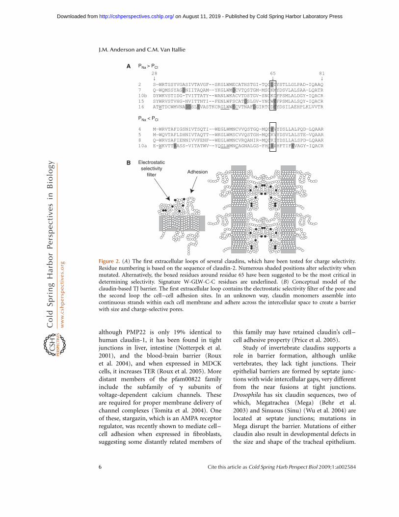
PNa > PCl
28 65 81↓ ↓ ↓
2 S—WRTSSYVGASIVTAVGF--SKGLWMECATHSTGI-TQCDIYSTLLGLPAD-IQAAQ7 Q—WQMSSYAGDNIITAQAM—-YKGLWMDCVTQSTGM-MSCKMYDSVLALSAA—LQATR10b DYWKVSTIDG-TVITTATY--WANLWKACVTDSTGV-SNCKDFPSMLALDGY-IQACR15 SYWRVSTVHG-NVITTNTI--FENLWFSCATDSLGV-YNCWEFPSMLALSQY-IQACR16 ATWTDCWMVNADDSLEVASTKCRGLWWECVTNAFDGIRTCDEYDSILAEHPLKLVVTR
PNa < PCl
4 M—WRVTAFIGSNIVTSQTI-—WEGLWMNCVVQSTGQ-MQCKVYDSLLALPQD—LQAAR5 M—WQVTAFLDHNIVTAQTT—-WKGLWMSCVVQSTGH-MQCKVYDSVLALSTE—VQAAR8 Q—WRVSAFIENNIVVFENF—-WEGLWMNCVRQANIR-MQCKIYDSLLALSPD—LQAAR10a E-WKVTTRASS-VITATWV—-YQGLWMNCAGNALGS-FHCRPHFTIFKVAGY-IQACR
Electrostaticselectivity
filter
A
BAdhesion
Figure 2. (A) The first extracellular loops of several claudins, which have been tested for charge selectivity.Residue numbering is based on the sequence of claudin-2. Numerous shaded positions alter selectivity whenmutated. Alternatively, the boxed residues around residue 65 have been suggested to be the most critical indetermining selectivity. Signature W-GLW-C-C residues are underlined. (B) Conceptual model of theclaudin-based TJ barrier. The first extracellular loop contains the electrostatic selectivity filter of the pore andthe second loop the cell–cell adhesion sites. In an unknown way, claudin monomers assemble intocontinuous strands within each cell membrane and adhere across the intercellular space to create a barrierwith size and charge-selective pores.
J.M. Anderson and C.M. Van Itallie
6 Cite this article as Cold Spring Harb Perspect Biol 2009;1:a002584
on August 11, 2019 - Published by Cold Spring Harbor Laboratory Press http://cshperspectives.cshlp.org/Downloaded from

Five claudin-like sequences have been identi-fied in Caenorhabditis elegans (Asano et al.2003) and RNAi-mediated ablation of claudin-like protein 1 disrupted the barrier betweenepithelial cells of the hypodermis.
Claudin Cell–Cell Adhesion and Assembly
There remains almost no information abouthow individual claudins assemble into higherorder structures to create 10-nm membraneparticles, polymerize into the strands observedin freeze-fracture EM, or adhere across thecell–cell interspace. Currently, two paperssuggest claudins, like connexins in the gap junc-tion, form hexamers. This is based on a study ofhuman claudin-4 expressed in insect cells (Miticet al. 2003) and on purified native MP20, thedistant claudin relative found in the lens(Jarvis and Louis 1995). There is consistentevidence that claudins can form homotypicadhesive plaques, and two studies provide evi-dence that this homotypic cell–cell adhesioncan occur through residues in the center of thesecond extracellular loop (Daugherty et al.2007; Piontek et al. 2007). Molecular modelingof claudin-5 complemented by mutagenesissuggests that adhesion requires an interactionof complementing hydrophobic residues alonga helix in the center of the second loop
(Piontek et al. 2007). A model of the barrier inFigure 2B is based on evidence that claudinsare cell-to-cell adhesion molecules and formsmall pores through the barrier, but there is cur-rently insufficient evidence to speculate on howthey oligomerize to form the particles seen infreeze fracture EM images.
Most cell types express several differentclaudins, which assemble into the sameadhesive strands, yet the generality of hetero-typic binding across the intercellular spaceremains an unresolved question. There islimited evidence that a subset of claudins caninteract heterotypically across cell contacts; forexample, claudin-1 can bind claudin-3 but notclaudin-2 (Furuse et al. 1999), but there iscurrently no information that allows generali-zations about which claudins can interactor if adhesion involves additional proteins(Fig. 2A,B). Some claudins are observed tocoimmunoprecipitate with a list of proteins(e.g., claudin-7 with EpCAM [Le Naour andZoller 2008]; claudin-11 with b1 integrin andtetraspanins [Tiwari-Woodruff et al. 2001]),although the biologic implications remainunknown. Further understanding of howvarious functions (selectivity, adhesion, andassembly) are organized within the protein isseverely limited by a current lack of structuralinformation.
Table 2. Genetic diseases of tight junction proteins
Gene Disease Pathology/Mechanism Ref.
Cldn-1 Ichthyosis, sclerosing cholangitis Affects skin and bile ducts (Hadj-Rabia et al. 2004)Cldn-14 Nonsyndromic deafness, DFNB29 Cochlear hair cell degeneration (Ben Yosef et al. 2003)Cldn-16
Human HHNCa Defective renal Mgþþ reabsorption (Simon et al. 1999)Bovine interstitial nephritis (Hirano et al. 2000)
Cldn-19 Renal Mgþþ loss and vision loss Similar to Cldn-16 (Konrad et al. 2006)PMP22 Peripheral polyneuropathies Demyelinization (Gabreels-Festen and
Wetering 1999)HNPPb Gene deletionCharcot-Marie-Tooth Type 1A Gene duplicationDejerine-Sottas syndrome Point mutations
ZO-2 Familial hypercholanemia Defective PDZ-claudin binding (Carlton et al. 2003)Tricellulin Nonsyndromic deafness Loss of ZO-1 binding (Riazuddin et al. 2006)
aHypomagnesemia hypercalciuria with nephrocalcinosisbHereditary neuropathy with liability to pressure palsies. Modified, with permission, from Van Itallie and Anderson 2006
(# Annual Reviews www.annualreviews.org).
Physiology and Function of the Tight Junction
Cite this article as Cold Spring Harb Perspect Biol 2009;1:a002584 7
on August 11, 2019 - Published by Cold Spring Harbor Laboratory Press http://cshperspectives.cshlp.org/Downloaded from

PERMSELECTIVITY AND THE TWO PATHWAYMODEL: SMALL SELECTIVE PORES ANDNONSELECTIVE BREAKS
The term “permselectivity” is used to describevariations among TJs in electrical resistance,ionic charge and size discrimination, and themagnitude of solute permeability (Table 1)(Powell 1981). TER and solute flux are in parta function of cell geometry. For example, amonolayer with smaller cells has more cell–cell contact length per unit area throughwhich electrical current and solutes can pass.Assuming the same claudins are expressed, amonolayer with smaller cells will show lowerTER and higher solute flux. Geometry of theindividual cell–cell contacts can also affectTJ length per unit area. Some cells touch withstraight junction contacts, whereas othersmake elaborate interdigitations. For example,those between cells in mammalian thin ascend-ing limb of the loop of Henle are extremelyredundant, which increases TJ length per unitarea of the epithelium at least 10-fold overstrength contacts. Presumably, this arrange-ment enhances the space for paracellulartransport without requiring larger epithelium.However, at the protein level, it is now recog-nized that a critical determinant of permselec-tivity is the profile and levels of differentclaudins expressed in a given tight junction.
Size
In general, description of paracellular size selec-tivity in the cell biology and physiology litera-ture has been rather imprecise, in contrast tostudies in the pharmaceutical literature, whichhave specifically focused on how transepithelialdrug absorption is influenced by solute size andchemistry. Research in the former fields is oftenconcerned only with TJ assembly or disassem-bly, rather than with subtleties of the permselec-tivity. In most studies, a single tracer size is usedto report paracellular flux, which limits theability to determine experimental changes inthe size-dependence of permeability. Tracercharge is usually ignored as is a possible trans-cellular contribution from transcytosis. Thesesubtleties are important because information
about alterations in size and charge dependencemay have important pathologic implications,for example in determining if specific cytokinesor bacterial toxins might have increased accessacross the paracellular space.
Now that the actual barrier-forming pro-teins have been discovered, it is instructive tocharacterize permeability as a continuous func-tion of solute size (Watson et al. 2001; Watsonet al. 2005; Van Itallie et al. 2008). This isperformed by characterizing the permeabilityfor solutes of progressively larger molecularradii. Apparent Permeability [Papp ¼ (dQ/dt)/(Concentration Gradient � Area)] takes intoaccount the chemical driving force for eachsolute and surface area, and, unlike simple flux(dQ/dt), can be compared among tissues andlaboratories. When size profiling is performedusing a noncharged solute like graded poly-ethylene polymers (PEGs), it becomes obvi-ous that permeability has two components(Fig. 3). There is a higher capacity pathwaywith a steep size dependence for solutes lessthan about 4 A in radius (Knipp et al. 1997;Watson et al. 2001; Van Itallie et al. 2008). Thispore-pathway shows ionic charge discrimi-nation and its magnitude varies among epithe-lia. This is the pathway that carries most of the
Solute radius (Å)
Pap
p (c
m/s
ec)
× 1
06
Leak
Pores2
1
3 4 5 6 7
Figure 3. The two pathway model. Idealized datashow the permeability of noncharged polyethyleneglycol molecules of different sizes across anepithelium. The pathway for molecules below 4 A isformed by claudin pores that are size and chargeselective. The pathway for molecule above 4 A indiameter shows no charge selectivity and appears tobe defined by the status of cell-to-cell adhesion andthe cytoskeleton.
J.M. Anderson and C.M. Van Itallie
8 Cite this article as Cold Spring Harb Perspect Biol 2009;1:a002584
on August 11, 2019 - Published by Cold Spring Harbor Laboratory Press http://cshperspectives.cshlp.org/Downloaded from
electrical current during physiologic transportand during experimentally imposed electricalfield (reflected in the TER), thus its magnitudeand charge selectivity are key determinants ofphysiologic transport. As described below, clau-dins confer the charge selectivity, but there is lessinformation on what determines the magnitudeof flux through the pores. However, differentclaudins may control the level of porositybecause it has been observed that expression ofclaudin-2 in cultured epithelial cells selectivelyincreases the permeability for solutes smallerthan 4 A, whereas claudins 4 and 18 do not(Van Itallie et al. 2008). In vivo, deletion ofclaudin-5 from mice results in a size-selectiveincrease in permeability of TJs in blood vessels(Nitta et al. 2003). It is important to recognizethat the permeability for noncharged andcharged solutes are not necessarily directlycorrelated. Because pores in different epitheliacan have different charge discrimination, onecan observe an apparent paradox in which thesolute permeability is very high but the ionpermeability is very low (high TER). Takentogether, the present data suggest that claudinscreate the system of small discriminatingpores, and by an unknown mechanism regulatethe magnitude of flux (Fig. 3).
Solutes that are larger than 4 A can alsoget through intact TJs. The magnitude ofthis component varies among epithelia andshows no charge discrimination (Knipp. et al.1997; Artursson et al. 1993). The distinctionbetween the pore and nonpore pathways hasbeen overlooked in most of the literaturebecause permeability is typically measuredonly by tracers, which are larger than thepores, namely mannitol (4.2 A in radius), inulin(15 A), and a graded series of fluorescentlylabeled dextrans (4 kDa, 10 kDa, 40 kDa, etc).This larger pathway is speculated to representsmall temporary breaks in the otherwisecontinuous TJ contacts. Interestingly, fluxthrough this pathway can be enhanced byproinflammatory cytokines without alteringthe pore pathway (Watson et al. 2005). Thenonpore pathway is sensitive to cytoskeletaldisruption (Bruewer et al. 2004; Ivanov et al.2005) and any form of cellular injury (Nusrat
et al. 2000). There is significant evidence thatthe “intactness” of the TJ is controlled by cyto-skeletal dynamics (Hartsock and Nelson 2008),myosin light chain activity (Ma et al. 2005),and any factor affecting cell homeostasis.As investigators dissect the role of individualproteins and signals in regulating the barrier,it will be useful to characterize barriers interms of the pore and nonpore pathways.
Claudin Charge Selectivity and TER
All of the available data supports the idea thatthe first extracellular loop of claudins createsan “electrostatic selectivity filter,” controllingoverall resistance and charge selectivity of thesmall pores (Fig. 2). In contrast, admittedlylimited data suggest that the second loop isinvolved in cell–cell adhesion (Blasig et al.2006; Piontek et al. 2007). Charged amino acidside chains on the claudins limit similarlycharged ions in solution without opposingpermeability of oppositely charged ions. Mostof these studies have been performed by expres-sing a single foreign claudin in the backgroundof all the other claudins expressed in a culturedepithelial cell monolayer, most often MDCKand LLC-PK1 cells or by siRNA knockdown ofselected claudins (Hou et al. 2006). A changefrom the baseline TER and dilution potential(a measure of cation/anion selectivity) is inter-preted to reveal selectivity of the transfected orsilenced claudin compared with the back-ground. These studies are somewhat qualitative.Lacking the electrophysiologic tools used tocharacterize membrane channels, such as patchclamping, it has been impossible to assign aspecific conductance to individual claudins.
A total of 15 claudins have now been testedwith good consensus. Claudins 2 (Furuse et al.2001; Colegio et al. 2002; Amasheh et al.2002) and 10 (Van Itallie et al. 2006) tend tomake tight monolayers leakier. Claudins 1(Inai et al. 1999; McCarthy et al. 2000), 4(Van Itallie et al. 2001), 5 (Wen et al. 2004;Amasheh et al. 2005), 7 (Alexandre et al.2005), 8 (Angelow et al. 2006; Yu et al. 2003;Jeansonne et al. 2003), 11 (Van Itallie et al.2003), 14 (Ben Yosef et al. 2003), 15 (Colegio
Physiology and Function of the Tight Junction
Cite this article as Cold Spring Harb Perspect Biol 2009;1:a002584 9
on August 11, 2019 - Published by Cold Spring Harbor Laboratory Press http://cshperspectives.cshlp.org/Downloaded from

et al. 2003; Van Itallie et al. 2003), 16 (Hou et al.2005; Ikari et al. 2008), 18 (Jovov et al. 2007),and 19 (Hou et al. 2008) tend to make leakymonolayers tighter. Claudins 2, 15 (Amashehet al. 2002; Colegio et al. 2003), 16, and 19(Hou et al. 2008) have a preference for cationsover anions. Claudin-10a (Van Itallie et al.2006) prefers anions. Where tested, the effecton TER is dose-dependent and each claudinreaches a saturating influence on TER (VanItallie et al. 2001), although the reason for thisis not clear. Most reports simply express thechange in barrier permselectivity that resultsfrom the maximal induction level. Somestudies suggest that the transfected claudinadds to those already present, whereas a singlestudy convincingly shows that claudin-8 canreplace endogenous claudin-2 (Yu et al. 2003).Potencies vary, with claudins 14 (Ben Yosefet al. 2003) and 18 (Jovov et al. 2007) producingthe electrically tightest barriers. A single studyhas simultaneously expressed two differentclaudins, showing that claudins 16 and 19exert properties in a cooperative and not justadditive fashion, consistent with the idea thatclaudins might form heterotypic pores (Houet al. 2008).
There is a general relationship between thechemistry of the first loop and charge selectivity(Fig. 2A). However, the relative influence ofspecific positions is still under debate. Forexample, claudin-16 has the highest proportionof negatively charged residues (Fig. 2A, shapedpositions) and when expressed in culturedepithelial cell monolayers, results in a strongenhancement in permeability for Naþ andMgþþ but has no affect on PCl (Hou et al.2005). In the opposite direction, claudin-10ahas more basic residues and reduces PNa, whilesimultaneously increasing PCl (Van Itallie et al.2006). Each claudin has shown a signaturepattern of whether it will increase or decreaseTER and increase, decrease, or not alter theindividual PNa and PCl. The ability to increaseTER can be based on limiting permeability forjust cations (claudin-4 [Van Itallie et al.2001]) or both cations and anions (claudin-7[Alexandre et al. 2005]). There is a reassuringcorrelation between the physiology of particular
tissues, the claudins they express, and the physi-ology of those claudin as determined exper-imentally (Holmes et al. 2006). This is bestdocumented along the nephron, where TERand ion selectivity are very well documented.For example, “leaky” claudin-2 is the dominantform in the leaky proximal tubule and isexcluded from tighter segments; “tight”claudin-4 and -8 are restricted to the tightcollecting ducts (Angelow et al. 2008).
The role of individual positions in influen-cing charge selectivity has most often beentested by expressing charge-reversing muta-tions. This approach has led to the conclusionthat positions along much of the first loopcan influence selectivity and thus this loop isfolded to line the pore space through whichsoluble ions pass. This has been most exten-sively studied for claudins 15 (Van Itallie et al.2003) and 16 (Hou et al. 2005). In the case ofclaudin-15, three widely spaced and negativelycharged residues were sequentially reversedand shown to have additive effects (Van Itallieet al. 2003). The “distributed filter” model hasbeen criticized because it is based on intro-ducing abnormal charges rather than simplyneutralizing them. Extensive characterizationof charge-neutralizing mutations of claudin-2convincingly showed that the only chargedresidue that provides the high PNa is the aspar-tic acid at position 65 (Fig. 2A) (Yu et al.2009). Resolution of the mechanism of selec-tivity must await high-resolution structuralinformation.
The TJ pore is slightly wider than that oftransmembrane pores and consequently showsless ionic discrimination. For comparison,some Naþ channels (ENaC) show a per-meability ratio for Naþ to Kþ (PNa/PK) ashigh as 1000 to 1 (Hille 2001). In contrast,claudin pores show almost no discriminationbetween Kþ and Naþ and the PCl/PNa ratioranges only from about 10 to 0.1 whencompared among various in vivo epithelia andcultured cell monolayers. Membrane channelsare of a size where the associated water mol-ecules must be stripped off so the ion can fitthrough the pore (Hille 2001; Yu et al. 2009).The modest ability of TJs in MDCK cells to
J.M. Anderson and C.M. Van Itallie
10 Cite this article as Cold Spring Harb Perspect Biol 2009;1:a002584
on August 11, 2019 - Published by Cold Spring Harbor Laboratory Press http://cshperspectives.cshlp.org/Downloaded from
discriminate among the alkali metal cations(K . Rb . Na . Li . Cs, so-called Eisenmanselectivity sequence V–VIII) does actuallysuggest that they are being partially dehydratedas they pass through the pore. The observedEisenman sequence is slightly different fromthe rank order of their free mobilities insolution, leading to the conclusion that thepore radius is about 3.6 A (Yu et al. 2009).This is concordant with the 4 A radius esti-mated using noncharged solutes (Watson et al.2001). It is remarkable that this degree of sizeand charge selectivity can be maintained whilecells in an epithelial sheet continuously moverelative to one another.
Several claudins have now been deletedfrom mice and the resulting phenotypes areconsistent with a barrier role for claudins 1(Ladwein et al. 2005), 5 (Nitta et al. 2003), 11(Gow et al. 2000), 14 (Ben Yosef et al. 2003),and 15 (Tamura et al. 2008), although thephenotypes are not easily explained as changesin permselectivity. In contrast, deletions of16 (Himmerkus et al. 2008) and 19 (Houet al. 2008) phenocopy their human diseasemutants and are more easily explained as ionselectivity defects, as described in the followingsection.
In zebrafish, reduction in claudin-15through morphlino treatment produces a devel-opmental intestinal defect, which very likelyresults from loss of ion transport selectivity(Bagnat et al. 2007). In the absence ofclaudin-15, the gut remains as multiplelumens, which fail to fuse. It is proposed thatactive ion and water secretion is required forthe lumens to enlarge, come into contact, andfuse. In the absence of claudin-15, there maybe a back leak of Naþ, preventing fluid accumu-lation and expansion. Interestingly, claudin-15KO mice develop a megaintestine, but thisphenotype appears to be a proliferation andnot a permeability defect (Tamura et al. 2008).
Claudin-16 Mutants and Other InheritedDiseases of Tight Junction Proteins
Currently, there are seven human diseasesknown to be caused by mutations in genes
encoding tight junction proteins (Table 2).The basis for ZO-2 (Carlton et al. 2003) andtricellulin-based defects (Riazuddin et al.2006) remains unclear, but the phenotypes ofseveral of the recessive claudin mutantsprovide additional evidence that claudinsprovide selectivity for the barrier. We describethe claudin-16 defect in some detail because itprovides the best evidence that claudins invivo create ion selective pores.
The first disease-causing claudin mutationwas identified through positional cloning ofthe locus responsible for a rare form of renalmagnesium loss, hypomagnesemia hypercalce-mia with nephrocalcinosis (HHNC) (Simonet al. 1999; Kaushansky et al. 2007). Patientswith this defect show increased urinary loss ofMgþþ and Caþþ, and reduced plasma Mgþþ
levels, leading to weakness and seizures. Thegene responsible encodes claudin-16 (originallyreferred to a Paracellin-1). Claudin-16 is highlyrestricted to the thick ascending limb of theloop of Henle, the segment of the nephronmost involved in reabsorbing filtered Mgþþ.Other claudins are also expressed in the sameTJs. Paracellular reabsorption is driven by anintralumenal positive electrical potential withrespect to the peritubular interstitium. Theseobservations led to an initial hypothesis thatclaudin-16 is a Mgþþ pore and when absent,Mgþþ remains in the tubule and is lost in theurine. When expressed in cultured monolayers,wild-type claudin-16 does form a highly cation-selective pore (Hou et al. 2005). Consistentwith the electrostatic claudin pore model, thefirst loop contains many negatively chargedresidues (Fig. 2), and neutralizing mutationsreduce cation permeability (Hou et al. 2005).Many of the human mutations are missensemutations, which fail to traffic properly to theplasma membrane when expressed in culturedcell models (Kausalya et al. 2006) suggestingthe defect results from the selective absenceof claudin-16, leaving other claudins to definethe barrier’s ion selectivity. Recently, mutationsin the human claudin-19 gene were shownto cause renal Mgþþ wasting by a verysimilar mechanism. In addition, claudin-19 isexpressed in the retina and affected individuals
Physiology and Function of the Tight Junction
Cite this article as Cold Spring Harb Perspect Biol 2009;1:a002584 11
on August 11, 2019 - Published by Cold Spring Harbor Laboratory Press http://cshperspectives.cshlp.org/Downloaded from
have defects in retinal development and visionloss (Konrad et al. 2006) (Table 2).
The Mg-pore model is probably too sim-plistic. The intralumenal positive potentialdriving Mgþþ results from a paracellular backdiffusion of Naþ down its concentration gradi-ent into the lumen. If Naþ entry is limited in theabsence of claudin-16 pores, there would be areduced electrical gradient to drive Mgþþ out.Although this can not be tested in humans,study of isolated perfused nephron segmentsfrom claudin-16 KO mice is consistent witha defect in Naþ permeability (Himmerkuset al. 2008). Whatever the full explanation,claudin-16 is clearly a cation pore and HHNCremains the best example of a disease causedby an ion-selective TJ defect.
It is tempting to also explain hearing lossin the human claudin-14 mutants as an ionselectivity defect, but the evidence is onlycircumstantial. Claudin-14 is expressed in TJslining the intrachoclear space, which containsfluid uniquely high in Kþ. This high gradientpromotes rapid entry of Kþ into the outer haircells; this is required for their depolarizationduring acoustic mechanotransduction. Otherforms of deafness are caused by mutationsin the transporters that normally secrete Kþ
into this space, suggesting that the loss ofclaudin-14 might cause deafness by allowingthe Kþ gradient to dissipate. In fact, whenclaudin-14 is expressed in cultured MDCKcells, the junctions become very tight by specifi-cally restricting cation permeability (Riazuddinet al. 2006). Finally, claudin-1 mutation resultsin neonatal ichthyosis-sclerosing cholangitis syn-drome (NISCH), but whether this pathophysiol-ogy results from a permeability defect is unclear(Hadj-Rabia et al. 2004). Although claudin-1 isvery widely expressed among all epithelia, it isdifficult to rationalize why patients with thesemutations develop predominately a scaling skindisease and obliteration of their bile ducts.
CONCLUSION
Significant progress has been made over the pastdecade in understanding the role of claudinsin regulation of TER and paracellular ionic
selectivity. In addition, the recognition of theexistence of two pathways for paracellularsolute flux should allow a more sophisticatedanalysis of the roles individual TJ proteinsplay in physiologic and pathologic regulationof permeability of nonionic solutes. However,further insights are hampered by the lack ofa three-dimensional model of TJ structure.Studies on the synaptic junction may provideclues and methods for studying TJ organiz-ation, but ultimately we will need structuralinformation about the integral membrane pro-teins of the TJ to ask informative questionsabout how this complex is organized andphysiologically regulated.
ACKNOWLEDGMENTS
This work was supported by grants from theNational Institutes of Health, DK45134 (JMA,CVI), P30 DK034987, and DK61397 (JMA).
REFERENCES
Alexandre MD, Lu Q, Chen YH. 2005. Overexpression ofclaudin-7 decreases the paracellular Cl2 conductanceand increases the paracellular Naþ conductance inLLC-PK1 cells. J Cell Sci 118: 2683–2693.
Amasheh S, Meiri N, Gitter AH, Schoneberg T, Mankertz J,Schulzke JD, Fromm M. 2002. Claudin-2 expressioninduces cation-selective channels in tight junctions ofepithelial cells. J Cell Sci 115: 4969–4976.
Amasheh S, Schmidt T, Mahn M, Florian P, Mankertz J,Tavalali S, Gitter AH, Schulzke JD, Fromm M. 2005.Contribution of claudin-5 to barrier properties in tightjunctions of epithelial cells. Cell Tissue Res 321: 89–96.
Angelow S, Ahlstrom R, Yu AS. 2008. Biology of claudins.Am J Physiol Renal Physiol 295: F867–F876.
Angelow S, Kim KJ, Yu AS. 2006. Claudin-8 modulates para-cellular permeability to acidic and basic ions in MDCK IIcells. J Physiol 571: 15–26.
Artursson P, Ungell AL, Lofroth JE. 1993. Selective paracel-lular permeability in two models of intestinal absorption:Cultured monolayers of human intestinal epithelial cellsand rat intestinal segments. Pharm Res 10: 1123–1129.
Asano A, Asano K, Sasaki H, Furuse M, Tsukita S. 2003.Claudins in Caenorhabditis elegans: Their distributionand barrier function in the epithelium. Curr Biol 13:1042–1046.
Bagnat M, Cheung ID, Mostov KE, Stainier DY. 2007.Genetic control of single lumen formation in the zebra-fish gut. Nat Cell Biol 9: 954–960.
Balda MS, Matter K. 2003. Epithelial cell adhesion and theregulation of gene expression. Trends Cell Biol 13:310–318.
J.M. Anderson and C.M. Van Itallie
12 Cite this article as Cold Spring Harb Perspect Biol 2009;1:a002584
on August 11, 2019 - Published by Cold Spring Harbor Laboratory Press http://cshperspectives.cshlp.org/Downloaded from

Barrios-Rodiles M, Brown KR, Ozdamar B, Bose R, Liu Z,Donovan RS, Shinjo F, Liu Y, Dembowy J, Taylor IW,et al. 2005. High-throughput mapping of a dynamic sig-naling network in mammalian cells. Science 307: 1621–1625.
Bazzoni G. 2003. The JAM family of junctional adhesionmolecules. Curr Opin Cell Biol 15: 525–530.
Behr M, Riedel D, Schuh R. 2003. The claudin-like megatra-chea is essential in septate junctions for the epithelialbarrier function in Drosophila. Dev Cell 5: 611–620.
Ben Yosef T, Belyantseva IA, Saunders TL, Hughes ED,Kawamoto K, Van Itallie CM, Beyer LA, Halsey K,Gardner DJ, Wilcox ER, et al. 2003. Claudin 14 knockoutmice, a model for autosomal recessive deafness DFNB29,are deaf due to cochlear hair cell degeneration. Hum MolGenet 12: 2049–2061.
Blasig IE, Winkler L, Lassowski B, Mueller SL, Zuleger N,Krause E, Krause G, Gast K, Kolbe M, Piontek J. 2006.On the self-association potential of transmembranetight junction proteins. Cell Mol Life Sci 63: 505–514.
Boron W, Boulpaep EL. 2005. Medical physiology. A cellularand molecular approach, pp. 931–946. Elsevier Saunders,Philidelphia.
Brancolini C, Edomi P, Marzinotto S, Schneider C. 2000.Exposure at the cell surface is required for Gas3/PMP22 to regulate both cell death and cell spreading:Implication for the Charcot-Marie-Tooth type 1A andDejerine-Sottas diseases. Mol Biol Cell 11: 2901–2914.
Bronstein JM. 2000. Function of tetraspan proteins in themyelin sheath. Curr Opin Neurobiol 10: 552–557.
Bruewer M, Hopkins AH, Hobert ME, Nusrat A, Madara JL.2004. RhoA, Rac1, and Cdc42 exert distinct effects onepithelial barrier via selective structural and biochemicalmodulation of junctional proteins and F-actin. Am JPhysiol Cell Physiol 287: C327–C335.
Carlton VE, Harris BZ, Puffenberger EG, Batta AK, KniselyAS, Robinson DL, Strauss KA, Shneider BL, Lim WA,Salen G, et al. 2003. Complex inheritance of familialhypercholanemia with associated mutations in TJP2and BAAT. Nat Genet 34: 91–96.
Cereijido M, Anderson JM. 2001. Introduction: Evolutionof ideas on the tight junction. In Tight junctions (ed.Cereijido M, Anderson JM), pp. 1–18. CRC Press, BocaRaton.
Cereijido M, Valdes J, Shoshani L, Contreras RG. 1998. Roleof tight junctions in establishing and maintaining cellpolarity. Annu Rev Physiol 60: 161–177.
Claude P. 1978. Morphological factors influencing transe-pithelial permeability: A model for the resistance of thezonula occludens. J Membr Biol 39: 219–232.
Colegio OR, Van Itallie CM, Mccrea HJ, Rahner C,Anderson JM. 2002. Claudins create charge-selectivechannels in the paracellular pathway between epithelialcells. Am J Physiol Cell Physiol 283: C142–C147.
Colegio OR, Van Itallie C, Rahner C, Anderson JM. 2003.Claudin extracellular domains determine paracellularcharge selectivity and resistance but not tight junctionfibril architecture. Am J Physiol Cell Physiol 284:C1346–C1354.
Coyne CB, Bergelson JM. 2005. CAR: Avirus receptor withinthe tight junction. Adv Drug Deliv Rev 57: 869–882.
Crone C, Christensen O. 1981. Electrical resistance of acapillary endothelium. J Gen Physiol 77: 349–371.
Daugherty BL, Ward C, Smith T, Ritzenthaler JD, Koval M.2007. Regulation of heterotypic claudin compatibility.J Biol Chem 282: 30005–30013.
Diamond JM. 1977. Twenty-first Bowditch lecture. The epi-thelial junction: Bridge, gate, and fence. Physiologist 20:10–18.
Diamond JM. 1978. Channels in epithelial cell membranesand junctions. Fed Proc 37: 2639–2644.
Farquhar MG, Palade GE. 1963. Junctional complexes invarious epithelia. J Cell Biol 17: 375–412.
Furuse M. 2009. Molecular basis of the core structure of tightjunctions. Cold Spring Harb Perspect Biol 2: a002907.
Furuse M, Hirase T, Itoh M, Nagafuchi A, Yonemura S,Tsukita S, Tsukita S. 1993. Occludin - A Novel IntegralMembrane-Protein Localizing at Tight Junctions. J CellBiol 123: 1777–1788.
Furuse M, Fujita K, Hiiragi T, Fujimoto K, Tsukita S. 1998.Claudin-1 and -2: Novel integral membrane proteinslocalizing at tight junctions with no sequence similarityto occludin. J Cell Biol 141: 1539–1550.
Furuse M, Sasaki H, Tsukita S. 1999. Manner of interactionof heterogeneous claudin species within and betweentight junction strands. J Cell Biol 147: 891–903.
Furuse M, Furuse K, Sasaki H, Tsukita S. 2001. Conversionof Zonulae occludentes from tight to leaky strand type byintroducing claudin-2 into Madin-Darby Canine KidneyI cells. J Cell Biol 153: 263–272.
Gabreels-Festen A, Wetering RV. 1999. Human nerve pathol-ogy caused by different mutational mechanisms of thePMP22 gene. Ann NY Acad Sci 883: 336–343.
Gonzalez-Mariscal L, Betanzos A, Nava P, Jaramillo BE.2003. Tight junction proteins. Prog Biophys Mol Biol 81:1–44.
Gonzalez-Mariscal L, Tapia R, Chamorro D. 2008. Crosstalkof tight junction components with signaling pathways.Biochim Biophys Acta 1778: 729–756.
Gow A, Southwood CM, Li JS, Pariali M, Riordan GP,Danias J, Bronstein JM, Brodie SE, Kachar B, LazzariniRA. 2000. CNS myelin and sertoli cell tight junctionstands are absent in osp/claudin 11-null mice.J Neurochem 74: S35.
Hadj-Rabia S, Baala L, Vabres P, Hamel-Teillac D, JacqueminE, Fabre M, Lyonnet S, De Prost Y, Munnich A, HadchouelM, et al. 2004. Claudin-1 gene mutations in neonatalsclerosing cholangitis associated with ichthyosis: A tightjunction disease. Gastroenterology 127: 1386–1390.
Hamazaki Y, Itoh M, Sasaki H, Furuse M, Tsukita S. 2002.Multi-PDZ domain protein 1 (MUPP1) is concentratedat tight junctions through its possible interaction withclaudin-1 and junctional adhesion molecule. J BiolChem 277: 455–461.
Hartsock A, Nelson WJ. 2008. Adherens and tight junctions:Structure, function and connections to the actin cytoske-leton. Biochim Biophys Acta 1778: 660–669.
Hille B. 2001. Ion channels of excitable membranes, pp.573–574. Sinauer Associates, Sunderland, Massachusetts.
Himmerkus N, Shan Q, Goerke B, Hou J, Goodenough DA,Bleich M. 2008. Salt and acid-base metabolismin claudin-16 knockdown mice: Impact for the
Physiology and Function of the Tight Junction
Cite this article as Cold Spring Harb Perspect Biol 2009;1:a002584 13
on August 11, 2019 - Published by Cold Spring Harbor Laboratory Press http://cshperspectives.cshlp.org/Downloaded from
pathophysiology of FHHNC patients. Am J Physiol RenalPhysiol 295: F1641–F1647.
Hirano T, Kobayashi N, Itoh T, Takasuga A, Nakamaru T,Hirotsune S, Sugimoto Y. 2000. Null mutation ofPCLN-1/Claudin-16 results in bovine chronic interstitialnephritis. Genome Res 10: 659–663.
Holmes JL, Van Itallie CM, Rasmussen JE, Anderson JM.2006. Claudin profiling in the mouse during postnatalintestinal development and along the gastrointestinaltract reveals complex expression patterns. Gene ExprPatterns 6: 581–588.
Hou J, Paul DL, Goodenough DA. 2005. Paracellin-1 and themodulation of ion selectivity of tight junctions. J Cell Sci118: 5109–5118.
Hou J, Gomes AS, Paul DL, Goodenough A. 2006. Study ofclaudin function by RNA interference. J Biol Chem 281:36117–36123.
Hou J, Renigunta A, Konrad M, Gomes AS, SchneebergerEE, Paul DL, Waldegger S, Goodenough DA. 2008.Claudin-16 and claudin-19 interact and form a cation-selective tight junction complex. J Clin Invest 118:619–628.
Ikari A, Ito M, Okude C, Sawada H, Harada H, Degawa M,Sakai H, Takahashi T, Sugatani J, Miwa M. 2008.Claudin-16 is directly phosphorylated by protein kinaseA independently of a vasodilator-stimulatedphosphoprotein-mediated pathway. J Cell Physiol 214:221–229.
Ikenouchi J, Furuse M, Furuse K, Sasaki H, Tsukita S,Tsukita S. 2005. Tricellulin constitutes a novel barrier attricellular contacts of epithelial cells. J Cell Biol 171:939–945.
Inai T, Kobayashi J, Shibata Y. 1999. Claudin-1 contributesto the epithelial barrier function in MDCK cells. Eur JCell Biol 78: 849–855.
Itoh M, Furuse M, Morita K, Kubota K, Saitou M, Tsukita S.1999. Direct binding of three tight junction-associatedMAGUKs, ZO-1, ZO-2 and ZO-3, with the COOHtermini of claudins. J Cell Biol 147: 1351–1363.
Ivanov AI, Hunt D, Utech M, Nusrat A, Parkos CA. 2005.Differential roles for actin polymerization and a myosinII motor in assembly of the epithelial apical junctionalcomplex. Mol Biol Cell 16: 2636–2650.
Jarvis LJ, Louis CF. 1995. Purification and oligomeric state ofthe major lens fiber cell membrane proteins. Curr Eye Res14: 799–808.
Jeansonne B, Lu Q, Goodenough DA, Chen YH. 2003.Claudin-8 interacts with multi-PDZ domain protein 1(MUPP1) and reduces paracellular conductance in epi-thelial cells. Cell Mol Biol(Noisy-le-grand) 49: 13–21.
Jetten AM, Suter U. 2000. The peripheral myelin protein 22and epithelial membrane protein family. Prog Nucl AcidRes Mol Biol 64: 97–129.
Jovov B, Van Itallie CM, Shaheen NJ, Carson JL, GamblingTM, Anderson JM, Orlando RC. 2007. Claudin-18: ADominant Tight Junctional Protein in Barrett’sEsophagus and Potential Contributor to its AcidResistance. Am J Physiol GI 293: G1106–G1113.
Kausalya PJ, Amasheh S, Gunzel D, Wurps H, Muller D,Fromm M, Hunziker W. 2006. Disease-associatedmutations affect intracellular traffic and paracellular
Mg transport function of Claudin-16. J Clin Invest 116:878–891.
Kaushansky N, Hemo R, Eisenstein M, Ben Nun A. 2007.OSP/claudin-11-induced EAE in mice is mediated bypathogenic T cells primarily governed by OSP192Yresidue of major encephalitogenic region OSP179-207.Eur J Immunol 37: 2018–2031.
Kearsey J, Petit S, De Oliveira C, Schweighoffer F. 2004. Anovel four transmembrane spanning protein, CLP24.Eur J Biochem 271: 2584–2592.
Knipp GT, Ho NF, Barsuhn CL, Borchardt RT. 1997.Paracellular diffusion in Caco-2 cell monolayers: Effectof perturbation on the transport of hydrophilic com-pounds that vary in charge and size. J Pharm Sci 86:1105–1110.
Koefeld-Johnsen V, Ussing HH. 1958. The nature of the frogskin potential. Acta Physiol Scand 42: 298–308.
Konrad M, Schaller A, Seelow E, Pandey AV, Waldegger S,Lesslauer A, Vitzthum H, Suzuki Y, Luk JM, Becker C,et al. 2006. Mutations in the tight-junction geneclaudin 19 (CLDN19) are associated with renal mag-nesium wasting, renal failure, and severe ocular involve-ment. Am J Hum Genet 79: 949–957.
Krause G, Winkler L, Mueller SL, Haseloff F, Piontek J, BlasigIE. 2008. Structure and function of claudins. BiochimBiophys Acta 1778: 631–645.
Kubota K, Furuse M, Sasaki H, Sonoda N, Fujita K,Nagafuchi A, Tsukita S. 1999. Ca2þ-independentcell-adhesion activity of claudins, a family of integralmembrane proteins localized at tight junctions. CurrBiol 9: 1035–1038.
Ladwein M, Pape UF, Schmidt DS, Schnolzer M, Fiedler S,Langbein L, Franke WW, Moldenhauer G, Zoller M.2005. The cell-cell adhesion molecule EpCAM interactsdirectly with the tight junction protein claudin-7. ExpCell Res 309: 345–357.
Laukoetter MG, Nava P, Lee WY, Severson EA, Capaldo CT,Babbin BA, Williams IR, Koval M, Peatman E, CampbellJA, et al. 2007. JAM-A regulates permeability and inflam-mation in the intestine in vivo. J Exp Med 204:3067–3076.
Le Naour F, Zoller M. 2008. The tumor antigen EpCAM:Tetraspanins and the tight junction protein claudin-7,new partners, new functions. Front Biosci 13: 5847–5865.
Liu Y, Nusrat A, Schnell FJ, Reaves TA, Walsh S, Pochet M,Parkos CA. 2000. Human junction adhesion moleculeregulates tight junction resealing in epithelia 18. J CellSci 113: 2363–2374.
Ma TY, Boivin MA, Ye D, Pedram A, Said HM. 2005.Mechanism of TNF-{alpha} modulation of Caco-2 intes-tinal epithelial tight junction barrier: Role of myosinlight-chain kinase protein expression. Am J PhysiolGastrointest Liver Physiol 288: G422–G430.
Madara JL, Trier JS, Neutra MR. 1980. Structural-Changes in the Plasma-Membrane AccompanyingDifferentiation of Epithelial-Cells in Human andMonkey Small-Intestine. Gastroenterology 78: 963–975.
Matsuda M, Kubo A, Furuse M, Tsukita S. 2004. A peculiarinternalization of claudins, tight junction-specificadhesion molecules, during the intercellular movementof epithelial cells. J Cell Sci 117: 1247–1257.
J.M. Anderson and C.M. Van Itallie
14 Cite this article as Cold Spring Harb Perspect Biol 2009;1:a002584
on August 11, 2019 - Published by Cold Spring Harbor Laboratory Press http://cshperspectives.cshlp.org/Downloaded from
McCarthy KM, Francis SA, McCormack JM, Lai J, RogersRA, Skare IB, Lynch RD, Schneeberger EE. 2000.Inducible expression of claudin-1-myc but notoccludin-VSV-G results in aberrant tight junctionstrand formation in MDCK cells. J Cell Sci 113:3387–3398.
Mitic LL, Unger VM, Anderson JM. 2003. Expression, solu-bilization, and biochemical characterization of the tightjunction transmembrane protein claudin-4. Protein Sci12: 218–227.
Nitta T, Hata M, Gotoh S, Seo Y, Sasaki H, Hashimoto N,Furuse M, Tsukita S. 2003. Size-selective looseningof the blood-brain barrier in claudin-5-deficient mice.J Cell Biol 161: 653–660.
Notterpek L, Roux KJ, Amici SA, Yazdanpour A, Rahner C,Fletcher BS. 2001. Peripheral myelin protein 22 is a con-stituent of intercellular junctions in epithelia. Proc NatlAcad Sci 98: 14404–14409.
Nusrat A, Turner JR, Madara JL. 2000. Molecular physiologyand pathophysiology of tight junctions. IV. Regulation oftight junctions by extracellular stimuli: Nutrients, cyto-kines, and immune cells. Am J Physiol Gastrointest LiverPhysiol 279: G851–G857.
Nusrat A, Eichel-Streiber C, Turner JR, Verkade P, MadaraJL, Parkos CA. 2001. Clostridium difficile toxinsdisrupt epithelial barrier function by altering membranemicrodomain localization of tight junction proteins.Infect Immun 69: 1329–1336.
Piontek J, Winkler L, Wolburg H, Muller SL, Zuleger N,Piehl C, Wiesner B, Krause G, Blasig IE. 2007.Formation of tight junction: Determinants of homophi-lic interaction between classic claudins. FASEB J 22:146–158.
Powell DW. 1981. Barrier function of epithelia. Am J Physiol241: G275–G288.
Price MG, Davis CF, Deng F, Burgess DL. 2005. Thealpha-amino-3-hydroxyl-5-methyl-4-isoxazolepropion-ate receptor trafficking regulator “stargazin” is related tothe claudin family of proteins by Its ability to mediatecell-cell adhesion. J Biol Chem 280: 19711–19720.
Riazuddin S, Ahmed ZA, Fanning AS, Lagziel A, Kitajiri S,Ramzan K, Khan SN, Chattaraj P, Friedman PL,Anderson JM, et al. 2006. Tricellulin is a tight-junctionprotein necessary for hearing. Am J Hum Genet 79:1040–1051.
Roh MH, Liu CJ, Laurinec S, Margolis B. 2002. The carboxylterminus of zona occludens-3 binds and recruits a mam-malian homologue of discs lost to tight junctions. J BiolChem 277: 27501–27509.
Roux KJ, Amici SA, Notterpek L. 2004. The temporospatialexpression of peripheral myelin protein 22 at the develop-ing blood-nerve and blood-brain barriers. J Comp Neurol474: 578–588.
Roux KJ, Amici SA, Fletcher BS, Notterpek L. 2005.Modulation of epithelial morphology, monolayer per-meability, and cell migration by growth arrest specific3/peripheral myelin protein 22. Mol Biol Cell 16:1142–1151.
Saitou M, Furuse M, Sasaki H, Schulzke JD, Fromm M,Takano H, Noda T, Tsukita S. 2000. Complex phenotypeof mice lacking occludin, a component of tight junctionstrands. Mol Biol Cell 11: 4131–4142.
Schmitz H, Barmeyer C, Fromm M, Runkel N, Foss HD,Bentzel CJ, Riecken EO, Schulzke JD. 1999. Alteredtight junction structure contributes to the impairedepithelial barrier function in ulcerative colitis.Gastroenterology 116: 301–309.
Schneeberger EE, Lynch RD. 2004. The tight junction: Amultifunctional complex. Am J Physiol-Cell Physiol 286:C1213–C1228.
Schulzke JD, Gitter AH, Mankertz J, Spiegel S, Seidler U,Amasheh S, Saitou M, Tsukita S, Fromm M. 2005.Epithelial transport and barrier function in occludin-deficient mice. Biochim Biophys Acta 1669: 34–42.
Simon DB, Lu Y, Choate KA, Velazquez H, Al Sabban E,Praga M, Casari C, Bettinelli C, Colussi C,Rodriguez-Soriano J, et al. 1999. Paracellin-1, a renaltight junction protein required for paracellular Mg2þ
resorption. Science 285: 103–106.
Steele EC, Lyon MF, Favor J, Guillot PV, Boyd Y, Church RL.1998. A mutation in the connexin 50 (Cx50) gene is acandidate for the No2 mouse cataract. Curr Eye Res 17:883–889.
Tamura A, Kitano Y, Hata M, Katsuno T, Moriwaki K,Sasaki H, Hayashi H, Suzuki Y, Noda T, Furuse M,et al. 2008. Megaintestine in claudin-15-deficientmice. Gastroenterology 134: 523–534.
Tiwari-Woodruff SK, Buznikov AG, Vu TQ, Micevych PE,Chen K, Kornblum HI, Bronstein JM. 2001. OSP/claudin-11 forms a complex with a novel member ofthe tetraspanin super family and beta 1 integrin and regu-lates proliferation and migration of oligodendrocytes.J Cell Biol 153: 295–305.
Tomita S, Fukata M, Nicoll RA, Bredt DS. 2004. Dynamicinteraction of stargazin-like TARPs with cycling AMPAreceptors at synapses. Science 303: 1508–1511.
Tsukita S, Yamazaki Y, Katsuno T, Tamura A, Tsukita S. 2008.Tight junction-based epithelial microenvironment andcell proliferation. Oncogene 27: 6930–6938.
Turner JR. 2006. Molecular basis of epithelial barrierregulation: From basic mechanisms to clinical appli-cation. Am J Pathol 169: 1901–1909.
Ussing HH, Windhager EE. 1964. Nature of shunt path andactive sodium transport path through frog skinepithelium. Acta Physiol Scand 61: 484–504.
Ussing HH, Zerahn K. 1951. Active transport of sodiumas the source of electric current in the short-circuited isolated frog skin. Acta Physiol Scand 23:110–127.
Van Itallie CM, Fanning AS, Anderson JM. 2003. Reversal ofcharge selectivity in cation or anion-selective epitheliallines by expression of different claudins. Am J PhysiolRenal Physiol 285: F1078–F1084.
Van Itallie C, Rahner C, Anderson JM. 2001. Regulatedexpression of claudin-4 decreases paracellular conduc-tance through a selective decrease in sodium per-meability. J Clin Invest 107: 1319–1327.
Van Itallie CM, Anderson JM. 2004. The Role of Claudins inDetermining Paracellular Charge Selectivity. Proc AmThorac Soc 1: 38–41.
Van Itallie CM, Anderson JM. 2006. Claudins andEpithelial Paracellular Transport. Annu Rev Physiol 68:403–429.
Physiology and Function of the Tight Junction
Cite this article as Cold Spring Harb Perspect Biol 2009;1:a002584 15
on August 11, 2019 - Published by Cold Spring Harbor Laboratory Press http://cshperspectives.cshlp.org/Downloaded from
Van Itallie CM, Rogan S, Yu A, Vidal LS, Holmes JL,Anderson JM. 2006. Two splice variants of claudin-10in the kidney create paracellular pores with differention selectivities 1. Am J Physiol Renal Physiol 291:F1288–F1299.
Van Itallie CM, Holmes J, Bridges A, Gookin JL, CoccaroMR, Proctor W, Colegio OR, Anderson JM. 2008.The density of small tight junction pores varies amongcell types and is increased by expression of claudin-2.J Cell Sci 121: 298–305.
Vetrano S, Rescigno M, Cera MR, Correale C, Rumio C,Doni A, Fantini M, Sturm A, Borroni E, Repici A, et al.2008. Unique role of junctional adhesion molecule-a inmaintaining mucosal homeostasis in inflammatorybowel disease. Gastroenterology 135: 173–184.
Wang Z, Mandell KJ, Parkos CA, Mrsny RJ, Nusrat A. 2005.The second loop of occludin is required for suppression ofRaf1-induced tumor growth. Oncogene 24: 4412–4420.
Watson CJ, Rowland M, Warhurst G. 2001. Functional mod-eling of tight junctions in intestinal cell monolayers usingpolyethylene glycol oligomers. Am J Physiol-Cell Physiol281: C388–C397.
Watson CJ, Hoare CJ, Garrod DR, Carlson GL, Warhurst G.2005. Interferon-gamma selectively increases epithelialpermeability to large molecules by activating differentpopulations of paracellular pores. J Cell Sci 118:5221–5230.
Wen HJ, Watry DD, Marcondes MCG, Fox HS. 2004.Selective decrease in paracellular conductance of tightjunctions: Role of the first extracellular domain ofclaudin-5. Mol Cell Biol 24: 8408–8417.
Wright EM, Diamond JM. 1968. Effects of pH and poly-valent cations on the selective permeability of gall-bladder epithelium to monovalent ions. BiochimBiophys Acta 163: 57–74.
Wu VM, Schulte J, Hirschi A, Tepass U, Beitel GJ. 2004.Sinuous is a Drosophila claudin required for septatejunction organization and epithelial tube size control.J Cell Biol 164: 313–323.
Yamazaki Y, Okawa K, Yano T, Tsukita S, Tsukita S. 2008.Optimized proteomic analysis on gels of cell-celladhering junctional membrane proteins. Biochem 47:5378–5386.
Yeaman C, Grindstaff KK, Nelson WJ. 2004. Mechanism ofrecruiting Sec6/8 (exocyst) complex to the apicaljunctional complex during polarization of epithelialcells. J Cell Sci 117: 559–570.
Yu AS, Enck AH, Lencer WI, Schneeberger EE. 2003.Claudin-8 expression in Madin-Darby canine kidneycells augments the paracellular barrier to cation per-meation. J Biol Chem 278: 17350–17359.
Yu AS, McCarthy KM, Francis SA, McCormack JM, Lai J,Rogers RA, Lynch RD, Schneeberger EE. 2005. Knockdown of occludin expression leads to diverse phenotypicalterations in epithelial cells. Am J Physiol Cell Physiol 288:C1231–C1241.
Yu AS, Cheng MH, Angelow S, Gunzel D, Kanzawa SA,Schneeberger EE, Fromm M, Coalson RD. 2009.Molecular Basis for Cation Selectivity inClaudin-2-based Paracellular Pores: Identification ofan Electrostatic Interaction Site. J Gen Physiol 133:111–127.
J.M. Anderson and C.M. Van Itallie
16 Cite this article as Cold Spring Harb Perspect Biol 2009;1:a002584
on August 11, 2019 - Published by Cold Spring Harbor Laboratory Press http://cshperspectives.cshlp.org/Downloaded from
2009; doi: 10.1101/cshperspect.a002584Cold Spring Harb Perspect Biol James M. Anderson and Christina M. Van Itallie Physiology and Function of the Tight Junction
Subject Collection Cell-Cell Junctions
Adherens Junctions, and Vascular DiseaseVascular Endothelial (VE)-Cadherin, Endothelial
Costanza GiampietroMaria Grazia Lampugnani, Elisabetta Dejana and and Networks
Junctions: Protein Interactions Building Control Cell−Signaling by Small GTPases at Cell
Vania Braga
An Evolutionary PerspectiveOrchestrate Tissue Morphogenesis and Function:Coordinate Mechanics and Signaling to Adherens Junctions and Desmosomes
Wickström, et al.Matthias Rübsam, Joshua A. Broussard, Sara A.
Cell Junctions−Receptors at Nonneural Cell Making Connections: Guidance Cues and
KennedyIan V. Beamish, Lindsay Hinck and Timothy E.
SignalingCell Contact and Receptor Tyrosine Kinase−Cell
McClatcheyChristine Chiasson-MacKenzie and Andrea I.
AssemblyThe Cadherin Superfamily in Neural Circuit
James D. Jontes
Junctions in AngiogenesisCell−Endothelial Cell−−Hold Me, but Not Too Tight
Anna Szymborska and Holger GerhardtCell Junctions−Cell
Mechanosensing and Mechanotransduction at
Alpha S. Yap, Kinga Duszyc and Virgile ViasnoffConnexins and Disease
al.Mario Delmar, Dale W. Laird, Christian C. Naus, et Cadherins for Hearing and Balance
Cell Adhesion: Sensational−Beyond Cell
Araya-Secchi, et al.Avinash Jaiganesh, Yoshie Narui, Raul
Cell Junctions in Hippo SignalingRuchan Karaman and Georg Halder Signaling Networks
Cell Junctions Organize Structural and−Cell
ChavezMiguel A. Garcia, W. James Nelson and Natalie
and the Development and Progression of CancerCell Adhesion−Loss of E-Cadherin-Dependent Cell
Heather C. Bruner and Patrick W.B. Derksenand Mucosal DiseaseCell Biology of Tight Junction Barrier Regulation
Aaron Buckley and Jerrold R. Turner
http://cshperspectives.cshlp.org/cgi/collection/ For additional articles in this collection, see
Copyright © 2009 Cold Spring Harbor Laboratory Press; all rights reserved
on August 11, 2019 - Published by Cold Spring Harbor Laboratory Press http://cshperspectives.cshlp.org/Downloaded from

Consequences for Tissue MechanicsDesmosomes and Intermediate Filaments: Their
MaginMechthild Hatzfeld, René Keil and Thomas M.
JunctionsAdherensCytoskeleton at Integration of Cadherin Adhesion and
René Marc Mège and Noboru Ishiyama
http://cshperspectives.cshlp.org/cgi/collection/ For additional articles in this collection, see
Copyright © 2009 Cold Spring Harbor Laboratory Press; all rights reserved
on August 11, 2019 - Published by Cold Spring Harbor Laboratory Press http://cshperspectives.cshlp.org/Downloaded from