permeability transition in human mitochondria persists in the … · permeability transition in...
TRANSCRIPT

Permeability transition in human mitochondria persistsin the absence of peripheral stalk subunits ofATP synthaseJiuya Hea, Joe Carrolla, Shujing Dinga, Ian M. Fearnleya, and John E. Walkera,1
aMedical Research Council Mitochondrial Biology Unit, University of Cambridge, Cambridge CB2 0XY, United Kingdom
Contributed by John E. Walker, July 12, 2017 (sent for review June 21, 2017; reviewed by Michael R. Duchen and David G. Nicholls)
The opening of a nonspecific channel, known as the permeabilitytransition pore (PTP), in the inner membranes ofmitochondria can betriggered by calcium ions, leading to swelling of the organelle,disruption of the inner membrane and ATP synthesis, and cell death.Pore opening can be inhibited by cyclosporin A mediated viacyclophilin D. It has been proposed that the pore is associated withthe dimeric ATP synthase and the oligomycin sensitivity conferralprotein (OSCP), a component of the enzyme’s peripheral stalk, pro-vides the site at which cyclophilin D interacts. Subunit b contributesa central α-helical structure to the peripheral stalk, extending fromnear the top of the enzyme’s catalytic domain and crossing themembrane domain of the enzyme via two α-helices. We investi-gated the possible involvement of the subunit b and the OSCP inthe PTP by generating clonal cells, HAP1-Δb and HAP1-ΔOSCP, lack-ing the membrane domain of subunit b or the OSCP, respectively, inwhich the corresponding genes, ATP5F1 and ATP5O, had been dis-rupted. Both cell lines preserve the characteristic properties of thePTP; therefore, the membrane domain of subunit b does not contrib-ute to the PTP, and the OSCP does not provide the site of interactionwith cyclophilin D. The membrane subunits ATP6, ATP8, and subunitc have been eliminated previously from possible participation in thePTP; thus, the only subunits of ATP synthase that could participate inpore formation are e, f, g, diabetes-associated protein in insulin-sensitive tissues (DAPIT), and the 6.8-kDa proteolipid.
human mitochondria | ATP synthase | permeability transition pore |ATP5F1 subunit b | ATP5O oligomycin sensitivity conferral protein
In 1976, Hunter et al. (1) demonstrated that bovine heart mito-chondria respond to the elevation of the concentration of ex-
ogenous Ca2+ ions to high levels by opening a nonspecific channel,now known as the mitochondrial permeability transition pore(PTP). Consequently, the mitochondria take up water, their cristaebecome swollen, and their membranes are disrupted. Since then,these observations have been replicated in mitochondria in situ inmany cell types, and other effectors of PTP opening besides an el-evated Ca2+ ion concentration have been identified, includingphosphate, adenine nucleotide depletion, and thiol oxidants (2).Today, it is well established that opening of the PTP disrupts ion
homeostasis and ATP synthesis, and the mitochondrial membraneslose their integrity, leading to cell death (3). The PTP in isolatedmitochondria can be opened experimentally by the introduction ofthapsigargin (4), an inhibitor of the Ca2+-ATPase in the sarco-plasmic and endoplasmic reticula, at high nonspecific concentra-tions. In cultured human cells, the PTP can be opened by providinga route for ingress of exogenous Ca2+ ions by permeabilizing theplasma membrane either with ionophores, such as ferutinin (5), orwith the mild detergent digitonin (6). The cytoplasmic Ca2+ ionsare taken up into the mitochondrial matrix by the Ca2+ uniporter(7, 8), a component of the inner membrane, and when the totalconcentration of Ca2+ ions in the mitochondrial matrix is suffi-ciently elevated, the PTP opens. A characteristic feature of thePTP is that its opening can be inhibited by cyclosporin A (CsA) (9)via the binding of the drug to cyclophilin D (10–13). Cyclophilin D
is a prolyl cis-trans isomerase found in the mitochondrial matrix,and it is thought to interact with and modulate the PTP rather thanbeing an integral component (14).PTP opening and its associated effects have been linked to various
human diseases, including cardiac ischemia, neurodegeneration,cancer, and muscle dystrophy, and thus knowledge of the proteinsforming the PTP has considerable medical relevance (15). Severalpossible protein constituents of the PTP have been proposed, in-cluding the ADP/ATP translocase, which is the predominant trans-port protein in the inner membranes of mitochondria, and thevoltage dependent anion channel found in the outer membraneof the organelle, but neither of these has withstood scrutiny (16,17). An alternative idea, that another component of the inner mi-tochondrial membrane, the AAA-protease SPG7, participates information of the PTP has been disputed (18, 19); (AAA is ATPaseassociated with diverse cellular activities, and SPG7 is a parapleginmatrix AAA-peptidase subunit). Another proposition, which weinvestigated in the present story, is that the PTP is associated withthe dimeric ATP synthase complex (20), another abundant con-stituent of the inner mitochondrial membrane.Each monomer of the dimeric mammalian complex is an as-
sembly of 28 proteins of 18 different types organized into twodomains (Fig. 1). The F1-catalytic domain sits above the mem-brane domain, and the two domains are linked by the central stalk(subunits γ, δ, and e) and the peripheral stalk [subunits b, d, and F6and oligomycin sensitivity conferral protein (OSCP)] (21). TheOSCP has been proposed to provide the site of PTP–cyclophilin Dinteraction (20).If the PTP is associated with the ATP synthase complex, then
it likely will involve one or more of the membrane subunits of the
Significance
Mitochondria generate the cellular fuel ATP to sustain complexlife. Production of ATP depends on the oxidation of energy-richcompounds to produce a chemical potential difference for hy-drogen ions, the proton motive force (pmf), across the innermitochondrial membrane (IMM). Disruption of the IMM, dissi-pation of the pmf, and cell death occur if the concentration ofcalcium ions inside mitochondria is sufficiently elevated toopen a pore in the IMM. The identity of the pore is disputed.One proposal is that the pore is in the enzyme that makes ATP.Here, we show that proteins in the enzyme’s peripheral stalkare not involved in the formation or regulation of the pore.
Author contributions: J.E.W. designed research; J.E.W. supervised the project; J.H., J.C.,S.D., and I.M.F. performed research; J.H., J.C., S.D., I.M.F., and J.E.W. analyzed data; andJ.E.W. wrote the paper.
Reviewers: M.R.D., University College London; and D.G.N., Buck Center for Researchon Aging.
The authors declare no conflict of interest.
Freely available online through the PNAS open access option.1To whom correspondence should be addressed. Email: [email protected].
This article contains supporting information online at www.pnas.org/lookup/suppl/doi:10.1073/pnas.1711201114/-/DCSupplemental.
9086–9091 | PNAS | August 22, 2017 | vol. 114 | no. 34 www.pnas.org/cgi/doi/10.1073/pnas.1711201114
Dow
nloa
ded
by g
uest
on
June
2, 2
020

enzyme. One specific proposal, that the PTP is provided by a ringof eight c subunits in the membrane sector of the enzyme’s rotor(22, 23), has been disproved in a clonal cell line in which thethree genes encoding subunit c have been disrupted (24). Al-though these cells are incapable of making subunit c, the char-acteristic properties of the PTP persist (24). Another idea, thattwo other membrane components of the ATP synthase, subunitsa (or ATP6) and A6L (or ATP8), might participate in PTPformation has been disproved as well. In human ρo cells, whichlack the mitochondrial genome and thus are devoid of bothsubunits, the PTP persists (24, 25).Here, we tested the possible participation of subunit b in the
PTP. This subunit has two transmembrane α-helices that help holdsubunit a against the c-ring (Fig. 1) (26–28). The remainder of theprotein is folded into a single α-helix 150 Å long, extending awayfrom the inner membrane toward the α3β3 domain and providingthe core of the peripheral stalk (29, 30). The associated d and F6subunits are largely α-helical as well, and form a bundle of parallelα-helices with subunit b (26–30). We disrupted the correspondinggene, ATP5F1, in a near haploid cell line, and studied whetherremoval of subunit b affects the functioning of the PTP. In addition,
we investigated whether the OSCP provides the site of bindingfor cyclophilin D. The OSCP is located at the upper end of theperipheral stalk (Fig. 1) and has two domains. The N-terminalα-helical domain is joined to the α3β3 domain via interactions withthe N-terminal regions of the three α subunits, and the C-terminaldomain connects the OSCP with the C-terminal region of subunit b(26–28, 30). In the experiments described below, we disruptedATP5O, the gene encoding the OSCP, and examined the effect of
Fig. 1. Organization of subunits in one of the monomers of the dimeric ATPsynthase complex in mammalian mitochondria. Black horizontal lines repre-sent the limits of the inner membrane between the matrix and the in-termembrane space (IMS). The F1 catalytic domain (subunit compositionα3β3γδe) is above the membrane; one of the α subunits (red) has been removedto expose the γ subunit (dark blue), lying approximately along the central axisof the spherical α3β3 domain. The γ, δ, and e subunits are bound to the c8-ring(gray), and together these subunits constitute the rotor. Rotation is generatedby the translocation of protons through the interface between the c8-ring andATP6 (or subunit a; light blue). The peripheral stalk (subunits OSCP, b, d, andF6) is on the right; b has two N-terminal transmembrane α-helices. The mem-brane domain also contains subunits ATP8 (or A6L), e, f, g, DAPIT and a 6.8-kDaproteolipid (6.8 kDa, or 6.8PL), each with a predicted transmembrane α-helix.The C-terminal region of ATP8, extends into the peripheral stalk; subunitsATP8 and b help keep subunit a in contact with the rotating c8-ring. Subunitse, f, g, DAPIT and 6.8PL are “supernumerary,” with no known roles in thegeneration or hydrolysis of ATP. In the dimeric complex, subunits e and g likelyform the interface between monomers.
A B
Fig. 2. Expression of the b andOSCP subunits of humanATP synthase in HAP1-WTand mutated clonal cells. Mitoplasts from HAP1-WT cells (A) and from HAP1-Δband HAP1-ΔOSCP cells (B), extracted with dodecylmaltoside, fractionated by SDS/PAGE, and subjected to Western blotting with antibodies against the corre-sponding subunits b and OSCP. Citrate synthase (CS) served as a loading control.
A
B
Fig. 3. Opening of the PTP in HAP1 cells. PTP opening induced with 40 μMthapsigargin (A) and with 25 μM ferutinin (B). HAP1-WT, HAP1-Δb, and HAP1-ΔOSCP cells were stained with calcein and tetramethylrhodamine methyl ester(TMRM) and then incubated for 1 h in the presence of either thapsigargin or fer-utinin. Duplicate samples were incubated first in the presence of 5 μMCsA and thentreatedwith either thapsigargin or ferutinin. Gray andwhite columns correspond tothe retention ratios for calcein and TMRM, respectively, compared with cells treatedwith the vehicle dimethyl sulfoxide only. The data are mean ± SD (n = 4).
He et al. PNAS | August 22, 2017 | vol. 114 | no. 34 | 9087
BIOCH
EMISTR
Y
Dow
nloa
ded
by g
uest
on
June
2, 2
020

removing the OSCP on the susceptibility to inhibition of PTPopening by CsA mediated via cyclophilin D.
ResultsHuman Cells Devoid of Subunit b and the OSCP. HAP1 cells have ahaploid karyotype, but a fragment of chromosome 15 is located inchromosome 19, and there is a reciprocal translocation betweenchromosomes 9 and 22 (31, 32). Neither of these features affectsATP5F1 andATP5O encoding subunit b and the OSCP, respectively,because ATP5F1 is on chromosome 1 and AT5PO is on chromo-some 21. Pairs of guide RNA molecules characteristic of exon I andintron A in ATP5F1 and ATP5O genes were selected (SI Appendix,Fig. S1 and Table S1). Each pair was introduced independently intoHAP1-WT cells, and clones arising from single cells, identified asexpressing Cas9, were screened for the absence of either subunit b orthe OSCP. In this way, HAP1-Δb and HAP1-ΔOSCP cells, lackingsubunit b and the OSCP, respectively, were identified (Fig. 2).Analysis of the DNA sequences in the regions of the human genomewhere ATP5F1 is found in HAP1-Δb and ATP5O is found in HAP1-ΔOSCP (SI Appendix, Table S2 and Fig. S1) showed deletion of62 and 214 bp, respectively. In addition, a single base had beeninserted at the deletion site in ATP5O. Each deletion had arisenfrom two gRNAs and nonhomologous end-joining of the deletedgenomic DNA. Human cells encode a precursor of subunit b where
the mature protein is preceded by a mitochondrial import sequenceof 42 residues (33). The deletion in ATP5F1 removed the trans-lational initiator codon of the precursor, 20 bases upstream, codons2–13 plus the first base of codon 14, and extended 2 bases into intronA. The OSCP has an N-terminal mitochondrial import sequence of23 amino acids (34), and the deletion in ATP5O also removed thetranslational initiator codon plus 88 bases upstream and codons2–12, and extended 90 bases into intron A.
Characteristics of HAP1-Δb and HAP1-ΔOSCP Cells. The HAP1-Δband HAP1-ΔOSCP cells grew more slowly than the HAP1-WT cells (SI Appendix, Fig. S2A), and the copy numbers of mi-tochondrial DNA were reduced by 8% in the former and by 30%in the latter (SI Appendix, Fig. S2B). Relative to HAP1-WT cells,the levels of complexes I, III, and IV, but not of complex II, werereduced in both derivative cell lines (SI Appendix, Fig. S2C), andthus they have a lower respiratory capacity (SI Appendix, Fig. S2D and E).
1000
800
600
400
0 200 400 600 800 1000 1200Time (s)
1000
800
600
400Fluo
resc
ence
(a.
u.)
0 200 400Time (s)
1000
800
600
400
1000
800
600
400Fluo
resc
ence
(a.
u.)
0 200 400 600 800Time (s)
0 200Time (s)
1000
800
600
400Fluo
resc
ence
(a.
u.)
0 200Time (s)
1000
800
600
400
0 200 400Time (s)
Ca2+ Ca2+
Ca2+ Ca2+
Ca2+ Ca2+
A B
C D
E F
Fig. 4. Calcium-induced opening of the PTP in permeabilized HAP1 cells. (Aand B) HAP1-WT cells. (C and D) HAP1-Δb cells. (E and F) HAP1-ΔOSCP cells.The calcium retention capacity of mitochondria in digitonin-permeabilizedcells (20 × 106 cells/mL) was examined in response to pulses of 10 μM CaCl2.Extramitochondrial Ca2+ was measured with Calcium green-5N fluorescence(a.u., arbitrary unit). Shown is the calcium retention capacity in the absence(A, C, and E) and presence (B, D, and F) of CsA (1 μM).
0 50 100 150 200 2501.24
1.28
1.32
1.36
1.40
Time (s)
Abs
orba
nce
(540
nm
)
0 50 100 150 200 250
1.42
1.44
1.46
1.48
1.50
Time (s)
Abs
orba
nce
(540
nm
)
0 50 100 150 200 250
1.77
1.79
1.81
1.83
Time (s)
Abs
orba
nce
(540
nm
)untreated
+Ca2+/CsA
+Ca2+
untreated
+Ca2+/CsA
+Ca2+
untreated+Ca2+/CsA
+Ca2+
Ca2+
A
B
C
1.40
Ca2+
1.75
1.73
Ca2+
Fig. 5. Swelling of mitochondria in permeabilized HAP1 cells associated withopening of the PTP. Swelling of 30 × 106 digitonin-permeabilized cells/mLwas induced by the addition of 200 μM CaCl2 and monitored by the decreasein absorbance at 540 nm measured in the presence or absence of 1 μM CsA.(A) HAP1-WT cells. (B) HAP1-Δb cells. (C) HAP1-ΔOSCP cells.
9088 | www.pnas.org/cgi/doi/10.1073/pnas.1711201114 He et al.
Dow
nloa
ded
by g
uest
on
June
2, 2
020

Pore Opening in HAP1-Δb and HAP1-ΔOSCP Cells. Under the opti-mum conditions established previously (24), PTP opening in intactHAP1-WT cells was demonstrated in the presence of both thap-sigargin and the calcium ionophore ferutinin, and was prevented bythe addition of CsA. Similar results were obtained with HAP1-Δband HAP1-ΔOSCP cells (Fig. 3). Other experiments on PTPopening were conducted with HAP1-WT, HAP1-Δb, and HAP1-ΔOSCP cells in which their plasma membranes had been per-meabilized with digitonin. In one set of experiments, the responsesof the cells to successive pulses of Ca2+ were monitored withCalcium green-5N in the absence and presence of CsA (Fig. 4 andSI Appendix, Tables S3–S5). On average, the ratios of the numberof calcium pulses required to induce the PTP in the presence andabsence of CsA were similar: 2.63 ± 0.48 in HAP1-WT cells (n =8), 2.48 ± 0.42 in HAP1-Δb cells (n = 4), and 2.22 ± 0.36 in HAP1-ΔOSCP cells (n = 6). Thus, in response to pulses of exogenousCa2+, there was no significant difference in PTP opening in thepresence and in the absence of either subunit b or the OSCP. Asexpected, in HAP1-WT and HAP1-Δb cells, inhibition of the mi-tochondrial calcium uniporter with Ru360 immediately after asingle calcium injection prevented any further uptake of Ca2+ bymitochondria (SI Appendix, Fig. S3).In a second set of experiments with HAP1-WT, HAP1-Δb, and
HAP1-ΔOSCP cells, with their plasma membranes permeabilizedwith digitonin, the decrease in absorbance at 540 nm following theaddition of exogenous Ca2+, was consistent with the opening of thePTP and the swelling of the mitochondria in all three cell types. Ineach case, in the presence of CsA, the addition of exogenous Ca2+
was not accompanied by a decrease in absorption at 540 nm (Fig. 5).
Vestigial ATP Synthase Complexes in HAP1-Δb and HAP1-ΔOSCP Cells.Despite the significant effect of the removal of either subunit bor the OSCP on cellular respiration, the mitochondria of bothHAP1-Δb and HAP1-ΔOSCP cells still retain an assembled ves-tigial ATP synthase complex. Analysis of this complex by SDS/PAGE and MS of the bands revealed a complete complement ofthe subunits that form the F1-catalytic domain (subunits α, β γ, δ,and e) plus subunit c; these are the components of the F1-c8 ringsubcomplex (Fig. 6A). In HAP1-ΔOSCP cells, an elevated level ofone mature form of IF1, IF1-M1, was associated with the com-plex. Examination of the vestigial complexes by quantitativeMS confirmed their subunit compositions (Fig. 6 B and C and SIAppendix, Fig. S4 and Datasets S1 and S2). Also detected wereelevated levels of mature IF1 in the complexes from both HAP1-Δband HAP1-ΔOSCP cells, and of the import precursor of IF1, IF1-P,in the complex from HAP1-ΔOSCP cells. There was a smallamount of OSCP in the complex from HAP1-Δb cells, but pe-ripheral stalk subunits d and F6; supernumerary subunits e, f,g, DAPIT and 6.8PL; and the mitochondrial encoded subunitsATP6 and ATP8 were not detected at significant levels. Relativeto the levels of intact ATP synthase present in HAP1-WT cells, thelevel of the vestigial complex was reduced to ≈30% in HAP1-Δbcells and 65% in HAP1-ΔOSCP cells. The quantitative analysis ofsamples of mitoplasts from HAP1-Δb and HAP1-ΔOSCP cells (SIAppendix, Figs. S4 and S5 and Datasets S3 and S4) confirmed the
A
B
C
Fig. 6. Effects of the separate deletion of subunit b and the OSCP inHAP1 cells on the human ATP synthase complex. (A) Impact of removal ofsubunit b and the OSCP on the subunit compositions of the vestigial ATPsynthase complexes. The complexes were purified from mitoplasts derivedfrom HAP1-WT, HAP1-Δb, and HAP1-ΔOSCP cells and fractionated by SDS/PAGE. Proteins were stained with Coomassie blue dye and identified by massspectrometry analysis of tryptic digests of gel bands. In the ΔOSCP track,band X contained peptides from α and γ subunits, and from TMED9,PRDX3 and Rab-7a. (B and C) Relative abundances of subunits of ATP syn-thase and of forms of the ATPase inhibitor protein, IF1 (24). The intact ATPsynthase and the vestigial complexes were purified from 1:1 mixtures ofSILAC-labeled cells with HAP1-WT and HAP1-Δb cells (B) and HAP1-WT andHAP1-ΔOSCP cells (C). Tryptic peptides were analyzed by quantitative massspectrometry. The experiment was performed twice with reciprocal protein
labeling. The bars represent median values of both relative abundance ratiosdetermined for proteins identified in the complementary SILAC labelingexperiments. Error bars show the range of the two values. In B, for subunit b,peptide data assigned to the truncated form detected in HAP1-Δb cells wereexcluded from the calculation of the abundance ratio. In C, the OSCP proteinratio for the control light/ΔOSCP heavy mixture was calculated with allavailable peptide values (n = 174), rather than by the standard procedure,which limits the calculation to four values assigned for peptide pairs with anidentified control sequence and an isotopic cluster for the ΔOSCP partner.The relative abundance of subunit c in HAP1-ΔOSCP samples was un-changed, with error bars smaller than the abscissa line. The histograms arederived from SI Appendix, Fig. S4 and Datasets S1 and S2.
He et al. PNAS | August 22, 2017 | vol. 114 | no. 34 | 9089
BIOCH
EMISTR
Y
Dow
nloa
ded
by g
uest
on
June
2, 2
020
findings with the purified vestigial complexes, but significant (albeitlow) levels of peripheral stalk subunits (F6 and OSCP in HAP1-Δbcells and F6 in HAP1-ΔOSCP cells) remained in the mitoplastsfrom both cells as well. In addition, supernumerary subunits (e, f, g,DAPIT and 6.8PL) and ATP8 were present at low levels in HAP1-ΔOSCP cells.The most surprising aspect of these quantitative MS analyses is
that they provide evidence for the presence at low levels (0.6%,based on peptide intensities) in the HAP1-Δb cells of five trypticpeptides representing residues 74–89, 90–97, 98–112, 122–129, and130–141 of the membrane extrinsic region of the mature subunit b.These peptides are derived from a region of the SDS/PAGE gelcorresponding to a truncated subunit b (apparent molecular weight17.5 kDa); however, there was no evidence of peptides originatingfrom the N-terminal region of the b protein. To understand theorigin of these peptides, RNA transcripts covering the region thatcould code for these peptides in HAP1-Δb cells were amplified byPCR and sequenced. These analyses showed that transcription hadoccurred in HAP1-Δb cells by use of an alternative splice site inintron A (SI Appendix, Fig. S6), allowing translation to begin fromthe ATG codon encoding methionine-67 in the WT subunit b,
thereby producing a truncated subunit b (residues 67–214) lackingthe membrane-intrinsic region of the mature subunit b. Thequantitative MS experiments showed no evidence of the pro-duction of a truncated OSCP subunit by a similar mechanism.
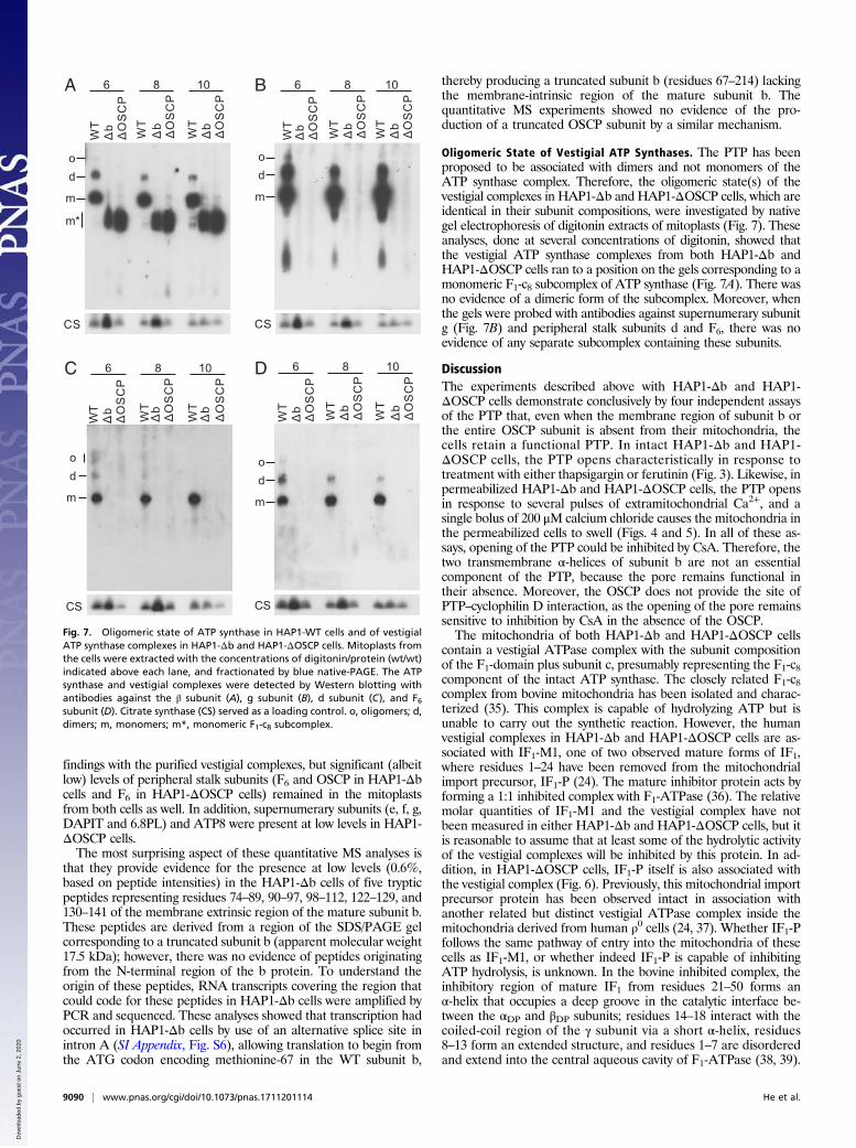
Oligomeric State of Vestigial ATP Synthases. The PTP has beenproposed to be associated with dimers and not monomers of theATP synthase complex. Therefore, the oligomeric state(s) of thevestigial complexes in HAP1-Δb and HAP1-ΔOSCP cells, which areidentical in their subunit compositions, were investigated by nativegel electrophoresis of digitonin extracts of mitoplasts (Fig. 7). Theseanalyses, done at several concentrations of digitonin, showed thatthe vestigial ATP synthase complexes from both HAP1-Δb andHAP1-ΔOSCP cells ran to a position on the gels corresponding to amonomeric F1-c8 subcomplex of ATP synthase (Fig. 7A). There wasno evidence of a dimeric form of the subcomplex. Moreover, whenthe gels were probed with antibodies against supernumerary subunitg (Fig. 7B) and peripheral stalk subunits d and F6, there was noevidence of any separate subcomplex containing these subunits.
DiscussionThe experiments described above with HAP1-Δb and HAP1-ΔOSCP cells demonstrate conclusively by four independent assaysof the PTP that, even when the membrane region of subunit b orthe entire OSCP subunit is absent from their mitochondria, thecells retain a functional PTP. In intact HAP1-Δb and HAP1-ΔOSCP cells, the PTP opens characteristically in response totreatment with either thapsigargin or ferutinin (Fig. 3). Likewise, inpermeabilized HAP1-Δb and HAP1-ΔOSCP cells, the PTP opensin response to several pulses of extramitochondrial Ca2+, and asingle bolus of 200 μM calcium chloride causes the mitochondria inthe permeabilized cells to swell (Figs. 4 and 5). In all of these as-says, opening of the PTP could be inhibited by CsA. Therefore, thetwo transmembrane α-helices of subunit b are not an essentialcomponent of the PTP, because the pore remains functional intheir absence. Moreover, the OSCP does not provide the site ofPTP–cyclophilin D interaction, as the opening of the pore remainssensitive to inhibition by CsA in the absence of the OSCP.The mitochondria of both HAP1-Δb and HAP1-ΔOSCP cells
contain a vestigial ATPase complex with the subunit compositionof the F1-domain plus subunit c, presumably representing the F1-c8component of the intact ATP synthase. The closely related F1-c8complex from bovine mitochondria has been isolated and charac-terized (35). This complex is capable of hydrolyzing ATP but isunable to carry out the synthetic reaction. However, the humanvestigial complexes in HAP1-Δb and HAP1-ΔOSCP cells are as-sociated with IF1-M1, one of two observed mature forms of IF1,where residues 1–24 have been removed from the mitochondrialimport precursor, IF1-P (24). The mature inhibitor protein acts byforming a 1:1 inhibited complex with F1-ATPase (36). The relativemolar quantities of IF1-M1 and the vestigial complex have notbeen measured in either HAP1-Δb and HAP1-ΔOSCP cells, but itis reasonable to assume that at least some of the hydrolytic activityof the vestigial complexes will be inhibited by this protein. In ad-dition, in HAP1-ΔOSCP cells, IF1-P itself is also associated withthe vestigial complex (Fig. 6). Previously, this mitochondrial importprecursor protein has been observed intact in association withanother related but distinct vestigial ATPase complex inside themitochondria derived from human ρ0 cells (24, 37). Whether IF1-Pfollows the same pathway of entry into the mitochondria of thesecells as IF1-M1, or whether indeed IF1-P is capable of inhibitingATP hydrolysis, is unknown. In the bovine inhibited complex, theinhibitory region of mature IF1 from residues 21–50 forms anα-helix that occupies a deep groove in the catalytic interface be-tween the αDP and βDP subunits; residues 14–18 interact with thecoiled-coil region of the γ subunit via a short α-helix, residues8–13 form an extended structure, and residues 1–7 are disorderedand extend into the central aqueous cavity of F1-ATPase (38, 39).
WT
6 8 10
od
m
m*
od
m
od
m
od
m
CS CS
CS CS
A B
C D
bΔ Δ
OS
CP
WT b
Δ ΔO
SC
P
WT b
Δ ΔO
SC
P
WT
6 8 10
bΔ Δ
OS
CP
WT b
Δ ΔO
SC
PW
T bΔ Δ
OS
CP
WT
6 8 10
bΔ Δ
OS
CP
WT b
Δ ΔO
SC
P
WT b
Δ ΔO
SC
P
WT
6 8 10
bΔ Δ
OS
CP
WT b
Δ ΔO
SC
P
WT b
Δ ΔO
SC
P
Fig. 7. Oligomeric state of ATP synthase in HAP1-WT cells and of vestigialATP synthase complexes in HAP1-Δb and HAP1-ΔOSCP cells. Mitoplasts fromthe cells were extracted with the concentrations of digitonin/protein (wt/wt)indicated above each lane, and fractionated by blue native-PAGE. The ATPsynthase and vestigial complexes were detected by Western blotting withantibodies against the β subunit (A), g subunit (B), d subunit (C), and F6subunit (D). Citrate synthase (CS) served as a loading control. o, oligomers; d,dimers; m, monomers; m*, monomeric F1-c8 subcomplex.
9090 | www.pnas.org/cgi/doi/10.1073/pnas.1711201114 He et al.
Dow
nloa
ded
by g
uest
on
June
2, 2
020

The mode of binding of IF1-P to the subcomplex is not known,but it seems possible that the presence in IF1-P of the additional24 N-terminal residues might impede binding of the protein tothe site occupied by mature IF1 in the inhibited complex.The removal of either subunit b or the OSCP destabilizes
the peripheral stalk, and none of its four constituent subunits(OSCP, b, F6, and d) or the associated ATP8 subunit, is presentin the vestigial subcomplex from HAP1-ΔOSCP cells and also inHAP1-Δb cells, although traces of the OSCP were detected inthe latter. In the absence of the peripheral stalk and ATP8,ATP6 no longer has any support to maintain its contact with thec8-ring (26–28). Therefore, ATP6 and the supernumerary sub-units (e, f, g, DAPIT, and 6.8PL) associated with ATP6 and themembrane domain of subunit b are also absent from the sub-complex (Fig. 6).As depicted in Fig. 1, subunit f is likely associated with subunits
ATP6 and ATP8 (27), and in the dimeric complex, subunits e and glikely form the interfaces between monomers, with subunitsDAPIT and 6.8PL together in a more peripheral position, relativeto the dimer interface. Clearly, this dimer interface is not present inthe vestigial complexes from HAP1-Δb and HAP1-ΔOSCP cells.However, subunits e, f, and g (and other supernumerary subunits)are still present in the mitoplasts of HAP1-Δb and HAP1-ΔOSCPcells, albeit at reduced levels relative to HAP1-WT cells (SI Ap-pendix, Fig. S5). In native gels of digitonin extracts of mitoplasts(Fig. 7), although there was no evidence of a separate subcomplex,
monomeric or dimeric, involving subunit g, it remains possible thatsuch a subcomplex was present in the mitoplast membranes, andthat it was disrupted by the conditions of extraction with digitonin.As noted earlier, dimers of the integral dimeric ATP synthase itselfcan become disrupted artifactually by this process (24, 40).Therefore, to eliminate any possibility of the participation of sub-units e and g (and subunits f, DAPIT, and 6.8PL) in forming thePTP, it will be necessary to remove each of them by gene disrup-tion experiments and to examine the consequences of doing so.
Materials and MethodsHuman HAP1 and mutant cells derived from themwere grown under standardconditions. Oxygen consumption rates were measured using a Seahorse XF24analyzer (Agilent). ATP5F1 and ATP5Owere disrupted in HAP1 cells by CRISPR-Cas9 technology (41). ATP synthase and its subcomplexes were purified frommitoplasts by immunocapture. Proteins were subject to stable isotope labelingin cell culture (SILAC). Labeled proteins were quantitated by MS. Opening ofthe PTP was assayed by four methods. In intact HAP1 cells, PTP opening wasinduced by thapsigargin (4) or ferutinin (5). In HAP1 cells in which the plasmamembrane had been permeabilized with digitonin, PTP opening was assessedby examining the capacity of the mitochondria to retain Ca2+ introduced ex-ogenously (42) and monitoring the swelling of mitochondria in response to apulse of 200 μM calcium chloride (43) in the absence and presence of CsA.Fulldetails of these processes are provided in SI Appendix.
ACKNOWLEDGMENTS. This work was supported by Medical Research Council,United Kingdom Programme Grant MR/M009858/1 (to J.E.W.).
1. Hunter DR, Haworth RA, Southard JH (1976) Relationship between configuration,function, and permeability in calcium-treated mitochondria. J Biol Chem 251:5069–5077.
2. Zoratti M, Szabò I (1995) The mitochondrial permeability transition. Biochim BiophysActa 1241:139–176.
3. Kwong JQ, Molkentin JD (2015) Physiological and pathological roles of the mito-chondrial permeability transition pore in the heart. Cell Metab 21:206–214.
4. Korge P, Weiss JN (1999) Thapsigargin directly induces the mitochondrial permeabilitytransition. Eur J Biochem 265:273–280.
5. Abramov AY, Duchen MR (2003) Actions of ionomycin, 4-BrA23187 and a novelelectrogenic Ca2+ ionophore on mitochondria in intact cells. Cell Calcium 33:101–112.
6. Chauvin C, et al. (2001) Rotenone inhibits the mitochondrial permeability transition-induced cell death in U937 and KB cells. J Biol Chem 276:41394–41398.
7. De Stefani D, Raffaello A, Teardo E, Szabò I, Rizzuto R (2011) A forty-kilodalton proteinof the inner membrane is the mitochondrial calcium uniporter. Nature 476:336–340.
8. Baughman JM, et al. (2011) Integrative genomics identifies MCU as an essentialcomponent of the mitochondrial calcium uniporter. Nature 476:341–345.
9. Crompton M, Ellinger H, Costi A (1988) Inhibition by cyclosporin A of a Ca2+-dependentpore in heart mitochondria activated by inorganic phosphate and oxidative stress.Biochem J 255:357–360.
10. Tanveer A, et al. (1996) Involvement of cyclophilin D in the activation of a mito-chondrial pore by Ca2+ and oxidant stress. Eur J Biochem 238:166–172.
11. Basso E, et al. (2005) Properties of the permeability transition pore in mitochondriadevoid of Cyclophilin D. J Biol Chem 280:18558–18561.
12. Nakagawa T, et al. (2005) Cyclophilin D-dependent mitochondrial permeabilitytransition regulates some necrotic but not apoptotic cell death. Nature 434:652–658.
13. Schinzel AC, et al. (2005) Cyclophilin D is a component of mitochondrial permeabilitytransition and mediates neuronal cell death after focal cerebral ischemia. Proc NatlAcad Sci USA 102:12005–12010.
14. Elrod JW, Molkentin JD (2013) Physiologic functions of cyclophilin D and the mito-chondrial permeability transition pore. Circ J 77:1111–1122.
15. Rasola A, Bernardi P (2007) The mitochondrial permeability transition pore and itsinvolvement in cell death and in disease pathogenesis. Apoptosis 12:815–833.
16. Kokoszka JE, et al. (2004) The ADP/ATP translocator is not essential for the mito-chondrial permeability transition pore. Nature 427:461–465.
17. Baines CP, Kaiser RA, Sheiko T, CraigenWJ, Molkentin JD (2007) Voltage-dependent anionchannels are dispensable for mitochondrial-dependent cell death. Nat Cell Biol 9:550–555.
18. Shanmughapriya S, et al. (2015) SPG7 is an essential and conserved component of themitochondrial permeability transition pore. Mol Cell 60:47–62.
19. König T, et al. (2016) The m-AAA protease associated with neurodegeneration limitsMCU activity in mitochondria. Mol Cell 64:148–162.
20. Giorgio V, et al. (2013) Dimers of mitochondrial ATP synthase form the permeabilitytransition pore. Proc Natl Acad Sci USA 110:5887–5892.
21. Walker JE (2013) The ATP synthase: The understood, the uncertain and the unknown.Biochem Soc Trans 41:1–16.
22. Bonora M, et al. (2013) Role of the c subunit of the FO ATP synthase in mitochondrialpermeability transition. Cell Cycle 12:674–683.
23. Alavian KN, et al. (2014) An uncoupling channel within the c-subunit ring of the F1FOATP synthase is the mitochondrial permeability transition pore. Proc Natl Acad SciUSA 111:10580–10585.
24. He J, et al. (2017) Persistence of the mitochondrial permeability transition in theabsence of subunit c of human ATP synthase. Proc Natl Acad Sci USA 114:3409–3414.
25. Masgras I, Rasola A, Bernardi P (2012) Induction of the permeability transition pore incells depleted of mitochondrial DNA. Biochim Biophys Acta 1817:1860–1866.
26. Zhou A, et al. (2015) Structure and conformational states of the bovine mitochondrialATP synthase by cryo-EM. eLife 4:e10180.
27. Vinothkumar KR, Montgomery MG, Liu S, Walker JE (2016) Structure of the mito-chondrial ATP synthase from Pichia angusta determined by electron cryo-microscopy.Proc Natl Acad Sci USA 113:12709–12714.
28. Hahn A, et al. (2016) Structure of a complete ATP synthase dimer reveals the mo-lecular basis of inner mitochondrial membrane morphology. Mol Cell 63:445–456.
29. Dickson VK, Silvester JA, Fearnley IM, Leslie AGW, Walker JE (2006) On the structureof the stator of the mitochondrial ATP synthase. EMBO J 25:2911–2918.
30. Rees DM, Leslie AGW, Walker JE (2009) The structure of the membrane extrinsic re-gion of bovine ATP synthase. Proc Natl Acad Sci USA 106:21597–21601.
31. Carette JE, et al. (2011) Ebola virus entry requires the cholesterol transporterNiemann-Pick C1. Nature 477:340–343.
32. Essletzbichler P, et al. (2014) Megabase-scale deletion using CRISPR/Cas9 to generatea fully haploid human cell line. Genome Res 24:2059–2065.
33. Vaca Jacome AS, et al. (2015) N-terminome analysis of the human mitochondrialproteome. Proteomics 15:2519–2524.
34. Gevaert K, et al. (2003) Exploring proteomes and analyzing protein processing by massspectrometric identification of sorted N-terminal peptides. Nat Biotechnol 21:566–569.
35. Watt IN, Montgomery MG, Runswick MJ, Leslie AGW, Walker JE (2010) Bioenergeticcost of making an adenosine triphosphate molecule in animal mitochondria. ProcNatl Acad Sci USA 107:16823–16827.
36. Gomez-Fernandez JC, Harris DA (1978) A thermodynamic analysis of the interactionbetween the mitochondrial coupling adenosine triphosphatase and its naturally oc-curring inhibitor protein. Biochem J 176:967–975.
37. Wittig I, et al. (2010) Assembly and oligomerization of human ATP synthase lackingmitochondrial subunits a and A6L. Biochim Biophys Acta 1797:1004–1011.
38. Gledhill JR, Montgomery MG, Leslie AGW, Walker JE (2007) How the regulatoryprotein, IF1, inhibits F1-ATPase from bovine mitochondria. Proc Natl Acad Sci USA 104:15671–15676.
39. Bason JV, Montgomery MG, Leslie AGW, Walker JE (2014) Pathway of binding of theintrinsically disordered mitochondrial inhibitor protein to F1-ATPase. Proc Natl AcadSci USA 111:11305–11310.
40. Arnold I, Pfeiffer K, Neupert W, Stuart RA, Schägger H (1998) Yeast mitochondrialF1Fo-ATP synthase exists as a dimer: Identification of three dimer-specific subunits.EMBO J 17:7170–7178.
41. Ran FA, et al. (2013) Genome engineering using the CRISPR-Cas9 system. Nat Protoc 8:2281–2308.
42. Murphy AN, Bredesen DE, Cortopassi G, Wang E, Fiskum G (1996) Bcl-2 potentiatesthe maximal calcium uptake capacity of neural cell mitochondria. Proc Natl Acad SciUSA 93:9893–9898.
43. Clarke SJ, McStay GP, Halestrap AP (2002) Sanglifehrin A acts as a potent inhibitor ofthe mitochondrial permeability transition and reperfusion injury of the heart bybinding to cyclophilin-D at a different site from cyclosporin A. J Biol Chem 277:34793–34799.
He et al. PNAS | August 22, 2017 | vol. 114 | no. 34 | 9091
BIOCH
EMISTR
Y
Dow
nloa
ded
by g
uest
on
June
2, 2
020