new enzyme immunoassay with visual detection based on membrane photoimmobilized antibodies
TRANSCRIPT

This article was downloaded by: [Temple University Libraries]On: 14 November 2014, At: 22:30Publisher: Taylor & FrancisInforma Ltd Registered in England and Wales Registered Number: 1072954Registered office: Mortimer House, 37-41 Mortimer Street, London W1T 3JH,UK
Analytical LettersPublication details, including instructions forauthors and subscription information:http://www.tandfonline.com/loi/lanl20
New Enzyme Immunoassaywith Visual Detection Based onMembrane PhotoimmobilizedAntibodiesB. M. Gorovits a , A. P. Osipov a , V. V. Doseeva a & A.M. Egorov aa Division for Chemical Enzymology, Departmentof Chemistry , M. V. Lomonosov Moscow StateUniversity , 119899, Moscow, U.S.S.R.Published online: 23 Oct 2006.
To cite this article: B. M. Gorovits , A. P. Osipov , V. V. Doseeva & A. M. Egorov(1991) New Enzyme Immunoassay with Visual Detection Based on MembranePhotoimmobilized Antibodies, Analytical Letters, 24:11, 1937-1966, DOI:10.1080/00032719108053026
To link to this article: http://dx.doi.org/10.1080/00032719108053026
PLEASE SCROLL DOWN FOR ARTICLE
Taylor & Francis makes every effort to ensure the accuracy of all theinformation (the “Content”) contained in the publications on our platform.However, Taylor & Francis, our agents, and our licensors make norepresentations or warranties whatsoever as to the accuracy, completeness,or suitability for any purpose of the Content. Any opinions and viewsexpressed in this publication are the opinions and views of the authors, andare not the views of or endorsed by Taylor & Francis. The accuracy of theContent should not be relied upon and should be independently verified withprimary sources of information. Taylor and Francis shall not be liable for anylosses, actions, claims, proceedings, demands, costs, expenses, damages,and other liabilities whatsoever or howsoever caused arising directly or

indirectly in connection with, in relation to or arising out of the use of theContent.
This article may be used for research, teaching, and private study purposes.Any substantial or systematic reproduction, redistribution, reselling, loan,sub-licensing, systematic supply, or distribution in any form to anyone isexpressly forbidden. Terms & Conditions of access and use can be found athttp://www.tandfonline.com/page/terms-and-conditions
Dow
nloa
ded
by [
Tem
ple
Uni
vers
ity L
ibra
ries
] at
22:
30 1
4 N
ovem
ber
2014
ANALYTICAL LETTERS, 24(11), 1937-1966 (1991)
NEW ENZYME IMMUNOASSAY WITH VISUAL DETECTION BASED ON
MEMBRANE PHOTOIMMOBILIZED ANTIBODIES
Key words: enzyme immunoassay, dot-analysis, IgG,
Shigella Sonnei, thyroxine, human chorionic
gonadotropin.
B.M.Gorovits, A.P.Osipov, V.V.Doseeva and A.M.Egorov
Division for Chemical Enzymology, Department of
Chemistry, M.V.Lomonosov Moscow State University,
119899 MOSCOW, U.S .S .R .
ABSTRACT
New method for visual enzyme immunoassay of some
model antigens in solution by using covalently
photoimmobilized antibodies has been developed. This
approach is based on the quantitative photoimmobiliza-
tion of antibodies on the surface of porous matrixes.
It is easy to control the dimensions and shape of the
activated zones and the quantity of the active groups
1937
Copyright 0 1991 by Marcel Dekker, Inc.
Dow
nloa
ded
by [
Tem
ple
Uni
vers
ity L
ibra
ries
] at
22:
30 1
4 N
ovem
ber
2014

1938 GOROVITS ET AL.
on it by this technique. A strip of the membrane im-
pregnated with p-azidobenzaldehyde was illuminated by
the light. A s a result, .the quantity of aldehyde groups
developed on the surface of membrane is proportional to
the time of illumination. After the covalent immobi-
lization of antibodies, the membrane has separate zones
with an exact surface concentration of antibodies. The
antigens of different types were assayed: human IgG,
human chorionic gonadotropin, S h i g s l l a Sonnei. The
lowest detection limit was 1 fig/ml, 20 U/l, l x104
cells/ml. The method allows measure of thyroxir,e con-
centrations in the range 5 0 to 200 nM: the precision of
replicate measurements has the coefficient of variation
7%. The reasons for the background signal appearance
were accurately analyzed. The choice of support was
substantiated, the optimal conditions of it.s pretreat-
ment being defined. This method makes possible the
visualization of the results based on comparison the
color intensity of zones with the control.
INTRODUCTION
The use of porous supports in the enzyme immunoas-
say (EIA) resulted in the creation of so called "on-
site" or "doctor-officeP1 kits for a fast visual quan-
tification of different substances in biological
fluids. When preparing any type of solid phase im-
Dow
nloa
ded
by [
Tem
ple
Uni
vers
ity L
ibra
ries
] at
22:
30 1
4 N
ovem
ber
2014

MEMBRANE P H O T O I M M O B I L I Z E D A N T I B O D I E S 1939
munoassay, it is important to choose the optimal sur-
face antibody concentration. In this respect, the
covalent immobilization 31: proteins on the matrixes
seems most convenient. There are different techniques
of covalent binding, for example the use of benzo-
quinone as activation reagent for filter paper . The application of porous support allows one to detect the
result visually. However, the visual detection which
takes into account dot size and intensity of stain-
ing is rather difficult. For examples, the dot coloring
may not be uniform, the dot shape may be vague. There-
fore it is complicated to define the exact dot size and
to assess its intensity compared to the standard. This
article describes the application of photoimmobiliza-
tion method previously elaborated for enzymephotography
1
L processes . The main idea of the method developed is the following. Impregnated with p-azidobenzaldehyde,
the membrane is UV-illuminated by constant inten-
sity. A s a result on the membrane surface aldehyde
groups are developed, the apparent surface concentra-
tion of which is proportional to the time of zone il-
lumination. After incubation of this activated membrane
in solution of antibodies, the antibodies were
covalently immobilized and zones with proportional an-
tibody surface concentration were developed. The
analyte can be quantified in sandwich or competitive
scheme of immunoassay.
Dow
nloa
ded
by [
Tem
ple
Uni
vers
ity L
ibra
ries
] at
22:
30 1
4 N
ovem
ber
2014

1940
EXPERIMENTAL
GOROVITS ET A L .
Materials and methods
Horseradish peroxidase (HRP), specific activity
8 2 9 U/ng, RZ=A403nM/A280nM=3.0, was purchased from
Biolar. Sodium periodate was obtained from Merck.
Potassium 4,4~-diazidostilbene-2,2~-disulfonate and
diethylacetate p-azidobenzaldehyde were originally syn-
thesized by A.A.Cshegolev at the division of chemical
enzymology, Moscow State University. The following
matrixes were used: chromatography paper FN-11
(Filtrak, Germany), regenerated cellulose MFC,
regenerated cellulose on nylon. net KS-49, nylon
membrane (Polymersynthesis, Vladimir, USSR), nuclear
(track) filters (Institute for Nuclear Research, Dubna,
USSR), nitrocellulose (Chemapol, CSFR). Thyroxine (T4),
bovine serum albumin (BSA), morpholinoethyl car-
bodiimide and diaminobenzidine dihydrochloride (DAB)
were purchased from Sigma.
To prepare peroxidase-labeled anti-peroxidase an-
tibodies, antiperoxidase serum was fractionated with
ammonium sulfate, followed by ion-exchange chromatog-
raphy on DEAE-Toyoperl 650M with a subsequent purifica-
tion of IgG fraction coupled with peroxidase by the
periodate method 3
For sensitization, the matrixes were placed in
the solution of photoreagent, than dried in the air at
Dow
nloa
ded
by [
Tem
ple
Uni
vers
ity L
ibra
ries
] at
22:
30 1
4 N
ovem
ber
2014
MEMBRANE PHOTOIMMOBILIZED ANTIBODIES 1941
room temperature in the dark. Two substances were used
as photoreagents: potassium 4,4'-dia~idostilbene-2,2~-
disulfonate (PDAS) (10 mg/ml water solution) and
diethylacetate p-azidobenzaldehyde (DABA) (0.45 mg/ml
benzene solution). Nonfiltered light of mercury lamp
and a Shimadzu CS-9000 deuterium lamp were used for
illumination. The matrixes were washed with water or
ethyl alcohol, respectively. DABA-activated matrixes
were additionally treated with 0.1M HC1 for 15-30 min.
IaG immobilization on photoactivated supports.
a) PDAS activated matrixes were placed in mor-
pholinoethyl carbodiimide solution (5 mg/ml) for 30 min
at room temperature then replaced into 0.01 M phosphate
buffer, pH 7.2, containing 0.15 M NaCl and im-
munoglobulin of different concentrations. Incubation
was carried out at room temperature for 30-120 min.
Noncovalently bound proteins were extracted by using
PBS with 0.05% Tween-20. In other cases, 0.1 M Tris-HC1
and pH 9.2 buffer containing 1 M NaC1, 0.5% Triton
X-100 and 0.2% potassium deoxycholate were used.
b) DABA activated matrixes were placed into im-
munoglobulin solution ( 1 ~ 1 0 - ~ - 1 ~ 1 0 - ~ M) in 0.01 M
phosphate buffer (pH 7.4; 0.14 M NaC1) (PBS). Incuba-
tion was carried out for 3-120 min at room temperature
or 12-14 h at 4OC. Then supports were treated with
NaBH4 (lmg/ml) solution in PBS for 15 min at room tem-
Dow
nloa
ded
by [
Tem
ple
Uni
vers
ity L
ibra
ries
] at
22:
30 1
4 N
ovem
ber
2014

1942 GOROVITS ET AL.
perature. The noncovalently bound proteins were ex-
tracted as described above.
Assav txocedures.
Photoactivation (by DABA), pretreatment, immobi-
lization of specific antibodies and removal of non-
covalently bound proteins were performed as mentioned
above.
a) Human chorionic UonadotroDin IhCG) assay. Sup-
ports containing immobilized anti-p-subunit of hCG IgG
were placed into the standard hCG solution in PBS con-
taining 0.05% Tween-20 (PBST). The incubation time was
varied. After washing the supports were placed into
anti-hCG (a-subunit) IgG-enzyme conjugate solutions in
PBST (5x10‘9 M) for 10-60 min. After washing, thereof,
the substrate solution containing 0.5 mg/ml DAB + H202 + 0.02% C O + ~ in PBS was used for visualization.
The intensity of dyeing was measured by a Shimadzu cs- 9000 instrument.
b) Assav of human and rabbit immunoslobulins G.
Matrixes were incubated successively in the solutions
of IgG-standards, IgG-enzyme conjugate (5x10-’ - lxlO-’ M) and substrate. The solution of 4-chloro-1-naphtol 4
was used for the detection.
c) S h i q e l l a S o n n e i assay. Matrixes were incubated
successively in the solutions of S h i g e l l a S o n n e i stan-
dards, protein A-enzyme conjugate (1 ampule in 10 ml
Dow
nloa
ded
by [
Tem
ple
Uni
vers
ity L
ibra
ries
] at
22:
30 1
4 N
ovem
ber
2014

MEMBRANE PHOTOIMMOBILIZED ANTIBODIES 1943
PBST, 1 h, room temperature) and substrate. Diaminoben-
zidine solution was used for the detection.
d) Thvroxine assav. Matrixes were successively in-
cubated in two solutions: one containing thyroxine
standards and horse radish peroxidase labeled antigen
(15-30 ng/ml) in PBST and the other containing DAB as
substrate.
RESULTS AND DISCUSSION
IuG immobilization on Rhotoactivated supports.
The earlier developed method for protein
photoimmobilization allows one to easily control the
size and shape of the activated matrix zone as well as
the degree of its activation. The prospects for ap-
plication of photoimmobilization to construct the sys-
tems for quantitative and semiquantitative im-
munodetection (with and without instrument respec-
tively) of some model antigens will be discussed fur-
ther. We selected the photoactivation reagent, support,
light source and the method for pretreatment of
matrixes to design the appropriate - to - exploit sys- tem possessing the maximum ratio, useful signal-
background.
Choice of DhotocouRlinu reaaent.
Some photoactive compounds known can be applied
for photoimmobilization of proteins on solid supports.
Dow
nloa
ded
by [
Tem
ple
Uni
vers
ity L
ibra
ries
] at
22:
30 1
4 N
ovem
ber
2014

1944 GOROVITS ET AL.
Two reagents of these tested in our work were sodium
4,41-diazidostilbene-2,21-disu1fonate and p-azido-
benzaldehyde. The first compound is water-soluble and
is characterized by a high rate of photodegradation
A reagent was covalently bound to the matrix,
nitrocellulose membranes or chromatographic paper, im-
pregnated with sodium 4,41-diazostilbene-2,2~-disul-
fonate by W-illumination. The proteins were subse-
quently immobilized by the activation of one or two
sulfo-groups of a photo-binding reagent with carbodi-
bide and by the reaction with amino-groups of protein.
In these conditions, rabbit G-immunoglobulin was
immobilized on nitrocellulose matrix. For the quantita-
tivedetermination 05 protein binding efficiency, the
modified matrix was treated with A-protein marked with
horse radish peroxidase (A-HRP). After evaluating the
nonspecifically bonded protein (see IlMaterials and
methodsgg) the matrix was allowed to react with the sub-
strate mixture to detect the enzyme label. The inten-
sity of support colour is related to the amount of im-
munoglobulins in solution analyzed.
5
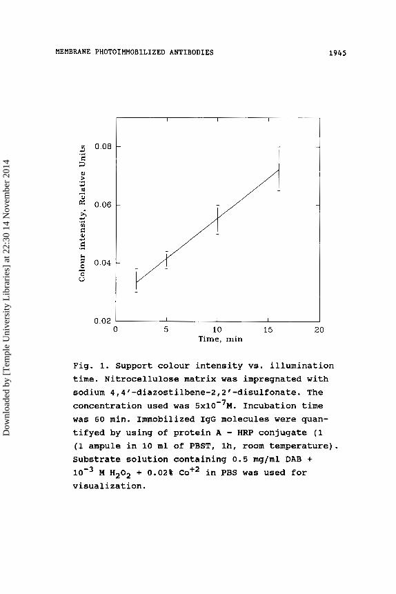
It follows from the dependency obtained (Fig.1)
that the binding.efficiency vs time is linear if the
illumination time is about 15 min or less. It should be
noted, however, that the rather high background level
seems to be due to the activation of some acid groups
on the matrix by carbodiimide and to the following
Dow
nloa
ded
by [
Tem
ple
Uni
vers
ity L
ibra
ries
] at
22:
30 1
4 N
ovem
ber
2014
MEMBRANE PHOTOIMMOBILIZED ANTIBODIES
v1 0.08
F: 5
3 .r(
z .” 3 m 4
0.06 i, 3
m .r(
E 4 d
2 0.04
.r(
h - 0 u
1945
-
-
-
I I I
0.02 ‘ I I I
0 5 10 15 20 Time, min
Fig. 1. Support colour intensity vs. illumination time. Nitrocellulose matrix was impregnated with sodium 4,41-diazostilbene-2,2~-disulfonate. The concentration used was 5x10-’M. Incubation time was 60 min. Immobilized IgG molecules were quan- tifyed by using of protein A - HRP conjugate (1 (1 ampule in 10 ml of PBST, lh, room temperature). Substrate solution containing 0.5 mg/ml DAB +
M H202 + 0.02% C O + ~ in PBS was used for visualization.
Dow
nloa
ded
by [
Tem
ple
Uni
vers
ity L
ibra
ries
] at
22:
30 1
4 N
ovem
ber
2014
1946 GOROVITS ET AL.
binding of IgG-molecules. The decrease in im-
munoglobulin concentration in solution as well as in
the immobilization time reduces the colour density of
both the background and the exposed sites (table 1).
The better results were obtained by using
p-azidobenzaldehyde as a photoimmobilization agent. It
can be used in organic solvent, but not in water, which
obviously restricts a possible matrix range. When the
impregnated membrane is W-illuminated the photosensi-
tive molecule transforms into the nitrene biradical
which can effectively interact with the matrix.
The protein immobilization was performed in
this case by the Schiff's reaction of aldehyde groups
on the matrix with amino groups of immunoglobulins.
It follows from the kinetic dependence of IgG
binding on the photomodified matrix (Fig.2) obtained
that if the immunoglobulin concentration ranges from
3 ~ 1 0 - ~ M to ~ x I O - ~ M the basic amount of protein binds to
thesupport faster than in 5 min. After 10-20 min in-
cubation, the reaction was complete . Figure 2 shows the results correspond to the covalently immobilized
proteins only. At all immobilization time intervals,
the surface concentration of immunoglobulins deviates
from one site of a matrix to the other in accordance
with variable activation degrees.
It was reported that in the process of
photoimmobilization on the porous supports the ratio of
Dow
nloa
ded
by [
Tem
ple
Uni
vers
ity L
ibra
ries
] at
22:
30 1
4 N
ovem
ber
2014

MEMBRANE PHOTOIMMOBILIZED ANTIBODIES 1947
Table 1. Influence of immobilization time on the colour density of both background ( D 6 5 0 R , 0 ) and 15 min exposed sites (D650R,15) of matrix impregnated with sodium 4,4'-diazostilbene-2,2'- disulfonate.
D650 R t O R , 15 Immo b i 1 i z a t i on
time, hours D
aldehyde groups to protein molecules was 5 0 0 : l . It is
obvious that in this case the amount of protein would
be the same on all activated sites. It is likely for
most aldehyde groups to exist in spatially unapproach-
able areas of porous matrix or to be protected by al-
ready immobilized immunoglobulins. Therefore the matrix
with a higher regular porous structure increases the
concentration of protein per unit of surface area.
Choice of matrix
The properties of the matrix chosen as solid
support have a large influence on the results of enzyme
immunoassay. These matrixes should be a neutral polymer
with minimal nonspecific interactions with biomolecules
and components of staining solution. They are able to
absorb qualitatively the final insoluble product of en-
Dow
nloa
ded
by [
Tem
ple
Uni
vers
ity L
ibra
ries
] at
22:
30 1
4 N
ovem
ber
2014

1948 GOROVITS ET AL.
0.03 I 3 I I I I I
a. 1 1
-
I 3 I 1 I I
0 20 40 60 80 100 120 Time, min
0.03 5 - 0, p:
0.02 .I 5 z
2
0, .Q 0.01 d .I
0 6 0.00
b.
I I I
cs 0 20 40 60 80 100 120
Time, min
Fig. 2. Kinetics of the immunoglobulin immobiliza- tion on the photoactivated support FN-11. Plots were obtained for the following initial concentra- tions of IgG: a) 3 . 0 ~ 1 0 - ~ M, b) ~ . O X ~ O - ~ M, and photoactivation times: 1) 10 min, 2) 3 min, 3) 0.5 min. Matrixes were impregnated with DABA. Attached IgG molecules were quantified by using A - HRP conjugate (1 ampule in 10 ml of PBST, 1 h, room temperature). Substrate solution containing 0.5 mg/ml DAB + 10% H202 + 0.02% C O + ~ in PBS was used for visualization.
protein
Dow
nloa
ded
by [
Tem
ple
Uni
vers
ity L
ibra
ries
] at
22:
30 1
4 N
ovem
ber
2014

MEMBRANE PHOTOIMMOBILIZED A N T I B O D I E S 1949
zyme reaction. Other requirements are the mechanical
strength and the chemical stability during activation
and functioning conditions. The porous matrix structure
provides high specific surface and therefore high
protein capacity.
Table 2 shows the results demonstrating the pos-
sibilities of the maximal binding of immunoglobulins by
various porous supports. Some supports (nitrocellulose
membranes, for example) can not withstand the treatment
with a photoreagent solution in organic solvent. Others
have a sufficiently large specific surface for the high
efficiency binding (for instance, nuclear filters with
a pore diameter of about 0.2-2 pm). For such membranes,
the summary section area of the pores obtained is about
a few percent of the whole film surface . The
highest ratio of signal to background was obtained with
the matrixes manufactured from the regenerated cel-
lulose (MFC) and cellulose matrix FN-11.
7
The absence of necessary mechanical strength of
MFC membranes hinders their application in analysis.
Application of regenerated cellulose matrix onto nylon
network (KS-49) provides a tensile or bending strength
to the matrix, a stability upon photochemical modifica-
tion and EIA. The ratio of signal to background is high
enough for KS-49 too (table 2).
Satisfactory results were obtained with chromatog-
raphy paper support Filtrak FN-11, a highly porous cel-
Dow
nloa
ded
by [
Tem
ple
Uni
vers
ity L
ibra
ries
] at
22:
30 1
4 N
ovem
ber
2014

1950 GOROVITS ET AL.
Table 2. Application of the various supports for photoimmobilization of immunoglobulins.
Regenerated cellulose on nylon net KS-49
0.02-0.05 0.6-0.9
Nylon membrane
0.3-0.5 1 - 2
Nuclear filters - I 1 - 0.01 0.03
lulose material. However, such matrix can not be suc-
cessfully used in all cases, as will be shown below.
Choice of W-liaht source.
Any source of W-light can be applied for the sup-
port photoactivation with the help of DABA. Mercury
lamp and deuterium lamp of CS-9000 were used in this
case. It should be noted that the differences in the
dependence of the matrix activation degrees from the
W-illumination time can be caused both by the conse-
quence of light source change and by the change of the
matrix. For example, larger W-illumination times are
required for regenerated cellulose matrix activation
Dow
nloa
ded
by [
Tem
ple
Uni
vers
ity L
ibra
ries
] at
22:
30 1
4 N
ovem
ber
2014

MEMBRANE PHOTOIMMOBILIZED ANTIBODIES 1951
compared to FN-11. This should be caused, in par-
ticular, by glycerine widely used as stabilizer with
some types of membranes, which can effectively interact
with DABA upon its photoactivation. As follows from the
dependence of surface protein concentration on FN-11
matrix versus illumination time (Fig.3), the most ef-
fective binding of immunoglobulin density on the matrix
takes place during the period of W-illumination by
this source for about 4-5 minutes. After 20 min il-
lumination, the surface protein concentration is
highest (>2x1O-l2 mole/cm2) . The quantity of covalently bound protein on the matrix was determined by subtrac-
tion of noncovalently bound protein from its total
quantity . JJ
The availability of the internal control section
is important for verifying the total nonspecific bind-
ing in any type of immunoassay. The method of photoim-
mobilization permits combination of internal control
with signal section on one matrix. The ratio of colour
intensities for signal and internal control sections
corresponds to signal/noise value. Influencing factors
are considered below.
ou antibodies.
This binding can be realized as a result of hydrogen,
electrostatic, hydrophobic and dispersion bonds. For
Dow
nloa
ded
by [
Tem
ple
Uni
vers
ity L
ibra
ries
] at
22:
30 1
4 N
ovem
ber
2014

1952 GOROVITS ET AL.
3
N
E 0
4 2 z 2 0 4 - c .d
' P I 4
a E l Y
0
I I I I I I
0 200 400 600 800 1000 1200 1400 Time, sec
F i g . 3 Surface protein (conjugate I g G - HRP) concentration on FN-11 matrix versus illumination time. Deuterium lamp of CS-9000 was used in this case. Matrixes were impregnated with DABA.
Dow
nloa
ded
by [
Tem
ple
Uni
vers
ity L
ibra
ries
] at
22:
30 1
4 N
ovem
ber
2014

MEMBRANE PHOTOIMMOBILIZED ANTIBODIES 1953
Table 3. Composition of solutions for removing noncovalently bound immunoglobulins from different porous supports.
.................................................. Matrix composition .................................................. Chromatog- 1M NaC1, 1% Triton X-100, 0.2% raphy paper sodium deoxycholate, TRIS-HC1
buffer pH 9.2 .................................................. Nylon - I1 - membrane
Regenerated 0.05% Tween-20, 0.01 M phosphate cellulose buffer, pH 7.2
Regenerated 0.1% Triton X-100, 0.01M phosphate cellulose on buffer, pH 7.2 the nylon net
..................................................
..................................................
removal of antibodies noncovalently bound with matrix,
the detergent solutions (Tween-20, Triton X-100, sodium
deoxycholate) were used. The compositions of solutions
to obtain the best results for different porous sup-
ports are presented in Table 3.
The antigen studied as well as the double antibody
conjugate with an enzyme (in sandwich assay) could be
adsorbed on the non-illuminated sites of the matrix. To
decrease this type of interaction we blocked the matrix
surface using BSA solutions (0.1-1.2%). The dependence
of a reference signal value for FN-11 matrix is shown
in Fig.4. It is obvious that 0.5% BSA solutions could
be successfully used. To shorten the treatment dura-
tion, we used 2-5% protein solutions.
Dow
nloa
ded
by [
Tem
ple
Uni
vers
ity L
ibra
ries
] at
22:
30 1
4 N
ovem
ber
2014

1954
P
s
2 0.1
s
.4 4 m *
.d 5 0, 4
C .4
0 0 u 4
0.0
GOROVITS ET AL.
- 0
0
\ @
\ I T
0.0 0.4 0.8 1.2
[BSA], %
Fig. 4. Reference signal values versus concentra- tions of blocking BSA solution for FN-11 matrix. Matrixes were impregnated with DABA. Blocking time was 30 min. Unoccupied sites for sorption were titred by IgG - HRP conjugate ( 5 ~ 1 0 - ~ M ) , incuba- tion time was 30 min. Substrate solution contain- ing 0.5 mg/ml DAB + 10'3M H202 + 0.02% C O + ~ in PBS was used for visualization.
Dow
nloa
ded
by [
Tem
ple
Uni
vers
ity L
ibra
ries
] at
22:
30 1
4 N
ovem
ber
2014

MEMBRANE PHOTOIMMOBILIZED ANTIBODIES 1955
Table 4 . Application of FN-11 matrix in hCG assay. Investigation of background signals.
This method was used for creation of enzyme im-
munoassay systems for detection of different model an-
tigens in solutions. Antigens of different types were
analyzed: high molecular weight antigens - human IgG, human chorionic gonadotropin, cells of Shigella Sonnei
and low molecular weight analyte thyroxine.
Immunoenzymatic determination of human chorionic
aonadotropin.
As follows from the results of Table 4 , the sig-
nals of high (500 U/1) and zero concentrations are
practically equal for FN-11 support. The best results
were obtained with the regenerated cellulose MFC
matrix, no nonspecific protein sorption taking place
actually.
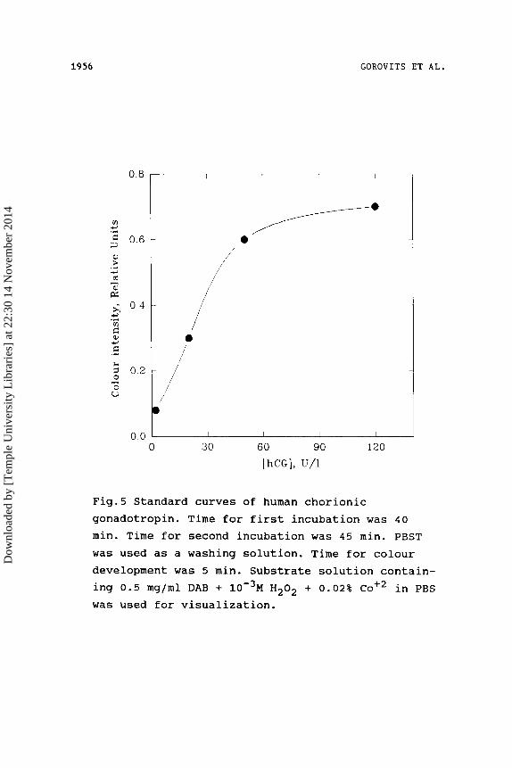
The calibration curve for hCG determination in
solution is presented on Figure 5. In practice the
critical concentration of hCG is about 50 U/1. It fol-
lows from Fig.5 that this concentration can be
detected with ease. This appears from the kinetic de-
pendences of the hormone binding to the specific an-
Dow
nloa
ded
by [
Tem
ple
Uni
vers
ity L
ibra
ries
] at
22:
30 1
4 N
ovem
ber
2014
1956 GOROVITS ET AL.
0.0 I I 1 I
0 30 60 90
[hCG], U/1
Fig.5 Standard curves of human chorionic
L 120
gonadotropin. Time for first incubation was 40 min. Time for second incubation was 45 min. PBST was used as a washing solution. Time for colour development was 5 min. Substrate solution contain- ing 0.5 mgjml DAB + 10% H202 + 0.02% C O + ~ in PBS was used for visualization.
Dow
nloa
ded
by [
Tem
ple
Uni
vers
ity L
ibra
ries
] at
22:
30 1
4 N
ovem
ber
2014

MEMBRANE PHOTOIMMOBILIZED ANTIBODIES 1957
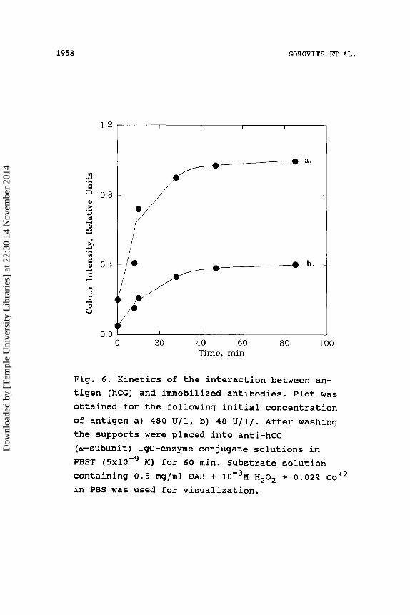
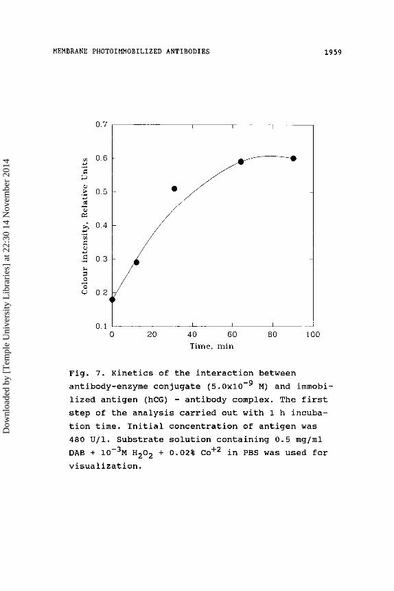
tibodies immobilized on the porous support (Fig.6) and
to the specific antibodies conjugated with HRP via im-
munological complex on the porous support (Fig.7). The
maximal signal develops in 20-30 minutes depending on
variable concentrations ( 4 8 0 U/1, 4 8 U/1) of original
protein under study. Thus, the whole analysis time was
estimated to be 1.5-2 hours.
To simplify the analytical procedure and to
decrease its time, the immobilized antibodies to
p-subunit of the hormone containing strip were treated
with the mixture of the hormone and conjugate solu-
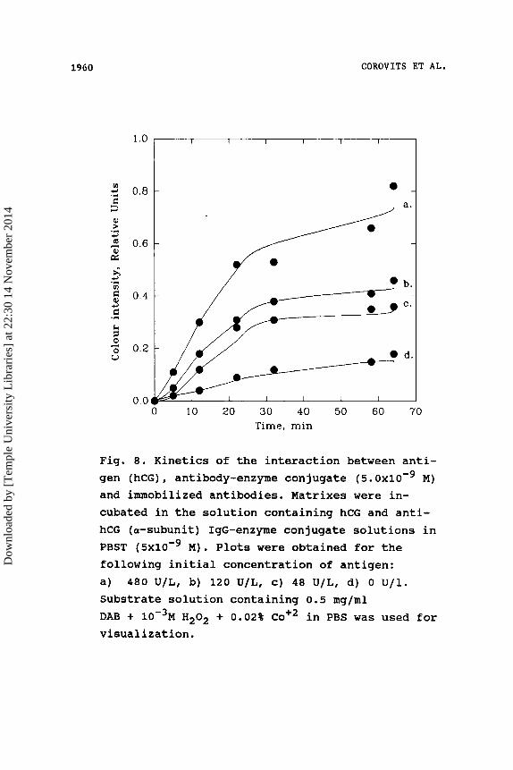
tions. The dependences of the signal versus the incuba-
tion time of the strip in this mixture are shown in
Fig.8. Curves a, b, c, d correspond to hCG concentra-
tions of 480, 120, 48, 0 U/l, respectively.
The analysis of these kinetic data allows us to con-
sider that 20-30 min incubation time is sufficient to
obtain the signals differing from the background even
if the antigen concentration is significantly low.
Therefore, the analysis time was reduced to 30 minutes a s
a result 6f optimization of the steps.
Enzyme immunoassay of human IqG.
IgG enzyme immunoassay was carried out using
chromatography paper FN-11 as a solid matrix. The
matrix was photoactivated in a manner, which al-
lows obtaining the maximal concentration of immobilized
antibodies.
Dow
nloa
ded
by [
Tem
ple
Uni
vers
ity L
ibra
ries
] at
22:
30 1
4 N
ovem
ber
2014
1958 GOROVITS ET AL.
-0 a. -
b.
I 0.0 I I I I 1
0 20 40 60 80 100 Time, min
Fig. 6. Kinetics of the interaction between an- tigen (hCG) and immobilized antibodies. Plot was obtained for the following initial concentration of antigen a) 480 U / 1 , b) 48 U / l / . After washing the supports were placed into anti-hCG (a-subunit) IgG-enzyme conjugate solutions in PBST (5x10-' M) for 60 min. Substrate solution containing 0.5 mg/ml DAB + ~ o - ~ M H202 + 0.02% co+2 in PBS was used for visualization.
Dow
nloa
ded
by [
Tem
ple
Uni
vers
ity L
ibra
ries
] at
22:
30 1
4 N
ovem
ber
2014
MEMBRANE PHOTOIMMOBILIZED ANTIBODIES 1959
0.7
0.6 3 .- C D al
3 6 al a:
,% 0.5 - 0.4
4
m .-
5 .: 0 3
2 : 0.2
4
L - I
0.1
I r I I
I I I I
0 20 40 60 ao 100 Time, min
Fig. 7. Kinetics of the interaction between antibody-enzyme conjugate (5. Oxlo-’ M ) and immobi- lized antigen (hCG) - antibody complex. The first step of the analysis carried out with 1 h incuba- tion time. Initial concentration of antigen was 480 U / l . Substrate solution containing 0.5 mg/ml DAB + 10m3M H 2 0 2 + 0 . 0 2 % C O + ~ in PBS was used for visualization.
Dow
nloa
ded
by [
Tem
ple
Uni
vers
ity L
ibra
ries
] at
22:
30 1
4 N
ovem
ber
2014
1960 GOROVITS ET AL.
3 9 ." rn
4 c .r(
h
m 0 V
0.8
I / 0
0 10 20 30 40 50 60
b.
C.
d.
-
70 Time, min
Fig. 8. Kinetics of the interaction between anti- gen (hCG) , antibody-enzyme con jugate (5. Oxlo-' M) and immobilized antibodies. Matrixes were in- cubated in the solution containing hCG and anti- hCG (a-subunit) IgG-enzyme conjugate solutions in PBST (5x10-' M) . Plots were obtained for the following initial concentration of antigen: a) 480 U/L, b) 120 U/L, c) 48 U/L, d) 0 U/1. Substrate solution containing 0.5 mg/ml DAB + 10-3M H202 + 0.02% C O + ~ in PBS was used for visualization.
Dow
nloa
ded
by [
Tem
ple
Uni
vers
ity L
ibra
ries
] at
22:
30 1
4 N
ovem
ber
2014

MEMBRANE PHOTOIMMOBILIZED ANTIBODIES 1961
The calibration curve obtained is shown in Fig.9.
The lower detection limit was defined as a concentra-
tion corresponding to the signal differing from the
background level on the double value of the absolute
error in observation (the variation coefficient varied
from 6 to 9 % (n=6) in this method). It was 0 . 5 pg/ml
in this case. The linear part of the dependence is from
2 to 60 pglml.
Enzvme immunoassav of Shisella Sonnei cells.
From the calibration curve of the dependence of
the matrix staining versus antigen concentration
(measured by the appropriate number of cells) is shown
in Fig.10. The fact follows that the concentration
range for Shigella Sonnei cells analyzable is from
1*103 to 1*106 cells/ml. It should be noted that a low
background signal (about 10% of the maximal) was ob-
served only with the regenerated cellulose as porous
support. In the experiments with nylon membrane and
chromatography paper FN-11 extremely high values of
the background signal were observed.
ComDetitive enzvme immunoassav of thvroxine.
For the choice of optimal surface antibody con-
centration, the influence of activation conditions on
the calibration curve was studied. Support (FN-11) was
W-illuminated using a mercury lamp for 1, 4 and 6 sec.
After that, the immobilization of specific antibodies,
the incubation of the matrixes in solutions containing
Dow
nloa
ded
by [
Tem
ple
Uni
vers
ity L
ibra
ries
] at
22:
30 1
4 N
ovem
ber
2014

1962 GOROVITS ET AL.
Fig. 9. Standard curves for human IgG. Initial concentration of antibody-enzyme conjugate was 5.0x10-’ M. Time for first incubation was 45 min. Time for second incubation was 45 min. PBST was used as a washing solution. Time for colour development was 20 min. The solution of 4-chloro- 1-naphtol was used for the detection.
Dow
nloa
ded
by [
Tem
ple
Uni
vers
ity L
ibra
ries
] at
22:
30 1
4 N
ovem
ber
2014

MEMBRANE PHOTOIMMOBILIZED ANTIBODIES 1963
1.2
v) 1.0 fi 3
.% 0.8
4 .d
al
c, m b, 0: 4
0.6 2 9 .5 0 .4
0
4
k
I I I I I I
0.0 0 1 2 3 4 5 6
lg[number of cells/ml]
Fig. 10. Standard curves for Shigella Sonnei cells. Time for first incubation was 60 min. Time for second incubation was 45 min. PBST was used as a washing solution. Time for colour development was 5 min. Substrate solution contain- ing 0.5 mg/ml DAB + I O - ~ M H202 + 0 . 0 2 % C O + ~ in PBS was used for visualization.
Dow
nloa
ded
by [
Tem
ple
Uni
vers
ity L
ibra
ries
] at
22:
30 1
4 N
ovem
ber
2014

1964 GOROVITS ET AL.
labeled and analyzed antigens, the washing of matrixes
and the detection enzyme label in substrate solution
were performed successively. It follows from the
calibration curves presented in Fig.11 that the in-
crease in matrix activation degree owing to its longer
exposition at the photoactivation step leads to dis-
placement of the detectable concentration range to
higher values.
The same result was reached at large illumination
times when the inert protein (BSA) in different con-
centrations was introduced into IgG solution under im-
mobilization. The lower detection limit was 50 nM, the
variation coefficient varied from 4 to 9 % (n=8) in
this method.
Thus, the possibilities of photoimmobilization
technique in regulation of quantity of surface-
immobilized protein quantity was used for the develop-
ment of T4 competitive analysis systems in different
concentration ranges.
CONCLUSION
The immunoenzymatic systems for quantitative in-
strumental determination of several model antigens have
been developed on the basis of antibodies photoim-
mobilized on porous membrane-like supports. This
technique can be effectively applied for
semiquantitative visual detection. The prospects of
using the photoactive reagent - p-azidobenzaldehyde in
Dow
nloa
ded
by [
Tem
ple
Uni
vers
ity L
ibra
ries
] at
22:
30 1
4 N
ovem
ber
2014

MEMBRANE PHOTOIMMOBILIZED ANTIBODIES 1965
2.5 I I I 1 1 I I
I \
0.0 I I I I I I I I
0 100 200 300 400 500 600 700
T4, nM
Fig. 11. Standard curves for T4 determination. Matrixes were impregnated with DABA. Support (FN-11) was W-illuminated using a mercury lamp for a) 6 sec, b) 4 sec, c) 1 sec. Incubation time was 60 min. PBST was used as a washing solution. Time for colour development was 5 min. Substrate solution containing 0.5 mg/ml DAB + 10-3M H202 + 0 . 0 2 % C O + ~ in PBS was used for visualization.
Dow
nloa
ded
by [
Tem
ple
Uni
vers
ity L
ibra
ries
] at
22:
30 1
4 N
ovem
ber
2014
1966 GOROVITS ET AL.
enzyme immunoassay were shown. The reasons for the
background signal appearance were analyzed in detail.
The best results were observed with the regenerated
cellulose matrixes as support. The method was verified
on different model objects: low-molecular antigen - thyroxine (the concentration range determined 50-200
nM); high-molecular antigens - human IgG, human
chorionic gonadotropin, S h i g e l l a S o n n e i cells (lower
detection limit being 1 p g / m l , 20 U/ml, 1x10⁴ cells/ml
respectively) .
REFERENCES 1. Chemla Y., Cherny Y., Bracha M. and Sperling J.,
J. of Immunological Methods, 1986, v.94, pp.263 - 269.
2. Alric M., Cheyvialle D., Renand M., Anal. Biochem., 1986, v.155, N2, pp.328-334
3. Kazanskaya N.F., Manenkova M.V., Pozharsky S.B. and Berezin I.V., Enzyme Microb. Technol., 1989,
4. Nakane P.K., Kawaoi A., J. Histochem. Cytochem,
5. Lamsina N.A., Borisova O.V., Kost O.V.,
V. 11, pp.507-510
1974, V. 22, pp.1064-1091.
Kazanskaya N.F., Pricladnaja biochimia i microbiologia, 1986, v.V+ part 3, pp.320-326 (Russ.).
6. Manenkova M.A., Kazanskaya N.F., Berezin I.V., Vestnik Moskovskogo Universiteta, 1985, v.26, part 2, pp.256-260 (Russ.).
7. Brock T.D., Membrane filtration. A User Guide and Reference Manual - A publication of Science Tech., Inc. Madison, WI, 1983, p.60.
Received March 18, 1991 Accepted J u l y 1, 1991
Dow
nloa
ded
by [
Tem
ple
Uni
vers
ity L
ibra
ries
] at
22:
30 1
4 N
ovem
ber
2014