ligand binding mass spectrometric immunoassay (lb-msia ... · ligand binding mass spectrometric...
TRANSCRIPT

Ligand Binding Mass Spectrometric Immunoassay (LB-MSIA™) Workflow with Deglycosylation for Therapeutic Antibodies Kwasi Antwi, Amanda Ribar, Urban A. Kiernan, and Eric E. Niederkofler, Thermo Fisher Scientific, Tempe AZ
Ap
plicatio
n N
otes M
SIA
100
4
Goal:To illustrate how pre-analytical deglycosylation of the antibody decreases data complexity and increases sensitivity that further improves upon the LB-MSIA work flow; a pre-clinical bio-analytical solution, based on mass spectrometric detection, specific for the bioanalysis of humanized and fully human therapeutic monoclonal antibodies using adalimumab in rodent plasma as a model biological system.
IntroductionA systematic study was performed to develop a universal workflow solution for the targeted analysis of humanized and fully human therapeutic mAbs that provides characterization information necessary to keep pace with new mAb therapeutic innovation and increased biological complexity. Focusing on the enablement of preclinical discovery and development research, the resultant automated and high throughput Ligand Binding-Mass Spectrometric Immunoassay (LB-MSIA™) combines the robust nature of traditional ligand binding assays with HRAM (High Resolution/Accurate Mass) mass spectrometric detection of intact mAbs. This hybrid bio-analytical workflow is specifically enabled by Streptavidin MSIA D.A.R.T.’S technology; a proprietary product that contains molecular trapping micro-columns covalently derivatized with streptavidin within a pipette housing. When the MSIA D.A.R.T.’S are coupled with a high affinity reagent, such as biotinylated anti-human IgG Fc affinity ligands, the workflow is able to selectively analyze for fully human and humanized therapeutic mAbs within rodent model plasma. Furthermore, the addition of a pre-analytical deglycosylation to this hybrid work-flow also helps to meet the demanding data requirements for biotransformation assessments. For instance, a new class of mAb based therapeutics, Antibody-Drug Conjugates (ADC), has unique data requirements for the establishment of Drug-Antibody Ratios (DAR) that are lacking standardized methodologies.
This LB-MSIA that includes the deglycosylation demonstrated percent coefficients of variation (% CV) and sensitivity akin to a traditional ligand binding assay.
Specifically, the reproducible detection of the therapeutic mAb, adalimumab, was achieved at concentrations aslow as 20 ng/mL directly from mouse plasma, an improve-ment over the 125 ng/mL limit-of-detection obtained with the non-deglycosylated intact workflow demonstrated in the previous application note (APAAMSIALB0615).
Materials• Thermo Scientific™ Streptavidin MSIA™ D.A.R.T.’S,
PN: 991STR12
• Thermo Scientific™ Finnpipette™ Novus i Multichannel Electronic Pipette, PN: 991SP12
• Alternative High-Through-Put Option: Thermo Scientific™ Versette™ Automated Liquid Handler
• Thermo Scientific™ Finnpipette™ F1 Adjustable-Volume Pipettes, PN: 4700850
• CaptureSelect™ Biotin Anti-IgG-Fc (Human) Conjugate, PN: 7103262100
• Abbvie™ Humira® (adalimumab)
• Mouse Plasma (K2 EDTA)
• Thermo Scientific™ BupH™ Modified Dulbecco’s Phosphate Buffered Saline (PBS) Packs, PN: 28374
• MSIA™ Elution Buffer

2 Materials cont.
• Fisher Chemical™ Optima™ Ammonium Hydroxide, PN: A470
• Fisher BioReagents™ Ammonium Bicarbonate, P N: BP2413500
• New England BioLabs™ glycerol-free PNGase F, PN: P0705
• Thermo Scientific™ HERAtherm™ Advanced Protocol Microbiological Incubator (37 °C)
• Fisher Chemical™ Optima™ LC/MS Grade Water, PN: W6
• Fisher Chemical™ Optima™ LC/MS Grade Formic Acid, PN: A117
• Fisher Chemical™ Optima™ LC/MS Grade Acetonitrile, PN: A955
• Thermo Scientific™ Nunc™ 500µL 95-Well Plates, Polypropylene, PN: 12-565-368
• Thermo Scientific™ Nunc™ 1.3 and 2.0mL DeepWell Plates with Shared-Wall Technology, 96 DeepWell, Polypropylene, PN: 12-565-650
• Thermo Scientific™ ProSwift™ RP-4H Monolith Column, 1.0 x 250 mm, PN: 066640
• Thermo Scientific™ Dionex™ UltiMate® 3000 UHPLC System
• Thermo Scientific™ Q Exactive™ Hybrid Quadrupole-Orbitrap Mass Spectrometer
• Thermo Scientific™ XCalibur™ Software, Version 2.2
• Thermo Scientific™ Protein Deconvolution Software, Version 3.0 with the ReSpect™ algorithm
MethodThe LB-MSIA workflow for the bio-analysis of therapeutic antibodies may be broken down into five major steps as illustrated in Figure 1. A Thermo Scientific™ Novus i electronic pipette was used to provide the repetitive bi-directional pipetting (aspirating and dispensing cycles) necessary to pass solutions through the micro-column housed within each of the MSIA D.A.R.T.’S. The Streptavidin MSIA D.A.R.T.’S are first derivatized with a biotin-conjugated anti-IgG Fc, an affinity ligand that specifically binds to the Fc portion of all four human IgG subclasses. The next step is to assay for the fully human therapeutic monoclonal antibody (adalimumab) from rodent plasma samples by incubating the samples with the anti-IgG-Fc-derivatized Streptavidin MSIA D.A.R.T.’S. The affinity bound adalimumab is subsequently released from the micro-column by treatment with the elution buffer and then deglycosylated using PNGase F. The ensuing eluate containing deglycosylated intact adalimumab is then analyzed using LC-MS (HRAM). Utilizing Thermo Scientific’s™ XCalibur™ (Version 2.2) and Protein Deconvolution (Version 3.0) Software the resulting raw HRAM MS data is processed to provide high content qualitative data.
Figure 1: A schematic showing the five major steps of the LB-MSIA Workflow

3 Method cont.Pre-AnalyticalDerivatization of Streptavidin MSIA D.A.R.T.’S with Affinity LigandTo enable the Streptavidin MSIA D.A.R.T.’S to have a specific affinity for humanized and fully human mAbs, each of the streptavidin derivatized micro-columns were loaded with 125 µL of 4 µg/mL CaptureSelect™ biotin anti-IgG-Fc (Human) conjugate, a single domain antibody (Life Technologies), prepared in PBS (BupH™ Modified Dulbecco’s PBS). This was accomplished by following the steps provided in Table 1 utilizing a Thermo Scientific™ Novus i electronic pipette equipped with Streptavidin MSIA D.A.R.T.’S.
Sample PreparationAll samples prepared consisted of 200 µL of mouse plasma supplemented with varying concentrations of adalimumab within the range of 20 - 8000 ng/mL. Prior to incubation of the samples with the anti-IgG-Fc- derivatized Streptavidin MSIA D.A.R.T.’S each sample was further diluted with 200 µL PBS. Using the Novus i, the following steps outlined in Table 2 were performed to capture adalimumab from the samples.
Sample ElutionFollowing the selective capture of adalimumab with the anti-IgG-Fc-derivatized Streptavidin MSIA D.A.R.T.’S, each device was treated with 50 µL of the MSIA™ Elution Buffer liberating the adalimumab. Reference Table 3 for the specifics of the repetitive pipetting used to elute captured adalimumab from the D.A.R.T.’S.
DeglycosylationTo deglycosylate the adalimumab present in each eluate, 7 µl of 50% ammonium hydroxide was added followed by 5 µL of 50 units/µL PNGase F prepared in 100mM ammonium bicarbonate, pH 8.5. The microplate containing the eluates was then sealed and incubated at 37 °C overnight for 15-18 hours. To quench the deglycosylation process and have the sample ready for LC-MS analysis, an additional 38 µL of elution buffer was added to each eluate in order to reduce the pH below 7. The deglycosylated intact adalimumab was then detected by LC-MS (HRAM).
Analytical DetectionLiquid ChromatographyThe affinity-purified adalimumab eluates were separated on a Thermo Scientific™ Dionex™ UltiMate® 3000 RSLC (Rapid Separation Liquid Chromatography) system utilizing a Thermo Scientific™ ProSwift™ RP-4H (1 x 250 mm) column heated to 60 °C. Separation was performed utilizing a gradient of 10% - 32% of 0.2% formic acid in acetonitrile over 12 minutes at a flow rate of 200 µL/min.
Mass SpectrometryFor all samples, full-scan MS data were acquired over the range of m/z 2,000 - 4,500 in positive-ion mode on a Thermo Scientific™ Q Exactive™ Hybrid Quadrupole-Orbitrap mass spectrometer with a resolving power of 17,500 (FWHM) at m/z 200 and the AGC (Automatic Gain Control) set to a target value of 3.00E6.
Data AnalysisAll LC-MS raw data was collected using Thermo Scientific’s XCalibur™ Software, Version 2.2. From the raw MS data an extracted ion chromatogram was generated for the five most abundant charge states of the deglycosylated intact adalimumab (Figure 2B and 2C), which were then integrated to obtain the AUC (Area Under the Curve) value for each sample analyzed.
Further characterization of adalimumab, specifically in reference to the presence of glycosylation, was obtained from processing the MS raw data using Thermo Scientific’s™ Protein Deconvolution™ Software Version 3.0 utilizing the ReSpect™ algorithm.
Assay StepAssay
Solution
Total Well Volume
(µL)
Asp/Disp Volume
(µL)Asp/Disp Cycles
▲ / ▼Speed
1 Elution Elution Buffer 50 30 20x 4
Table 3 – Novus i Protocol for Eluting Affinity-Captured Adalimumab from Anti-IgG-Fc-Derivatized D.A.R.T.’S.
Assay StepAssay
Solution
Total Well Volume
(µL)
Asp/Disp Volume
(µL)Asp/Disp Cycles
▲ / ▼Speed
1 Buffer Pre-Rinse
PBS 200 150 10x 4
2 Immobilization of anti-IgG-Fc
Biotin anti-IgG Fc conjugate
antibody125 70 500x 1
3 Buffer Rinse PBS 200 150 10x 4
4 Buffer Rinse PBS 200 150 10x 4
Table 1 – Derivatization of Streptavidin MSIA D.A.R.T.’S with Biotinylated Anti-Human IgG Fc; Novus i Protocol in Descending Order
Assay StepAssay
Solution
Total Well Volume
(µL)
Asp/Disp Volume
(µL)Asp/Disp Cycles
▲ / ▼Speed
1
Adalimumab Capture by Anti-
IgG-Fc MSIA D.A.R.T.’S
Sample Solution
400 300 500x 1
2 Buffer Rinse PBS 200 150 10x 4
3 Buffer Rinse PBS 200 150 10x 4
4 Water Rinse Water 200 150 10x 4
5 Water Rinse Water 200 150 10x 4
Table 2 – Adalimumab Capture; Novus i Protocol in Descending Order

4 Results and DiscussionThe MSIA workflow provides a unique push button and automated solution for therapeutic mAb bio-analytics. By combining the performance characteristics of traditional ligand binding assays with the benefits of HRAM MS detection, high value data content is produced that is highly sensitive, robust, and reproducible. The molecular trapping technology of the MSIA D.A.R.T.’S creates an ideal scenario to assay therapeutics from plasma utilizing high affinity anti-IgG-Fc binders specific for fully human mAbs. The additional pre-analytical deglycosylation further improves upon the ability to identify post- translational modifications that may be present in complex in vivo biotransformation studies and Drug Antibody Ratio (DAR) determination. The integration of the Q Exactive for HRAM detection helps provide additional analytical flexibility over other developing triple quadrupole methods reliant on peptide analysis.
Detection of Deglycosylated Intact Adalimumab The data presented in Figure 2 shows the analysis of deglycosylated intact adalimumab purified from a sample containing 100ng adalimumab at a concentration of 500 ng/mL in mouse plasma. The LC elution profile shown in
Figure 2A indicates a retention time of eleven minutes for the deglycosylated intact adalimumab. No mouse IgG or other mouse proteins were detected when the entire LC elution gradient (2-14 minutes) was scanned in the MS and data deconvolved using the Protein Deconvolution software (data not shown). This indicates the high selectivity that can be achieved with the LB-MSIA workflow. Figures 2B and 2C respectively show the resulting mass spectrum of deglycosylated intact adalim-umab and the corresponding zoom-in of m/z 2600-2880 to highlight the five most abundant charge states.
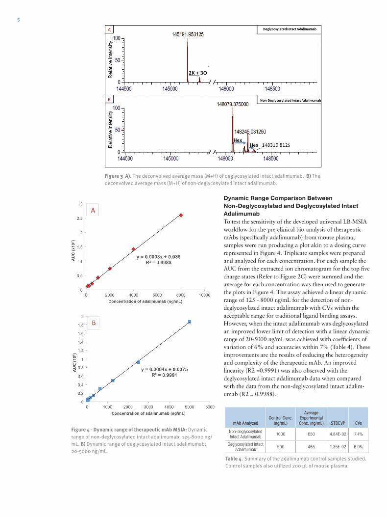
The mass spectrum was then deconvolved resulting in Figure 3A. The deconvolved data showed two masses with the 145191 mass representing deglycosylated intact adalimumab, while the other mass, 145318, is shifted by ~127 Da, suggestive of the presence of additional two potassium and three oxidations. As a comparison to the deglycosylated intact adalimumab data, Figure 3B shows deconvolved mass spectrum data for the non-deglycosyl-ated intact adalimumab. The non-deglycosylated adalimumab data shows three masses, with 148080, 148244, and 148407 representing the addition of hexose groups (162 Da) to adalimumab.
Figure 2.Deglycosylated analysis of adalimumab: 100ng adalimumab sample at a concentration of 500 ng/mL purified from mouse plasma. A) Base peak chromatogram of adalimumab showing the elution profile of deglycosylated adalimumab. B) and C) Raw MS spectra and corresponding zoom-in of the region m/z 2600-2880 around the five most abundant charge states, respectively.
5
Dynamic Range Comparison Between Non-Deglycosylated and Deglycosylated Intact AdalimumabTo test the sensitivity of the developed universal LB-MSIA workflow for the pre-clinical bio-analysis of therapeutic mAbs (specifically adalimumab) from mouse plasma, samples were run producing a plot akin to a dosing curve represented in Figure 4. Triplicate samples were prepared and analyzed for each concentration. For each sample the AUC from the extracted ion chromatogram for the top five charge states (Refer to Figure 2C) were summed and the average for each concentration was then used to generate the plots in Figure 4. The assay achieved a linear dynamic range of 125 - 8000 ng/mL for the detection of non-deglycosylated intact adalimumab with CVs within the acceptable range for traditional ligand binding assays. However, when the intact adalimumab was deglycosylated an improved lower limit of detection with a linear dynamic range of 20-5000 ng/mL was achieved with coefficients of variation of 6% and accuracies within 7% (Table 4). These improvements are the results of reducing the heterogeneity and complexity of the therapeutic mAb. An improved linearity (R2 =0.9991) was also observed with the deglycosylated intact adalimumab data when compared with the data from the non-deglycosylated intact adalim-umab (R2 = 0.9988).
Figure 3 A). The deconvolved average mass (M+H) of deglycosylated intact adalimumab. B) The deconvolved average mass (M+H) of non-deglycosylated intact adalimumab.
Figure 4 - Dynamic range of therapeutic mAb MSIA: Dynamic range of non-deglycosylated intact adalimumab; 125-8000 ng/mL. B) Dynamic range of deglycosylated intact adalimumab; 20-5000 ng/mL.
Table 4. Summary of the adalimumab control samples studied. Control samples also utilized 200 µL of mouse plasma.
mAb Analyzed Control Conc.
(ng/mL)
Average Experimental Conc. (ng/mL) STDEVP CVs
Non-deglycosylated Intact Adalimumab
1000 650 4.84E-02 7.4%
Deglycosylated Intact Adalimumab
500 465 1.35E-02 6.0%

6 ConclusionA hybrid approach for the universal bio-analysis of fully human and humanized therapeutic mAbs in pre-clinical research was demonstrated and improved upon by the addition of a pre-analytical deglycosylation. The developed LB-MSIA provided a highly sensitive, robust, and reproducible method for the generation of high value data content for the bio-analysis of therapeutic antibodies. Both the raw and the deconvolved HRAM mass spectra generated in this study clearly showed the presence of the post-translational modifications of adalimumab, thus making MSIA amiable for additional bio-analyses, including complex in vivo biotransformation studies and Drug Antibody Ratio (DAR) determination. The high
selectivity of the CaptureSelect™ biotin anti-IgG-Fc (human) conjugate combined with the molecular trapping technology of the MSIA D.A.R.T.’S creates an ideal scenario to assay low abundant (ng/mL) intact human therapeutic mAbs from rodent plasma. As a hybrid approach, the use of the Q Exactive for HRAM detection helps provide additional analytical flexibility and data content over other developing triple quadrupole methods that are reliant on peptide analysis. As shown, the combined benefits of the LB-MSIA enable the characteriza-tion of a deglycosylated mAb over a wide dynamic range (20-5000 ng/mL) while maintaining coefficients of variation of <15% and accuracies within 20%.

7
thermoscientific.com/msia Products are intended for research use only.
© 2015 Thermo Fisher Scientific Inc. All rights reserved.
APAAMSIATA1015
North America: +1 800 995 2787 • [email protected] North America: +1 858 453 7551 • [email protected]
Ordering InformationMSIA D.A.R.T.’S for Immunoaffinity Capture
Compatible with the Thermo Scientific Versette Automated Liquid Handler and Thermo Scientific Finnpipette® Novus i Multichannel Electronic Pipette
Cat. No. Description Packaging
991CUS02 300µl MSIA D.A.R.T.’S, Custom Pack of 96 units
991PRT11 300µl MSIA D.A.R.T.’S, Protein A Pack of 96 units
991PRT12 300µl MSIA D.A.R.T.’S, Protein A Pack of 24 units
991PRT13 300µl MSIA D.A.R.T.’S, Protein G Pack of 96 units
991PRT14 300µl MSIA D.A.R.T.’S, Protein G Pack of 24 units
991PRT15 300µl MSIA D.A.R.T.’S, Protein A/G Pack of 96 units
991PRT16 300µl MSIA D.A.R.T.’S, Protein A/G Pack of 24 units
991STR11 300µl MSIA D.A.R.T.’S, Streptavidin Pack of 96 units
991STR12 300µl MSIA D.A.R.T.’S, Streptavidin Pack of 24 units
991001096 300µl MSIA D.A.R.T.’S, Insulin Pack of 96 units
991001024 300µl MSIA D.A.R.T.’S, Insulin Pack of 24 units
991R 300 µL MSIA D.A.R.T.’S, Reloadable Rack 1 reloadable rack, D.A.R.T.’S are not included
Automated Liquid Handling Platform
Cat. No. Description
650-MSIA MSIA Versette Automated Liquid Handler
Multichannel Pipettes and Pipette Stand
Cat. No. Description Packaging
991S Finnpipette Novus i Adjustable Pipette Stand 1 pipette stand
991SP12 Finnpipette Novus i Electronic 12-Channel Pipette, 30-300µl and Pipette Stand
1 pipette and 1 pipette stand
Liquid Chromatography
Cat. No. Description
Thermo Scientific™ Dionex™ UltiMate® 3000 UHPLC System
066640 ProSwift™ RP-4H Monolith Column, 1.0 x 250 mm
Mass Spectrometry and Software
Description
Thermo Scientific™ Q Exactive™ Hybrid Quadrupole-Orbitrap Mass Spectrometer
Thermo Scientific™ TSQ Vantage Triple Stage Quadrupole Mass Spectrometer
Thermo Scientific™ Pinpoint Software
Thermo Scientific™ XCalibur™ Software
Thermo Scientific™ Protein Deconvolution Software, Version 3.0 with the ReSpect™ algorithm