quantitative fluorescent immunoassay of antibodies to, and surface
TRANSCRIPT
JOURNAL OF CLINICAL MICROBIOLOGY, Feb. 1978, p. 202-2080095-1 137/78/0007-0202$02.00/0Copyright (© 1978 American Society for Microbiology
Vol. 7, No. 2
Printed in U.S.A.
Quantitative Fluorescent Immunoassay of Antibodies to, andSurface Antigens of, Actinomyces viscosus
T. P. GILLIS AND J. J. THOMPSON*
Department ofMicrobiology, Louisiana State University Medical Center, New Orleans, Louisiana 70119
Received for publication 19 August 1977
Optimal conditions for a fluorescence immunoassay of antibodies to, and surfaceantigens of, Actinomyces viscosus ATCC 19246 are described. In the standardfluorescence immunoassay, 108 colony-forming units of A. viscosus reacted withan antibody preparation, were washed, and then were treated with an excess offluorescein-conjugated goat anti-rabbit immunoglobulin G. After another set ofwashes, fluorescence was determined in a spectrofluorometer; in most casesexcitation was at 485 nm, with emission measured at 525 nm. These conditionsminimized interference from light scatter and stray light. Under appropriateconditions, antibodies to A. viscosus could be readily determined, with thefluorescence of the specific antibody-treated cells more than five times thefluorescence of controls treated with normal rabbit serum. Organisms coated withspecific antibody could be detected at levels approaching 10" colony-forming unitsper ml. The standard fluorescence immunoassay procedure was readily adaptedto the measurement of either particulate or soluble surface antigens of A. viscosusby competition of the antigen with a fixed amount of antibody in the standardassay system; the competition resulted in an antigen dose-dependent inhibition offluorescence. The fluorescent immunoassay system thus appears to be a generalone that could be applied to other microbial systems as well.
The immunofluorescent technique originallydescribed by Coons et al. (9, 10) has many ap-plications in the rapid qualitative detection ofmicroorganisms in clinical materials, includingdetection ofActinomyces species in human den-tal-plaque samples (8, 19). Quantitation of flu-orescent microorganisms from such specimens isdifficult. Recent work with solid-phase fluores-cent immunoassays (FIA), however, has dem-onstrated that antigens or antibodies can bequantitated by the measurement of immuno-specific fluorescence associated with individual,derivatized carrier beads (2, 4, 17) or suspensionsof such beads (3). We report here that suspen-sions of the microorganism A. viscosus, a sus-pected periodontal pathogen of humans and an-imals (1, 14, 15), can be used to directly assayantibodies to, and surface antigens of, that or-ganism by simple FIA procedures. This FIAmethodology thus appears to be a general onepotentially applicable in the clinical setting toquantitative determinations of microorganismsor of antibodies directed to their surface com-ponents.
MATERIALS AND METHODS
Organisms. A. viscosus ATCC 19246 was obtaineddirectly from the American Type Culture Collectionand maintained aerobically by passage on blood agar
plates. The organism was characterized periodicallyby colonial morphology, Gram stain, and biochemicalreactions. Bacterial suspensions were prepared from60-h cultures of the organism grown aerobically at37°C in Actinomyces broth (Baltimore Biological Lab-oratory, Cockeysville, Md.) by centrifugation (1,256x g for 10 min) and three washes with 100 mMphosphate buffer (PB), pH 7.0. For plate counts, serial10-fold dilutions of the stock bacterial suspension werediluted in PB, and portions were plated in triplicateon brain heart infusion agar (Baltimore BiologicalLaboratory) plates supplemented with 2% yeast ex-tract (Difco Laboratories, Detroit, Mich.). Absorbanceat 660 nm was determined on serial twofold dilutionsof the stock bacterial suspension with a double-beamspectrophotometer (GCA/McPherson Instrument,Acton, Mass.). Under these conditions, an absorbanceof 0.32 corresponds to 10' colony-forming units (CFU)per ml.
Lactobacillus acidophilus ATCC 4356 was ob-tained directly from the American Type Culture Col-lection and maintained aerobically by passage on Ro-gosa SL agar (Difco Laboratories) plates. Bacterialsuspensions were prepared from 60-h cultures of theorganism grown aerobically at 37°C in Rogosa SLbroth (Difco Laboratories) by centrifugation (1,256 xg for 10 min) and three washes with PB.
Ra-a-19246 production. A suspension of washedA. viscosus ATCC 19246 was adjusted to 1010 CFU/ml.A 1-ml amount of this suspension was injected intra-venously into the peripheral ear vein of New Zealandwhite rabbits three times per week for 1 month. After
202
FIA FOR BACTERIAL ANTIBODIES AND ANTIGENS
a 1-week rest period, the rabbits were exsanguinated,and the sera (Ra-a-19246) were collected and storedat -200C.
Soluble antigen preparation. A pellet of A. vis-cosus (300 mg wet weight) was resuspended in deion-ized water (15 ml) and disrupted with an ultratipsonic oscillator (New Brunswick Scientific Co., NewBrunswick, N.J.) at full power for 15 min in an icebath. The disrupted suspension was then clarified bytwo centrifugations (800 x g for 10 min), and theclarified supernatant fluid was centrifuged (100,000 xg for 90 min). The supernatant fluid from the lastcentrifugation step was used as the "soluble" antigenpreparation. Antigen preparations were stored at-200C.FIA. A fluorescein-conjugated goat anti-rabbit im-
munoglobulin G serum (Fl-Go-a-RaG) (Miles Labo-ratories, Inc., Elkhart, Ind.) was used for FIA of anti-bodies bound to A. viscosus. In brief, a standardizedamount of A. viscosus (108 CFU) was pelleted bycentrifugation (1,256 x g for 5 min) in test tubes (6 by50 mm). The pellet was resuspended in 300 pl of eitheran Ra-a-19246 dilution in PB or 300 pi of the corre-sponding dilution of pooled normal rabbit serum (Pel-Freez, Rogers, Ark.) as control. After reaction for 30min at room temperature with intermittent mixing,the cells were washed three times (500 pl/wash) in PBby centrifugation (1,256 x g for 5 min). The washedpellets were resuspended in an Fl-Go-a-RaG dilution(300 Ai) and incubated for 15 min at room temperaturewith intermittent mixing. The suspensions were thencentrifuged (1,256 x g for 5 min) and washed (500ul/wash) three times with PB. The washed cells werefinally resuspended in 2.5 ml of PB, and the fluores-cence of the mixture was determined as detailed below.It must be noted that centrifugation steps in this FIAare critical; incomplete resuspension of the pellets orloss of materials during washes leads to erratic results.For this reason, it is essential to use "swinging-bucket"rotor types for the centrifugation steps.The FIA described above for the detection of anti-
bodies to A. viscosus can be modified to detect A.viscosus surface antigens by the incorporation of apreincubation step of antigen with a standardizedamount of Ra-a-19246 for 30 min at room temperature.After centrifugation (1,256 x g for 5 min) to removewhole cells if they were used as inhibitors, the mixturewas added to a standardized pellet of A. viscosus, andthe FIA was completed as described above. A fluores-cence control was included with buffer instead of an-tigen in the preincubation step. Inhibition was calcu-lated as follows: percent inhibition = (1- experimentalfluorescence/control fluorescence) x 100.
Fluorescence measurements. Most fluorescencemeasurements were made on an unmodified MKIspectrofluorometer (Farrand Optical Co., Inc., Val-halla, N.Y.) and recorded as measurements of photo-current in nanoamperes. Some readings were alsomade by replacing the photomultiplier of the MKIwith a model 1140 quantum photometer (PrincetonApplied Research Corp., Princeton, N.J.) and R212photomultiplier tube (Hamamatsu Corp., Middlesex,N.J.) and measuring fluorescence as photon-countingrate in the digital mode (at root mean square percent-age of deviation of 1.3%) or as nanoamperes in the
electrometer mode. Crossed film polarizers (FarrandOptical Co.) were used in most cases in an attempt tominimize interference from light scatter (5, 11). How-ever, comparison studies not detailed here showedlittle or no effect of polarizers on experimental-controlfluorescence ratios under the optimum conditions de-scribed below. Similarly, comparison studies of theeffect of slit width on experimental-control fluores-cence ratios (21) showed little advantage in the use ofnarrow slits. Accordingly, 10-nm band pass slits wereused for most of the experiments described below.Spectra were obtained by scanning the analyzer mono-chromator at 20 nm/min and recording fluorescenceon a SR-255B strip chart recorder (Heath Co., BentonHarbor, Mich.).
RESULTSStability of Fl-Go-a-RaG-labeled bacte-
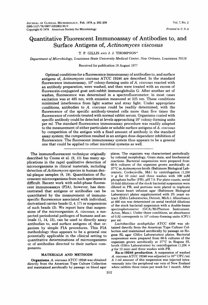
rial suspensions. To determine optimum con-ditions for the FIA, it was necessary to deter-mine excitation and emission spectra with la-beled microorganisms. A suspension of A. vis-cosus was reacted with Ra-a-19246 (1:100 dilu-tion) and Fl-Go-a-RaG (1:80 dilution) in thestandard FIA procedure. Fluorescence emissionwas excited at 485 nm and analyzed at 525 nm.As shown in Fig. 1, fluorescence of the cellsuspension was virtually constant over 10 min ofcontinuous observation. This suggests that nei-ther quenching of fluorescence of the fluoro-phore nor settling of the organisms was signifi-cant over a time period longer than that neces-sary to obtain the spectra shown below. Stabilityof the fluorescence was seen with or withoutcrossed polarizers.Choice of optimum excitation wave-
length of Fl-Go-a-RaG-labeled A. viscosus.Emission spectra were obtained for a suspensionof labeled organisms prepared as described inthe "stability" studies, with excitation at two
100-
90-
80-
70-
. 60-z 50-uJ 40-
3 30-20-10-
Ra-a-19246 +
Fl -Go -a-RaG
NRS+FI -Go-a-RaGCELLS+BUFFER
2 4 6 8 10TIME, MINUTES
FIG. 1. Stability of Fl-Go-a-RaG-labeled A. visco-sus. Excitation was at 485 nm, and emission wasanalyzed at 525 nm with crossed polarizers.
203VOI.. 7, 1978
204 GILLIS AND THOMPSON
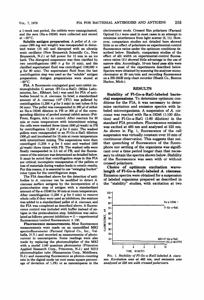
different wavelengths. As Fig. 2A shows, excita-tion at 495 nm gave slightly more fluorescencesignal than at 520 nm (the emission maximumof fluorescein [8]), but the peak was poorly re-solved from the large scatter signal centered at495 nm. In contrast, excitation at 485 nm (Fig.2B) permitted resolution of the fluorescence sig-nal from scatter, albeit with some apparent lossof fluorescence intensity. We felt that this smallloss of intensity was minimal compared with therelative absence of scatter interference and forthis reason chose 485 nm as our standard exci-tation wavelength for the studies described be-low. The use of crossed polarizers gave no betterresolution of the fluorescence peak when exci-tation was at 495 nm.Choice of optimum emission wavelength
ofFl-Go-a-RaG-labeled A. viscosus. An emis-sion spectrum was obtained for a suspension oflabeled organisms prepared as described in the
40 - A B
10
470 510 550 470 510 550
l,nm
FIG. 2. Effect ofexcitation wavelength on emissionspectrum. (A) Excitation at 495 nm. (B) Excitation at485 nm. Both spectra were obtained without polariz-ers at a scan rate of 20 nm/min.
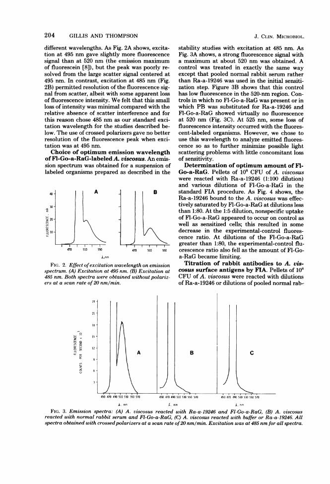
stability studies with excitation at 485 nm. AsFig. 3A shows, a strong fluorescence signal witha maximum at about 520 nm was obtained. Acontrol was treated in exactly the same wayexcept that pooled normal rabbit serum ratherthan Ra-a-19246 was used in the initial sensiti-zation step. Figure 3B shows that this controlhas low fluorescence in the 520-nm region. Con-trols in which no Fl-Go-a-RaG was present or inwhich PB was substituted for Ra-a-19246 andFl-Go-a-RaG showed virtually no fluorescenceat 520 nm (Fig. 3C). At 525 nm, some loss offluorescence intensity occurred with the fluores-cent-labeled organisms. However, we chose touse this wavelength to analyze emitted fluores-cence so as to further minimize possible lightscattering problems with little concomitant lossof sensitivity.Determination of optimum amount of Fl-
Go-a-RaG. Pellets of 108 CFU of A. viscosuswere reacted with Ra-a-19246 (1:100 dilution)and various dilutions of Fl-Go-a-RaG in thestandard FIA procedure. As Fig. 4 shows, theRa-a-19246 bound to the A. viscosus was effec-tively saturated by Fl-Go-a-RaG at dilutions lessthan 1:80. At the 1:5 dilution, nonspecific uptakeof Fl-Go-a-RaG appeared to occur on control aswell as sensitized cells; this resulted in somedecrease in the experimental-control fluores-cence ratio. At dilutions of the Fl-Go-a-RaGgreater than 1:80, the experimental-control flu-orescence ratio also fell as the amount of Fl-Go-a-RaG became limiting.Titration of rabbit antibodies to A. vis-
cosus surface antigens by FIA. Pellets of 108CFU of A. viscosus were reacted with dilutionsof Ra-a-19246 or dilutions of pooled normal rab-
2oi / 151
,z
o 6
450 470 490 510 530 550 570 450 470 490 510 530 550 570
J~
C
450 470 490 510 530 550 570
A. nm A nm (. nm
FIG. 3. Emission spectra: (A) A. viscosus reacted with Ra-a-19246 and Fl-Go-a-RaG, (B) A. viscosusreacted with normal rabbit serum and Fl-Go-a-RaG, (C) A. viscosus reacted with buffer or Ra-a-19246. Allspectra obtained with crossedpolarizers at a scan rate of20 nm/min. Excitation was at 485 nm for all spectra.
J. ClIAN. MICROBIOL.
FIA FOR BACTERIAL ANTIBODIES AND ANTIGENS
bit serum and Fl-Go-a-RaG (1:80). Figure 5shows that the bacterial suspension appeared tobe saturated with antibody at dilutions of 1:80or less of the Ra-a-19246. At higher dilutions,the amount of specific fluorescence dropped rap-idly; this resulted in a decrease in experimental-control fluorescence ratios.
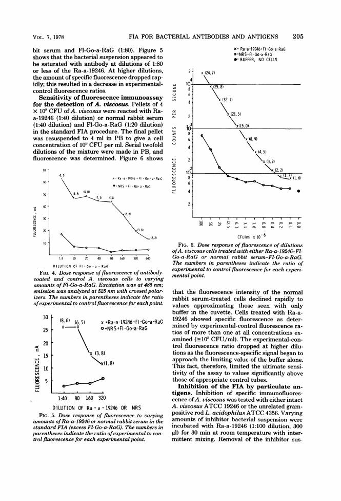
Sensitivity of fluorescence immunoassayfor the detection of A. viscosus. Pellets of 4x 10' CFU of A. viscosus were reacted with Ra-a-19246 (1:40 dilution) or normal rabbit serum
(1:40 dilution) and Fl-Go-a-RaG (1:20 dilution)in the standard FIA procedure. The final pelletwas resuspended to 4 ml in PB to give a cellconcentration of 108 CFU per ml. Serial twofolddilutions of the mixture were made in PB, andfluorescence was determined. Figure 6 shows
70
13. 71x x Ra -a-19246 +Fl -Go-a-RaG
60 -
\ NRS + Fl -Go-a -RaG
54)~(35.8) (I 1)
< 40-
(6. 6)
2 30-
O20-1\g43. 8)
(2.2)
1:5 10 20 40 80 160 320 640
D LUT ION OF Fl - Go - a - RaG
FIG. 4. Dose response of fluorescence of antibody-coated and control A. viscosus cells to varyingamounts of Fl-Go-a-RaG. Excitation was at 485 nm;emission was analyzed at 525 nm with crossed polar-izers. The numbers in parentheses indicate the ratioofexperimental to control fluorescence for eachpoint.
30
25
20c
LiJ
Lii
0
15
(8.6) (6.5) x =Ra-a-19246+FI -Go-a-RaGx x ° =NRS+FI-Go-a-RaG
x (3.8)
\x(l. 8)10
5
1:40 80 160 320
D ILUTION OF Ra - a - 19246 OR NRSFIG. 5. Dose response of fluorescence to varying
amounts ofRa-a-19246 or normal rabbit serum in thestandard FIA (excess Fl-Go-a-RaG). The numbers inparentheses indicate the ratio ofexperimental to con-
trol fluorescence for each experimental point.
x= Ra-a-19246+FI-Go-a-RaGo =NRS+FI -Go-a-RaG
BUFFER, NO CELLS
cY
cL'
LJLl.
N
00li m Na- CO
CFU/ml x 10 6
FIG. 6. Dose response of fluorescence of dilutionsofA. viscosus cells treated with either Ra-a-19246-Fl-Go-a-RaG or normal rabbit serum-Fl-Go-a-RaG.The numbers in parentheses indicate the ratio ofexperimental to control fluorescence for each experi-mental point.
that the fluorescence intensity of the normalrabbit serum-treated cells declined rapidly tovalues approximating those seen with onlybuffer in the cuvette. Cells treated with Ra-a-19246 showed specific fluorescence as deter-mined by experimental-control fluorescence ra-
tios of more than one at all concentrations ex-
amined (-105 CFU/ml). The experimental-con-trol fluorescence ratio dropped at higher dilu-tions as the fluorescence-specific signal began toapproach the limiting value of the buffer alone.This fact, therefore, limited the ultimate sensi-tivity of the assay to values significantly abovethose of appropriate control tubes.Inhibition of the FIA by particulate an-
tigens. Inhibition of specific immunofluores-cence ofA. viscosus was tested with either intactA. viscosus ATCC 19246 or the unrelated gram-positive rod L. acidophilus ATCC 4356. Varyingamounts of inhibitor bacterial suspension were
incubated with Ra-a-19246 (1:100 dilution, 300pl) for 30 min at room temperature with inter-mittent mixing. Removal of the inhibitor sus-
VOL. 7, 1978 205
206 GILLIS AND THOMPSON
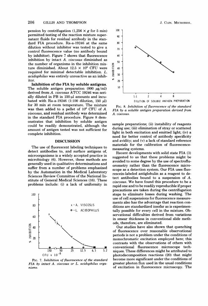
pension by centrifugation (1,256 x g for 5 min)permitted testing of the reaction mixture super-natant fluids for residual antibody in the stan-dard FIA procedure. Ra-a-19246 at the samedilution without inhibitor was tested to give acontrol fluorescence value (no antibody boundby inhibitor). Figure 7 shows that fluorescenceinhibition by intact A. viscosus diminished asthe number of organisms in the inhibition mix-ture diminished. About 12.5 x 10⁶ CFU wererequired for minimal detectable inhibition. L.acidophilus was entirely unreactive as an inhib-itor.Inhibition of the FIA by soluble antigens.
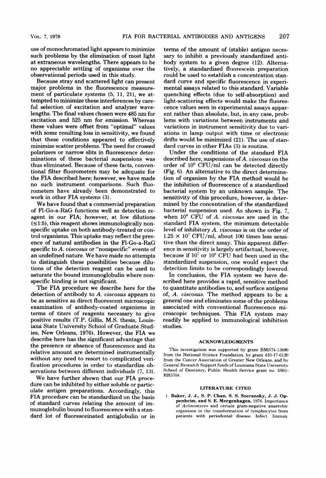
The soluble antigen preparation (800 jig/ml)derived from A. viscosus ATCC 19246 was seri-ally diluted in PB in 150-Ml amounts and incu-bated with Ra-a-19246 (1:100 dilution, 150 Il)for 30 min at room temperature. The mixturewas then added to a pellet of 108 CFU of A.viscosus, and residual antibody was determinedin the standard FIA procedure. Figure 8 dem-onstrates that inhibition by soluble antigencould be readily demonstrated, although theamount of antigen tested was not sufficient forcomplete inhibition.
DISCUSSIONThe use of fluorescent labeling techniques to
detect antibodies to, and surface antigens of,microorganisms is a widely accepted practice inmicrobiology (6). However, these methods aregenerally used in qualitative determinations andsuffer from a number of problems emphasizedby the Automation in the Medical LaboratorySciences Review Committee of the National In-stitute of General Medical Sciences (16). Theseproblems include: (i) a lack of uniformity in
100
80
60
040
- 20
x =A. VISCOSUS
° = L. AC IDOPHILUSx
X~~~~~\x
, -v a
150 '50 25 12.5 6.3 3.2
CFU x 1o-6FIG. 7. Inhibition of fluorescence of the standard
FIA by intact A. viscosus or L. acidophilus orga-nzsms.
100
90
0
It:
80 -
70 -
60 -
50 -
40 -
30 -
20 -
10 -
1:1 2 4 8
DILUTION OF SOLUBLE ANTIGEN PREPARATION
FIG. 8. Inhibition of fluorescence of the standardFIA by a soluble antigen preparation derived fromA. viscosus.
sample preparations; (ii) instability of reagentsduring use; (iii) elimination of stray or scatteredlight in both excitation and emitted light; (iv) aneed for better control of antibody specificityand avidity; and (v) a lack of standard referencematerials for the calibration of fluorescence-measuring systems.Recent developments with solid state FIA (3)
suggested to us that these problems might beavoided to some degree by the use of spectroflu-orometry rather than the fluorescence micro-scope as a detection system. Our FIA uses fluo-rescein-labeled antiglobulin as a reagent to de-tect antibodies bound to a suspension of A.viscosus. We have found this procedure to be arapid one and to be readily reproducible if properprecautions are taken during the centrifugationsteps to eliminate losses during washing. Theuse of cell suspensions for fluorescence measure-ments also has the advantage that reaction con-ditions are standardized insofar as is experimen-tally possible for every cell in the mixture. Ob-servational difficulties derived from variationsin smear thickness in conventional slide meth-ods, therefore, are eliminated.Our studies have also shown that quenching
of fluorescence over reasonable observationalperiods is not a problem under the conditions ofmonochromatic excitation employed here; thiscontrasts with the observations of others withconventional fluorescence microscope tech-niques. These differences might be attributed tophotodecomposition reactions (20) that mightbecome more significant under the conditions ofgreater photon flux used in the usual conditionsof excitation in fluorescence microscopy. The
J. CI.IN. MICROBIOL.
FIA FOR BACTERIAL ANTIBODIES AND ANTIGENS
use of monochromated light appears to minimizesuch problems by the elimination of most lightat extraneous wavelengths. There appears to beno appreciable settling of organisms over theobservational periods used in this study.Because stray and scattered light can present
major problems in the fluorescence measure-ment of particulate systems (5, 11, 21), we at-tempted to minimize these interferences by care-ful selection of excitation and analyzer wave-lengths. The final values chosen were 485 nm forexcitation and 525 nm for emission. Whereasthese values were offset from "optimal" valueswith some resulting loss in sensitivity, we foundthat these conditions appeared to effectivelyminimize scatter problems. The need for crossedpolarizers or narrow slits in fluorescence deter-minations of these bacterial suspensions wasthus eliminated. Because of these facts, conven-tional filter fluorometers may be adequate forthe FIA described here; however, we have madeno such instrument comparisons. Such fluo-rometers have already been demonstrated towork in other FIA systems (3).We have found that a commercial preparation
of Fl-Go-a-RaG functions well as detection re-agent in our FIA; however, at low dilutions(c1:5), this reagent shows immunologically non-specific uptake on both antibody-treated or con-trol organisms. This uptake may reflect the pres-ence of natural antibodies in the Fl-Go-a-RaGspecific to A. viscosus or "nonspecific" events ofan undefined nature. We have made no attemptsto distinguish these possibilities because dilu-tions of the detection reagent can be used tosaturate the bound immunoglobulin where non-specific binding is not significant.The FIA procedure we describe here for the
detection of antibody to A. viscosus appears tobe as sensitive as direct fluorescent microscopicexamination of antibody-coated organisms interms of titers of reagents necessary to givepositive results (T.P. Gillis, M.S. thesis, Louis-iana State University School of Graduate Stud-ies, New Orleans, 1976). However, the FIA wedescribe here has the significant advantage thatthe presence or absence of fluorescence and itsrelative amount are determined instrumentallywithout any need to resort to complicated veri-fication procedures in order to standardize ob-servations between different individuals (7, 13).We have further shown that our FIA proce-
dure can be inhibited by either soluble or partic-ulate antigen preparations. Accordingly, thisFIA procedure can be standardized on the basisof standard curves relating the amount of im-munoglobulin bound to fluorescence with a stan-dard lot of fluoresceinated antiglobulin or in
terms of the amount of (stable) antigen neces-sary to inhibit a previously standardized anti-body system to a given degree (12). Alterna-tively, a standardized fluorescein preparationcould be used to establish a concentration stan-dard curve and specific fluorescence in experi-mental assays related to this standard. Variablequenching effects (due to self-absorption) andlight-scattering effects would make the fluores-cence values seen in experimental assays appar-ent rather than absolute, but, in any case, prob-lems with variations between instruments andvariations in instrument sensitivity due to vari-ations in lamp output with time or electronicdrifts would be minimized (21). The use of stan-dard curves in other FIAs (3) is routine.Under the conditions of the standard FIA
described here, suspensions ofA. viscosus on theorder of 105 CFU/ml can be detected directly(Fig. 6). An alternative to the direct determina-tion of organism by the FIA method would bethe inhibition of fluorescence of a standardizedbacterial system by an unknown sample. Thesensitivity of this procedure, however, is deter-mined by the concentration of the standardizedbacterial suspension used. As shown in Fig. 7,when 108 CFU of A. viscosus are used in thestandard FIA system, the minimum detectablelevel of inhibitory A. viscosus is on the order of1.25 x 107 CFU/ml, about 100 times less sensi-tive than the direct assay. This apparent differ-ence in sensitivity is largely artifactual, however,because if 107 or 106 CFU had been used in thestandardized suspension, one would expect thedetection limits to be correspondingly lowered.
In conclusion, the FIA system we have de-scribed here provides a rapid, sensitive methodto quantitate antibodies to, and surface antigensof, A. viscosus. The method appears to be ageneral one and eliminates some of the problemsassociated with conventional fluorescence mi-croscopic techniques. This FIA system mayreadily be applied to immunological inhibitionstudies.
ACKNOWLEDGMENTSThis investigation was supported by grant BMS74-13680
from the National Science Foundation, by grant 410-17-6120from the Cancer Association of Greater New Orleans, and byGeneral Research Support funds of Louisiana State UniversitySchool of Dentistry, Public Health Service grant no. 5S01-RR5704.
LITERATURE CITED
1. Baker, J. J., S. P. Chan, S. S. Socransky, J. J. Op-penheim, and S. E. Mergenhagen. 1976. Importanceof Actinomyces and certain gram-negative anaerobicorganisms in the transformation of lymphocytes frompatients with periodontal disease. Infect. Immun.
207VOL. 7, 1978
208 GILLIS AND THOMPSON
13:1363-1368.2. Bloemmen, F. J., J. Radl, J. J. Haaijman, P. van den
Berg, H. R. E. Schuit, and W. Hijmans. 1976. Micro-fluorometric evaluation of the specificity of fluorescentantisera against mouse immunoglobulins with definedantigen substrate (DASS) system. J. Immunol. Meth-ods 10:337-355.
3. Burgett, M. W., S. J. Fairfield, and J. F. Monthony.1977. A solid phase fluorescent immunoassay for thequantitation of the C4 component of human comple-ment. J. Immunol. Methods 16:211-219.
4. Capel, A. J. A. 1974. A quantitative immunofluorescencemethod based on the covalent coupling of protein tosepharose beads. J. Immunol. Methods 5:165-178.
5. Chen, R. F. 1966. Reduction of light scatter in fluorometryby the use of horizontally polarized excitation. Anal.Biochem. 14:497-499.
6. Cherry, W. B. 1974. Immunofluorescence techniques, p.29-44. In E. H. Lennette, E. H. Spaulding, and J. P.Truant (ed.), Manual of clinical microbiology, 2nd ed.Amnerican Society for Microbiology, Washington, D.C.
7. Cherry, W. B., and C. B. Reimer. 1973. Diagnosticimmunofluorescence. Bull. W. H. 0. 48:737-746.
8. Collins, P. A., M. A. Gerencser, and J. M. Slack. 1973.Enumeration and identification of Actinomycetaceae inhuman dental calculus using the fluorescent antibodytechnique. Arch. Oral Biol. 18:145-153.
9. Coons, A. H., H. J. Creech, and R. N. Jones. 1941.Immunological properties of antibody containing a flu-orescent group. Proc. Soc. Biol. Med. 47:200-202.
10. Coons, A. H., and M. H. Kaplan. 1950. Localization ofantigen in tissue cells. II. Improvements in a methodfor the detection of antigen by means of fluorescentantibody. J. Exp. Med. 91:1-13.
11. Davis, R. P., and M. Canessa-Fischer. 1965. Spectro-fluorometric identification of reduced pyridine nucleo-tide in the intact isolated urinary bladder of the toad.Anal. Biochem. 10:325-343.
12. Fagraeus, A., and N. R. Berquist. 1975. The raison
d'etre of standards in indirect immunofluorescence.Ann. N.Y. Acad. Sci. 254:69-76.
13. Jongsma, P. M., W. Hijmans, and J. S. Ploem. 1971.Quantitative immunofluorescence. Histochemie25:329-343.
14. Jordan, H. V., and P. H. Keyes. 1964. Aerobic, gram-positive filamentous bacteria as etiologic agents of ex-perimental periodontal disease in hamsters. Arch. OralBiol. 9:401-414.
15. Jordan, H. V., P. H. Keyes, and S. Bellack. 1972.Periodontal lesions in hamsters and gnotobiotic ratsinfected with Actinomyces of human origin. J. Perio-dontal Res. 7:21-28.
16. Kinney, T. D., and R. S. Melville (ed.). 1976. Mecha-nization, automation, and increased effectiveness of theclinical laboratory, p. 95-96. In Automation in medicallaboratory sciences-review committee. Department ofHealth, Education, and Welfare publication no. (NIH)77-145, Washington, D.C.
17. Knapp, W., and J. S. Ploem. 1974. Microfluorometry ofantigen-antibody interactions in immunofluorescenceusing the defined antigen substrate (DASS) system.Sensitivity, specificity and variables of the method. J.Immunol. Methods 5:259-273.
18. Peters, J. H., and A. H. Coons. 1976. Fluorescent anti-body as a specific cytochemical reagent, p. 424-444. InC. A. Williams and M. W. Chase (ed.), Methods inimmunology and immunochemistry, vol. 5. AcademicPress Inc., New York.
19. Slack, J. M., S. Landfried, and M. A. Gerencser. 1971.Identification of Actinomyces and related bacteria indental calculus by the fluorescent antibody technique.J. Dent. Res. 50:78-82.
20. Wick, G., K. Schauenstein, F. Herzog, and A. Stein-batz. 1975. Investigations of the recovery phenomenonafter laser excitation in immunofluorescence. Ann. N.Y.Acad. Sci. 254:172-174.
21. Udenfriend, S. 1962. Fluorescence assay in biology andmedicine, p. 110-124. Academic Press Inc., New York.
J. CI,IN. MICROBIOI,.