human/mouse/rat bnp enzyme immunoassay kit · raybio® human/mouse/rat bnp enzyme immunoassay kit...
TRANSCRIPT
RayBio® Human/Mouse/Rat BNP Enzyme Immunoassay Kit
Catalog #: EIA-BNP, EIAM-BNP, EIAR-BNP
User ManualLast revised August 7, 2017
Caution:Extraordinarily useful information enclosed
ISO 13485 Certified
3607 Parkway Lane, Suite 100Norcross, GA 30092
Tel: 1-888-494-8555 (Toll Free) or 770-729-2992, Fax:770-206-2393 Web: www.RayBiotech.com, Email: [email protected]
1
Table of Contents
Section Page #
I. Introduction 3
II. General Description 4
III. How It Works 4
IV. Storage 5
V. Reagents 5
VI. Additional Materials Required 6
VII. Reagent Preparation A. Preparation of Plate and Anti-BNP Antibody B. Preparation of Biotinylated Peptide (Item F) C. Preparation of Standards D. Preparation of Positive Control E. Preparation of Samples F. Preparation of Wash Buffer and HRP-Strep
6 6 7 8 9 9 10
VIII. Assay Procedure 10
IX. Assay Procedure Summary 11
X. Calculation of Results A. Typical Data B. Sensitivity C. Standard Curve Range D. Reproducibility E. Assay Diagram
12 12 12 12 12 13
XI. Specificity 14
XII. Select Publications 14
XIII. Troubleshooting Guide 15
Please read the entire manual carefully before starting your experiment
2
I. Introduction
Brain natriuretic peptide (BNP), (aka B-type natriuretic peptide), is a 32 amino acid polypeptide secreted by the ventricles of the heart in response to excessive stretching of myocytes in the ventricles. BNP was originally identified in extracts of porcine brain, but in humans it is produced mainly in the cardiac ventricles. Its counterpart in rats is a 45 amino acid peptide hormone. At the time of release, a co-secreted 76 amino acid N-terminal fragment (NT-proBNP) is also released with BNP.
BNP binds to and activates NPRA in a similar fashion to atrial natriuretic peptide (ANP) but with 10-fold lower affinity. The biological half-life of BNP, however, is twice as long as that of ANP. Both ANP and BNP have limited ability to bind and activate NPRB.
Physiologic actions of BNP include decrease in systemic vascular resistance and central venous pressure as well as an increase in natriuresis. Thus, the resulting effect of BNP is a decrease in cardiac output and a decrease in blood volume.
Tests showing elevated levels of BNP or NT-proBNP in blood are used as a diagnosis of heart failure and may be useful to establish prognosis in heart failure, as both markers are typically higher in patients with poorer outcome.
Both BNP and NT-proBNP have been approved as a marker for acute congestive heart failure (CHF). The plasma concentrations of both BNP are increased in patients with asymptomatic and symptomatic left ventricular dysfunction. There is no level of BNP that perfectly separates patients with and without heart failure.
3
II. General Description
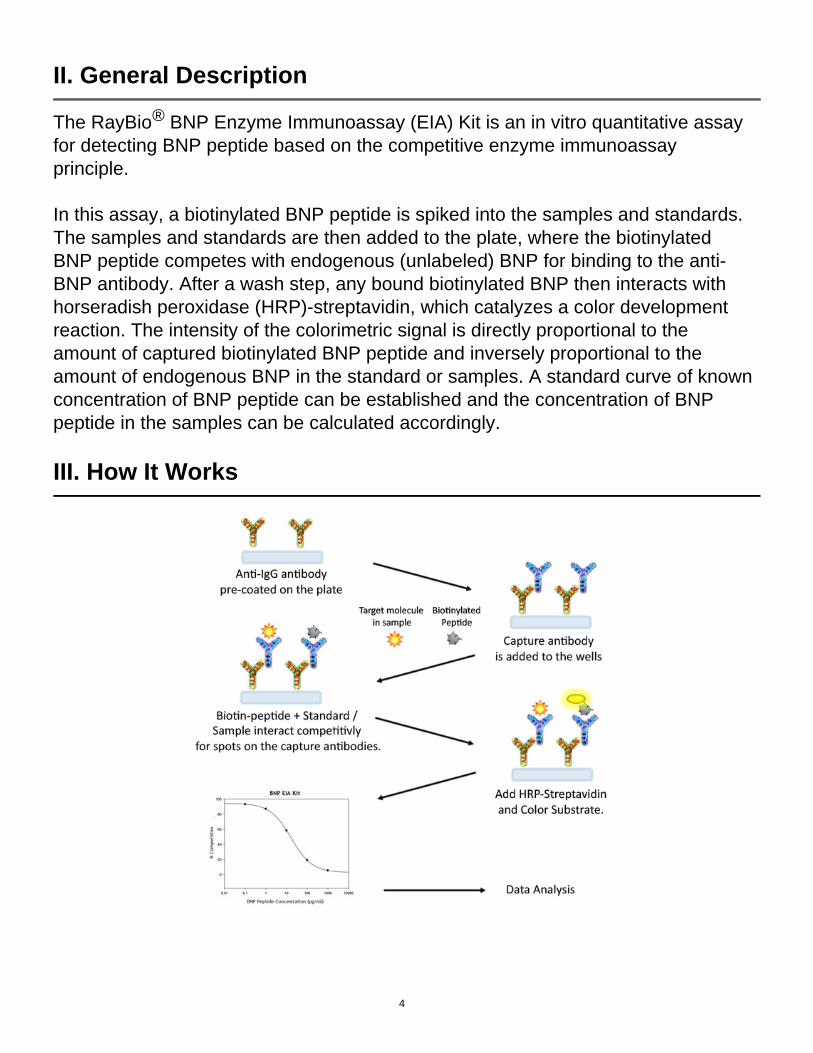
The RayBio® BNP Enzyme Immunoassay (EIA) Kit is an in vitro quantitative assay for detecting BNP peptide based on the competitive enzyme immunoassay principle.
In this assay, a biotinylated BNP peptide is spiked into the samples and standards. The samples and standards are then added to the plate, where the biotinylated BNP peptide competes with endogenous (unlabeled) BNP for binding to the anti-BNP antibody. After a wash step, any bound biotinylated BNP then interacts with horseradish peroxidase (HRP)-streptavidin, which catalyzes a color development reaction. The intensity of the colorimetric signal is directly proportional to the amount of captured biotinylated BNP peptide and inversely proportional to the amount of endogenous BNP in the standard or samples. A standard curve of known concentration of BNP peptide can be established and the concentration of BNP peptide in the samples can be calculated accordingly.
III. How It Works
4
IV. Storage
The entire kit may be stored at -20°C to -80°C for up to 6 months from the date of shipment. For extended storage, it is recommended to store at -80°C. Avoid repeated freeze-thaw cycles. For prepared reagent storage, see table below.
V. Reagents
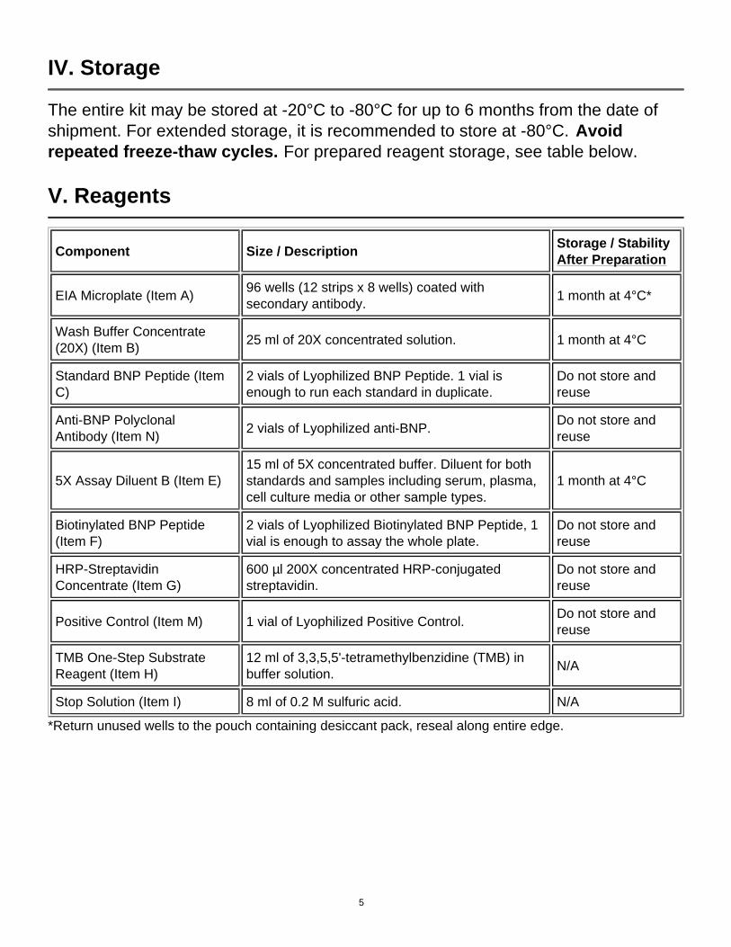
Component Size / DescriptionStorage / Stability After Preparation
EIA Microplate (Item A)96 wells (12 strips x 8 wells) coated with secondary antibody.
1 month at 4°C*
Wash Buffer Concentrate (20X) (Item B)
25 ml of 20X concentrated solution. 1 month at 4°C
Standard BNP Peptide (Item C)
2 vials of Lyophilized BNP Peptide. 1 vial is enough to run each standard in duplicate.
Do not store and reuse
Anti-BNP Polyclonal Antibody (Item N)
2 vials of Lyophilized anti-BNP.Do not store and reuse
5X Assay Diluent B (Item E)15 ml of 5X concentrated buffer. Diluent for both standards and samples including serum, plasma, cell culture media or other sample types.
1 month at 4°C
Biotinylated BNP Peptide (Item F)
2 vials of Lyophilized Biotinylated BNP Peptide, 1 vial is enough to assay the whole plate.
Do not store and reuse
HRP-Streptavidin Concentrate (Item G)
600 µl 200X concentrated HRP-conjugated streptavidin.
Do not store and reuse
Positive Control (Item M) 1 vial of Lyophilized Positive Control.Do not store and reuse
TMB One-Step Substrate Reagent (Item H)
12 ml of 3,3,5,5'-tetramethylbenzidine (TMB) in buffer solution.
N/A
Stop Solution (Item I) 8 ml of 0.2 M sulfuric acid. N/A
*Return unused wells to the pouch containing desiccant pack, reseal along entire edge.
5
VI. Additional Materials Required
1. Microplate reader capable of measuring absorbance at 450 nm2. Precision pipettes to deliver 2 µl to 1 ml volumes3. Adjustable 1-25 ml pipettes for reagent preparation4. 100 ml and 1 liter graduated cylinders5. Absorbent paper6. Distilled or deionized water7. SigmaPlot software (or other software which can perform four-parameter
logistic regression models)8. Tubes to prepare standard or sample dilutions9. Orbital shaker
10. Aluminum foil11. Plastic wrap
VII. Reagent Preparation
Keep kit reagents on ice during reagent preparation steps.
A. Preparation of Plate and Anti-BNP Antibody
1. Equilibrate plate to room temperature before opening the sealed pouch.
2. Label removable 8-well strips as appropriate for your experiment.
3. 5X Assay Diluent B (Item E) should be diluted 5-fold with deionized or distilled water.
4. Briefly centrifuge the anti-BNP antibody vial (Item N) and reconsititute with 55 µl of 1X Assay Diluent B to prepare the antibody concentrate. Pipette up and down to mix gently.
5. The antibody concentrate should then be diluted 100-fold with 1X Assay Diluent B. This is your anti-BNP antibody working solution, which will be used in step 2 of Assay Procedure (Section VIII).
Note: The following steps may be done during the antibody incubation procedure (step 2 of Assay Procedure)
6
B. Preparation of Biotinylated BNP (Item F)
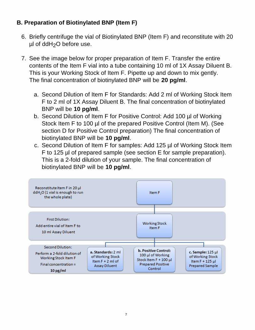
6. Briefly centrifuge the vial of Biotinylated BNP (Item F) and reconstitute with 20 µl of ddH2O before use.
7. See the image below for proper preparation of Item F. Transfer the entire contents of the Item F vial into a tube containing 10 ml of 1X Assay Diluent B. This is your Working Stock of Item F. Pipette up and down to mix gently. The final concentration of biotinylated BNP will be 20 pg/ml.
a. Second Dilution of Item F for Standards: Add 2 ml of Working Stock Item F to 2 ml of 1X Assay Diluent B. The final concentration of biotinylated BNP will be 10 pg/ml.
b. Second Dilution of Item F for Positive Control: Add 100 µl of Working Stock Item F to 100 µl of the prepared Positive Control (Item M). (See section D for Positive Control preparation) The final concentration of biotinylated BNP will be 10 pg/ml.
c. Second Dilution of Item F for samples: Add 125 µl of Working Stock Item F to 125 µl of prepared sample (see section E for sample preparation). This is a 2-fold dilution of your sample. The final concentration of biotinylated BNP will be 10 pg/ml.
7
C. Preparation of Standards
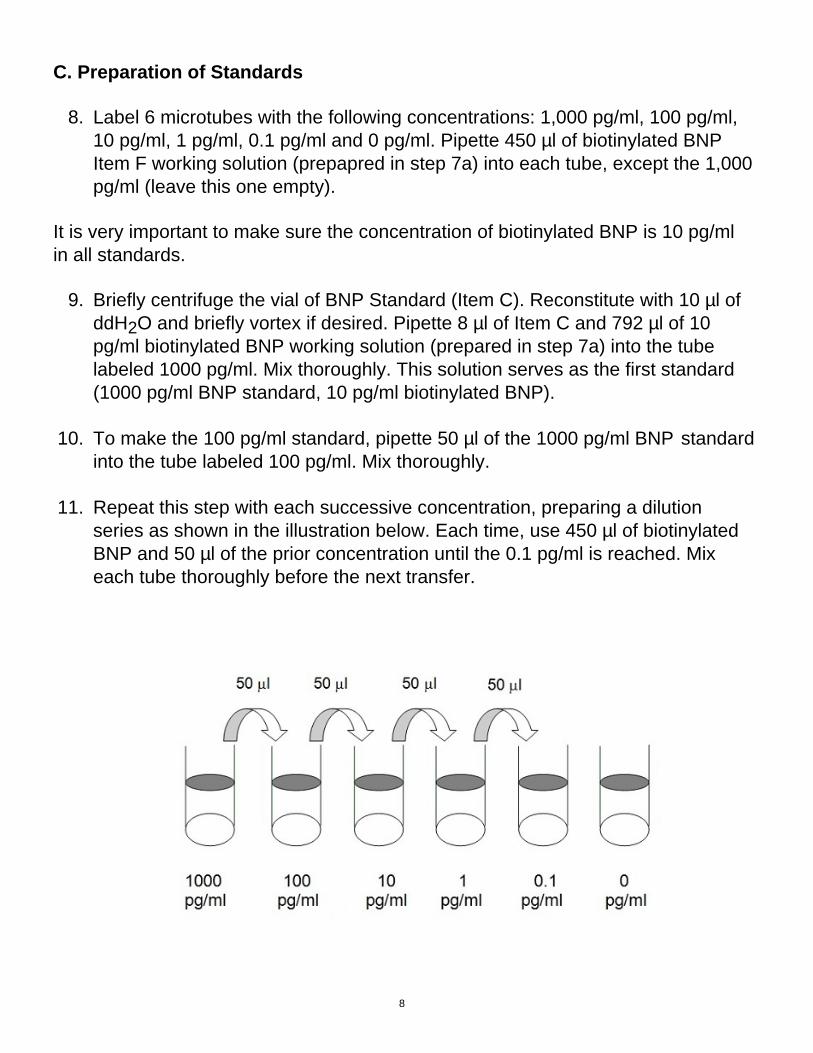
8. Label 6 microtubes with the following concentrations: 1,000 pg/ml, 100 pg/ml, 10 pg/ml, 1 pg/ml, 0.1 pg/ml and 0 pg/ml. Pipette 450 µl of biotinylated BNP Item F working solution (prepapred in step 7a) into each tube, except the 1,000 pg/ml (leave this one empty).
It is very important to make sure the concentration of biotinylated BNP is 10 pg/ml in all standards.
9. Briefly centrifuge the vial of BNP Standard (Item C). Reconstitute with 10 µl of ddH2O and briefly vortex if desired. Pipette 8 µl of Item C and 792 µl of 10 pg/ml biotinylated BNP working solution (prepared in step 7a) into the tube labeled 1000 pg/ml. Mix thoroughly. This solution serves as the first standard (1000 pg/ml BNP standard, 10 pg/ml biotinylated BNP).
10. To make the 100 pg/ml standard, pipette 50 µl of the 1000 pg/ml BNP standard into the tube labeled 100 pg/ml. Mix thoroughly.
11. Repeat this step with each successive concentration, preparing a dilution series as shown in the illustration below. Each time, use 450 µl of biotinylated BNP and 50 µl of the prior concentration until the 0.1 pg/ml is reached. Mix each tube thoroughly before the next transfer.
8
D. Positive Control Preparation
12. Briefly centrifuge the Positive Control vial (Item M) and reconstitute with 100 µl of ddH2O.
13. Refer to step 7b. This is a 2-fold dilution of the Positive Control. The final concentration of biotinylated BNP should still be 10 pg/ml.
The Positive Control is a cell culture media sample that serves as a system control to verify that the kit components are working. The resulting OD will not be used in any calculations; if no positive competition is observed please contact RayBiotech Technical Support. The Positive Control may be diluted further if desired, but be sure the final concentration of biotinylated BNP is 10 pg/ml.
E. Sample Preparation
14. If you wish to perform a 2-fold dilution of your sample, proceed to step 7c. If you wish to perform a higher dilution of your sample, dilute your sample with 1X Assay Diluent B before performing step 7c.EXAMPLE (to make a 4-fold dilution of sample):
a. Dilute sample 2-fold (62.5 µl of sample + 62.5 µl of 1X Assay Diluent B.).b. Perform step 7c (125 µl of working solution Item F + 125 µl of sample
prepared above).
The total volume is 250 µl, enough for duplicate wells on the microplate.
It is very important to make sure the final concentration of the biotinylated BNP is 10 pg/ml.
Note: Optimal sample dilution factors should be determined empirically, however you may reference below for recommended dilution factors for serum: Human=2X Mouse=2X Rat=2X. If you have any questions regarding the recommendended dilutions you may contact technical support at 888-494-8555 or [email protected].
9
F. Preparation of Wash Buffer and HRP
15. If Item B (20X Wash Concentrate) contains visible crystals, warm to room temperature and mix gently until dissolved.
16. Dilute 20 ml of Wash Buffer Concentrate into deionized or distilled water to yield 400 ml of 1X Wash Buffer.
17. Briefly centrifuge the HRP-Streptavidin vial (Item G) before use.
18. Dilute the HRP-Streptavidin concentrate 200-fold with 1X Assay Diluent B.
VIII. Assay Procedure
1. Keep kit reagents on ice during reagent preparation steps. It is recommended that all standards and samples be run at least in duplicate.
2. Add 100 µl of Anti-BNP Antibody (Item N) (See Reagent Preparation step 5) to each well. Incubate for 1.5 hours at room temperature with gentle shaking (1-2 cycle/sec). You may also incubate overnight at 4ºC.
3. Discard the solution and wash wells 4 times with 1X Wash Solution Buffer (200-300 µl each). Washing may be done with a multichannel pipette or an automated plate washer. Complete removal of liquid at each step is essential to good assay performance. After the last wash, remove any remaining Wash Buffer by aspirating or decanting. Invert the plate and blot it against clean paper towels.
4. Add 100 µl of each standard (see Reagent Preparation Section C), Positive Control (see Reagent Preparation Section D) and sample (see Reagent Preparation Section E) to appropriate wells. Be sure to include a blank well (Assay Diluent only). Cover wells and incubate for 2.5 hours at room temperature with gentle shaking (1-2 cycles/sec) overnight or at 4ºC.
5. Discard the solution and wash 4 times as directed in Step 3.
10
6. Add 100 µl of prepared HRP-Streptavidin solution (see Reagent Preparation step 18) to each well. Incubate for 45 minutes at room temperature with gentle shaking. It is recommended that incubation time should not be shorter or longer than 45 minutes.
7. Discard the solution and wash 4 times as directed in Step 3.
8. Add 100 µl of TMB One-Step Substrate Reagent (Item H) to each well. Incubate for 30 minutes at room temperature in the dark with gentle shaking (1-2 cycles/sec).
9. Add 50 µl of Stop Solution (Item I) to each well. Read at 450 nm immediately.
IX. Assay Procedure Summary
1. Prepare all reagents, samples and standards as instructed.
2. Add 100 µl anti-BNP to each well. Incubate 1.5 hours at room temperature or overnight at 4ºC.
3. Add 100 µl standard or sample to each well. Incubate 2.5 hours at room temperature or overnight at 4ºC.
4. Add 100 µl prepared Streptavidin solution. Incubate 45 minutes at room temperature.
5. Add 100 µl TMB One-Step Substrate Reagent to each well. Incubate 30 minutes at room temperature.
6. Add 50 µl Stop Solution to each well. Read at 450 nm immediately.
11
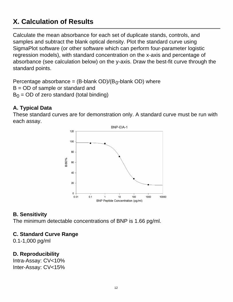
X. Calculation of Results
Calculate the mean absorbance for each set of duplicate stands, controls, and samples and subtract the blank optical density. Plot the standard curve using SigmaPlot software (or other software which can perform four-parameter logistic regression models), with standard concentration on the x-axis and percentage of absorbance (see calculation below) on the y-axis. Draw the best-fit curve through the standard points.
Percentage absorbance = (B-blank OD)/(B0-blank OD) where B = OD of sample or standard and B0 = OD of zero standard (total binding)
A. Typical DataThese standard curves are for demonstration only. A standard curve must be run with each assay.
B. Sensitivity The minimum detectable concentrations of BNP is 1.66 pg/ml.
C. Standard Curve Range 0.1-1,000 pg/ml
D. Reproducibility Intra-Assay: CV<10%Inter-Assay: CV<15%
12
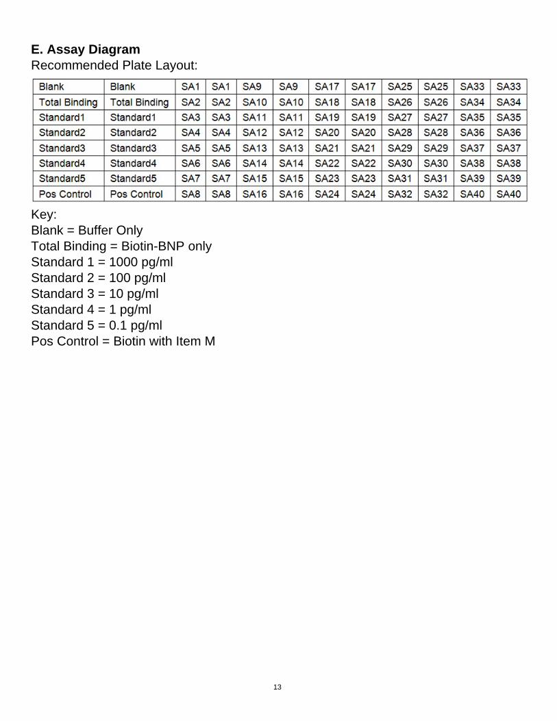
E. Assay Diagram Recommended Plate Layout:
Key:Blank = Buffer OnlyTotal Binding = Biotin-BNP onlyStandard 1 = 1000 pg/ml Standard 2 = 100 pg/ml Standard 3 = 10 pg/ml Standard 4 = 1 pg/ml Standard 5 = 0.1 pg/ml Pos Control = Biotin with Item M
13
XI. Specificity
Cross Reactivity: This EIA kit shows no cross-reactivity with any of the adipokines tested: Ghrelin, Nesfatin, Angiotensin II, NPY and APC.
XIV. Publications Citing This Product
1. Martín R, Miana M, Jurado-López R, Martínez-Martínez E, Gómez-Hurtado N, et al. DIOL Triterpenes Block Profibrotic Effects of Angiotensin II and Protect from Cardiac Hypertrophy. PLoS ONE. 2012;7(7):e41545. doi:10.1371/journal.pone.0041545Species: MouseSample Type: Serum
2. Ku HC., et al. DPP4 deficiency preserves cardiac function via GLP-1 signaling in rats subjected to myocardial ischemia/reperfusion. Naunyn Schmiedebergs Arch Pharmacol. 2011 Aug;384(2):197-207. doi: 10.1007/s00210-011-0665-3.Species: RatSample Type: Plasma
3. Martin, R., Cordova C., San Roman JA., Gutierrez B., Cachofeiro V., Nieto ML. Oleanolic acid Modulates the Immune-Inflammatory Response in Mice with Experimental Autoimmune Myocarditis and Protects from Cardiac Injury. J Mol Cell Cardiol. 2014 Apr 13;72C:250-262Species: MouseSample Type: Serum
4. Martin, R., Cordova C., San Roman JA., Gutierrez B., Cachofeiro V., Nieto ML. Oleanolic acid Modulates the Immune-Inflammatory Response in Mice with Experimental Autoimmune Myocarditis and Protects from Cardiac Injury. J Mol Cell Cardiol. 2014 Apr 13;72C:250-262Species: MouseSample Type: Conditioned Media
5. Liu L., Aquirre SA., Evering WE., Hirakawa BP., May JR., Palacio K., Wang J., Zhang Y., Stevens GJ. mir-208a as a Biomarker of isoproterenol-induced Cardiac Injury in Sod 2+/- and C57BL/6 Wild-Type Mice. Toxicol Pathol. 2014 Apr 8. [Epub ahead of print]Species: MouseSample Type: Plasma
For additional publications citing this product please contact technical support at 888-494-8555 or [email protected].
14
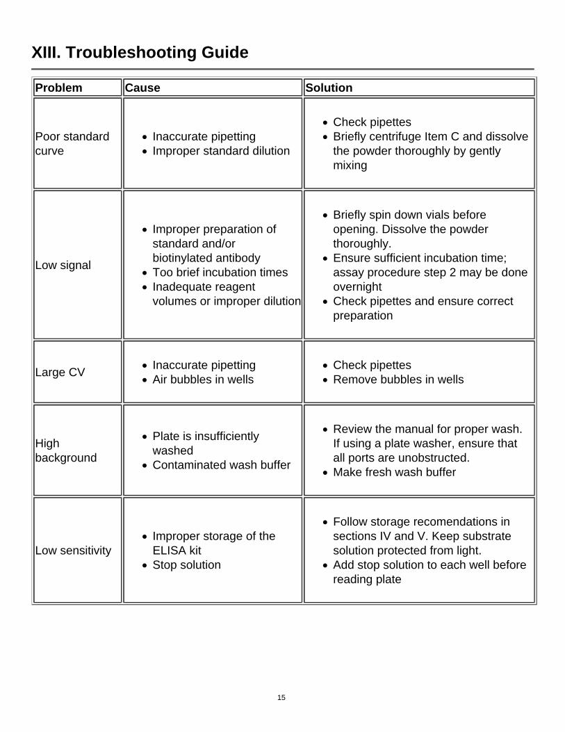
XIII. Troubleshooting Guide
Problem Cause Solution
Poor standard curve
Inaccurate pipettingImproper standard dilution
Check pipettesBriefly centrifuge Item C and dissolve the powder thoroughly by gently mixing
Low signal
Improper preparation of standard and/or biotinylated antibodyToo brief incubation timesInadequate reagent volumes or improper dilution
Briefly spin down vials before opening. Dissolve the powder thoroughly.Ensure sufficient incubation time; assay procedure step 2 may be done overnightCheck pipettes and ensure correct preparation
Large CVInaccurate pipettingAir bubbles in wells
Check pipettesRemove bubbles in wells
High background
Plate is insufficiently washedContaminated wash buffer
Review the manual for proper wash. If using a plate washer, ensure that all ports are unobstructed.Make fresh wash buffer
Low sensitivityImproper storage of the ELISA kitStop solution
Follow storage recomendations in sections IV and V. Keep substrate solution protected from light.Add stop solution to each well before reading plate
15

RayBio® ELISA Kits
Over 2,000 ELISA kits available, visit www.RayBiotech.com/ELISA-Kits.html for details.
This product is for research use only.
©2015 RayBiotech, Inc
16