multiple myeloma 101 · antibodies. slide created by redmeded for mmrf pt summit series \⠀㈀...
TRANSCRIPT
MULTIPLE MYELOMA 101 David H. Vesole, MD, PhD
Co-Director, Myeloma Division
Director, Myeloma Research
John Theurer Cancer Center
Hackensack University Medical Center
Director, Myeloma Program
Professor of Medicine, Georgetown University

What is multiple myeloma?Multiple myeloma
Normal plasma cellsM proteins
Multiple myeloma cells
BoneBone
marrow
Lightchain
Heavychains
Lightchain
Antibodies

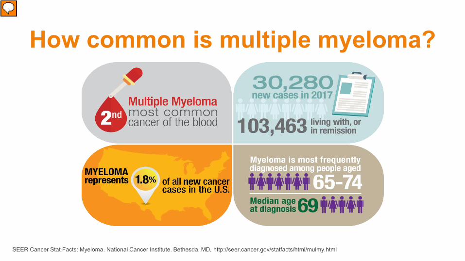
How common is multiple myeloma?
SEER Cancer Stat Facts: Myeloma. National Cancer Institute. Bethesda, MD, http://seer.cancer.gov/statfacts/html/mulmy.html
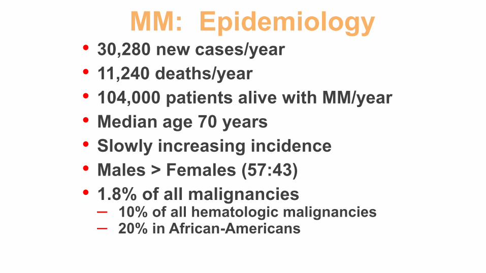
MM: Epidemiology• 30,280 new cases/year• 11,240 deaths/year • 104,000 patients alive with MM/year• Median age 70 years• Slowly increasing incidence• Males > Females (57:43)• 1.8% of all malignancies
– 10% of all hematologic malignancies– 20% in African-Americans
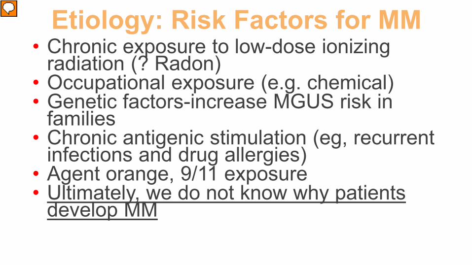
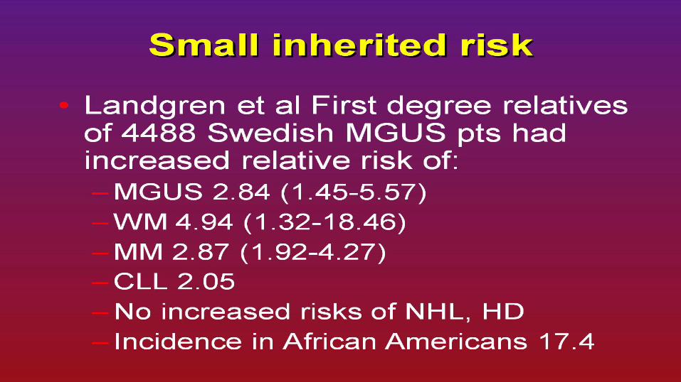
Etiology: Risk Factors for MM• Chronic exposure to low-dose ionizing
radiation (? Radon)• Occupational exposure (e.g. chemical)• Genetic factors-increase MGUS risk in
families• Chronic antigenic stimulation (eg, recurrent
infections and drug allergies)• Agent orange, 9/11 exposure• Ultimately, we do not know why patients
develop MM
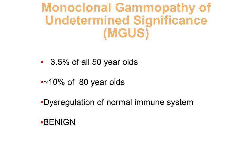
Monoclonal Gammopathy of Undetermined Significance
(MGUS)
•~ 3.5% of all 50 year olds
•~10% of 80 year olds
•Dysregulation of normal immune system
•BENIGN
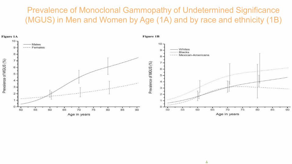
Prevalence of Monoclonal Gammopathy of Undetermined Significance (MGUS) in Men and Women by Age (1A) and by race and ethnicity (1B)
Landgren et al Leukemia. 2014. 28: 1537-1542.
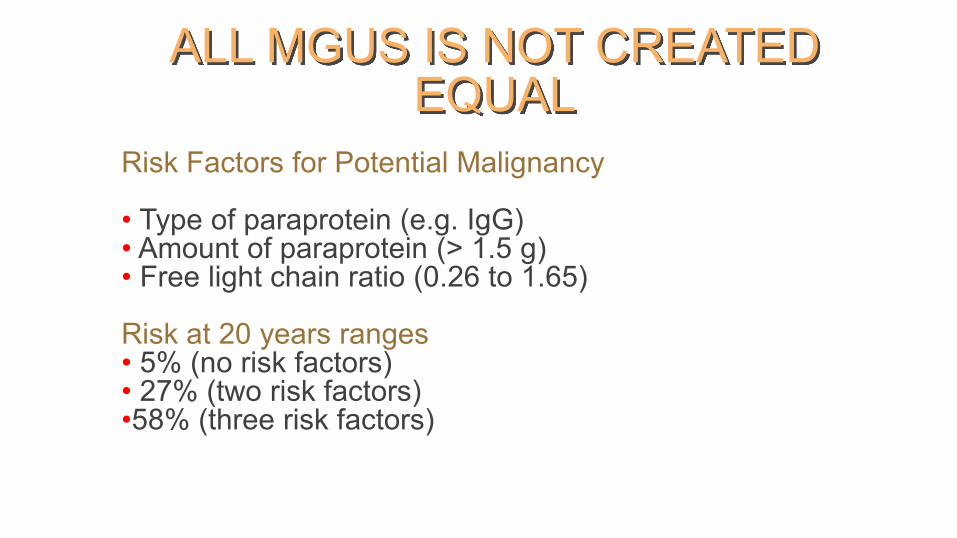
ALL MGUS IS NOT CREATED EQUAL
Risk Factors for Potential Malignancy
• Type of paraprotein (e.g. IgG)• Amount of paraprotein (> 1.5 g)• Free light chain ratio (0.26 to 1.65)
Risk at 20 years ranges • 5% (no risk factors)• 27% (two risk factors)•58% (three risk factors)

Smoldering Myeloma
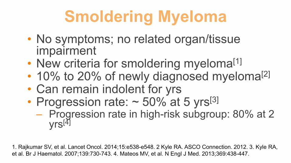
Smoldering Myeloma• No symptoms; no related organ/tissue
impairment• New criteria for smoldering myeloma[1]
• 10% to 20% of newly diagnosed myeloma[2]
• Can remain indolent for yrs• Progression rate: ~ 50% at 5 yrs[3]
– Progression rate in high-risk subgroup: 80% at 2 yrs[4]
1. Rajkumar SV, et al. Lancet Oncol. 2014;15:e538-e548. 2 Kyle RA. ASCO Connection. 2012. 3. Kyle RA, et al. Br J Haematol. 2007;139:730-743. 4. Mateos MV, et al. N Engl J Med. 2013;369:438-447.
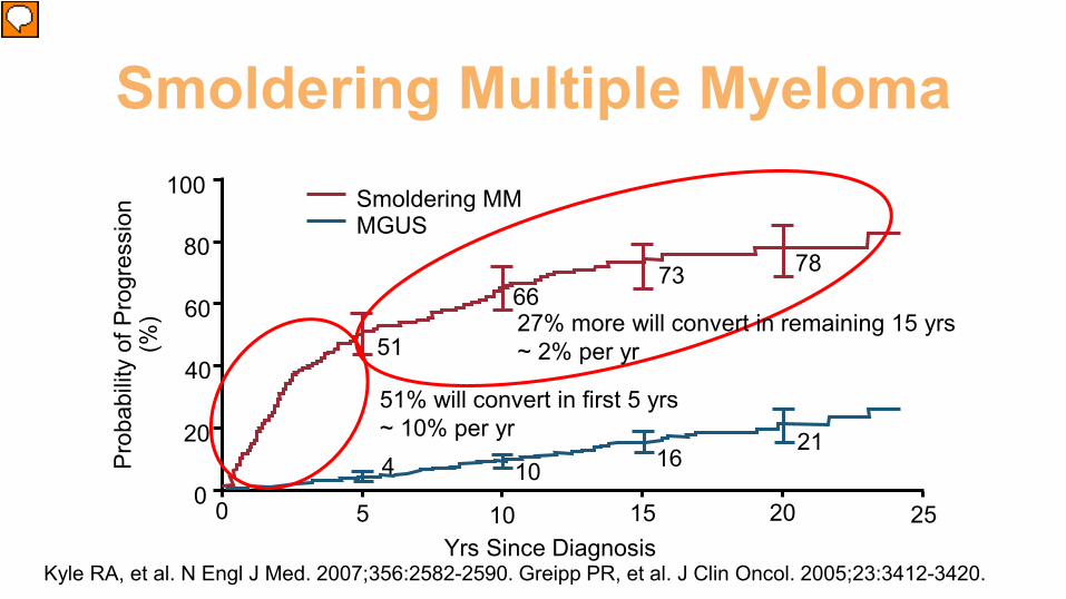
100
80
60
40
20
0
51% will convert in first 5 yrs~ 10% per yr
0 5 10 15 20 25
Prob
abilit
y of
Pro
gres
sion
(%
)
51
6673 78
4 10 1621
MGUSSmoldering MM
Smoldering Multiple Myeloma
Kyle RA, et al. N Engl J Med. 2007;356:2582-2590. Greipp PR, et al. J Clin Oncol. 2005;23:3412-3420.Yrs Since Diagnosis
27% more will convert in remaining 15 yrs~ 2% per yr
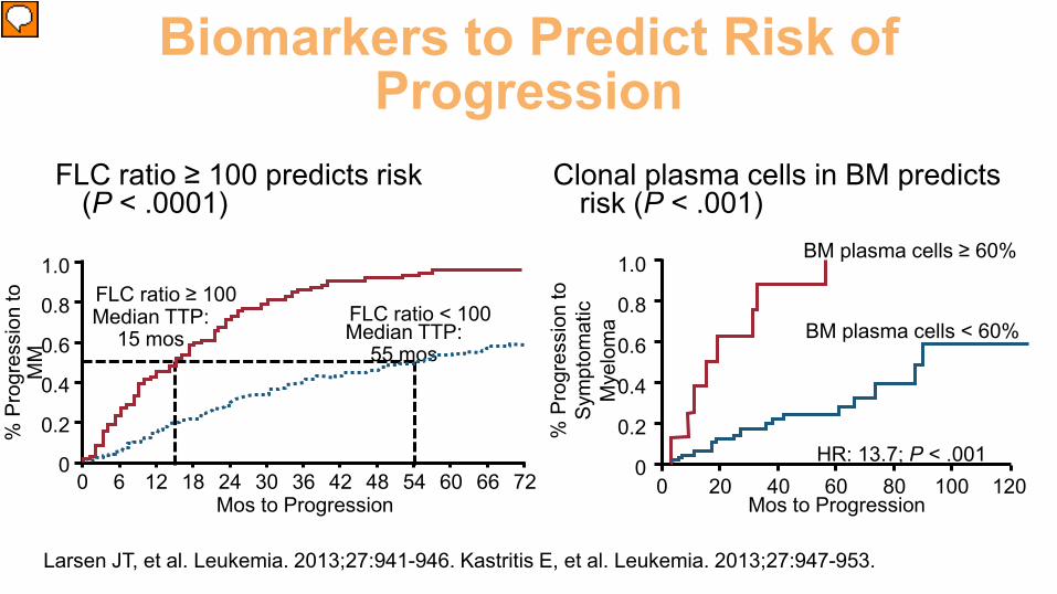
HR: 13.7; P < .001
Biomarkers to Predict Risk of Progression
FLC ratio ≥ 100 predicts risk (P < .0001)
Clonal plasma cells in BM predicts risk (P < .001)
Larsen JT, et al. Leukemia. 2013;27:941-946. Kastritis E, et al. Leukemia. 2013;27:947-953.
FLC ratio ≥ 100FLC ratio < 100Median TTP:
15 mos Median TTP:55 mos
1.0
0.8
0.6
0.4
0.2
00 20 40 60 80 100 120
Mos to Progression
BM plasma cells < 60%
BM plasma cells ≥ 60%
% P
rogr
essi
on to
Sy
mpt
omat
ic
Mye
lom
a
% P
rogr
essi
on to
M
M
Mos to Progression720 6 12 18 24 30 36 42 48 54 60 66
1.0
0.8
0.6
0.4
0.2
0
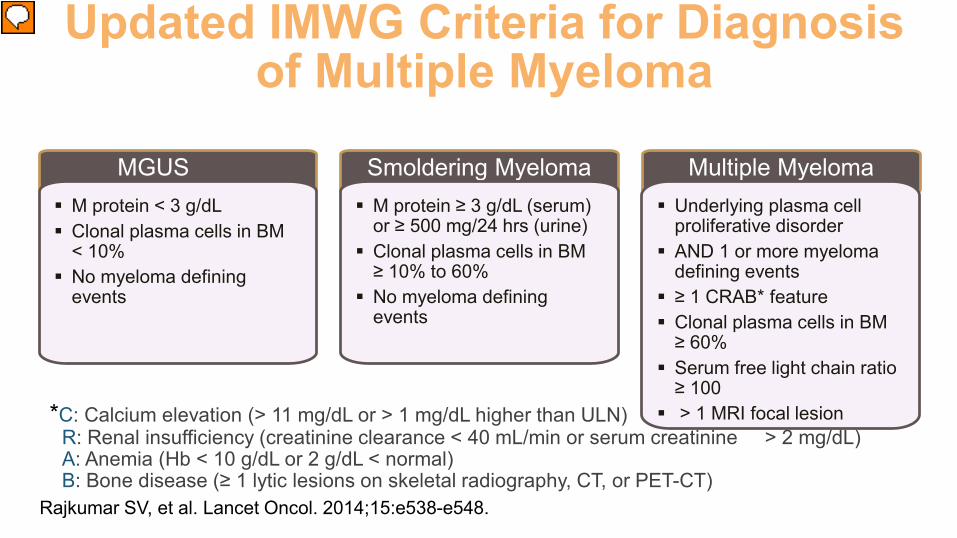
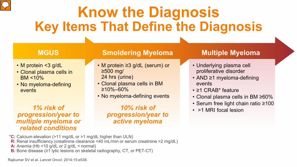
Updated IMWG Criteria for Diagnosis of Multiple Myeloma
*C: Calcium elevation (> 11 mg/dL or > 1 mg/dL higher than ULN)R: Renal insufficiency (creatinine clearance < 40 mL/min or serum creatinine > 2 mg/dL)A: Anemia (Hb < 10 g/dL or 2 g/dL < normal)B: Bone disease (≥ 1 lytic lesions on skeletal radiography, CT, or PET-CT)
Rajkumar SV, et al. Lancet Oncol. 2014;15:e538-e548.
MGUS M protein < 3 g/dL Clonal plasma cells in BM
< 10% No myeloma defining
events
Smoldering Myeloma M protein ≥ 3 g/dL (serum)
or ≥ 500 mg/24 hrs (urine) Clonal plasma cells in BM
≥ 10% to 60% No myeloma defining
events
Multiple Myeloma Underlying plasma cell
proliferative disorder AND 1 or more myeloma
defining events ≥ 1 CRAB* feature Clonal plasma cells in BM
≥ 60% Serum free light chain ratio
≥ 100 > 1 MRI focal lesion
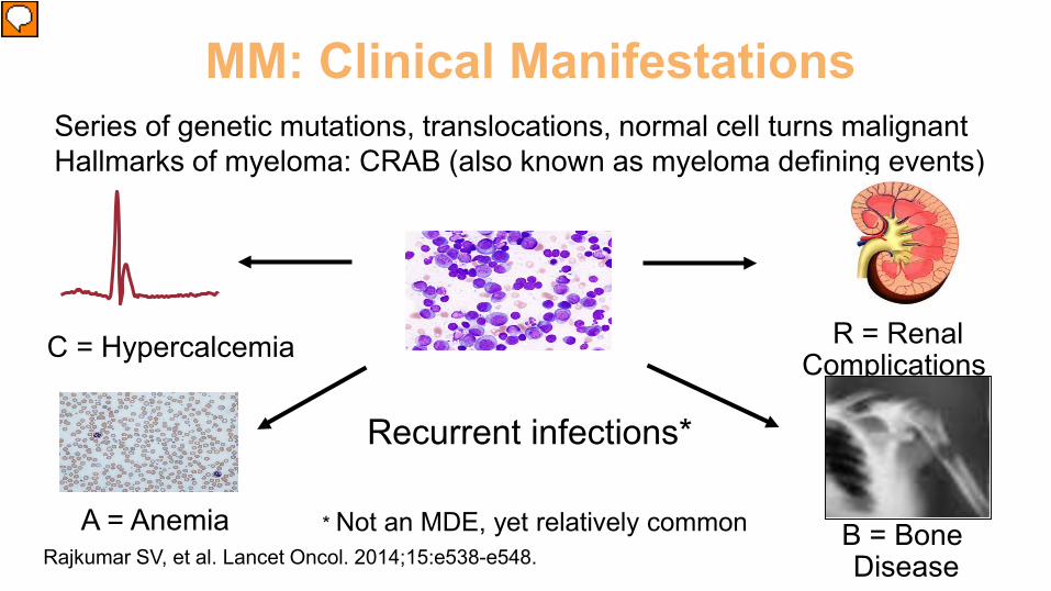
MM: Clinical ManifestationsSeries of genetic mutations, translocations, normal cell turns malignantHallmarks of myeloma: CRAB (also known as myeloma defining events)
A = Anemia B = Bone Disease
C = Hypercalcemia R = RenalComplications
* Not an MDE, yet relatively common
Recurrent infections*
Rajkumar SV, et al. Lancet Oncol. 2014;15:e538-e548.

Effects of Myeloma and Common Symptoms
Multiple Myeloma Symptoms, Side Effects, and Complications. https://themmrf.org/multiple-myeloma/symptoms-side-effects-and-complications/. Accessed February 19, 2019.Campbell K. Nurs Times. 2014;110:12.Kyle R et al. Mayo Clin Proc. 2003;78:21.
About 10% to 20% of patients with newly diagnosed myeloma do
not have any symptoms.
Low blood counts• Weakness• Fatigue• Infection
Decreased kidneyfunction
Weakness
Bone damage Bone pain
Bone turnover • Loss of appetite• Weight loss
Genetic changes occur
Ghobrial IM, et al. Blood. 2014;124:3380-3388. Rajkumar SV, et al. Lancet Oncol. 2014;15:e538-3548. Faiman B. Clin Lymphoma Myeloma Leuk. 2014;14:436-440.
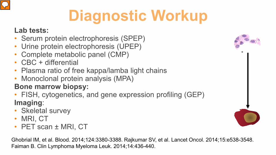
Diagnostic Workup Lab tests: • Serum protein electrophoresis (SPEP)• Urine protein electrophoresis (UPEP)• Complete metabolic panel (CMP)• CBC + differential• Plasma ratio of free kappa/lamba light chains• Monoclonal protein analysis (MPA)Bone marrow biopsy: • FISH, cytogenetics, and gene expression profiling (GEP)Imaging: • Skeletal survey• MRI, CT• PET scan ± MRI, CT
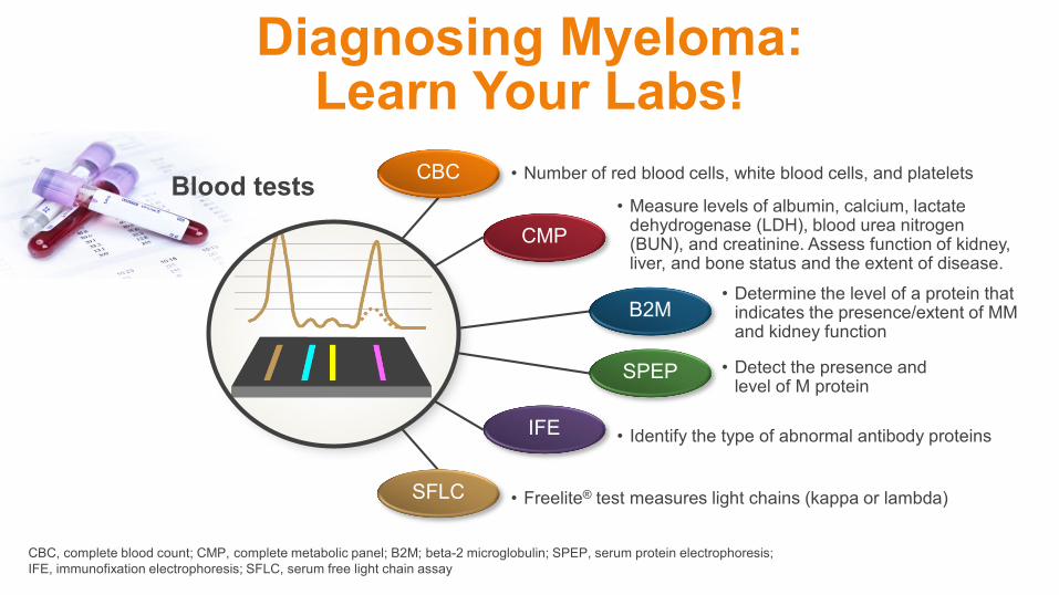
Diagnosing Myeloma: Learn Your Labs!
• Number of red blood cells, white blood cells, and platelets
• Measure levels of albumin, calcium, lactate dehydrogenase (LDH), blood urea nitrogen (BUN), and creatinine. Assess function of kidney, liver, and bone status and the extent of disease.
• Determine the level of a protein that indicates the presence/extent of MM and kidney function
• Identify the type of abnormal antibody proteins
• Detect the presence and level of M protein
• Freelite® test measures light chains (kappa or lambda)
CBC, complete blood count; CMP, complete metabolic panel; B2M; beta-2 microglobulin; SPEP, serum protein electrophoresis;IFE, immunofixation electrophoresis; SFLC, serum free light chain assay
Blood tests CBC
CMP
B2M
SPEP
IFE
SFLC

Diagnosing Myeloma: Learn Your Labs!
UPEP, urine protein electrophoresis
Urine tests • Detect Bence Jones proteins (otherwise known as myeloma light chains)
• Determine the presence and levels of M protein and Bence Jones protein
24-hr Urine Analysis
UPEP

Types of Monoclonal Protein (M Protein) in Multiple Myeloma
Light chain only• Also known as Bence
Jones protein• 20% of all myeloma
cases• Renal failure more
common in light chain multiple myeloma; creatinine >2 mg/dLin 1/3 of cases
Non-secretory• No monoclonal
protein present • 3% of cases of
multiple myeloma
Intact immunoglobulin• For example:
• IgG+kappa• IgG+lambda• IgA+kappa• IgA+lambda• etc…
• 80% of myeloma cases
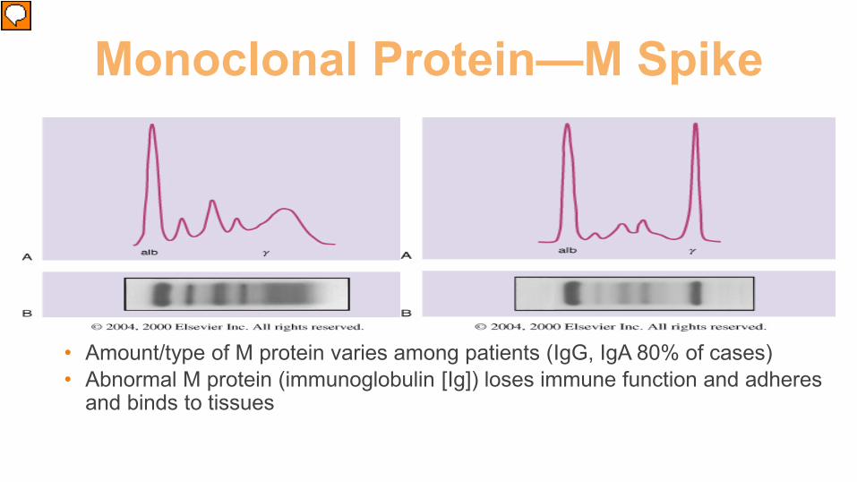
Monoclonal Protein—M Spike
• Amount/type of M protein varies among patients (IgG, IgA 80% of cases)• Abnormal M protein (immunoglobulin [Ig]) loses immune function and adheres
and binds to tissues
Normal SPEP Abnormal SPEP

Kyle RA and Rajkumar SV. Cecil Textbook of Medicine, 22nd Edition, 2004
Immunofixation to Determine Type of Monoclonal Protein
IgG kappa M protein Lambda Light Chains

Intact Immunoglobulin
Exposed surfaceHidden surface
Free Light Chain
Previouslyhiddensurface
FLC reference range:κ 3.3 – 19.4 mg/L λ 5.7 – 26.3 mg/L κ/λ ratio 0.26 - 1.65

Myeloma Cells
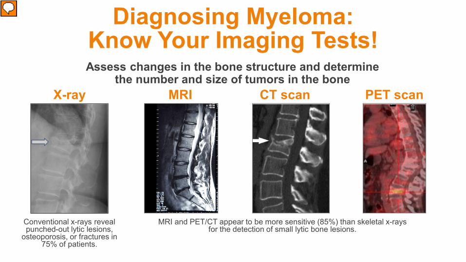
Diagnosing Myeloma: Know Your Imaging Tests!
X-ray MRI CT scan PET scan
Conventional x-rays reveal punched-out lytic lesions,
osteoporosis, or fractures in 75% of patients.
MRI and PET/CT appear to be more sensitive (85%) than skeletal x-raysfor the detection of small lytic bone lesions.
Assess changes in the bone structure and determine the number and size of tumors in the bone
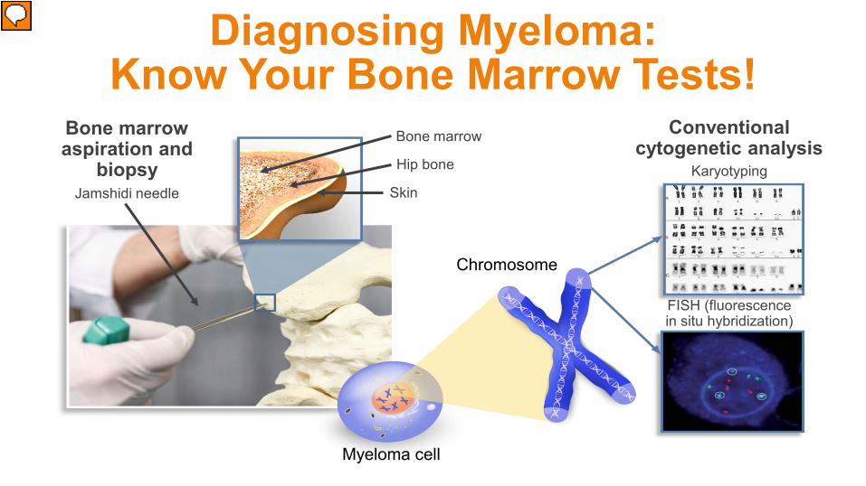
Diagnosing Myeloma: Know Your Bone Marrow Tests!
Karyotyping
FISH (fluorescence in situ hybridization)
Myeloma cell
Chromosome
Jamshidi needle
Bone marrow
Skin
Hip bone
Bone marrow aspiration and
biopsy
Conventional cytogenetic analysis

How aggressive is my myeloma?
Based on the Updated Mayo Stratification of Myeloma and Risk-Adapted Therapy (mSMART) Consensus Guidelines 2013aTrisomies may ameliorate; bBy FISH or equivalent method; cCut-offs vary; dt(11;14) may be associated with plasma cell leukemiaMikhael JR et al. Mayo Clin Proc. 2013;88:360.
Currently cannot predict with great certainty all high-risk patients.
High Risk• High-risk genetic abnormalitiesa,b
− t(4;14)− t(14;16)− t(14;20)− Del 17p− p53 mutation− Gain 1q
• RISS Stage 3• High plasma cell S-phasec
• GEP: high-risk signature
• Double-hit myeloma: any two high-risk genetic abnormalities
• Triple-hit myeloma: three or more high-risk genetic abnormalities
Standard Riska
• All others including:− Trisomies− t(11;14)d
− t(6;14)
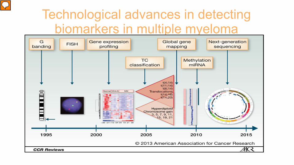
Technological advances in detecting biomarkers in multiple myeloma
Landgren, and Gareth. Clin Cancer Res 2014;20:804-813
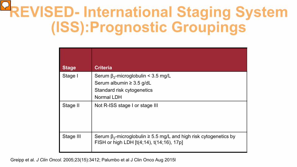
REVISED- International Staging System (ISS):Prognostic Groupings
Stage CriteriaStage I Serum β2-microglobulin < 3.5 mg/L
Serum albumin ≥ 3.5 g/dLStandard risk cytogeneticsNormal LDH
Stage II Not R-ISS stage I or stage III
Stage III Serum β2-microglobulin ≥ 5.5 mg/L and high risk cytogenetics by FISH or high LDH [t(4;14), t(14;16), 17p]
Greipp et al. J Clin Oncol. 2005;23(15):3412; Palumbo et al J Clin Onco Aug 2015l
Better prognosis
Poorer prognosis
• Underlying plasma cell proliferative disorder
• AND ≥1 myeloma-defining events
• ≥1 CRAB* feature• Clonal plasma cells in BM ≥60%• Serum free light chain ratio ≥100• >1 MRI focal lesion
• M protein ≥3 g/dL (serum) or ≥500 mg/24 hrs (urine)
• Clonal plasma cells in BM ≥10%–60%
• No myeloma-defining events
• M protein <3 g/dL• Clonal plasma cells in
BM <10%• No myeloma-defining
events
Know the DiagnosisKey Items That Define the Diagnosis
*C: Calcium elevation (>11 mg/dL or >1 mg/dL higher than ULN)R: Renal insufficiency (creatinine clearance <40 mL/min or serum creatinine >2 mg/dL)A: Anemia (Hb <10 g/dL or 2 g/dL < normal)B: Bone disease (≥1 lytic lesions on skeletal radiography, CT, or PET-CT)
Rajkumar SV et al. Lancet Oncol. 2014;15:e538.
MGUS Smoldering Myeloma Multiple Myeloma
10% risk of progression/year to
active myeloma
1% risk of progression/year tomultiple myeloma or
related conditions
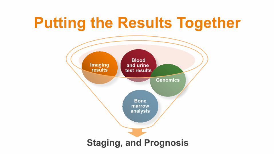
Putting the Results Together
Staging, and Prognosis
Bone marrow analysis
Imaging results
Bloodand urine test results
Genomics

Relapse can occur when:
Existing clone no longer has to compete for space with the formerly dominant clone
Acquires additional mutation(s) providing a growth and/or survival advantage
Clonal Evolution and ClonalCompetition
Multiple clones may be present at the time of diagnosis The predominant clone may change over time, especially after treatment roundsHypothesis: effective treatment reduces or eliminates the dominant clone; however, other clones can still exist
Keats JJ, et al. Blood. 2012;120:1067-1076.
Clone 1.1Clone 1.2Clone 2.1Clone 2.2Misc
Diagnosis~ 2N
Remission ~ 2N Relapse 1
~ 2N
Relapse 2 ~ 2N
Relapse 3 ~ 2N
Plasma cell leukemia ~ 3N Relapse 4 ~ 3N
clg-high37%
clg-high66%
clg-low34% clg-low
63%
72%11%
10%
31%64%
64%
21%9%
19%58%
71%
17%
78%95%
96%96%

Treatment Overview
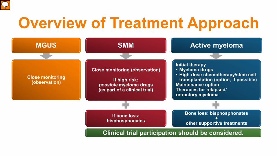
Overview of Treatment ApproachMGUS
Close monitoring (observation)
SMM
Close monitoring (observation)
If high risk: possible myeloma drugs(as part of a clinical trial)
Active myeloma
Initial therapy• Myeloma drugs• High-dose chemotherapy/stem cell
transplantation (option, if possible)Maintenance optionTherapies for relapsed/refractory myeloma
If bone loss: bisphosphonates
Bone loss: bisphosphonates+
other supportive treatments
Clinical trial participation should be considered.

History of Treatment• 1844: Rhubarb and infusions of orange peel
for Sarah Newbury• 1845: Phlebotomy, then leeches for
maintenance therapy (William McBean)• 1947: Urethane reported by Alwall• 1958: Blohkin reports sarcolysin (melphalan)
in 3 of 6 patients• 1962: Bergsagel expands use of melphalan• 1962: Maas report on prednisone


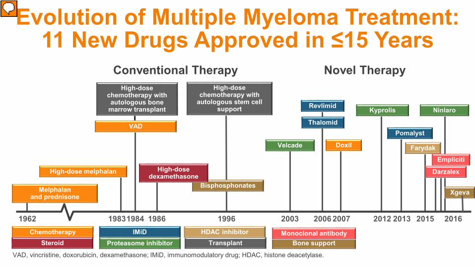
1962 1983 1986 1996 2012
Evolution of Multiple Myeloma Treatment: 11 New Drugs Approved in ≤15 Years
1984 2003 2006 2007
VAD, vincristine, doxorubicin, dexamethasone; IMiD, immunomodulatory drug; HDAC, histone deacetylase.
2013Chemotherapy
Steroid Transplant IMiD
Bone supportProteasome inhibitor HDAC inhibitor
2015
Conventional Therapy Novel Therapy
BisphosphonatesMelphalan and prednisone
VAD
High-dose dexamethasone
High-dose chemotherapy with
autologous stem cell support Kyprolis
High-dose melphalan
High-dose chemotherapy with autologous bone
marrow transplant
Velcade
Thalomid
Revlimid
Doxil
Pomalyst
Farydak
Ninlaro
2016
Empliciti
Darzalex
Monoclonal antibody
Xgeva
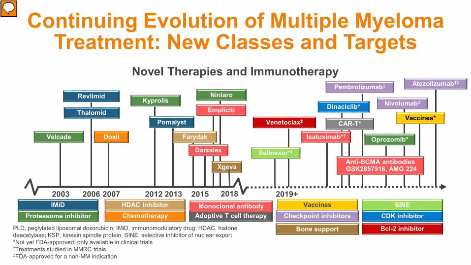
Continuing Evolution of Multiple Myeloma Treatment: New Classes and Targets
PLD, peglylated liposomal doxorubicin; IMiD, immunomodulatory drug; HDAC, histone deacetylase; KSP, kinesin spindle protein, SINE, selective inhibitor of nuclear export*Not yet FDA-approved; only available in clinical trials†Treatments studied in MMRC trials‡FDA-approved for a non-MM indication
Novel Therapies and Immunotherapy
20122003 2006 2007 2013 2015 2019+
Doxil
Kyprolis
Velcade
Thalomid
Revlimid
Pomalyst
Farydak Isatuximab*†
Nivolumab‡
Vaccines*
Ninlaro
Darzalex
Empliciti
Pembrolizumab‡
Dinaciclib*
CAR-T*
Oprozomib*
Proteasome inhibitor IMiD
ChemotherapyVaccines
Adoptive T cell therapy Checkpoint inhibitorsHDAC inhibitor Monoclonal antibody SINE
CDK inhibitor
Venetoclax‡
Atezolizumab†‡
Bcl-2 inhibitor
Anti-BCMA antibodiesGSK2857916, AMG 224Xgeva
Bone support
2018
Selinexor*†

Measuring Response to Therapy
Response Type
Abbreviation
TestsM-Protein Reduction
Immunofixation
Bone Marrow
FreeliteBlood Urine PC
Immuno-fluorescence Other
Complete response
CR 0 0 Negative <5% __ __ __
Stringent complete response
sCR 0 0 Negative <5% Negative __ Normal
Very good partial response
VGPR >90% <100 mg/24 hrs
__ __ __ __ __
Partial response
PR >50% >90% __ __ __ __ __
Stable response
SD Does not meet criteria for response or progressive disease
Progressive disease
PD An increase of 25% in M-protein; an increase of 10% in bone marrow plasma cells
Degree (or depth) of response is usually associated with better prognosis. Some patients do well despite never achieving a complete response (CR). Kumar S, Paiva B, Anderson KC, et al. Lancet Oncol. 2016;17(8):e328-e346.
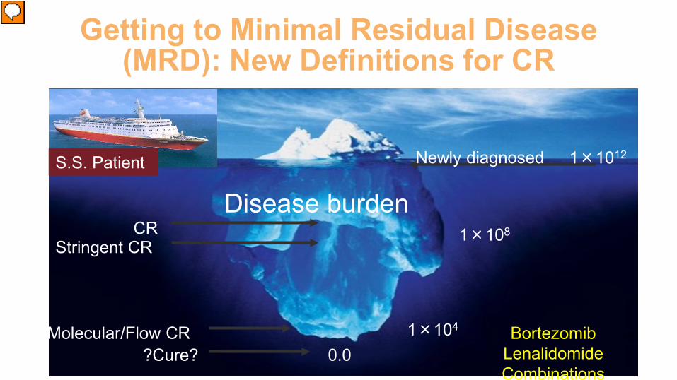
Getting to Minimal Residual Disease (MRD): New Definitions for CR
S.S. Patient 1×10¹²
Stringent CR
Molecular/Flow CR?Cure?
Disease burden
Newly diagnosed
1×10⁸
1×10⁴
0.0Bortezomib
LenalidomideCombinations
CR
Rawstron A C et al. JCO 2013;31:2540-2547
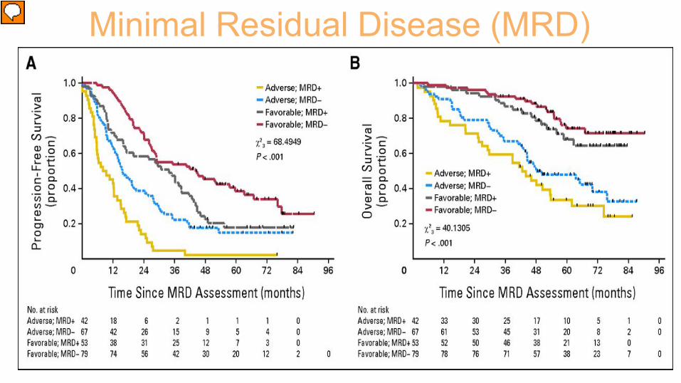
Minimal Residual Disease (MRD)
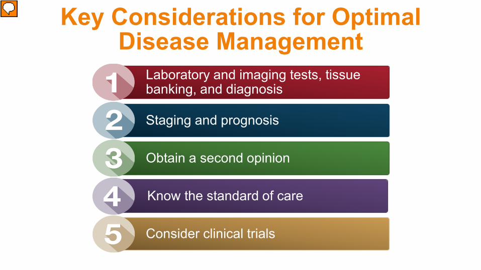
Key Considerations for Optimal Disease Management
Laboratory and imaging tests, tissue banking, and diagnosis
Staging and prognosis
Obtain a second opinion
Know the standard of care
Consider clinical trials

Myeloma 101: Summary
Be an informed and empowered patient!
Multiple myeloma can have numerous effects on the body.
Genomics is growing and may lead to personalized treatments.
Survival improving because of new drugs and new combinations of drugs.
Treatment paradigm will continue to change with the approval of additional novel agents.