mechanism of xenopus cranial neural crest cell migration
TRANSCRIPT

www.landesbioscience.com Cell Adhesion & Migration 553
Cell Adhesion & Migration 4:4, 553-560; October/November/December 2010; © 2010 Landes Bioscience
SPECIAL FOCUS: RECENT ADVANCES IN MOLECULAR AND CELLULAR MECHANISMS GOVERNING NEURAL CREST CELL MIGRATION, REVIEW
Introduction
The neural crest is a transient population of cells that is induced by the canonical Wnt, BMB, FGF, Notch and the retinoic acid signaling pathways1-5 at the border between the neural and non-neural ectoderm. Neural crest cells undergo extensive ventral migration to participate in the formation of multiple tissues and organs. In Xenopus laevis, detailed observations of the different phases of cranial neural crest (CNC) cell migration have shown that these cells initiate the ventral migration as a cohesive tissue.6 Due to the cohesive nature of this cell population, it is possible to dissect the CNC before they initiate migration and either graft them back into a host embryo or place them on various substrates in vitro.7-9 In both cases, cells from the CNC explant start migrating as a cohesive sheet maintaining contact with each other. This initial phase lasts between 3–5 h until finally the cells lose con-tact and migrate as individuals. These two distinct migratory phases make the CNC a powerful model to study how cohesive cell migration is regulated, as well as the switch involved in the transition to single cell migration. In Xenopus, contrary to what is known about the trunk neural crest, the CNC is never included in the neural tube, but instead migrates from the border of the
*Correspondence to: Dominique Alfandari; Email: [email protected]: 02/17/10; Accepted: 05/03/10Previously published online:www.landesbioscience.com/journals/celladhesion/article/12202DOI: 10.4161/cam.4.4.12202
This Review focuses on recent advances in the field of cranial neural crest cell migration in Xenopus laevis with specific emphasis on cell adhesion and the regulation of cell migration. Our goal is to combine the understanding of cell adhesion to the extracellular matrix with the regulation of cell-cell adhesion and the involvement of the planar cell polarity signaling-pathway in guiding the migration of cranial neural crest cells during embryogenesis.
Mechanism of Xenopus cranial neural crest cell migration
Dominique Alfandari,1,* Hélène Cousin1 and Mungo Marsden2
1Department of Veterinary and Animal Sciences; University of Massachusetts Amherst; Amherst, MA USA; 2Department of Biology; University of Waterloo; Waterloo, Ontario Canada
Key words: neural crest, cell migration, extracellular matrix, cell adhesion, Wnt, planar cell polarity
Abbreviation: CNC, cranial neural crest; FN, fibronectin; PCP, planar cell polarity; Hep II, second heparin-binding site of fibronectin; MO, morpholino; EC1-3, cadherin-11 extracellular domains 1 to 3; GFP, green fluorescent protein; Fz-7, frizzled-7; Dsh, dishevelled; PKCa, protein kinase C alpha; GEF, guanine exchange factor; EMT, epithelium to mesenchyme transition;
FRET, fluorescence resonance energy transfer; PTK7, protein tyrosine kinase 7; ECM, extracellular matrix
neural plate before neural tube closure.6 It is not clear if a true epithelium to mesenchyme transition (EMT) is involved during the first phase of CNC migration.
Single cell migration has been studied extensively in vitro on various substrates and the mechanics underlying cell motility are well understood. For example, a single cell needs to distin-guish the front (leading edge) from the back (trailing edge). The adhesion to the substrate, as well as the polymerization of actin, is increased at the leading edge; both of which are regulated by small GTPases of the Rho family (reviewed in ref. 10). In a single cell situation, cues from the environment have to define the polar-ity by specifying either the leading or the trailing edge. On the other hand, in cohesive cell migration, it may be easier to define the trailing edge as the one in contact with other cells, so that the polarity of the tissue defines the polarity of each cell.
Members of the planar cell polarity (PCP) pathway are clearly involved in contact-mediated inhibition of CNC migra-tion and thus are the best candidates to define the trailing edge in the cohesive cell migration. The notion of contact inhibition is about 50 years old and describes the process by which two fibroblasts migrating in vitro stop and change direction after they contact each other.11 This review will focus on the recent findings obtained in Xenopus with particular attention to the role and regulation of cell adhesion. For a recent review on the various pathways involved in neural crest cell migration, see reference 12. To avoid confusion, we will use dorso/ventral and back/front axis when describing the polarity of the CNC tissue, while we will use leading and trailing edge only when describ-ing the polarity within a cell.
Cell-Matrix Adhesion
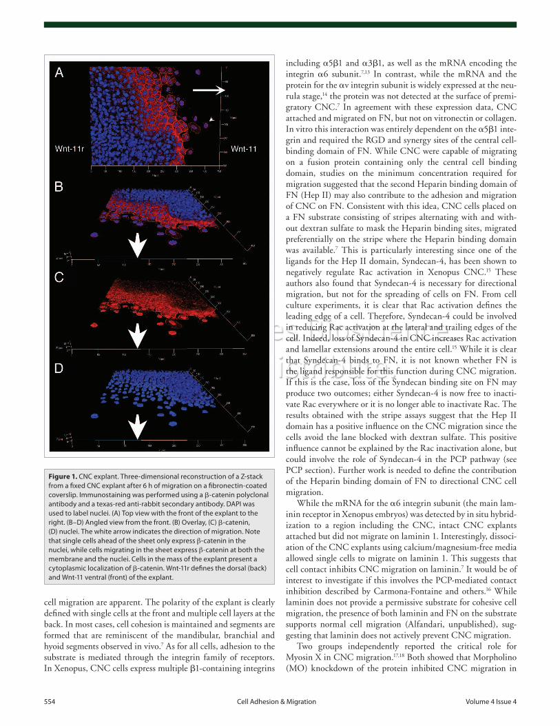
In vitro, CNC explants, which are initially about three cells thick, attach and spread when placed on a permissive substrate such as fibronectin (FN). During the initial phase, cells that are at the border of the explant migrate away increasing the overall surface of the explant and decreasing its thickness. Figure 1 shows one CNC explant fixed after six hours of migration on a FN-coated coverslip. In this example, both the cohesive sheet and single
554 Cell Adhesion & Migration Volume 4 Issue 4
including a5b1 and a3b1, as well as the mRNA encoding the integrin a6 subunit.7,13 In contrast, while the mRNA and the protein for the av integrin subunit is widely expressed at the neu-rula stage,14 the protein was not detected at the surface of premi-gratory CNC.7 In agreement with these expression data, CNC attached and migrated on FN, but not on vitronectin or collagen. In vitro this interaction was entirely dependent on the a5b1 inte-grin and required the RGD and synergy sites of the central cell-binding domain of FN. While CNC were capable of migrating on a fusion protein containing only the central cell binding domain, studies on the minimum concentration required for migration suggested that the second Heparin binding domain of FN (Hep II) may also contribute to the adhesion and migration of CNC on FN. Consistent with this idea, CNC cells placed on a FN substrate consisting of stripes alternating with and with-out dextran sulfate to mask the Heparin binding sites, migrated preferentially on the stripe where the Heparin binding domain was available.7 This is particularly interesting since one of the ligands for the Hep II domain, Syndecan-4, has been shown to negatively regulate Rac activation in Xenopus CNC.15 These authors also found that Syndecan-4 is necessary for directional migration, but not for the spreading of cells on FN. From cell culture experiments, it is clear that Rac activation defines the leading edge of a cell. Therefore, Syndecan-4 could be involved in reducing Rac activation at the lateral and trailing edges of the cell. Indeed, loss of Syndecan-4 in CNC increases Rac activation and lamellar extensions around the entire cell.15 While it is clear that Syndecan-4 binds to FN, it is not known whether FN is the ligand responsible for this function during CNC migration. If this is the case, loss of the Syndecan binding site on FN may produce two outcomes; either Syndecan-4 is now free to inacti-vate Rac everywhere or it is no longer able to inactivate Rac. The results obtained with the stripe assays suggest that the Hep II domain has a positive influence on the CNC migration since the cells avoid the lane blocked with dextran sulfate. This positive influence cannot be explained by the Rac inactivation alone, but could involve the role of Syndecan-4 in the PCP pathway (see PCP section). Further work is needed to define the contribution of the Heparin binding domain of FN to directional CNC cell migration.
While the mRNA for the a6 integrin subunit (the main lam-inin receptor in Xenopus embryos) was detected by in situ hybrid-ization to a region including the CNC, intact CNC explants attached but did not migrate on laminin 1. Interestingly, dissoci-ation of the CNC explants using calcium/magnesium-free media allowed single cells to migrate on laminin 1. This suggests that cell contact inhibits CNC migration on laminin.7 It would be of interest to investigate if this involves the PCP-mediated contact inhibition described by Carmona-Fontaine and others.16 While laminin does not provide a permissive substrate for cohesive cell migration, the presence of both laminin and FN on the substrate supports normal cell migration (Alfandari, unpublished), sug-gesting that laminin does not actively prevent CNC migration.
Two groups independently reported the critical role for Myosin X in CNC migration.17,18 Both showed that Morpholino (MO) knockdown of the protein inhibited CNC migration in
cell migration are apparent. The polarity of the explant is clearly defined with single cells at the front and multiple cell layers at the back. In most cases, cell cohesion is maintained and segments are formed that are reminiscent of the mandibular, branchial and hyoid segments observed in vivo.7 As for all cells, adhesion to the substrate is mediated through the integrin family of receptors. In Xenopus, CNC cells express multiple b1-containing integrins
Figure 1. CNC explant. Three-dimensional reconstruction of a Z-stack from a fixed CNC explant after 6 h of migration on a fibronectin-coated coverslip. Immunostaining was performed using a b-catenin polyclonal antibody and a texas-red anti-rabbit secondary antibody. DAPI was used to label nuclei. (A) Top view with the front of the explant to the right. (B–D) Angled view from the front. (B) Overlay, (C) b-catenin, (D) nuclei. The white arrow indicates the direction of migration. Note that single cells ahead of the sheet only express β-catenin in the nuclei, while cells migrating in the sheet express β-catenin at both the membrane and the nuclei. Cells in the mass of the explant present a cytoplasmic localization of b-catenin. Wnt-11r defines the dorsal (back) and Wnt-11 ventral (front) of the explant.
www.landesbioscience.com Cell Adhesion & Migration 555
outcomes. Overexpression of a Cadherin-11 lacking the cytoplas-mic domain prevents CNC migration, most likely by increasing cell-cell adhesion. Overexpression of the wild-type Cadherin-11 blocks CNC migration as well, however it also decreases CNC markers like Twist. This can be rescued by overexpression of b-catenin, suggesting that the decrease in CNC markers is due to sequestration of b-catenin at the plasma membrane (Fig. 2). This in turn reduces available b-catenin levels that can translo-cate into the nucleus to activate downstream target genes such as Twist.22 Interestingly, CNC migration can also be rescued in embryos overexpressing Cadherin-11 by co-expressing the ADAM13 metalloprotease, which binds and cleaves the extra-cellular domain of Cadherin-11 in vivo.23 Thus, the ADAM13 metalloprotease can cleave and separate the extracellular domain from the transmembrane and cytoplasmic domains of the pro-tein. Consistent with this observation, ADAM13 knockdown also reduces CNC migration and increases Cadherin-11 protein levels. Cell migration can then be rescued by either reintroduc-ing the ADAM13 protease, or injecting an mRNA that encodes the N-terminal portion of Cadherin-11 containing the first three cadherin repeats (EC1-3), in or next to the CNC.23 This sug-gests that a precise balance between the full-length and cleaved Cadherin-11 protein is required to promote CNC cell migration (Fig. 3). Cleavage of the extracellular domain of Cadherin-11 allows cells to decrease cell adhesion without affecting cell sig-naling, since the cleaved transmembrane fragment is stable and can still interact with b-catenin.23 Reduction of Cadherin-11 by MO knockdown prevents the second phase of migration in vivo, while in vitro, cells do not display filopodia and spread less effi-ciently compared to those derived from control embryos. In both cases, the effects can be rescued by expression of either wild-type Cadherin-11 or a truncated protein containing the transmem-brane and cytoplasmic domains, suggesting that the extracellu-lar domain may not be critical.24 As the efficiency of the MO knockdown was about 80% in vivo, it is possible that the level of protein remaining in the knockdown is sufficient to achieve the extracellular function, while a higher level of protein is critical for the cytoplasmic domain function. This is consistent with the fact that only a fraction of Cadherin-11 is cleaved by the ADAM13 protease in vivo. Using a C-terminal fusion of green fluores-cent protein (GFP) to Cadherin-11, the authors showed that the protein localizes to the filopodia of CNC when placed on FN. Additionally, the cytoplasmic domain of Cadherin-11 was bound to the guanine exchange factor (GEF)-Trio, which is known in mammalian cells to activate RhoA, Rac and Cdc42.25 Using site directed mutation analysis, they showed that both the Trio and b-catenin binding sites on the Cadherin-11 cytoplasmic domain were critical for the migration of CNC (Fig. 3). In addition, over-expression of human Trio, as well as constitutively active forms of RhoA, Rac1 and Cdc42, was sufficient to rescue CNC cell spreading and migration in Cadherin-11 knockdown embryos, suggesting that Cadherin-11 is critical for the activation of small GTPases in CNC.24 The use of fluorescence resonance energy transfer (FRET), as described by Matthews and colleagues,15 could show which of these (RhoA, Rac, Cdc42) are affected by the reduction in Cadherin-11 protein levels.
vivo as well as in vitro. Nie and colleagues17 used a MO to pre-vent Myosin X mRNA translation, affecting both maternal and zygotic expression of the protein, and found that it affected early neural crest gene expression (Slug and Sox10). This is consistent with a role for Myosin X during early embryogenesis prior to CNC migration. In addition to the role of Myosin X in CNC migration, they also showed that cells were less adhesive and formed less aggregates in vitro, suggesting that overall cell adhe-sion was decreased.17 Using a MO to target splicing of the pre-mRNA, Hwang and colleagues were able to prevent selectively the zygotic expression of Myosin X, without affecting transla-tion from maternally stored mRNA that plays essential roles during mitosis.19,20 Using this elegant strategy, they were able to show no defect in Slug induction, an early marker for the CNC, but showed a defect in migration when CNC from knockdown embryos were grafted into host embryos or placed on a FN sub-strate. Both defects could be rescued by overexpressing Myosin X.18 All of the observed phenotypes are consistent with the previ-ously known role of Myosin X in regulating cytoskeleton and integrin localization, but the precise role of this protein in CNC remains to be elucidated.
While the use of MO has allowed researchers to effectively and rapidly study gene function by loss-of-function experi-ments in Xenopus, it is essential to keep in mind that these experiments knockdown protein expression and are not the equivalent of a genetic knockout. As such these data need to be interpreted cautiously. In most studies, the level of endog-enous protein is not directly evaluated; instead the efficiency of a given MO is determined by its ability to prevent translation of synthetic mRNA encoding a tagged protein construct. The efficacy of MO knockdown varies with the protein in ques-tion and investigators use from 1–100 ng of Morpholino to achieve the desired result. The “gold standard” is the rescue of the MO phenotype through expression of a modified mRNA that does not complement the sequence of the MO. While this demonstrates that the MO itself is not toxic, it does not demon-strate that the phenotypes are not due to off-target inhibition. The rescue itself results in ectopic expression, as it is nearly impossible to target only the tissues being studied. The use of MOs is continually evolving and elegant experiments with splice-specific MO, such as that described above, have given us insights into the temporal regulation of protein expression. Given these limitations and careful interpretations, MOs are a powerful tool to investigate protein function during Xenopus embryogenesis.
Cell-Cell Adhesion
Among the cell-cell adhesion molecules, Cadherin-11 and the protocadherin PCNS have been shown to be critical for CNC migration both in vivo and in vitro. The mechanism by which Cadherin-11 controls CNC migration has been extensively stud-ied in Xenopus and has revealed some unexpected and excit-ing novel activities. Cadherin-11 is expressed in the CNC cells before and throughout migration.21 Both overexpression and MO knockdown interfere with CNC migration, but with different
556 Cell Adhesion & Migration Volume 4 Issue 4
Planar Cell Polarity
One of the defining features of the NC cells is that they migrate directionally in vivo and in vitro. In vivo, defined pathways of ECM and molecules that repel NC cells such as Ephrins, Semaphorins and Robo/Slit appear to guide NC cells along migration pathways without providing directional cues.12 In vitro, NC cells retain persistent migration even in the absence of external cues, indicating that in vivo they are unlikely to be drawn towards an attractant. Recent work has revealed the non-canonical Wnt planar cell polarity (PCP) pathway as the primary source of signals that promote directional CNC migration in Xenopus. The various proteins that can regulate the canonical and non-canonical Wnt pathways during Xenopus CNC speci-fication and migration are represented in Figure 2 and discussed in detail below.
The PCP pathway was first described in Drosophila where it acts to align bristles and hairs in the adult. Disruption of
The protocadherin PCNS is expressed from stage 12 to 27 in both mesoderm and neural crest cells. Using MO to prevent pro-tein translation, the authors showed that a decrease in PCNS pro-tein affected the position of the CNC markers Slug and Sox10, which suggests that CNC migration in the hyoid and branchial segments was inhibited. This was confirmed by placing the CNC explants on FN in vitro and showing that CNC explants with reduced PCNS did not spread or migrate.26 At this point, it is unclear whether PCNS is directly affecting cell adhesion to the extracellular matrix (ECM) by controlling integrin receptor acti-vation or if it affects the survival of explanted cells, as both situ-ations would generate a similar rounded cell shape. The fact that CNC cells still expressed the proper molecular markers argues for a direct role on the control of cell migration. Given the critical role of the PCP pathway in CNC migration and the established role of the protocadherin PAPC in this pathway,27 it is tempting to speculate that PCNS may influence the PCP signaling during CNC migration.
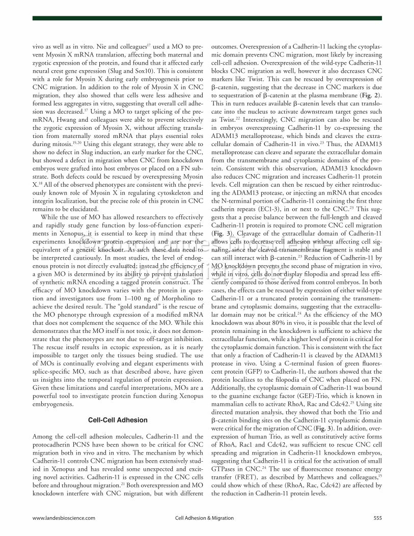
Figure 2. Regulation of Wnt signaling in Xenopus CNC. Wnt signaling is involved in the induction and specification of the CNC via the canonical pathway mediated by Wnt-11, Fz-7 and Dsh, stabilizing β-catenin to promote Lef/TCF-dependent gene transcription. Alternatively, Dsh stabilizes p120-Cat to promote expression of these same genes, including Wnt-11; thus, providing a positive feedback loop. Cadherin-11 binds to both β-catenin and p120-catenin maintaining a membrane bound pool. In addition, Cadherin-11 binds to the (GEF)-Trio and activates small GTPases of the Rho family to promote cell protrusion. Fz-7 and Dsh can complex with both PTK-7 and Synd-4 to mediate either the activation of RhoA and Rac or the inhibition of Rac, respectively. In the CNC, Pescadillo expression depends on the presence of Wnt-4 and Fz-3. Knockdown of Pescadillo increases apoptosis and decreases cell proliferation and cell migration. Apoptosis and cell proliferation defects can be rescued by knocking down p53.

www.landesbioscience.com Cell Adhesion & Migration 557
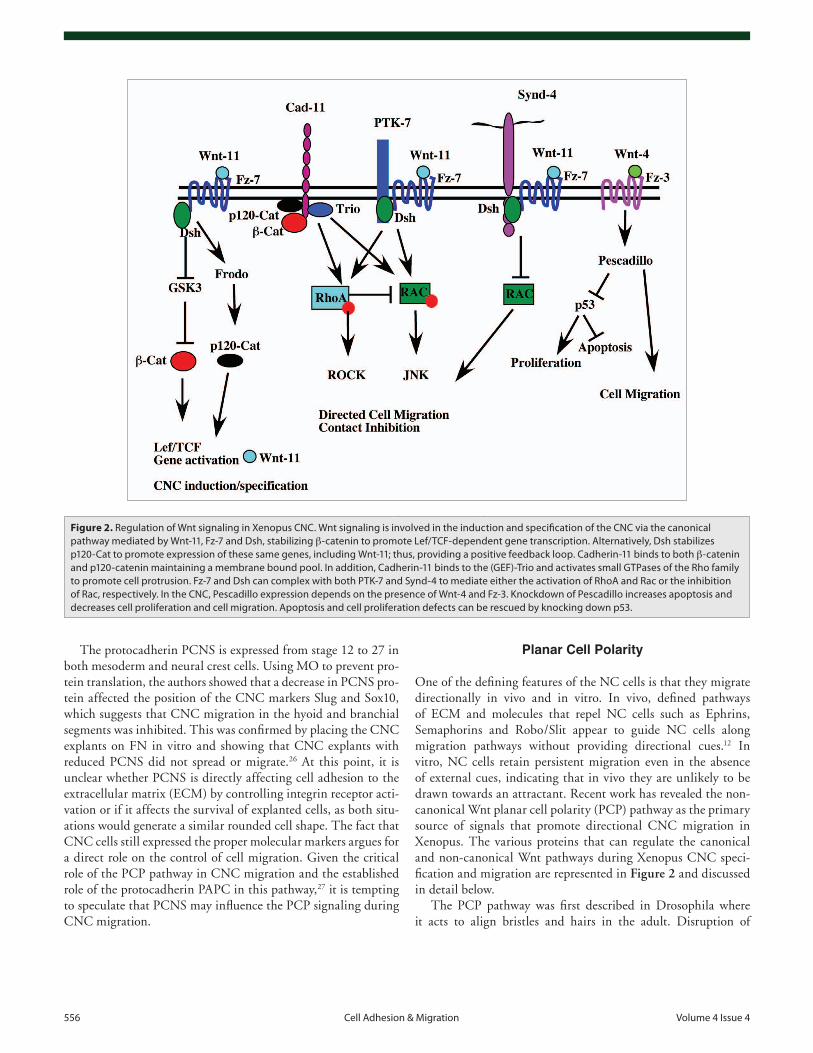
Figure 3. Protein control of CNC cell migration. Schematic representation of two migrating CNC cells showing the various proteins involved in the control of cell migration. The red arrow represents the direction of migration. Rac activation (red dot) is maximal at the leading edge. Activation is mediated at least in part by non-canonical Wnt signaling (Wnt-11, Fz-7 and Dsh). Traction is provided by the α5β1 integrin binding to the central cell-binding domain of FN. Cell protrusions and filopodia, in particular, depend on the presence of Cadherin-11 associated to the (GEF)-Trio and β-catenin to promote activation of Rho GTP-ase. Cadherin-11 also binds the ADAM13 metalloprotease that cleaves its extracellular domain. The cleaved extracellular domain (EC1-3) promotes CNC migration in embryos lacking ADAM13 or overexpressing Cadherin-11. Contact between two CNC cells localizes Wnt-11, Fz-7 and Dsh at the point of contact. This induces the local activation of RhoA and the destabilization of the cell protrusion, possibly by inhibiting Rac. At the lateral edge of the CNC, Syndecan-4 binding to FN recruits Fz-7 and Dsh and reduces Rac activation and cell protrusion.
558 Cell Adhesion & Migration Volume 4 Issue 4
precise and localized dose of the protein is required to promote directional migration. As proposed for Wnt-11r, it is also possible that the initial Wnt-11 expression at the ventral edge of the neural crest may act to restrict the movement of CNC prior to the initia-tion of migration.
During the second phase of CNC migration, individual cells leave the CNC aggregate and migrate ventrally over ectoderm or mesoderm. These isolated CNC cells use PCP to regulate contact inhibition with other CNC cells but not with other cell populations. Contact between individually migrating CNC cells results in the accumulation of Wnt-11, Fz-7 and Dsh at the site of contact.16 This results in the activation of Rho, a reduction in protrusive activity and a subsequent reversal in the migratory path of each cell. Interestingly, contact inhibition is closely tied to signals that stem from the extracellular matrix. Syndecan-4 interacts directly with the FN found in the migration pathway, as well as with Fz-7 and Dsh at the plasma membrane, and is required for the localized decrease in Rac seen upon CNC con-tact. While Syndecan-4 signaling is intimately tied to the PCP pathway, the importance of the PKCa binding site, in the cyto-plasmic domain, for its function suggest that it may integrate multiple signaling pathways.15 Carmona-Fontaine and colleagues have proposed an elegant and simple model whereby Wnt and Syndecan signaling converge to regulate the directional move-ment of the CNC. As CNC cells begin to displace ventrally, they express Wnt-11r (or Wnt-11) and initiate individual migrations. At the leading edge of the CNC, individual cells migrate along pathways defined by the extracellular matrix and inhibitory mol-ecules. Their ventral progression is driven by contact inhibition with cells that lie more dorsal. In the dorsal CNC, contact inhibi-tion provides the driving force to “push” the CNC ventrally. This simple model accounts for the ability of individual CNC cells to move persistently in vitro as they move away from denser popula-tions of CNC cells.16
Other proteins have been shown to be critical for the PCP in CNC. In particular, Protein tyrosine kinase 7 (PTK7), a trans-membrane protein containing seven extracellular immunoglobu-lin domains, forms a complex with Fz-7 and Dsh that is necessary for the membrane localization of Dsh and its activation.38 There is evidence that canonical and non-canonical Wnt signaling play intertwined roles in CNC cell migration. MO knockdown of p120-catenin39 disrupts CNC migration, resulting in defects in craniofacial cartilage at the tailbud stage. While these experi-ments do not reveal what phase of CNC migration is disturbed in p120-catenin knockdown, these phenotypes are interesting as p120-catenin can activate the canonical and non canonical Wnt pathways by modulating the transcription inhibitor Kaiso, Cadherin adhesion and activation of Rho GTPases.40-44 Thus, p120-catenin knockdown could affect CNC migration at many different levels. Another example is the expression of the cell cycle associated protein Pescadillo in CNC that depends on the presence of Wnt-4 and its receptor Fz-3.45 Pescadillo is critical for CNC in two distinct but essential roles. First, Pescadillo knock-down induces apoptosis and decreases cell proliferation via a p53 dependent mechanism. Second, Pescadillo knockdown affects CNC migration but not induction, as indicated by the analysis
any member of the PCP pathway results in a loss of cell polar-ity and consequently a randomization of hair orientation.28 In vertebrates, the PCP pathway also regulates cell polarity orient-ing cells during convergent extension in mesoderm, neural tube formation, cochlear stereocilia orientation and primary ciliogen-esis.29-34 In Xenopus, PCP signaling originating in the neural and non-neural ectoderm defines the medial and ventral edges of the CNC. Once the CNC initiates ventral migration, PCP within the cell mediates contact inhibition between CNC cells result-ing in directional migration in vivo and persistent migration in vitro.16,35
Two non-canonical Wnt are expressed in neighboring tissues during the initial specification of the CNC in Xenopus. Wnt-11r is expressed in the neural plate dorsal and adjacent to Snail-expressing CNC. The ventral margin of the CNC is defined by the expression of Wnt-11 in the dorsal epidermis.36 MO knockdown of Wnt-11r inhibits CNC migration in a cell non-autonomous fashion. This effect is specific as CNC migration can be rescued by the expression of Wnt-11r but not Wnt-11. These experiments further indicate that the two closely related Wnts have distinct receptors and function in non-redundant pathways. Wnt-11r may provide a molecular “push” that moves the CNC away from the neural plate as knockdowns result in the accumulation of the CNC near the neural tube. A role for Wnt-11r acting as a repellent is supported by the observation that contact mediated inhibition and the collapse of cell pro-trusion is absent in CNC from embryos injected with the Wnt-11r MO.16 In addition, members of the PCP pathway, including Frizzled-7 (Fz-7) and Dishevelled (Dsh) are localized to cell contacts between CNC cells and mediate the localized activa-tion of RhoA. Experiments utilizing a ROCK inhibitor sug-gest that activated Rho antagonizes Rac, decreasing protrusive activity at cell-cell contacts. This suggests a model where high Rac activation at the ventral free edge of CNC promotes direc-tional movement while high Rho and low Rac activity at cell-cell contact sites acts to prevent dorsal movement of the sheet of cells. Wnt-11r may well play a similar role in the neural plate resulting in the segregation and mobilization of the CNC from other cell populations prior to ventral migration.
The role played by Wnt-11 is less clear as experimental evi-dence was collected using a dominant negative Wnt-11 construct known to also block Wnt-5a37 and therefore likely to also inhibit Wnt-11r signaling. Taking into account this potential lack of specificity, there is evidence that Wnt-11 may regulate the initial phase of ventral migratory behavior of the CNC. During the ini-tial migratory phase, the CNC moves as a coherent sheet of cells, and grafts of explanted ectoderm that express Wnt-11 cannot reverse the direction of migration if positioned dorsal from the CNC, suggesting that it does not act as a chemoattractant. On the other hand, this graft does prevent CNC migration either by removing the Wnt-11r expressing tissue or providing an opposite (dorsal instead of ventral) source of Wnt-11.35 If the explanted ectoderm expressing Wnt-11 is grafted in the pathway, cells do migrate towards it but appear to stop at the site of expression. Given the fact that overexpression of Wnt-11 or most members of the PCP pathway prevent CNC migration, it is likely that a very

www.landesbioscience.com Cell Adhesion & Migration 559
respect to the dorso/ventral polarity translated into a rear/front axis. The integrity of the tissue can be reinforced by homophylic adhesion that could involve Cadherin-11, as well as the synthesis of a dense extracellular matrix gel that encapsulates the entire tis-sue.7 Cell protrusions require multiple proteins on the cell surface including a5b1 integrins, Syndecan-4, Cadherin-11 and PTK-7; all of which affect the level of activation of small GTPases of the Rho family. The actual traction is mostly provided by the interaction between a5b1 and FN in the pathway, while the ori-entation of cell protrusions is controlled by the PCP pathway. Cells at the front of the CNC that are exposed to Wnt-11 express Fz-7 and PTK-7, bringing Dsh to the membrane and activating Rac and RhoA. At the lateral edges, Syndecan-4 modifies this signaling cascade so that Rac is now inactivated, thereby decreas-ing the number of lateral protrusions. Cells within the CNC can receive the same signaling, but would also have Cadherin-11-mediated contact so that the general level of GTPase activity is greater within the CNC population than outside. Once the second phase of migration starts, the ADAM13 metalloprotease cleaves the Cadherin-11 extracellular domain to reduce cell-cell adhesion and promote single cell migration. This does not influ-ence signaling through either b-catenin or the (GEF)-Trio, as the transmembrane “stub” remains at the plasma membrane. As cells begin to migrate as individuals, the velocity of cell migration increases, potentially mediated through the contact inhibition controlled by PCP. Each time two single CNC cells collide, the membranes in contact collapse, re-polarizing the cells and allow-ing them to accelerate and change direction. The same mecha-nism ensures that no CNC cell turns around to migrate dorsally. At this point, it is likely that production of chemoattractant(s) by the target tissues guides CNC to their final destinations. Because so many different targets exist, it may be more efficient to initiate the migration by first moving away from the neural tube, then away from other CNC cells, and then finally toward a target. While many aspects of the proposed model are speculative, it is consistent with the current, published observations.
Acknowledgements
This work was supported by a grant from the National Institute of Health (NIDCR) RO1DE16289 to D.A. and a grant from the Canadian Institutes of Health Research (CIHR) to M.M. The authors would like to thank Erin Kerdavid, Genevieve Abbruzzese, Russell Neuner and Drs. Kate McCusker and Sam Black for comments and corrections on the manuscript.
of CNC markers and later defects in craniofacial cartilages. The mechanism by which Pescadillo may control cell migration is not known yet, but since the CNC that remain dorsal maintain strong gene expression, it is likely to be independent of its role in cell proliferation and apoptosis.
Regulation of Transcription and Cell Migration
Snail and Slug are the first factors that determine neural crest specification. The epistatic relationship between Snail and Slug is well established,46 and it is clear that the expression and func-tion of the two are intimately tied.47 This makes it difficult to specifically address the function of the individual molecules. Nevertheless, gain- and loss-of-function experiments, as well as dominant negative constructs that can be turned on (hormone inducible) at a specific time, have made it possible to show that these transcription factors are used both for the induction and the migration of CNC.46-48 Unfortunately, since they have many targets, it is unclear at the moment which gene products need to be repressed or activated to promote CNC migration. In tis-sue culture cells, Slug has been shown to control EMT in part by inhibiting the expression of E-Cadherin.49 Although this has not been shown in the context of CNC, EMT is a step necessary for neural crest cells to emigrate.50 Interestingly, Mayor and col-leagues have shown that, in Xenopus, Slug inhibition leads to the loss of expression of ADAM13.51 Since ADAM13 is required for CNC migration, it is possible that the effects for loss-of-func-tion of Snail and Slug are due, in part, to a lack of ADAM13 expression.52
A Model for CNC Cell Migration
With these recent advances in the field, it is tempting to propose a simplified model that would take into account all of the previ-ously described work. The various proteins involved and their putative position within the cell are presented in Figure 3. We show two CNC cells that contact each other so that the leading and trailing edge, as well as one site of cell contact, are visible.
The CNC is induced at the border between the neural plate and the neural tube turning on CNC-specific transcription fac-tors such as Slug, Snail, Twist and Sox8. The dorsal-most border of the CNC is defined by Wnt-11r expressed by the neural plate, while the ventral-most border is defined by Wnt-11. Both of these signals appear critical for the synchronized movement of the CNC as a cohesive sheet. They are also likely to polarize the tissue with
References1. Barembaum M, Bronner-Fraser M. Early steps in
neural crest specification. Semin Cell Dev Biol 2005; 16:642-6.
2. Huang X, Saint-Jeannet JP. Induction of the neural crest and the opportunities of life on the edge. Dev Biol 2004; 275:1-11.
3. LaBonne C. Vertebrate development: Wnt signals at the crest. Curr Biol 2002; 12:743-4.
4. Aybar MJ, Mayor R. Early induction of neural crest cells: lessons learned from frog, fish and chick. Curr Opin Genet Dev 2002; 12:452-8.
5. Mayor R, Aybar MJ. Induction and development of neural crest in Xenopus laevis. Cell Tissue Res 2001; 305:203-9.
6. Sadaghiani B, Thiebaud CH. Neural crest development in the Xenopus laevis embryo, studied by interspecific transplantation and scanning electron microscopy. Dev Biol 1987; 124:91-110.
7. Alfandari D, Cousin H, Gaultier A, Hoffstrom BG, DeSimone DW. Integrin alpha5beta1 supports the migration of Xenopus cranial neural crest on fibronec-tin. Dev Biol 2003; 260:449-64.
8. Borchers A, Epperlein HH, Wedlich D. An assay sys-tem to study migratory behavior of cranial neural crest cells in Xenopus. Dev Genes Evol 2000; 210:217-22.
9. DeSimone DW, Davidson L, Marsden M, Alfandari D. The Xenopus embryo as a model system for studies of cell migration. Methods Mol Biol 2005; 294:235-45.
10. Petrie RJ, Doyle AD, Yamada KM. Random versus directionally persistent cell migration. Nat Rev Mol Cell Biol 2009; 10:538-49.
11. Abercrombie M, Heaysman JE. Observations on the social behaviour of cells in tissue culture I. Speed of movement of chick heart fibroblasts in relation to their mutual contacts. Exp Cell Res 1953; 5:111-31.
12. Kuriyama S, Mayor R. Molecular analysis of neural crest migration. Philos Trans R Soc Lond B Biol Sci 2008; 363:1349-62.
560 Cell Adhesion & Migration Volume 4 Issue 4
40. Park JI, Ji H, Jun S, Gu D, Hikasa H, Li L, et al. Frodo links Dishevelled to the p120-catenin/Kaiso pathway: distinct catenin subfamilies promote Wnt signals. Dev Cell 2006; 11:683-95.
41. Park JI, Kim SW, Lyons JP, Ji H, Nguyen TT, Cho K, et al. Kaiso/p120-catenin and TCF/beta-catenin complexes coordinately regulate canonical Wnt gene targets. Dev Cell 2005; 8:843-54.
42. Hatzfeld M. The p120 family of cell adhesion mol-ecules. Eur J Cell Biol 2005; 84:205-14.
43. Kim SW, Park JI, Spring CM, Sater AK, Ji H, Otchere AA, et al. Non-canonical Wnt signals are modulated by the Kaiso transcriptional repressor and p120-catenin. Nat Cell Biol 2004; 6:1212-20.
44. Anastasiadis PZ, Reynolds AB. Regulation of Rho GTPases by p120-catenin. Curr Opin Cell Biol 2001; 13:604-10.
45. Gessert S, Maurus D, Rossner A, Kuhl M. Pescadillo is required for Xenopus laevis eye development and neural crest migration. Dev Biol 2007; 310:99-112.
46. Aybar MJ, Nieto MA, Mayor R. Snail precedes slug in the genetic cascade required for the specification and migration of the Xenopus neural crest. Development 2003; 130:483-94.
47. Carl TF, Dufton C, Hanken J, Klymkowsky MW. Inhibition of neural crest migration in Xenopus using antisense slug RNA. Dev Biol 1999; 213:101-15.
48. LaBonne C, Bronner-Fraser M. Snail-related transcrip-tional repressors are required in Xenopus for both the induction of the neural crest and its subsequent migra-tion. Dev Biol 2000; 221:195-205.
49. Bolos V, Peinado H, Perez-Moreno MA, Fraga MF, Esteller M, Cano A. The transcription factor Slug represses E-cadherin expression and induces epithelial to mesenchymal transitions: a comparison with Snail and E47 repressors. J Cell Sci 2003; 116:499-511.
50. Duband JL, Monier F, Delannet M, Newgreen D. Epithelium-mesenchyme transition during neural crest development. Acta Anat (Basel) 1995; 154:63-78.
51. Mayor R, Guerrero N, Young RM, Gomez-Skarmeta JL, Cuellar C. A novel function for the Xslug gene: con-trol of dorsal mesendoderm development by repressing BMP-4. Mech Dev 2000; 97:47-56.
52. Alfandari D, Cousin H, Gaultier A, Smith K, White JM, Darribere T, et al. Xenopus ADAM 13 is a metallo-protease required for cranial neural crest-cell migration. Curr Biol 2001; 11:918-30.
26. Rangarajan J, Luo T, Sargent TD. PCNS: a novel pro-tocadherin required for cranial neural crest migration and somite morphogenesis in Xenopus. Dev Biol 2006; 295:206-18.
27. Unterseher F, Hefele JA, Giehl K, De Robertis EM, Wedlich D, Schambony A. Paraxial protocadherin coordinates cell polarity during convergent extension via Rho A and JNK. EMBO J 2004; 23:3259-69.
28. Klein TJ, Mlodzik M. A conserved signaling cassette regulates hair patterning from Drosophila to man. Proc Natl Acad Sci USA 2004; 101:9173-4.
29. Jenny A, Mlodzik M. Planar cell polarity signaling: a common mechanism for cellular polarization. Mt Sinai J Med 2006; 73:738-50.
30. Karner C, Wharton KA Jr, Carroll TJ. Planar cell polar-ity and vertebrate organogenesis. Semin Cell Dev Biol 2006; 17:194-203.
31. Jones C, Chen P. Planar cell polarity signaling in verte-brates. Bioessays 2007; 29:120-32.
32. Goodrich LV. The plane facts of PCP in the CNS. Neuron 2008; 60:9-16.
33. Jones C, Chen P. Primary cilia in planar cell polarity regulation of the inner ear. Curr Top Dev Biol 2008; 85:197-224.
34. Tada M, Kai M. Noncanonical Wnt/PCP signaling during vertebrate gastrulation. Zebrafish 2009; 6: 29-40.
35. De Calisto J, Araya C, Marchant L, Riaz CF, Mayor R. Essential role of non-canonical Wnt signalling in neural crest migration. Development 2005; 132:2587-97.
36. Matthews HK, Broders-Bondon F, Thiery JP, Mayor R. Wnt11r is required for cranial neural crest migration. Dev Dyn 2008; 237:3404-9.
37. Tada M, Smith JC. Xwnt11 is a target of Xenopus Brachyury: regulation of gastrulation movements via Dishevelled, but not through the canonical Wnt path-way. Development 2000; 127:2227-38.
38. Shnitsar I, Borchers A. PTK7 recruits dsh to regu-late neural crest migration. Development 2008; 135: 4015-24.
39. Ciesiolka M, Delvaeye M, Van Imschoot G, Verschuere V, McCrea P, van Roy F, et al. p120 catenin is required for morphogenetic movements involved in the for-mation of the eyes and the craniofacial skeleton in Xenopus. J Cell Sci 2004; 117:4325-39.
13. Lallier TE, Whittaker CA, DeSimone DW. Integrin alpha6 expression is required for early nervous system development in Xenopus laevis. Development 1996; 122:2539-54.
14. Joos TO, Reintsch WE, Brinker A, Klein C, Hausen P. Cloning of the Xenopus integrin alpha(v) subunit and analysis of its distribution during early development. Int J Dev Biol 1998; 42:171-9.
15. Matthews HK, Marchant L, Carmona-Fontaine C, Kuriyama S, Larrain J, Holt MR, et al. Directional migration of neural crest cells in vivo is regulated by Syndecan-4/Rac1 and non-canonical Wnt signaling/RhoA. Development 2008; 135:1771-80.
16. Carmona-Fontaine C, Matthews HK, Kuriyama S, Moreno M, Dunn GA, Parsons M, et al. Contact inhibition of locomotion in vivo controls neural crest directional migration. Nature 2008; 456:957-61.
17. Nie S, Kee Y, Bronner-Fraser M. Myosin-X is critical for migratory ability of Xenopus cranial neural crest cells. Dev Biol 2009; 335:132-42.
18. Hwang YS, Luo T, Xu Y, Sargent TD. Myosin-X is required for cranial neural crest cell migration in Xenopus laevis. Dev Dyn 2009; 238:2522-9.
19. Woolner S, O’Brien LL, Wiese C, Bement WM. Myosin-10 and actin filaments are essential for mitotic spindle function. J Cell Biol 2008; 182:77-88.
20. Weber KL, Sokac AM, Berg JS, Cheney RE, Bement WM. A microtubule-binding myosin required for nuclear anchoring and spindle assembly. Nature 2004; 431:325-9.
21. Vallin J, Girault JM, Thiery JP, Broders F. Xenopus cad-herin-11 is expressed in different populations of migrat-ing neural crest cells. Mech Dev 1998; 75:171-4.
22. Borchers A, David R, Wedlich D. Xenopus cadherin-11 restrains cranial neural crest migration and influ-ences neural crest specification. Development 2001; 128:3049-60.
23. McCusker C, Cousin H, Neuner R, Alfandari D. Extracellular cleavage of cadherin-11 by ADAM metal-loproteases is essential for Xenopus cranial neural crest cell migration. Mol Biol Cell 2009; 20:78-89.
24. Kashef J, Kohler A, Kuriyama S, Alfandari D, Mayor R, Wedlich D. Cadherin-11 regulates protrusive activity in Xenopus cranial neural crest cells upstream of Trio and the small GTPases. Genes Dev 2009; 23:1393-8.
25. Bateman J, Van Vactor D. The Trio family of guanine-nucleotide-exchange factors: regulators of axon guid-ance. J Cell Sci 2001; 114:1973-80.