cranial neural crest cells contribute to connective … neural crest cells contribute to connective...
TRANSCRIPT
a
Developmental Biology 237, 354–367 (2001)doi:10.1006/dbio.2001.0377, available online at http://www.idealibrary.com on
Cranial Neural Crest Cells Contribute toConnective Tissue in Cranial Muscles in theAnuran Amphibian, Bombina orientalis
Lennart Olsson,* ,† ,1 Pierre Falck,† Kristin Lopez,‡ Jared Cobb,‡nd James Hanken§
*Institut fur Spezielle Zoologie und Evolutionsbiologie, Friedrich-Schiller-Universitat Jena,Erbertstrasse 1, D-07743 Jena, Germany; †Evolutionary Biology Centre, Uppsala University,Norbyv. 18A, SE-75239 Uppsala, Sweden; ‡Department of Environmental, Population, andOrganismic Biology, University of Colorado, Boulder, Colorado 80309-0334; and §Museum ofComparative Zoology, Harvard University, 26 Oxford Street, Cambridge, Massachusetts 02138
The contribution of cranial neural crest cells to the development and patterning of cranial muscles in amphibians wasinvestigated in the phylogenetically basal and morphologically generalized frog, Bombina orientalis. Experimental methodsincluded fluorescent marking of premigratory cranial neural crest and extirpation of individual migratory streams. Neuralcrest cells contributed to the connective tissue component, but not the myofibers, of many larval muscles within the firsttwo branchial arches (mandibular and hyoid), and complex changes in muscle patterning followed neural crest extirpation.Connective tissue components of individual muscles of either arch originate from the particular crest migratory stream thatis associated with that arch, and this relationship is maintained regardless of the segmental identity—or embryonicderivation—of associated skeletal components. These developmental relations define a pattern of segmentation in the headof larval anurans that is similar to that previously described in the domestic chicken, the only vertebrate that has beenthoroughly investigated in this respect. The fundamental role of the neural crest in patterning skeleton and musculaturemay represent a primitive feature of cranial development in vertebrates. Moreover, the corresponding developmentalprocesses and cell fates appear to be conserved even when major evolutionary innovations—such as the novel cartilages andmuscles of anuran larvae—result in major differences in cranial form. © 2001 Academic Press
Key Words: neural crest; cell migration; cell fate; Bombina; extirpation; vital dye labeling; cranial muscles.
madcccvactgSqc
INTRODUCTION
The neural crest is a distinct population of embryoniccells that is unique to vertebrates. It gives rise to a large anddiverse array of adult tissues, including nerves, pigmentcells, and much of the cranial skeleton (Hall, 1999). Manyneural crest derivatives were first reported as the results ofembryological studies conducted during the late 19th andearly 20th centuries, yet several important features havebeen described only recently. The full extent of neural crestcontribution to the vertebrate body is still to be defined.
1 To whom correspondence should be addressed at: Institut furSpezielle Zoologie und Evolutionsbiologie, Friedrich-Schiller-Universitat Jena, Erbertstrasse 1, D-07743 Jena, Germany. Fax:
n0049–3641949 162. E-mail: [email protected].
354
A direct role of neural crest in cranial muscle develop-ent was first reported by Le Lievre and Le Douarin (1975),
nd later by Noden (1983a,b) and Couly et al. (1992), whoescribed the neural crest derivation of connective tissueomponents of several branchial arch muscles in quail–hick chimeras. This role is now known to be only oneomponent of a comprehensive mechanism of cranial de-elopment and patterning, in which positional relationsmong hindbrain segments (rhombomeres), the neuralrest, and musculoskeletal derivatives are maintainedhroughout crest migration, pattern formation, and histo-enesis (Kontges and Lumsden, 1996; Graham et al., 1996;chilling, 1997; Schilling and Kimmel, 1997). One conse-uence of these relations is that the connective tissueomponents of a given muscle and its corresponding con-
ective tissue (skeletal) attachment site(s) typically are0012-1606/01 $35.00Copyright © 2001 by Academic Press
All rights of reproduction in any form reserved.
spvd
emtuTsatqtrt
355Cranial Neural Crest Fate in B. orientalis
derived from the same migratory crest stream. It also leadsto segmental boundaries that may lie within individualskeletal or connective tissue elements, instead of coincid-ing with discrete anatomical boundaries. In chickens, forexample, distal portions of the lower jaw are derived fromthe mandibular (first arch) neural crest stream, whereas themost proximal portion of the jaw is derived from the hyoid(second arch) crest stream (Kontges and Lumsden, 1996).
Much of the classical research assessing the embryonicorigins of cranial tissues in vertebrates, including deriva-tives of the neural crest, is based on amphibians (reviewedin De Beer, 1937; Edgeworth, 1935; Goodrich, 1930; Halland Horstadius, 1988; Holtfreter, 1968). These early studiesprecisely and accurately define the extensive contributionof neural crest to many cranial tissues in both frogs andsalamanders (e.g., cranial cartilages; Stone, 1926, 1929), yetnone report a direct contribution of neural crest to cranial
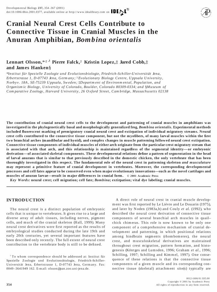
FIG. 1. Larval skull and cranial musculature of Bombina oriencartilages are shaded according to the migratory stream from whicmandibular stream; medium gray, hyoid stream; dark gray, branchiamuscles are depicted schematically; only muscles of interest for the(second) arch muscles are blue. Paired muscles are depicted oceratobranchials I–IV; CH, ceratohyal; CT, cornua trabecula (trapalatoquadrate; SR, suprarostral; TP, trabecular plate. Muscles: lev,levator mandibulae anterior articularis; mlmas, levator mandibulaetwo parts; superficialis and profundus); ang, angularis group—mhyoideus group—mih, interhyoideus; moh, orbitohyoideus; msh, suintermandibularis anterior; mip, intermandibularis posterior; mm(1999).
muscle in any species. This fact, in light of the recent w
Copyright © 2001 by Academic Press. All right
tudies in birds summarized above, immediately raises theossibility of variation among the major living groups ofertebrates with respect to the role of neural crest in cranialerivation and patterning.At the same time, results of two separate studies provide
vidence that neural crest might contribute to cranialuscle development in amphibians in a manner similar to
hat seen in amniotes, but that this important feature wentndetected by the classical embryologists and anatomists.hese results are rarely, if ever, cited in contemporarytudies. In the axolotl, cranial muscle patterning is severelyffected by neural crest extirpation and transplantation, andhese effects cannot be explained simply as indirect conse-uences of altered skeletal morphology (Hall, 1950). Abla-ion of the hyoid crest stream, for example, severely dis-upts patterning of the depressor mandibulae muscle, evenhough the (first arch) cartilages to which this muscle
depicted in dorsal (left) and ventral views. Neural crest-derivedey originate (redrawn from Olsson and Hanken, 1996): light gray,am. The few noncrest-derived cartilages are lightly shaded. Cranialent study are shown. Mandibular (first) arch muscles are red, hyoidne side only. Cartilages: BB, basibranchial; BH, basihyal; CB,lar horn); IR, infrarostral; MC, Meckel’s; OC, otic capsule; PQ,or mandibulae group—mlma, levator mandibulae anterior; mlmaa,rior subexternus; mlmp, levator mandibulae posterior (comprisingyoangularis; mqa, quadratoangularis; msa, suspensorioangularis;soriohyoideus; osh, orbito- and suspensoriohyoideus; others—mia,andibulolabialis. Anatomical nomenclature follows Cannatella
talis,h thl strepresn o
beculevatante
ha, hspenl, m
ould normally attach are intact. These effects are exactly
s of reproduction in any form reserved.
ebiv
356 Olsson et al.
what one would expect if neural crest is mediating cranialmuscle patterning directly. In chimaeric Xenopus larvaethat developed from embryos that received heterospecific
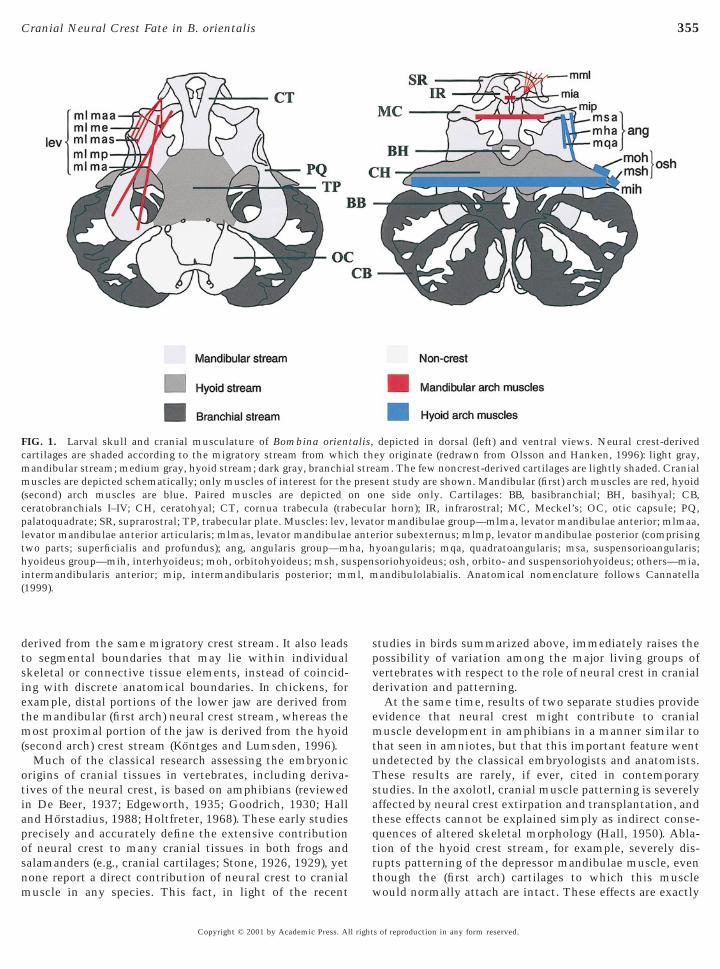
FIG. 2. (A) A late stage-16 B. orientalis embryo showing thextirpation technique. A flap of epidermis overlying the stream toe extirpated (here the hyoid stream) is folded up and the underly-ng neural crest stream removed using tungsten needles. Lateraliew; anterior to the left. (B) Stage-14 (neurula) embryo of B.
orientalis. The left side depicts six regions of the cranial neuralfold; regions 2–6 were injected with DiI. The right side depicts theapproximate origins of neural crest cells that contribute to thethree cranial migratory streams—mandibular (M), hyoid (H), andbranchial (B). Dorsal view; anterior at top.
grafts of cranial neural crest, labeled (crest-derived) cells
Copyright © 2001 by Academic Press. All right
were observed within several cranial muscles (Sadaghianiand Thiebaud, 1987). Thus, the possibility of a neural crestcontribution to connective tissue components of cranialmusculature in living amphibians remains unresolved andworthy of investigation. Anuran amphibians are especiallyinteresting in this regard because of the extremely special-ized larval head, which includes a unique, complex feedingapparatus comprised of novel upper and lower jaw cartilagesand muscle configurations found in no other vertebrates(Cannatella, 1999; Hanken, 1999; Haas, 2001).
The primary goals of the present study are to determine:(1) whether neural crest contributes to the development ofconnective tissue components of larval cranial musculaturein amphibians, and (2) if it does, whether the pattern of crestcontribution to branchial arch musculoskeletal and connec-tive tissues reveals the same segmental relations describedin amniotes (chicken). We focus on mandibular and hyoidarch components in larval anurans because of their impor-tance to cranial function and evolution (Cannatella, 1999;De Jongh, 1968; Hanken et al., 1997), because their post-hatching (metamorphic) development is relatively wellknown (Alley, 1989; Alley and Omerza, 1999), and becauseof novel musculoskeletal configurations that offer conve-nient means of assessing underlying developmental rela-tions. We employ two different experimental methods,neural crest ablation and fate mapping with a vital dye.These experiments provide direct evidence of neural crestcontribution to connective tissue components of branchialarch muscles in amphibians, including the connective tis-sue attachments to cranial cartilages. They also implicate aprominent role for neural crest in cranial muscle pattern-ing. Furthermore, as in amniotes, the connective tissuecomponents of individual muscles and their associatedconnective tissue (skeletal) attachments are derived fromthe same neural crest migratory stream, which need not bethe same crest stream that gives rise to the correspondingskeletal component (cartilage). This suggests that theserelations are an evolutionarily primitive and conservedfeature of cranial development in vertebrates.
MATERIALS AND METHODS
Choice of Species
We assessed neural crest contribution to cranial musculature inthe fire-bellied toad, Bombina orientalis. This is a phylogeneticallybasal taxon among living frogs, and both adults and larvae have arelatively generalized morphology (Cannatella, 1999; Cannatellaand de Sa, 1993; Fig. 1). Both features make it an especiallyattractive subject for investigation of presumed ancestral, or primi-tive, features of anurans. The neural crest contribution to thecartilaginous larval skull was evaluated in an earlier study (Olssonand Hanken, 1996).
EmbryosEggs were obtained from laboratory matings among wild-caught
adults (Charles D. Sullivan Co., Inc.), which were maintained as a
s of reproduction in any form reserved.
owGt
357Cranial Neural Crest Fate in B. orientalis
breeding colony at the University of Colorado. Breeding andhusbandry followed established procedures (Carlson and Ellinger,1980; Frost, 1982). Adults received dorsal subcutaneous injectionsof chorionic gonadotropin (Sigma, cat. no. CG-5) and were allowedto spawn overnight in the dark at room temperature. Fertilized eggswere reared in 10% Holtfreter solution (Hamburger, 1960) at10–25°C. Embryos were dejellied either chemically (0.63 g cysteineHCl, 0.12 g NaCl, 24 ml H2O, buffered to pH 8.0 with 5 N NaOH)r manually with watchmaker’s forceps. Embryos and tadpolesere staged from external morphology according to the scheme ofosner (1960), which defines a total of 46 stages from fertilization
o metamorphosed froglet.
Surgical Procedures
Neural crest extirpations were performed at late stage 16 (neuraltube, gill plates), when the cranial neural crest cells are migratingventrally in four main streams (mandibular, hyoid, and twobranchial, Fig. 2A). Embryos were placed in trenches cut in 2%agar-coated Petri dishes, which were filled with 10% Holtfretersolution plus antibiotic (50 mg gentamicin sulfate per liter; Sigma,cat. no. G-1264). Surgery was performed by using watchmaker’sforceps and tungsten needles. A flap of ectoderm was prepared bycutting through three sides of a rectangular portion overlying thetargeted neural crest stream and leaving the dorsal side intact. Theflap was folded up dorsally, and the mandibular, hyoid, or branchialstream removed (Fig. 2A). In many anuran species, and especiallyBombina orientalis, neural crest cells at this stage are more darklypigmented than neighboring cells and are readily seen against thelighter, underlying tissues (Stone 1932; Olsson and Hanken, 1996).This makes possible complete removal of all crest cells, and onlycrest cells. Finally, the flap of ectoderm was pressed back into placeand the extirpated embryos allowed to heal for 1–2 h before beingtransferred to agar-coated dishes filled with 10% Holtfreter solu-tion plus antibiotic. Additional sham-operated embryos receivedthe same surgery, except for removal of the neural crest, and servedas a control for crest extirpation. All operations were unilateral,usually on the right side of the embryo; the opposite side was leftintact and served as an additional control.
Embryos were reared until larval stage 26 or 27, killed in 30%aqueous chloretone (1,1,1-trichloro-2-methyl-2-propanol; Sigma,cat. no. T-5138), fixed for 2 h in 4% paraformaldehyde (PFA) in 0.1M phosphate-buffered saline (PBS), run through a graded methanolseries (25, 50, and 75%; 5 min each), and fixed overnight or longerin Dent fixative [1 part dimethyl sulfoxide (DMSO): 4 partsmethanol; Dent et al., 1989]. After immersion in Dent bleach (1part 30% hydrogen peroxide: 2 parts Dent fixative; Dent et al.,1989) for 4 days to lighten skin pigmentation, specimens werestored in 100% methanol at 220°C.
Each type of extirpation (mandibular, hyoid, or branchial stream)was performed on 20 embryos, and 26 additional embryos served ascontrols. Thirty-four of 60 crest-extirpated embryos (9 mandibular,11 hyoid, and 14 branchial) and 25 of 26 controls survived and werepreserved as described above.
Cartilage and Muscle Double Staining
Preserved larvae were first stained for cartilage as whole mounts(Klymkowsky and Hanken, 1991). Specimens were immersed inAlcian blue solution [20 mg Alcian blue 8GX (C.I. 74240), 30 mlabsolute ethanol, 70 ml glacial acetic acid] for about 3 h and
returned to Dent fixative. The same specimens were then immu-Copyright © 2001 by Academic Press. All right
nostained according to the technique of Klymkowsky and Hanken(1991), which was slightly modified to minimize nonspecific back-ground staining, as follows. After rehydration in a graded methanolseries, specimens were washed three times for 5 min in “salinecocktail” [0.1 M Niu-Twitty saline, 0.1 M phosphate buffer (K/Na;pH 7.4), 0.4% Triton X-100], preincubated for 1–2 h in “serumcocktail” (saline cocktail containing 5% newborn-calf serum, 5%DMSO, and 0.1% thimerosal), and incubated for 20–24 h with amonoclonal antibody for newt skeletal muscle (12/101; Kintnerand Brockes, 1984) that was diluted 1:500 with serum cocktail.After six washes in serum cocktail (5 h total), specimens wereincubated for 20–24 h in secondary antibody [horseradish peroxi-dase (HRP)-conjugated, goat anti-mouse IgG, diluted with serumcocktail at 1:1000]. Specimens were washed two times for 30 minand then overnight in serum cocktail, further washed three timesfor 1 h in saline cocktail, and then reacted for 1–2 h with 0.5 mg/mldiaminobenzidine (DAB) in saline cocktail containing 0.02% hy-drogen peroxide. The reaction was stopped by dehydration withmethanol two times for 5 min. Finally, larvae were cleared bystepwise transfer from 100% methanol to BABB (1 part benzylalcohol: 2 parts benzyl benzoate) and examined with a Wild M5dissecting microscope. The 12/101 monoclonal antibody cross-reacts widely with striated muscle from a wide variety of verte-brates (e.g., other amphibians, rodents and chicken; Klymkowskyand Hanken, 1991). It does not bind to smooth muscle, and bindsonly weakly to cardiac muscle. Monoclonal antibody 12/101,developed by Dr. J. P. Brockes, was obtained from the Developmen-tal Studies Hybridoma Bank, which was developed under theauspices of the NICHD and is maintained by The University ofIowa, Department of Biological Sciences (Iowa City, IA).
Vital Labeling
Embryos were labeled with vital dye at stage 14 (neurula; Fig. 2B),immediately before the onset of cranial neural crest cell migration.Dejellied and decapsulated embryos were immobilized in shallowtrenches cut into 2% agar gelled at the bottom of Petri dishes. A 0.5%stock solution of the lipophilic dye DiI (1,19-dioctadecyl-3,3,39,39-tetramethylindocarbocyanine, perchlorate; Molecular Probes, Inc.,cat. no. D-282) was prepared in 100% ethanol and stored at 4°C.Immediately before injection, it was diluted in 0.3 M sucrose toworking concentrations of 0.1 and 0.05%. Micropipets pulled fromthin-walled, 1.2-mm diameter glass microfilaments were filled withdye and attached to a Picospritzer II (General Valve). Micropipet tipswere broken to a diameter of ca. 20 mm. DiI was injected into any offive different sites in the left neural fold (Fig. 2B, sites 2–6) by insertinga micropipet and expelling a small amount of dye solution. Injectionswere made by hand or with a micromanipulator. The right side wasnot injected and served as a control. Following injection, embryoswere reared individually in 24-well tissue culture plates in 10%Holtfreter solution plus antibiotic at 25°C.
All neural crest and presumptive neural tube cells within a givensite appeared to be labeled by the injection procedure. A total of sixsites were injected in an earlier study that assessed the chondro-genic fate of cranial neural crest cells in this species (Olsson andHanken, 1996; Fig. 2B). Because the first site (transverse neural fold)did not produce neural crest, we injected only sites 2–6 in thepresent study. These five sites span all three cranial neural creststreams. Great care was taken to avoid labeling any mesodermcells. Fortunately, in Bombina orientalis the unpigmented (white)mesoderm is readily distinguished from the pigmented (gray)
cranial neural crest, and eggs and embryos are large (relative tos of reproduction in any form reserved.
fmil
umcwtnl
dostm(AansaVmac“T
p
m in F
358 Olsson et al.
those of other anurans, e.g., Xenopus laevis). Also, cranial neuralcrest migration begins relatively early in Bombina, when neuralolds and neural crest lie dorsomedial to, and distinct from, paraxial
esoderm (Olsson and Hanken, 1996). Thus, it is relatively easy tonject DiI into neural crest at this stage without accidentallyabeling paraxial mesoderm.
Fluorescence Microscopy of Living Embryos andCryostat Sections
For fluorescence imaging, living embryos were mounted tempo-rarily in rectangles cut into gelled agar at the bottom of custom-made brass slides with coverslip floors (M. Klymkowsky, Univer-sity of Colorado). Frozen sections of older specimens (stage 26) wereprepared by immersing tadpoles in 30% aqueous chloretone andfixing them overnight or longer in 4% paraformaldehyde/0.25%glutaraldehyde in PBS at 4°C. Specimens then were washed thor-oughly in PBS, soaked overnight in 15% aqueous sucrose, trans-ferred through two changes of a solution of 15% sucrose/7.5%gelatin at 37°C for a total of at least 5 h, and embedded in freshsucrose/gelatin solution at 4°C. Once set, the specimens werefrozen and stored at 220°C. Sections (20 mm) were cut with a LeicaCM 1800 cryostat. Live whole mounts and sections were viewedwith a Leitz Dialux 20 or Leica DMRXE epifluorescence micro-scope equipped with a rhodamine (N2) filter block. Sections alsowere viewed with Nomarsky differential interference contrast(DIC) microscopy.
Documentation
Photographs and digital images were obtained with either a WildMPS55 Photoautomat and Kodak T-MAX P3200 or EktachromeP1600 film, or a Photonic Science Colour Coolview digital videocamera. Images were analyzed and enhanced with Adobe Photo-shop on Macintosh computers. Corresponding images obtainedfrom fluorescence and DIC microscopy were combined to docu-ment the exact histological location of the fluorescent signal in a
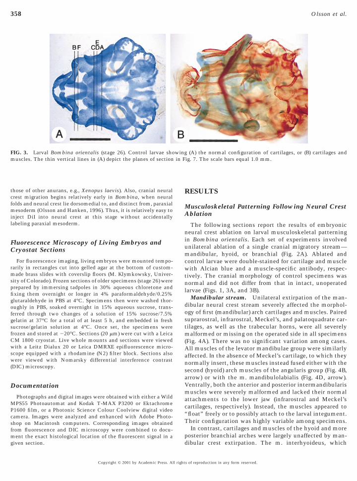
FIG. 3. Larval Bombina orientalis (stage 26). Control larvae shouscles. The thin vertical lines in (A) depict the planes of section
given section. d
Copyright © 2001 by Academic Press. All right
RESULTS
Musculoskeletal Patterning Following Neural CrestAblation
The following sections report the results of embryonicneural crest ablation on larval musculoskeletal patterningin Bombina orientalis. Each set of experiments involved
nilateral ablation of a single cranial migratory stream—andibular, hyoid, or branchial (Fig. 2A). Ablated and
ontrol larvae were double-stained for cartilage and muscleith Alcian blue and a muscle-specific antibody, respec-
ively. The cranial morphology of control specimens wasormal and did not differ from that in intact, unoperatedarvae (Figs. 1, 3A, and 3B).
Mandibular stream. Unilateral extirpation of the man-ibular neural crest stream severely affected the morphol-gy of first (mandibular) arch cartilages and muscles. Paireduprarostral, infrarostral, Meckel’s, and palatoquadrate car-ilages, as well as the trabecular horns, were all severelyalformed or missing on the operated side in all specimens
Fig. 4A). There was no significant variation among cases.ll muscles of the levator mandibulae group were similarly
ffected. In the absence of Meckel’s cartilage, to which theyormally insert, these muscles instead fused either with theecond (hyoid) arch muscles of the angularis group (Fig. 4B,rrow) or with the m. mandibulolabialis (Fig. 4D, arrow).entrally, both the anterior and posterior intermandibularisuscles were severely malformed and lacked their normal
ttachments to the lower jaw (infrarostral and Meckel’sartilages, respectively). Instead, the muscles appeared tofloat” freely or to possibly attach to the larval integument.heir configuration was highly variable among specimens.In contrast, cartilages and muscles of the hyoid and more
osterior branchial arches were largely unaffected by man-
(A) the normal configuration of cartilages, or (B) cartilages andig. 7. The scale bars equal 1.0 mm.
wing
ibular crest extirpation. The m. interhyoideus, which
s of reproduction in any form reserved.
359Cranial Neural Crest Fate in B. orientalis
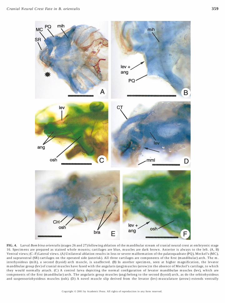
FIG. 4. Larval Bombina orientalis (stages 26 and 27) following ablation of the mandibular stream of cranial neural crest at embryonic stage16. Specimens are prepared as stained whole mounts; cartilages are blue, muscles are dark brown. Anterior is always to the left. (A, B)Ventral views; (C–F) Lateral views. (A) Unilateral ablation results in loss or severe malformation of the palatoquadrate (PQ), Meckel’s (MC),and suprarostral (SR) cartilages on the operated side (asterisk). All three cartilages are components of the first (mandibular) arch. The m.interhyoideus (mih), a second (hyoid) arch muscle, is unaffected. (B) In another specimen, seen at higher magnification, the levatormandibulae group (lev) of cranial muscles have fused with the angularis (ang) muscles (arrow) in the absence of Meckel’s cartilage, to whichthey would normally attach. (C) A control larva depicting the normal configuration of levator mandibulae muscles (lev), which arecomponents of the first (mandibular) arch. The angularis group muscles (ang) belong to the second (hyoid) arch, as do the orbitohyoideus
and suspensoriohyoideus muscles (osh). (D) A novel muscle slip derived from the levator (lev) musculature (arrow) extends ventrallyCopyright © 2001 by Academic Press. All rights of reproduction in any form reserved.
360 Olsson et al.
connects paired ceratohyal cartilages, was normal (Figs. 4Aand 4B). The m. orbitohyoideus and m. suspensoriohyoid-eus appeared largely intact, even though the palatoquadratecartilage from which they both originate was severelymalformed (cf. Figs. 4C and 4E). Muscles of the angularisgroup also were surprisingly normal in appearance, despitetheir frequent fusion with the levator mandibulae group(Fig. 4F, arrow; for control configuration, see Fig. 4C).
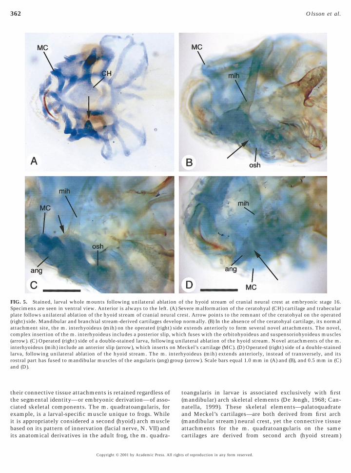
Hyoid stream. In all 11 cases, extirpation of the hyoidneural crest stream caused severe malformation of theceratohyal cartilage and the cartilaginous trabecular plate,and of several of their associated muscles. Both cartilageswere reduced on the operated side; only a small fragmentremained of the ceratohyal, leaving a big gap in the ventralskeleton in this area (Fig. 5A, arrow). All three pairedmuscles of the angularis group retained their normal inser-tion on Meckel’s cartilage, but their respective origins wereabnormal: instead of originating from the ceratohyal (m.hyoangularis) or palatoquadrate (m. quadratoangularis andm. suspensorioangularis) cartilages, each muscle fused tothe m. interhyoideus (Fig. 5D) and, occasionally, to the m.orbitohyoideus and m. suspensoriohyoideus. The m. inter-hyoideus normally attaches to the lateral end of eachceratohyal cartilage on either side of the head. In theabsence of the ceratohyal on the operated side of treatedembryos, the m. interhyoideus instead projected anteriorlyto attach to Meckel’s cartilage (Fig. 5C). The m. interhyoi-deus also frequently fused to both the m. orbitohyoideusand m. suspensoriohyoideus (Fig. 5B, arrow). Finally, the m.orbitohyoideus and m. suspensoriohyoideus retained theirnormal origin from the palatoquadrate cartilage. However,instead of inserting on the (now absent) ceratohyal carti-lage, each muscle fused to the m. interhyoideus. Cartilagesand muscles of the mandibular and posterior branchialarches were normal.
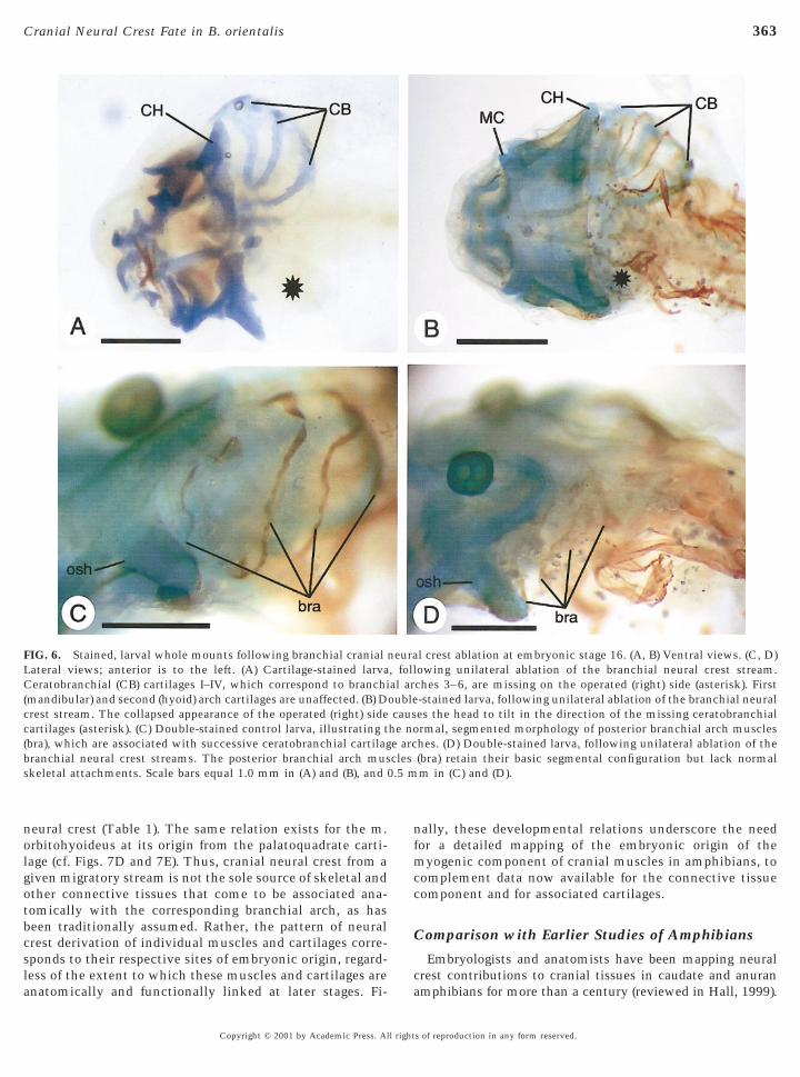
Branchial stream. Unilateral extirpation of the branchialneural crest stream led to absence of the entire cartilaginousbranchial basket (arches 3–6) on the operated side (Fig. 6A,asterisk). Yet, despite the loss of their skeletal attachments,the corresponding branchial arch muscles were present and,although shrunken (Fig. 6B, asterisk), retained their basicsegmental configuration (cf. Figs. 6C and 6D). The musclesappeared to originate from dorsal fascia, but there were noobvious insertions at the opposite ends. Mandibular and hyoidarch cartilages and muscles were intact (Figs. 6A and 6B).
to fuse with the m. mandibulolabialis (mml). (This embryo was opeis to the left.) (E) The operated (left) side of a larva, following unilatunaffected by the treatment. Orbitohyoideus and suspensoriohyoiceratohyal cartilage (CH). Branchial muscles (bra) also are normallyof the mandibular stream. The angularis (ang) group muscles fuseof Meckel’s cartilage, to which they would normally attach. The sca
and the scale bar in (F) is 0.3 mm.Copyright © 2001 by Academic Press. All right
DiI Fate Mapping
DiI injected into the left cranial neural fold of stage-14(neurula) embryos bound to the cell membranes of neuralcrest cells, which emerged to form three migratory streams(Fig. 2B). Later, DiI was readily identified in cryostat sec-tions of early larvae (stage 26), where it labeled severalstructures within the head. The pattern of neural crestcontribution to cranial cartilages is identical to what wereported earlier (Olsson and Hanken, 1996) and is identicalto that inferred from the results of ablation experimentsdescribed above. The following account, therefore, empha-sizes results that pertain to the contribution of neural crestto muscle connective tissue. In Fig. 7, results are presentedas DIC micrographs overlain by the DiI stain taken fromfluorescence images of the same section. Whereas thisimaging technique obviates the need to show both imagesside by side, the connective tissue morphology (which isclearly visible with DIC microscopy alone) is somewhatobscured by the DiI staining.
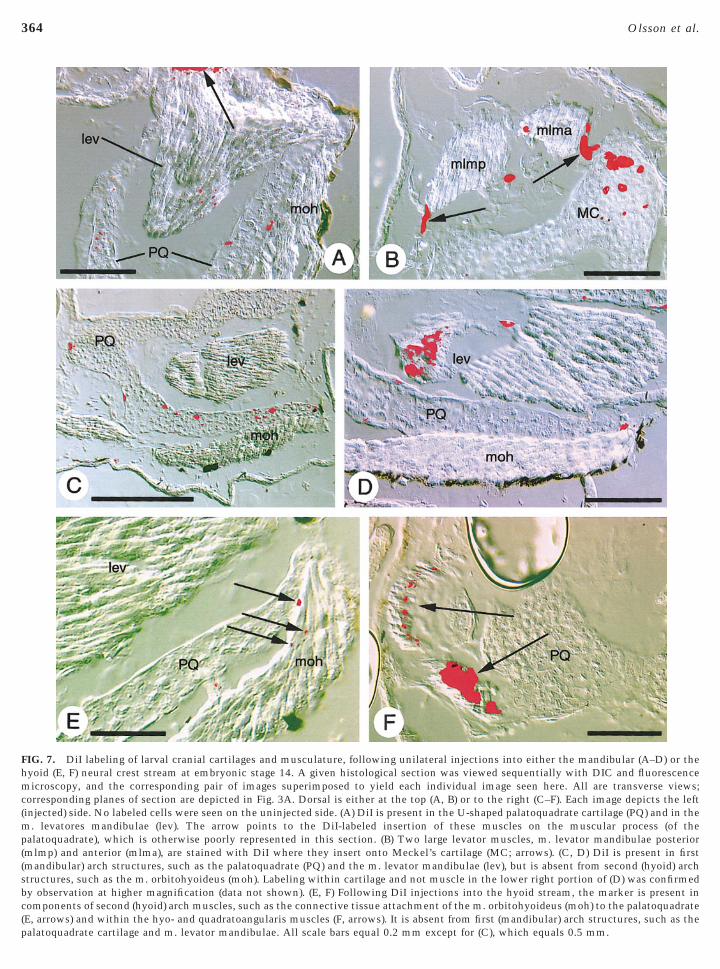
The mandibular neural crest stream originates from re-gion 2 and the anterior portion of region 3 (Fig. 2B). Inlarvae, DiI-labeled cells from this stream were seen in thepalatoquadrate and Meckel’s cartilages (Figs. 7A–7D), twoprominent skeletal components of the first (mandibular)arch. Moreover, neural-crest contributions to the attach-ment points of the levator mandibulae muscles were seen atboth their origins from the palatoquadrate (Fig. 7A, arrow)and their insertions on Meckel’s cartilage (Fig. 7B, arrows).Consistently, only cartilages derived from the mandibularneural crest stream and first (mandibular) arch muscles,such as the m. levatores mandibulae (Fig. 7D), ever con-tained DiI. Second (hyoid) arch muscles, e.g., m. orbitohy-oideus (Figs. 7C and 7D), never displayed the marker. Notall mandibular muscles, however, were labeled. No neuralcrest contribution was detected in any attachment point ofthe m. intermandibularis anterior or posterior, two trans-verse, ventral muscles that attach exclusively to the man-dibular skeleton (infrarostral and Meckel’s cartilages, re-spectively). Also, DiI was never visible within the tinyattachment points of anterior slips of levator mandibulaemuscles that insert on the suprarostral cartilage, or withinconnective tissue at the origin of the m. mandibulolabialis.
The hyoid neural crest stream originates from the poste-rior portion of region 3 and all of region 4 (Fig. 2B). DiIinjected into these cells in embryos labeled connective
on the right side and the image has been reversed so that anteriorblation of the mandibular stream. Second (hyoid) arch muscles aremuscles (osh), for example, retain their normal insertions on the
loped. (F) Operated (left) side of a larva following unilateral ablationthose of the levator mandibulae (lev) group (arrow) in the absencers in (A), (B), and (D) equal 1.0 mm, those in (C) and (E) are 0.5 mm,
ratederal adeusdevewithle ba
s of reproduction in any form reserved.
birsowsldeDaaacpci11v
ncitpVaf
vnfandmtrfboollep1s
tmsdopwlslftrrmnr
361Cranial Neural Crest Fate in B. orientalis
tissue in several cranial muscles in larvae. Connectivetissue at both the origin (palatoquadrate) and insertion(ceratohyal) of the m. orbitohyoideus contained the marker,as did connective tissue surrounding the correspondingmyofibers (Fig. 7E). Similar results were seen in the m.suspensoriohyoideus and m. interhyoideus (not illustrated).DiI was also detected within connective tissues at theorigin and insertion of two of the three muscles of theangularis group, the m. hyoangularis and m. quadratoangu-laris (Fig. 7F). No label was detected within the m. suspen-sorioangularis, the third member of the angularis group, orin any first (mandibular) arch muscle.
DiI injected into neural fold regions 5 and 6 was carriedwithin the branchial crest stream or in trunk neural crestcells. No contribution to any muscle attachment points orassociated connective tissue components was seen follow-ing these injections.
DISCUSSION
Neural Crest Contribution to the ConnectiveTissue Component of Cranial Musculature
Our results provide both direct and indirect evidence thatembryonic neural crest is important for the development ofconnective tissue components of cranial musculature in atleast one species of amphibian, the fire-bellied toad, Bom-
ina orientalis. Following injection of the lipophilic dye DiInto premigratory cranial neural crest, fluorescent label waseadily localized in the connective tissue component ofeveral cranial muscles and associated skeletal attachmentsf larvae prepared as frozen sections. These contributionsere derived from two of the three principal migratory
treams of cranial neural crest, and they included muscu-oskeletal components of both the first and second (man-ibular and hyoid) branchial arches (Table 1). As witharlier results in the domestic chicken (Le Lievre and Leouarin, 1975; Noden, 1986; Couly et al., 1992; Kontges
nd Lumsden, 1996), the neural crest contribution waslways localized to connective tissue components withinnd adjacent to cranial muscles and was not traced toontractile elements, viz., myofibers. The correspondingattern of neural crest contribution to cranial cartilages isoncordant with earlier accounts for several anuran species,ncluding B. orientalis (Olsson and Hanken 1996; Hanken,999; Fig. 1), as well as other vertebrates (reviewed in Hall,999 and LeDouarin and Kalcheim, 1999). This furtheralidates the precision of our labeling procedure.Extirpation of individual streams of migrating cranial
eural crest cells resulted in predictable and complexhanges in cranial muscle patterning. Such results must benterpreted cautiously because of the experimental artifactshat may follow neural crest ablation and because of theossibility of crest regeneration (Scherson et al., 1993;aglia and Hall, 1999). Nevertheless, when combined withnalyses that are less sensitive to such experimental arti-
acts, such as fate mapping using vital dyes, ablation may beCopyright © 2001 by Academic Press. All right
aluable and informative. In this study, results followingeural crest ablation are entirely concordant with thoserom vital labeling in terms of the evidence they provide of
neural crest contribution to connective tissue compo-ents of cranial musculature. Ablation of either the man-ibular or hyoid crest stream results in altered patterning ofuscles that vital labeling shows normally receive a con-
ribution from that stream. Muscles that do not normallyeceive a contribution from the ablated stream are unaf-ected. Moreover, these effects on muscle patterning cannote explained simply as artifacts of altered skeletal morphol-gy following neural crest ablation, since in many instancesf altered muscle patterning the associated skeleton isargely, if not completely, intact. Finally, both ablation andabeling data in Bombina are consistent with the hypoth-sis that neural crest is helping to mediate cranial muscleatterning (Noden, 1983a, 1986; Schilling and Kimmel,997), although such a patterning role in this or otherpecies of amphibians awaits more explicit proof.Evidence for neural crest contribution to muscle connec-
ive tissue is most compelling for the first two cranialigratory streams, but weaker for the third, or branchial
tream. Unilateral extirpation of the branchial stream led toisrupted development of the gill musculature on theperated side, such that the muscles were small and lackedroper attachments. Yet, no fluorescent label was observedithin any posterior branchial arch muscle following vital
abeling of the branchial stream, which is the principalource of the associated skeleton (ceratobranchial carti-ages; Olsson and Hanken, 1996). Because our analysis ofrozen sections focused on musculoskeletal components ofhe first two branchial arches, components of more poste-ior arches were not assessed comprehensively; thus, fluo-escent label in the connective tissue components of theseuscles may have gone undetected. The full extent of
eural crest contribution to gill musculature in amphibiansemains to be adequately documented.
Developmental Relations between Musculatureand Skeleton
Combined data from extirpation and fate-mapping experi-ments in Bombina reveal that the neural crest-derivedconnective tissue components of individual larval musclesof the first two branchial arches originate from the particu-lar crest migratory stream that is associated with each arch.In other words, connective tissues of first (mandibular) archmuscles are derived from mandibular stream neural crest,whereas connective tissues of second (hyoid) arch musclesare derived from the hyoid crest stream. These developmen-tal relations, which are established during embryonic de-velopment, define a pattern of segmentation in the head oflarval anurans that is similar to that previously described inthe domestic chicken, the only vertebrate that has beenthoroughly investigated in this respect (Kontges and Lums-den, 1996; Graham et al., 1996).
Fidelity between individual branchial arch muscles and
s of reproduction in any form reserved.
t(na(a
362 Olsson et al.
their connective tissue attachments is retained regardless ofthe segmental identity—or embryonic derivation—of asso-ciated skeletal components. The m. quadratoangularis, forexample, is a larval-specific muscle unique to frogs. Whileit is appropriately considered a second (hyoid) arch musclebased on its pattern of innervation (facial nerve, N. VII) and
FIG. 5. Stained, larval whole mounts following unilateral ablatiSpecimens are seen in ventral view. Anterior is always to the left.plate follows unilateral ablation of the hyoid stream of cranial neur(right) side. Mandibular and branchial stream-derived cartilages devattachment site, the m. interhyoideus (mih) on the operated (rightcomplex insertion of the m. interhyoideus includes a posterior slip,(arrow). (C) Operated (right) side of a double-stained larva, followininterhyoideus (mih) include an anterior slip (arrow), which inserts olarva, following unilateral ablation of the hyoid stream. The m. irostral part has fused to mandibular muscles of the angularis (ang)and (D).
its anatomical derivatives in the adult frog, the m. quadra- c
Copyright © 2001 by Academic Press. All right
oangularis in larvae is associated exclusively with firstmandibular) arch skeletal elements (De Jongh, 1968; Can-atella, 1999). These skeletal elements—palatoquadratend Meckel’s cartilages—are both derived from first archmandibular stream) neural crest, yet the connective tissuettachments for the m. quadratoangularis on the same
f the hyoid stream of cranial neural crest at embryonic stage 16.vere malformation of the ceratohyal (CH) cartilage and trabecular
est. Arrow points to the remnant of the ceratohyal on the operatednormally. (B) In the absence of the ceratohyal cartilage, its normalextends anteriorly to form several novel attachments. The novel,h fuses with the orbitohyoideus and suspensoriohyoideus muscles
ilateral ablation of the hyoid stream. Novel attachments of the m.eckel’s cartilage (MC). (D) Operated (right) side of a double-stainedyoideus (mih) extends anteriorly, instead of transversely, and its(arrow). Scale bars equal 1.0 mm in (A) and (B), and 0.5 mm in (C)
on o(A) Seal crelop
) sidewhicg unn M
nterhgroup
artilages are derived from second arch (hyoid stream)
s of reproduction in any form reserved.
nfmcc
0.5 m
363Cranial Neural Crest Fate in B. orientalis
neural crest (Table 1). The same relation exists for the m.orbitohyoideus at its origin from the palatoquadrate carti-lage (cf. Figs. 7D and 7E). Thus, cranial neural crest from agiven migratory stream is not the sole source of skeletal andother connective tissues that come to be associated ana-tomically with the corresponding branchial arch, as hasbeen traditionally assumed. Rather, the pattern of neuralcrest derivation of individual muscles and cartilages corre-sponds to their respective sites of embryonic origin, regard-less of the extent to which these muscles and cartilages are
FIG. 6. Stained, larval whole mounts following branchial cranialLateral views; anterior is to the left. (A) Cartilage-stained larvaCeratobranchial (CB) cartilages I–IV, which correspond to branchi(mandibular) and second (hyoid) arch cartilages are unaffected. (B) Dcrest stream. The collapsed appearance of the operated (right) sidecartilages (asterisk). (C) Double-stained control larva, illustrating t(bra), which are associated with successive ceratobranchial cartilagbranchial neural crest streams. The posterior branchial arch musskeletal attachments. Scale bars equal 1.0 mm in (A) and (B), and
anatomically and functionally linked at later stages. Fi-
Copyright © 2001 by Academic Press. All right
ally, these developmental relations underscore the needor a detailed mapping of the embryonic origin of the
yogenic component of cranial muscles in amphibians, toomplement data now available for the connective tissueomponent and for associated cartilages.
Comparison with Earlier Studies of Amphibians
Embryologists and anatomists have been mapping neuralcrest contributions to cranial tissues in caudate and anuran
l crest ablation at embryonic stage 16. (A, B) Ventral views. (C, D)owing unilateral ablation of the branchial neural crest stream.ches 3–6, are missing on the operated (right) side (asterisk). First-stained larva, following unilateral ablation of the branchial neurales the head to tilt in the direction of the missing ceratobranchialrmal, segmented morphology of posterior branchial arch muscleshes. (D) Double-stained larva, following unilateral ablation of the(bra) retain their basic segmental configuration but lack normalm in (C) and (D).
neura, follal aroublecaus
he noe arccles
amphibians for more than a century (reviewed in Hall, 1999).
s of reproduction in any form reserved.
hmc(mp((sbc(p
364 Olsson et al.
FIG. 7. DiI labeling of larval cranial cartilages and musculature, following unilateral injections into either the mandibular (A–D) or theyoid (E, F) neural crest stream at embryonic stage 14. A given histological section was viewed sequentially with DIC and fluorescenceicroscopy, and the corresponding pair of images superimposed to yield each individual image seen here. All are transverse views;
orresponding planes of section are depicted in Fig. 3A. Dorsal is either at the top (A, B) or to the right (C–F). Each image depicts the leftinjected) side. No labeled cells were seen on the uninjected side. (A) DiI is present in the U-shaped palatoquadrate cartilage (PQ) and in the
. levatores mandibulae (lev). The arrow points to the DiI-labeled insertion of these muscles on the muscular process (of thealatoquadrate), which is otherwise poorly represented in this section. (B) Two large levator muscles, m. levator mandibulae posterior
mlmp) and anterior (mlma), are stained with DiI where they insert onto Meckel’s cartilage (MC; arrows). (C, D) DiI is present in firstmandibular) arch structures, such as the palatoquadrate (PQ) and the m. levator mandibulae (lev), but is absent from second (hyoid) archtructures, such as the m. orbitohyoideus (moh). Labeling within cartilage and not muscle in the lower right portion of (D) was confirmedy observation at higher magnification (data not shown). (E, F) Following DiI injections into the hyoid stream, the marker is present inomponents of second (hyoid) arch muscles, such as the connective tissue attachment of the m. orbitohyoideus (moh) to the palatoquadrateE, arrows) and within the hyo- and quadratoangularis muscles (F, arrows). It is absent from first (mandibular) arch structures, such as the
alatoquadrate cartilage and m. levator mandibulae. All scale bars equal 0.2 mm except for (C), which equals 0.5 mm.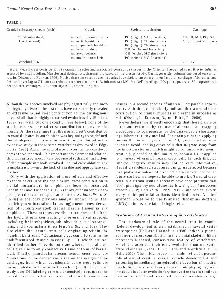
letatilamuicw“cisn
tmcw
tpicttcteNtfclpma(
365Cranial Neural Crest Fate in B. orientalis
Although the species involved are phylogenetically and mor-phologically diverse, these studies have consistently revealedan extensive neural crest contribution to the cartilaginouslarval skull that is highly conserved evolutionarily (Hanken,1999). Yet, with but one exception (see below), none of thestudies reports a neural crest contribution to any cranialmuscle. At the same time that the neural crest’s contributionto cranial tissues in amphibians was beginning to be defined,the development of cranial musculature was the subject ofextensive study in these same vertebrates (reviewed in Edge-worth, 1935). Again, no role of neural crest in muscle devel-opment was reported. This important developmental relation-ship was missed most likely because of technical limitationsof the principle methods involved—neural crest ablation andtransplantation—and the lack of a stable and appropriate cellmarker.
Only with the application of more reliable and effectivemethods of cell labeling has a neural crest contribution tocranial musculature in amphibians been demonstrated.Sadaghiani and Thiebaud’s (1987) study of chimaeric Xeno-pus (neural crest transplantation from X. borealis to X.aevis) is the only previous analysis known to us thatxplicitly mentions (albeit in passing) a neural crest deriva-ion of (undifferentiated) cranial muscle tissues in anymphibian. These authors describe neural crest cells fromhe hyoid stream contributing to several larval muscles,ncluding the interhyoideus, orbitohyoideus, quadratoangu-aris, and hyoangularis (their Figs. 9a, 9c, and 10a). Theylso claim that neural crest cells originating within theandibular stream, “Occasionally . . . could be seen in the
ndifferentiated muscle masses” (p. 99), which are notdentified further. They do not state whether neural crestells give rise to only connective tissue or to myofibers asell. Finally, mandibular stream neural crest cells arenumerous in the connective tissue on the margin of theartilages, from which occasionally some crest cells arencorporated into the attached muscles” (p. 99). The presenttudy uses DiI-labeling to more extensively document the
TABLE 1
Cranial migratory stream (arch) Muscle
Mandibular (first) m. levatores mandibulaeHyoid (second) m. orbitohyoideus
m. suspensoriohyoideusm. interhyoideusm. hyoangularism. quadratoangularis
Branchial (3–6)
Note. Neural crest contributions to cranial muscles and associatassessed by vital labeling. Muscles and skeletal attachments are baresults (Olsson and Hanken, 1996). Notice that some second arch mfirst arch cartilages: CT, cornua trabecula (trabecular horn); IR, infSecond arch cartilages. CH, ceratohyal; TP, trabecular plate.
eural crest contribution to cranial muscle connective
Copyright © 2001 by Academic Press. All right
issues in a second species of anuran. Comparable experi-ents with the axolotl clearly indicate that a neural crest
ontribution to cranial muscles is present in urodeles asell (Olsson, L., Ericsson, R., and Falck, P., 2000).Nevertheless, we strongly encourage that these claims be
ested and extended by the use of alternate fate-mappingrocedures, to compensate for the unavoidable shortcom-ngs inherent in any method. For example, when applyingertain fluorescent dyes such as DiI, great care has to beaken to avoid labeling other cells that migrate away fromhe injection site and which might be confused with neuralrest, e.g., placodal epithelium. Also, because DiI is appliedo a subset of cranial neural crest cells in each injectedmbryo, negative results may not be very informative.eural crest-derived structures can go undetected because
hat particular subset of crest cells was never labeled. Inuture studies, we hope to be able to mark all neural crestells in a stream using a recently developed protocol thatabels premigratory neural crest cells with green fluorescentrotein (GFP; Carl et al., 1999, 2000), and which avoidsany of the potential artifacts identified above. Another
pproach would be to use lysinated rhodamine dextransLRDs) to follow the fate of single cells.
Evolution of Cranial Patterning in Vertebrates
The fundamental role of the neural crest in cranialskeletal development is well established in several verte-brate species (Hall and Horstadius, 1988). Indeed, a promi-nent neural crest contribution to the cranial skeleton likelyrepresents a shared, conservative feature of vertebrates,which characterized their early evolution from nonverte-brate chordates (Gans, 1989; Gans and Northcutt 1983;Hall, 1999). The initial report—in birds—of an importantrole of neural crest in cranial muscle development andpatterning immediately introduced the question of whetherthis feature also evolved early in vertebrate phylogeny, or if,instead, it is a later evolutionary innovation that is confined
Skeletal attachment Cartilage
PQ (origin), MC (insertion) CT, IR, MC, PQ, SRPQ (origin), CH (insertion) CH, TP (anterior part)PQ (origin), CH (insertion)CH (origin and insertion)CH (origin), MC (insertion)PQ (origin), MC (insertion)
CB I–IV
nnective tissues in the Oriental fire-bellied toad, B. orientalis, asn the present study. Cartilages (right column) are based on earliers have skeletal attachments on first arch cartilages: Abbreviations:tral; MC, Meckel’s cartilage; PQ, palatoquadrate; SR, suprarostral.
ed cosed ouscleraros
to a more recent and restricted clade of vertebrates, e.g.,
s of reproduction in any form reserved.
A
C
C
C
C
C
H
H
H
H
H
K
K
K
L
366 Olsson et al.
tetrapods. This question can only be answered definitivelyby additional, detailed experimental studies of neural crestfates and muscle origins in a taxonomically diverse series oftaxa, especially more “basal” lineages such as fishes.
Nevertheless, data from the present study of anuranamphibians, combined with limited, initial evidence ofcranial muscle development in the zebrafish (Schilling andKimmel, 1994) and in urodeles (Olsson et al., 2000), suggestthat a neural crest contribution to the development ofbranchial-arch muscles is in fact a primitive vertebrate traitthat is widespread—but largely undetected—in most livingclades. Moreover, they suggest that the essential role of theneural crest in cranial musculoskeletal patterning (Noden,1991; Graham et al., 1996; Schilling, 1997; Schilling andKimmel, 1997) may represent a fundamental, conservedfeature of cranial development in vertebrates generally.Finally, the presence of similar developmental relationsbetween musculature and skeleton in tetrapods as differentas frogs and birds demonstrates how developmental pro-cesses and cell fates can be conserved even when majorevolutionary innovations—such as the novel cartilages andmuscles of anuran larvae—result in major anatomical dif-ferences in cranial form.
ACKNOWLEDGMENTS
We thank Olle Håstad and Margret Roser for help with illusta-tions. Detailed comments by three anonymous reviewers on earlierversions of this paper are gratefully acknowledged. Financial sup-port was provided by the Wenner-Gren Foundations and theSwedish Institute (to L.O.), and by the U.S. National ScienceFoundation (IBN 94-19407, to J.H.).
REFERENCES
Alley, K. E. (1989). Myofiber turnover is used to retrofit frog jawmuscles during metamorphosis. Am. J. Anat. 184, 1–12.lley, K. E., and Omerza, F. F. (1999). Neuromuscular remodelingand myofiber turnover in Rana pipiens jaw muscles. Cells TissueOrg. 164, 46–58.annatella, D. C. (1999). Architecture: cranial and axial musculo-skeleton. In “Tadpoles: The Biology of Anuran Larvae” (R. W.McDiarmid and R. Altig, Eds.), pp. 52–91. University of ChicagoPress, Chicago.annatella, D. C., and de Sa, R. O. (1993). Xenopus laevis as amodel organism. Syst. Biol. 42, 476–507.arl, T. F., Dufton, C., Hanken, J., and Klymkowsky, M. W. (1999).Inhibition of neural crest migration in Xenopus using antisenseSlug RNA. Dev. Biol. 213, 101–115.arl, T. F., Vourgourakis, Y., Klymkowsky, M., and Hanken, J.(2000). Green fluorescent protein used to assess cranial neuralcrest derivatives in the frog, Xenopus laevis. In “RegulatoryProcesses in Development: The Legacy of Sven Horstadius(1898–1996)” (C.-O. Jacobson and L. Olsson, Eds.), Wenner-Gren International Series, Vol. 76, pp. 167–172. Portland Press,London.arlson, J. T., and Ellinger, M. S. (1980). The reproductive biologyof Bombina orientalis, with notes on care. Herpetol. Rev. 11,
11–12.Copyright © 2001 by Academic Press. All right
Couly, G. F., Coltey, P. M., and LeDouarin, N. M. (1992). Thedevelopmental fate of the cephalic mesoderm in quail-chickchimeras. Development 114, 1–15.
De Beer, G. R. (1937). “The Development of the Vertebrate Skull.”Oxford Univ. Press, Oxford.
De Jongh, H. J. (1968). Functional morphology of the jaw apparatusof larval and metamorphosing Rana temporaria L. Neth. J. Zool.18, 1–103.
Dent, J. A., Polson, A. G., and Klymkowsky, M. W. (1989). Awhole-mount immunocytochemical analysis of the expression ofthe intermediate filament protein vimentin in Xenopus. Devel-opment 105, 61–74.
Edgeworth, F. H. (1935). “The Cranial Muscles of Vertebrates.”Cambridge Univ. Press, Cambridge.
Frost, J. R. (1982). A time efficient, low cost method for thelaboratory rearing of frogs. Herpetol. Rev. 13, 75–77.
Gans, C. (1989). Stages in the origin of vertebrates: Analysis bymeans of scenarios. Biol. Rev. Camb. Philos. Soc. 64, 221–268.
Gans, C., and Northcutt, G. (1983). Neural crest and the origin ofvertebrates: A new head. Science 220, 268–274.
Goodrich, E. S. (1930). “Studies on the Structure and Developmentof Vertebrates.” Macmillan, London.
Gosner, K. L. (1960). A simplified table for staging anuran embryosand larvae with notes on identification. Herpetologica 16, 183–190.
Graham, A., Kontges, G., and Lumsden, A. (1996). Neural crestapoptosis and the establishment of craniofacial pattern: Anhonorable death. Mol. Cell. Neurosci. 8, 76–83.
Haas, A. (2001). Mandibular arch musculature of anuran tadpoles,with comments on homologies of amphibian jaw muscles. J.Morphol. 247, 1–33.
Hall, B. K. (1999). “The Neural Crest in Development and Evolu-tion.” Springer-Verlag, New York.
Hall, B. K., and Horstadius, S. (1988). “The Neural Crest.” OxfordUniv. Press, Oxford.
Hall, E. K. (1950). Experimental modifications of muscle develop-ment in Amblystoma punctatum. J. Exp. Zool. 113, 355–377.amburger, V. (1960). “A Manual of Experimental Embryology.”University of Chicago Press, Chicago.anken, J. (1999). Larvae in amphibian development and evolution. In“The Origin and Evolution of Larval Forms” (B. K. Hall and M. H.Wake, Eds.), pp. 61–108. Academic Press, San Diego and London.anken, J., Klymkowsky, M. W., Alley, K. E., and Jennings, D. H.(1997). Jaw muscle development as evidence for embryonicrepatterning in direct- developing frogs. Proc. R. Soc. London Ser.B Biol. Sci. 264, 1349–1354.arrison, R. G. (1935). Heteroplastic grafting in embryology. Har-vey Lect. 29, 116–157.oltfreter, J. (1968). On mesenchyme and epithelia in inductiveand morphogenetic processes. In “Epithelial–Mesenchymal In-teractions” (R. Fleischmajer and R.-E. Billingham, Eds.), pp. 1–30.Williams and Wilkins, Baltimore.intner, C. R., and Brockes, J. P. (1984). Monoclonal antibodiesidentify blastemal cells derived from dedifferentiating muscle innewt limb regeneration. Nature 308, 67–69.
lymkowsky, M. W., and Hanken, J. (1991). Whole-mount staining ofXenopus and other vertebrates. Methods Cell Biol. 36, 419–441.ontges, G., and Lumsden, A. (1996). Rhombencephalic neuralcrest segmentation is preserved throughout craniofacial ontog-eny. Development 122, 3229–3242.
e Douarin, N. M., and Kalcheim, C. (1999). “The Neural Crest,”
2nd Ed. Cambridge Univ. Press,Cambridge, U.K.s of reproduction in any form reserved.
S
S
S
S
S
S
S
S
V
367Cranial Neural Crest Fate in B. orientalis
Le Lievre, C., and Le Douarin, N. M. (1975). Mesenchymal deriva-tives of the neural crest: Analysis of chimaeric quail and chickembryos. J. Embryol. Exp. Morphol. 34, 125–154.
Noden, D. M. (1983a). The embryonic origins of avian cephalic andcervical muscles and associated connective tissues. Am. J. Anat.168, 257–276.
Noden, D. M. (1983b). The role of the neural crest in patterning ofavian cranial skeletal, connective, and muscle tissues. Dev. Biol.96, 144–165.
Noden, D. M. (1986). Origins and patterning of craniofacial mes-enchymal tissues. J. Craniofac. Genet. Dev. Biol. Suppl. 2, 15–31.
Noden, D. M. (1991). Vertebrate crraniofacial development: Therelation between ontogenetic process and morphological out-come. Brain Behav. Evol. 38, 190–225.
Olsson, L., Ericsson, R., and Falck, P. (2000). Neural crest contri-butions to cranial muscle fate and patterning in the Mexicanaxolotl (Ambystoma mexicanum). In “Regulatory Processes inDevelopment: The Legacy of Sven Horstadius (1898–1996)”(C.-O. Jacobson and L. Olsson, Eds.), Wenner-Gren InternationalSeries, Vol. 76, pp. 159–166. Portland Press, London.
Olsson, L., and Hanken, J. (1996). Cranial neural-crest migrationand chondrogenic fate in the Oriental fire-bellied toad, Bombinaorientalis: Defining the ancestral pattern of head development inanuran amphibians. J. Morphol. 229, 105–120.
adaghiani, B., and Thiebaud, C. H. (1987). Neural crest develop-ment in the Xenopus laevis embryo, studied by interspecifictransplantation and scanning electron microscopy. Dev. Biol.
124, 91–110.Copyright © 2001 by Academic Press. All right
cherson, T., Serbedzija, G., Fraser, S., and Bronner-Fraser, M.(1993). Regulative capacity of the cranial neural tube to formneural crest. Development 118, 1049–1062.
chilling, T. F. (1997). Genetic analysis of craniofacial developmentin the vertebrate embryo. BioEssays 19, 459–468.
chilling, T. F., and Kimmel, C. B. (1994). Segment and cell typelineage restrictions during pharyngeal arch development in thezebrafish embryo. Development 120, 483–494.
chilling, T. F., and Kimmel, C. B. (1997). Musculoskeletal pattern-ing in the pharyngeal segments of the zebrafish embryo. Devel-opment 124, 2945–2960.
tone, L. S. (1926). Further experiments on the extirpation andtransplantation of mesectoderm in Amblystoma punctatum. J.Exp. Zool. 44, 95–131.
tone, L. S. (1929). Experiments showing the role of migratingneural crest (mesectoderm) in the formation of head skeleton andloose connective tissue in Rana palustris. Roux’s Arch. Entw.Mech. Org. 118, 40–77.
tone, L. S. (1932). Selective staining of the neural crest and itspreservation for microscopic study. Anat. Rec. 51, 267–273.
aglia, J. L., and Hall, B. K. (1999). Regulation of neural crest cellpopulations: Occurrence, distribution and underlying mecha-nisms. Int. J. Dev. Biol. 43, 95–110.
Received for publication April 17, 2001Revised June 26, 2001
Accepted June 28, 2001
Published online August 9, 2001s of reproduction in any form reserved.