branchial anomalies - university of texas medical · pdf filearches each arch contains...
TRANSCRIPT

BRANCHIAL ANOMALIES
David Gleinser, MD Faculty Advisor: Harold Pine, MD
The University of Texas Medical Branch (UTMB Health) Department of Otolaryngology
Grand Rounds Presentation September 30, 2011
Embryology
Branchial anomalies result from improper development of the branchial apparatus
Branchial apparatus develops 2nd-6th week Neck is shaped like a hollow tube with
circumferential ridges = Arches (mesoderm)
Ridges between arches = Clefts and Pouches Clefts = outside (ectoderm)
Pouches = inside (endoderm)
“CAP”


Arches
Each arch contains Cartilage Cranial nerve Artery Muscle component
All neural crest origin
6 arches, only 5 form structures in humans 1, 2, 3, 4, and 6 5th fails to develop
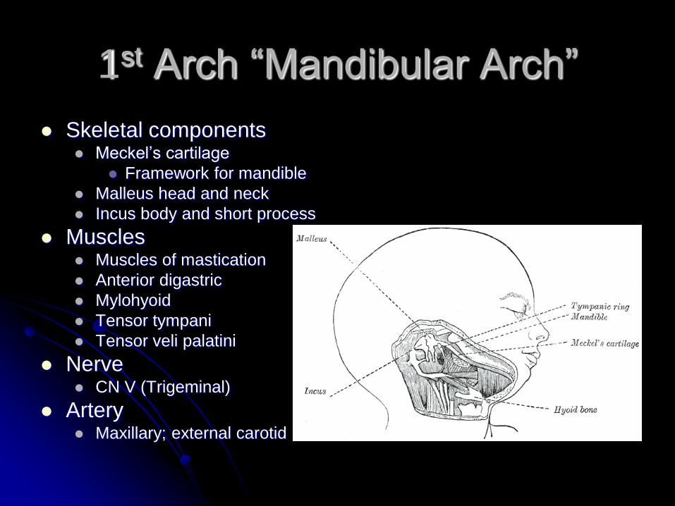
1st Arch “Mandibular Arch”
Skeletal components Meckel’s cartilage
Framework for mandible
Malleus head and neck
Incus body and short process
Muscles Muscles of mastication
Anterior digastric
Mylohyoid
Tensor tympani
Tensor veli palatini
Nerve CN V (Trigeminal)
Artery Maxillary; external carotid
2nd Arch “Hyoid Arch”
Skeletal components Reichert’s cartilage
Stapes Malleus manubrium Incus long process Styloid process Hyoid bone (lesser horn and upper body)
Muscles Facial expression, buccinator, platysma, stapedius, stylohyoid,
posterior digastric
Nerve CN VII (Facial)
Artery
Stapedial

3rd Arch
Skeletal components
Hyoid (greater horn and lower body)
Muscles
Stylopharyngeus
Nerve
CN IX (Glossopharyngeal)
Artery
Common/Internal carotid

4th Arch
Skeletal components
Thyroid, epiglottic, cuneiform cartilages
Muscles
Cricothyroid, inferior constrictors
Nerve
Superior laryngeal
Artery
Subclavian, aortic arch

6th Arch
Skeletal components
Cricoid, arytenoids, corniculate
Muscles
All intrinsic muscles of larynx (except
cricothyroid)
Nerve
Recurrent laryngeal
Artery
Pulmonary artery

Branchial Clefts and Pouches
4 clefts and 4 pouches
5th and 6th contribute to the 4th
Clefts provide “covering” to structures of
the corresponding arch and pouch

Pouches
1st Pouch Eustachian tube, middle ear, mastoid, inner layer
of tympanic membrane
2nd Pouch Tonsils, root of tongue, foramen cecum,
pharynx(part)
3rd Pouch – ventral and dorsal wings Ventral wing – Thymus
Dorsal wing – inferior parathyroid glands
Pouches
4th Pouch Superior parathyroid glands
Parafollicular C-cells of thyroid gland
5th Pouch Contributes to Parafollicular C-cells
6th Pouch Contributes to laryngeal musculature and
cartilage

1st Arch Anomalies
Involves malformations of eyes, ears,
palate, and mandible
2 main manifestations of “First Arch
Syndrome”
Treacher Collins Syndrome
Pierre Robin Syndrome

Treacher Collins Syndrome
Mandibulofacial dysostosis
Inherited AD
Features Midface and mandibular hypoplasia
Ear anomalies: microtia, anotia, stenotic or atresia of EAC, malformation of malleus and incus (CHL)
Eye anomalies: coloboma of lower lids, down-slopping palpebral fissures
Cleft palate

Treacher Collins Syndrome
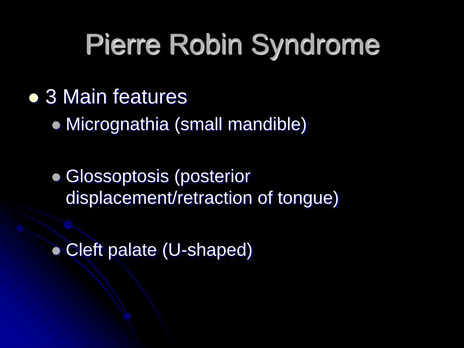
Pierre Robin Syndrome
3 Main features
Micrognathia (small mandible)
Glossoptosis (posterior
displacement/retraction of tongue)
Cleft palate (U-shaped)

Pierre Robin Syndrome

2nd Arch Anomalies
Malformed auricle
Microtia
Ossicular malformation
Stapes, malleus, incus
CHL
Muscular asymmetry of face
Hyoid malformation
lesser horn and upper body
3rd Arch Anomalies
Hyoid anomalies
Lower body
Greater horn
Aneurysm of carotid artery

4th Arch Anomalies
Laryngeal stenosis
Laryngoptosis (low position of larynx)
Chondromalacia
Double aortic arch
Pulmonary artery sling
Left pulmonary artery originates from right
pulmonary artery
Slings around right main-stem bronchus
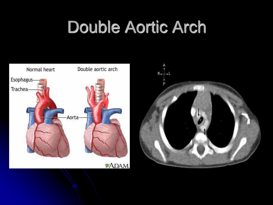
Double Aortic Arch

Pulmonary Artery Sling

1st Pouch Anomalies
Atretic eustachian tube -> recurrent OM
ET diverticuli
Absence
Tympanic cavity
Mastoid antrum
Perforated TM
Bifid tongue
Branchiogenic nasopharyngeal cysts (very rare)

2nd Pouch Anomalies
Thyroglossal duct cyst
7% of population
Failure of ablation of TGD
Anywhere from base of tongue to upper
mediastinum
Typical finding
Cystic lesion just below hyoid in midline that
moves with deglutination and tongue protrusion

TGD Cyst
May contain thyroid tissue
Potentially the only functioning thyroid
Perform U/S or CT to look for thyroid and to
assess lesion
Treatment – surgical
May contain cancer
1%
Papillary carcinoma

TGD Cyst

2nd Pouch Anomalies
Lingual Thyroid
Failure of decent of thyroid -> atopic 90% of cases at the base of tongue (lingual
thyroid)
4:1 female:male
Usually not noted until teenage or young adult
Asymptomatic (most cases); dysphagia, airway compromise
Reddish mass (well vascularized) at base of tongue

Lingual Thyroid
Hypothyroidism – 70% of cases
2/3 cases – only functioning thyroid tissue
Thyroid function study prior to treatment
Treatment
Asymptomatic – Monitor
Symptomatic
Excise +/- transplant tissue into muscles of neck
Radioiodine therapy (destroys all thyroid tissue)
Usually require lifelong thyroid replacement

Lingual Thyroid

Lingual Thyroid

Lingual Thyroid
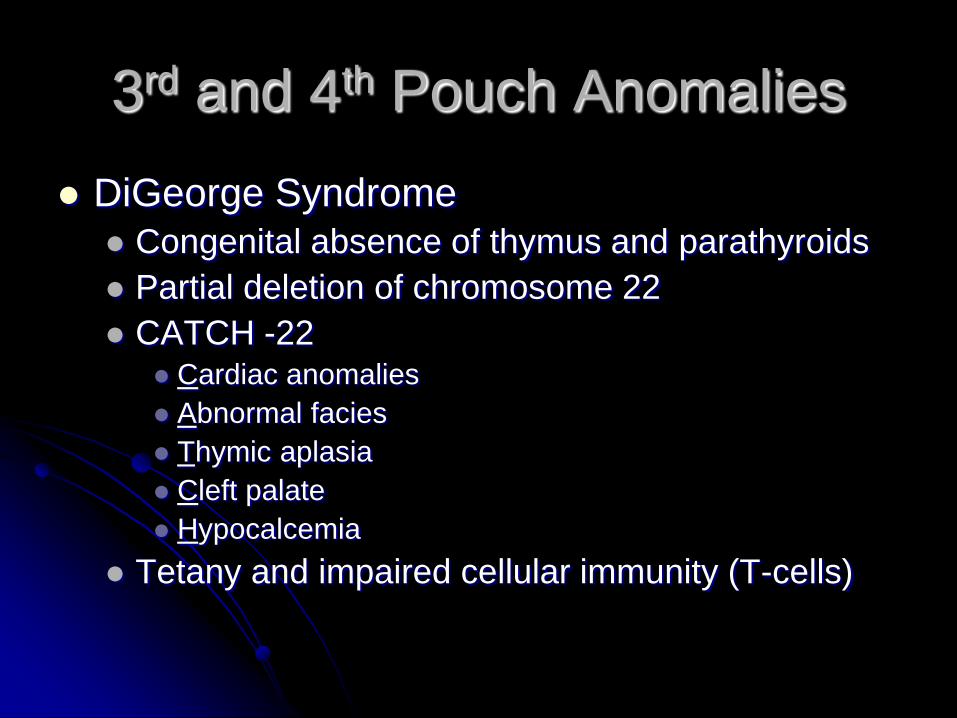
3rd and 4th Pouch Anomalies
DiGeorge Syndrome
Congenital absence of thymus and parathyroids
Partial deletion of chromosome 22
CATCH -22 Cardiac anomalies
Abnormal facies
Thymic aplasia
Cleft palate
Hypocalcemia
Tetany and impaired cellular immunity (T-cells)
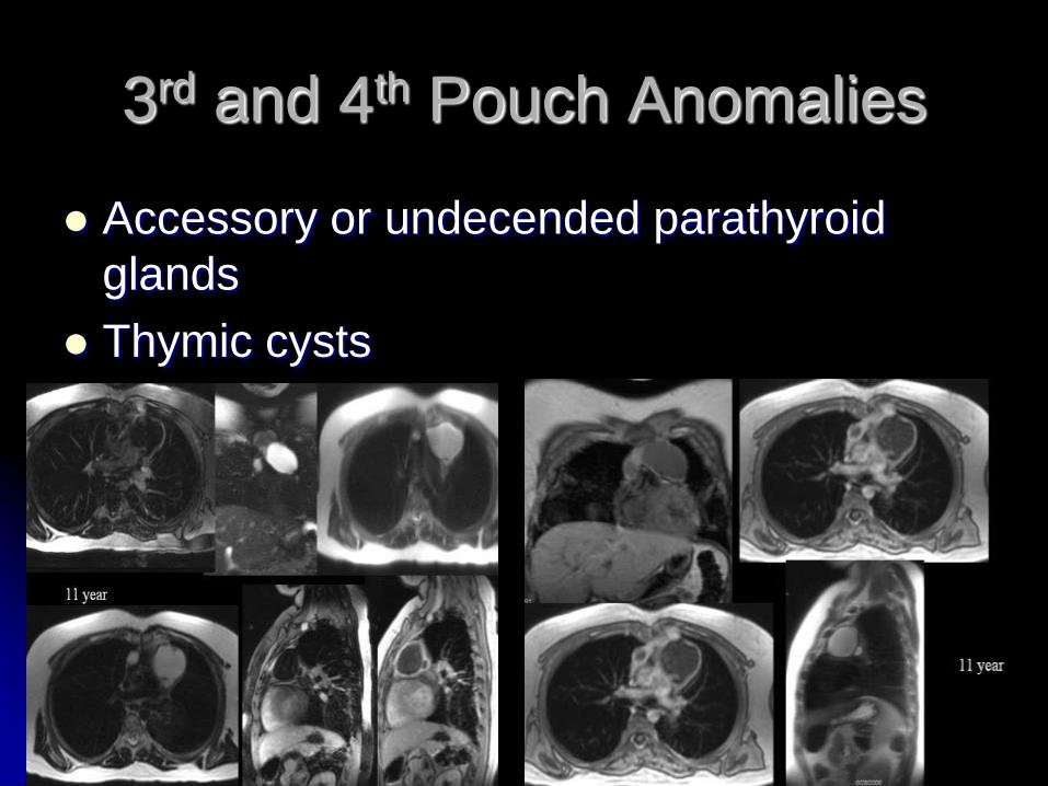
3rd and 4th Pouch Anomalies
Accessory or undecended parathyroid
glands
Thymic cysts
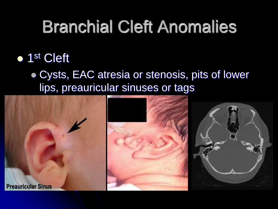
Branchial Cleft Anomalies
1st Cleft
Cysts, EAC atresia or stenosis, pits of lower
lips, preauricular sinuses or tags

Branchial Cleft Anomalies
2nd Cleft Cysts Cervical sinuses
3rd Cleft Cysts (rare) Thymic cysts
4th Cleft Cysts (extremely rare) Cysts on the Vagus nerve -> cough
Branchial Cleft Cysts
Results from failed obliteration of branchial
clefts
2-3% are bilateral
2nd cleft cyst is the most common type
~95% of cases

1st Branchial Cleft Cyst
Work Classification
Type I Preauricular mass or sinus
Ectoderm
Sinus tract is anterior and medial to the EAC
Preauricular region Lateral to CN VII Parallels EAC
Ends in EAC or middle ear
Type II More common than Type I
Presents at the angle of mandible or submandibular region
Angle of mandible -> Lateral or medial to CN VII -> Ends
in concha or bony-cartilaginous junction of EAC.
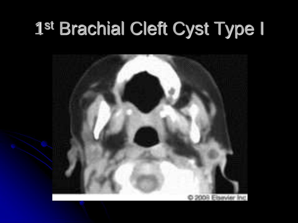
1st Brachial Cleft Cyst Type I

2nd Branchial Cleft Cyst
Most common branchial cyst
Presents as a mass just anterior and
medial to the SCM in the neck
Tract
Anterior neck -> Along carotid sheath ->
Between external and internal carotid arteries
-> superficial to CN IX and XII -> Opens into
tonsillar fossa

2nd Branchial Cleft Cyst

3rd Branchial Cleft Cyst
Closely associated with the thyroid gland
If patient with recurrent thyroid abscesses, consider
diagnosis
Usually on the left
Tract:
Lateral neck (similar or lower location than 2nd) ->
Deep to carotids -> Deep CN IX, superficial to CN XII,
Superficial to superior laryngeal nerve -> Pierces
thyrohyoid membrane -> Opens into apex of pyriform
sinus

3rd Branchial Cleft Cyst

4th Branchial Cleft Cyst
Very rare ~ 200 cases reported in the literature
Also associated with recurrent thyroid
abscesses
Theoretical path of tract: Low in neck (anterior to SCM) -> Deep to common
carotid -> Loops around aortic arch on the left
(subclavian on the right) -> Deep to superior laryngeal
nerve -> Superficial to recurrent laryngeal nerve ->
Opens into pyriform sinus

Work-up
• Ultrasound • Round mass with uniform low echogenicity and lack of internal
septations
• Advantages: No radiation, no sedation for children, low cost
• Not typically ordered alone
• CT • Homogeneous lesion with low attenuation centrally and a
smooth enhancing rim
• Often part of the work-up
• More radiation, higher cost, may require sedation (children)

Work-up
MRI Hypointense on T1 and hyperintense on T2
Advantages: No radiation
Disadvantages: Sedation for children, very expensive
Fluroscopic fistulography or CT fistulography Inject radiopaque dye into the fistula or sinus to delineate course
Barium swallow esophagography Help locate fistula tract in type 3 and 4 anomalies
FNA Usually only done if suspect cancer
May cause cyst to collapse -> much harder to remove at time of surgery

Treatment – Infected Cyst
Antibiotics Should cover respiratory flora and Staph aureus
(broad spectrum)
Cover 2-4 weeks
Abscess Consider needle aspiration to drain
May work without causing as much scaring as I&D
I&D if needle aspiration doesn’t work
Once infection cleared, operate
Treatment - Surgical
Complete surgical excision of tract and cyst is treatment of choice in most cases
1st cysts Must identify facial nerve as tract is usually
associated with it
If possible, wait till 2 years of age Mastoid tip defined
Facial nerve larger and deeper
Controversy: waiting can lead to more infections more scar more difficult surgery
Lacrimal probes can help locate tract

Treatment - Surgical
3rd and 4th cysts
Must identify the recurrent laryngeal nerve as
closely associated (will be deep to tract)
Removal of ipsilateral thyroid lobe is
advocated to ensure complete removal of
tract
Perform DL to examine pyriform sinus
Fogarty vascular catheter can be placed through
the sinus tract
Endoscopic Cauterization of Pyriform
Sinus Opening
Literature describes this for treatment of
4th sinus tracts, but has been performed
with 3rd cleft anomalies
Recommendation
Performed alone
Performed with surgical resection of cyst and
tract

Endoscopic Cauterization of Pyriform
Sinus Opening
Verret et al
Performed endoscopic cauterization of sinus
in 10 children with 4th branchial cleft
anomalies (no surgical excision!)
Dilated sinus opening with balloon catheter
cautery with electrocautery ball coagulator
7 showed no recurrent disease after 3 years
3 lost to F/U

Sources Branstetter BF, Davis LM, Coombs BD, et al. Branchial Cleft Cysts Imaging. eMedicine by
WebMD. 2011 May. Available from: http://emedicine.medscape.com/article/382803-overview.
Schoen JD and Edmonds JL. Branchial Anomalies. Children’s ENT of Houston. 2011 Sept.
Available from: http://www.childrensenthouston.com/branchial-anomalies.
Rodriguez-Vazquez JF, Merida-Velasco JR, Verdugo-Lopez S, et al. Morphogenesis of the
second pharyngeal arch cartilage (Reichert’s cartilage) in human embryos. J Anat. 2006 February;
208(2): 179–189.
Marino TA. Development and Fate of the Primitive Pharynx, Branchial Arches, and the Tongue.
Temple University. 2011 Sept. Available from:
http://isc.temple.edu/marino/embryology/parch98/parch_text.htm.
Verret DJ, McClay J, Murray A, et al. Endoscopy Cauterization of Fourth Branchial Cleft Sinus
Tracts. Arch Otolaryngol Head Neck Surg. 2004 April; 130: 465-468.
Bawle EV, Jyonouchi H, Park CL, et al. DiGeorge Syndrome. eMedicine by WebMD. 2010 Aug.
Available from: http://emedicine.medscape.com/article/886526-overview.
Propst EJ, Willging JP, and Alessandro de Alarcon. Branchial Arch Anomaly. Otolaryngology
Cases. New York: Thieme, 2010.