novel tfap2-mediated control of soxe expression...
TRANSCRIPT
RESEARCH ARTICLE720
INTRODUCTIONThe neural crest (NC) and cranial placodes are present in allvertebrates (including the basal lamprey and hagfish) but are absentfrom the basal chordate amphioxus, suggesting that these structureswere not present in the first chordates. Although recent worksuggests that evolutionary precursors of these tissues were probablypresent in the proximate vertebrate ancestor, bona fide NC and afull complement of cranial placodes are nevertheless considered tobe vertebrate synapomorphies (Canestro et al., 2003; Mazet andShimeld, 2005). Derivatives of the NC and cranial placodes,including jaws and paired sensory structures, contribute to thepredatory behavior of vertebrates, which is not exhibited byprotochordates (Gans and Northcutt, 1983; Northcutt and Gans,1983). These NC-derived features were key to the success ofvertebrates, which adds interest to identifying the genomic changesthat resulted in the emergence of this novel cell type.
The development of the NC is controlled by a gene regulatorynetwork (GRN) composed of transcription factors and otherregulatory molecules (Sauka-Spengler and Bronner-Fraser, 2008).Importantly, at least some regulatory interactions within the NCGRN must be evolutionarily novel because many of thetranscription factors contributing to the NC GRN were recruited to
the neural-plate border from other tissues (Meulemans andBronner-Fraser, 2005). The evolution of novel regulatoryinteractions can result from mutations causing ‘protein neo-functionalization’, or those causing ‘regulatory neo-functionalization’. In the first scenario, mutations in protein-codingsequence would imbue an existing transcription factor with newfunctionality (e.g. new DNA-binding specificity or affinity for newco-factors), permitting it to make new regulatory connections. Along-standing hypothesis is that protein neo-functionalization isfacilitated by gene duplication events, which would relieveconstraints on transcription factors imposed by pleiotropy. Indeed,gene duplication followed by protein neo-functionalization hasbeen proposed as a driving force in vertebrate evolution (Ohno,1970). According to this model, duplicated proteins with initiallyidentical biochemical functions diverge as one copy performs theancestral functions and the other gains the ability to carry outadditional functions (Wagner, 1998). There is support for thismodel because many examples of paralogous transcription factorswith distinct functions have been documented (reviewed byWagner and Lynch, 2008). However, it remains to be testedwhether gene duplication followed by protein neo-functionalizationcontributed to the emergence of the NC GRN, or to any othervertebrate novelty.
In the alternative scenario, regulatory neo-functionalization,mutations lead to the appearance of a transcription factor’s cognateDNA-binding site in cis-regulatory sequences of a gene notpreviously regulated by that transcription factor. In many cases, atranscription factor from one species can replace the function of itsortholog in another species. This has often been shown in thecontext of evolutionarily conserved GRNs. For example, Pax6function is conserved in the photoreceptor GRN across all phyla(Onuma et al., 2002). However, evidence for regulatory neo-
Development 139, 720-730 (2012) doi:10.1242/dev.071308© 2012. Published by The Company of Biologists Ltd
1Department of Anatomy and Cell Biology, Carver College of Medicine, University ofIowa IA 52242, USA. 2Interdisciplinary Graduate Program in Genetics, University ofIowa, IA 52242, USA. 3Department of Ecology and Evolutionary Biology, Universityof Colorado, CO 80309, USA.
*Author for correspondence ([email protected])
Accepted 6 December 2011
SUMMARYGene duplication has been proposed to drive the evolution of novel morphologies. After gene duplication, it is unclear whetherchanges in the resulting paralogs’ coding-regions, or in their cis-regulatory elements, contribute most significantly to theassembly of novel gene regulatory networks. The Transcription Factor Activator Protein 2 (Tfap2) was duplicated in the chordatelineage and is essential for development of the neural crest, a tissue that emerged with vertebrates. Using a tfap2-depletedzebrafish background, we test the ability of available gnathostome, agnathan, cephalochordate and insect tfap2 paralogs todrive neural crest development. With the exception of tfap2d (lamprey and zebrafish), all are able to do so. Together withexpression analyses, these results indicate that sub-functionalization has occurred among Tfap2 paralogs, but that neo-functionalization of the Tfap2 protein did not drive the emergence of the neural crest. We investigate whether acquisition ofnovel target genes for Tfap2 might have done so. We show that in neural crest cells Tfap2 directly activates expression of sox10,which encodes a transcription factor essential for neural crest development. The appearance of this regulatory interaction is likelyto have coincided with that of the neural crest, because AP2 and SoxE are not co-expressed in amphioxus, and because neuralcrest enhancers are not detected proximal to amphioxus soxE. We find that sox10 has limited ability to restore the neural crest inTfap2-deficient embryos. Together, these results show that mutations resulting in novel Tfap2-mediated regulation of sox10 andother targets contributed to the evolution of the neural crest.
KEY WORDS: Neural crest, Evolution, Tfap2, Transcription factor
Novel Tfap2-mediated control of soxE expression facilitatedthe evolutionary emergence of the neural crestEric Van Otterloo1, Wei Li2, Aaron Garnett3, Maria Cattell3, Daniel Meulemans Medeiros3 and Robert A. Cornell1,2,*
DEVELO
PMENT

721RESEARCH ARTICLETfap2 in neural crest evolution
functionalization requires a transcription factor from one species toreplace functionally its ortholog in another species in anevolutionarily novel GRN. The best example to date is thatDrosophila melanogaster SoxE (Sox100B) can partially replacevertebrate Sox10 in the NC GRN (Cossais et al., 2010); however,whether this example is typical or exceptional remains unclear.
The transcription factor Activator Protein 2 (Tfap2) family,members of which promote growth or differentiation in a varietyof cell types (Eckert et al., 2005), presents an excellent opportunityto test whether the formation of one node in the NC GRN requiredprotein neo-functionalization. This family is optimal for such a testbecause Tfap2 proteins have been duplicated in the chordatelineage, and because they are essential for the early steps of NCdevelopment (Hoffman et al., 2007; Li and Cornell, 2007; Luo etal., 2003). Our results support the model that sub-functionalizationhas contributed to retention of the Tfap2 paralogs in evolution.However, they do not support a role for protein neo-functionalization of Tfap2 in the assembly of the NC GRN. Insteadof gaining novel protein function, Tfap2 acquired novel targets,including sox10 and other genes, and this contributed to theevolution of the NC.
MATERIALS AND METHODSFish maintenanceZebrafish embryos and adults were reared as described previously(Westerfield, 1993). Embryos were staged by hours or days post-fertilization (hpf or dpf, respectively) at 28.5°C (Kimmel et al., 1995).Because of the poor effectiveness of tfap2a morpholinos (MOs) beyond 48hpf (our unpublished observations), we used homozygous tfap2a mutants,generated from heterozygous adults harboring a presumed null allele oftfap2a (lockjaw, tfap2ats213) (Knight et al., 2003), in analyses ofmelanophores.
Generation of cDNAs for in situ hybridization and mis-expressionexperimentsFirst-strand cDNA was generated from total RNA extracted from zebrafishembryos at 24 hpf as described (O’Brien et al., 2004) or lamprey andamphioxus embryos/adults as described (Meulemans and Bronner-Fraser,2002). Partial or full length cDNA clones were amplified (supplementarymaterial Table S1). Digoxigenin (DIG)-labeled probes (Roche Diagnostics,Mannheim, Germany) were synthesized, and whole-mount in situhybridization was performed following standard methods (Thisse andThisse, 2008).
For mis-expression experiments, full-length coding regions of eachzebrafish gene studied were amplified from a pool of 24 hpf first-strandcDNA. PCR products were cloned into pSCA (Stratagene/Agilent, SantaClara, CA, USA), pCR4-TOPO (Invitrogen, Carlsbad, CA, USA) orpENTR/D-TOPO (Invitrogen) shuttling vectors, as indicated(supplementary material Table S1). Coding regions were then shuttled intopCS2+ either using standard cloning methods (pSCA and pCR4-TOPO)(Sambrook et al., 1989) or Gateway cloning using LR Clonase II (pENTR-D/TOPO, Invitrogen). All final plasmids were confirmed by sequencing.Plasmids were linearized and capped mRNA synthesized in vitro using theSP6 mMessage mMachine kit (Ambion, Austin, TX, USA) andconcentrated using Microcon Spin Columns (Millipore, Billerica, MA,USA).
For forced expression of sox10, embryos were injected withhsp70:sox10 (Elworthy et al., 2003). At 80% epiboly, embryos weretransferred to 37°C water and incubated at this temperature for 60 minutesthen allowed to develop at room temperature.
Amphioxus soxE enhancer analysisRegions of amphioxus genomic DNA proximal to soxE (see supplementarymaterial Table S2 for genomic coordinates) were amplified and, with theexception of soxE promoter region 2, cloned into a tol-2 based GFPreporter vector described previously (Fisher et al., 2006). Several F0
embryos injected with construct containing the soxE region 4 were raised,and two independent stable transgenic strains were isolated that hadidentical GFP expression to transient transgenic embryos. The amphioxussoxE promoter region 2 was amplified and ligated into the sox10:eGFPplasmid (Wada et al., 2005) in place of the zebrafish sox10 element usingstandard cloning methods (Sambrook et al., 1989).
For analysis of conserved transcription factor binding sites the onlinetool ConSite (Sandelin et al., 2004) was used. Search parameters includeda conservation cut-off of 38%, a window size of 50 and a TF scorethreshold of 80%.
Morpholinos and microinjectionPlasmids used for in vitro mRNA synthesis are described above. Plasmidsand MOs were injected at the one-cell stage and mRNA at the two- to four-cell stage. Approximately 125 pg of tfap2 mRNAs, 125 pg of all plasmidsand 5 ng of MOs were injected. MOs targeting tfap2a (tfap2a e2i2) andtfap2c (tfap2c e3i3) used here have been described (Li and Cornell, 2007).Supplementary material Table S1 contains sequences.
Chromatin immunoprecipitation (ChIP)A modified ChIP protocol for fish embryos was used (Lindeman et al.,2009). Chromatin was sheared on ice using a probe-tip sonicator (VirTisVirsonic 600) with the following settings: power setting, 5; pulse time,20 seconds; number of pulses, ten; break between pulses, 2 minutes.Anti-Tfap2a(CT) (Anaspec, Fremont, CA, USA) or Rabbit-IgG(Millipore) were used for immunoprecipitation. ChIP experiments wereperformed in triplicate, on newly isolated embryos. PCR reactions (10l) following ChIP were prepared with immunoprecipitated DNA, usingthe SYBR Green kit (Applied Biosystems, Foster City, CA, USA)following the manufacturer’s instructions. Primers were used at a finalconcentration of 200 nM in separate PCR reactions. Quantitative real-time PCR in Low 96-well plates (Bio-Rad, Hercules, CA, USA) wasconducted using a Bio-Rad thermal cycler (CFX96 Real-Time PCRDetection System) following the default protocol; triplicates of eachreaction were carried out simultaneously and mean and standard errorwere calculated. Melt-curve analysis was also performed to confirmspecificity of primers. Figure 5B shows a representative run from one ofthe three repeats.
Cycloheximide experimentsEmbryos at 90% epiboly were incubated for ~2 hours in 100 g/mlcycloheximide (diluted in fish water from a 40 mg/ml stock dissolved inDMSO) (Sigma-Aldrich), a dose several times the level previouslyshown to strongly reduce protein translation in 1.5 hpf zebrafishembryos (Leung et al., 2003), or in diluted DMSO as a negative control.Subsequently, embryos were incubated for an additional 2.5 hours indexamethasone (100 M in fish water from a 40 mM stock dissolved inethanol) (Sigma-Aldrich), or in diluted ethanol as a negative control.Next, RNA was isolated (Trizol, Invitrogen) following themanufacturer’s instructions and treated with DNaseI to remove genomicDNA. Quantitative reverse-transcriptase PCR (qRT-PCR) analysis ofmRNA levels was conducted, starting with 1 g of total RNA, aspreviously described (Van Otterloo et al., 2010). Primer efficiencieswere confirmed using a tenfold series dilution of cDNA and generatinga standard curve. All primers were designed to span a large intron,preventing amplification of genomic DNA. The 2Ct method was usedto calculate fold-enrichment over wild-type samples and all groups werenormalized to -actin (Dussault and Pouliot, 2006). Each experimentwas repeated in triplicate, starting from injections. Results are shownfrom a representative experiment.
Western blottingWestern blotting of zebrafish embryos was carried out essentially aspreviously described (Link et al., 2006). Antibodies 9E 10 (anti-myc,1:100) and AA4.3 (alpha-tubulin 1:100) were developed by J. MichaelBishop and Charles Walsh, respectively, and were obtained from theDevelopmental Studies Hybridoma Bank (Iowa, USA). D
EVELO
PMENT

722
RESULTSSequential sub-functionalization of tfap2duplicates in the vertebrate lineageWe first sought to establish the relationships among Tfap2 paralogspresent in amphioxus, lamprey, mammals and zebrafish.Previously, we had identified five tfap2 homologs in zebrafish(Danio rerio, tfap2a-e), one in lamprey (Petromyzon marinus, Pm-tfap2) and one in amphioxus (Branchiostoma floridae, Bf-tfap2) (Liand Cornell, 2007; Meulemans and Bronner-Fraser, 2002).However, after the pre-assembly lamprey genome becameavailable, we found a second lamprey tfap2 gene. Phylogeneticanalysis of the Drosophila (D. melanogaster) and deuterostomeTfap2 proteins grouped this new lamprey Tfap2 with gnathostomeTfap2d genes. The previously described lamprey Tfap2 fell withina clade that includes the genes encoding gnathostome Tfap2a,Tfap2b, Tfap2c and Tfap2e (Fig. 1A). Given the evidence for twogenome duplication events in the vertebrate lineage (Holland et al.,1994), these results support a scenario in which whole-genomeduplication in the common ancestor of lamprey and gnathostomesgenerated two paralogs, Tfap2d and Tfap2a/b/c/e, from an ancestralTfap2. Subsequent duplication events, including a second round ofwhole-genome duplication, generated all of the gnathostome Tfap2paralogs (except Tfap2d) from the Tfap2a/b/c/e ancestor. Bycontrast, any duplicates of Tfap2d were lost. Although there isevidence for a genome duplication event within the teleost lineage(Postlethwait et al., 2004), given the one-to-one correspondence oftetrapod and zebrafish Tfap2 homologs, it appears that Tfap2duplicates generated in this genome duplication event were lost. Insummary, these findings provide an evolutionary scenario for theorigin of Tfap2 paralogs present in vertebrates.
We asked whether the duplication-degeneration-complementation model explains the retention of vertebrate tfap2dparalogs (Force et al., 1999). Under this model, sub-functionalization refers to the partitioning of an ancestral gene’sexpression pattern, or its functional domains, between itsduplicates, leading to selective pressure to retain both duplicates(Force et al., 1999). For this analysis, we consider amphioxus torepresent the vertebrate ancestor. Amphioxus tfap2 is expressed inthe anterior neural tube and in the non-neural ectoderm(Meulemans and Bronner-Fraser, 2002). Similarly, lamprey tfap2is expressed in the brain and the non-neural ectoderm, includingthe neural plate border/NC (Meulemans and Bronner-Fraser, 2002).However, lamprey tfap2d is expressed only in the brain (Fig. 1B-E) [tfap2d expression persists from larval stages into adulthood asobserved by RT-PCR (data not shown)]. Similarly, mouse Tfap2d(Zhao et al., 2003) and zebrafish tfap2d are expressed solely withinthe brain (Fig. 1F-M,P). Together, these data are consistent withsub-functionalization of an ancestral tfap2 expression domainbetween two duplicates (tfap2 and tfap2d) pre-dating the split ofagnathans and gnathostomes.
We next investigated whether sub-functionalization might alsoexplain the retention of tfap2a, tfap2b, tfap2c and tfap2e ingnathostomes. In this analysis, we considered lamprey tfap2expression to represent the ancestral condition. Zebrafish tfap2a,tfap2b and tfap2c are also expressed in this tissue but with distincttiming or spatial restrictions (supplementary material Fig. S1).Thus, during the gastrula stage zebrafish tfap2a and tfap2c arestrongly expressed in the ventral ectoderm (presumptive skin) butby 24 hpf tfap2c expression remains high in embryonic skin,whereas tfap2a expression is relatively low, particularly in theperiderm (Hoffman et al., 2007; Li and Cornell, 2007). Expressionof tfap2b is absent from ventral ectoderm during gastrulation but
RESEARCH ARTICLE Development 139 (4)
is present in discrete regions of anterior skin at 24 hpf (Knight etal., 2005). In addition, lamprey tfap2 is expressed in premigratoryand migratory NC (Meulemans and Bronner-Fraser, 2002). Inzebrafish embryos at the gastrula stage, tfap2a and tfap2c areexpressed in lateral neural plate cells, which later give rise to theNC (Li and Cornell, 2007). By the neurula stage and somitogenesisstages, however, only tfap2a is expressed at high levels in thepremigratory NC. In NC derivatives, tfap2e is expressed inmelanoblasts, whereas tfap2b is expressed in spinal sensoryneurons, some of which are derived from the NC (Knight et al.,2005; Knight et al., 2003; O’Brien et al., 2004; Van Otterloo et al.,2010) (supplementary material Fig. S1 and Table S3). Finally,lamprey tfap2 is expressed in the brain, and zebrafish tfap2a,tfap2b, tfap2c and tfap2e are also all expressed in the brain, but inimperfectly overlapping patterns (Fig. 1N-Q) [tfap2c is expressedin the brain at 24 hpf (Li and Cornell, 2007)]. In summary,expression of modern tfap2a, tfap2b, tfap2c and tfap2e can beviewed as a partitioning of the ancestral pattern. These results areconsistent with a model in which multiple rounds of duplicationand sub-functionalization have resulted in retention of these fourtfap2 duplicates.
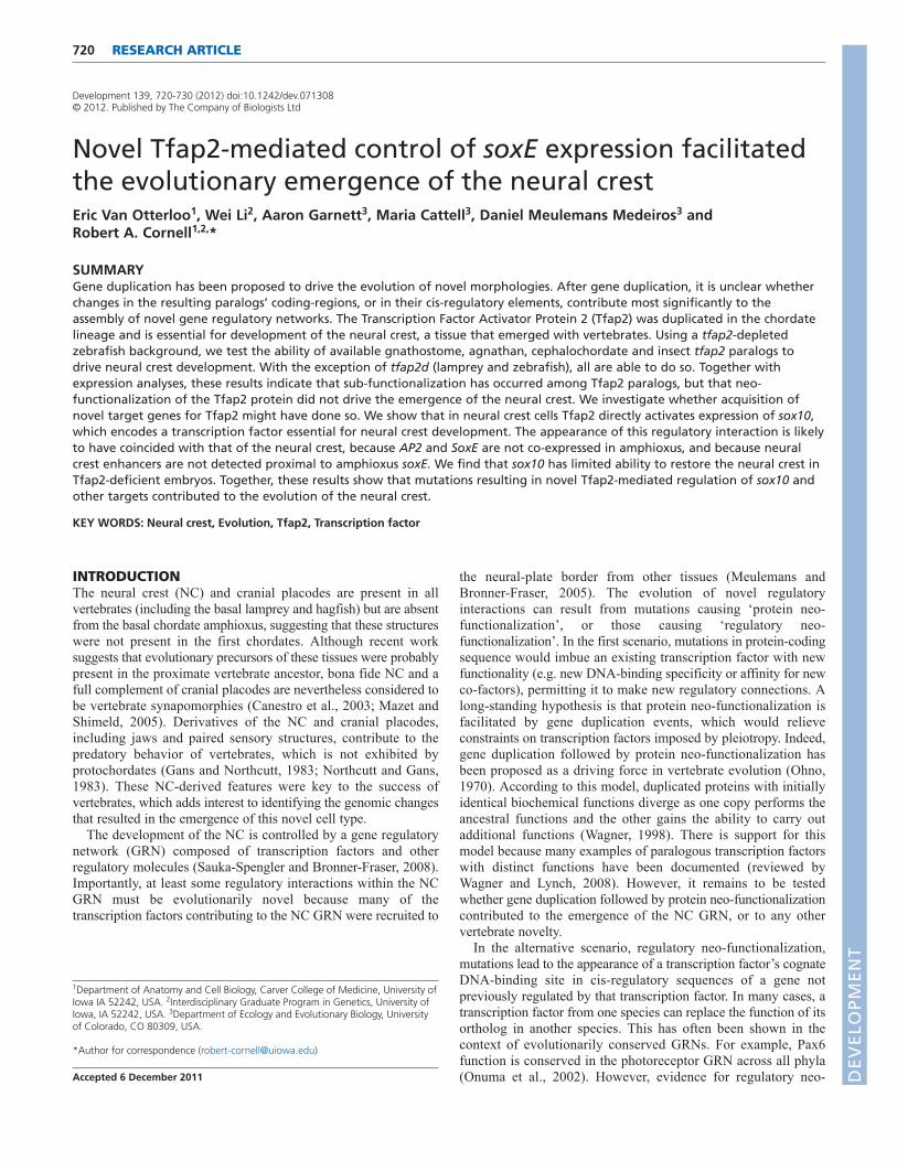
Tfap2d is functionally distinct from other Tfap2family membersThe proteins encoded by paralogs that have undergone sub-functionalization are under relaxed selection. Specifically, they arefreed to lose functions appropriate for domains in which they areno longer expressed and to become functionally optimized forexpression domains they retain (Conant and Wagner, 2003; Lynchand Wagner, 2008). We tested whether paralogs Tfap2b, Tfap2dand Tfap2e, which have all lost ancestral expression in the lateralneural border, have also lost a function appropriate for thisdomain, i.e. the ability to generate the NC. Starting with tfap2a/c-deficient embryos (which are embryos depleted of both tfap2a andtfap2c by mutation or by morpholino), we injected mRNAsencoding the various Tfap2 paralogs. We raised such embryos toappropriate stages then fixed and processed them to reveal amarker of premigratory NC, foxd3 (Kelsh et al., 2000; Odenthaland Nusslein-Volhard, 1998), or migratory NC, dlx2a (Akimenkoet al., 1994). As expected, mRNAs encoding Tfap2a or Tfap2cefficiently rescued foxd3 expression (Fig. 2G,M), dlx2a expression(Fig. 2H,N) and also melanophores (Fig. 2I,O). Interestingly,injection of the tfap2b and tfap2e mRNAs also rescued foxd3expression (Fig. 2J,S), dlx2a expression (Fig. 2K,T) andmelanophores (Fig. 2L,U) in tfap2a/c-deficient embryos. Furthertests revealed that overexpression of each of these Tfap2 paralogssimilarly rescued in tfap2a/c-deficient embryos the NC derivativesperipheral glia, marked by foxd3 at 28 hpf (Kelsh et al., 2000), andsensory neuron precursors, marked by expression of tlxa at 22 hpf(Andermann and Weinberg, 2001) (supplementary material Fig.S2). By contrast, injection of a similar dose of the tfap2d mRNAinto tfap2a/c-deficient embryos failed to rescue expression offoxd3, dlx2a or melanophores (Fig. 2P-R). Western blot analysisof epitope-tagged variants of Tfap2d and Tfap2a revealed similarstability in zebrafish embryos (supplementary material Fig. S3).Finally, we found that injection of lamprey tfap2 (56% and 55%protein similarity to Tfap2a and Tfap2c, respectively) alsoefficiently rescued foxd3 expression, dlx2a expression andmelanophores in tfap2a/c-deficient embryos (Fig. 3J-L), whereasinjection of the lamprey tfap2d (45% and 39% protein similarityto Tfap2a and Tfap2c, respectively) at a similar dose failed to doso (Fig. 3M-O) (all rescue experiments are summarized in Fig. 4). D
EVELO
PMENT

In summary, Tfap2b and Tfap2e, retain the ability to promote NCdevelopment, while Tfap2d virtually lacks this ability. As discussedabove, it appears that tfap2d lost enhancers possessed by the tfap2
723RESEARCH ARTICLETfap2 in neural crest evolution
ancestor that drive expression in the non-neural ectoderm, and didso prior to the split of agnathans and gnathostomes. However, thefunctional experiments just described do not resolve whether
Fig. 1. Characterization of embryonic tfap2d expression in zebrafish and lamprey. (A)Phylogenetic tree of deuterostome and Drosophilamelanogaster tfap2 genes, constructed using the Maximum Likelihood method (Schmidt et al., 2002). A similar tree topology was obtained usingthe Neighbor-Joining method (not shown) (Saitou and Nei, 1987). Confidence values for both methods are shown at branch points with quartet-puzzling reliability scores in black and Neighbor-Joining bootstrap values in green. Each gene name includes a prefix with the initials of thecorresponding species name. Ci, Ciona intestinalis; Pm, Petromyzon marinus; Mm, Mus musculus; Dr, Danio rerio; Bf, Branchiostoma floridae; Sp,Strongylocentrotus purpuratus; Dm, Drosophila melanogaster. (B-E)Lamprey embryos, fixed at the stage indicated and processed to reveal tfap2dexpression. (B,C)Expression of tfap2d is first observed in bilateral spots in the presumed forebrain after neurulation (Tahara st. 25) in lateral (B) anddorsal (C) views. (D,E)Expression in the brain at late larval stage (st. 26.5) in (D) lateral and (E) dorsal views. (F-Q)Wild-type zebrafish embryos, fixedat the stages indicated and processed to reveal expression of tfap2d (F-M) or the indicated tfap2 family member (N-Q) by RNA in situ hybridization.(F)Dorsal view of the head showing tfap2d expression in a distinct structure within the presumptive mesencephalon. (G)Lateral view showingtfap2d expression within two distinct domains of the tegmentum; lines represent field of view for optical sections shown in H and I. (H,I)Dorsalviews of the head, expression is in presumed tegmentum. (J)Higher magnification of embryo shown in G. (K)Lateral view of the head of a 48 hpfembryo, showing expression in tegmentum and optic tectum and retina (arrow). (L,M)Dorsal (L) and ventral (M) views of embryo shown in K,focusing on tfap2d expression within the optic tectum and retina, respectively. (N-Q)Embryos at 48 hpf, revealing shared and distinct expressiondomains of tfap2 family members in the brain. Embryos shown with anterior to the left, unless otherwise indicated. e, eye. Scale bars: 50m.
DEVELO
PMENT

724
Tfap2d subsequently lost the ability to induce NC, i.e. because ofthe absence of selective pressure to retain this function, oralternatively, whether the other Tfap2 paralog, which retainedexpression in non-neural ectoderm, gained this ability, i.e. viaprotein neo-functionalization.
Tfap2 from amphioxus can induce neural crestTo determine whether the ability of Tfap2 to induce NC resultedfrom protein neo-functionalization, we generated a full-lengthamphioxus tfap2 cDNA and generated mRNA from it in vitro.Interestingly, injection of amphioxus tfap2 mRNA (48% and 46%protein similarity to Tfap2a and Tfap2c, respectively) into tfap2a/c-deficient embryos efficiently rescued expression of foxd3 (in NCand in peripheral glia), expression of dlx2a, expression of tlxa andmelanophores (Fig. 3G-I; supplementary material Fig. S2). Testingan even more ancient AP2 homolog, we injected Drosophila tfap2mRNA (40% and 39% protein similarity to Tfap2a and Tfap2c,respectively) into tfap2a/c-deficient zebrafish embryos, andobserved robust foxd3 expression in the lateral neural plate at 11hpf (supplementary material Fig. S4G); however, in such embryos,expression of dlx2a at 22 hpf and melanophores at 48 hpf wererarely present (supplementary material Fig. S4H,I, legend). Theless robust rescue of NC by Drosophila tfap2 might reflectinstability of Drosophila Tfap2 relative to other Tfap2 proteinstested, or possibly the absence in Drosophila Tfap2 of a functionessential for NC maintenance or other later events in NCdevelopment. Nonetheless, because amphioxus efficiently rescuedNC in tfap2a/c-deficient embryos, we conclude that recruitment ofTfap2 into a GRN that controls induction of NC at the neural plateborder did not require Tfap2 to gain new protein function.
RESEARCH ARTICLE Development 139 (4)
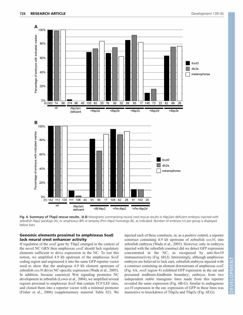
sox10 is targeted directly by Tfap2a and forcedexpression of sox10 partially restores trunkneural crest in tfap2a/c-deficient embryosWe sought to identify the novel regulatory connections gainedby Tfap2 in the context of the NC GRN. In amphioxus, theorthologs of multiple NC regulatory proteins (e.g. SoxE, FoxD,Twist, ID) are expressed in the mesoderm but not in the neuralplate border (Sauka-Spengler et al., 2007). Mutations in theregulatory regions of the genes encoding such regulatoryproteins might have introduced Tfap2 binding sites, therebyrecruiting their expression to the neural plate border. Weinvestigated this possibility for sox10, a gene necessary for earlysteps of NC induction (Dutton et al., 2001; Honore et al., 2003)and expression of which in NC is lost in tfap2a/c-deficientembryos (Hoffman et al., 2007; Li and Cornell, 2007). Weconducted anti-Tfap2a chromatin immunoprecipitation (ChIP) on4.9 kb upstream of the start codon of sox10, a region previouslyshown to drive reporter expression in the NC (Wada et al., 2005).We started with lysates of zebrafish embryos at 12 hpf, a stageat which both tfap2a and sox10 are expressed at high levels inthe NC, using an antibody generated against zebrafish Tfap2a,specificity of which was confirmed in tfap2a mutants(supplementary material Fig. S5). A pair of predicted Tfap2binding sites are present proximal to the transcription start site(Fig. 5A). On the precipitated chromatin we performedquantitative PCR (qPCR) with primer pairs positioned near thesepredicted Tfap2a binding sites, or with primer pairs positioned1.2 kb downstream, as a negative control (off-target site) (Fig.5A). We observed 12- to 22-fold enrichment at the target site inanti-Tfap2a-precipitated chromatin versus IgG-precipitated
Fig. 2. Assessment of the ability ofgnathostome Tfap2 paralogs to restore neuralcrest in tfap2a/c-deficient zebrafish embryos.(A,D,G,J,M,P,S) Dorsal views of flat-mounted wild-type zebrafish embryos (A) or embryos injected withthe indicated mRNA and/or MO (D,G,J,M,P,S), fixedat 11 hpf and processed to reveal foxd3 expression.Restored foxd3 expression was found on left, right,or both sides as a result of mosaic injection ofmRNA. White asterisks indicate premigratory neuralcrest. Red asterisks indicate non-neural crest-derivedtailbud. (B,E,H,K,N,Q,T) Lateral views of wild-typezebrafish embryos (B) or embryos injected with theindicated mRNA and/or MO (E,H,K,N,Q,T), fixed at22 hpf and processed to reveal dlx2a expression.With the exception of tfap2d, no trend was seen inthe spatial extent of dlx2a expression and the tfap2paralog used. White asterisks indicate migratoryneural crest. Yellow asterisks indicate brain.(C,F,I,L,O,R,U) Lateral views of live embryos at 48hpf that are wild type (C) or tfap2a mutants injectedwith tfap2c MO (F,I,L,O,R,U), and injected with theindicated mRNA. Embryos shown with anterior tothe left, unless otherwise indicated. Scale bars: in A,100m for A,D,G,J,M,P,S; in B, 50m forB,E,H,K,N,Q,T; in C, 100m for C,F,I,L,O,R,U.
DEVELO
PMENT

chromatin, and minimal enrichment at the off-target site (Fig.5B). These findings indicate that Tfap2a binds the zebrafishsox10 promoter at a time when sox10 is expressed in the NC.
Next, we investigated whether Tfap2a overexpression willelevate sox10 expression when protein translation is inhibited.We co-injected embryos with tfap2a MO, tfap2c MO, andmRNA encoding a dexamethasone-inducible variant of Tfap2a(Tfap2a fused to glucocorticoid-receptor, tfap2a-GR) (Luo et al.,2002). At 10 hpf, we treated the embryos with cycloheximide toblock translation (Leung et al., 2003). Two hours later, we addeddexamethasone to induce nuclear transport of Tfap2a-GR.Finally, 2.5 hours later (~14 hpf), we harvested RNA from theseembryos, generated first-strand cDNA and used qPCR tomeasure the levels of sox10 and zgc:85942 (ednrab – ZebrafishInformation Network), an ednra homolog which, like sox10, isexpressed at high levels in the NC at 13 hpf (Thisse and Thisse,2004). As expected, in tfap2a/c-deficient embryos the levels ofboth sox10 and zgc:85942 mRNA were reduced relative to thosein control embryos (Fig. 5C,D). In tfap2a/c-deficient embryosinjected with tfap2a-GR and not treated with dexamethasone,sox10 and zgc:85942 expression were barely elevated above thelevels detected in tfap2a/c-deficient embryos (Fig. 5C,D),confirming that Tfap2a-GR is only minimally active in theabsence of dexamethasone. By contrast, in embryos treated withdexamethasone but not cycloheximide, the expression of bothNC markers was significantly elevated (Fig. 5C,D).
725RESEARCH ARTICLETfap2 in neural crest evolution
Interestingly, in embryos treated with dexamethasone in thepresence of cycloheximide, the expression of sox10, but not thatof zgc:85942, was raised to levels significantly higher than thosein tfap2a/c-deficient embryos (Fig. 5C,D). Together, thesefindings strongly suggest that Tfap2 activates sox10 expressiondirectly.
To assess whether activation of sox10 is the sole requirement ofTfap2a in NC development, we re-introduced sox10 into tfap2a/c-deficient embryos. To do so, we used the hsp70:sox10 plasmid,which contains a heat-shock-inducible promoter upstream of thesox10 coding region (Elworthy et al., 2003). In hsp70:sox10-injected, tfap2a/c-deficient embryos exposed to heat-shock at 9 hpfand fixed at 12 hpf, snai1b and sox9b were detected in a scatteredpattern around the embryo (mosaic expression is typical oftransient transgenesis) (Thisse et al., 1995; Yan et al., 2005) (Fig.5M,N). However, foxd3 expression was absent in these embryos,and crestin expression was absent from similarly treated embryosfixed at 22 hpf (Rubinstein et al., 2000) (Fig. 5O; data not shown).Interestingly, in similarly treated embryos melanophores werepresent at 48 hpf, but only in the trunk, not in the head (Fig. 5P).Control experiments revealed the requirement for both thehsp70:sox10 plasmid and the heat-shock for the results describedin this section. Because forced expression of sox10 was insufficientto restore a full NC program in tfap2a/c-deficient embryos, it isvery likely that there are Tfap2 regulatory targets other than sox10that are essential in the NC GRN.
Fig. 3. Assessment of the ability of lamprey and amphioxus Tfap2 paralogs to restore neural crest in tfap2a/c-deficient zebrafishembryos. (A,D,G,J,M) Dorsal views of flat-mounted wild-type zebrafish embryos (A) or embryos injected with the indicated mRNA and/or MO(D,G,J,M), fixed at 11 hpf and processed to reveal foxd3 expression. White asterisks indicate premigratory neural crest. Red asterisks indicate non-neural crest-derived tailbud. (B,E,H,K,N) Lateral views of wild-type zebrafish embryos (B) or embryos injected with the indicated mRNA and/or MO(E,H,K,N), fixed at 22 hpf and processed to reveal dlx2a expression. White asterisks indicate migratory neural crest. Yellow asterisks indicate brain.(C,F,I,L,O) Lateral views of live embryos at 48 hpf that are wild type (C) or tfap2a mutants injected with tfap2c MO (F,I,L,O), and injected with theindicated mRNA. No trend was seen in the ability of Bf-tfap2 versus Pm-tfap2 to rescue more melanophores per embryos. Insets in figure I and L arehigher magnification images of boxed melanophores. Embryos shown with anterior to the left, unless otherwise indicated. Scale bars: in A, 100mfor A,D,G,J,M; in B, 50m for B,E,H,K,N; in C, 100m for C,F,I,L,O.
DEVELO
PMENT
726
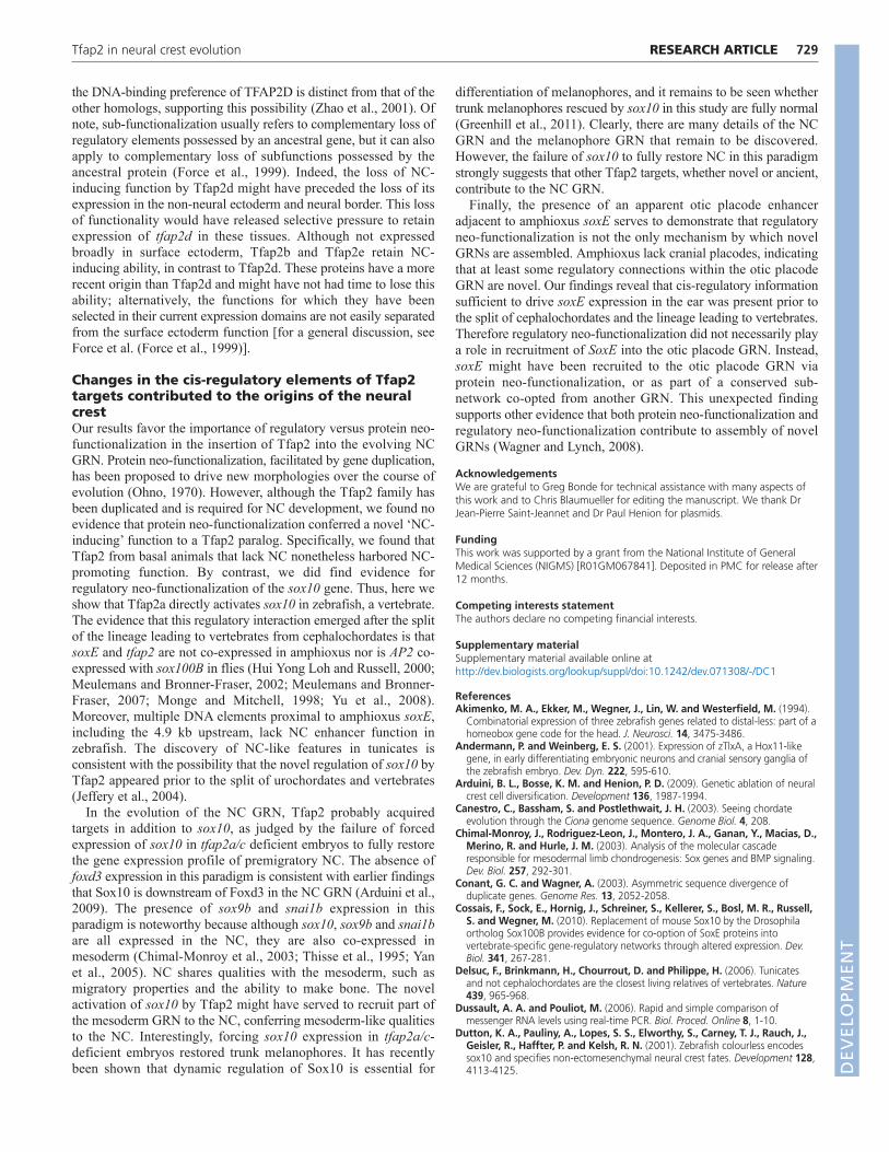
Genomic elements proximal to amphioxus SoxElack neural crest enhancer activityIf regulation of the soxE gene by Tfap2 emerged in the context ofthe novel NC GRN then amphioxus soxE should lack regulatoryelements sufficient to drive expression in the NC. To test thisnotion, we amplified 4.9 kb upstream of the amphioxus SoxEcoding region and engineered it into the same GFP reporter vectorused to show that the analogous 4.9 kb element upstream ofzebrafish sox10 drives NC-specific expression (Wada et al., 2005).In addition, because canonical Wnt signaling promotes NCdevelopment in zebrafish (Lewis et al., 2004), we amplified severalregions proximal to amphioxus SoxE that contain TCF/LEF sites,and cloned them into a reporter vector with a minimal promoter(Fisher et al., 2006) (supplementary material Table S2). We
RESEARCH ARTICLE Development 139 (4)
injected each of these constructs, or, as a positive control, a reporterconstruct containing 4.9 kb upstream of zebrafish sox10, intozebrafish embryos (Wada et al., 2005). However, only in embryosinjected with the zebrafish construct did we detect GFP expressionconcentrated in the NC, as recognized by anti-Sox10immunoreactivity (Fig. 6H,I). Interestingly, although amphioxusembryos are believed to lack ears, zebrafish embryos injected witha construct containing an element downstream of amphioxus soxE(Fig. 6A, soxE region 4) exhibited GFP expression in the ear andpresumed midbrain-hindbrain boundary; embryos from twoindependent stable transgenic lines made from this reporterrevealed the same expression (Fig. 6B-G). Similar to endogenoussox10 expression in the ear, expression of GFP in these lines wasinsensitive to knockdown of Tfap2a and Tfap2c (Fig. 6F,G).
Fig. 4. Summary of Tfap2 rescue results. (A,B)Histograms summarizing neural crest rescue results in tfap2a/c-deficient embryos injected withzebrafish tfap2 paralogs (A), or amphioxus (Bf) or lamprey (Pm) tfap2 homologs (B), as indicated. Number of embryos (n) per group is displayedbelow bars.
DEVELO
PMENT
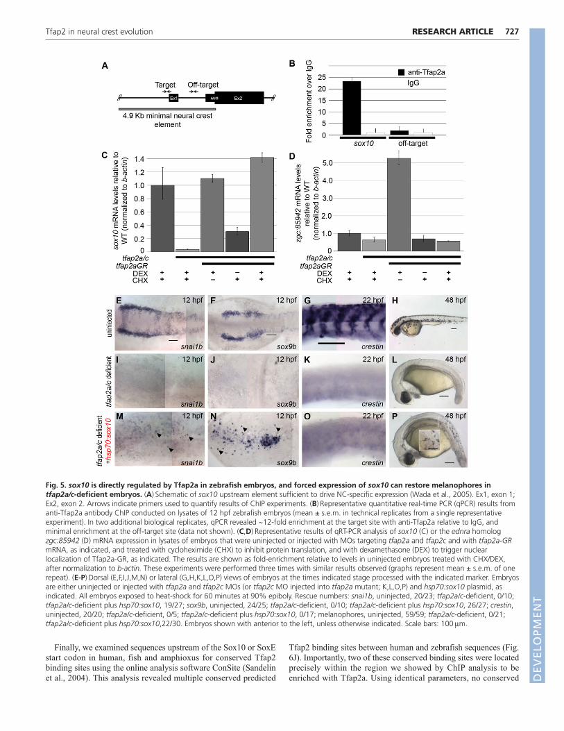
Finally, we examined sequences upstream of the Sox10 or SoxEstart codon in human, fish and amphioxus for conserved Tfap2binding sites using the online analysis software ConSite (Sandelinet al., 2004). This analysis revealed multiple conserved predicted
727RESEARCH ARTICLETfap2 in neural crest evolution
Tfap2 binding sites between human and zebrafish sequences (Fig.6J). Importantly, two of these conserved binding sites were locatedprecisely within the region we showed by ChIP analysis to beenriched with Tfap2a. Using identical parameters, no conserved
Fig. 5. sox10 is directly regulated by Tfap2a in zebrafish embryos, and forced expression of sox10 can restore melanophores intfap2a/c-deficient embryos. (A)Schematic of sox10 upstream element sufficient to drive NC-specific expression (Wada et al., 2005). Ex1, exon 1;Ex2, exon 2. Arrows indicate primers used to quantify results of ChIP experiments. (B)Representative quantitative real-time PCR (qPCR) results fromanti-Tfap2a antibody ChIP conducted on lysates of 12 hpf zebrafish embryos (mean ± s.e.m. in technical replicates from a single representativeexperiment). In two additional biological replicates, qPCR revealed ~12-fold enrichment at the target site with anti-Tfap2a relative to IgG, andminimal enrichment at the off-target site (data not shown). (C,D)Representative results of qRT-PCR analysis of sox10 (C) or the ednra homologzgc:85942 (D) mRNA expression in lysates of embryos that were uninjected or injected with MOs targeting tfap2a and tfap2c and with tfap2a-GRmRNA, as indicated, and treated with cycloheximide (CHX) to inhibit protein translation, and with dexamethasone (DEX) to trigger nuclearlocalization of Tfap2a-GR, as indicated. The results are shown as fold-enrichment relative to levels in uninjected embryos treated with CHX/DEX,after normalization to b-actin. These experiments were performed three times with similar results observed (graphs represent mean ± s.e.m. of onerepeat). (E-P)Dorsal (E,F,I,J,M,N) or lateral (G,H,K,L,O,P) views of embryos at the times indicated stage processed with the indicated marker. Embryosare either uninjected or injected with tfap2a and tfap2c MOs (or tfap2c MO injected into tfap2a mutant; K,L,O,P) and hsp70:sox10 plasmid, asindicated. All embryos exposed to heat-shock for 60 minutes at 90% epiboly. Rescue numbers: snai1b, uninjected, 20/23; tfap2a/c-deficient, 0/10;tfap2a/c-deficient plus hsp70:sox10, 19/27; sox9b, uninjected, 24/25; tfap2a/c-deficient, 0/10; tfap2a/c-deficient plus hsp70:sox10, 26/27; crestin,uninjected, 20/20; tfap2a/c-deficient, 0/5; tfap2a/c-deficient plus hsp70:sox10, 0/17; melanophores, uninjected, 59/59; tfap2a/c-deficient, 0/21;tfap2a/c-deficient plus hsp70:sox10,22/30. Embryos shown with anterior to the left, unless otherwise indicated. Scale bars: 100m.
DEVELO
PMENT
728
Tfap2 binding sites were detected when comparing human withamphioxus sequences or zebrafish with amphioxus sequences (Fig.6J; data not shown). In combination with the enhancer analysis ofsoxE discussed above, and the absence of co-expression of soxEand tfap2 in amphioxus (and Drosophila), these findings supportthe hypothesis that sox10 became a regulatory target of Tfap2 afterthe split of the cephalochordates from the lineage leading tovertebrates.
DISCUSSIONSelective pressure to retain Tfap2 paralogsresulted from sub-functionalizationHere, we began by showing that fish and mammalian Tfap2dparalogs resulted from an ancient duplication of an ancestral Tfap2prior to the split of agnathans and gnathostomes, potentially duringa genome-duplication event. Interestingly, both MaximumLikelihood and Neighbor Joining (not shown) analyses groupedamphioxus Tfap2, sea urchin Tfap2 and ascidian Tfap2B togetherin one clade, and placed ascidian Tfap2A as an outgroup to allother chordate Tfap2s with moderate to high support. Thisgrouping contradicts recent phylogenomic analyses showing thaturochordates are the vertebrate sister group (Delsuc et al., 2006).These results probably reflect the rapid evolution of ascidian tfap2genes, as has been seen with many other urochordatedevelopmental regulators (Holland and Gibson-Brown, 2003).However, regardless of the phylogenetic position of the ascidiangenes, a comparison of the expression pattern of the single tfap2
RESEARCH ARTICLE Development 139 (4)
homolog in amphioxus (a basal chordate) and the two homologs inlamprey (a basal vertebrate) supports the duplication-degeneration-complementation model (Force et al., 1999). This model posits thatselective pressure to retain duplicated, otherwise redundant, genesemerges through a complementary loss of gene-regulatory elementsinherited from the ancestral gene (a process termed sub-functionalization) (Force et al., 1999). Sub-functionalization alsoappears to have occurred among the tfap2a/b/c/e genes ingnathostomes. Like lamprey tfap2, the collective expression patternof zebrafish tfap2a/b/c/e paralogs includes the presumptive skin,the neural plate border, NC and NC derivatives. However, asdescribed here and elsewhere (Hoffman et al., 2007; Knight et al.,2005; Knight et al., 2003; Li and Cornell, 2007; Meulemans andBronner-Fraser, 2002; O’Brien et al., 2004; Van Otterloo et al.,2010), the individual patterns of these paralogs are not identical.Thus, it appears that through the loss of tissue-specific enhancersassociated with an ancestral tfap2a/b/c/e, initially redundant tfap2duplicates have acquired distinct expression territories, resulting inselective pressure to retain all four paralogs.
Relaxed selective pressure on Tfap2d permittedthe loss of its neural crest-promoting functionWe have also tested the notion that expression-domain partitioningrelaxes selective pressure on duplicated proteins. We showed thatTfap2d has lost, to a great degree, the ability to generate NC.Whether Tfap2d has also become optimized for its brain function,relative to the ancestral Tfap2, will require further tests. However,
Fig. 6. Amphioxus soxE elements fail to drive neural crest specific expression in zebrafish embryos. (A)Schematic of amphioxus soxElocus showing location of DNA elements tested in zebrafish reporter analysis. Red box, coding region of soxE; red arrow, initial AUG. Genomiccoordinates are shown in supplementary material Table S2. (B,C)Anterior dorsal (B) and lateral (C) views of a Tg(Bf-soxE-E4:eGFP) zebrafish embryofixed at 12 hpf and processed for gfp expression. gfp expression in midbrain-hindbrain (arrows) and otic placodes (arrowheads) is apparent. (D-G)Dorsal (D,F) and lateral (E,G) views of live, 48 hpf Tg(Bf-soxE-E4:eGFP) embryos. Arrowheads in D and E indicate GFP expression in otic vesicle(observed in five of 20 injected embryos). (F,G)A Tg(Bf-soxE-E4:eGFP) embryo injected with MOs targeting tfap2a and tfap2c, revealing eGFPexpression in otic vesicle (arrowheads) despite an absence of melanophores (otic vesicle expression seen in three of 14 similarly injected transgenicembryos). (H,I)Lateral views of 22 hpf fixed embryos injected with (H) sox10:egfp plasmid (Wada et al., 2005), showing many GFP-positive, anti-Sox10-positive NC cells (arrowheads) in the dorsum (18/22 injected embryos showed dorsal GFP-positive cells), or (I) an analogous plasmidharboring 4.9 kb upstream of amphioxus soxE (Bf-soxE-p2:eGFP), with very few such cells (4/22 embryos showed few dorsal GFP-positive cells all ofwhich were negative for anti-Sox10 immunoreactivity). (J)Modified output of ConSite software showing conservation of sequence and Tfap2transcription factor binding sites in 4.9 kb of genomic DNA upstream of the start of the initiator AUG in human SOX10 (–4.9 Hs-SOX10) andzebrafish sox10 (–4.9 Dr-sox10). Arrows, region tested in ChIP analysis. An identical comparison of amphioxus soxE (–4.9 Bf-soxE) and zebrafishsox10 using identical parameters fails to detect conserved Tfap2 binding sites. Embryos shown with anterior to the left, unless otherwise indicated.
DEVELO
PMENT
the DNA-binding preference of TFAP2D is distinct from that of theother homologs, supporting this possibility (Zhao et al., 2001). Ofnote, sub-functionalization usually refers to complementary loss ofregulatory elements possessed by an ancestral gene, but it can alsoapply to complementary loss of subfunctions possessed by theancestral protein (Force et al., 1999). Indeed, the loss of NC-inducing function by Tfap2d might have preceded the loss of itsexpression in the non-neural ectoderm and neural border. This lossof functionality would have released selective pressure to retainexpression of tfap2d in these tissues. Although not expressedbroadly in surface ectoderm, Tfap2b and Tfap2e retain NC-inducing ability, in contrast to Tfap2d. These proteins have a morerecent origin than Tfap2d and might have not had time to lose thisability; alternatively, the functions for which they have beenselected in their current expression domains are not easily separatedfrom the surface ectoderm function [for a general discussion, seeForce et al. (Force et al., 1999)].
Changes in the cis-regulatory elements of Tfap2targets contributed to the origins of the neuralcrestOur results favor the importance of regulatory versus protein neo-functionalization in the insertion of Tfap2 into the evolving NCGRN. Protein neo-functionalization, facilitated by gene duplication,has been proposed to drive new morphologies over the course ofevolution (Ohno, 1970). However, although the Tfap2 family hasbeen duplicated and is required for NC development, we found noevidence that protein neo-functionalization conferred a novel ‘NC-inducing’ function to a Tfap2 paralog. Specifically, we found thatTfap2 from basal animals that lack NC nonetheless harbored NC-promoting function. By contrast, we did find evidence forregulatory neo-functionalization of the sox10 gene. Thus, here weshow that Tfap2a directly activates sox10 in zebrafish, a vertebrate.The evidence that this regulatory interaction emerged after the splitof the lineage leading to vertebrates from cephalochordates is thatsoxE and tfap2 are not co-expressed in amphioxus nor is AP2 co-expressed with sox100B in flies (Hui Yong Loh and Russell, 2000;Meulemans and Bronner-Fraser, 2002; Meulemans and Bronner-Fraser, 2007; Monge and Mitchell, 1998; Yu et al., 2008).Moreover, multiple DNA elements proximal to amphioxus soxE,including the 4.9 kb upstream, lack NC enhancer function inzebrafish. The discovery of NC-like features in tunicates isconsistent with the possibility that the novel regulation of sox10 byTfap2 appeared prior to the split of urochordates and vertebrates(Jeffery et al., 2004).
In the evolution of the NC GRN, Tfap2 probably acquiredtargets in addition to sox10, as judged by the failure of forcedexpression of sox10 in tfap2a/c deficient embryos to fully restorethe gene expression profile of premigratory NC. The absence offoxd3 expression in this paradigm is consistent with earlier findingsthat Sox10 is downstream of Foxd3 in the NC GRN (Arduini et al.,2009). The presence of sox9b and snai1b expression in thisparadigm is noteworthy because although sox10, sox9b and snai1bare all expressed in the NC, they are also co-expressed inmesoderm (Chimal-Monroy et al., 2003; Thisse et al., 1995; Yanet al., 2005). NC shares qualities with the mesoderm, such asmigratory properties and the ability to make bone. The novelactivation of sox10 by Tfap2 might have served to recruit part ofthe mesoderm GRN to the NC, conferring mesoderm-like qualitiesto the NC. Interestingly, forcing sox10 expression in tfap2a/c-deficient embryos restored trunk melanophores. It has recentlybeen shown that dynamic regulation of Sox10 is essential for
729RESEARCH ARTICLETfap2 in neural crest evolution
differentiation of melanophores, and it remains to be seen whethertrunk melanophores rescued by sox10 in this study are fully normal(Greenhill et al., 2011). Clearly, there are many details of the NCGRN and the melanophore GRN that remain to be discovered.However, the failure of sox10 to fully restore NC in this paradigmstrongly suggests that other Tfap2 targets, whether novel or ancient,contribute to the NC GRN.
Finally, the presence of an apparent otic placode enhanceradjacent to amphioxus soxE serves to demonstrate that regulatoryneo-functionalization is not the only mechanism by which novelGRNs are assembled. Amphioxus lack cranial placodes, indicatingthat at least some regulatory connections within the otic placodeGRN are novel. Our findings reveal that cis-regulatory informationsufficient to drive soxE expression in the ear was present prior tothe split of cephalochordates and the lineage leading to vertebrates.Therefore regulatory neo-functionalization did not necessarily playa role in recruitment of SoxE into the otic placode GRN. Instead,soxE might have been recruited to the otic placode GRN viaprotein neo-functionalization, or as part of a conserved sub-network co-opted from another GRN. This unexpected findingsupports other evidence that both protein neo-functionalization andregulatory neo-functionalization contribute to assembly of novelGRNs (Wagner and Lynch, 2008).
AcknowledgementsWe are grateful to Greg Bonde for technical assistance with many aspects ofthis work and to Chris Blaumueller for editing the manuscript. We thank DrJean-Pierre Saint-Jeannet and Dr Paul Henion for plasmids.
FundingThis work was supported by a grant from the National Institute of GeneralMedical Sciences (NIGMS) [R01GM067841]. Deposited in PMC for release after12 months.
Competing interests statementThe authors declare no competing financial interests.
Supplementary materialSupplementary material available online athttp://dev.biologists.org/lookup/suppl/doi:10.1242/dev.071308/-/DC1
ReferencesAkimenko, M. A., Ekker, M., Wegner, J., Lin, W. and Westerfield, M. (1994).
Combinatorial expression of three zebrafish genes related to distal-less: part of ahomeobox gene code for the head. J. Neurosci. 14, 3475-3486.
Andermann, P. and Weinberg, E. S. (2001). Expression of zTlxA, a Hox11-likegene, in early differentiating embryonic neurons and cranial sensory ganglia ofthe zebrafish embryo. Dev. Dyn. 222, 595-610.
Arduini, B. L., Bosse, K. M. and Henion, P. D. (2009). Genetic ablation of neuralcrest cell diversification. Development 136, 1987-1994.
Canestro, C., Bassham, S. and Postlethwait, J. H. (2003). Seeing chordateevolution through the Ciona genome sequence. Genome Biol. 4, 208.
Chimal-Monroy, J., Rodriguez-Leon, J., Montero, J. A., Ganan, Y., Macias, D.,Merino, R. and Hurle, J. M. (2003). Analysis of the molecular cascaderesponsible for mesodermal limb chondrogenesis: Sox genes and BMP signaling.Dev. Biol. 257, 292-301.
Conant, G. C. and Wagner, A. (2003). Asymmetric sequence divergence ofduplicate genes. Genome Res. 13, 2052-2058.
Cossais, F., Sock, E., Hornig, J., Schreiner, S., Kellerer, S., Bosl, M. R., Russell,S. and Wegner, M. (2010). Replacement of mouse Sox10 by the Drosophilaortholog Sox100B provides evidence for co-option of SoxE proteins intovertebrate-specific gene-regulatory networks through altered expression. Dev.Biol. 341, 267-281.
Delsuc, F., Brinkmann, H., Chourrout, D. and Philippe, H. (2006). Tunicatesand not cephalochordates are the closest living relatives of vertebrates. Nature439, 965-968.
Dussault, A. A. and Pouliot, M. (2006). Rapid and simple comparison ofmessenger RNA levels using real-time PCR. Biol. Proced. Online 8, 1-10.
Dutton, K. A., Pauliny, A., Lopes, S. S., Elworthy, S., Carney, T. J., Rauch, J.,Geisler, R., Haffter, P. and Kelsh, R. N. (2001). Zebrafish colourless encodessox10 and specifies non-ectomesenchymal neural crest fates. Development 128,4113-4125. D
EVELO
PMENT

730 RESEARCH ARTICLE Development 139 (4)
Eckert, D., Buhl, S., Weber, S., Jager, R. and Schorle, H. (2005). The AP-2family of transcription factors. Genome Biol. 6, 246.
Elworthy, S., Lister, J. A., Carney, T. J., Raible, D. W. and Kelsh, R. N. (2003).Transcriptional regulation of mitfa accounts for the sox10 requirement inzebrafish melanophore development. Development 130, 2809-2818.
Fisher, S., Grice, E. A., Vinton, R. M., Bessling, S. L., Urasaki, A., Kawakami,K. and McCallion, A. S. (2006). Evaluating the biological relevance of putativeenhancers using Tol2 transposon-mediated transgenesis in zebrafish. Nat.Protoc. 1, 1297-1305.
Force, A., Lynch, M., Pickett, F. B., Amores, A., Yan, Y. L. and Postlethwait, J.(1999). Preservation of duplicate genes by complementary, degenerativemutations. Genetics 151, 1531-1545.
Gans, C. and Northcutt, R. G. (1983). Neural crest and the origin of vertebrates:a new head. Science 220, 268-273.
Greenhill, E. R., Rocco, A., Vibert, L., Nikaido, M. and Kelsh, R. N. (2011). Aniterative genetic and dynamical modelling approach identifies novel features ofthe gene regulatory network underlying melanocyte development. PLoS Genet.7, e1002265.
Hoffman, T. L., Javier, A. L., Campeau, S. A., Knight, R. D. and Schilling, T. F.(2007). Tfap2 transcription factors in zebrafish neural crest development andectodermal evolution. J. Exp. Zool. B Mol. Dev. Evol. 308, 679-691.
Holland, L. Z. and Gibson-Brown, J. J. (2003). The Ciona intestinalis genome:when the constraints are off. BioEssays 25, 529-532.
Holland, P. W., Garcia-Fernandez, J., Williams, N. A. and Sidow, A. (1994).Gene duplications and the origins of vertebrate development. DevelopmentSuppl. 125-133.
Honore, S. M., Aybar, M. J. and Mayor, R. (2003). Sox10 is required for theearly development of the prospective neural crest in Xenopus embryos. Dev. Biol.260, 79-96.
Hui Yong Loh, S. and Russell, S. (2000). A Drosophila group E Sox gene isdynamically expressed in the embryonic alimentary canal. Mech. Dev. 93, 185-188.
Jeffery, W. R., Strickler, A. G. and Yamamoto, Y. (2004). Migratory neural crest-like cells form body pigmentation in a urochordate embryo. Nature 431, 696-699.
Kelsh, R. N., Dutton, K., Medlin, J. and Eisen, J. S. (2000). Expression ofzebrafish fkd6 in neural crest-derived glia. Mech. Dev. 93, 161-164.
Kimmel, C. B., Ballard, W. W., Kimmel, S. R., Ullmann, B. and Schilling, T. F.(1995). Stages of embryonic development of the zebrafish. Dev. Dyn. 203, 253-310.
Knight, R. D., Nair, S., Nelson, S. S., Afshar, A., Javidan, Y., Geisler, R., Rauch,G. J. and Schilling, T. F. (2003). lockjaw encodes a zebrafish tfap2a required forearly neural crest development. Development 130, 5755-5768.
Knight, R. D., Javidan, Y., Zhang, T., Nelson, S. and Schilling, T. F. (2005).AP2-dependent signals from the ectoderm regulate craniofacial development inthe zebrafish embryo. Development 132, 3127-3138.
Leung, T., Bischof, J., Soll, I., Niessing, D., Zhang, D., Ma, J., Jackle, H. andDriever, W. (2003). bozozok directly represses bmp2b transcription andmediates the earliest dorsoventral asymmetry of bmp2b expression in zebrafish.Development 130, 3639-3649.
Lewis, J. L., Bonner, J., Modrell, M., Ragland, J. W., Moon, R. T., Dorsky, R. I.and Raible, D. W. (2004). Reiterated Wnt signaling during zebrafish neuralcrest development. Development 131, 1299-1308.
Li, W. and Cornell, R. A. (2007). Redundant activities of Tfap2a and Tfap2c arerequired for neural crest induction and development of other non-neuralectoderm derivatives in zebrafish embryos. Dev. Biol. 304, 338-354.
Lindeman, L. C., Vogt-Kielland, L. T., Alestrom, P. and Collas, P. (2009). Fish’nChIPs: chromatin immunoprecipitation in the zebrafish embryo. Methods Mol.Biol. 567, 75-86.
Link, V., Shevchenko, A. and Heisenberg, C. P. (2006). Proteomics of earlyzebrafish embryos. BMC Dev. Biol. 6, 1.
Luo, T., Matsuo-Takasaki, M., Thomas, M. L., Weeks, D. L. and Sargent, T. D.(2002). Transcription factor AP-2 is an essential and direct regulator of epidermaldevelopment in Xenopus. Dev. Biol. 245, 136-144.
Luo, T., Lee, Y. H., Saint-Jeannet, J. P. and Sargent, T. D. (2003). Induction ofneural crest in Xenopus by transcription factor AP2alpha. Proc. Natl. Acad. Sci.USA 100, 532-537.
Lynch, V. J. and Wagner, G. P. (2008). Resurrecting the role of transcription factorchange in developmental evolution. Evolution 62, 2131-2154.
Mazet, F. and Shimeld, S. M. (2005). Molecular evidence from ascidians for theevolutionary origin of vertebrate cranial sensory placodes. J. Exp. Zool. B Mol.Dev. Evol. 304, 340-346.
Meulemans, D. and Bronner-Fraser, M. (2002). Amphioxus and lamprey AP-2genes: implications for neural crest evolution and migration patterns.Development 129, 4953-4962.
Meulemans, D. and Bronner-Fraser, M. (2005). Central role of gene cooption inneural crest evolution. J. Exp. Zool. B Mol. Dev. Evol. 304, 298-303.
Meulemans, D. and Bronner-Fraser, M. (2007). The amphioxus SoxB family:implications for the evolution of vertebrate placodes. Int. J. Biol. Sci. 3, 356-364.
Monge, I. and Mitchell, P. J. (1998). DAP-2, the Drosophila homolog oftranscription factor AP-2. Mech. Dev. 76, 191-195.
Northcutt, R. G. and Gans, C. (1983). The genesis of neural crest and epidermalplacodes: a reinterpretation of vertebrate origins. Q. Rev. Biol. 58, 1-28.
O’Brien, E. K., d’Alencon, C., Bonde, G., Li, W., Schoenebeck, J., Allende, M.L., Gelb, B. D., Yelon, D., Eisen, J. S. and Cornell, R. A. (2004). Transcriptionfactor Ap-2alpha is necessary for development of embryonic melanophores,autonomic neurons and pharyngeal skeleton in zebrafish. Dev. Biol. 265, 246-261.
Odenthal, J. and Nusslein-Volhard, C. (1998). fork head domain genes inzebrafish. Dev. Genes Evol. 208, 245-258.
Ohno, S. (1970). Evolution by Gene Duplication. Berlin, New York: Springer-Verlag.
Onuma, Y., Takahashi, S., Asashima, M., Kurata, S. and Gehring, W. J.(2002). Conservation of Pax 6 function and upstream activation by Notchsignaling in eye development of frogs and flies. Proc. Natl. Acad. Sci. USA 99,2020-2025.
Postlethwait, J., Amores, A., Cresko, W., Singer, A. and Yan, Y. L. (2004).Subfunction partitioning, the teleost radiation and the annotation of the humangenome. Trends Genet. 20, 481-490.
Rubinstein, A. L., Lee, D., Luo, R., Henion, P. D. and Halpern, M. E. (2000).Genes dependent on zebrafish cyclops function identified by AFLP differentialgene expression screen. Genesis 26, 86-97.
Saitou, N. and Nei, M. (1987). The neighbor-joining method: a new method forreconstructing phylogenetic trees. Mol. Biol. Evol. 4, 406-425.
Sambrook, J., Maniatis, T. and Fritsch, E. F. (1989). Molecular Cloning: ALaboratory Manual. Cold Spring Harbor, New York: Cold Spring HarborLaboratory Press.
Sandelin, A., Wasserman, W. W. and Lenhard, B. (2004). ConSite: web-basedprediction of regulatory elements using cross-species comparison. Nucleic AcidsRes. 32, W249-W252.
Sauka-Spengler, T. and Bronner-Fraser, M. (2008). A gene regulatory networkorchestrates neural crest formation. Nat. Rev. Mol. Cell. Biol. 9, 557-568.
Sauka-Spengler, T., Meulemans, D., Jones, M. and Bronner-Fraser, M. (2007).Ancient evolutionary origin of the neural crest gene regulatory network. Dev.Cell 13, 405-420.
Schmidt, H. A., Strimmer, K., Vingron, M. and von Haeseler, A. (2002). TREE-PUZZLE: maximum likelihood phylogenetic analysis using quartets and parallelcomputing. Bioinformatics 18, 502-504.
Thisse, B. and Thisse, C. (2004). Fast Release Clones: A High ThroughputExpression Analysis. ZFIN Direct Data Submission (http://www.zfin.org).
Thisse, C. and Thisse, B. (2008). High-resolution in situ hybridization to whole-mount zebrafish embryos. Nat. Protoc. 3, 59-69.
Thisse, C., Thisse, B. and Postlethwait, J. H. (1995). Expression of snail2, asecond member of the zebrafish snail family, in cephalic mesendoderm andpresumptive neural crest of wild-type and spadetail mutant embryos. Dev. Biol.172, 86-99.
Van Otterloo, E., Li, W., Bonde, G., Day, K. M., Hsu, M. Y. and Cornell, R. A.(2010). Differentiation of zebrafish melanophores depends on transcriptionfactors AP2 alpha and AP2 epsilon. PLoS Genet. 6, e1001122.
Wada, N., Javidan, Y., Nelson, S., Carney, T. J., Kelsh, R. N. and Schilling, T. F.(2005). Hedgehog signaling is required for cranial neural crest morphogenesisand chondrogenesis at the midline in the zebrafish skull. Development 132,3977-3988.
Wagner, A. (1998). The fate of duplicated genes: loss or new function? BioEssays20, 785-788.
Wagner, G. P. and Lynch, V. J. (2008). The gene regulatory logic of transcriptionfactor evolution. Trends Ecol. Evol. 23, 377-385.
Westerfield, M. (1993). The Zebrafish Book. Eugene, OR: University of OregonPress.
Yan, Y. L., Willoughby, J., Liu, D., Crump, J. G., Wilson, C., Miller, C. T.,Singer, A., Kimmel, C., Westerfield, M. and Postlethwait, J. H. (2005). Apair of Sox: distinct and overlapping functions of zebrafish sox9 co-orthologs incraniofacial and pectoral fin development. Development 132, 1069-1083.
Yu, J. K., Meulemans, D., McKeown, S. J. and Bronner-Fraser, M. (2008).Insights from the amphioxus genome on the origin of vertebrate neural crest.Genome Res. 18, 1127-1132.
Zhao, F., Satoda, M., Licht, J. D., Hayashizaki, Y. and Gelb, B. D. (2001).Cloning and characterization of a novel mouse AP-2 transcription factor, AP-2delta, with unique DNA binding and transactivation properties. J. Biol. Chem.276, 40755-40760.
Zhao, F., Lufkin, T. and Gelb, B. D. (2003). Expression of Tfap2d, the geneencoding the transcription factor Ap-2 delta, during mouse embryogenesis.Gene Expr. Patterns 3, 213-217.
DEVELO
PMENT