lupus anticoagulant testing made simple - tacls.org · w pregnancy complications such as recurrent...
TRANSCRIPT

Generate Knowledge
Lupus Anticoagulant Testing Made Simple
Paul Riley, PhD, MBA

Learning objectives
Describe antiphospholipid syndrome (aPS) and role of the lupus anticoagulant (LA) in thrombosis
Present current guidelines for LA screening and confirmation
Discuss recommended LA testing methodologies
Correlate LA and aPS to specific clinical cases
Antiphospholipid Syndrome (aPS) Overview

Clinical Manifestations of aPS

Antiphospholipid Syndrome (aPS) Diagnosis
Giannakopoulos B, Passam F, Ioannou Y, Krilis SA. How we diagnose the antiphospholipid syndrome. Blood 2009; 113: 985-884.

Antiphospholipid Syndrom (aPS) is an auto-immune condition characterized by a hypercoagulable state:w Blood clots in arteries and veinsw Pregnancy complications such as recurrent miscarriages or severe preeclampsia
Rare syndrome, more prevalent in women than in men.
Primary aPS: absence of any other related diseaseSecondary aPS : with other auto-immune disease such as Lupus erythematosus (SLE)
In rare cases, aPS can leads to rapid organ failure due to generalized thrombosis = Catastrophic antiphospholipid syndrome
Lupus Anticoagulants are Part of aPS
TreatmentAnticoagulation (UFH) to reduce thrombosis risk & improve pregnancy prognosis (no VKA / teratogenic)

Antiphospholipid syndrome (aPS) is caused by antiphospholipid antibodies (APL):w Heterogeneous group of antibodiesw Able to bind phospholipids (PL) to prolong PL-dependent tests
In vivo APL cause Thrombosis: Modulate TF expression, enhance binding to platelets, and interfere with antithrombotic mechanisms
In vitro APL cause prolongation of clotting time: Normally thrombosis state = time shortening. In vitro artifact due to test principle: in clotting time factors are activated by PL in the assay. If PL are monopolized by APL, factor activation is decreased (prolongs clotting time).
Why we use PL dependent tests
Lupus Anticoagulants are Part of aPS

aPTT Reagent – PL Concentration
Routine aPTT Sensitive aPTT Confirmatory50% 25% 100%PL PL PL
= PL particles

LA Effect on Clotting Tests
FXa
FX
FXa
FXa
Clotting TimeNormal
FXa
Patient without LA
FXa
FX
FXa
Clotting TimeProlonged
Lupus
Patient with LA

fPS
TM
TM
TMIIa
Protein C
PC
APC
Va or VIIIa
VIIIai or Vai
free PS
IIa
fPS
Inactivated Cofactors
=PL
= PL
FXa/FVa/Ca++
Prothrombin =PL
Phospholipid Dependency of Protein C Pathway
= PL
=PL Indicates any phospholipid-dependent activation step

Lupus Anticoagulant Diagnostic Guidelines

LA Guidelines - Chronology
• 1995
• 2006
• 2009
• 2012
• 2014
ISTH
BCSH
CLSI
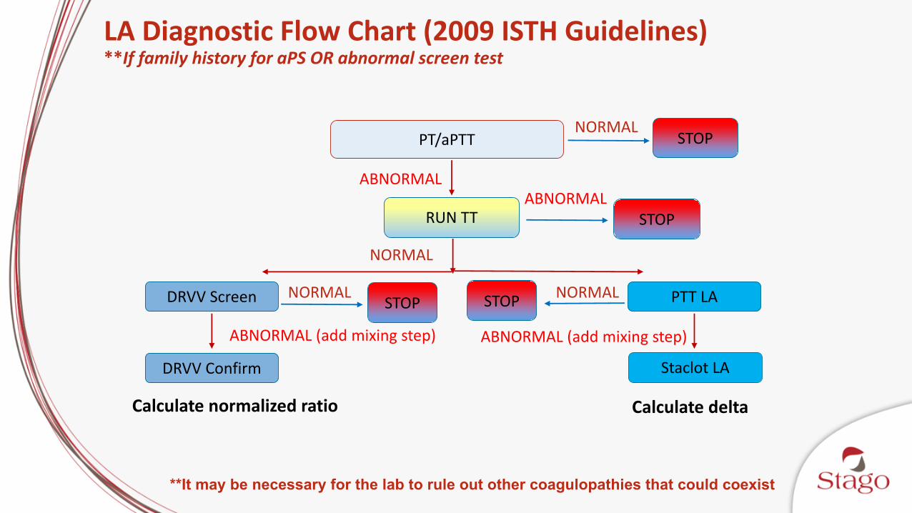
PT/aPTTNORMAL
ABNORMAL
STOP
DRVV Screen
RUN TT
NORMAL
STOP
ABNORMAL
NORMAL STOP PTT LANORMALSTOP
ABNORMAL (add mixing step) ABNORMAL (add mixing step)
Staclot LADRVV Confirm
Calculate normalized ratio Calculate delta
LA Diagnostic Flow Chart (2009 ISTH Guidelines)**If family history for aPS OR abnormal screen test
**It may be necessary for the lab to rule out other coagulopathies that could coexist

LA Testing Guidelines – ISTH SSC 2009Step 1 – Screen (low PL reagent)
LA sensitive aPTT reagentdRVVT screen reagentThrombin Time – eliminate prolonged clotting times due to anticoagulation
Step 2 – Mixing studiesRepeat screening tests using a patient 1:1 mix1:1 mix = 1 part patient + 1 part pool normal plasma
Step 3 – Confirm (high PL reagent)Hexagonal phase PL reagent (Staclot® LA)dRVVT confirm
Tests must be repeated > 12 weeks after initial testing; need to demonstrate persistence
Other Helpful Tests to Consider
Full laboratory aPS profile should include:LA testing (clotting based)Anti β2-GPI ELISA (aβ2-GPI) IgG, IgMAnticardiolipin (aCL) IgG, IgM
Presence of medium-high titers of aCL and aβ2-GPI of same isotype (i.e. IgG) is in agreement with positive LA and IDs patients with high thrombotic risk
Thrombin time can help to rule out heparin and other anticoagulant contamination

Triple Positive aPS Patients
Pengo V, Ruffatti A, Legnani C, Testa S, Fierro T, Marongui F, et al. Incidence of a first thromboembolic event in asymptomatic carriers of high-risk antiphospholipid antibody profile: a multicenter prospective study. Blood 2011; 118: 4714-4718.
Lupus anticoagulantsAnti-β2GPI Anticardiolipin

Persistence of Testing Results
Erkan D, Derksen WJM, Kaplan V, Sammaritano L, Pierangeli SS, Roubey R, Lockshin MD. Real world experience with antiphospholipid antibody tests: how stable are results over time? Ann Rheum Dis 2005; 64: 1321-1325.
Test % positive on repeat testing
Mean follow up time
LA 39 of 51 patients (77%) 2.4 years
Anticardiolipin antibody (moderate – high titer)
65 of 86 patients (75%) 3.5 years
Anti-β2GPI antibody (moderate –high titer)
11 of 15 patients (76%) 1.0 year
Repeat aPS results remain stable for at least ¾ of patients regardless of laboratory performing testVariation not correlated with aspirin, warfarin, or hydroxyquinoline useIndefinite anticoagulation is indicated for most aPS patients
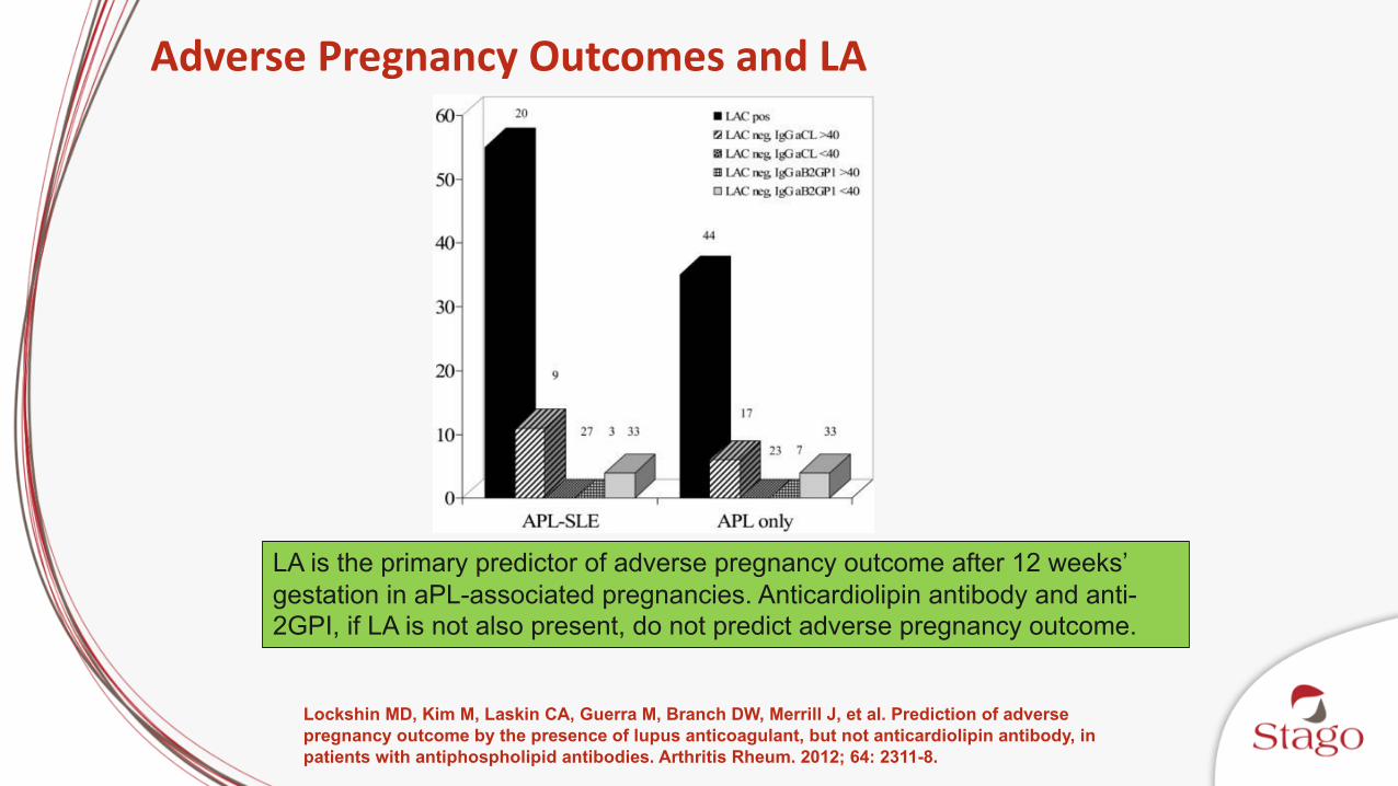
LA is the primary predictor of adverse pregnancy outcome after 12 weeks’ gestation in aPL-associated pregnancies. Anticardiolipin antibody and anti-2GPI, if LA is not also present, do not predict adverse pregnancy outcome.
Adverse Pregnancy Outcomes and LA
Lockshin MD, Kim M, Laskin CA, Guerra M, Branch DW, Merrill J, et al. Prediction of adverse pregnancy outcome by the presence of lupus anticoagulant, but not anticardiolipin antibody, in patients with antiphospholipid antibodies. Arthritis Rheum. 2012; 64: 2311-8.

Comparison of LA Guidelines
Preanalyticalconditions
Choice of tests&
Methodology
Results Interpretation
Moore GW. Recent Guidelines and Recommendations for Laboratory Detection
of Lupus Anticoagulants. Semin Thromb Hemost 2014; 40: 163-71.

Lupus Anticoagulant Laboratory Tests

LA Testing: Preanalytics
Double centrifugation (stay away from the platelet layer)
Platelet Poor Plasma (PPP); < 5000 platelets/µL
No hemolysis or traumatic draw, discard 1st tube
Pooled normal plasma (PNP) should be from multiple donors, well characterized for all coagulation factors and platelet poor
-70°C preferred for freezing; CLSI recommends no more than two weeks at -20°C (H21-A5, H57-A)
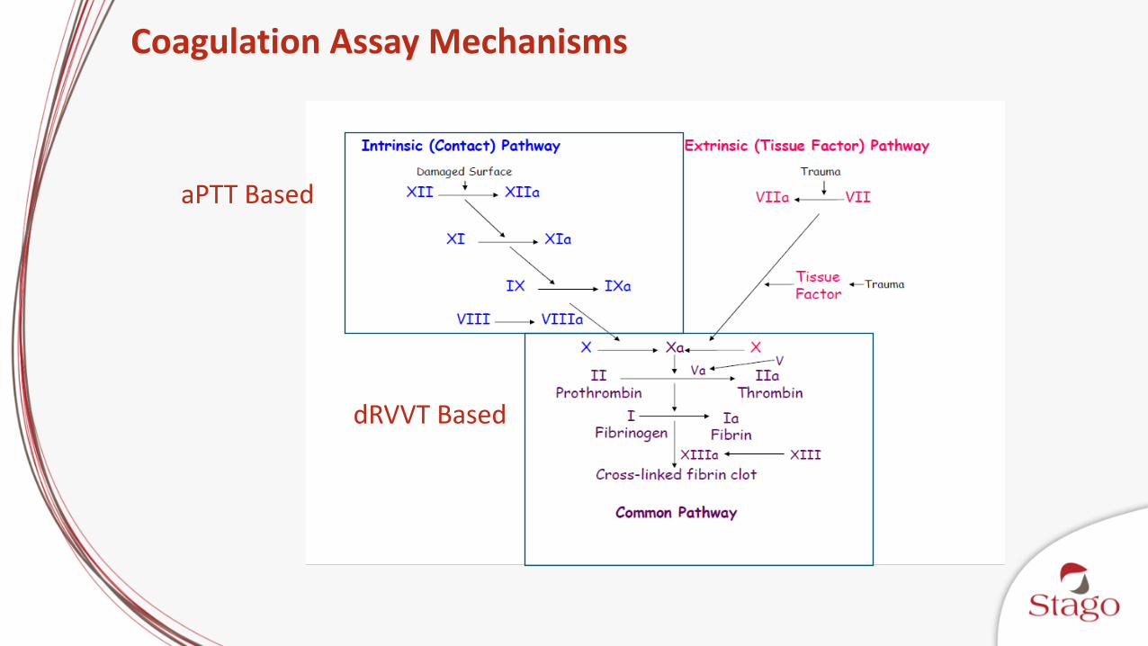
Coagulation Assay Mechanisms
aPTT Based
dRVVT Based

Activated Partial Thromboplastin Time (aPTT)Involves activation of FXII by PL and CaCl2Reagent composition
PLActivatorTests for factors VIII, IX, IX and XII
Clinical uses for aPTTFactor deficienciesHeparin therapyCirculating anticoagulantsDisseminated intravascular coagulation (DIC)
Potential interference from anticoagulant drugsWarfarinRivaroxaban, edoxaban (not sensitive to apixaban)Argatroban, dabigatran

Differential Diagnosis of Coagulation Inhibitors
Arnout J. Antiphospholipid syndrome: diagnostic aspects of lupus anticoagulants. Thromb Haemost. 2001; 86: 83-91.

dRVVT Screen & Confirm – Principle
FXa
FX
FXa
Clotting TimeNormal
Patient without LA – Low [PL] test
FXa
FX
FXa
FXa
FXa
Clotting TimeNormal
Patient without LA – High [PL] test
FXa
FXa
Ratio < 1.2
DRVV DRVV
Screening TestsDRVV Screen
Confirmation TestsDRVV Confirm

FXa
FX
FXa
Clotting TimeProlonged
Lupus
Patient with LA – Low [PL] test
FXa
FX
FXa
FXa
Clotting TimeShorter than low [PL] test
Lupus
Patient with LA – High [PL] test
Screening TestsDRVV Screen
Confirmation TestsDRVV Confirm
Ratio > 1.2
DRVV DRVV
dRVVT Screen & Confirm – Principle

Effect of LA (+) Sample on Screening aPTT
Screening aPTT
50% PL
= PL particles
= LA
In the cuvette, LA overwhelms the PL in the aPTT reagent Reduced concentration of PL results in prolonged clotting timesClotting times (example):
Normal plasma: 28.0 – 36.0 secondsLA positive: 55 seconds

Effect of LA (+) on aPTT and dRVVT Screen
aPTT/dRVVT screen
25%PL
= PL particles
= LA
In the cuvette, LA overwhelms the PL in the aPTT/dRVV reagent Reduced concentration of PL results in prolonged clotting timesClotting times (example):
PTT-LANormal: 34.3 – 40.4 secondsLA positive: 62.0 secs.
dRVVT screenNormal: 36.8 – 42.8LA positive: 58.0 secs
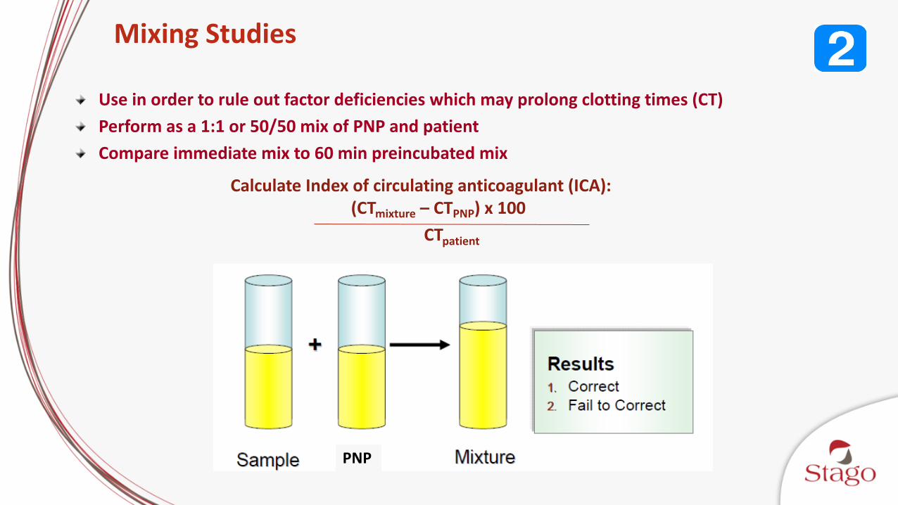
Mixing Studies
Use in order to rule out factor deficiencies which may prolong clotting times (CT)
Perform as a 1:1 or 50/50 mix of PNP and patient
Compare immediate mix to 60 min preincubated mix
Calculate Index of circulating anticoagulant (ICA):
(CTmixture – CTPNP) x 100
CTpatient
PNP

Mixing Study InterpretationAPTT normal range = 25-35 sec
Sample: Immediate 60 min Interpretation
Patient 1 46 48 Complete Correction
PNP 31 32 C/W Factor Deficiency
1:1 Mix 1 33 35
Patient 2 46 48 Incomplete Correction
PNP 31 32 C/W Inhibitor
1:1 Mix 2 36 38
Patient 3 46 60 Incomplete Correction
PNP 31 32 Prolongation at 60 min
1:1 Mix 3 35 48 C/W time dependent inhibitor (Factor VIII Inhibitor)

Borderline Screening aPTT Results (1 - 5 sec)
Use a LA sensitive aPTT (more sensitive than the routine aPTT) in a 4:1 patient:PNP mixing study
Arnout J, Meijer P, Vermylen J. Lupus anticoagulant testing in Europe: an analysis of results from the first European Concerted Action on Thrombophilia (ECAT) survey using plasmas spiked with monoclonal antibodies against human beta2-glycoprotein I. Thromb Haemost 1999; 81: 929-934.
Clo
ttin
g T
ime R
ati
o
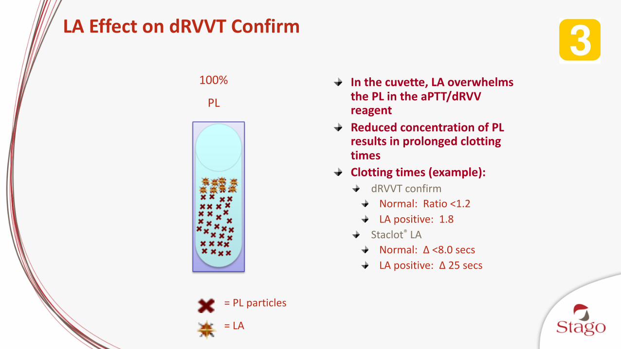
LA Effect on dRVVT Confirm
100%
PL
= PL particles
= LA
In the cuvette, LA overwhelms the PL in the aPTT/dRVV reagent Reduced concentration of PL results in prolonged clotting timesClotting times (example):
dRVVT confirmNormal: Ratio <1.2LA positive: 1.8
Staclot® LANormal: Δ <8.0 secsLA positive: Δ 25 secs

Staclot® LA – Integrated Test System
Steps and
PatientBufferPNP
aPTT-LSCaCl2
Low PL concentrationTube #1
Steps and
T1-T2 = ∆ Time
PatientHex Phos
PNPaPTT-LSCaCl2
High PL concentrationTube #2

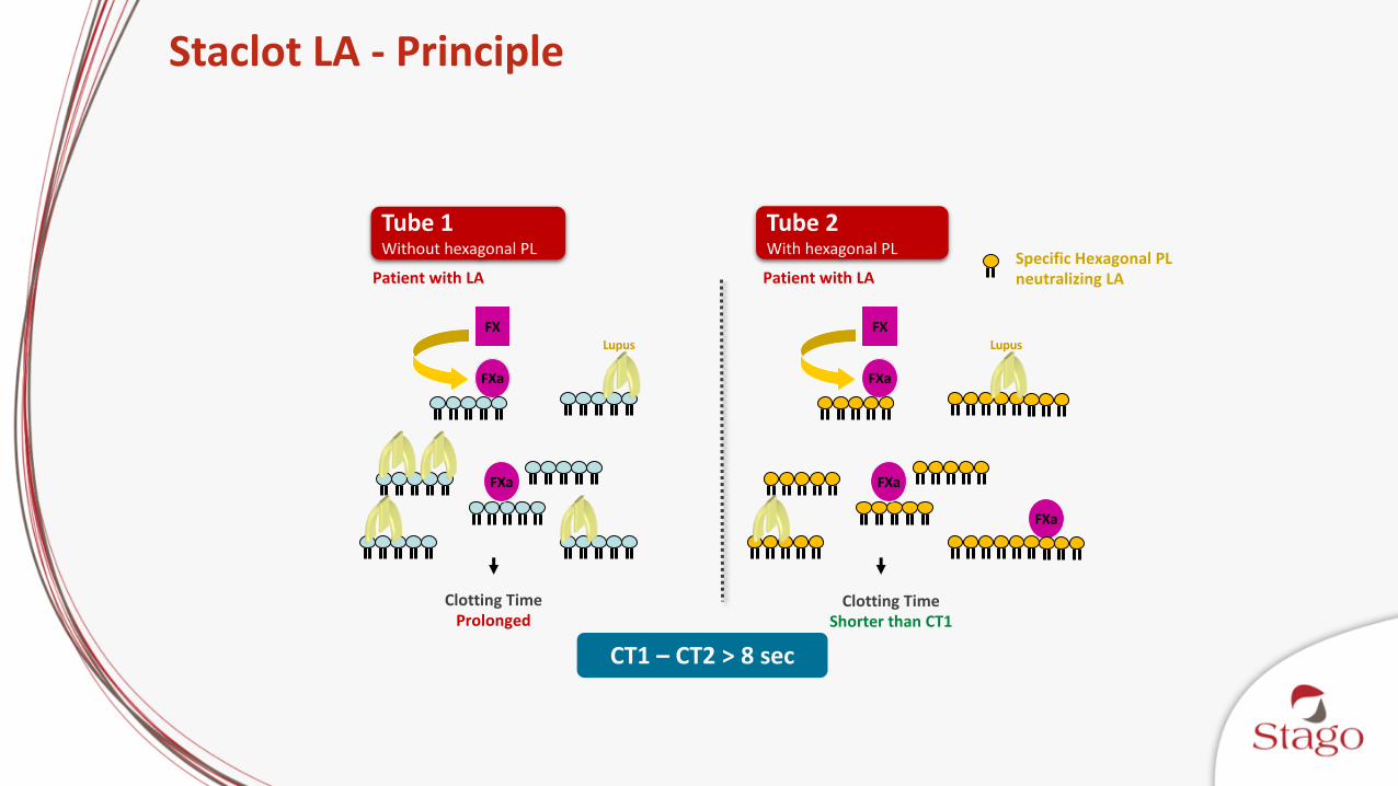
Staclot LA - Principle
Tube 1Without hexagonal PL
Tube 2With hexagonal PL
CT1 – CT2 < 8 sec
Specific Hexagonal PL neutralizing LA
FXa
FX
FXa
Clotting TimeNormal
Patient without Lupus
FXa
FX
FXa
Clotting TimeNormal
Patient without Lupus
Staclot LA - Principle
FXa
FX
FXa
Clotting TimeProlonged
Lupus
Patient with LA
FXa
FX
FXa
FXa
Clotting TimeShorter than CT1
Lupus
Patient with LA
Tube 1Without hexagonal PL
Tube 2With hexagonal PL
CT1 – CT2 > 8 sec
Specific Hexagonal PL neutralizing LA

Thrombin Time Principle and Applications
Principle: measure clotting time in presence of known thrombin concentration
Recommended by 2009 ISTH SSC guidelines to exclude contamination by anticoagulant drugs
Prolongation of the thrombin time indicates:Presence of UFH, LMWH, argatroban, dabigatranFibrinogen abnormalities
Qualitative (dysfibrinogenaemia)Quantitative could be from congenital or acquired hypofibrinogenemias
The presence of fibrin degradation products

TT and LA Testing Panels
Sangle NA, Rodgers GM, Smock KJ. Prevalence of heparin in samples submitted for lupus anticoagulant testing. Lab Hematol. 2011; 17: 6-11.
13% of cases diagnosed as LA positive had less than significant levels of heparin, but 4% had significant levels of heparin (or another anticoagulant).

Article Flow Chart
TT and Reptilase should be used together to rule out heparin or other anticoagulant contamination before specialized tests such as LA testing are performed.
Sangle NA, Rodgers GM, Smock KJ. Prevalence of heparin in samples submitted for lupus anticoagulant testing. Lab Hematol. 2011; 17: 6-11.

Influence of Anticoagulant Drugs on LA Testing
Martinuzzo ME, Barrera LH, D 'adamo MA, Otaso JC, Gimenez MI, Oyhamburu J. Frequent false-positive results of lupus anticoagulant tests in plasmas of patients receiving the new oral anticoagulants and enoxaparin. Int J Lab Hematol 2014; 36: 144-50.
Prevalence of abnormal results (%)
Dabigatran (110 mg bid)
Rivaroxaban (15 mg bid)
Rivaroxaban (10 mg/day)
Enoxaparin (40 mg)
PT activity <70% 95 100 77.2 53
TT ratio >1.2 100 0 0 28
aPTT > 40 sec 100 75 13.6 66.6
aPTT > 40 sec, ICA > 10%
100 100 0 66.6
dRVVT screen >40 sec
100 100 81.8 60
dRVVT >40 sec, ICA >13%
100 100 82.3 50
dRVVT screen/confirm NR >1.17
81.8 100 76.5 70

Conclusions
Nomenclature for LA can be confusing
Tests for aPSClotting studies to detect LAConfirm tests should be based on same format as screenDirect detection of the antibodies using ELISA
Diagnosis of aPS requires the presence of at least one of the clinical entities: thrombosis, pregnancy morbidity; and at least one positive test
The positive findings must be persistent when retesting at > 12 weeks

Case Studies

Case Study # 1
11 year old female presents with epistaxis and fever, along with malaise and anorexia. She had an unremarkable physical history with a tonsillectomy at age 9 without excessive bleeding, no relatives with bleeding histories, but taking aspirin for fever.
Physical exam showed she was afebrile and well nourished but cervical lympadenopathy present.
Adams AL, Audeh YM, de Luna R, Baker MS, Marques MB. Laboratory Evaluation of Coagulation Inhibitors. Lab Med 2003; 34: 584-588.

Case Study # 1 Laboratory Results
TEST RESULT REFERENCE RANGE
PT 13.8 sec 11.3 – 14.6 sec
aPTT 55.0 sec 25 – 34 sec
aPTT 1:1 mix 43.0 sec Correction to 25 – 34 sec
PTT-LA 108.0 sec 36 – 50.1 sec
dRVVT Screen 43.1 sec 29.6 – 42.9 sec
Prothrombin 82% 50 – 150%
Factor VIII 135% 60 – 160%
Staclot® LA 20.4 sec Negative < 8 sec
Adams AL, Audeh YM, de Luna R, Baker MS, Marques MB. Laboratory Evaluation of Coagulation Inhibitors. Lab Med 2003; 34: 584-588.

Case Study # 1 – Diagnosis
Probability of hemophilia A (FVIII deficiency) and hemophilia B (FIX deficiency) are low, along with factor inhibitors.
LA is most likely present; often are transient in children and associated with viral infections
LA not expected to cause thrombosis in this case
Cervical lymphadenopathy presence suggests infectious mononucleosis
Adams AL, Audeh YM, de Luna R, Baker MS, Marques MB. Laboratory Evaluation of Coagulation Inhibitors. Lab Med 2003; 34: 584-588.

Case Study # 2
43 year old male presents with an ischemic stroke.
History of hypertension, but no surgical history available.
Unremarkable family history, the only medication being taken is a multivitamin.
Adams AL, Audeh YM, de Luna R, Baker MS, Marques MB. Laboratory Evaluation of Coagulation Inhibitors. Lab Med 2003; 34: 584-588.
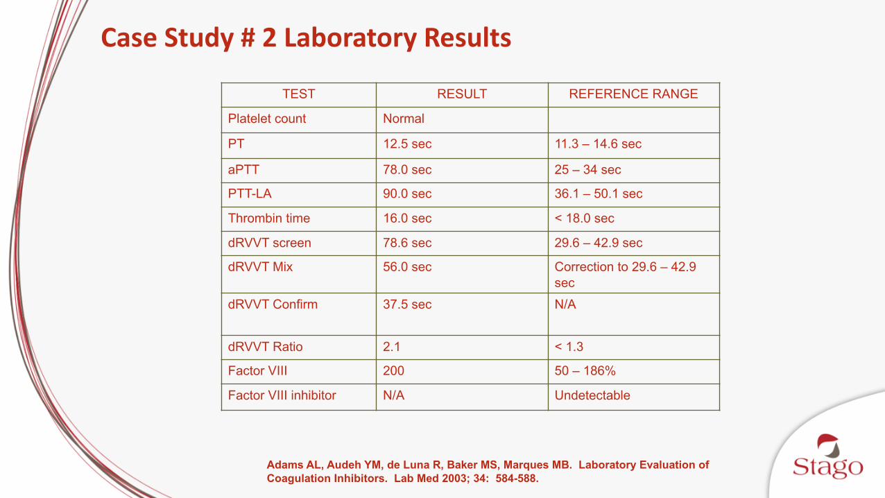
Case Study # 2 Laboratory Results
TEST RESULT REFERENCE RANGE
Platelet count Normal
PT 12.5 sec 11.3 – 14.6 sec
aPTT 78.0 sec 25 – 34 sec
PTT-LA 90.0 sec 36.1 – 50.1 sec
Thrombin time 16.0 sec < 18.0 sec
dRVVT screen 78.6 sec 29.6 – 42.9 sec
dRVVT Mix 56.0 sec Correction to 29.6 – 42.9 sec
dRVVT Confirm 37.5 sec N/A
dRVVT Ratio 2.1 < 1.3
Factor VIII 200 50 – 186%
Factor VIII inhibitor N/A Undetectable
Adams AL, Audeh YM, de Luna R, Baker MS, Marques MB. Laboratory Evaluation of Coagulation Inhibitors. Lab Med 2003; 34: 584-588.
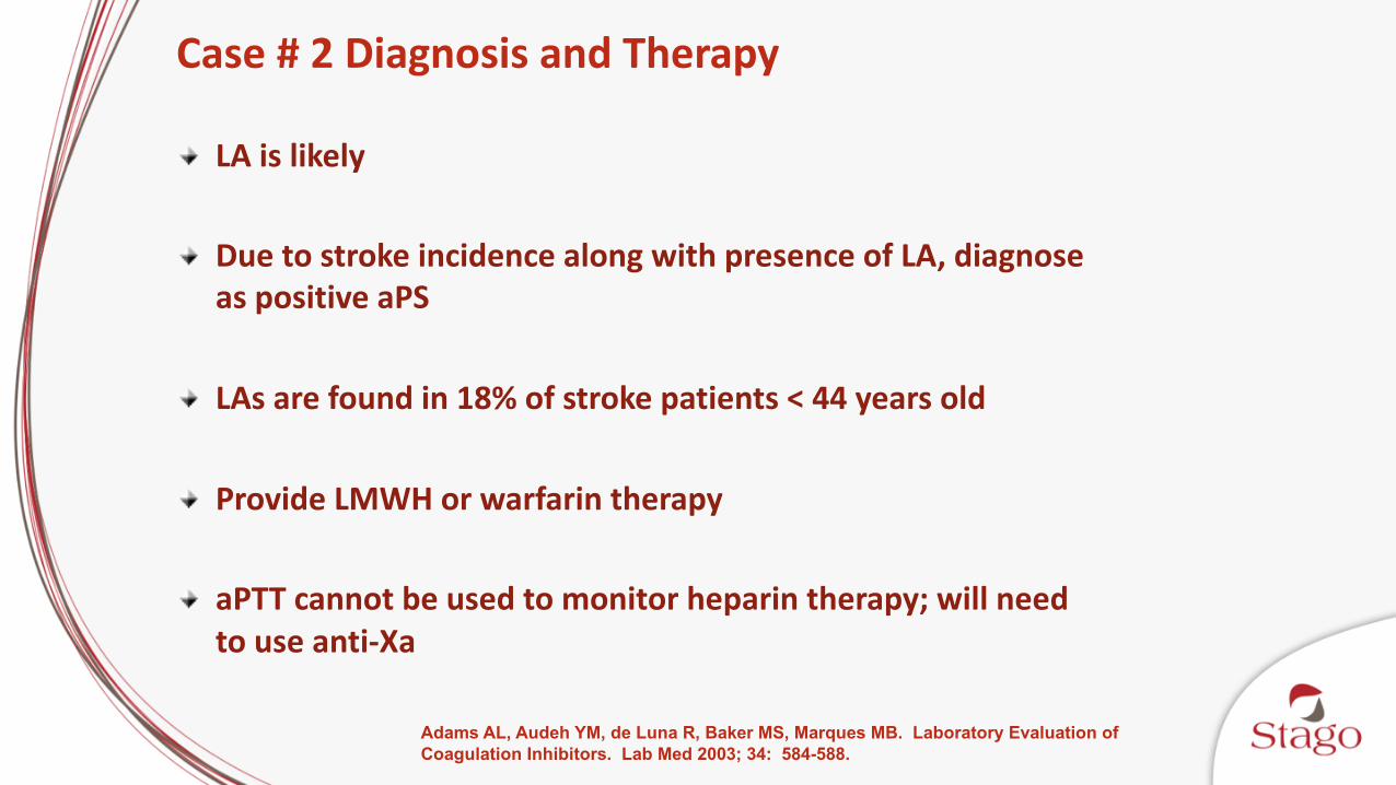
LA is likely
Due to stroke incidence along with presence of LA, diagnose
as positive aPS
LAs are found in 18% of stroke patients < 44 years old
Provide LMWH or warfarin therapy
aPTT cannot be used to monitor heparin therapy; will need
to use anti-Xa
Case # 2 Diagnosis and Therapy
Adams AL, Audeh YM, de Luna R, Baker MS, Marques MB. Laboratory Evaluation of Coagulation Inhibitors. Lab Med 2003; 34: 584-588.
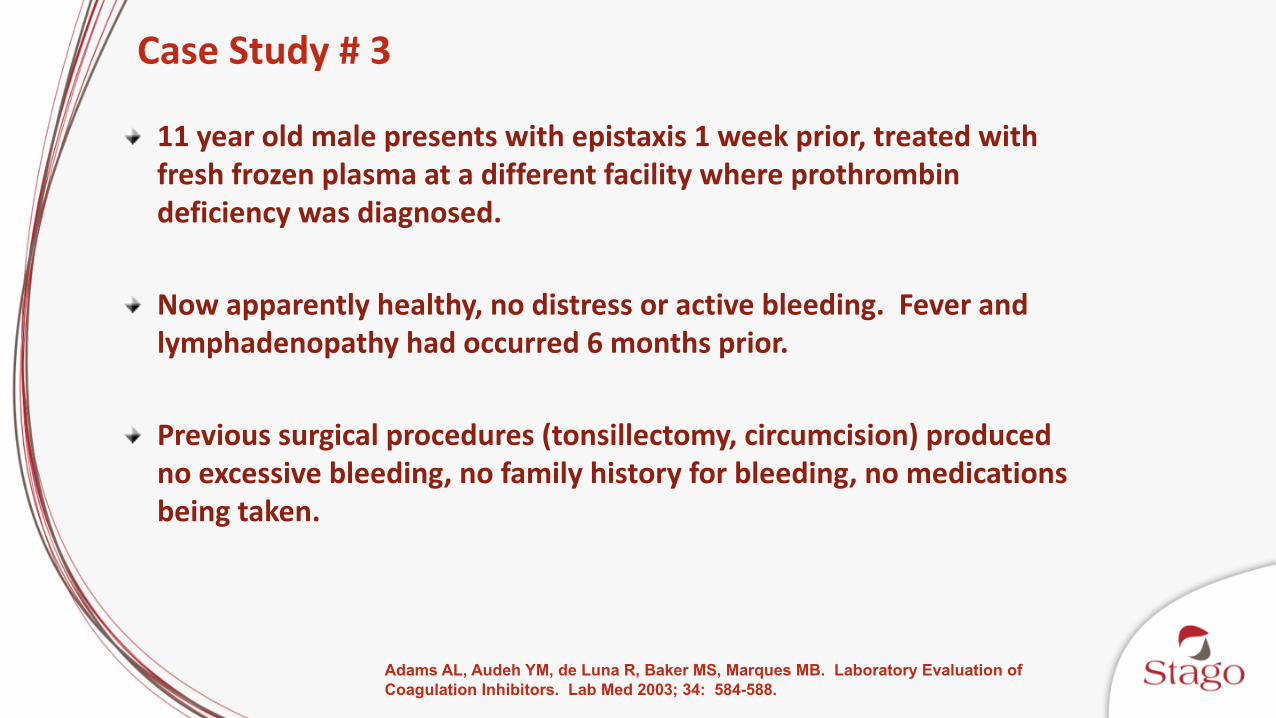
Case Study # 3
11 year old male presents with epistaxis 1 week prior, treated with fresh frozen plasma at a different facility where prothrombin deficiency was diagnosed.
Now apparently healthy, no distress or active bleeding. Fever and lymphadenopathy had occurred 6 months prior.
Previous surgical procedures (tonsillectomy, circumcision) produced no excessive bleeding, no family history for bleeding, no medications being taken.
Adams AL, Audeh YM, de Luna R, Baker MS, Marques MB. Laboratory Evaluation of Coagulation Inhibitors. Lab Med 2003; 34: 584-588.

Case Study # 3 Laboratory Results
TEST RESULT REFERENCE RANGE
Platelet count 287 130 – 400 x 103/μL
PT 25.0 sec 10.7 – 13.0 sec
INR 2.3 1.0 (before therapy);
2 – 3 (therapeutic range)
Prothrombin 6 50 – 150%
aPTT 62.0 sec 25.0 – 34.0 sec
PTT-LA 113.0 sec 36.1 – 50.1 sec
Staclot® LA 61.5 sec Negative < 8 sec
dRVVT screen 99.2 sec 29.6 – 42.9 sec
dRVVT Confirm 70.2 sec N/A
dRVVT Ratio 1.4 < 1.3
Factor VIII inhibitor N/A Undetectable
Adams AL, Audeh YM, de Luna R, Baker MS, Marques MB. Laboratory Evaluation of Coagulation Inhibitors. Lab Med 2003; 34: 584-588.
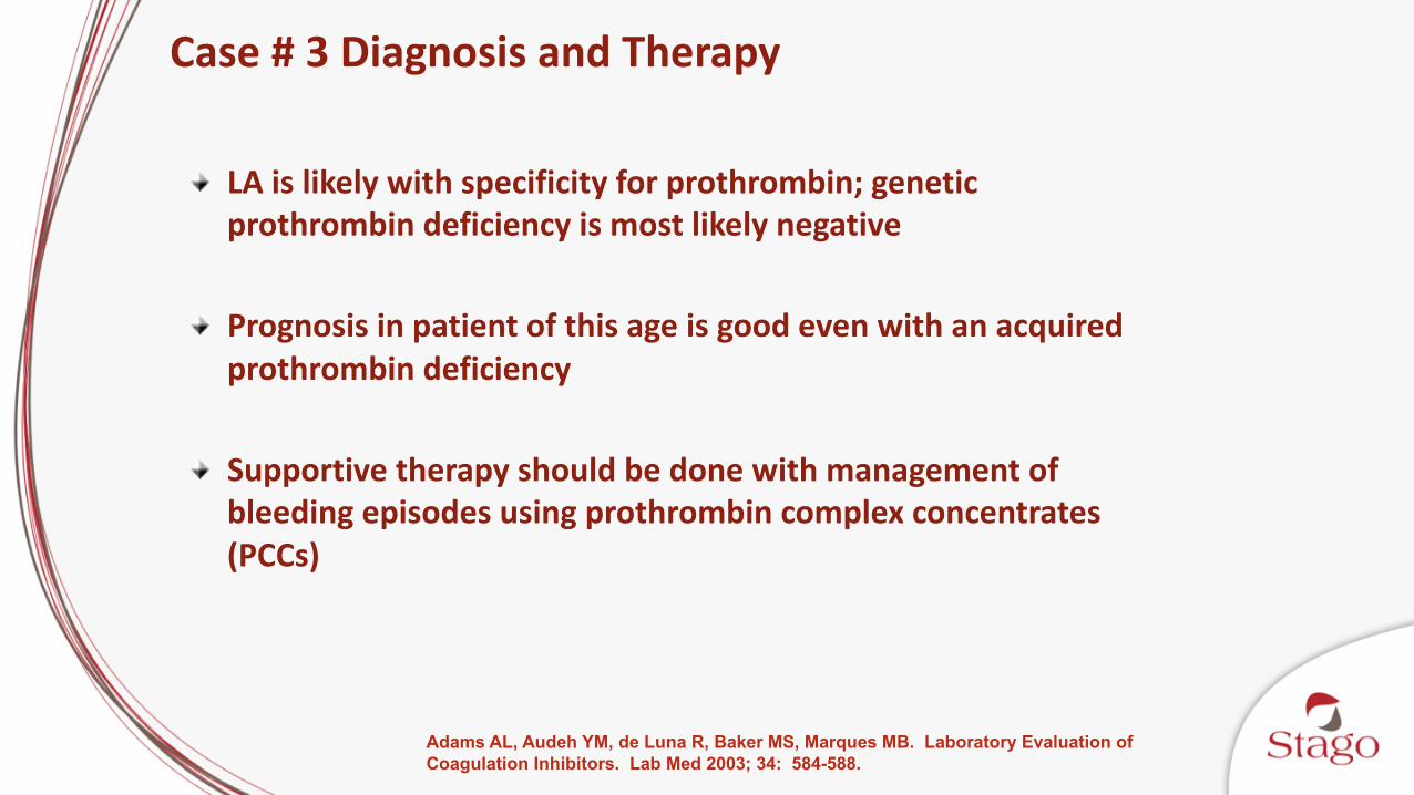
LA is likely with specificity for prothrombin; genetic prothrombin deficiency is most likely negative
Prognosis in patient of this age is good even with an acquired prothrombin deficiency
Supportive therapy should be done with management of bleeding episodes using prothrombin complex concentrates (PCCs)
Case # 3 Diagnosis and Therapy
Adams AL, Audeh YM, de Luna R, Baker MS, Marques MB. Laboratory Evaluation of Coagulation Inhibitors. Lab Med 2003; 34: 584-588.

Case Study # 4
69 year old female presents with easy bruising within past few weeks. Large hematoma at site of intramuscular injection; multiple ecchymoses in left arm with diffuse swelling.
History of hypertension, diabetes, arthritis, asthma, previous surgeries (cholecystectomy, hysterectomy) produced no excessive bleeding, no relatives with bleeding disorders.
Medications include Acetaminophen, atenolol, azithromycin, estradiol, glyburide, pravastatin sodium, prednisone, ramipril, and theophylline.
Adams AL, Audeh YM, de Luna R, Baker MS, Marques MB. Laboratory Evaluation of Coagulation Inhibitors. Lab Med 2003; 34: 584-588.
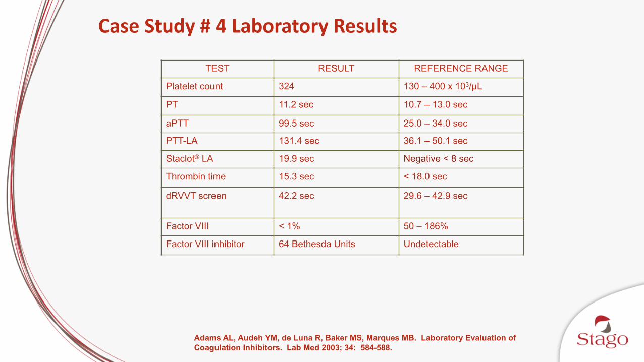
Case Study # 4 Laboratory Results
TEST RESULT REFERENCE RANGE
Platelet count 324 130 – 400 x 103/μL
PT 11.2 sec 10.7 – 13.0 sec
aPTT 99.5 sec 25.0 – 34.0 sec
PTT-LA 131.4 sec 36.1 – 50.1 sec
Staclot® LA 19.9 sec Negative < 8 sec
Thrombin time 15.3 sec < 18.0 sec
dRVVT screen 42.2 sec 29.6 – 42.9 sec
Factor VIII < 1% 50 – 186%
Factor VIII inhibitor 64 Bethesda Units Undetectable
Adams AL, Audeh YM, de Luna R, Baker MS, Marques MB. Laboratory Evaluation of Coagulation Inhibitors. Lab Med 2003; 34: 584-588.

Bleeding episode currently but not historically indicates presence of acquired hemophilia
Most patients with acquired hemophilia are > 50 years old and half have an associated disorder such as arthritis, SLE, malignancy, or a drug reaction
With such high inhibitor levels (> 5 BU), activated prothrombin complex concentrates or recombinant FVIIa are indicated to control bleeding
Laboratory should continue monitoring FVIII and FVIII inhibitors going forward
Case # 4 Diagnosis and Therapy
Adams AL, Audeh YM, de Luna R, Baker MS, Marques MB. Laboratory Evaluation of Coagulation Inhibitors. Lab Med 2003; 34: 584-588.

Stago 24/7 Educational Webinar Sites
www.stago-edvantage.comUS based KOLs1 hour; PACE accreditedAccessible from mobile devicesVirtual exhibit hall
www.stagowebinars.comMostly European KOLs30 – 45 min including 15 min discussionAccessible from mobile devices

Stago Educational Apps
HaemoscoreClinical scoring algorithmsApple and AndroidTablet or phone
iHemostasisCoagulation diagramsCase studiesApple & AndroidTablet only