low-grade osteosarcoma arising from cemento-ossifying ... · 48 low-grade osteosarcoma arising from...
TRANSCRIPT
48
Low-grade osteosarcoma arising from cemento-ossifying fibroma: a case report
Yong Bin Lee1, Nam-Kyoo Kim3, Jae-Young Kim4, Hyung Jun Kim1,2
1Department of Oral and Maxillofacial Surgery, 2Oral Cancer Research Institute, Yonsei University College of Dentistry, Seoul, 3Department of Dentistry, Catholic Kwandong University International St. Mary’s Hospital, Incheon,
4Department of Oral and Maxillofacial Surgery, Gangnam Severance Hospital, Yonsei University College of Dentistry, Seoul, Korea
Abstract (J Korean Assoc Oral Maxillofac Surg 2015;41:48-51)
Cemento-ossifying fibromas are benign tumors, and, although cases of an aggressive type have been reported, no cases of cemento-ossifying fibroma transforming into osteosarcoma have been documented previously. Low-grade osteosarcoma is a rare type of primary bone tumor, representing 1%-2% of all osteosarcomas. A 45-year-old female patient was diagnosed with cemento-ossifying fibroma, treated with mass excision several times over a period of two years and eight months, and followed up. After biopsy gathered because of signs of recurrence, she was diagnosed with low-grade osteo-sarcoma. The patient underwent wide excision, segmental mandibulectomy, and reconstruction with fibula free flap. The aim of this report is to raise awareness of the possibility that cemento-ossifying fibroma can transform into osteosarcoma and of the consequent necessity for careful diagnosis and treatment planning.
Key words: Cemento ossifying fibroma, Low-grade osteosarcoma[paper submitted 2014. 7. 21 / revised 2014. 9. 18 / accepted 2014. 9. 29]
differentiated intramedullary osteosarcomas and parosteal
osteosarcomas2. These low-grade osteosarcomas may be mis-
taken for fibro-osseous neoplasms3.
We report a case of low-grade osteosarcoma in the mandi-
ble that was transformed from a recurrent cemento-ossifying
fibroma. This is the first reported case of the transformation
of cemento-ossifying fibroma into malignant low-grade cen-
tral osteosarcoma.
II. Case Report
A 45-year-old female patient was referred for a radiolucent
lesion of the left posterior mandible, with complaints of a
three-month history gingival swelling and tooth pain in the
left mandibular second premolar. On intraoral examination,
the overlying mucosa was intact with no paresthesia on the
left lower lip. Bony swelling was suspected in the left pos-
terior vestibule. The second premolar was the vital tooth. A
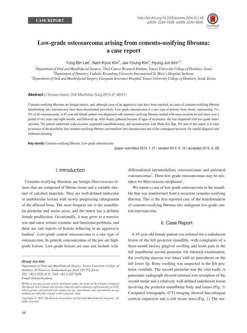
panoramic radiograph showed external root resorption of the
second molar and a relatively well-defined radiolucent lesion
involving the posterior mandibular body and ramus.(Fig. 1)
Computed tomography (CT) imaging showed buccolingual
cortical expansion and a soft tissue mass.(Fig. 1) The sur-
I. Introduction
Cemento-ossifying fibromas are benign fibro-osseous tu-
mors that are composed of fibrous tissue and a variable mix-
ture of calcified materials. They are well-defined unilocular
or multilocular lesions with slowly progressing enlargement
of the affected bone. The most frequent site is the mandibu-
lar premolar and molar areas, and the tumor has a definite
female predilection. Occasionally, it may grow to a massive
size and cause serious cosmetic and functional problems, and
there are rare reports of lesions behaving in an aggressive
fashion1. Low-grade central osteosarcoma is a rare type of
osteosarcoma. In general, osteosarcomas of the jaw are high-
grade lesions. Low-grade lesions are rare and include well-
CASE REPORT
Hyung Jun KimDepartment of Oral and Maxillofacial Surgery, Yonsei University College of Dentistry, 50 Yonsei-ro, Seodaemoon-gu, Seoul 120-752, KoreaTEL: +82-2-2228-3132 FAX: +82-2-2227-8256E-mail: [email protected]
This is an open-access article distributed under the terms of the Creative Commons Attribution Non-Commercial License (http://creativecommons.org/licenses/by-nc/3.0/), which permits unrestricted non-commercial use, distribution, and reproduction in any medium, provided the original work is properly cited.
CC
Copyright Ⓒ 2015 The Korean Association of Oral and Maxillofacial Surgeons. All rights reserved.
http://dx.doi.org/10.5125/jkaoms.2015.41.1.48pISSN 2234-7550·eISSN 2234-5930
Low-grade osteosarcoma arising from cemento-ossifying fibroma
49
bone, and cementum-like material.(Fig. 2) Conservative mass
excision under general anesthesia was performed. The surgi-
cal specimen was sent to a pathologist, and the pathologist
recommended close follow-up because of the high cellularity
of the specimen.
Five months after the first operation, the patient com-
rounding soft tissue did not appear to be involved. Under lo-
cal anesthesia, extraction of the second molar was performed,
and a biopsy was taken below the extraction socket for his-
topathological examination. A histopathological diagnosis
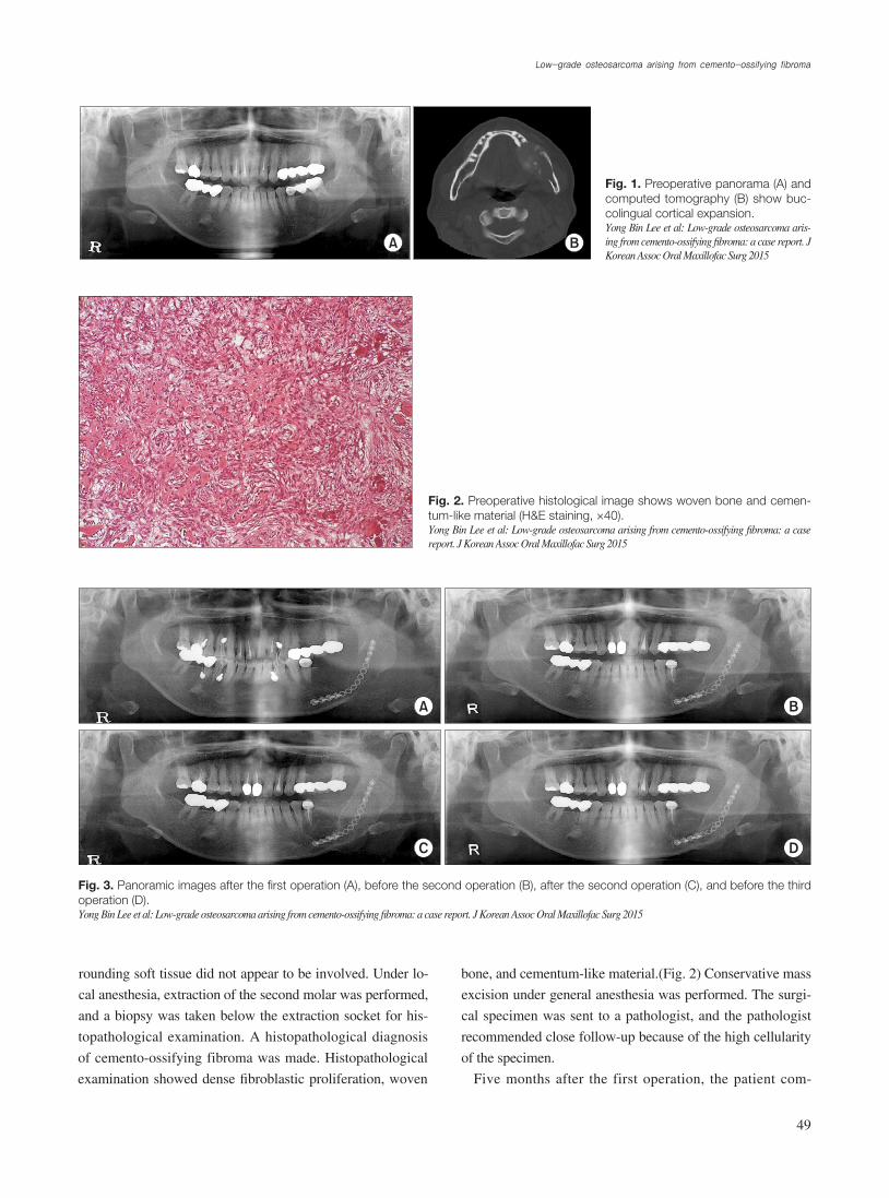
of cemento-ossifying fibroma was made. Histopathological
examination showed dense fibroblastic proliferation, woven
Fig. 1. Preoperative panorama (A) and computed tomography (B) show buc-colingual cortical expansion.Yong Bin Lee et al: Low-grade osteosarcoma aris-ing from cemento-ossifying fibroma: a case report. J Korean Assoc Oral Maxillofac Surg 2015
A B
Fig. 2. Preoperative histological image shows woven bone and cemen-tum-like material (H&E staining, ×40).Yong Bin Lee et al: Low-grade osteosarcoma arising from cemento-ossifying fibroma: a case report. J Korean Assoc Oral Maxillofac Surg 2015
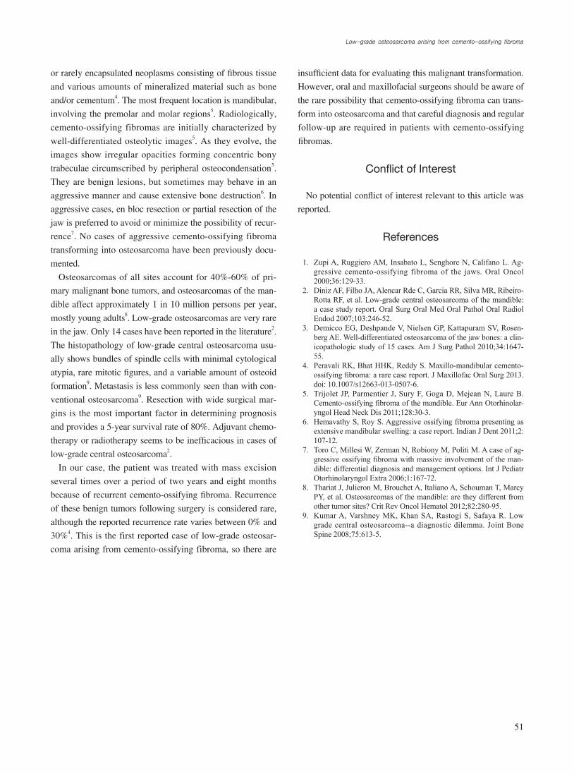
Fig. 3. Panoramic images after the first operation (A), before the second operation (B), after the second operation (C), and before the third operation (D).Yong Bin Lee et al: Low-grade osteosarcoma arising from cemento-ossifying fibroma: a case report. J Korean Assoc Oral Maxillofac Surg 2015
A B
C D

J Korean Assoc Oral Maxillofac Surg 2015;41:48-51
50
first premolar and mass excision were performed under gen-
eral anesthesia.(Fig. 3) The pathological diagnosis was the
same as previously, but the surgical specimen showed high
cellularity and an infiltrative growth pattern into normal bone
tissue without mitotic figures or cellular atypia.
Three months after the third operation, tumor recurrence
was suspected at the parasymphysis area on follow-up radio-
graph (Fig. 4), and a biopsy was taken from the buccal side of
the lesion. The specimen showed minimal cellular atypia and
irregular bony trabeculae with few mitotic figures (Fig. 5), so
a diagnosis of low-grade osteosarcoma was made. No region-
al or distant metastasis was shown on magnetic resonance
imaging or positron emission tomography-CT imaging. The
patient underwent wide excision, segmental mandibulectomy,
and reconstruction with fibula free flap.(Fig. 6)
III. Discussion
Cemento-ossifying fibromas are benign fibro-osseous le-
sions of the jaw which have been described as demarcated
plained of tooth pain in the left mandibular second premolar
and underwent root canal treatment. Periodic follow-up was
performed, and there were no signs of recurrence on either
clinical or radiographic examination for 16 months. After
16 months, however, follow-up panoramic radiograph and
CT displayed signs of recurrence, including a mixed high-
attenuation intrabony lesion and a partially multilocular le-
sion. Mass excision was performed under local anesthesia,
the surgical specimen was submitted for histological exami-
nation; the pathological diagnosis was cemento-ossifying
fibroma. One year after the second operation, the patient
complained of discomfort in the left mandibular canine area,
and CT revealed expansion of the lesion and signs of recur-
rence. Intentional root canal treatment of the left mandibular
Fig. 4. A, B. Before operation, panora-ma and computed tomography (CT). C, D. After the final operation, panorama and CT.Yong Bin Lee et al: Low-grade osteosarcoma aris-ing from cemento-ossifying fibroma: a case report. J Korean Assoc Oral Maxillofac Surg 2015
A B
C D
Fig. 5. Final histological image shows bundles of spindle cells with nuclear hyperchromasia and a variable amount of osteoid and ir-regular bone (H&E staining, ×40).Yong Bin Lee et al: Low-grade osteosarcoma arising from cemento-ossifying fibroma: a case report. J Korean Assoc Oral Maxillofac Surg 2015
Fig. 6. Follow-up panorama (postoperative day 7 months).Yong Bin Lee et al: Low-grade osteosarcoma arising from cemento-ossifying fibroma: a case report. J Korean Assoc Oral Maxillofac Surg 2015
Low-grade osteosarcoma arising from cemento-ossifying fibroma
51
insufficient data for evaluating this malignant transformation.
However, oral and maxillofacial surgeons should be aware of
the rare possibility that cemento-ossifying fibroma can trans-
form into osteosarcoma and that careful diagnosis and regular
follow-up are required in patients with cemento-ossifying
fibromas.
Conflict of Interest
No potential conflict of interest relevant to this article was
reported.
References
1. Zupi A, Ruggiero AM, Insabato L, Senghore N, Califano L. Ag-gressive cemento-ossifying fibroma of the jaws. Oral Oncol 2000;36:129-33.
2. Diniz AF, Filho JA, Alencar Rde C, Garcia RR, Silva MR, Ribeiro-Rotta RF, et al. Low-grade central osteosarcoma of the mandible: a case study report. Oral Surg Oral Med Oral Pathol Oral Radiol Endod 2007;103:246-52.
3. Demicco EG, Deshpande V, Nielsen GP, Kattapuram SV, Rosen-berg AE. Well-differentiated osteosarcoma of the jaw bones: a clin-icopathologic study of 15 cases. Am J Surg Pathol 2010;34:1647-55.
4. Peravali RK, Bhat HHK, Reddy S. Maxillo-mandibular cemento-ossifying fibroma: a rare case report. J Maxillofac Oral Surg 2013. doi: 10.1007/s12663-013-0507-6.
5. Trijolet JP, Parmentier J, Sury F, Goga D, Mejean N, Laure B. Cemento-ossifying fibroma of the mandible. Eur Ann Otorhinolar-yngol Head Neck Dis 2011;128:30-3.
6. Hemavathy S, Roy S. Aggressive ossifying fibroma presenting as extensive mandibular swelling: a case report. Indian J Dent 2011;2: 107-12.
7. Toro C, Millesi W, Zerman N, Robiony M, Politi M. A case of ag-gressive ossifying fibroma with massive involvement of the man-dible: differential diagnosis and management options. Int J Pediatr Otorhinolaryngol Extra 2006;1:167-72.
8. Thariat J, Julieron M, Brouchet A, Italiano A, Schouman T, Marcy PY, et al. Osteosarcomas of the mandible: are they different from other tumor sites? Crit Rev Oncol Hematol 2012;82:280-95.
9. Kumar A, Varshney MK, Khan SA, Rastogi S, Safaya R. Low grade central osteosarcoma--a diagnostic dilemma. Joint Bone Spine 2008;75:613-5.
or rarely encapsulated neoplasms consisting of fibrous tissue
and various amounts of mineralized material such as bone
and/or cementum4. The most frequent location is mandibular,
involving the premolar and molar regions5. Radiologically,
cemento-ossifying fibromas are initially characterized by
well-differentiated osteolytic images5. As they evolve, the
images show irregular opacities forming concentric bony
trabeculae circumscribed by peripheral osteocondensation5.
They are benign lesions, but sometimes may behave in an
aggressive manner and cause extensive bone destruction6. In
aggressive cases, en bloc resection or partial resection of the
jaw is preferred to avoid or minimize the possibility of recur-
rence7. No cases of aggressive cemento-ossifying fibroma
transforming into osteosarcoma have been previously docu-
mented.
Osteosarcomas of all sites account for 40%-60% of pri-
mary malignant bone tumors, and osteosarcomas of the man-
dible affect approximately 1 in 10 million persons per year,
mostly young adults8. Low-grade osteosarcomas are very rare
in the jaw. Only 14 cases have been reported in the literature2.
The histopathology of low-grade central osteosarcoma usu-
ally shows bundles of spindle cells with minimal cytological
atypia, rare mitotic figures, and a variable amount of osteoid
formation9. Metastasis is less commonly seen than with con-
ventional osteosarcoma9. Resection with wide surgical mar-
gins is the most important factor in determining prognosis
and provides a 5-year survival rate of 80%. Adjuvant chemo-
therapy or radiotherapy seems to be inefficacious in cases of
low-grade central osteosarcoma2.
In our case, the patient was treated with mass excision
several times over a period of two years and eight months
because of recurrent cemento-ossifying fibroma. Recurrence
of these benign tumors following surgery is considered rare,
although the reported recurrence rate varies between 0% and
30%4. This is the first reported case of low-grade osteosar-
coma arising from cemento-ossifying fibroma, so there are