localization of the 92 kd gelatinase mrna in squamous cell and
TRANSCRIPT

American Journal of Pathology, Vol. 144, No. 3, March 1994Copynght C) American Societyfor Investigative Pathology
Localization of the 92 kd Gelatinase mRNA inSquamous Cell and Adenocarcinomas of theLung Using in Situ Hybridization
Rafaela Canete-Soler, Leslie Litzky,Irina Lubensky, and Ruth J. MuschelFrom the Department ofPathology and LaboratoryMedicine, University of Pennsylvania,Philadelphia, Pennsylvania
We have used in situ hybridizationforRNA to lo-calize ceUs containing mRNAfor the 92 kd gelati-nase in carcinomas ofthe lung. We used archivalmaterial to analyze sections from 12 cases ofsquamous ceUl carcinomas of the lung includingsix stage Iand three stage HI andfrom three casesofadenocarcinoma ofthe lung. Presence ofmRNAin the tissue was verified by in situ hybridizationforgamma actin. The 92 kdgelatinase mRNA wasfound in aU 12 squamous ceU carcinomas tumorsandwas highly expressed in the tumor ceUs them-selves. In addition, it wasfound in host stromalceUs surrounding the tumor, but not in normallungfibroblasts. In contrast itwas notfound in theadenocarcinomas ofthe lung or in the stroma sur-rounding these tumors. The mRNAfor the 92 kdgelatinase was present in normalpulmonary tis-sue, bronchial epithelium, basal ceU hyperplasiaof bronchial epithelium, alveolar macrophages,andfocally in bronchialmucousglands. It was notpresent in normal alveoli, vascular ceUs, carti-lage, or most lymphocytes. We corroborated thepresence ofthe mRNAfor the 92 kdgelatinase byribonuclease protection assay. The levels ofmRNAfor the 92 kd gelatinase in two specimensof squamous ceU carcinoma were 6- to 10-foldgreater than in the nonneoplastic tissue and twoadenocarcinoma specimens. (AmJ Pathol 1994,144:518-527)
The metalloproteinase family has been implicated inthe proteolysis of extracellular matrix that is requiredfor tumor cell invasion and metastasis.1-3 This familyof enzymes includes three members that have thecapability of cleaving type IV collagen, stromelysin,
the 72 kd gelatinase, and the 92 kd gelatinase. Ex-pression of each of these has been found to correlatewith malignant potential in different systems.4 10
The 92 kd gelatinase is capable of degrading col-lagens type IV, V, VII, and X as well as gelatin, elastin,and casein.11-15 The cDNA for this enzyme wascloned by Wilhelm et al.11 The sequence revealedseveral common features of the matrix metalloprotein-ase family including the cleavage site for activationand the catalytic domain. There is also a regionunique to the 92 kd gelatinase in the COOH-terminalend, which is homologous to alpha 11 type V collagen.This enzyme is secreted from various normalcell types including monocytes and activatedmacrophages, early trimester trophoblast, fibro-blasts treated with phorbol esters and mammary epi-thelial cells.' 1,12,15-19 Various tumor cell lines also se-crete the 92 kd gelatinase and in some transformedlines, epidermal growth factor, interleukin-1 B, or tu-mor necrosis factor-a can induce the production ofthe 92 kd gelatinase.20,21 The region 5' to the codingregion has been cloned and was found to have tworegions with sequence homology to AP-1 transcrip-tional activation sites.22 Recently, Sato and Seiki23characterized the promoter region and identifiedthree cis elements as AP-1, SP-1, and NFKB sites.
There is evidence that the release of this enzymeis important for invasion in some cases. Trophoblastcells isolated from early first trimester fetuses secretethe 92 kd gelatinase and are invasive in in vitro as-says. An antibody to the 92 kd gelatinase can inhibitthis invasion in vitro.18 Third trimester placental cellsdo not release the enzyme and are not invasive. Anantibody to the 92 kd gelatinase can also inhibit thein vitro invasion of a rat prostate cell line.24 Furtherevidence correlates the production of the 92 kd gelati-
This work was supported by NIH grant CA46830 and by a fellow-ship from the Direcci6n General de Investigacidn Cientifica y T6c-nica del Ministerio de Educaci6n y Ciencia EspanOl to RCS.
Accepted for publication November 22, 1993.
Address reprint requests to Dr. Ruth J. Muschel, Clinical Re-search Building 520, University of Pennsylvania, 422 Curie Boule-vard, Philadelphia, PA 19104.
518

Localization of 92 kd Gelatinase mRNA 519AJP March 1994, Vol. 144, No. 3
nase with tumor cell metastasis.7 Metastatic rat em-bryo cells that had been transformed by H-ras plusv-myc released the 92 kd gelatinase, whereas tumori-genic but nonmetastatic rat embryo cells transformedby H-ras plus ElA did not. These experiments sug-gested that the 92 kd gelatinase might be involved intumor cell invasion and raised the question of whetherit can be found in human cancers and which normaltissues contain the 92 kd gelatinase. We used in situhybridization to examine carcinomas of the lung forthe presence of 92 kd gelatinase mRNA and to lo-calize that message and evaluated its levels by ribo-nuclease protection assay.
Materials and Methods
Tissue Preparation
Thirteen cases of squamous cell carcinoma of thelung were selected from the files of the Surgical Pa-thology Division at the Hospital of the University ofPennsylvania. These had been accessioned from 1year to 1 month before in situ hybridization was per-formed. All of the blocks has been fixed either inbuffered formalin or Bouin's. Three blocks were spe-cifically included because they had been pro-cessed as part of an inhouse protocol that limits thetime in buffered formalin to under 12 hours. Fiveblocks were selected because the samples hadbeen taken fresh for frozen section and then beenimmediately fixed in buffered formalin. Fixationtimes were not known for the other blocks. Clinicalhistory was available for 11 cases. All specimenswere obtained at the time of primary resection. Sixwere from patients with stage 1, three with stage 11,one stage Illa, and one case was from a chest wallexcision from a patient with stage IV disease. Of thestage cases, five were T2, NO.
In situ hybridization was performed in all caseson the main tumor mass and on two lymph nodemetastases in the stage Illa case. In three of thestage cases, multiple blocks from the same tumormasses were examined, two from one and threefrom the other two, and on two of the stage 11 casestwo and three blocks from the same tumor were ex-amined. All blocks from different areas of the sametumor gave the same results. Normal lung tissuewas obtained from a lung removed at transplanta-tion for primary pulmonary hypertension as well asareas adjacent to the tumors. Six cases of adeno-carcinoma of the lung were also selected. Onlythree of these proved to contain sufficient RNA tobe suitable for analysis (see results). All of thecases that were classified as squamous cell carci-
nomas of the lung had clear-cut morphological evi-dence of squamous differentiation and all of the ad-enocarcinomas had glandular differentiation. Noambiguous cases were used.
From each paraffin-embedded sample, 5-pmsections were cut and placed on Superfrost/PlusSlides (Fisher Scientific, Springfield, NJ). One sec-tion was stained with hematoxylin and eosin. Sec-tions were deparaffinized by two 10-minute immer-sions in 100% xylene followed by two 5-minuteimmersions in 100% ethanol and then were air-dried. The tissue was permeabilized by incubationin pepsin (2.5 mg/ml in 0.12 N HCI and 0.3% Brij35, pH 2) at 45 C for 20 minutes. The concentrationof pepsin used was determined through titration foroptimal signal. The pepsin reaction was stopped bywashing the slides twice with phosphate-bufferedsaline at room temperature.
In Situ Hybridization
In situ hybridization was performed using ribo-probes labeled with digoxigenin and then detectedwith antibodies to digoxigenin coupled to alkalinephosphatase.25 Sections were prehybridized for 2hours at 48 C with moderate shaking in a Coplin jarin hybridization buffer (50% v/v formamide, FisherScientific, Springfield, NJ; 5x standard saline cit-rate (SSC), 2% w/v blocking reagent, BMB, India-napolis, IN; 0.1% w/v N-lauroylsarcosine, 0.02% w/vsodium dodecyl sulfate (SDS), 0.5 mg/ml dena-tured, sheared salmon sperm DNA, and 50 pg/mltRNA). Hybridization was performed in 25 p1 of hy-bridization buffer on each section with 4 pg/ml RNAof the indicated probe. All probes were riboprobesincorporating digoxigenin. Probes were preheatedto 85 C for 5 minutes before hybridization. Titrationof the probes between 0.04 and 10 pg/ml had indi-cated that 4 pg/ml was the optimal probe concen-tration for maximal signal with minimal background.Hybridization was conducted in a moist chamberfor at least 6 hours at 48 C. The slides were thenwashed for 5 minutes with 2x SSC, 0.1% SDS atroom temperature followed by two 15-minutewashes with 0.1 x SSC, 0. 1% SDS at 58 C. The sig-nal was detected with an alkaline phosphatasecolor reaction using antidigoxigenin antibodycoupled to alkaline phosphatase (BMB). Slideswere pretreated with 0.1 M maleic acid and 0.15 MNaCI, pH 7.5, and then incubated for 30 minutes atroom temperature with 20 ml of 2% blocking buffer(BMB) containing 50 pg/ml tRNA (GIBCO-BRL,Gaithersburg, MD), followed by 30 minutes incuba-

520 Canete-Soler et alAJP March 1994, Vol. 144, No. 3
tion at room temperature with the antibody at1:3500 dilution.
Excess antibody was removed by washing twicewith 0.1 M maleic acid and 0.15 M NaCI, pH 7.5.Slides were then incubated for 2 minutes in 100 mMTris-HCI, 50 mM MgCI2, pH 9.5. The color reactionwas performed by incubation in 15 ml of 100 mMTris-HCI, 50 mM MgCI2, pH 9.5, with 0.1 mM levami-sole (Sigma Chemical Co., St. Louis, MO), 0.338mg/ml 4-nitroblue tetrazolium chloride (BMB), and0.173 mg/ml 5-bromo 4-chloro 3-indolyl-phosphate(BMB). This incubation was performed in the darkfor 10 to 15 hours at 37 C. The reaction wasstopped by washing for 5 minutes at room tempera-ture with 10 mM Tris-HCI, 1 mM EDTA, pH 8. Slideswere counterstained with hematoxylin stain Gill's for-mulation 2 (Fisher Scientific) for 30 seconds andmounted with Cristal Mount (Biomeda Corp., FosterCity, CA).
RNA Probes
pBS92 containing the cDNA for the 92 kd gelatin-ase cloned into pBluescript was provided by Dr. G.Goldberg. The antisense probe from 1955 to 2334(379 bases) was generated after a BamHl cleavageand the antisense from 1854 to 2334 (480 bases)by a BstEIl cleavage. A sense probe from nucleo-tides 1 to 145 (145 bases) was generated after aPvull cleavage. A fragment extending from nucleo-tides 1750 to 2121 from that cDNA was subclonedinto the Hindlll and PstI sites of the pBluescript SKpolylinker. This was used to generate a sense probeand an antisense probe of 371 bases. All of theseregions are without known significant homology toother sequences in the GenBank. Furthermore, theregion from nucleotide 1011 until the end of the 3'region has less than 40% homology to other mem-bers of the metalloproteinase family. Thus, these an-tisense probes should not cross-hybridize with otherknown members of that family.
All linearized templates were purified by treatmentwith 150 mg/ml proteinase K (BMB)/0.5% SDS for 1hour at 37 C followed by phenol/chlorophorm extrac-tion and ethanol precipitation. Run-off sense and an-tisense transcripts incorporating digoxigenin-UTP(BMB) were synthesized from 5 pg of purified DNAtemplate with the T7 or T3 RNA polymerases (GIBCO-BRL) and in the presence of 10 pi of 1Ox NTP labelingmixture (10 mM each ATP, CTP, GTP, 6.5 mM UTP, 3.5mM DIG-UTP, pH 7.5). A 100 pl reaction was con-ducted in transcription buffer at 37 C for 2 hours. Thereaction was stopped by precipitation with 4 M LiCI
and ethanol. Typical riboprobe yield was 30 to 40 pgof RNA as assessed by spectrophotometry. Probesize was confirmed by denaturing polyacrylamide gelelectrophoresis. The newly synthesized RNA was ali-quoted and stored at -20 C. Sense and antisenseriboprobes were adjusted to the same concentrationand applied to slides as described above. The 333nucleotide sense and antisense riboprobes derivedfrom human gamma actin cDNA (provided by Dr.Mats Gafuel) were used as a control.
Ribonuclease Protection Assay
Run-off sense and antisense transcripts incorporat-ing 32P-CTP (Du Pont-NEN, Boston MA) were syn-thesized from 1 pg of purified template with the T7and T3 RNA polymerases (GIBCO-BRL) in the pres-ence of 10 mM DTT, 0.5 mM of each ATP, GTP, andUTP, 5 pM cold CTP, and 50 pC 32P-CTP (800 mCi/mmol, 10 mCi/ml, Du Pont-NEN). The reaction wasconducted in transcription buffer at 37 C for 1 hour.Transcripts were separated from nonincorporatednucleotides in a G-50 Sephadex spin column (BMB)and subsequent precipitation with 5 M ammoniumacetate. Typical specific activity was 5 x 10⁸ cpm/pg. A 333 nucleotide antisense riboprobe derivedfrom human gamma actin cDNA (provided by Dr.Mats Gafuel) was labeled in the same way andused as a control. The protected fragment from thisprobe was predicted to be 286 bases. The probefor the 92 kd gelatinase was 562 bases and afterprotection was expected to be 490 bases.
Total cellular RNA was isolated from a cell linegenetically engineered to express the human 92 kdgelatinase that was used as an internal control, anonneoplastic lung, two invasive squamous cell car-cinomas of the lung, and two adenocarcinomas ofthe lung. RNA was isolated according to themethod of Chomczynski and Sacchi.26
Antisense 92 kd gelatinase and gamma actinriboprobes (3 x 10⁴-6 x 10⁴ cpm/tube) obtained asdescribed above were denatured for 5 minutes at85 C and hybridized at 44 C to 15 pg of total cellu-lar RNA. Hybridizations were conducted overnightin 20 pl hybridization buffer (50% formamide, 0.5 MNaCI, 40 mM Pipes, pH 6.4, 1 mM EDTA). The reac-tion was then digested at 37 C for 30 minutes with20 units of ribonuclease Ti (Ambion, TX). RNAase-resistant hybrids were purified and analyzed on a5% urea polyacrylamide gel.
These riboprobes were also used in Northernblotting against RNA from both tumor cell lines andfrom a cell line RA3.1S7 containing an expression
Localization of 92 kd Gelatinase mRNA 521AJP March 1994, Vol. 144, No. 3
vector for the 92 kd gelatinase (Bernhard et al,Proc. Natl. Acad. Sci. USA, in press). The antisenseprobe for the 92 kd gelatinase gave rise to a singleband of the appropriate size (approximately 2.4 kb)using these RNAs under the same conditions usedfor in situ hybridization (data not shown). The tumorcell lines produce several gelatinases in culture, yetno cross-reactivity was seen.
Results
In Situ Hybridization with a Gamma ActinProbe
To verify that each sample retained sufficient RNAto yield a strong positive signal, each block wastested for hybridization using a probe for gammaactin, a housekeeping gene expressed in many celltypes.27 Because the processing of surgical speci-mens may vary, some specimens may remain un-fixed for a longer time than others. This could resultin loss of mRNA in some samples. Two of the sec-tions were used for hybridization with gamma actinsense and antisense probes as described in Mate-rials and Methods. Thirty-one total blocks were ex-amined from the 13 cases. Twelve cases were posi-tive for gamma actin and sections from 30 of the 31blocks were positive. In these cases, virtually all ofthe tumor cells stained with the antisense probe forgamma actin, whereas necrotic areas did not stain.Fibroblasts, macrophages, and endothelial cells ofthe tumor stroma were positive as were some butnot all infiltrating lymphocytes. Alveolar macro-phages and alveolar cells, bronchial epithelium,cartilage, and bronchial mucous glands were posi-tive for gamma actin. None of the normal or the tu-mor cells stained with the sense probe for gammaactin (data not shown). Both the antisense and thesense probe also showed some background stain-ing of the collagenous fibrous tissue (Figure 1D).Because this occurs in the absence of any probe,the suggestion is that this fibrous tissue nonspecifi-cally binds some alkaline phosphatase activity thatcannot be inhibited by the blocking reagents.
Of the six cases of adenocarcinoma of the lungselected, only three had sufficient mRNA for furtherstudy as judged by the reaction with gamma actinprobes. The macrophages in these specimens alsoreacted strongly with the 92 kd gelatinase probes,also confirming that these samples would not yieldfalse-negative results. Of the squamous cell carci-nomas, 12 had strong staining with the gamma ac-
tin probe. The staining intensity appeared to beequivalent in the adenocarcinomas and the squa-mous cell carcinomas.
In Situ Hybridization with a 92 kdGelatinase Probe
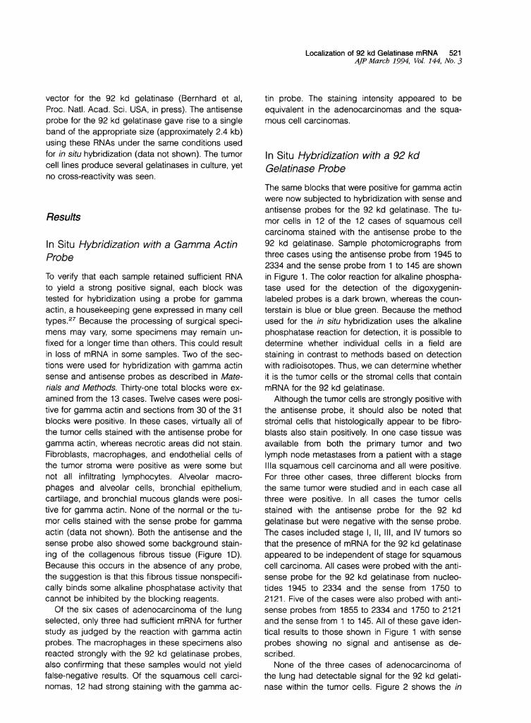
The same blocks that were positive for gamma actinwere now subjected to hybridization with sense andantisense probes for the 92 kd gelatinase. The tu-mor cells in 12 of the 12 cases of squamous cellcarcinoma stained with the antisense probe to the92 kd gelatinase. Sample photomicrographs fromthree cases using the antisense probe from 1945 to2334 and the sense probe from 1 to 145 are shownin Figure 1. The color reaction for alkaline phospha-tase used for the detection of the digoxygenin-labeled probes is a dark brown, whereas the coun-terstain is blue or blue green. Because the methodused for the in situ hybridization uses the alkalinephosphatase reaction for detection, it is possible todetermine whether individual cells in a field arestaining in contrast to methods based on detectionwith radioisotopes. Thus, we can determine whetherit is the tumor cells or the stromal cells that containmRNA for the 92 kd gelatinase.
Although the tumor cells are strongly positive withthe antisense probe, it should also be noted thatstromal cells that histologically appear to be fibro-blasts also stain positively. In one case tissue wasavailable from both the primary tumor and twolymph node metastases from a patient with a stageIlla squamous cell carcinoma and all were positive.For three other cases, three different blocks fromthe same tumor were studied and in each case allthree were positive. In all cases the tumor cellsstained with the antisense probe for the 92 kdgelatinase but were negative with the sense probe.The cases included stage 1, 11, 111, and IV tumors sothat the presence of mRNA for the 92 kd gelatinaseappeared to be independent of stage for squamouscell carcinoma. All cases were probed with the anti-sense probe for the 92 kd gelatinase from nucleo-tides 1945 to 2334 and the sense from 1750 to2121. Five of the cases were also probed with anti-sense probes from 1855 to 2334 and 1750 to 2121and the sense from 1 to 145. All of these gave iden-tical results to those shown in Figure 1 with senseprobes showing no signal and antisense as de-scribed.None of the three cases of adenocarcinoma of
the lung had detectable signal for the 92 kd gelati-nase within the tumor cells. Figure 2 shows the in

522 Canete-Soler et alAJP March 1994, Vol. 144, No. 3
I.I
Figure 1. In situ hybridization ofsquamous cell carcinomas of the lung with a 92 kd gelatinase probe. Sectionsfrom three different squamous cellcarcinomas of the lung are shown. A, C, and E were probed with the antisense 92 kd gelatinase probe (1854 to 2334 [480 bases]), whereas B, D,and F were probed with the sense 92 kd gelatinase probe (1 to 145). Positive staining is seen as deep brown. The counterstain is blue. Although thecolor balance used to print all panels was identical, the film to photograph some of the sections was different resulting in somewhat different in-tensities of blue. The white Ts are centered within masses of tumor cells; the S indicates a stromal area where background staining of acellularconnective tissue can be seen. The arrous point to cells within stroma adjacent to the tumor cells that histologically are fibroblasts.
situ hybridization with the 92 kd gelatinase probefor two adenocarcinomas of the lung. The alveolarmacrophages were positive in each case and thesignal was also found in bronchial epithelium thatserved as an internal positive control. However, noother stromal component of the tumor was positive.Components of normal host tissue were also
found to stain for the 92 kd gelatinase mRNA in insitu hybridization. Tissue macrophages appeared to
stain as reported by Pyke et al28,29 (Figure 2). Al-veolar macrophages also stained. Most alveoli werenegative (Figure 3A and B). Although normal alveoliseemed to be consistently negative, we did noticethat in areas of extensive fibrotic disease positivecells could sometimes be seen. Focal-staining in al-veolar type 11 cells was occasionally noticed seem-ingly in areas with increased fibrosis and may rep-resent damaged areas (Figure 3C). Blood vessels
140-
R
F..........
Ai. "Aft"

Localization of 92 kd Gelatinase mRNA 523AJV March 1994, Vol. 144, No. 3
Figure 2. In situ hybridization ofadenocarcinomas of the lung with a 92 kdgelatinaseprobe. Sectionsfrom two adenocarcinomas of the lung are
shown. All are stained with the antisense probe for the 92 kd gelatinase. C shows a high power view of the section shown in A and D shows a high
power viewfrom the section shown in B. The broad arrows indicate clusters of tumor cells, whereas the narrow arrows show cells that stain posi-
tively and histologically are consistent with macrophages. The S indicates stromal areas adjacent to the tumor. Positive staining is brown as de-
scribed in Figure 1.
did not stain. Neutrophils and most lymphocyteswere negative. Ciliated bronchial epithelium waspositive and underlying basal cell hyperplasiastained with a stronger intensity (Figure 3E and F).Only some of the cells in bronchial mucosal glandsstained positively (Figure 3G and H). Tissue stromafrom normal tissue including fibroblasts were nega-tive as was cartilage (Figure 3D). Only strong brownstaining has been regarded as positive. Althoughother trace alterations can be seen, these are notclearly significant. All three antisense and the twosense probes gave identical results on normal lung(data not shown).
Ribonuclease Protection Assay
Tissue for RNA isolation was not available for mostspecimens. However, two samples from the adeno-carcinomas of the lung and specimens from two ofthe squamous cell carcinomas were obtained forRNA extraction. Also, one specimen of normal lungtissue was obtained for this purpose. The levels of
the mRNA for the 92 kd gelatinase in the two speci-mens of squamous cell carcinoma were at least 6-to 10-fold greater in the nonneoplastic and the ad-enocarcinoma specimens, which contained essen-tially equivalent amounts of mRNA as judged fromthe control ribonuclease protection for gamma actinthat was included in the reaction (Figure 4). Whenthe assay was performed with the sense riboprobeno signal was detectable in any sample (data notshown). This data corroborates the results found byin situ hybridization.
DiscussionIn this study we used in situ hybridization to localizemRNA for the 92 kd gelatinase in squamous cellcarcinomas, adenocarcinomas of the lung, and innonneoplastic lung tissue. In all of the 12 cases ofsquamous cell lung carcinomas examined, the ma-lignant cells were found to contain mRNA for the 92kd gelatinase within their cytoplasm. This appearedto be true regardless of stage, although only one
A W.11..-I.-
....W .M., ,-6.
W&

524 Canete-Soler et alAJP Marcb 1994, Vol. 144, No. 3
r r. ~ i!
Figure 3. In situ hybnidization oflung tissue with a 92 kdgelatinaseprobe. A to E and G show sections oflung that wereprobed with the antisenseprobefor the 92 kd gelatinase, whereas F and H wereprobed with the senseprobe. A shows normal alveoli that do not stain. B shows an area withsomefibrosis that also did not stain, whereas C showed another area offibrosis in which afew cells histologically identified as type IIpneumocytesstained. Some of these cells are indicated with arrows. D shows bronchial cartilage. E and F arefrom bronchial mucosa; E probed with the anti-sense probe and F the control sense probe. E shows strongly staining basal cells with epitbelial cells less positive. Thefibroblasts appear negative. Gand H arefrom bronchial mucous glands in which there arefocally positive cells, some indicated by arrows. H shows the control probed with thesense probe. Positive staining is brown as described in Figure 1.
.....

C: X
~ ~ ~ C)(ou)
0 0o0
o
cc X. j 4 4 aa cn
stage Ill and stage IV case were obtained. The dis-tribution of cases reflects the greater usefulness ofsurgery for early stage disease. In addition, metas-tases from the stage Ill A case also had mRNA forthe 92 kd gelatinase. Pyke et al28'29 have foundmRNA for the 92 kd gelatinase in tumor cells fromsquamous cell carcinomas of the skin. The findingthat both the tumor cells and the adjacent fibro-blasts contain the mRNA for the 92 kd gelatinase isnotable in that a number of the metalloproteinasesthat have been associated with carcinomas proveto be induced at the mRNA level in the stroma.Stromelysin 3 was described as being induced inthe stroma of breast carcinomas, and the 72 kdcollagenase/gelatinase has been found in thestroma surrounding squamous cell carcinomas ofthe skin and colon.28'30,31 However, the 92 kdgelatinase could be both tumor and stromally de-rived in squamous cell carcinomas.The presence of the 92 kd gelatinase mRNA in
host stromal fibroblasts is mimicked by our in vitromodel in which a metastatic rat embryo cell linetransformed by ras plus myc is co-cultured with nor-mal fibroblasts, the release of the 92 kd gelatinaseis induced in the fibroblasts, but co-culture with atumorigenic nonmetastatic line does not result inproduction of the 92 kd gelatinase (Himelstein et al,J Cell Sci, in press). These findings were furthersupported in that in situ hybridization on sections
Localization of 92 kd Gelatinase mRNA 525AJP March 1994, Vol. 144, No. 3
Figure 4. mRNA levels for the 92 kd gelatinasefrom tumor specimens. Total RNA was preparedfrom two adenocarcinomas of the lung, twosquamous cell carcinomas, and normal lungtissue from specimens also used for in situ by-bnidization. The ribonuclease protection assaywas performed as described in Materials andMethods using the antisense 92 kd gelatinaseprobe nucleotides 1854 to 2334 and the anti-sense gamma actin. The lanes are as marked.The probes lane contains reaction mixture with
__ 500 both riboprobes, but without any added RNA.Probes plus Ti contains both probes withoutRNA and after digestion with Ti under the con-
-400 ditions usedfor the samples. The RA3 lane con-tains RNA from a transformed rat embryo cell
300 line that does not express any 92 kd mRNA. The____ band seen at 286 is of the predicted size for200 protection of the gamma actin. Although the200-UV probe for the 92 kd gelatinase (562) is larger
than the expected fragment (490), the gel at
100 this concentration does not resolve those twobands. The mRNA from a cell line expressinghuman 92 kd gelatinase RA3.S7 yields a frag-ment of the same size as the squamous cell car-cinomas (data not shown).
obtained from tumors from the metastatic cell linerevealed that the fibroblasts adjacent to the tumorcontained mRNA for the 92 kd gelatinase, whereasfibroblasts adjacent to the nonmetastatic tumor didnot (Himelstein et al, J Cell Sci, in press). Thesedata suggest a possible in vivo role of tumor cells inthe induction of the 92 kd gelatinase in the hoststroma.
The 92 kd gelatinase mRNA was also found innormal host tissue. Macrophages are known to pro-duce the 92 kd gelatinase and as expected wefound mRNA for the 92 kd in alveolar macrophages.mRNA for the 92 kd gelatinase also was present inthe bronchial epithelium and the underlying cells ofbasal cell hyperplasia. Perhaps this is not surprisingbecause the latter cells are at sites of alveolar injuryand repair. The presence of the 92 kd gelatinasemRNA in the normal tissue that gives rise to the tu-mors rules out the potential use of the presence ofthis mRNA as a marker for tumor progression. Theabsence of expression in the adenocarcinomassuggests that there may be a significant differencein 92 kd gelatinase expression based on the histo-logical subtype of the tumor, but more cases needto be analyzed to determine whether this is a con-sistent finding. Interestingly, a previous study on theexpression of collagenase-related MMP genes inhuman lung tumors reported no expression of ST2in 3 of 3 adenocarcinomas analyzed and very low

526 Canete-Soler et alAJP March 1994, Vol. 144, No. 3
levels for collagenase and Pump-1,32 whereasthese enzymes were found in squamous cell carci-nomas. Because it is not known with certainty whichcell is the progenitor for adenocarcinomas of thelung, its cellular origin cannot be evaluated.The role of the 92 kd gelatinase in both normal
tissue and tumors is unclear. The evidence from invivo models strongly supports the suggestion of apotential action in tumor invasion. However, thepresence of the mRNA in normal epithelium is puz-zling and indicates a more complex action. It will beinteresting to determine whether these tissues syn-thesize or secrete 92 kd gelatinase protein andwhether the enzyme is in an active form. It is alsopossible that an imbalance between the 92 kdgelatinase expression and tissue inhibitor ofmetalloproteases-1 or 2 expression could accountfor the greater invasiveness in carcinoma cells.33'34TIMP-1 is found complexed to the 92 kd gelatinaseduring purification and completely inhibits its enzy-matic activity.34-36 It will be interesting to examinethe distribution of TIMP in the squamous carcino-mas of the lung and in normal tissue.
AcknowledgmentsWe thank Edward Hodge, the histotechnologist inthe Division of Surgical Pathology who cut the sec-tions for this work. We also thank Dr. G. Pietra forhelp in obtaining specimens.
References1. Liotta L, Stetler-Stevernson WG: Metalloproteinases
and cancer invasion. Sem Cancer Biol 1990, 1:99-1062. Matrisian LM, Bowdern GT: Stromelyisn/transin and tu-
mor progression. Sem Cancer Biol 1990, 1:107-1163. Murphy G, Docherty AJP: The matrix metalloprotein-
ases and their inhibitors. Am J Respir Cell Mol Biol1992, 7:120-125
4. Monteagudo C, Merino MJ, San-Juan J, Liotta LA,Stetler-Stevenson WG: Immunohistochemical distribu-tion of type IV collagenase in normal, benign and ma-lignant breast tissue. Am J Pathol 1990, 136:585-592
5. Levy AT, Cioce V, Sobel ME, Garbisa S, Grigioni WF,Liotta LA, Stetler-Stevenson WG: Increased expres-sion of the Mr 72,000 type-IV collagenase in humancolonic adenocarcinoma. Cancer Res 1991, 51:439-44
6. Matrisian LM, Bowden GT, Krieg P, Furstenberger G,Briand JP, Leroy P, Breathnach R: The mRNA codingfor the secreted protease transin is expressed moreabundantly in malignant than in benign tumors. ProcNatl Acad Sci USA 1986, 83:9413-9417
7. Bernhard EJ, Muschel RJ, Hughes EN: 92 kDa gelati-nase release correlates with the metastatic phenotypein transformed rat embryo cells. Cancer Res 1990, 50:3872-3877
8. Yamagata S, Yoshika I, Tanaka R, Shimizu S: Gelatin-ases of metastatic cell lines of murine colonic carci-noma as detected by substrate gel electrophoresis.Biochem Biophys Res Commun 1988, 151:158-162
9. Ballin M, Gomez DE, Sinha CC, Thorgeirsson UP: Rasoncogene mediated induction of a 92kDa metallopro-teinase: strong correlation with the malignant pheno-type. Biochem Biophys Res Commun 1988, 154:832-838
10. Kubota S, Mitsudomi T, Yamada Y: Invasive human fi-brosarcoma DNA mediated induction of a 92 kDagelatinase/type IV collagenase leads to an invasivephenotype. Biochem Biophys Res Commun 1991,181:1539-1547
11. Mainardi CL, Hibbs MS, Hasty KA, Seyer JM: Purifica-tion of a type V collagen degrading metalloproteinasefrom rabbit alveolar macrophages. Collagen Relat Res1984, 4:479-492
12. Hibbs MS, Hoidal JR, Kang AH: Expression of a me-talloproteinase that degrades native type V collagenand denatured collagens by cultured human alveolarmacrophages. J Clin Invest 1987, 80:1644-50
13. Wilhelm SM, Collier IE, Marmer BL, Eisen AZ, GrantGA, Goldberg GI: SV-40-transformed human lung fi-broblasts secrete a 92-kDa type IV collagenase whichis identical to that secreted by normal human macro-phages. J Biol Chem 1989, 264:17213-17221
14. Senior RM, Griffin GL, Fliszar CJ, Shapiro SD, Gold-berg GI, Welgus HG: Human 92- and 72-kilodaltontype IV collagenases are elastases. J Biol Chem 1991,266:7870-7875
15. Murphy G, Cockett Ml, Ward RV, Docherty AJP: Matrixmetalloproteinase degradation of elastin, type IV colla-gen and proteoglycan. Biochem J 1991, 277:277-279
16. Welgus HG, Campbell EJ, Cury JD, Eisen AZ, SeniorRM, Wilhelm SM, Golberg GI: Neutral metalloprotein-ases produced by human mononuclear phagocytes. JClin Invest 1990, 86:1496-1502
17. Fisher SJ, Cui T-Y, Zhang L, Hartman L, Grahl K,Zhang G-Y, Tarpey J, Damsky CH: Adhesive and deg-radative properties of human placental cytotropho-blast cells in vitro. J Cell Biol 1989, 109:891-902
18. Librach CL, Werb Z, Fitzgerald ML, Chiu K, Corwin NM,Esteves RA, Grobelny D, Galardy R, Damsky CH, FisherSJ: 92-kD type IV collagenase mediates invasion of hu-man cytotrophoblasts. J Cell Biol 1991, 113:437-449
19. Talhouk RS, Chin JR, Unemori EN, Werb Z, Bissell MJ:Proteinases of the mammary gland: developmentalregulation in vivo and vectorial secretion in culture.Development 1991, 112:439-449
20. Unemori EN, Hibbs MS, Amento EP: Constitutive ex-pression of a 92-kD gelatinase (type V collagenase)by rheumatoid synovial fibroblasts and its induction innormal human fibroblasts by inflammatory cytokines. JClin Invest 1991, 88:1656-1662

Localization of 92 kd Gelatinase mRNA 527AJP March 1994, Vol. 144, No. 3
21. Okada Y, Tsuchiya H, Shimizu H, Tomita K, NakanishiI, Sato H, Seiki M, Yamashita K, Hayakawa T: Induc-tion and stimulation of 92 kDa gelatinase/Type IV col-lagenase production in osteosarcoma and fibrosar-coma cell lines by tumor necrosis factor alpha.Biochem Biophys Res Commun 1990, 171:610-617
22. Huhtala P, Tuuttila A, Chow LT, Lohi J, Keski-Oja J,Tryggvason K: Complete structure of the human genefor the 92-kDa type IV collagenase. J Biol Chem 1991,266:16485-16490
23. Sato H, and Seiki M: Regulatory mechanism of 92 kDatype IV collagenase expression which is associatedwith invasiveness of tumor cells. Oncogene 1993,8:395-405
24. Wang M, Stearns ME: Blocking of collagenase secre-tion by estramustine during in vitro tumor cell invasion.Cancer Res 1988, 48:6262-6271
25. Dooley S, Radtke J, Blin N, Unteregger G: Rapid de-tection of DNA-binding factors using protein blottingand digoxigenin-UTP marked probes. Nucleic AcidsRes 1988, 16:11839-11945
26. Chomczynski P, Sacchi N: Single-step method of RNAisolation by acid guanidium thiocyanate-phenol-chloroform extraction. Anal Biochem 1987, 162:156-159
27. Erba HP, Eddy R, Shows T, Kedes L, Gunning P:Structure, chromosome location and expression of thehuman gamma actin gene: differential evolution, loca-tion and expression of the cytoskeletal beta andgamma actin genes. Mol Cell Biol 1988, 8:1775-1789
28. Pyke C, Ralfkiaer E, Huhtala P, Hurskainen T, Dano K,Tryggvason K: Localization of messenger RNA for Mr72,000 and 92,000 type IV collagenases in humanskin cancers by in situ hybridization. Cancer Res1992, 52:1336-1341
29. Pyke C, Ralfkiaer E, Tryggvason K, Dano K: Messen-ger RNA for two type IV collagenases is located instromal cells in human colon cancer. Am J Pathol1993, 142:359-365
30. Basset P, Bellocq JP, Wolf C, Stoll I, Hutin P, LimacherJM, Podhajcer OL, Chenard MP, Rio MC, Chambon P:A novel metalloproteinase gene specifically ex-pressed in stromal cells of breast carcinomas. Nature1990, 348:699-704
31. Poulsom R, Pignatelli M, Stetler-Stevenson WG, LiottaLA, Wright PA, Jeffery RE, Longcraft JM, Rogers L,Stamp GWH: Stromal expression of 72 Kda type IVcollagenase (MMP-2) and TIMP-2 mRNAs in colorec-tal neopplase. Am J Pathol 1992, 141:389-396
32. Muller D, Breathnach R, Engelmann A, Milton R, Bron-ner G, Flesh H, Dumont P, Eber M, Abescassis J: Ex-pression of collagenase related metalloproteinasegenes in human lung or head and neck tumors. Int JCancer 1991, 48:550-556
33. Goldberg GI, Marmer BL, Grant GA, Eisen AZ,Wilhelm S, He C: Human 72-kilodalton type IV colla-genase forms a complex with a tissue inhibitor of me-talloproteases designated TIMP-2. Proc Natl Acad SciUSA 1989, 86:8207-8211
34. Liotta LA, Steeg PS, Stetler-Stevenson WG: Cancermetastasis and angiogenesis: an imbalance of posi-tive and negative regulation. Cell 1991, 64:327-336
35. Sellers A, Murphy G, Meikle MC, Reynolds JJ: Rabbitbone collagenase inhibitor blocks the activity of otherneutral metalloproteinases. Biochem Biophys ResCommun 1991, 87:581-587
36. Murphy G, Koklitis P, Carne AF: Dissociation of tissueinhibitor of metalloproteinase inhibitor (TIMP) from en-zyme complexes yields fully active inhibitor. BiochemJ 1989, 261:1031-1034