cloning of a 72 kda matrix metalloproteinase (gelatinase) from
TRANSCRIPT
Biochem. J. (1994) 300, 729-736 (Printed in Great Britain)
Cloning of a 72 kDa matrix metalloproteinase (gelatinase) from chickenembryo fibroblasts using gene family PCR: expression of the gelatinaseincreases upon malignant transformationRonald T. AIMES,* Deborah L. FRENCHt and James P. QUIGLEYttDepartment of *Biochemistry and Cell Biology and tPathology, State University of New York at Stony Brook, Stony Brook, New York 11794, U.S.A.
Chicken embryo fibroblasts secrete a 72 kDa progelatinase thatdisplays all of the characteristics of a matrix metalloproteinase.Employing reverse-transcription PCR and degenerate oligo-nucleotide primers that are specific for two highly conservedsequences found in all matrix metalloproteinases, a DNA frag-ment specific for the chicken gelatinase was generated. Using thisPCR product as a probe, cDNA clones were isolated from achicken embryo cDNA library and the entire protein codingsequence was determined. The chicken progelatinase is 84 %identical, at the amino acid level, with human and mouse 72 kDaprogelatinase/type-IV procollagenase, with the greatest degreeof similarity occurring in the propeptide and catalytic domains.The avian and mammalian proteinases diverge significantly inthe C-terminal, hemopexin-like domain. The last 100 residues ofthe chicken gelatinase are only 66 % identical with mammalian
INTRODUCTION
Extracellular matrix (ECM) remodelling represents an importantevent in many normal and pathological processes. One majorproteinase family, the matrix metalloproteinases (MMPs), appear
to play a central role in degrading components of the ECM.Their functions in embryo implantation (Librach et al., 1991),inflammatory diseases (Case et al., 1990), angiogenesis andtumour invasion and metastasis (Liotta et al., 1991) have beenwell documented. Several members of this proteinase family havebeen described and include: two collagenases (MMP-1 andMMP-8) (Macartney and Tschesche, 1983; Goldberg et al.,1986); two gelatinases/type-IV collagenases (MMP-2 and MMP-9) (Collier et al., 1988; Wilhelm et al., 1989); three stromelysins(MMP-3, -10 and -11) (Chin et al., 1985; Matrisian et al., 1985;Nicholson et al., 1989; Basset et al., 1990); matrilysin (MMP-7)(Woessner and Taplin, 1988); and a recently cloned mouse
macrophage elastase (Shapiro et al., 1992). Individual membersof the MMP family degrade various matrix proteins, includinginterstitial and basement membrane collagens, fibronectin, elastinand proteoglycan core protein (Docherty and Murphy, 1990).Although their substrate specificities and patterns ofexpression
vary, MMPs share functional and structural features. Theseproteinases function at neutral pH values, are inhibited by a
family of protein inhibitors [the tissue inhibitors of
gelatinases. Mouse 72 kDa progelatinase, however, does notdiverge significantly (> 98 % identity) from human progelatinasein the hemopexin-like domain. The divergence in this domain ofthe chicken progelatinase may explain some of the distinctcatalytic and inhibitory properties of the 72 kDa chickenprogelatinase. Northern-blot analysis reveals that steady-statelevels of the chicken progelatinase mRNA are increased 5-foldupon malignant transformation of chicken embryo fibroblastswith Rous sarcoma virus (RSV) and 3-fold by treatment with thetumour-promoting phorbol ester, phorbol 12-myristate 13-acetate (PMA) This represents the first reported cloning of anavian matrix metalloproteinase. The increased expression of thechicken progelatinase by RSV transformation and the tumourpromoter PMA suggests that the progelatinase is regulateddifferently in chicken cells.
metalloproteinases (TIMPs)] (Winyard et al., 1991), require anintrinsic zinc ion for catalytic activity, and are secreted asinactive zymogens (Woessner, 1991). A highly ordered domainstructure confers both the shared and unique properties to eachof these enzymes. All MMPs contain at least two domains: apropeptide and a catalytic domain. In addition, a large C-terminal domain, which shows some sequence similarity tohemopexin (Muller et al., 1988) and the ECM-protein vitronectin(Matrisian et al., 1986), is present in all members except MMP-7 (Quantin et al., 1989). The catalytic domains of MMP-2 andMMP-9 are interrupted by a large fibronectin-like gelatin-bindingdomain and MMP-9 contains a small type-V collagen-likedomain.A well-studied member of the MMP family is MMP-2, which
has been described in humans and other mammals as well(Seltzer et al., 1981; Salo et al., 1983; Murphy et al., 1985).Human MMP-2 was shown to cleave gelatin (denatured collagen)with high specific activity and basement-membrane type-IVcollagen at a reduced, yet significant rate (Collier et al., 1988).Considerable controversy still exists over the ability of MMP-2and MMP-9 to cleave native type-IV collagen; some studies havedemonstrated that these gelatinases are weak type-IVcollagenases (Mackay et al., 1990; Okada et al., 1990; Nagase etal., 1991; Morodomi et al., 1992). Recently our laboratory hasreported the isolation and characterization of a gelatinolytic
Abbreviations used: MMP, matrix metalloproteinase; TIMP, tissue inhibitor of metalloproteinases; CEF, chicken embryo fibroblast; RSV, Roussarcoma virus; RT, reverse transcription; SSPE, (0.18 M NaCI/10 mM Na2HPO4, pH 7.4/1 mM EDTA); CG62, degenerate oligonucleotide to the N-terminus of the active chicken gelatinase; APMA, p-aminophenylmercuric acetate; M-MuLV, Moloney murine leukaemia virus; ECM, extracellularmatrix; PMA, phorbol 12-myristrate 13-acetate.
I To whom correspondence should be addressed.The nucleotide sequence data reported will appear in GenBank, EMBL and DDBJ Nucleotide Sequence Databases under the accession number
U07775.
729
730 R. T. Aimes, D. L. French and J. P. Quigley
enzyme secreted by Rous sarcoma virus (RSV)-transformedchicken embryo fibroblasts (CEF) that appears to be a memberof the MMP family (Chen et al., 1991). Partial N-terminalsequencing and the presence of a 21 kDa protein that co-purifieswith the chicken progelatinase suggested that this enzyme is anavian homologue of human MMP-2. Although this gelatinaserapidly degrades denatured collagens it does not appear to cleavenative type-IV collagen (Chen et al., 1991). A direct comparisonwith MMP-2 proteinases in various other species may help toelucidate the basis for these differences in substrate specificityand may also help reveal the basis of substrate specificity in allof the MMP-family members.Two separate sequences are believed to be responsible for the
co-ordination of the catalytic zinc ion in the MMP zymogens:the cysteine-switch and the zinc-binding regions (Springman etal., 1990; Van Wart and Birkedal-Hansen, 1990; Windsor et al.,1991). The sequence conservation between the MMPs in theseregions is remarkable and was exploited to clone the cDNAencoding the chicken progelatinase. A similar approach has beenutilized successfully to identify serine- and cysteine-proteinasecDNAs from the parasite Anisakis simplex (Sakanari et al.,1989). Comparison of the nucleotide and deduced amino acidsequences of the chicken cDNA clone with those of the mam-malian proMMP-2 reveals a high level of identity. Interestingly,this identity is not distributed homogeneously over the entiremolecule, as chicken and human MMP-2 appear to divergesignificantly at the C-terminus. This divergence is interestingbecause this region of human MMP-2 has been shown to becrucial for TIMP binding (Murphy et al., 1992b) and has beenimplicated in the substrate specificity of collagenase (Murphy etal., 1992a; Sanchez-Lopez et al., 1993).
MATERIAL AND METHODS
MaterialsPathogen-free, complement fixation avian leukosis (COFAL)-negative eggs were purchased from SPAFAS (Norwich, CT,U.S.A.). LiCl, urea, cell culture reagents, ethidium bromide,Tris, EDTA, sodium citrate and NaCl were purchased fromSigma Chemical Company (St. Louis, MO, U.S.A.). Phenol,formamide, nucleic acid molecular-mass markers, T4 poly-nucleotide kinase, sequencing primers and restriction endo-nucleases were purchased from BRL (Grand Island, NY,U.S.A.). Radiolabelled deoxyribonucleotides were purchasedfrom Amersham (Arlington Heights, IL, U.S.A.). Moloneymurine leukaemia virus (M-MuLV) reverse transcriptase, andrandom prime DNA labelling reagents were purchased fromBoehringer-Mannheim (Indianapolis, IN, U.S.A.). Reagents forPCR were purchased from Perkin-Elmer Cetus (Norwalk, CT,U.S.A.). Magna nylon membranes were from Micron Separations(Westboro, MA, U.S.A.). Methylene Blue stain was obtainedfrom Molecular Research Center (Cincinnati, OH, U.S.A.).Erase-a-base nested deletion kit and Magic miniprep kits were
purchased from Promega (Madison, WI, U.S.A.). The T/Acloning kit was from Invitrogen (San Diego, CA, U.S.A.). DNA-sequencing reagents were purchased from United States Bio-chemical (Cleveland, OH, U.S.A.). Fetal bovine serum was
purchased from Hyclone (Logan, UT, U.S.A.). A 10-day-oldchicken embryo Agtl 1 cDNA library was purchased from Clon-tech (Palo Alto, CA, U.S.A.). Oligonucleotides were obtainedfrom either Operon Technologies (Alameda, CA, U.S.A.) or
Genosys Biotechnologies (The Woodlands, TX, U.S.A.). pBlue-script II KS' was purchased from Stratagene (La Jolla, CA,U.S.A.) and phorbol 12-myristate 13-acetate was obtained fromChemsyn Science Laboratories (Lenexa, KS, U.S.A.).
Cell culturePrimary cultures of CEF were prepared from 10-day-oldpathogen-free, COFAL-negative fertilized eggs as previouslydescribed (Fairbairn et al., 1985; Sullivan and Quigley, 1986;Chen et al., 1991). Secondary cultures of CEF were infected withRSV as described previously (Sullivan and Quigley, 1986; Chenet al. 1991). Alternatively, non-transformed cells were maintainedessentially as above without infection. PMA was included inculture medium at a concentration of 100 ng/ml where indicated.
RNA isolation and analysisTotal cellular RNA was isolated from cells using a modifiedLiCl/urea extraction procedure (Auffray and Rougeon, 1980;Bothwell et al., 1990). RNA was dissolved in 10 mM Tris(pH 7.5)/1 mM EDTA (pH 8.0). Northern blots were performedusing formaldehyde/agarose denaturing gels (Maniatis et al.,1982). After electrophoresis, RNA was transferred to nylonmembranes and was stained with Methylene Blue (Maniatis etal., 1982) following the manufacturer's recommendations toverify equal loading. Membranes were prehybridized in 50%(v/v) formamide/5 x Denhardt's solution [1 x Denhardt's =0.02% BSA serum albumin/0.02% Ficoll 400/0.02%poly(vinylpyrrolidine)]/5 x SSPE [1 x SSPE = 0.18 M NaCl/10 mM Na2HPO4 (pH 7.4)/1 mM EDTA]/0.2 % SDS/100 ,ug/ml salmon sperm DNA at 42 °C and hybridized withDNA probes labelled with [a-32P]-dCTP using random priming.Blots were washed in a 0.2 x SSPE/0.1 % SDS solution andexposed to Fuji XAR film.
DNA isolation and analysisPlasmid DNA was isolated using a Magic miniprep kit accordingto the manufacturer's recommendations. A phage DNA wasisolated using poly(ethylene glycol) precipitation as previouslydescribed (Maniatis et al., 1982). For Southern-blot analysis(Southern, 1975), gels were denatured, neutralized and the DNAwas transferred to nylon membranes by capillary blotting.Oligonucleotide probes were hybridized in 1 x SSPE/5 x Den-hardt's solution/100,g/ml salmon sperm DNA at 25 'C. Blotswere washed in 0.2 x SSPE/0. 1 % SDS as indicated.
Labelling of DNA probesOligonucleotides were end-labelled with [y-32P]dATP using T4polynucleotide kinase as previously described (Maniatis et al.,1982). Larger probes were random prime labelled with [a-32P]dCTP according to the manufacturer's protocol.
Oligonucleotide constructionThree degenerate oligonucleotides were used to clone the cDNAencoding the 72 kDa chicken gelatinase. Two of these were usedas primers for PCR and were constructed to hybridize to portionsof the nucleotide sequences that encode the cysteine-switchregion (5' primer) and the zinc-binding region (3' primer). Thesequence of the 5' primer is 5'-GATCGAGCTCTGYGG-NAAYCCNGAYGT-3' (degenerate nucleotide positions areindicated by the standard IUPAC one-letter code. Y = T or C;R = A or G; D = A, G or T; M = A or C) and contains thenucleotide sequence that encodes the consensus amino acidsequence C73GNPDV. The 3' primer sequence is 5'-CGACGTCGACTGNCCRAAYTCRTGDGC-3' and is comp-lementary to the nucleotide sequence that encodes the consensusamino acid sequence A373HEFGH. The underlined ten base
cDNA cloning of the 72 kDa chicken gelatinase
extensions were added to facilitate subcloning and encode a SacIand Sall site (in bold) respectively. The third oligonucleotide(CG62) is a 17-base degenerate oligonucleotide and representsthe sequences that can encode the N-terminal six residues of thep-aminophenylmercuric acetate (APMA)-activated chicken gela-tinase as determined by protein microsequencing (Chen et al.,1991). The sequence 5'-TAYAAYTTYTTYCCNMG-3'represents the sequence of the N-terminus Y81NFFPR.
PCRPCR was preformed as described previously (Lee et al., 1988;Rappolee et al., 1988). Briefly, cDNA was prepared using 3 ,ug oftotal RNA from RSV-transformed CEF. Reverse transcription(RT) was carried out in a 20 ,1 volume of 10 mM Tris (pH 8.3),50 mM KCI, 5 mM MgCl2, 4 mM dNTP, 1 ,uM 3' PCR primer,and 1 unit/,u M-MuLV reverse transcriptase at 42 °C for 30 min.PCR was performed using the cDNA generated above in 100 ,ulcontaining 10 mM Tris (pH 8.3), 50 mM KC1, 2 mM MgCl2,0.8 mM dNTP, 0.2 ,uM of each PCR primer and 25 units/ml TaqDNA polymerase. A hot start (D'Aquila et al., 1991) was usedfollowed by three low-annealing-temperature cycles of 1 min at94 °C and 1 min at 55 'C. This was followed immediately by 27cycles of 1 min at 94 'C, 1 min at 65 'C and 1 min at 72 'C. PCRproducts were subcloned using the T/A cloning kit (Mead et al.,1991) according to the manufacturer's protocol.
DNA sequencingDNA was sequenced using the dideoxy-chain-terminationmethod (Sanger et al., 1977) and Sequenase version 2.0 accordingto the manufacturer's protocol. For sequencing large fragmentsof DNA a series of nested deletions (Henikoff, 1984) weregenerated using the Erase-a-base system.
Isolation and characterization of cDNA clonesA 10-day-old chicken embryo Agtl 1 cDNA library was screenedwith 32P-labelled DNA probes as described (Maniatis et al.,1982). Phage plaques (2.5 x 10⁵) were generated in bacterialstrain Y1090 after overnight incubation at 37 'C. The DNAfrom the phage plaques was transferred to nylon membranes andprobed with DNA probes as described above for Northern blots.The filters were washed in 0.2 x SSPE/0. 1 % SDS at 55 'C andexposed to film overnight. Positive plaques were isolated andpurified using standard procedures (Maniatis et al., 1982).
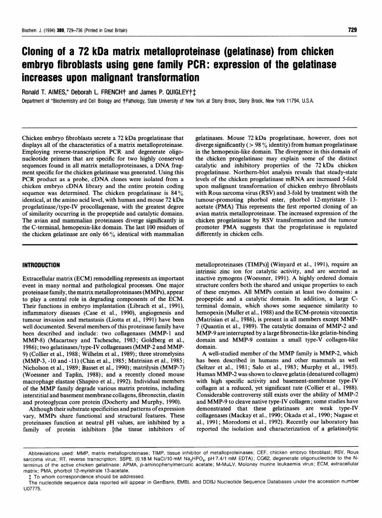
RESULTSRT-PCR of RSV-transformed CEF RNA yields a 0.95 kb productthat encodes a portion of the 72 kDa chicken gelatinaseMembers of the MMP family are composed of a series offunctional domains. Two of these domains contain the highlyconserved sequence motifs of the cysteine-switch (PRCGXPD)and the zinc-binding regions (AAHEXGH), which are involvedin the co-ordination of a zinc ion within the proenzymes(Springman et al., 1990; Vallee and Auld, 1990). In order togenerate a DNA probe for use in screening a chicken embryocDNA library, total RNA from RSV-transformed CEF was usedas a template in RT-PCR (Rappolee et al., 1988). Two degenerateoligonucleotide primers were designed to hybridize to thenucleotide sequences encoding the conserved motifs mentionedabove (see the Materials and methods section). On the basis of
human and mouse proMMP-2 cDNA sequences, the predictedsize of the PCR fragment representative of the chickenprogelatinase would be 937 bp (Collier et al., 1988; Reponen etal., 1992). A major 0.95 kb PCR product was generated, alongwith a 0.48 kb and a minor 0.42 kb product (Figure la, lane 1).To determine which of the PCR products represented the
chicken progelatinase, Southern blots (Southern, 1975) wereperformed and probed with 32P-labelled CG62. Thisoligonucleotide probe is specific for the nucleotide sequence thatencodes the N-terminal six residues of the 62 kDa active chickengelatinase previously determined (Chen et al., 1991) and whichare located between the cysteine-switch and zinc-binding regions.CG62 hybridized to the major band at 0.95 kb, to a 0.48 kb
kb3.0 -
2.0 -1.6 -
1.0 -
0.5 -0.4 -
1 2 3 4
(a) kb
m 3.0 -(b)
2.0 -1.6-
0.5 -0.4 -
1 2 3 4
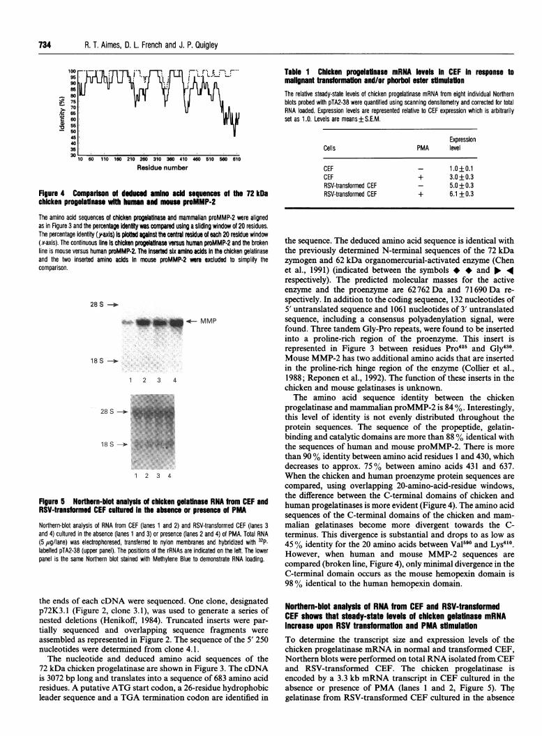
Figure 1 Ethidium bromide-stained agarose gel electrophoresis andSouthern-blot analysis of RSV-transformed CEF RT-PCR product
(a) DNA samples were electrophoresed on a 0.7% agarose gel containing 1 #sg/ml ethidiumbromide. Lane 1, 10 ,ul of the RT-PCR reaction; lane 2, T/A clone pTA2-38 digested with EcoRland HindlIl; lane 3, T/A vector cut with EcoRl and Hindlll; lane 4, gel-purified EcoRl/Hindlllfragment from pTA2-38. (b) Southern blot of the same gel shown in (a) probed with a 32plabelled degenerate oligonucleotide CG62 which hybridizes to the nucleotides encoding the 6N-terminal residues of the 62 kDa active chicken gelatinase. DNA standards (in kb) areindicated on the left.
ATG TGA AATAAA AAAAAAI////////////////////////// II
1.1 (1.0)
3.1 (2.8)
4.1 (3.0)
4.2 (2.3)
PCR(0.9) ............................
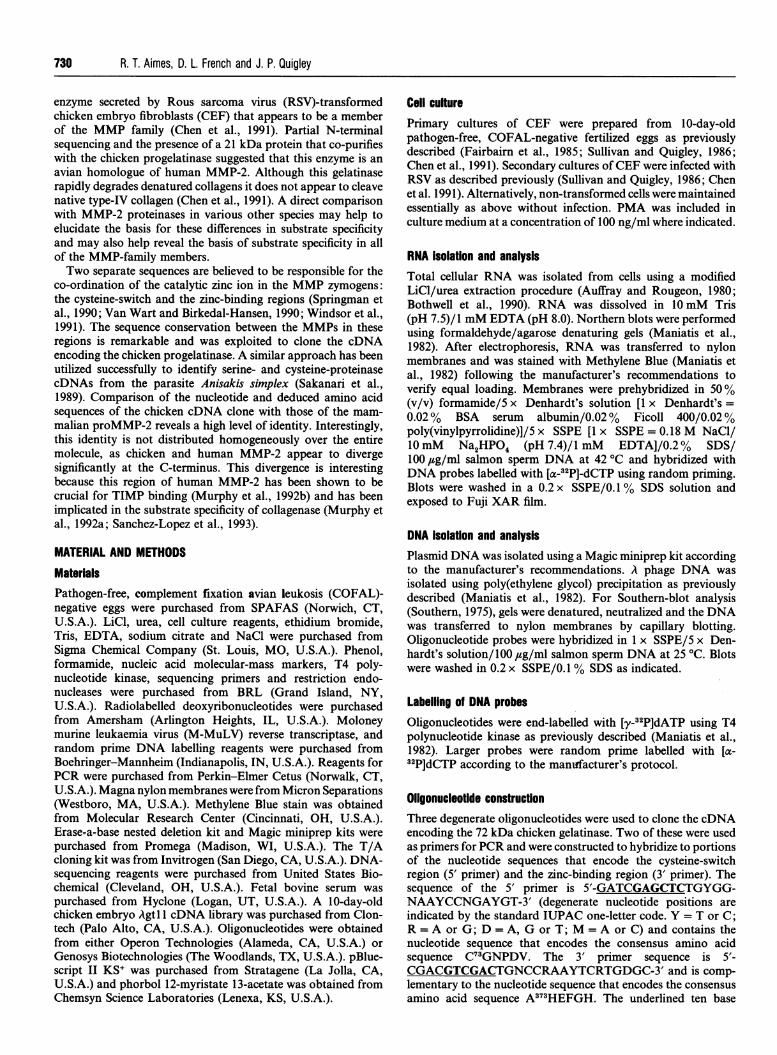
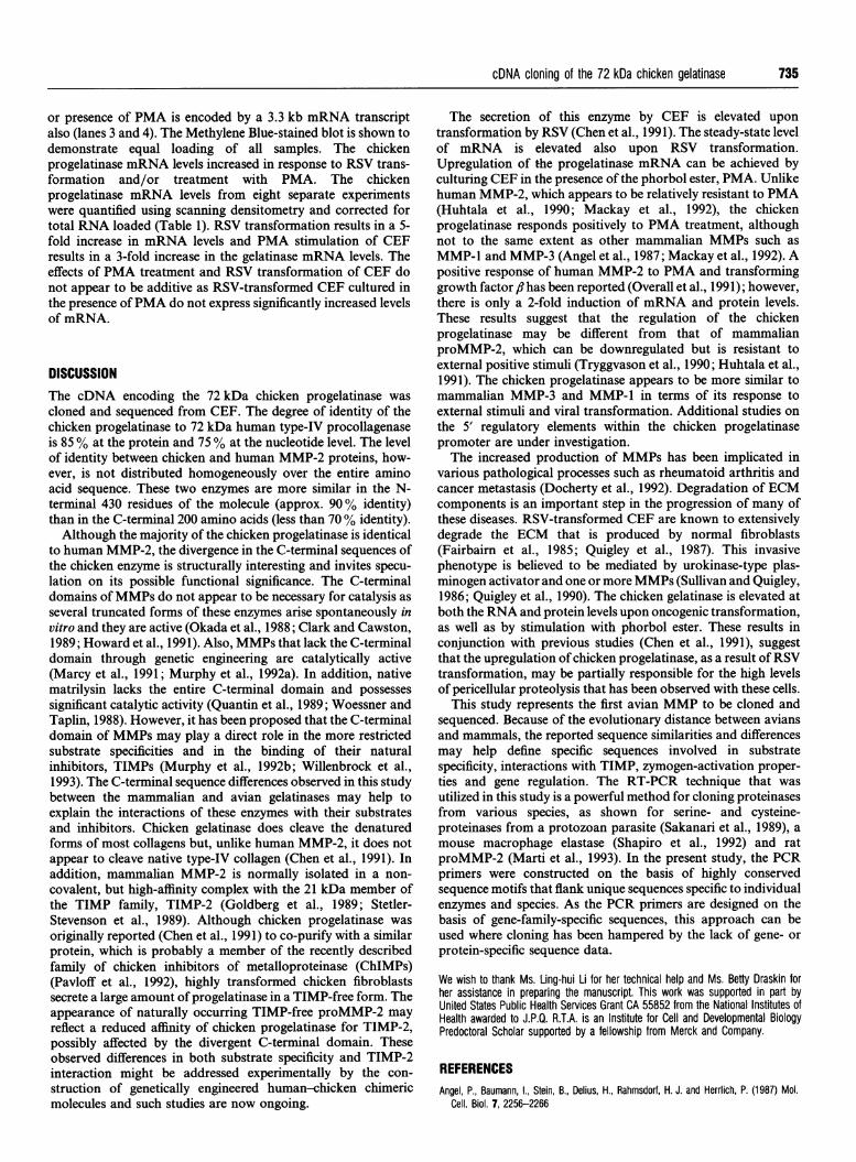
Figure 2 Schematic of 72 kDa chicken gelatinase cDNA, cDNA clones andsequencing strategy
A schematic representation of the 72 kDa chicken gelatinase cDNA is shown. The protein-coding region of the cDNA is represented by the box, with the stippled portion representing thesignal peptide. The relative positions of the start translation signal (ATG), stop codon (TGA),polyadenylation signal (AATAAA) and poly(A) tail (AAAAA) are indicated. Lines below the boxrepresent the four cDNA clones iso!ated from the chicken embryo cDNA library and arenumbered on the left (size, in kb, indicated in parentheses). The broken line represents the PCRproduct generated from RSV-transformed CEF RNA that was used as a probe for screening thecDNA library. The arrows above the box represent the nested deletions of clone 3.1 that wereused for sequence assembly. The sequence of the 5' end of the cDNA was determined fromclone 4.1. The entire DNA sequence was determined from both cDNA strands.
731
732 R. T. Aimes, D. L. French and J. P. Quigley
AATTCCGGCAAAAGAGAAACGGTGCAGAGAGTTAAGATGTGCAGATAAGCAACTAGTGCACTGTGCAGCCAAAGTAMCTGACAGTCAGTCAGAGAAATC -110
TTTTAAAGAGGATTGCAAAATATAGGCAGAATGAAGACTCACAGTGTTTTTG,GCTTCTTTTTTAAAGTACTATTAATCCAAGTGTATCTTTTTAACAAA -10M K T H S V F G F F F K V L L I Q V Y L F N K -4
ACTTTAGCTGCACCGTCACCAATCATTAAGTTCCCTGGAGACAGCACTCCAAAAACAGACAAAGAGCTAGCAGTG-CAATACCTGAATAAATATTATGGAT +91T L A *A P S P I I K F P G* D S T P K T D K E L A V Q Y L N K Y Y G +30
* * * * * * * * * V A * * * * * * * * a * * * * 1 F * * +30* * * * * * * * * V A * * * * * * * * * * * * * T F * * +30
GCCCAAAA&GACAATTGCAACTTATTTGTATTGAAAGATACTTTGAAGAAATGCAGAAMTTTTTTGGGCTGCCTGAAACAGGAGATTTGGATCAAAACAC +191C PKD NC N LF VL KD TL K KM QKFF GL PE TG DL DQ N +63* E S * *.* * *. Q+63* ES * * * * * * * *. * * * . * * ** . * .Q ** * * * . +63
AATTGAGACAATGAAGAAACCCCGCTGTGGTAACCCCGATGTG,GCCAATTACAACTTCTTTCCAAGAAAGCCAAAATGGGAAAAGAATCATATAACATAC +291T IE T MKK PR CGERP D V A NYNF F PR K P K WEKNH IT Y +97* * * * R *..9a9o o . . * * * * D * Q * * +97** * * R * * * * * * * * * * * . * . * . * * * . D * * Q . . . +97
AGGATTATAGGCTATACCCCGGATTTGGATCCTGAGACAGTAGATGATGCCTTTGCCCGAGCCTTTAAAGTCTGGAGTGATGTCACGCCACTGAGATTTA +391R I I G Y T P D L D P E T V D D A F A R A F K V W S D V T P L R F +130
* * * * * * * . * . . . * * . * Q . *. * . * * +130* **.* * * * * * * * ~~~~~~~L** * ** * * * * ** +130
ACCGAATAAATGATGGAGAGGCAGACATTATGATTAATTTTGGCCGATGGGAACATGGTGATGGCTATCCATTTGATGGCAAAGATGGTCTCCTGGCTCA +491N RI ND GE AD I MI N FGRW E HG DG YP FD G KD GLLA +1635 . . H.00 0 0* 000 0 *0** 00 **0 0* 0+163
CGCCTTTGCACCGGGGCCAGGAATTGGAGGAGACTCCCATTTTGATGATGATGAACTGTGGACTCTTGGAGAAGGGCAAGTGGTTAGAGTAAAGTATGGA +591H A F A P G P G I G G D D S H F D D E L W T L G E G Q V V R V K YG +197
T * V . .. . . . . . . .. oo o 9 +197T * V . .. .* . . . . .. * 9 9 &o +197
AATGCAGATGGTGAATACTGCAAATTTCCCTTCTGGTTCAATGGTAAGGAATACAACAGCTGCACAGATGCAGGACGTAATGATGGATTCCTCTGGTGTT +691N A D G E Y C K F P F W F N G K E Y N S C T D A G R N D G F L W C +230
L . * . . . . * . . . a T & . S * . & * . a +230L . . . R . . 5 . . . . T * . S . . . * . . +230
CCACAACCAAAGACTTTGATGCAGATGGCAAATATGGCTTTTGTCCCCATGAGTCACTTTTTACAATGGGTGGCAATGGTGATGGACAGCCCTGCAAGTT +791S T T K D F D A D G K Y G F C P H E S L F T M G G N G D G Q P C K +263
Y N * E K . . . . * . . . . . A . . . . A E . +263Y N * E K * . o . . . . . . . A..o00 A . +263
TCCCTTTAAATTTCAAGGCCAGTCCTATGACCAGTGTACAACAGAAGGCAGGACAGATGGATACAGATGGTGTGGAACCACTGAAGACTATGATAGAGAT +891F P F K F Q G Q S Y D Q C T T E G R T D G Y R W C G T T E D Y D R D +297
R * 0 T * . S . . * . . . . . . . . . . . * . . * * . . * +297R o . T . . N 5 . . * . . . * . . . . . . & . . . * . . . * +297
AAGAAATACGGATTCTGCCCAGAMACTGCCATGTCAACAGTTGGTGGAAATTCAGAAGGAGCTCCTTGTGTATTCCCCTTCATCTTCCTTGGGAATAAAT +991K K Y G F C P E T A M S T V G G N S E G A P C V F P F I F L G N K +330
T . . . . . +330
ACGACTCCTGTACAAGTGCAGGTCGCAATGATGGCAAGCTGTGGTGTGCTTCTACCAGCAGCTATGATGATGACCGCAAGTGGGGCTTTTGTCCAGATCA +1091YDS C T SA GR ND GK L WCA ST SS YDD DR K WG FC PD +363.E . * . . . * * S . . * M . * . T * A N . . * . ...* 9 * +363E . * . . * * . . * & . V . . . T * A N . * . . * . . * . . . . +363
AGGATACAGTCTCTTCTTGGTTGCTGCCCACGAATTTGGCCATGCGATGGGATTAGAGCACTCCGAGGACCCAGGAGCTCTCATGGCCCCGATCTACACC +1191Q G Y S L F L V A A H E F G 8 A H G L E H S E D P G A L M A P I Y T +397
. ~~~ ~ ~~~~~Q. ...0a0 0 0 0+397.. . . . . . Q . . . . * ~~~~~~~~~~~~~~~~~+397
Y T K N F R L S Q 0 0 I K G I Q E L Y E V S P D V E IG FG G +430* o ~~~G A - - - I D L- _- +424
* .. . * . . *H * G P . . . A D 7 D T +426
Figure 3 For caption see facing page.
cDNA cloning of the 72 kDa chicken gelatinase 733
G3GCCAGGACCACGTCCTACCCTTGGACCTGTCACTCCAGOACTCTGCAAGCACGACATTGTATTTGATGGAGTTGCACAAATTAGAGGAGAAATATTTTT +1391G P G P R P T L G P V T P E L C K H D I V F D G V A Q I R G E I F +463* T * * T * * a * * I **Q.o9o0o0I .+457* T * * T * * * a * I **Q * * * * * * I **.+459
CTTCAAAGACAGATTCATGTGGAGGACTGTAAACCCTCGAGGAAMACCCACAGGTCCTCTTCTCGTTGCTACATTCTGGCCTGATCTGCCAGAGAAAATC +1491F F K D R F M W R T V N P R G K P T G P L L V A T F W P D L P E K I +497
* * * * * I * * * *T * * D * * M E * * * * * +491* **** I ** *V ** * ****T* * D * * * E * * * * * +493
GATGCTGTCTACGAGTCCCCTCAG-GATGAGAAGGCTGTATTVTTTTGCAGGAAATGAGTACTGGGTTTATACAGCCAGCAACCTGGATAGGGGCTATCCAA +1591D A V Y E S P Q D E K A V F F A G N E Y W V Y T A S N L D R G Y P +530* * A * * E * * * * * * * * * * I * S * * T * E * * a* +524* * A * * E * * * * * * * * * * * * * * S * * T * E * * * * +526
AGAAACTCCCAGCCTGGACTACCCCTGATGTGAACGCATTATGCAGCCTT1LAJACTGGGGCAGAAACAAGAAGACATATATTTTCTCTGGAGACAG +1691K KLTS L GL P P DVQR I DAAF N WG RN KK TYI F SG D* P *.** * 0 0 0 V * * * * * S K * * * * * * * A* P * * * * * * * * * * Q V * * * * * * S K * * * * * * * A *
ATACGGAGTACATGAGAAAGAAAAATGGGCTGCAACCCAAATCATGCGATTCTGGATGGGTTCAGTAACTCGTGC +179179R Y W K Y N E E K K K M E L A T P K F I A D S W N G V P D N L D A V +597K F * R * * * V * * * * D P G F * * L * * * A a * A I * * * a * * * +591K F a R * * * V * * * * 0 P G F * * L * * * * * * A I * * * * * * * +593
CTGGTCTACTGCAGGGGTCACTATTTTTAAAACCATACATCACAATGGAGACAGATTTGAGATGTAAAATGGAAG +189189L G L T D S G Y T Y F F K D Q Y Y L Q M E D K S L K I V K I G K I +630V D * Q G G * H 5 - * G A * * * K L * N Q * * * S * * F * S * +624V D * Q G G * H S * * * * G A * * K L * N Q * * * Sa * F * S * +626
GTTCTGACTGGTTGGGTTGCTGAACTGTAGAATATATTAATAACCAAATATTTACTTTTTTGTTATATACCTTATCTGTAATTAGAAATAGATCTGAATGS SD WL GC *
K * * * * *
K, * * * * *
TCAATTACTGGACCAGTCAGGTACTGGCACCCAAACATTAAGTACCAGTACTGTACACATCTGATAATTTAAAAGTGTTTGCCTCCTCAACATACAAAAG +2091ATGTTTACGTATCTATTCTGGTACAATTTTTCAGTTTTTGTGCTAGTCATAGCAATGCTAGTAGCAGAACACGCACGGTTCTTTATTCCCCTGGTGCTTG +2191AAGAAAGCATTGAAATCATTGGAGACTGGACCAGACCCCTGTGTTAAATCCATCTCAAATAGCCTCAAGTAAGATGTACCAGCAGTGCACTTTGATTTTT +2291TTTTAATTTTTTTTTTTAAATACAACTCTTTTATAAMTAGATGGTTTGTGTTAATGTTTTAACCCTCTTGCGAGTTTGATCATTACTGCCATTTATTACA +2391TAATGATACAAAAGACTGTCAAAGGCACAGGTTGCCACGGGATTTGATTTGTATTGATGCATGATGGAATCATGGCAAGAAACTGTATTAAAGTATTGTT +2491TTAAAGTACTTTCTATTTTAAAACCTTAGTTCAACTTAGCAATTTGCTTCCTGCACTTTGTTACTACTCTATAACTTGTCTATACAGACAGAATGACTCA +2591GATGTTTTAAATCAATGTATTATACACAGAAGTCAAGTATTACTGTTTACCTTGTTTTATTTTTCTGTTCTTTATAATATTGAGTTTTTGTTTGGTTAGG +2691CTTTCTGCTTAGGCATTGGGCAACAGAAGGTGTTCCTTTCACATATGATGGAATGTTTCTCATTAAAAAAAAAAAAGGTTAAAAAAATCCAGAAAACAAT +2791GTCCCAAACATGAAATGCCAATGCTACAAATAAAATTAAATTACACTGTCAAAAAAAAAAAAAAACGGTTAAC +2864
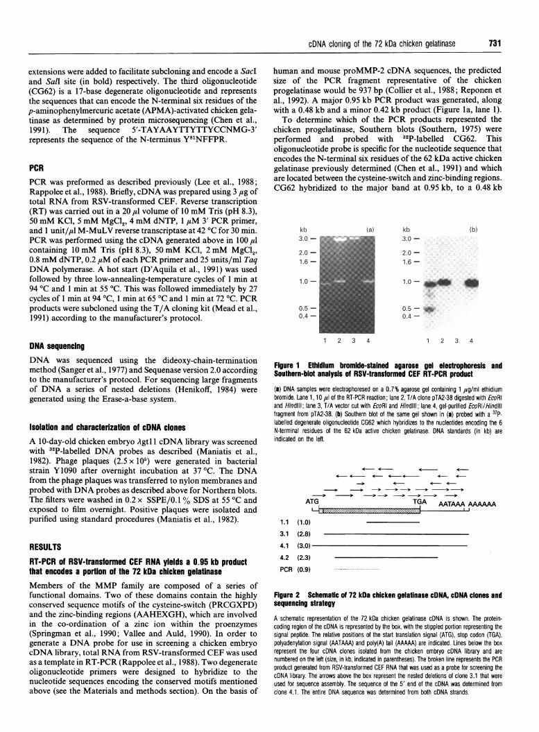
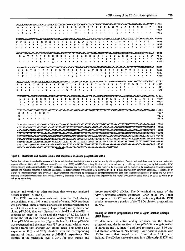
Figure 3 Nucleotide and deduced amino acid sequences of chicken progelatinase cDNA
The first line indicates the nucleotide sequence and the second line shows the deduced amino acid sequence of the chicken gelatinase. The third and fourth lines show the deduced amino acidsequence of human (Collier et al., 1988) and mouse (Reponen et al., 1992) proMMP-2 respectively. Identical residues are indicated by (.), differing residues are given by their one-letfer fUPACleffering. Missing residues are indicated by (-). The numbering of the amino acid residues starts from the first residue of fhe proenzyme, with the residues oftfhe signal peptide being given negativenumbers. The nucleotide sequence is numbered accordingly. The putative initiation of translation (ATG) is marked by (No No lo.) and the translation termination signal (TGA) is indicafed by anasterisk (*). The polyadenylation signal (AATAAA) is double underlined. The additional 18 nucleotides and corresponding six amino acids found in the chicken gelatinase are boxed. The PCR product(excluding the oligonucleotide primer) is underlined. Previously determined (Chen et al., 1991) N-terminal sequences for the chicken proenzyme and active enzyme are contained within**and No. .4 respectively.
product and weakly to other products that were not analysed
further (Figure lb, lane 1).
The PCR products were subcloned into the T/A cloning
vector (Mead et al., 1991) and a panel of cloned PCR products
was generated. Three of these clones tested positive when probed
with CG62 (results not shown). Figure 1(a) (lane 2) shows one
clone, pTA2-38, that was digested with EcoRI and Hindlll to
generate an insert of 1.0 kb and the vector of 3.0 kb. Lane 3
shows the 3.0 kb T/A vector alone. When probed with CG62,
clone pTA2-38 was positive (Figure lb, lane 2). Clone pTA2-38
was sequenced and the insert was 937 bp in length with an open
reading frame that encodes 294 amino acids. This amino acid
sequence is 91 % and 90% identical with the corresponding
regions of human and mouse proMMP-2 respectively. The
identity at the nucleotide level is 7800 for both human and
mouse proMMP-2 cDNA. The N-terminal sequence of the
APMA-activated chicken gelatinase (Chen et al., 1991) that
corresponds to CG62 was identified, confirming that the PCR
product represents a portion of the 72 kDa chicken progelatinase
cDNA.
Cloning of chicken progelatinase from a )Agtl 1 chicken embryo
cDNA library
To determine the entire coding sequence for the chicken
progelatinase, the insert from clone pTA2-38 was gel-purified
(Figures Ila and Ib, lanes 4) and used to screen a Agtl 1I0-day-old chicken embryo cDNA library. Four positive clones, with
cDNA inserts that ranged in size from 1.0 to 3.0 kb, were
isolated. The cDNAs were subcloned into pBluescript KS' and
+563+557+559
+1991+637+631+633
734 R. T. Aimes, D. L. French and J. P. Quigley
75 V70656055
45403530
10 60 110 160 210 260 310 360 410 460 510 560 610
Residue number
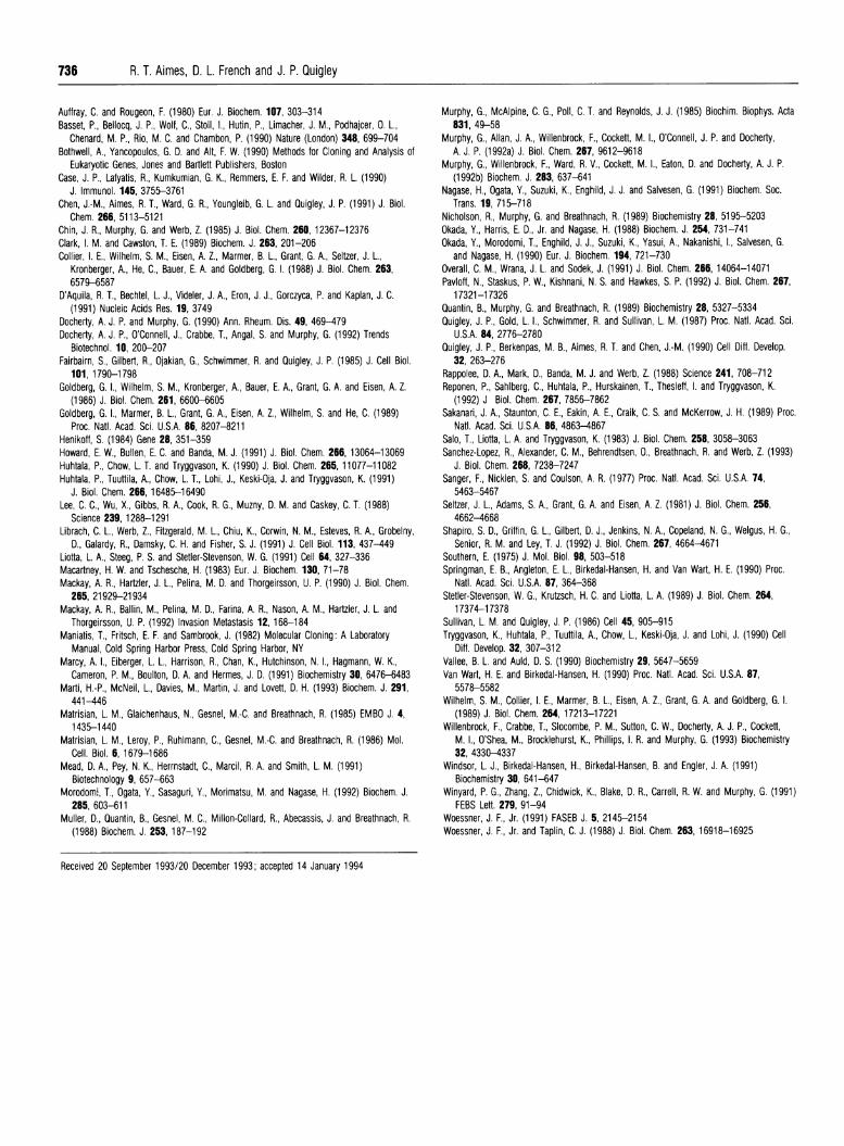
Figure 4 Comparison of deduced amino acid sequences of the 72 kDachicken progelatnase with human and mouse proMMP-2
Table 1 Chicken progelailnase mRNA levels In CEF In response tomalignant transformation and/or phorbol ester stimulationThe relative steady-state levels of chicken progelatinase mRNA from eight individual Northernblots probed with pTA2-38 were quantified using scanning densitometry and corrected for totalRNA loaded. Expression levels are represented relative to CEF expression which is arbitrarilyset as 1.0. Levels are means+S.E.M.
ExpressionCells PMA level
CEFCEFRSV-transformed CEFRSV-transformed CEF
- 1.0+0.1+ 3.0+0.3- 5.0+0.3+ 6.1 +0.3
The amino acid sequences of chicken progelatinase and mammalian proMMP-2 were alignedas in Figure 3 and the percentage identity was compared using a sliding window of 20 residues.The percentage identity ('axis) is plotted against the central residue of each 20 residue window(x-axis). The continuous line Is chicken progelatinase versus human proMMP-2 and the brokenline is mouse versus human proMMP-2. The inserted six amino acids In the chicken gelatinaseand the two inserted amino acids In mouse proMMP-2 were excluded to simplify thecomparison.
28S
q*4p-4_4-MMP
18S-
1 2 3 4
28 S
18S
1 2 3 4
Figure 5 Northern-blot analysis of chicken gelatinase RNA from CEF andRSV-transformed CEF cultured in the absence or presence of PMA
Northern-blot analysis of RNA from CEF (lanes 1 and 2) and RSV-transformed CEF (lanes 3and 4) cultured in the absence (lanes 1 and 3) or presence (lanes 2 and 4) of PMA. Total RNA(5 1ug/lane) was electrophoresed, transferred to nylon membranes and hybridized with 32p-labelled pTA2-38 (upper panel). The positions of the rRNAs are indicated on the left. The lowerpanel is the same Northern blot stained with Methylene Blue to demonstrate RNA loading.
the sequence. The deduced amino acid sequence is identical withthe previously determined N-terminal sequences of the 72 kDazymogen and 62 kDa organomercurial-activated enzyme (Chenet al., 1991) (indicated between the symbols * * and * 4respectively). The predicted molecular masses for the activeenzyme and the proenzyme are 62762 Da and 71690 Da re-spectively. In addition to the coding sequence, 132 nucleotides of5' untranslated sequence and 1061 nucleotides of 3' untranslatedsequence, including a consensus polyadenylation signal, werefound. Three tandem Gly-Pro repeats, were found to be insertedinto a proline-rich region of the proenzyme. This insert isrepresented in Figure 3 between residues Pro425 and Gly430.Mouse MMP-2 has two additional amino acids that are insertedin the proline-rich hinge region of the enzyme (Collier et al.,1988; Reponen et al., 1992). The function of these inserts in thechicken and mouse gelatinases is unknown.The amino acid sequence identity between the chicken
progelatinase and mammalian proMMP-2 is 84 %. Interestingly,this level of identity is not evenly distributed throughout theprotein sequences. The sequence of the propeptide, gelatin-binding and catalytic domains are more than 88 % identical withthe sequences of human and mouse proMMP-2. There is morethan 90% identity between amino acid residues 1 and 430, whichdecreases to approx. 75% between amino acids 431 and 637.When the chicken and human proenzyme protein sequences arecompared, using overlapping 20-amino-acid-residue windows,the difference between the C-terminal domains of chicken andhuman progelatinases is more evident (Figure 4). The amino acidsequences of the C-terminal domains of the chicken and mam-malian gelatinases become more divergent towards the C-terminus. This divergence is substantial and drops to as low as45 % identity for the 20 amino acids between VaI590 and Lys610.However, when human and mouse MMP-2 sequences arecompared (broken line, Figure 4), only minimal divergence in theC-terminal domain occurs as the mouse hemopexin domain is98 % identical to the human hemopexin domain.
the ends of each cDNA were sequenced. One clone, designatedp72K3.1 (Figure 2, clone 3.1), was used to generate a series ofnested deletions (Henikoff, 1984). Truncated inserts were par-tially sequenced and overlapping sequence fragments wereassembled as represented in Figure 2. The sequence of the 5' 250nucleotides were determined from clone 4.1.The nucleotide and deduced amino acid sequences of the
72 kDa chicken progelatinase are shown in Figure 3. The cDNAis 3072 bp long and translates into a sequence of 683 amino acidresidues. A putative ATG start codon, a 26-residue hydrophobicleader sequence and a TGA termination codon are identified in
Northern-blot analysis of RNA from CEF and RSV-transformedCEF shows that steady-state levels of chicken gelatinase mRNAincrease upon RSV transformation and PMA stimulationTo determine the transcript size and expression levels of thechicken progelatinase mRNA in normal and transformed CEF,Northern blots were performed on total RNA isolated from CEFand RSV-transformed CEF. The chicken progelatinase isencoded by a 3.3 kb mRNA transcript in CEF cultured in theabsence or presence of PMA (lanes 1 and 2, Figure 5). Thegelatinase from RSV-transformed CEF cultured in the absence
cDNA cloning of the 72 kDa chicken gelatinase 735
or presence of PMA is encoded by a 3.3 kb mRNA transcriptalso (lanes 3 and 4). The Methylene Blue-stained blot is shown todemonstrate equal loading of all samples. The chickenprogelatinase mRNA levels increased in response to RSV trans-formation and/or treatment with PMA. The chickenprogelatinase mRNA levels from eight separate experimentswere quantified using scanning densitometry and corrected fortotal RNA loaded (Table 1). RSV transformation results in a 5-fold increase in mRNA levels and PMA stimulation of CEFresults in a 3-fold increase in the gelatinase mRNA levels. Theeffects of PMA treatment and RSV transformation of CEF donot appear to be additive as RSV-transformed CEF cultured inthe presence ofPMA do not express significantly increased levelsof mRNA.
DISCUSSIONThe cDNA encoding the 72 kDa chicken progelatinase wascloned and sequenced from CEF. The degree of identity of thechicken progelatinase to 72 kDa human type-IV procollagenaseis 85 % at the protein and 75 % at the nucleotide level. The levelof identity between chicken and human MMP-2 proteins, how-ever, is not distributed homogeneously over the entire aminoacid sequence. These two enzymes are more similar in the N-terminal 430 residues of the molecule (approx. 90% identity)than in the C-terminal 200 amino acids (less than 70% identity).Although the majority of the chicken progelatinase is identical
to human MMP-2, the divergence in the C-terminal sequences ofthe chicken enzyme is structurally interesting and invites specu-lation on its possible functional significance. The C-terminaldomains ofMMPs do not appear to be necessary for catalysis asseveral truncated forms of these enzymes arise spontaneously invitro and they are active (Okada et al., 1988; Clark and Cawston,1989; Howard et al., 1991). Also, MMPs that lack the C-terminaldomain through genetic engineering are catalytically active(Marcy et al., 1991; Murphy et al., 1992a). In addition, nativematrilysin lacks the entire C-terminal domain and possessessignificant catalytic activity (Quantin et al., 1989; Woessner andTaplin, 1988). However, it has been proposed that the C-terminaldomain of MMPs may play a direct role in the more restrictedsubstrate specificities and in the binding of their naturalinhibitors, TIMPs (Murphy et al., 1992b; Willenbrock et al.,1993). The C-terminal sequence differences observed in this studybetween the mammalian and avian gelatinases may help toexplain the interactions of these enzymes with their substratesand inhibitors. Chicken gelatinase does cleave the denaturedforms of most collagens but, unlike human MMP-2, it does notappear to cleave native type-IV collagen (Chen et al., 1991). Inaddition, mammalian MMP-2 is normally isolated in a non-covalent, but high-affinity complex with the 21 kDa member ofthe TIMP family, TIMP-2 (Goldberg et al., 1989; Stetler-Stevenson et al., 1989). Although chicken progelatinase wasoriginally reported (Chen et al., 1991) to co-purify with a similarprotein, which is probably a member of the recently describedfamily of chicken inhibitors of metalloproteinase (ChIMPs)(Pavloff et al., 1992), highly transformed chicken fibroblastssecrete a large amount of progelatinase in a TIMP-free form. Theappearance of naturally occurring TIMP-free proMMP-2 mayreflect a reduced affinity of chicken progelatinase for TIMP-2,possibly affected by the divergent C-terminal domain. Theseobserved differences in both substrate specificity and TIMP-2interaction might be addressed experimentally by the con-struction of genetically engineered human-chicken chimeric
The secretion of this enzyme by CEF is elevated upontransformation by RSV (Chen et al., 1991). The steady-state levelof mRNA is elevated also upon RSV transformation.Upregulation of the progelatinase mRNA can be achieved byculturing CEF in the presence of the phorbol ester, PMA. Unlikehuman MMP-2, which appears to be relatively resistant to PMA(Huhtala et al., 1990; Mackay et al., 1992), the chickenprogelatinase responds positively to PMA treatment, althoughnot to the same extent as other mammalian MMPs such asMMP-1 and MMP-3 (Angel et al., 1987; Mackay et al., 1992). Apositive response of human MMP-2 to PMA and transforminggrowth factor , has been reported (Overall et al., 1991); however,there is only a 2-fold induction of mRNA and protein levels.These results suggest that the regulation of the chickenprogelatinase may be different from that of mammalianproMMP-2, which can be downregulated but is resistant toexternal positive stimuli (Tryggvason et al., 1990; Huhtala et al.,1991). The chicken progelatinase appears to be more similar tomammalian MMP-3 and MMP-1 in terms of its response toexternal stimuli and viral transformation. Additional studies on
the 5' regulatory elements within the chicken progelatinasepromoter are under investigation.The increased production of MMPs has been implicated in
various pathological processes such as rheumatoid arthritis andcancer metastasis (Docherty et al., 1992). Degradation of ECMcomponents is an important step in the progression of many ofthese diseases. RSV-transformed CEF are known to extensivelydegrade the ECM that is produced by normal fibroblasts(Fairbairn et al., 1985; Quigley et al., 1987). This invasivephenotype is believed to be mediated by urokinase-type plas-minogen activator and one or more MMPs (Sullivan and Quigley,1986; Quigley et al., 1990). The chicken gelatinase is elevated atboth the RNA and protein levels upon oncogenic transformation,as well as by stimulation with phorbol ester. These results inconjunction with previous studies (Chen et al., 1991), suggestthat the upregulation of chicken progelatinase, as a result ofRSVtransformation, may be partially responsible for the high levelsof pericellular proteolysis that has been observed with these cells.
This study represents the first avian MMP to be cloned andsequenced. Because of the evolutionary distance between aviansand mammals, the reported sequence similarities and differencesmay help define specific sequences involved in substratespecificity, interactions with TIMP, zymogen-activation proper-ties and gene regulation. The RT-PCR technique that wasutilized in this study is a powerful method for cloning proteinasesfrom various species, as shown for serine- and cysteine-proteinases from a protozoan parasite (Sakanari et al., 1989), a
mouse macrophage elastase (Shapiro et al., 1992) and ratproMMP-2 (Marti et al., 1993). In the present study, the PCRprimers were constructed on the basis of highly conservedsequence motifs that flank unique sequences specific to individualenzymes and species. As the PCR primers are designed on thebasis of gene-family-specific sequences, this approach can beused where cloning has been hampered by the lack of gene- or
protein-specific sequence data.
We wish to thank Ms. Ling-hui Li for her technical help and Ms. Betty Draskin forher assistance in preparing the manuscript. This work was supported in part byUnited States Public Health Services Grant CA 55852 from the National Institutes ofHealth awarded to J.P.Q. R.T.A. is an Institute for Cell and Developmental BiologyPredoctoral Scholar supported by a fellowship from Merck and Company.
REFERENCESAngel, P., Baumann, I., Stein, B., Delius, H., Rahmsdorf, H. J. and Herrlich, P. (1987) Mol.
molecules and such studies are now ongoing. Cell. Biol. 7, 2256-2266
736 R. T. Aimes, D. L. French and J. P. Quigley
Auffray, C. and Rougeon, F. (1980) Eur. J. Biochem. 107, 303-314Basset, P., Bellocq, J. P., Wolf, C., Stoll, I., Hutin, P., Limacher, J. M., Podhajcer, 0. L.,
Chenard, M. P., Rio, M. C. and Chambon, P. (1990) Nature (London) 348, 699-704Bothwell, A., Yancopoulos, G. D. and Alt, F. W. (1990) Methods for Cloning and Analysis of
Eukaryotic Genes, Jones and Bartlett Publishers, BostonCase, J. P., Lafyatis, R., Kumkumian, G. K., Remmers, E. F. and Wilder, R. L. (1990)
J. Immunol. 145, 3755-3761Chen, J.-M., Aimes, R. T., Ward, G. R., Youngleib, G. L. and Quigley, J. P. (1991) J. Biol.
Chem. 266, 5113-5121Chin, J. R., Murphy, G. and Werb, Z. (1985) J. Biol. Chem. 260,12367-12376Clark, I. M. and Cawston, T. E. (1989) Biochem. J. 263, 201-206Collier, I. E., Wilhelm, S. M., Eisen, A. Z., Marmer, B. L., Grant, G. A., Seltzer, J. L.,
Kronberger, A., He, C., Bauer, E. A. and Goldberg, G. I. (1988) J. Biol. Chem. 263,6579-6587
D'Aquila, R. T., Bechtel, L. J., Videler, J. A., Eron, J. J., Gorczyca, P. and Kaplan, J. C.(1991) Nucleic Acids Res. 19, 3749
Docherty, A. J. P. and Murphy, G. (1990) Ann. Rheum. Dis. 49, 469-479Docherty, A. J. P., O'Connell, J., Crabbe, T., Angal, S. and Murphy, G. (1992) Trends
Biotechnol. 10, 200-207Fairbairn, S., Gilbert, R., Ojakian, G., Schwimmer, R. and Quigley, J. P. (1985) J. Cell Biol.
101, 1790-1798Goldberg, G. I., Wilhelm, S. M., Kronberger, A., Bauer, E. A., Grant, G. A. and Eisen, A. Z.
(1986) J. Biol. Chem. 261, 6600-6605Goldberg, G. I., Marmer, B. L., Grant, G. A., Eisen, A. Z., Wilhelm, S. and He, C. (1989)
Proc. Natl. Acad. Sci. U.S.A. 86, 8207-8211Henikoff, S. (1984) Gene 28, 351-359Howard, E. W., Bullen, E. C. and Banda, M. J. (1991) J. Biol. Chem. 266, 13064-13069Huhtala, P., Chow, L. T. and Tryggvason, K. (1990) J. Biol. Chem. 265, 11077-11082Huhtala, P., Tuuttila, A., Chow, L. T., Lohi, J., Keski-Oja, J. and Tryggvason, K. (1991)
J. Biol. Chem. 266,16485-16490Lee, C. C., Wu, X., Gibbs, R. A., Cook, R. G., Muzny, D. M. and Caskey, C. T. (1988)
Science 239, 1288-1291Librach, C. L., Werb, Z., Fitzgerald, M. L., Chiu, K., Corwin, N. M., Esteves, R. A., Grobelny,
D., Galardy, R., Damsky, C. H. and Fisher, S. J. (1991) J. Cell Biol. 113, 437-449Liotta, L. A., Steeg, P. S. and Stetler-Stevenson, W. G. (1991) Cell 64, 327-336Macartney, H. W. and Tschesche, H. (1983) Eur. J. Biochem. 130, 71-78Mackay, A. R., Hartzler, J. L., Pelina, M. D. and Thorgeirsson, U. P. (1990) J. Biol. Chem.
265, 21 929-21 934Mackay, A. R., Ballin, M., Pelina, M. D., Farina, A. R., Nason, A. M., Hartzler, J. L. and
Thorgeirsson, U. P. (1992) Invasion Metastasis 12, 168-184Maniatis, T., Fritsch, E. F. and Sambrook, J. (1982) Molecular Cloning: A Laboratory
Manual, Cold Spring Harbor Press, Cold Spring Harbor, NYMarcy, A. I., Eiberger, L. L., Harrison, R., Chan, K., Hutchinson, N. I., Hagmann, W. K.,
Cameron, P. M., Boulton, D. A. and Hermes, J. D. (1991) Biochemistry 30, 6476-6483Marti, H.-P., McNeil, L., Davies, M., Martin, J. and Lovett, D. H. (1993) Biochem. J. 291,
441-446Matrisian, L. M., Glaichenhaus, N., Gesnel, M.-C. and Breathnach, R. (1985) EMBO J. 4,
1435-1 440Matrisian, L. M., Leroy, P., Ruhlmann, C., Gesnel, M.-C. and Breathnach, R. (1986) Mol.
Cell. Biol. 6,1679-1686Mead, D. A., Pey, N. K., Herrnstadt, C., Marcil, R. A. and Smith, L. M. (1991)
Biotechnology 9, 657-663Morodomi, T., Ogata, Y., Sasaguri, Y., Morimatsu, M. and Nagase, H. (1992) Biochem. J.
285, 603-611Muller, D., Quantin, B., Gesnel, M. C., Millon-Collard, R., Abecassis, J. and Breathnach, R.
(1988) Biochem. J. 253, 187-1 92
Murphy, G., McAlpine, C. G., Poll, C. T. and Reynolds, J. J. (1985) Biochim. Biophys. Acta831, 49-58
Murphy, G., Allan, J. A., Willenbrock, F., Cockett, M. I., O'Connell, J. P. and Docherty,A. J. P. (1992a) J. Biol. Chem. 267, 9612-9618
Murphy, G., Willenbrock, F., Ward, R. V., Cockeft, M. I., Eaton, D. and Docherty, A. J. P.(1992b) Biochem. J. 283, 637-641
Nagase, H., Ogata, Y., Suzuki, K., Enghild, J. J. and Salvesen, G. (1991) Biochem. Soc.Trans. 19, 715-718
Nicholson, R., Murphy, G. and Breathnach, R. (1989) Biochemistry 28, 5195-5203Okada, Y., Harris, E. D., Jr. and Nagase, H. (1988) Biochem. J. 254, 731-741Okada, Y., Morodomi, T., Enghild, J. J., Suzuki, K., Yasui, A., Nakanishi, I., Salvesen, G.
and Nagase, H. (1990) Eur. J. Biochem. 194, 721-730Overall, C. M., Wrana, J. L. and Sodek, J. (1991) J. Biol. Chem. 266, 14064-14071Pavioff, N., Staskus, P. W., Kishnani, N. S. and Hawkes, S. P. (1992) J. Biol. Chem. 267,
17321-1 7326Quantin, B., Murphy, G. and Breathnach, R. (1989) Biochemistry 28, 5327-5334Quigley, J. P., Gold, L. I., Schwimmer, R. and Sullivan, L. M. (1987) Proc. Natl. Acad. Sci.
U.S.A. 84, 2776-2780Quigley, J. P., Berkenpas, M. B., Aimes, R. T. and Chen, J.-M. (1990) Cell Diff. Develop.
32, 263-276Rappolee, D. A., Mark, D., Banda, M. J. and Werb, Z. (1988) Science 241, 708-712Reponen, P., Sahlberg, C., Huhtala, P., Hurskainen, T., Thesleff, I. and Tryggvason, K.
(1992) J Biol. Chem. 267, 7856-7862Sakanari, J. A., Staunton, C. E., Eakin, A. E., Craik, C. S. and McKerrow, J. H. (1989) Proc.
Natl. Acad. Sci. U.S.A. 86, 4863-4867Salo, T., Liotta, L. A. and Tryggvason, K. (1983) J. Biol. Chem. 258, 3058-3063Sanchez-Lopez, R., Alexander, C. M., Behrendtsen, O., Breathnach, R. and Werb, Z. (1993)
J. Biol. Chem. 268, 7238-7247Sanger, F., Nicklen, S. and Coulson, A. R. (1977) Proc. Natl. Acad. Sci. U.S.A. 74,
5463-5467Seltzer, J. L., Adams, S. A., Grant, G. A. and Eisen, A. Z. (1981) J. Biol. Chem. 256,
4662-4668Shapiro, S. D., Griffin, G. L., Gilbert, D. J., Jenkins, N. A., Copeland, N. G., Welgus, H. G.,
Senior, R. M. and Ley, T. J. (1992) J. Biol. Chem. 267, 4664-4671Southern, E. (1975) J. Mol. Biol. 98, 503-518Springman, E. B., Angleton, E. L., Birkedal-Hansen, H. and Van Wart, H. E. (1990) Proc.
Natl. Acad. Sci. U.S.A. 87, 364-368Stetler-Stevenson, W. G., Krutzsch, H. C. and Liotta, L. A. (1989) J. Biol. Chem. 264,
17374-17378Sullivan, L. M. and Quigley, J. P. (1986) Cell 45, 905-915Tryggvason, K., Huhtala, P., Tuuttila, A., Chow, L., Keski-Oja, J. and Lohi, J. (1990) Cell
Diff. Develop. 32, 307-312Vallee, B. L. and Auld, D. S. (1990) Biochemistry 29, 5647-5659Van Wart, H. E. and Birkedal-Hansen, H. (1990) Proc. Natl. Acad. Sci. U.S.A. 87,
5578-5582Wilhelm, S. M., Collier, I. E., Marmer, B. L., Eisen, A. Z., Grant, G. A. and Goldberg, G. I.
(1989) J. Biol. Chem. 264,17213-17221Willenbrock, F., Crabbe, T., Slocombe, P. M., Sutton, C. W., Docherty, A. J. P., Cockett,
M. I., O'Shea, M., Brocklehurst, K., Phillips, I. R. and Murphy, G. (1993) Biochemistry32, 4330-4337
Windsor, L. J., Birkedal-Hansen, H., Birkedal-Hansen, B. and Engler, J. A. (1991)Biochemistry 30, 641-647
Winyard, P. G., Zhang, Z., Chidwick, K., Blake, D. R., Carrell, R. W. and Murphy, G. (1991)FEBS Lett. 279, 91-94
Woessner, J. F., Jr. (1991) FASEB J. 5, 2145-2154Woessner, J. F., Jr. and Taplin, C. J. (1988) J. Biol. Chem. 263, 16918-16925
Received 20 September 1993/20 December 1993; accepted 14 January 1994