curcumin suppresses gelatinase b mediated norepinephrine
TRANSCRIPT

Curcumin Suppresses Gelatinase B MediatedNorepinephrine Induced Stress in H9c2 CardiomyocytesShrey Kohli1¤, Aastha Chhabra1, Astha Jaiswal1, Yashika Rustagi1, Manish Sharma2, Vibha Rani1*
1 Department of Biotechnology, Jaypee Institute of Information Technology, Noida, Uttar Pradesh, India, 2 Peptide and Proteomics Division, Defence Institute ofPhysiology and Allied Sciences (DIPAS), DRDO, Delhi, India
Abstract
Background: Extracellular matrix (ECM) remodeling facilitates biomechanical signals in response to abnormalphysiological conditions. This process is witnessed as one of the major effects of the stress imposed bycatecholamines, such as epinephrine and norepinephrine (NE), on cardiac muscle cells. Matrix metalloproteinases(MMPs) are the key proteases involved in degradation of the ECM in heart.Objectives: The present study focuses on studying the effect of curcumin on Gelatinase B (MMP-9), an ECMremodeling regulatory enzyme, in NE-induced cardiac stress. Curcumin, a bioactive polyphenol found in the spiceturmeric, has been studied for its multi-fold beneficial properties. This study focuses on investigating the role ofcurcumin as a cardio-protectant.Methods: H9c2 cardiomyocytes were subjected to NE and curcumin treatments to study the response in stressconditions. Effect on total collagen content was studied using Picrosirus red staining. Gelatinase B activity wasassessed through Gel-Diffusion Assay and Zymographic techniques. RT-PCR, Western Blotting andImmunocytochemistry were performed to study effect on expression of gelatinase B. Further, the effect of curcuminon the localization of NF-κB, known to regulate gelatinase B, was also examined.Results: Curcumin suppressed the increase in the total collagen content under hypertrophic stress and was found toinhibit the in-gel and in-situ gelatinolytic activity of gelatinase B. Moreover, it was found to suppress the mRNA andprotein expression of gelatinase B.Conclusions: The study provides an evidence for an overall inhibitory effect of curcumin on Gelatinase B in NE-induced hypertrophic stress in H9c2 cardiomyocytes which may contribute in the prevention of ECM remodeling.
Citation: Kohli S, Chhabra A, Jaiswal A, Rustagi Y, Sharma M, et al. (2013) Curcumin Suppresses Gelatinase B Mediated Norepinephrine Induced Stressin H9c2 Cardiomyocytes. PLoS ONE 8(10): e76519. doi:10.1371/journal.pone.0076519
Editor: Effie C Tsilibary, National Center for Scientific Research Demokritos, Greece
Received May 18, 2013; Accepted August 30, 2013; Published October 7, 2013
Copyright: © 2013 Kohli et al. This is an open-access article distributed under the terms of the Creative Commons Attribution License, which permitsunrestricted use, distribution, and reproduction in any medium, provided the original author and source are credited.
Funding: This work was supported by the research grant awarded to Dr. Vibha Rani by the Department of Biotechnology, Government of India (BT/PR3978/17/766/2011). The funders had no role in study design, data collection and analysis, decision to publish, or preparation of the manuscript.
Competing interests: The authors have declared that no competing interests exist.
* E-mail: [email protected]
¤ Current address: Institute for Clinical Chemistry and Pathobiochemistry, Otto-Von Guericke University (Medical Faculty), Magdeburg, Sachsen-Anhlat,Germany
Introduction
The catecholamines, Epinephrine and Norepinephrine (NE),have been demonstrated to pose stress conditions on thecardiac cells deteriorating their structure and function [1]. NE,an adrenergic agonist has been shown to induce stress on theheart with and without adrenoreceptor blockade even inmicromolar concentrations [2]. Severe toxic insults cause celldeath instantly but in an early response to mild stimuli,hypertrophy occurs and involves enlargement ofcardiomyocytes as well as activation of counter-regulatorymechanisms including overexpression of fetal genes such asAtrial Natriuretic factor (ANF). Prolonged hypertrophy leads to
cardiotoxicity, causing cell death and ultimately cardiac failure[3]. One major effect of such stress is remodeling of themyocardial extracellular matrix (ECM) known to facilitate thebiomechanical signals in response to abnormal physiologicalconditions [4]. ECM turnover is regulated by matrixmetalloproteinases (MMPs) which are a family of calciumdependent, zinc containing, substrate specific endopeptidases,subdivided into six major classes including collagenases,gelatinases, stromelysins, matrilysins, membrane type MMPsand other unclassified MMPs [5,6]. The primary function ofthese proteases is to degrade ECM proteins. A comparativeoverview of the functional properties of the 23 membersreported in humans till date has been described by our group
PLOS ONE | www.plosone.org 1 October 2013 | Volume 8 | Issue 10 | e76519

previously [7]. ECM remodeling involves degradation as well asincreased synthesis of collagen. Collagen turnover is found tobe upregulated due to an imbalance between the rate of itsdegradation and synthesis under stress leading to deviationsfrom its normal content. The rate of collagen synthesisoverrides the rate of its degradation. In response to this, anupregulated enzymatic activity of MMPs mainly MMP-2 and 9(Gelatinase A & B respectively) is observed [8,9]. Althoughgelatinase A has been reported to play a role in cardiacdisease, we have focused our study to gelatinase B owing to itsmeager mechanistic information. Further, identifying aprospective therapeutic strategy for its inhibition would be ofgreat benefit. Curcuma longa, a traditional Indian medicinalherb, has been widely applied in clinical therapy for centuries.Curcumin, the principal curcuminoid obtained from this herb, isfound to have multifold pharmaceutical properties includinganti-microbial, anti-inflammatory, anti-ageing, anti-proliferative,anti-oxidative, neuroprotective and cardioprotective [10-17].Recently, it has been established that curcumin attenuatesmaladaptive cardiac repair and improves cardiac function byreducing degradation of ECM [18]. Our earlier studies haveevaluated its cardioprotective potential by targeting thetranscriptional pathway regulating the re-expression of fetalcardiac gene program [19]. We further explore our research toanalyze the effect of curcumin on the proteins involved in theECM remodeling in NE-induced stress in cardiomyocytes. Toour knowledge, the effect of curcumin on gelatinases in thiscondition has not been addressed till date. This studyexamines the influence of exogenous curcumin on gelatinase Bin NE-induced stress using H9c2 cardiomyocytes as the modelsystem [20]. Our studies reveal for the first time that curcuminsuppresses the upregulated activity and expression ofgelatinase B due to NE-induced stress in H9c2cardiomyocytes.
Methods
Cell culture and Curcumin TreatmentEmbryonic Rat Heart-derived H9c2 cells (NCCS, Pune,
India) were cultured and hypertrophic stress was induced using2 µM NE as previously described [19]. A concentration of 8 µMcurcumin as reported earlier by our group was addedsimultaneous to the NE induction [19]. Cell size was studiedand then analyzed by NIH ImageJ. Additionally, forward scatterof the cells was recorded in flow cytometry experiments usingFACS Calibur (BD Biosciences, USA).
RT-PCRTotal RNA was extracted using TRIzol reagent (Ambion)
after treatment under different experimental conditions andcDNA was synthesized using oligo-dT primers. The productwas PCR amplified under semi-quantitative conditions usinggene-specific primers (Table 1). To validate the results, qRT-PCR was performed in triplicates using SYBR Green chemistryand fold change in Gene expression was calculated usingΔΔCt Method after normalizing the data to β-Actin referencegene.
Estimation of collagen contentH9c2 cells were treated with NE and curcumin. Followed by
PBS wash and methanol fixation, cells were stained with 0.1%Sirius Red F3BA in saturated picric acid (w/v) for 1 hr at roomtemperature. The collagen bound stain was further eluted with0.1 N NaOH for 5 min. The absorbance of eluted stain wasrecorded at 540 nm in a microplate reader (Bio-Rad Labs). Astandard curve was plotted as quantity of collagen versusabsorbance and the collagen content of samples wasestimated.
Extraction of total cell proteinCell pellet was washed with ice-cold PBS and lysed using
RIPA buffer (20 mM Tris-HCl (pH 7.5), 150 mM NaCl, 1 mM Na2EDTA, 1 mM EGTA, 1% NP-40, 0.25% sodium deoxycholate,2.5 mM sodium pyrophosphate, 1 mM Na 3VO4, ProteaseInhibitors Cocktail) in ice for 1 hr for the extraction of total cellprotein. The total protein was obtained after centrifugation at13000g for 15 min in a refrigerated centrifuge. Quantitation oftotal protein obtained was done using Bicinchoninic acid (BCA)assay.
Extraction of Nuclear and Cytosolic proteinNuclear and cytosolic protein extracts of the cells were
prepared by incubating the cells firstly with Buffer A (20mMHEPES, 20% Glycerol, 10mM NaCl, 1.5 mM MgCl2, 0.2mMEDTA, 0.1% Triton X-100, 1mM DTT, 100mM PMSF, ProteaseInhibitors Cocktail) in ice for 15 minutes. This was followed bycentrifugation at low speed. Cytosolic extract was obtained inthe supernatant and the pellet obtained was resuspended inice-chilled Buffer B (20mM HEPES, 20% Glycerol, 500mMNaCl, 1.5 mM MgCl2, 0.2mM EDTA, 0.1% Triton X-100, DTT,PMSF, Protease Inhibitors Cocktail). The nuclei were lysed byintermittent tapping during an incubation of 60 minutes at 4°C.This was then centrifuged at high speed and nuclear proteinswere obtained in supernatant. The nuclear and cytosolicprotein extract thus obtained was utilized for western blotting.
Gel diffusion assayTotal cell protein (100 µg) was loaded into wells punched in a
1.5% agarose prepared in digestion buffer (50 mM Tris-Cl (pH7.4), 150 mM NaCl, 5mM CaCl2, 0.02% Brij-45) containing 1mg/ml gelatin and incubated overnight at 37°C. Zones ofgelatin digestion were detected by staining agarose gel in a -solution containing 0.25% Coomassie Brilliant Blue R-250. A
Table 1. Primers for RT-PCR.
Gene Sequence Ta
ANF (F) 5'-CTGCTAGACCACCTGGAGGA-3' 60°C (R) 5'-AAGCTGTTGCAGCCTAGTCC-3' MMP-9 (F) 5’-CACCGCTCACCTTCACCCG-3’ 66°C (R) 5’-TGCCGAGTTGCCCCCAGTTA-3’ β-Actin (F) 5'-CATCGTACTCCTGCTTGCTG-3' 57.5°C (R) 5'-CCTCTATGCCAACACAGTGC-3'
doi: 10.1371/journal.pone.0076519.t001
Curcumin Inhibits Gelatinase B in NE Stress
PLOS ONE | www.plosone.org 2 October 2013 | Volume 8 | Issue 10 | e76519

standard curve of the enzymatic activity as a function ofdiameter of digested zone was prepared using trypsin. Thegelatinase activity was calculated using the standard plot.
Gelatin zymographyTotal protein samples (40 µg) were mixed with an equal
volume of 2X sample buffer (0.005% Bromophenol Blue, 20%glycerol, 4% SDS, 100mM Tris-Cl (pH 6.8)) and subjected toelectrophoresis in 10% polyacrylamide gels containing gelatin(1 mg/ml) under non-reducing conditions. Gels were washedwith 2.5% Triton X-100 and incubated in digestion buffer (asabove) at 37°C for 24 hr. The gels were stained and destainedsubsequently.
in situ ZymographyCells were cultured on cover slip and treated under different
experimental conditions. After methanol fixation, they wereembedded in a mixture of 0.5% agarose and 0.1% fluoresceinconjugated gelatin spread on a glass slide and incubated at37°C for 1 hr in developing buffer (50 mM Tris-Cl (pH 7.4), 150mM NaCl, 5 mM CaCl2, 0.02% Brij-45). The liberation offluorescent signal as a result of gelatinolytic activity wasexamined using a fluorescent microscope (OlympusCorporation, Japan). Images were captured at 20Xmagnification
Western blottingEqual quantity of protein (20 µg) from various experimental
groups were separated on 10% SDS-polyacrylamide gel andtransferred to a polyvinylidenedifluoride (PVDF) membrane.The membrane was blocked in 5% Bovine Serum Albumin(BSA) followed by overnight incubation at 4°C with primaryantibody against MMP-9, NF- κB, Lamin-A/C & β-Actin andthen with secondary antibody for 1.5 hr at 37°C. Themembrane was developed by Enhanced Chemiluminescence(ECL) as described by the manufacturer (Amersham GEHealthcare). The intensity of protein bands was analyzed usingNIH ImageJ software. Fold change in expression underdifferent experimental conditions was calculated with respect tocontrol after normalizing the data with β-Actin or Lamin A/C.
ImmunocytochemistryCells cultured on coverslips under different experimental
conditions were methanol fixed and then blocked for 1 hr atroom temperature using 3% BSA followed by incubation withprimary antibody (Collagen-IV; MMP-9; NF-κB) for 1 hr at 37°C.They were then incubated in FITC conjugated secondaryantibody for 1 hr at 37°C. Nuclei were stained with 4',6-diamidino-2-phenylindole (DAPI) and viewed under fluorescentmicroscope. Overlay images of DAPI and FITC were createdfor interpretation of results.
Statistical analysisExperiments were carried out in triplicates and repeated
three times. All data were expressed as Mean+ SEM andsignificance was evaluated by student’s T-test as well as twoway ANOVA. P value was calculated on comparing the data
from control vs. NE-treated group and NE-treated group vs. NE+curcumin-treated group. A value of P<0.05 was consideredstatistically as significant.
Source of chemicalsAll antibodies were purchased from Santacruz Biotechnology
Inc., USA. All chemicals were purchased from Sigma-Aldrich,USA unless or otherwise stated.
Results
Curcumin prevents Norepinephrine induced cardiacstress in H9c2 cells
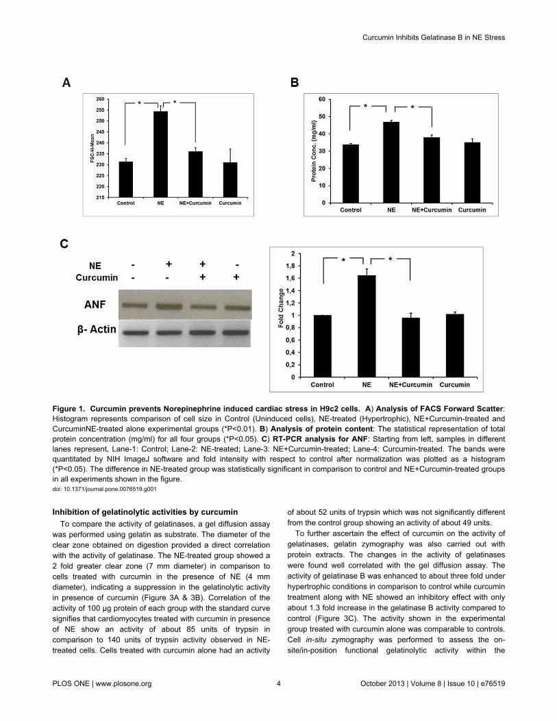
Optimal concentration of curcumin was determined throughMTT assay (Figure S1). H9c2 cells were treated with 8 µMcurcumin in the presence of 2 µM NE and incubated for 48 hrwithout any cytotoxicity. Besides this, cells were separatelytreated with curcumin alone without NE. Increase in size ofterminally differentiated cardiomyocytes, protein content andinduction of fetal genes such as ANF are indicators ofhypertrophic stress [19]. A reduction in the size ofcardiomyocytes was observed after treatment with curcuminunder hypertrophic conditions as reported earlier by our group(Figure S2). FACS data shows a decrease in the forwardscatter (FSC) with curcumin treatment in presence of NE-induced stress indicating a reduction in cell size after curcumintreatment (Figure 1A). A reduction in protein content was alsoobserved on treatment with curcumin in the presence of NE ascompared to the cardiomyocytes which were treated with NEalone (Figure 1B). Curcumin treatment along with NE resultedin downregulation of ANF gene expression in comparison tothe NE-treated experimental group (Figure 1C). These datasuggest that curcumin prevents NE-induced hypertrophicstress in H9c2 cardiomyocytes. Curcumin alone however didnot show any significant effect.
Increase in collagen content due to hypertrophic stressand effect of curcumin
The total collagen content and the effect of curcumin on itwere studied since collagen is a major ECM protein involved inhypertrophic remodeling (Figure 2A). Picrosirius staininganalysis showed a 3.5-4 fold increase in collagen contentunder hypertrophic conditions. Treatment of cells with curcuminin presence of NE, significantly prevented the increase incollagen content which was seen in NE-treated experimentalgroup. Treatment with curcumin alone was however seen to becomparable to control group. Immunocytochemistry wasperformed to study the expression of collagen-IV, the majorcollagen involved in hypertrophy (Figure 2B). Similarcorrelation was observed and curcumin treatment significantlysuppressed the expression of collagen-IV which was enhancedunder hypertrophic conditions. This emphasizes that theincrease in collagen turnover caused due to NE can beprevented by curcumin treatment.
Curcumin Inhibits Gelatinase B in NE Stress
PLOS ONE | www.plosone.org 3 October 2013 | Volume 8 | Issue 10 | e76519
Inhibition of gelatinolytic activities by curcuminTo compare the activity of gelatinases, a gel diffusion assay
was performed using gelatin as substrate. The diameter of theclear zone obtained on digestion provided a direct correlationwith the activity of gelatinase. The NE-treated group showed a2 fold greater clear zone (7 mm diameter) in comparison tocells treated with curcumin in the presence of NE (4 mmdiameter), indicating a suppression in the gelatinolytic activityin presence of curcumin (Figure 3A & 3B). Correlation of theactivity of 100 µg protein of each group with the standard curvesignifies that cardiomyocytes treated with curcumin in presenceof NE show an activity of about 85 units of trypsin incomparison to 140 units of trypsin activity observed in NE-treated cells. Cells treated with curcumin alone had an activity
of about 52 units of trypsin which was not significantly differentfrom the control group showing an activity of about 49 units.
To further ascertain the effect of curcumin on the activity ofgelatinases, gelatin zymography was also carried out withprotein extracts. The changes in the activity of gelatinaseswere found well correlated with the gel diffusion assay. Theactivity of gelatinase B was enhanced to about three fold underhypertrophic conditions in comparison to control while curcumintreatment along with NE showed an inhibitory effect with onlyabout 1.3 fold increase in the gelatinase B activity compared tocontrol (Figure 3C). The activity shown in the experimentalgroup treated with curcumin alone was comparable to controls.Cell in-situ zymography was performed to assess the on-site/in-position functional gelatinolytic activity within the
Figure 1. Curcumin prevents Norepinephrine induced cardiac stress in H9c2 cells. A) Analysis of FACS Forward Scatter:Histogram represents comparison of cell size in Control (Uninduced cells), NE-treated (Hypertrophic), NE+Curcumin-treated andCurcuminNE-treated alone experimental groups (*P<0.01). B) Analysis of protein content: The statistical representation of totalprotein concentration (mg/ml) for all four groups (*P<0.05). C) RT-PCR analysis for ANF: Starting from left, samples in differentlanes represent, Lane-1: Control; Lane-2: NE-treated; Lane-3: NE+Curcumin-treated; Lane-4: Curcumin-treated. The bands werequantitated by NIH ImageJ software and fold intensity with respect to control after normalization was plotted as a histogram(*P<0.05). The difference in NE-treated group was statistically significant in comparison to control and NE+Curcumin-treated groupsin all experiments shown in the figure.doi: 10.1371/journal.pone.0076519.g001
Curcumin Inhibits Gelatinase B in NE Stress
PLOS ONE | www.plosone.org 4 October 2013 | Volume 8 | Issue 10 | e76519
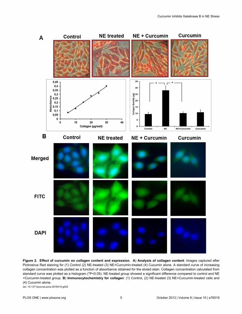
Figure 2. Effect of curcumin on collagen content and expression. A) Analysis of collagen content: Images captured afterPictrosirus Red staining for (1) Control (2) NE-treated (3) NE+Curcumin-treated (4) Cucumin alone. A standard curve of increasingcollagen concentration was plotted as a function of absorbance obtained for the eluted stain. Collagen concentration calculated fromstandard curve was plotted as a histogram (*P<0.05). NE-treated group showed a significant difference compared to control and NE+Curcumin-treated group. B) Immunocytochemistry for collagen: (1) Control, (2) NE-treated (3) NE+Curcumin-treated cells and(4) Cucumin alone.doi: 10.1371/journal.pone.0076519.g002
Curcumin Inhibits Gelatinase B in NE Stress
PLOS ONE | www.plosone.org 5 October 2013 | Volume 8 | Issue 10 | e76519

Figure 3. Effect of curumin on gelatinolytic activity. A) Gel-diffusion assay: Upper gel: Various concentrations of trypsin (1-Blank; 2-5 µg/µl; 3-10 µg/µl; 4-15 µg/µl; 5-20 µg/µl; 6-25 µg/µl; 7-30 µg/µl; 8-35 µg/µl) were added to different wells and proteaseactivity was observed as digested zones around it. A standard curve of the enzyme activity in units as a function of diameter of zonewas prepared. The enzyme activity for different samples shown in the lower gel (Control; NE-treated; NE+Curcumin-treated;Curcumin-treated alone) was calculated from the standard graph and represented as a histogram (*P<0.01). The difference of NE-treated was significant to control as well as NE+Curcumin-treated group. B) Gelatin Zymography: Samples in different lanes ofzymograms starting from the left represented as Lane-1: Control; Lane-2: NE-treated; Lane-3: NE+Curcumin-treated; Lane-4:Curcumin-treated alone. The fold change in the activity for bands corresponding to MMP-9 with respect to control was quantifiedusing ImageJ and plotted as histogram (*P<0.01, **P<0.05). C) in-situ Gelatin Zymography: The experiment was carried outunder different experimental conditions above and images captured by fluoresencent microscope at 20X magnifications arerepresented.doi: 10.1371/journal.pone.0076519.g003
Curcumin Inhibits Gelatinase B in NE Stress
PLOS ONE | www.plosone.org 6 October 2013 | Volume 8 | Issue 10 | e76519

cardiomyocytes. Significant difference was observed in theactivity shown by cells treated with NE alone. The activity wasinhibited remarkably after curcumin treatment in the presenceof NE (Figure 3D). Hence, these results emphasize thatcurcumin has an inhibitory effect on the activity of gelatinaseswhich was upregulated under NE-induced stress condition.
Curcumin decreases the expression of Gelatinase BOf the two members of the gelatinase family, gelatinase B
was chosen as the potential candidate for further analysis. Todetermine the effect of curcumin on the mRNA expression, weperformed semi-quantitative RT-PCR analysis. As shown inFigure 4A, curcumin-treated cardiac cells showed significantlylower mRNA expression levels of MMP-9 compared to the NE-induced cells. This was further confirmed by qRT-PCR analysiswhich showed about negligible increase on treatment withcurcumin along NE in comparison to 1.7 fold increase in mRNAexpression after NE treatment. To investigate if thedownregulation of MMP-9 was also at the protein level, westernblot was performed using MMP-9 antibody which showed thatcurcumin treatment along with NE prevented the increase inthe MMP-9 protein expression (Figure 4B). Similar results werealso observed in immunocytochemistry experiments (Figure4C). These results indicate that curcumin suppresses NE-induced MMP-9 expression in cardiomyocytes both at mRNAas well as protein levels.
Curcumin prevents nuclear localization of NF-κBGelatinase-B is transcriptionally regulated by NF-κB. In order
to find out the effect of curcumin on it, we carried outimmunofluorescence studies with NF-κB antibody to determineits localization under different experimental conditions. NEinduction resulted in localization of NF-κB inside the nucleus.This possibly triggers the transcriptional machinery and furtherexpression of MMP-9 gene. Curcumin-treated cells showed areduced localization of NF-κB inside the nucleus. NF-κB wasfound to be localized majorly in the cytoplasm (Figure 5A). Theresult was also confirmed by performing western blot of nuclearand cytoplasmic protein extracts separately. Expression of NF-κB was greater in the nuclear extract prepared from NE-induced cells and the cytosolic extracts of cells treated withboth curcumin and NE. Control as well as cells treated withcurcumin alone also showed cytoplasmic localization (Figure5B). These findings suggest that curcumin suppresses theentry of NF-κB triggered due to NE, inside the nucleus.
Discussion
The rationale of the present study is to assess the effect ofcurcumin on gelatinases under NE-induced stress conditions.In our study, it was observed that curcumin prevented anincrease in the collagen content and its expression. Moreover,it could effectively suppress the activity and expression ofgelatinase B.
H9c2 cells show almost identical hypertrophic responses tothose observed in primary cardiomyocytes and thus can beused as a model for in vitro studies of cardiac hypertrophy [20].Induction of hypertrophic stress is associated with an increase
in cell size and expression of marker genes such as ANF.Curcumin has a wide array of pharmacological effects but itsrole in cardiac hypertrophic remodeling is as yet largelyunknown. In our study, H9c2 cells undergoing NE-inducedstress show an increase in cell size, protein content and ANFgene expression which is prevented on treatment withcurcumin. This apparent difference suggests that curcumin isplaying a role in altering the cellular events in H9c2 cells inorder to circumvent the hypertrophic stress condition.
Previous studies have shown that cardiac hypertrophy ischaracterized by an overall imbalance of ECM turnover withmyocardial collagen accumulation [21,22]. It has been reportedthat under cardiac stress, an increase in ventricular collagendeposited leads to a rise in collagen concentration as well aschanges in collagen composition [23]. Curcumin treatment ofH9c2 cells under hypertrophic conditions prevented theincrease in collagen content and expression in cardiomyocytesindicating that curcumin is directly able to target the process ofcollagen synthesis. The increase in collagen synthesis isassociated with an increase in the enzymatic activity ofcollagen degrading enzymes. Degradation of collagen formsgelatin which is further digested by gelatinases [24]. Theenzymatic activity of gelatinases is thus elevated underhypertrophic stress. Henceforth, the effect of curcumin on theactivity and expression of gelatin degrading enzymes wasevaluated. Activity assays demonstrate that curcuminsuppresses the gelatinolytic activity of the proteases from cellsundergoing stress. Cell in situ zymography, whichdemonstrates in-position activity of these proteases showedcomparable effects to the activity assays.
Collagen turnover and ECM remodeling that occur duringvarious physiological and pathological processes are largelydependent on the regulation of MMP activity [25]. Thereduction in ECM remodeling mainly by suppressed MMPactivity, further preventing collagen deposition appears to be anattractive therapeutic intervention for heart failure and can be ofclinical utility to humans. MMPs, especially gelatinases, areresponsible for regulating most of the matrix turnover, sincethey can collectively degrade the basement membrane proteinslike gelatin, collagen type-IV, V and VII, elastin andproteoglycans [26]. MMP activities were known to be up-regulated in cardiac tissues by β-adrenergic stimulation [27].The gelatin zymography results clearly emphasize that activityof gelatinases is elevated under hypertrophic conditions and issuppressed on treatment with curcumin. An inhibition ofventricular hypertrophy by blocking MMP activity was earlierseen in the TNF-α transgenic mouse model of dilatedcardiomyopathy [28]. Use of natural compounds which caninhibit the activity of MMPs has been advocated as a potentialtherapeutic approach in treatment of cardiovascular disorders[29].
MMP-9 was chosen as a candidate gelatinase for furtheranalysis of the effect of curcumin. MMP-2 is known to beconstitutively expressed by many cell types in culture, whileMMP-9 expression is induced by cytokines, growth factors, etc.[30]. It is known that MMP-9 activity and expression levels areelevated in cardiomyopathies but still the mechanistic outlook isnot very clear [31]. The upregulation of MMP-9 expression in
Curcumin Inhibits Gelatinase B in NE Stress
PLOS ONE | www.plosone.org 7 October 2013 | Volume 8 | Issue 10 | e76519
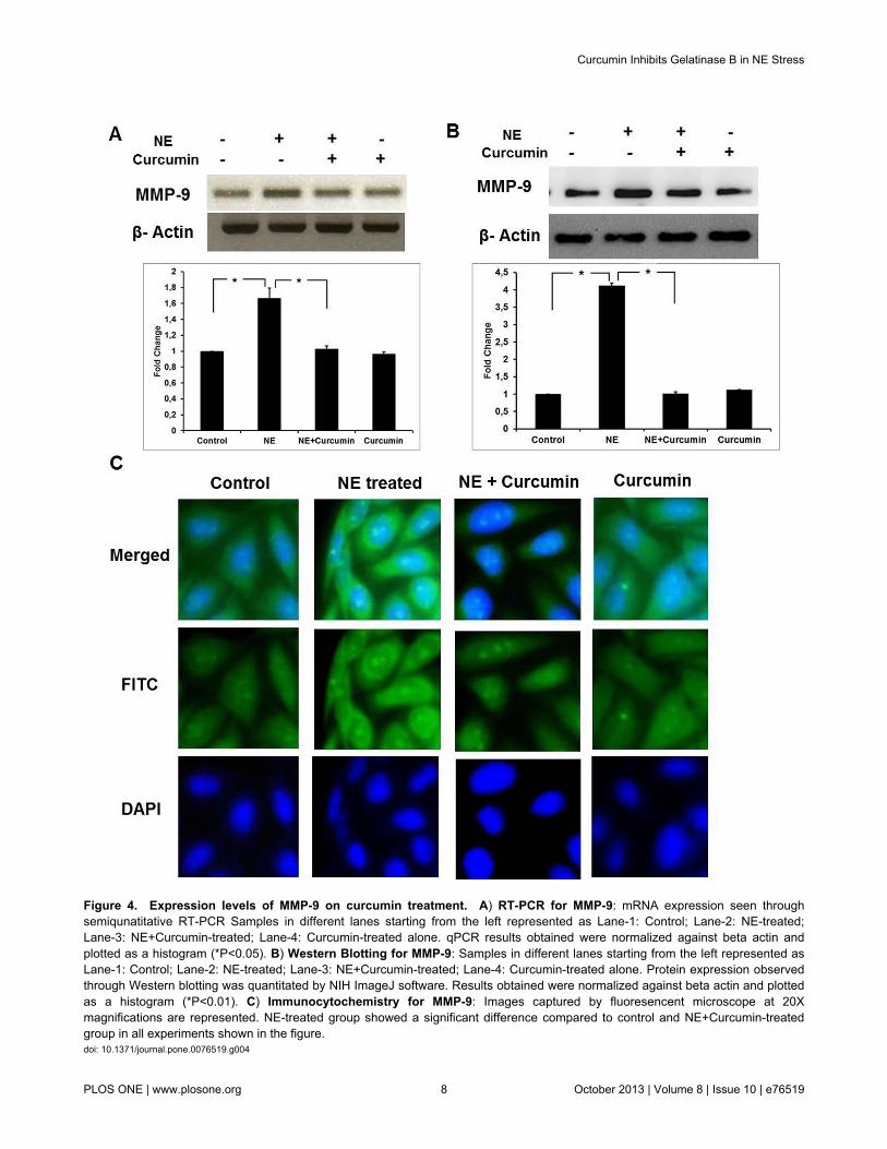
Figure 4. Expression levels of MMP-9 on curcumin treatment. A) RT-PCR for MMP-9: mRNA expression seen throughsemiqunatitative RT-PCR Samples in different lanes starting from the left represented as Lane-1: Control; Lane-2: NE-treated;Lane-3: NE+Curcumin-treated; Lane-4: Curcumin-treated alone. qPCR results obtained were normalized against beta actin andplotted as a histogram (*P<0.05). B) Western Blotting for MMP-9: Samples in different lanes starting from the left represented asLane-1: Control; Lane-2: NE-treated; Lane-3: NE+Curcumin-treated; Lane-4: Curcumin-treated alone. Protein expression observedthrough Western blotting was quantitated by NIH ImageJ software. Results obtained were normalized against beta actin and plottedas a histogram (*P<0.01). C) Immunocytochemistry for MMP-9: Images captured by fluoresencent microscope at 20Xmagnifications are represented. NE-treated group showed a significant difference compared to control and NE+Curcumin-treatedgroup in all experiments shown in the figure.doi: 10.1371/journal.pone.0076519.g004
Curcumin Inhibits Gelatinase B in NE Stress
PLOS ONE | www.plosone.org 8 October 2013 | Volume 8 | Issue 10 | e76519
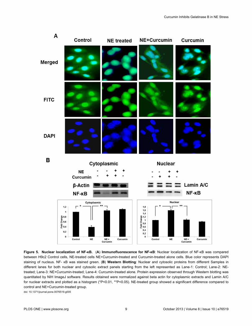
Figure 5. Nuclear localization of NF-κB. (A) Immunofluorescence for NF-κB: Nuclear localization of NF-κB was comparedbetween H9c2 Control cells, NE-treated cells NE+Curcumin-treated and Curcumin-treated alone cells. Blue color represents DAPIstaining of nucleus. NF- κB was stained green. (B) Western Blotting: Nuclear and cytosolic proteins from different Samples indifferent lanes for both nuclear and cytosolic extract panels starting from the left represented as Lane-1: Control; Lane-2: NE-treated; Lane-3: NE+Curcumin-treated; Lane-4: Curcumin-treated alone. Protein expression observed through Western blotting wasquantitated by NIH ImageJ software. Results obtained were normalized against beta actin for cytoplasmic extracts and Lamin A/Cfor nuclear extracts and plotted as a histogram (*P<0.01, **P<0.05). NE-treated group showed a significant difference compared tocontrol and NE+Curcumin-treated group.doi: 10.1371/journal.pone.0076519.g005
Curcumin Inhibits Gelatinase B in NE Stress
PLOS ONE | www.plosone.org 9 October 2013 | Volume 8 | Issue 10 | e76519
the current study agrees with earlier reported studies [32].From the present study, it can be suggested that curcumintargets the mRNA as well as protein expression of MMP-9.
The promoter of MMP-9 is highly conserved and carriesputative NF-κB binding sites [33]. Hence, there is aninvolvement of NF-κB in the process of MMP-9 upregulation[34]. By inhibiting NF-κB translocation from cytoplasm tonucleus, the MMP-9 transcriptional pathway is altered whichfurther alters the expression levels of this protein.
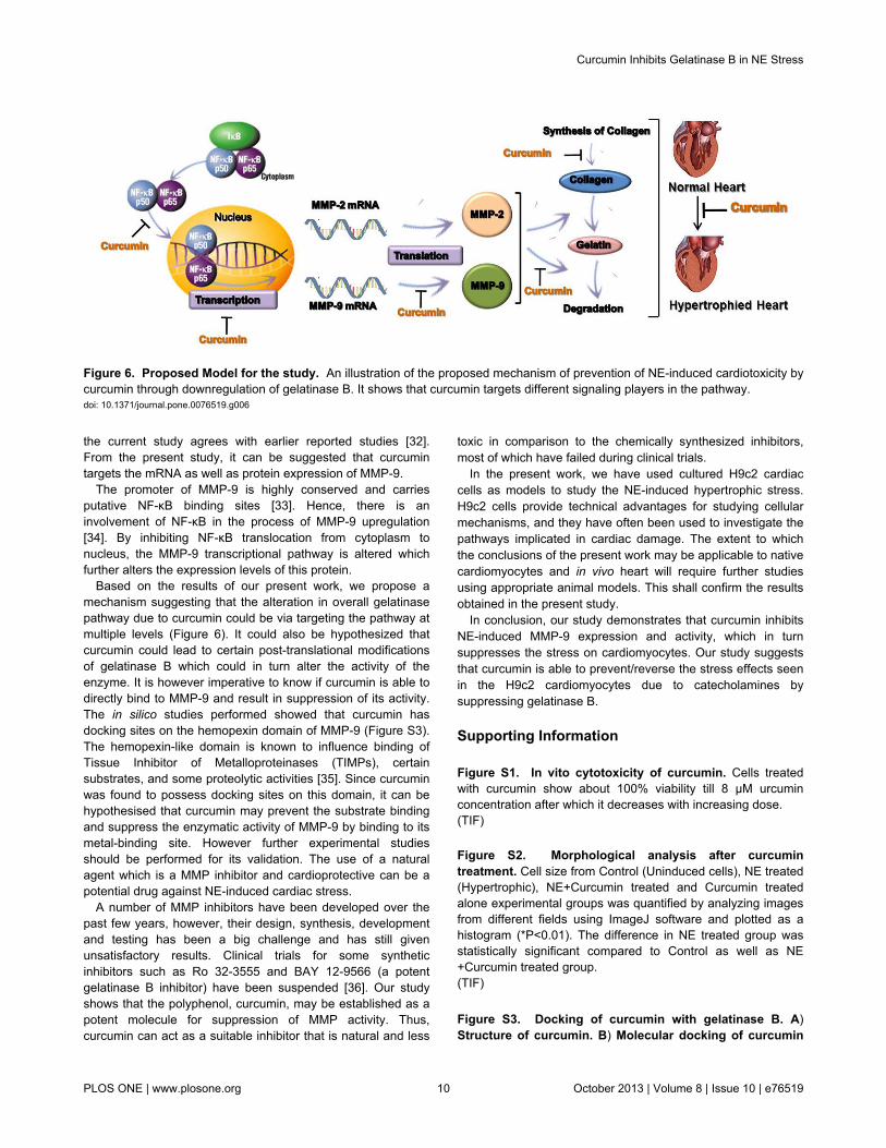
Based on the results of our present work, we propose amechanism suggesting that the alteration in overall gelatinasepathway due to curcumin could be via targeting the pathway atmultiple levels (Figure 6). It could also be hypothesized thatcurcumin could lead to certain post-translational modificationsof gelatinase B which could in turn alter the activity of theenzyme. It is however imperative to know if curcumin is able todirectly bind to MMP-9 and result in suppression of its activity.The in silico studies performed showed that curcumin hasdocking sites on the hemopexin domain of MMP-9 (Figure S3).The hemopexin-like domain is known to influence binding ofTissue Inhibitor of Metalloproteinases (TIMPs), certainsubstrates, and some proteolytic activities [35]. Since curcuminwas found to possess docking sites on this domain, it can behypothesised that curcumin may prevent the substrate bindingand suppress the enzymatic activity of MMP-9 by binding to itsmetal-binding site. However further experimental studiesshould be performed for its validation. The use of a naturalagent which is a MMP inhibitor and cardioprotective can be apotential drug against NE-induced cardiac stress.
A number of MMP inhibitors have been developed over thepast few years, however, their design, synthesis, developmentand testing has been a big challenge and has still givenunsatisfactory results. Clinical trials for some syntheticinhibitors such as Ro 32-3555 and BAY 12-9566 (a potentgelatinase B inhibitor) have been suspended [36]. Our studyshows that the polyphenol, curcumin, may be established as apotent molecule for suppression of MMP activity. Thus,curcumin can act as a suitable inhibitor that is natural and less
toxic in comparison to the chemically synthesized inhibitors,most of which have failed during clinical trials.
In the present work, we have used cultured H9c2 cardiaccells as models to study the NE-induced hypertrophic stress.H9c2 cells provide technical advantages for studying cellularmechanisms, and they have often been used to investigate thepathways implicated in cardiac damage. The extent to whichthe conclusions of the present work may be applicable to nativecardiomyocytes and in vivo heart will require further studiesusing appropriate animal models. This shall confirm the resultsobtained in the present study.
In conclusion, our study demonstrates that curcumin inhibitsNE-induced MMP-9 expression and activity, which in turnsuppresses the stress on cardiomyocytes. Our study suggeststhat curcumin is able to prevent/reverse the stress effects seenin the H9c2 cardiomyocytes due to catecholamines bysuppressing gelatinase B.
Supporting Information
Figure S1. In vito cytotoxicity of curcumin. Cells treatedwith curcumin show about 100% viability till 8 µM urcuminconcentration after which it decreases with increasing dose.(TIF)
Figure S2. Morphological analysis after curcumintreatment. Cell size from Control (Uninduced cells), NE treated(Hypertrophic), NE+Curcumin treated and Curcumin treatedalone experimental groups was quantified by analyzing imagesfrom different fields using ImageJ software and plotted as ahistogram (*P<0.01). The difference in NE treated group wasstatistically significant compared to Control as well as NE+Curcumin treated group.(TIF)
Figure S3. Docking of curcumin with gelatinase B. A)Structure of curcumin. B) Molecular docking of curcumin
Figure 6. Proposed Model for the study. An illustration of the proposed mechanism of prevention of NE-induced cardiotoxicity bycurcumin through downregulation of gelatinase B. It shows that curcumin targets different signaling players in the pathway.doi: 10.1371/journal.pone.0076519.g006
Curcumin Inhibits Gelatinase B in NE Stress
PLOS ONE | www.plosone.org 10 October 2013 | Volume 8 | Issue 10 | e76519

and gelatinase B: Amino acid residues SER80 and THR577were found to be critical in docking studies of curcumin andgelatinase B as indicated by the yellow dotted lines.(TIF)
Supporting Information S1. Supporting methods.(DOC)
Acknowledgements
We would like acknowledge Jaypee Institute of InformationTechnology, NOIDA, for providing infrastructural support.
Author Contributions
Conceived and designed the experiments: SK AC AJ MS VR.Performed the experiments: SK AC AJ YR. Analyzed the data:SK VR. Contributed reagents/materials/analysis tools: MS VR.Wrote the manuscript: SK AC AJ VR. Revised the manuscript:SK AC AJ MS VR.
References
1. Rona G (1985) Catecholamine cardiotoxicity. J Mol Cell Cardiol 17:291-306. doi:10.1016/S0022-2828(85)80130-9. PubMed: 3894676.
2. Rump AF, Schierholz J, Klaus W (2002) Studies on the cardiotoxicity ofNorepinephrine in isolated rabbit hearts. Arzneimittelforschung 52:543-551. PubMed: 12189778.
3. Kang YJ (2001) Molecular and cellular mechanisms of cardiotoxicity.Environ Health Perspect 109: 27-34. doi:10.2307/3434844. PubMed:11250803.
4. Berk BC, Fujiwara K, Lehoux S (2007) ECM remodeling in hypertensiveheart disease. J Clin Invest 117: 568-575. doi:10.1172/JCI31044.PubMed: 17332884.
5. Lindsey MK, Borg TK (2010) Understanding the role of the extracellularmatrix in cardiovascular development and disease: where do we gofrom here? J Mol Cell Cardiol 48: 431-432. doi:10.1016/j.yjmcc.2009.09.007. PubMed: 19781548.
6. Tyagi SC (1997) Proteinases and myocardial extracellular matrixturnover. Mol Cell Biochem 168: 1-12. doi:10.1023/A:1006850903242.PubMed: 9062888.
7. Jaiswal A, Chhabra A, Malhotra U, Kohli S, Rani V (2011) Comparativeanalysis of human matrix metalloproteinases: Emerging therapeutictargets in diseases. Bioinformation 6: 23-30. doi:10.6026/97320630006023. PubMed: 21464841.
8. Spinale FG, Coker ML, Bond BR, Zellner JL (2000) Myocardial matrixdegradation and metalloproteinase activation in the failing heart: apotential therapeutic target. Cardiovasc Res 46: 225-238. doi:10.1016/S0008-6363(99)00431-9. PubMed: 10773226.
9. Kizaki K, Ito R, Okada M, Yoshioka K, Uchide T et al. (2006) Enhancedgene expression of myocardial matrix metalloproteinases 2 and 9 afteracute treatment with doxorubicin in mice. Pharmacol Res 53: 341-346.doi:10.1016/j.phrs.2006.01.001. PubMed: 16455267.
10. Epstein JA (2008) Currying favor for the heart. J Clin Invest 118:850-852. PubMed: 18292806.
11. De R, Kundu P, Swarnakar S, Ramamurthy T, Chowdhury A et al.(2009) Antimicrobial activity of curcumin against Helicobacter pyloriisolates from India and during infections in mice. Antimicrob AgentsChemother 53: 1592-1597. doi:10.1128/AAC.01242-08. PubMed:19204190.
12. Sikora E, Scapagnini G, Barbagallo M (2010) Curcumin, inflammation,ageing and age-related diseases. Immun Ageing 7: 1. doi:10.1186/1742-4933-7-1. PubMed: 20205886.
13. Vanden WB (2012) Epigenetic impact of dietary polyphenols in cancerchemoprevention: Lifelong remodeling of our epigenomes. PharmacolRes 65: 565-576. doi:10.1016/j.phrs.2012.03.007. PubMed: 22465217.
14. Menon VP, Sudheer AR (2007) Antioxidant and anti-inflammatoryproperties of curcumin. Adv Exp Med Biol 595: 105-125. doi:10.1007/978-0-387-46401-5_3. PubMed: 17569207.
15. Yang F, Lim GP, Begum AN, Ubeda OJ, Simmons MR et al. (2005)Curcumin inhibits formation of amyloid beta oligomers and fibrils, bindsplaques, and reduces amyloid in vivo. J Biol Chem 280: 5892–5901.PubMed: 15590663.
16. Lim GP, Chu T, Yang F, Beech W, Frautschy SA et al. (2001) The curryspice curcumin reduces oxidative damage and amyloid pathology in anAlzheimer transgenic mouse. J Neurosci 21: 8370–8377. PubMed:11606625.
17. Hong D, Zeng X, Xu W, Ma J, Tong Y et al. (2010) Altered profiles ofgene expression in curcumin-treated rats with experimentally inducedmyocardial infarction. Pharmacol Res 61: 142-148. doi:10.1016/j.phrs.2009.08.009. PubMed: 19747544.
18. Wang NP, Wang ZF, Tootle S, Philip T, Zhao ZQ (2012) Curcuminpromotes cardiac repair and ameliorates cardiac dysfunction followingmyocardial infarction. Br J Pharmacol 167: 1550-1562. doi:10.1111/j.1476-5381.2012.02109.x. PubMed: 22823335.
19. Ahuja S, Kohli S, Krishnan S, Dogra D, Rani V et al. (2011) Curcumin:a potential therapeutic polyphenol, prevents noradrenaline-inducedhypertrophy in rat cardiac myocytes. J Pharm Pharmacol 63:1604-1612. doi:10.1111/j.2042-7158.2011.01363.x. PubMed:22060292.
20. Watkins SJ, Borthwick GM, Arthur HM (2011) The H9C2 cell line andprimary neonatal cardiomyocyte cells show similar hypertrophicresponses in-vitro. In Vitro Cell Dev Biol Anim 47: 125-131. doi:10.1007/s11626-010-9368-1. PubMed: 21082279.
21. Lombardi R, Betocchi S, Losi MA, Tocchetti CG, Aversa M et al. (2003)Myocardial collagen turnover in hypertrophic cardiomyopathy.Circulation 108: 1455-1460. doi:10.1161/01.CIR.0000090687.97972.10. PubMed: 12952838.
22. Weber KT (1989) Cardiac interstitium in health and disease: the fibrillarcollagen network. J Am Coll Cardiol 13: 1637-1652. doi:10.1016/0735-1097(89)90360-4. PubMed: 2656824.
23. Wang X, McLennan SV, Allen TJ, Twigg SM (2010) Regulation of pro-inflammatory and pro-fibrotic factors by CCN2/CTGF in H9c2cardiomyocytes. J Cell Commun Signal 4: 15-23. doi:10.1007/s12079-009-0083-1. PubMed: 20195389.
24. Bishop JE, Rhodes S, Laurent GJ, Low RB, Stirewalt WS (1994)Increased collagen synthesis and decreased collagen degradation inright ventricular hypertrophy induced by pressure overload. CardiovascRes 28: 1581-1585. doi:10.1093/cvr/28.10.1581. PubMed: 8001049.
25. Jugdutt BI (2003) Remodeling of the myocardium and potential targetsin the collagen degradation and synthesis pathways. Curr Drug TargetsCardiovasc Haematol Disord 3: 1-30. doi:10.2174/1568006033337276.PubMed: 12769643.
26. Kassiri Z, Khokha R (2005) Myocardial extra-cellular matrix and itsregulation by metalloproteinases and their inhibitors. Thromb Haemost93: 212-219. PubMed: 15711735.
27. Menon B, Singh M, Singh K (2005) Matrix metalloproteinases mediateβ-adrenergic receptor-stimulated apoptosis in adult rat ventricularmyocytes. Am J Physiol Cell Physiol 289: C168-C176. doi:10.1152/ajpheart.01235.2004. PubMed: 15728709.
28. Li YY, Kadokami T, Wang P, McTiernan CF, Feldman AM et al. (2002)MMP inhibition modulates TNF-alpha transgenic mouse phenotypeearly in the development of heart failure. Am J Physiol Heart CircPhysiol 282: H983-H989. PubMed: 11834496.
29. Sang QX, Jin Y, Newcomer RG, Monroe SC, Fang X et al. (2006)Matrix Metalloproteinase Inhibitors as Prospective Agents for thePrevention and Treatment of Cardiovascular and Neoplastic Diseases.Curr Top Med Chem 6: 289-316. doi:10.2174/156802606776287045.PubMed: 16611144.
30. Cawston TE (1995) Proteinases and inhibitors. Br Med Bull 51:385-401. PubMed: 7552071.
31. Liu P, Sun M, Sader S (2006) Matrix metalloproteinases incardiovascular disease. Can J Cardiol 22(Suppl B): 25B–30B. doi:10.1016/S0828-282X(06)70983-7. PubMed: 16498509.
32. Carvalho RF, Dariolli R, Justulin Junior LA, Sugizaki MM, Politi OkoshiM et al. (2006) Heart failure alters matrix metalloproteinase geneexpression and activity in rat skeletal muscle. Int J Exp Pathol 87:437-443. doi:10.1111/j.1365-2613.2006.00497.x. PubMed: 17222211.
33. Deschamps AM, Spinale FG (2006) Pathways of matrixmetalloproteinase induction in heart failure: Bioactive molecules and
Curcumin Inhibits Gelatinase B in NE Stress
PLOS ONE | www.plosone.org 11 October 2013 | Volume 8 | Issue 10 | e76519

transcriptional regulation. Cardiovasc Res 69: 666-676. doi:10.1016/j.cardiores.2005.10.004. PubMed: 16426590.
34. Bond M, Chase AJ, Baker AH, Newby AC (2001) Inhibition oftranscription factor NF-kB reduces matrix metalloproteinase-1, -3 and-9 productionby vascular smooth muscle cells. Cardiovasc Res 50:556–565. doi:10.1016/S0008-6363(01)00220-6. PubMed: 11376631.
35. Das S, Mandal M, Chakraborti T, Mandal A, Chakraborti S (2003)Structure and evolutionary aspects of matrix metalloproteinases: A brief
overview. Mol Cell Biochem 253: 31-40. doi:10.1023/A:1026093016148. PubMed: 14619953.
36. Brown PD (2000) Ongoing trials with matrix metalloproteinaseinhibitors. Expert Opin Investig Drugs 9: 2167-2177. doi:10.1517/13543784.9.9.2167. PubMed: 11060801.
Curcumin Inhibits Gelatinase B in NE Stress
PLOS ONE | www.plosone.org 12 October 2013 | Volume 8 | Issue 10 | e76519