li ca cervix
TRANSCRIPT
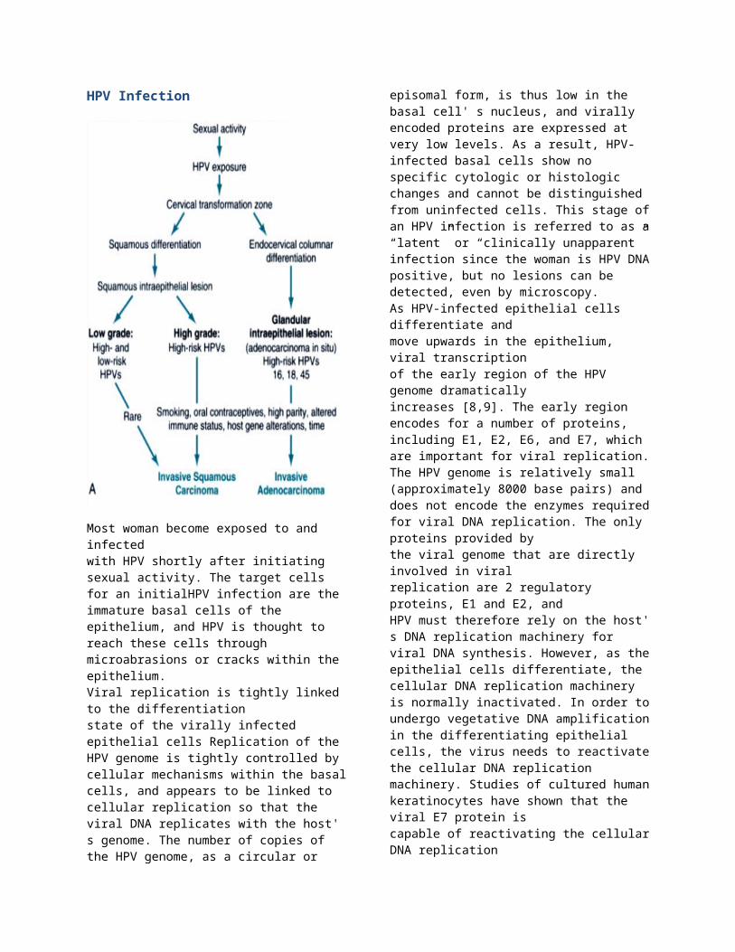
HPV Infection
Most woman become exposed to and infected with HPV shortly after initiating sexual activity. The target cells for an initialHPV infection are the immature basal cells of the epithelium, and HPV is thought to reach these cells through microabrasions or cracks within the epithelium.Viral replication is tightly linked to the differentiationstate of the virally infected epithelial cells Replication of the HPV genome is tightly controlled by cellular mechanisms within the basal cells, and appears to be linked to cellular replication so that the viral DNA replicates with the host' s genome. The number of copies of the HPV genome, as a circular or episomal form, is thus low in the basal cell' s nucleus, and virally encoded proteins are expressed at very low levels. As a result, HPV-infected basal cells show no specific cytologic or histologic changes and cannot be distinguished from uninfected cells. This stage of an HPV infection is referred to as a “latent” or “clinically unapparent” infection since the
woman is HPV DNA positive, but no lesions can be detected, even by microscopy.As HPV-infected epithelial cells differentiate andmove upwards in the epithelium, viral transcriptionof the early region of the HPV genome dramaticallyincreases [8,9]. The early region encodes for a number of proteins, including E1, E2, E6, and E7, which are important for viral replication. The HPV genome is relatively small (approximately 8000 base pairs) and does not encode the enzymes required for viral DNA replication. The only proteins provided bythe viral genome that are directly involved in viralreplication are 2 regulatory proteins, E1 and E2, andHPV must therefore rely on the host' s DNA replication machinery for viral DNA synthesis. However, as the epithelial cells differentiate, the cellular DNA replication machinery is normally inactivated. In order to undergo vegetative DNA amplification in the differentiating epithelial cells, the virus needs to reactivate the cellular DNA replication machinery. Studies of cultured human keratinocytes have shown that the viral E7 protein iscapable of reactivating the cellular DNA replicationmachinery in differentiated cells [11]. The viralE6 protein also appears to play an important roleby blocking the apoptosis that would normally occurin the differentiated cells. Together, thesechanges provide in the cell the synthetic phase environment necessary for vegetative viral DNA replication and complete virion formation. Infectious virus is eventually released as the differentiated cells are shed from the epithelium. In most women immunity develops against HPV after a period of months or years, and productive viral infection ceases. These women eventually become HPV DNA negative. In some HPV-infected women viral gene expression becomes unlinked to the state of differentiation of the infected epithelial cells, resulting in a change in the topography of viral gene expression within the epithelium. One of the results of this other mode of viral infection is the dramatic increase in the expression of E6 and E7 HPV in the lower layers of the epithelium due to the proteins' deregulated expression . The deregulated expression of HPV proteins E6 and E7 results in the disruption of normal cell cycle regulation; abrogation of apoptosis mechanisms; and genetic instability. Genetic instability, which is a characteristic feature of most malignant neoplasms, occurs early in the development of precancers, thereby allowing for the stepwise acquisition of multiple mutations.
Types of HPV Based on epithelial tissue
HPVs are small DNA viruses that infect various epithelial tissues including the epidermis (cutaneous types) and the epithelial linings of the upper respiratory system and anogenital tract (mucosotropic types)a. Mucosal HPV
A subset of mucosotropic HPVs that belong to the alpha genus, including the high-risk HPV types 16 and 18.Cervical epithelial cells that harbour integrated HPV 16 DNA have a selective growth advantage over cells that harbour normal extrachromosomal viral genomes; this growth advantage correlates with the increased expression of two viral genes in particular,E6 and E7. The early proteins, E6 and E7, bind and inactivate the tumour-suppressor gene product, p53, and the retinoblastoma tumour-suppressor protein (pRb), respectively). In cell lines derived from HPV-positive cervical cancers, these genes are not inactivated mutationally, whereas they are mutated in cell lines derived from HPV-negative cervical cancers. The expression of the E6 and E7 viral genes is required for the continued growth of cell lines derived from cervical cancers In addition, continuous expression of these early proteins can lead to the accumulation of mutations in the cellular genome that are required for malignant conversion. Both E6 and E7 co-operate to induce transformation of epithelial cells, however, a fully malignant phenotype is only observed after prolonged cultivation of the transformed cells (Hurlin et al., 1991; Dürst et al., 1995), which supports the multistep nature of HPV-induced transformation. In a transgenic mouse model, the expression of HPV16 E6 alone has been shown to be sufficient to induce carcinomas. In contrast, E6 and E7 of the low-risk mucosal types have very low transforming activities in vitro Exceptions include immortalization by the low-risk type HPV 6 of human mammary epithelial cells which are not the natural host cells of these viruses.
b. Cutaneous HPVsA subset of cutaneous HPV types classified into the beta genus (approximately 25 types, also called epidermodysplasia verruciformis (EV)-HPV types, have been sequenced so far) are commonly and consistently found in non-melanoma skin cancers. These skin tumours arise predominantly at sites exposed to the sun and, contrary to mucosal types, the EV-HPV DNA copy number appears to be much lower than one copy/cell Cutaneous HPVs have been studied less extensively than mucosal types and their capacity for cell transformation and molecular mechanisms are still largely unknown. Most cutaneous HPV types express E6 and E7 gene products that are structurally similar to those of the mucosal types but their genome does not harbour an identifiable E5 open-reading frame. Schmitt et al. (1994) performed a comparative analysis of various properties of the E6 and E7 proteins of EV-associated type 8 and non-EV-associated type 1 cutaneous HPVs by transfecting the genes into different cell lines. HPV 8 E6, HPV 16 E6 and E7 and HPV 1 E7 but not cottontail rabbit papillomavirus (CRPV) long E6 or HPV 8 E7 were able to transform immortalized mouse fibroblasts (C127 cell line) while cells that expressed HPV 1 E6 or CRPV short E6 exhibited a weak transformed phenotype. The in-vitro retinoblastoma protein (Rb)-binding affinity (relative to that of HPV 16 E7) was 66% for HPV 1 E7, 34% for HPV 8 E7 and 11% for CRPV E7. None of the E6 or E7 proteins of the cutaneous HPV types 1 or 8 or CRPVrevealed true immortalizing activities in primary human keratinocytes. In these cells, only a weak induction of proliferation was observed with HPV8 E7, and only HPV 8 E7 transformed primary rodent cells co-transfected with the EJ-Ras oncogene. HPV 5 and 8 E7s were shown to form complexes with the Rb protein, but with lower affinities than that of HPV 16 E7. Ciccolini found that HPV 1 E7 binds to pRb with an affinity similar to that of high-risk E7 proteins but has no transforming activity in primary rodent cells. HPV 8 E6 protein expressed in vitro was shown not to bind murine p53. Similarly, HPV 1 and 8 E6 proteins bound to neither human p53 nor E6-associated protein (E6-AP). Furthermore, HPV 8 E2, E4 and E6 were shown to interact with the TATA boxbinding protein (TBP) and a number of TBP-associated factors.Caldeira analysed the in-vitro properties of E7 proteins of cutaneous EV(HPV20 and 38) and non-EV- (HPV10) HPVtypes that are frequently detected in the skin. It was shown that HPV 38 E7 binds to and inactivates the tumour suppressor pRb and induces loss of G1/S transition control. In contrast, HPV 10 and HPV 20 E7 proteins do not display in-vitro transforming activities. Moreover, E6 and E7 of HPV 38 were shown to immortalize primary human keratinocytes, which suggests a role of HPV 38 infection in skin carcinogenesis.
Based on the ability to promote malignant transformationsa. high-risk , including 16, 18, 45, and 31, give contribution to invasive cancerb. moderate-risk account for the majority of cervical carcinomas, with smaller contributions by HPV 33, 35,
39, 45, 52, 56, 58, and 59. c. Low-risk , condylomas, which are benign lesions, are associated with infection by 6, 11, 42, and 44
Why does HPV affected in squamoucolumnar junction?
HPVs infect immature basal cells of the squamous ephitelium in areas of ephitelial breaks, or immature metaplastic squamous cells present at the squamoucolumnar junction. HPVs cannot infect the mature superficial squamous cells that cover the ectocervix, vagina, or vulva. Establishing HPV infection in these site requires damage to the surface ephitelium, which gives the virus access to the immature cells in basal layer of ephitelium. The cervix, with its correlatively large area of immature squamous metaplastic ephitelium, is particularly vulnerable to HPV infection as compared, for example, with vulvar skin and mucosa that covered by mature squamous cells. This difference in ephitelial susceptibility to HPV infection accounts for the marked difference in incidence of HPV-related cancers arising in different sites, and explains the high frequency of cervical cancer in woman or anal cancer in homosexual men and a relatively low frequency of vulvar and penile cancer.