jejunum and ileum location and...
TRANSCRIPT
The small intestine

DOUDENUM
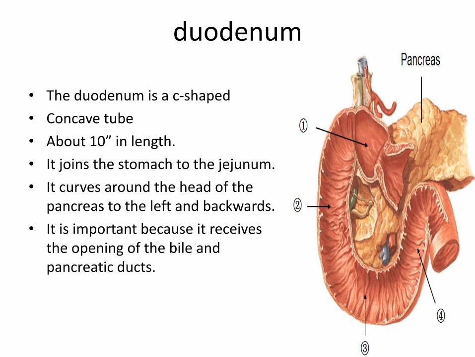
duodenum
• The duodenum is a c-shaped
• Concave tube
• About 10” in length.
• It joins the stomach to the jejunum.
• It curves around the head of the pancreas to the left and backwards.
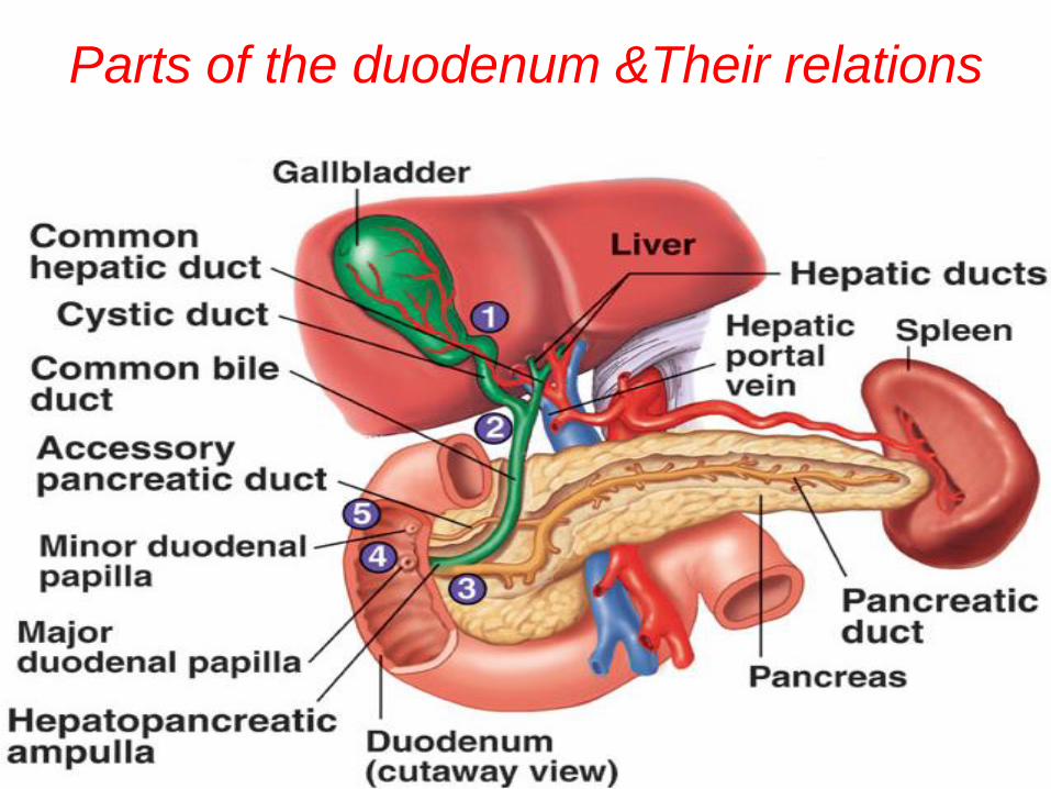
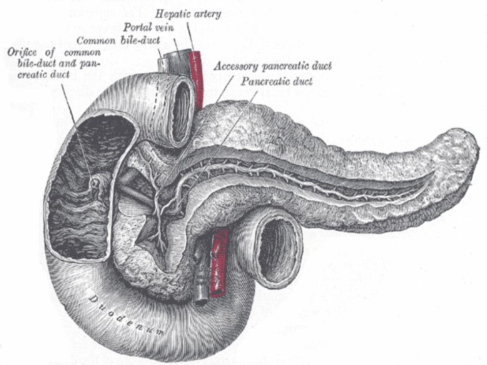
• It is important because it receives the opening of the bile and pancreatic ducts.
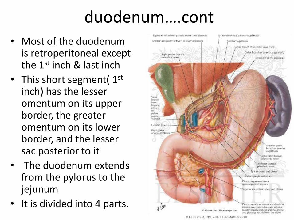
duodenum….cont • Most of the duodenum
is retroperitoneal except the 1st inch & last inch
• This short segment( 1st inch) has the lesser omentum on its upper border, the greater omentum on its lower border, and the lesser sac posterior to it
• The duodenum extends from the pylorus to the jejunum
• It is divided into 4 parts.
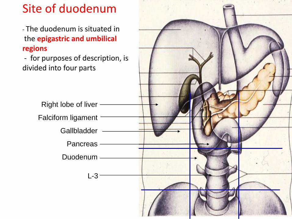
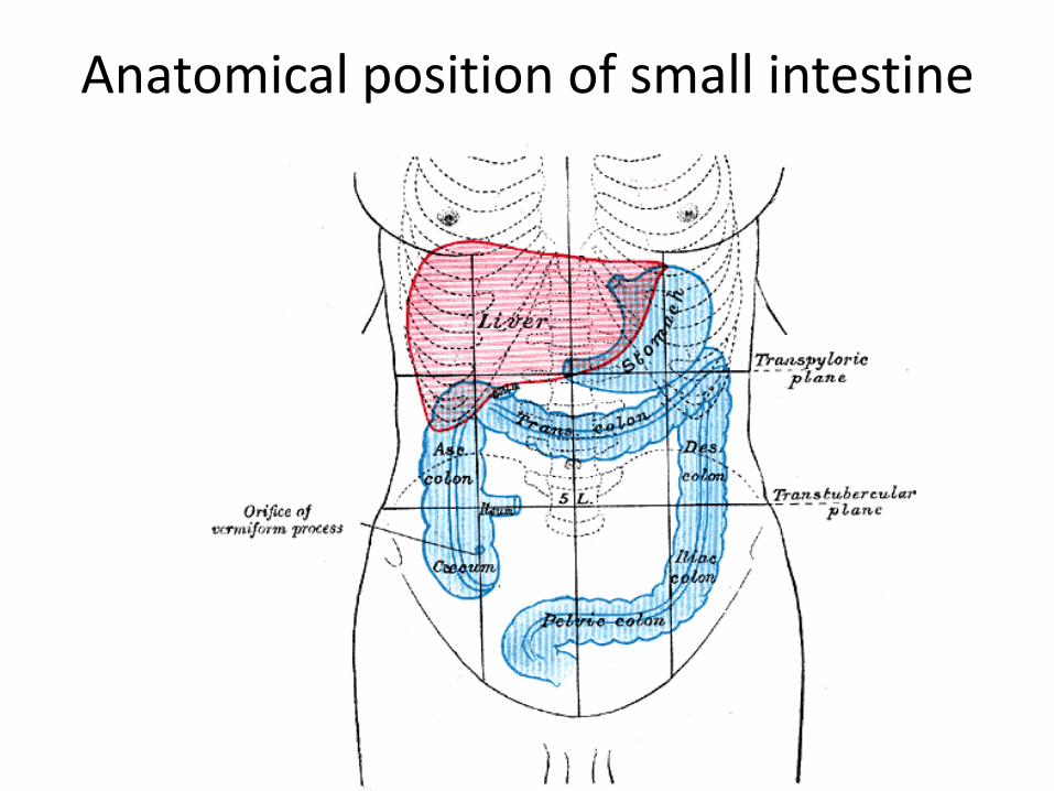
Site of duodenum
- The duodenum is situated in the epigastric and umbilical regions - for purposes of description, is divided into four parts
Right lobe of liver
Falciform ligament
Gallbladder
Pancreas
Duodenum
L-3
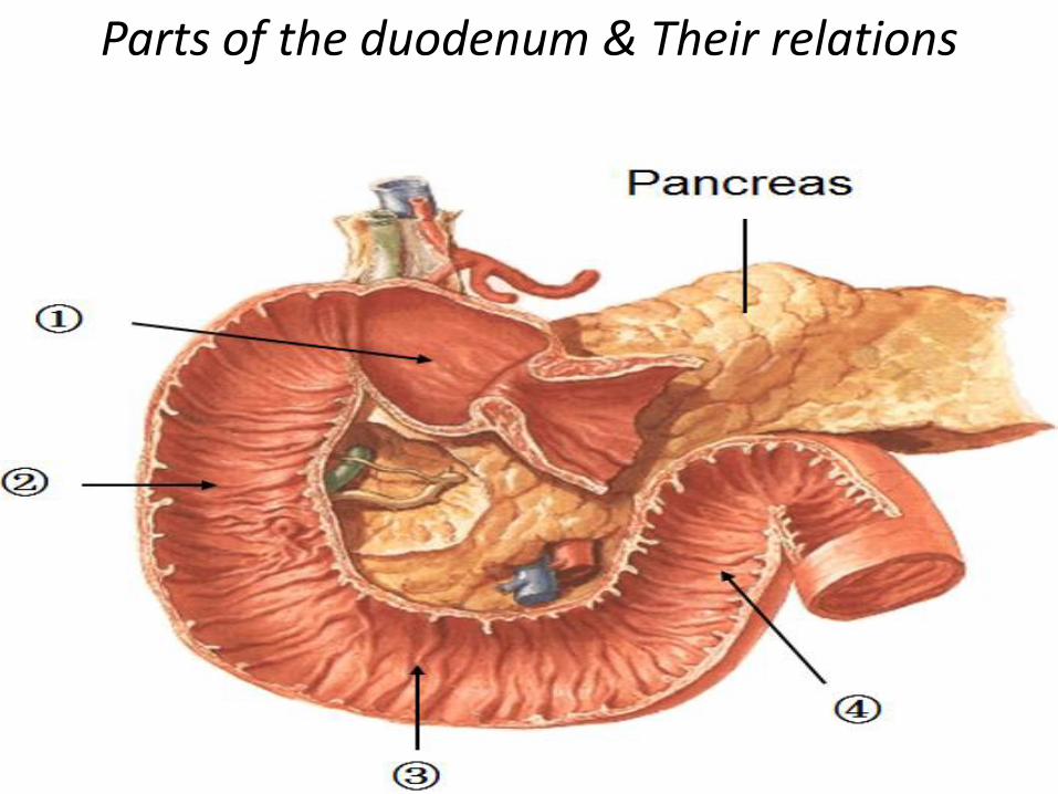
Parts of the duodenum & Their relations
Parts of the duodenum &Their relations
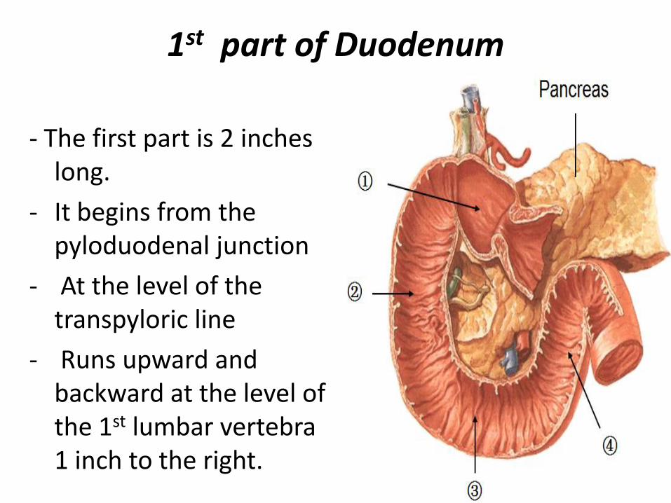
1st part of Duodenum
- The first part is 2 inches long.
- It begins from the pyloduodenal junction
- At the level of the transpyloric line
- Runs upward and backward at the level of the 1st lumbar vertebra 1 inch to the right.
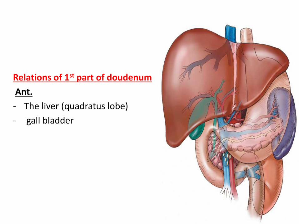
Relations of 1st part of doudenum
Ant.
- The liver (quadratus lobe)
- gall bladder
Relations of 1st part of duodenum……cont
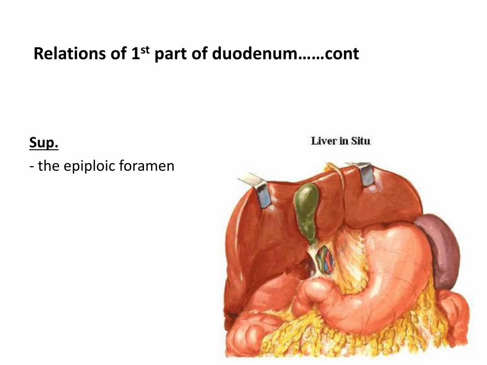
Sup.
- the epiploic foramen
Relations of 1st part duodenum……cont
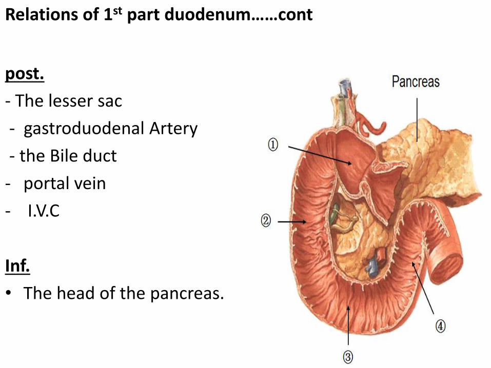
post.
- The lesser sac
- gastroduodenal Artery
- the Bile duct
- portal vein
- I.V.C
Inf.
• The head of the pancreas.
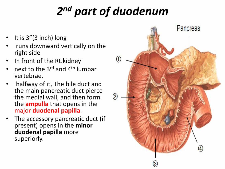
2nd part of duodenum
• It is 3”(3 inch) long • runs downward vertically on the
right side • In front of the Rt.kidney • next to the 3rd and 4th lumbar
vertebrae. • halfway of it, The bile duct and
the main pancreatic duct pierce the medial wall, and then form the ampulla that opens in the major duodenal papilla.
• The accessory pancreatic duct (if present) opens in the minor duodenal papilla more superiorly.
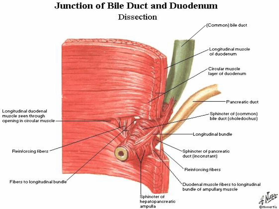
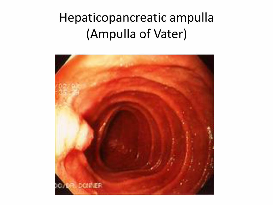
Hepaticopancreatic ampulla (Ampulla of Vater)
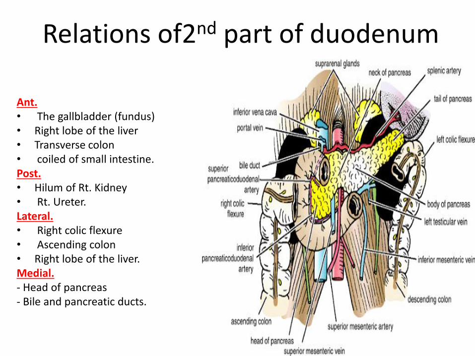
Relations of2nd part of duodenum
Ant. • The gallbladder (fundus) • Right lobe of the liver • Transverse colon • coiled of small intestine. Post. • Hilum of Rt. Kidney • Rt. Ureter. Lateral. • Right colic flexure • Ascending colon • Right lobe of the liver. Medial. - Head of pancreas - Bile and pancreatic ducts.
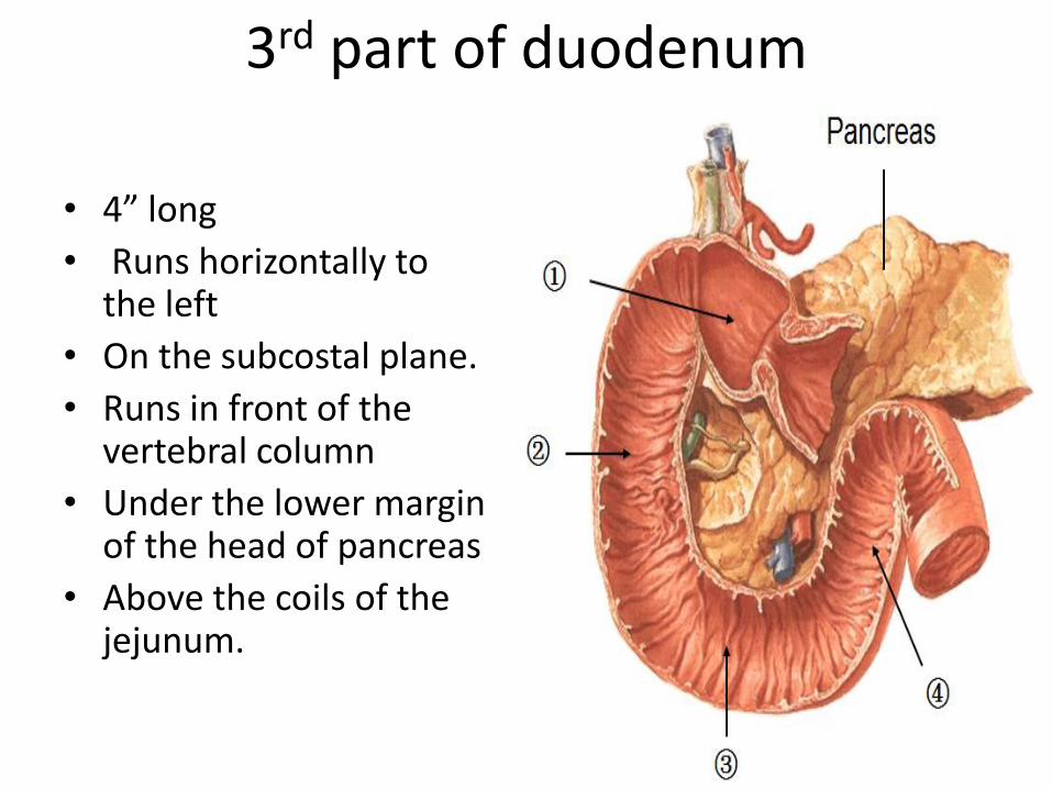
3rd part of duodenum
• 4” long
• Runs horizontally to the left
• On the subcostal plane.
• Runs in front of the vertebral column
• Under the lower margin of the head of pancreas
• Above the coils of the jejunum.
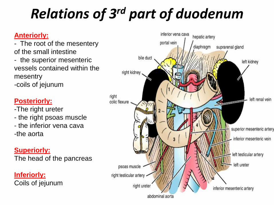
Relations of 3rd part of duodenum
Anteriorly:
- The root of the mesentery
of the small intestine
- the superior mesenteric
vessels contained within the
mesentry
-coils of jejunum
Posteriorly:
-The right ureter
- the right psoas muscle
- the inferior vena cava
-the aorta
Superiorly:
The head of the pancreas
Inferiorly:
Coils of jejunum
4th part of duodenum…..cont
• 1” long
• Runs upward to the left
• End in the duodejejunal junction at the level of the 2nd lumbar vertebrae 1” to the left.
• The junction (flexure) is held in position by the ligament of Treitz, which is attached to the right crus of the diaphragm (duodenal recess).

Relation of 4th part of duodenum Ant.
- The beginning of the root of the mesentery
- coils of the jejunum.
Post.
- Lt. psoas major
- the sympathetic chain
left margin of the aorta.
Sup.
- Uncinate process of the pancreas.
Blood supply of duodenum
• Arteries
1- upper half (1st part + upper1/2 of 2nd part) is supplied by the superior pancreaticoduodenal artery, a branch of the gastroduodenal artery .
2- The lower half (lower ½of 2nd part +3rd+4th part) is
supplied by the inferior pancreaticoduodenal artery, a branch of the superior mesenteric artery
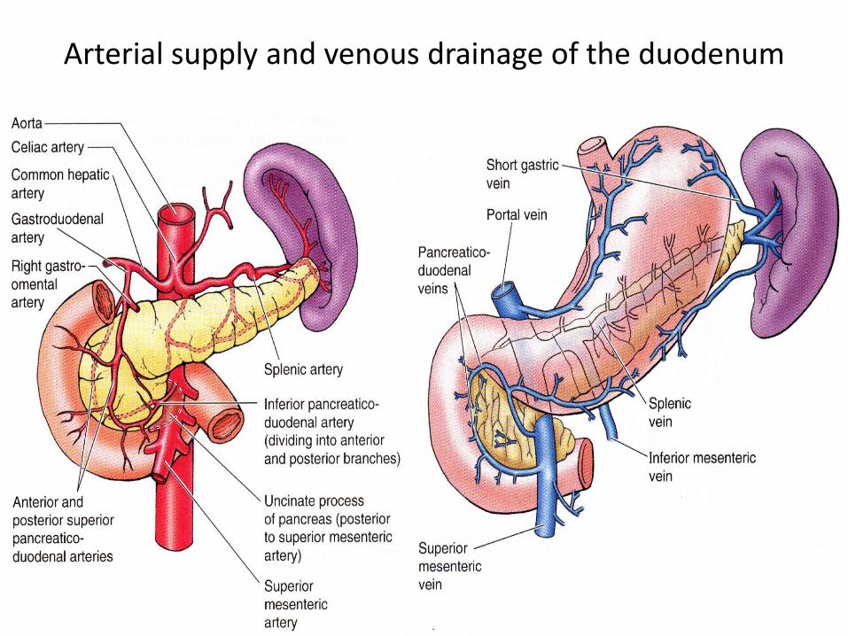
Arterial supply and venous drainage of the duodenum
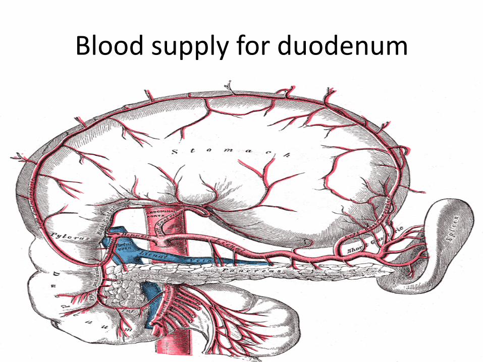
Blood supply for duodenum
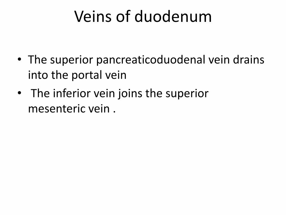
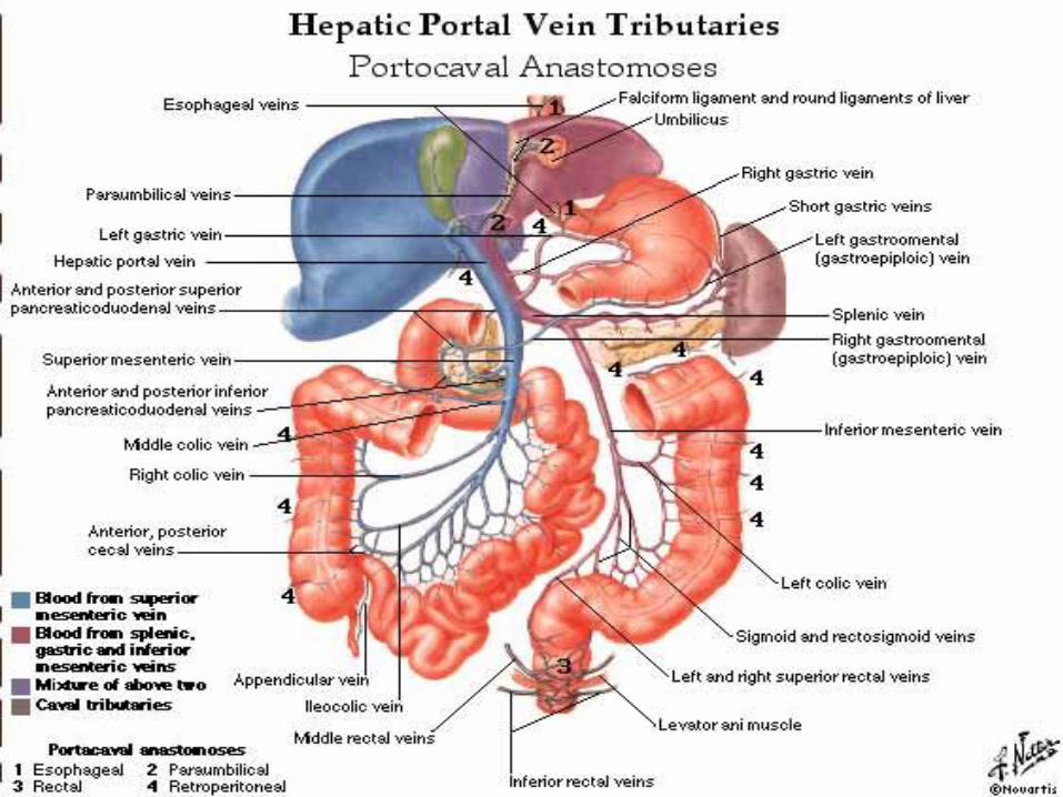
Veins of duodenum
• The superior pancreaticoduodenal vein drains into the portal vein
• The inferior vein joins the superior mesenteric vein .
Lymphatic drainage
• The lymph vessels follow the arteries
• drain upward via pancreaticoduodenal nodes the gastroduodenal nodes the celiac nodes
• drain downward via pancreaticoduodenal nodes the superior mesenteric nodes around the origin of the superior mesenteric artery.
Nerve supply
• Sympathetic nerve
• parasympathetic nerves from:
1- The celiac plexus
2- Superior mesenteric plexus.
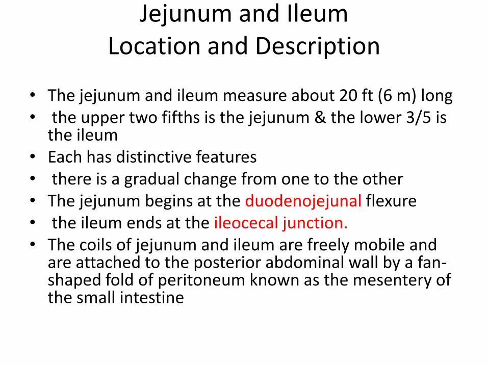
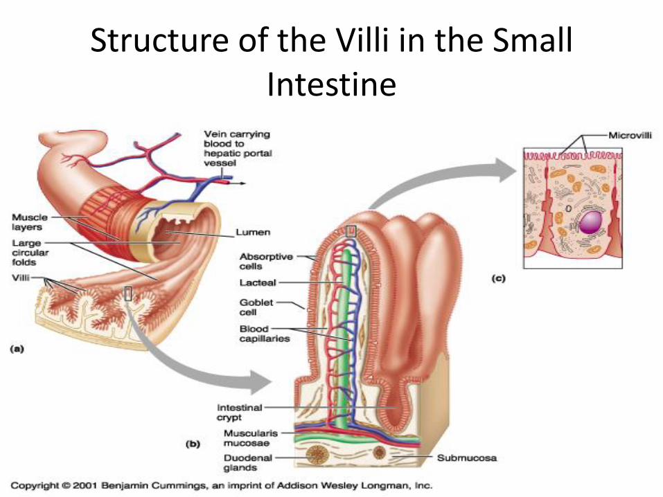
Jejunum and Ileum Location and Description
• The jejunum and ileum measure about 20 ft (6 m) long • the upper two fifths is the jejunum & the lower 3/5 is
the ileum • Each has distinctive features • there is a gradual change from one to the other • The jejunum begins at the duodenojejunal flexure • the ileum ends at the ileocecal junction. • The coils of jejunum and ileum are freely mobile and
are attached to the posterior abdominal wall by a fan-shaped fold of peritoneum known as the mesentery of the small intestine
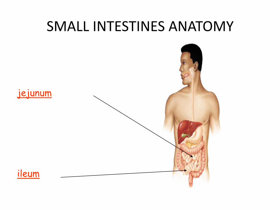
SMALL INTESTINES ANATOMY
jejunum
ileum

32
Structure of the Villi in the Small Intestine
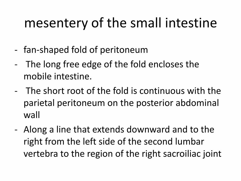
mesentery of the small intestine
- fan-shaped fold of peritoneum
- The long free edge of the fold encloses the mobile intestine.
- The short root of the fold is continuous with the parietal peritoneum on the posterior abdominal wall
- Along a line that extends downward and to the right from the left side of the second lumbar vertebra to the region of the right sacroiliac joint
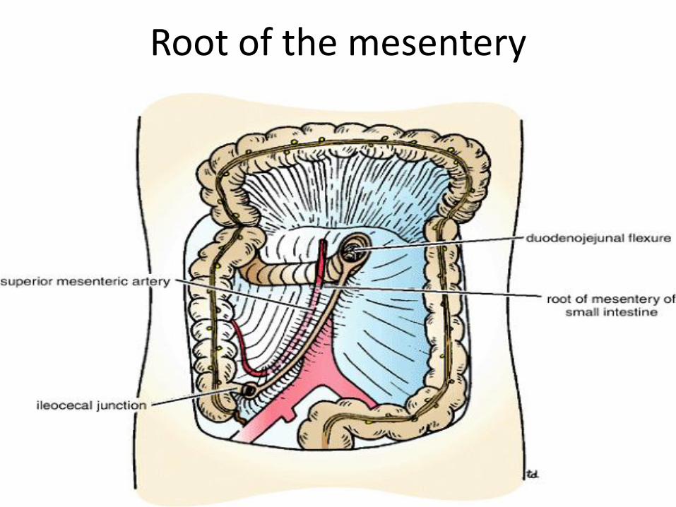
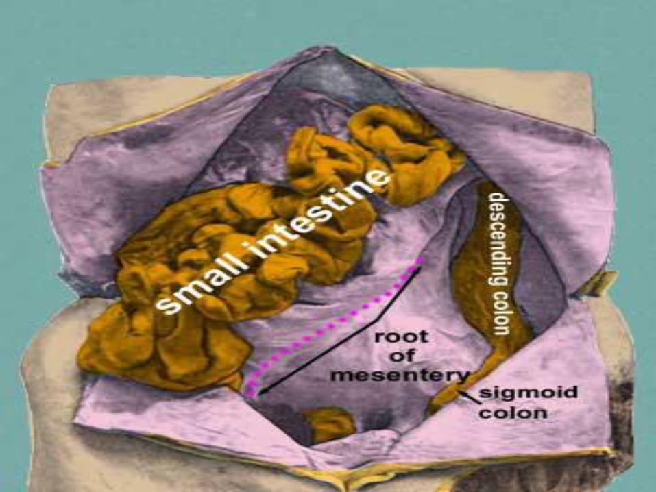
Root of the mesentery

Contents of the mesentery
- The branches of the superior mesenteric artery and vein
- Lymphatic vessels & lymphatic nodes
- nerves
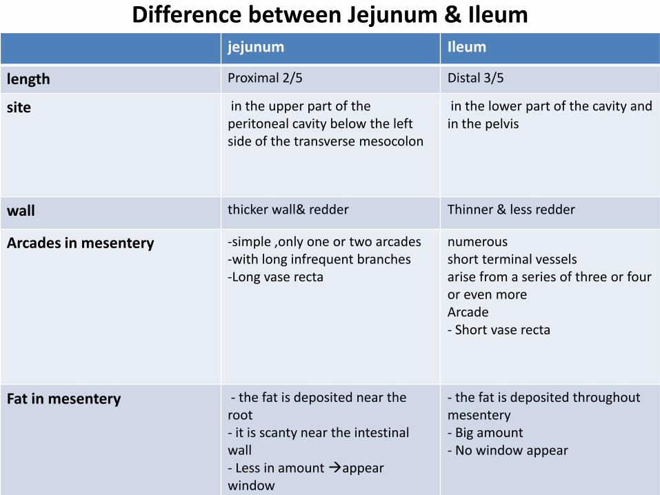
Difference between Jejunum & Ileum
Ileum jejunum
Distal 3/5 Proximal 2/5 length
in the lower part of the cavity and in the pelvis
in the upper part of the peritoneal cavity below the left side of the transverse mesocolon
site
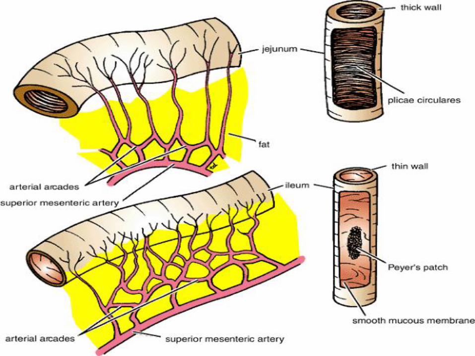
Thinner & less redder thicker wall& redder wall
numerous short terminal vessels arise from a series of three or four or even more Arcade - Short vase recta
-simple ,only one or two arcades -with long infrequent branches -Long vase recta
Arcades in mesentery
- the fat is deposited throughout mesentery - Big amount - No window appear
- the fat is deposited near the root - it is scanty near the intestinal wall - Less in amount appear window
Fat in mesentery
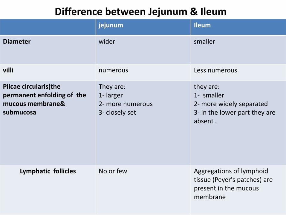
Difference between Jejunum & Ileum Ileum jejunum
smaller wider Diameter
Less numerous numerous villi
they are: 1- smaller 2- more widely separated
3- in the lower part they are absent .
They are:
1- larger
2- more numerous 3- closely set
Plicae circularis(the permanent enfolding of the mucous membrane& submucosa
Aggregations of lymphoid tissue (Peyer's patches) are present in the mucous membrane
No or few Lymphatic follicles
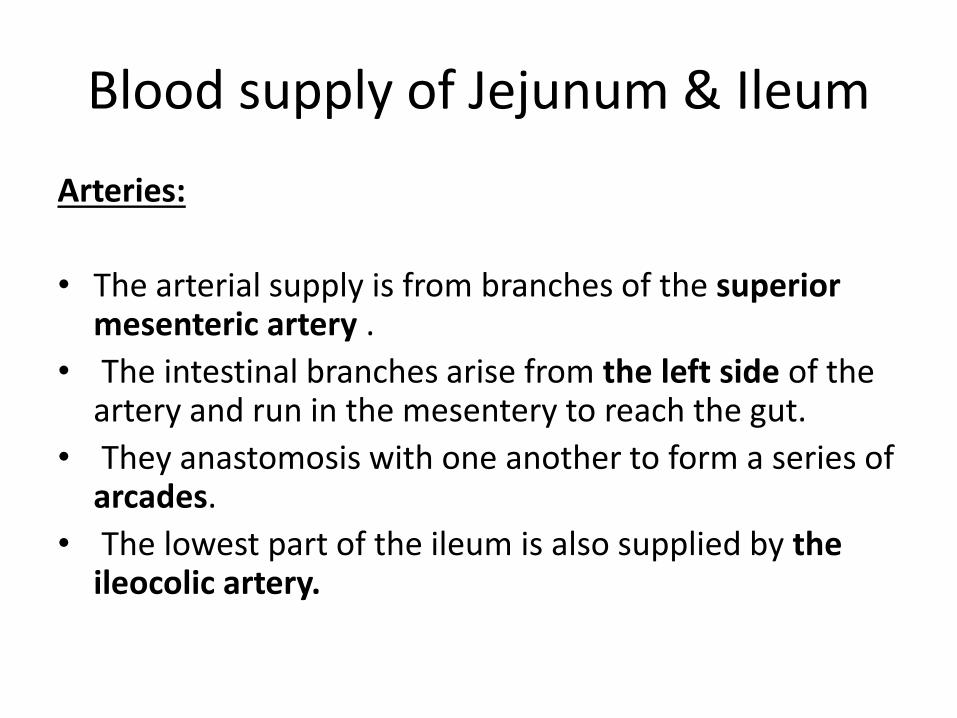
Blood supply of Jejunum & Ileum
Arteries:
• The arterial supply is from branches of the superior mesenteric artery .
• The intestinal branches arise from the left side of the artery and run in the mesentery to reach the gut.
• They anastomosis with one another to form a series of arcades.
• The lowest part of the ileum is also supplied by the ileocolic artery.
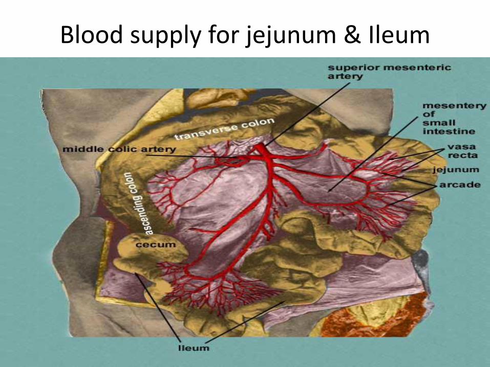
Blood supply for jejunum & Ileum
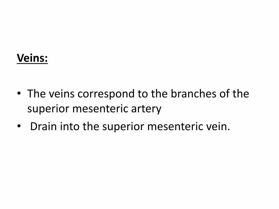
Veins:
• The veins correspond to the branches of the superior mesenteric artery
• Drain into the superior mesenteric vein.

Lymphatic Drainage of jejunum & ileum
• The lymph vessels pass through many intermediate mesenteric nodes
• Finally reach the superior mesenteric nodes around the origin of the superior mesenteric artery.
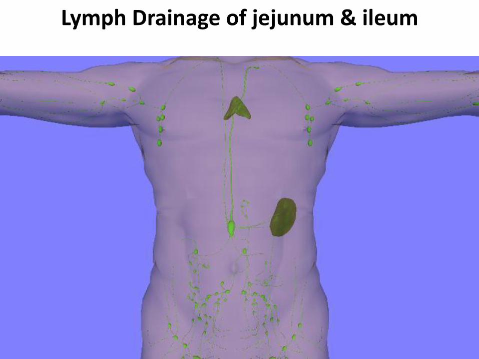
Lymph Drainage of jejunum & ileum
Nerve Supply of jejunum & Ileum
• The nerves are derived from the sympathetic and parasympathetic (vagus)
• Nerves from the superior mesenteric plexus.

Nerve supply for small intestine
Congenital anomaly of small intestine
Meckel's Diverticulum:
• a congenital anomaly of the ileum
• Present in 2% of people
• 2 feet from iliocecal junction
• 2 inch long
• contains gastric or pancreatic tissue
• Remains of vitelline duct of embryo
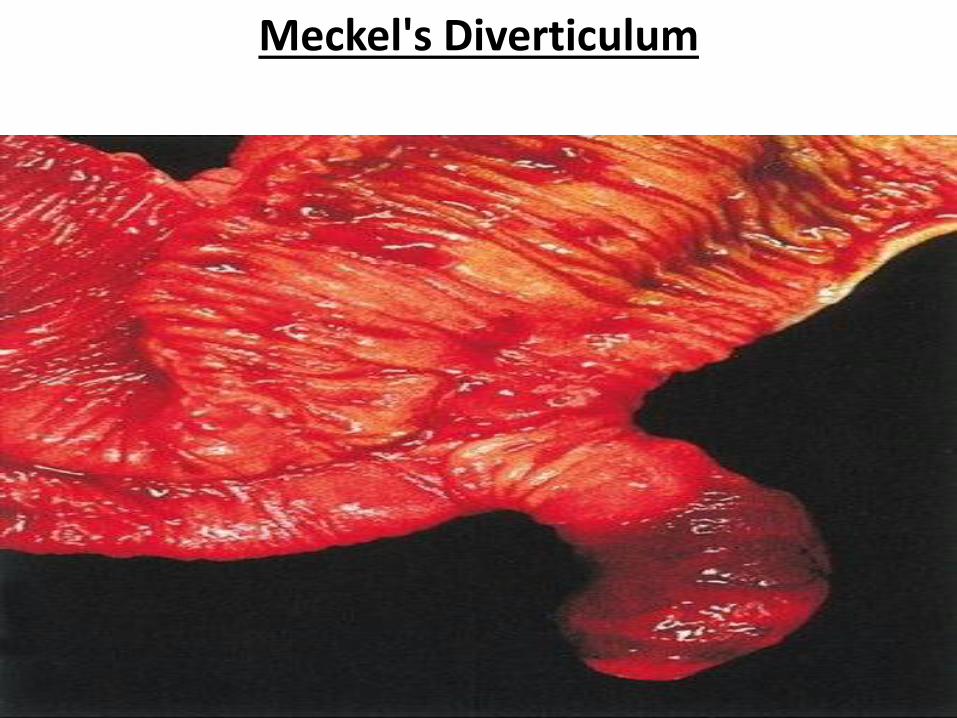
Meckel's Diverticulum