introduction - lms.ipb.ac.id bahan ajar fisiologi veteriner 2 (kardiovaskuler) 1 2/28/2017 bahan...
TRANSCRIPT
2/28/2017
1
D I V IS ION O F P H Y S IOLOGY
D E PARTEMEN T O F A N ATOYI , P H Y S IOLOGY A N D
P H A RMACOLOGY
FA C ULTY O F V E TERINARY M E DICINE
2/28/2017 BAHAN AJAR FISIOLOGI VETERINER 2 (KARDIOVASKULER) 1 2/28/2017 BAHAN AJAR FISIOLOGI VETERINER 2 (KARDIOVASKULER) 2
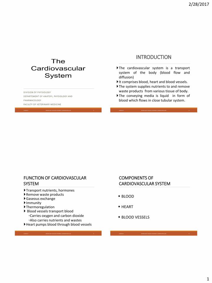
INTRODUCTION
The cardiovascular system is a transportsystem of the body (blood flow anddiffusion) It comprises blood, heart and blood vessels.The system supplies nutrients to and remove
waste products from various tissue of body.The conveying media is liquid in form of
blood which flows in close tubular system.
2/28/2017 BAHAN AJAR FISIOLOGI VETERINER 2 (KARDIOVASKULER) 3
FUNCTION OF CARDIOVASCULAR SYSTEM
Transport nutrients, hormonesRemove waste productsGaseous exchange ImmunityThermoregulation Blood vessels transport blood◦Carries oxygen and carbon dioxide◦Also carries nutrients and wastes
Heart pumps blood through blood vessels
2/28/2017 BAHAN AJAR FISIOLOGI VETERINER 2 (KARDIOVASKULER) 4
COMPONENTS OF CARDIOVASCULAR SYSTEM
BLOOD
HEART
BLOOD VESSELS
2/28/2017
2
2/28/2017 BAHAN AJAR FISIOLOGI VETERINER 2 (KARDIOVASKULER) 5
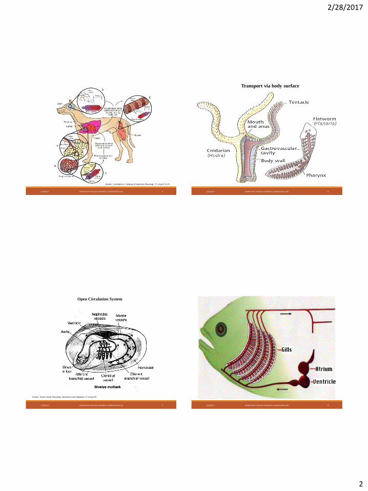
Sumber: Cunningham’s Textbook of Veterinary Physiology 5th ed pp:173-174
Transport via body surface
2/28/2017 BAHAN AJAR FISIOLOGI VETERINER 2 (KARDIOVASKULER) 6
Open Circulation System
2/28/2017 BAHAN AJAR FISIOLOGI VETERINER 2 (KARDIOVASKULER) 7
Sumber: Eckert Animal Physiology: Mechanism and Adaptation 4th ed pp:476
2/28/2017 BAHAN AJAR FISIOLOGI VETERINER 2 (KARDIOVASKULER) 8
2/28/2017
3
2/28/2017 BAHAN AJAR FISIOLOGI VETERINER 2 (KARDIOVASKULER) 9 2/28/2017 BAHAN AJAR FISIOLOGI VETERINER 2 (KARDIOVASKULER) 10
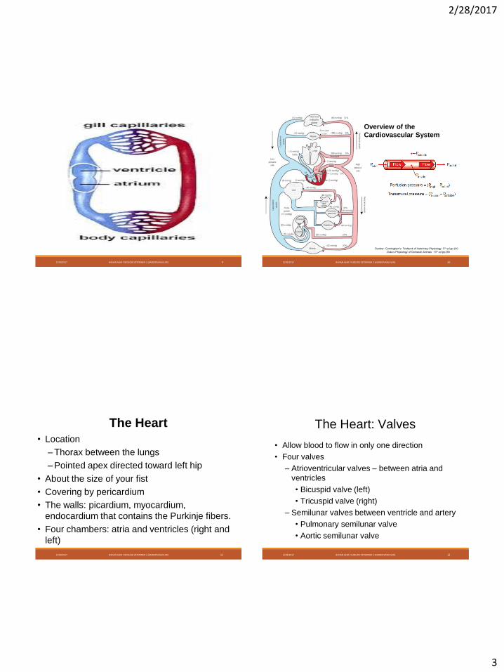
Duke’s Physiology of Domestic Animals 13th ed pp:289
Overview of the
Cardiovascular System
Sumber: Cunningham’s Textbook of Veterinary Physiology 5th ed pp:160
2/28/2017 BAHAN AJAR FISIOLOGI VETERINER 2 (KARDIOVASKULER) 11
The Heart
• Location
– Thorax between the lungs
– Pointed apex directed toward left hip
• About the size of your fist
• Covering by pericardium
• The walls: picardium, myocardium,
endocardium that contains the Purkinje fibers.
• Four chambers: atria and ventricles (right and
left)
2/28/2017 BAHAN AJAR FISIOLOGI VETERINER 2 (KARDIOVASKULER) 12
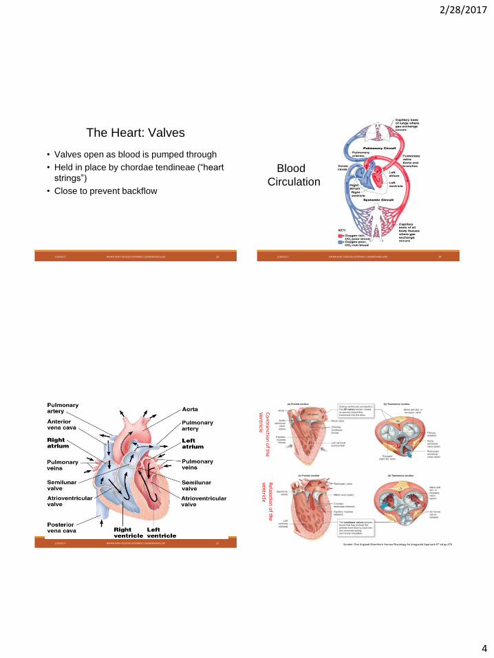
The Heart: Valves
• Allow blood to flow in only one direction
• Four valves
– Atrioventricular valves – between atria and
ventricles
• Bicuspid valve (left)
• Tricuspid valve (right)
– Semilunar valves between ventricle and artery
• Pulmonary semilunar valve
• Aortic semilunar valve
2/28/2017
4
2/28/2017 BAHAN AJAR FISIOLOGI VETERINER 2 (KARDIOVASKULER) 13
The Heart: Valves
• Valves open as blood is pumped through
• Held in place by chordae tendineae (“heart
strings”)
• Close to prevent backflow
2/28/2017 BAHAN AJAR FISIOLOGI VETERINER 2 (KARDIOVASKULER) 14
Blood
Circulation
2/28/2017 BAHAN AJAR FISIOLOGI VETERINER 2 (KARDIOVASKULER) 15 2/28/2017 BAHAN AJAR FISIOLOGI VETERINER 2 (KARDIOVASKULER) 16
Co
ntran
ction
of th
e V
entricle
Relaxatio
n o
f the
ventricle
Sumber: Dee Unglaub Silverthorn Human Physiology An Integrated Approach 6th ed pp 476
2/28/2017
5
2/28/2017 BAHAN AJAR FISIOLOGI VETERINER 2 (KARDIOVASKULER) 17
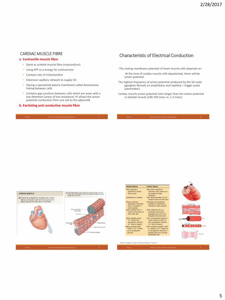
CARDIAC MUSCLE FIBREa. Contractile muscle fibre
• Same as sceletal muscle fibre (myocardium)
• Using ATP as a energy for contranction
• Contains lots of mitochondria
• Extensive capillary network to supply O2
• Having a specialized plasma membrane called desmosome linking between cells
• Contains gap junctions between cells which are areas with a low detention (areas of low resistance) allows the action potential conduction from one cell to the adjacentb
b. Excitating and conductive muscle fibre
2/28/2017 BAHAN AJAR FISIOLOGI VETERINER 2 (KARDIOVASKULER) 18
Characteristic of Electrical Conduction
-The resting membrane potential of heart muscle cells depends on:
At the time of cardiac muscle cells depolarized, there will be action potential
The highest frequency of action potential produced by the SA node (ganglion Remark on amphibians and reptiles) = trigger pulse (pacemaker)
Cardiac muscle action potential lasts longer than the action potential in skeletal muscle (100-250 msec vs. 1-2 msec)
2/28/2017 BAHAN AJAR FISIOLOGI VETERINER 2 (KARDIOVASKULER) 19 2/28/2017 BAHAN AJAR FISIOLOGI VETERINER 2 (KARDIOVASKULER) 20
Sumber: Cunningham’s Textbook of Veterinary Physiology 5th ed pp:172
2/28/2017
6
2/28/2017 BAHAN AJAR FISIOLOGI VETERINER 2 (KARDIOVASKULER) 21
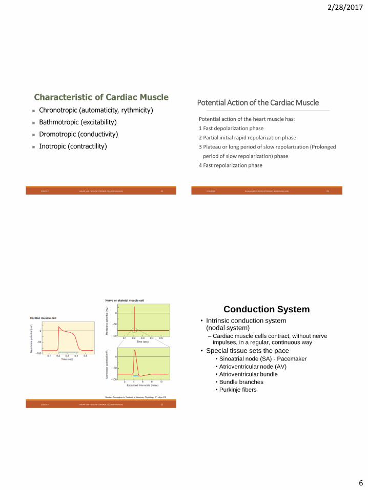
Characteristic of Cardiac Muscle
Chronotropic (automaticity, rythmicity)
Bathmotropic (excitability)
Dromotropic (conductivity)
Inotropic (contractility)
Potential Action of the Cardiac Muscle
Potential action of the heart muscle has:
1 Fast depolarization phase
2 Partial initial rapid repolarization phase
3 Plateau or long period of slow repolarization (Prolonged
period of slow repolarization) phase
4 Fast repolarization phase
2/28/2017 BAHAN AJAR FISIOLOGI VETERINER 2 (KARDIOVASKULER) 22
2/28/2017 BAHAN AJAR FISIOLOGI VETERINER 2 (KARDIOVASKULER) 23
Sumber: Cunningham’s Textbook of Veterinary Physiology 5th ed pp:176
Conduction System• Intrinsic conduction system
(nodal system)
– Cardiac muscle cells contract, without nerve impulses, in a regular, continuous way
• Special tissue sets the pace• Sinoatrial node (SA) - Pacemaker
• Atrioventricular node (AV)
• Atrioventricular bundle
• Bundle branches
• Purkinje fibers
2/28/2017
7
2/28/2017 BAHAN AJAR FISIOLOGI VETERINER 2 (KARDIOVASKULER) 25
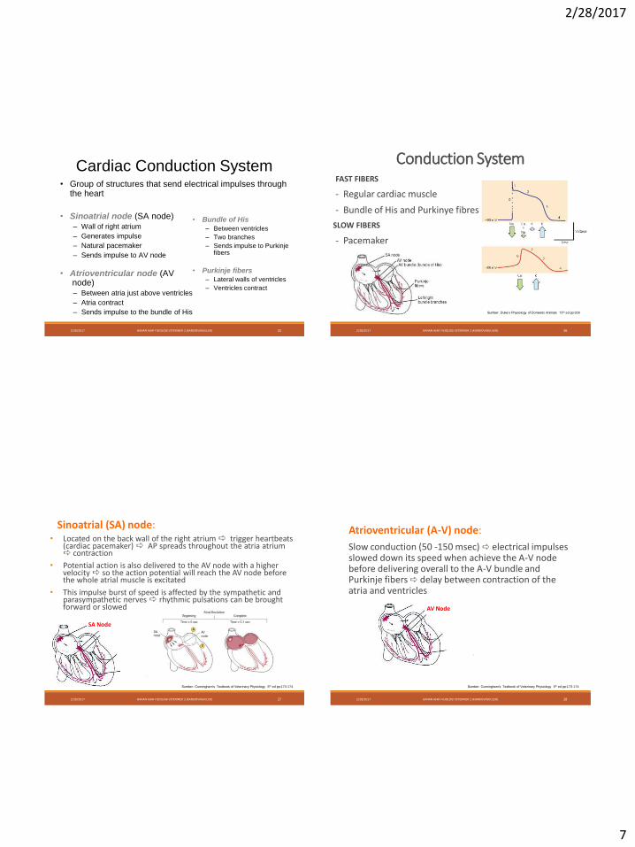
Cardiac Conduction System• Group of structures that send electrical impulses through
the heart
• Sinoatrial node (SA node)– Wall of right atrium
– Generates impulse
– Natural pacemaker
– Sends impulse to AV node
• Atrioventricular node (AVnode)– Between atria just above ventricles
– Atria contract
– Sends impulse to the bundle of His
• Bundle of His
– Between ventricles
– Two branches
– Sends impulse to Purkinje fibers
• Purkinje fibers
– Lateral walls of ventricles
– Ventricles contract
2/28/2017 BAHAN AJAR FISIOLOGI VETERINER 2 (KARDIOVASKULER) 26
Conduction SystemFAST FIBERS
- Regular cardiac muscle
- Bundle of His and Purkinye fibres
SLOW FIBERS
- Pacemaker
Sumber: Duke’s Physiology of Domestic Animals 13th ed pp:309
2/28/2017 BAHAN AJAR FISIOLOGI VETERINER 2 (KARDIOVASKULER) 27
Sinoatrial (SA) node:• Located on the back wall of the right atrium trigger heartbeats
(cardiac pacemaker) AP spreads throughout the atria atrium contraction
• Potential action is also delivered to the AV node with a higher velocity so the action potential will reach the AV node before the whole atrial muscle is excitated
• This impulse burst of speed is affected by the sympathetic and parasympathetic nerves rhythmic pulsations can be brought forward or slowed
SA Node
Sumber: Cunningham’s Textbook of Veterinary Physiology 5th ed pp:173-174
2/28/2017 BAHAN AJAR FISIOLOGI VETERINER 2 (KARDIOVASKULER) 28
Atrioventricular (A-V) node:
Slow conduction (50 -150 msec) electrical impulses slowed down its speed when achieve the A-V node before delivering overall to the A-V bundle and Purkinje fibers delay between contraction of the atria and ventricles
AV Node
Sumber: Cunningham’s Textbook of Veterinary Physiology 5th ed pp:173-174
2/28/2017
8
2/28/2017 BAHAN AJAR FISIOLOGI VETERINER 2 (KARDIOVASKULER) 29
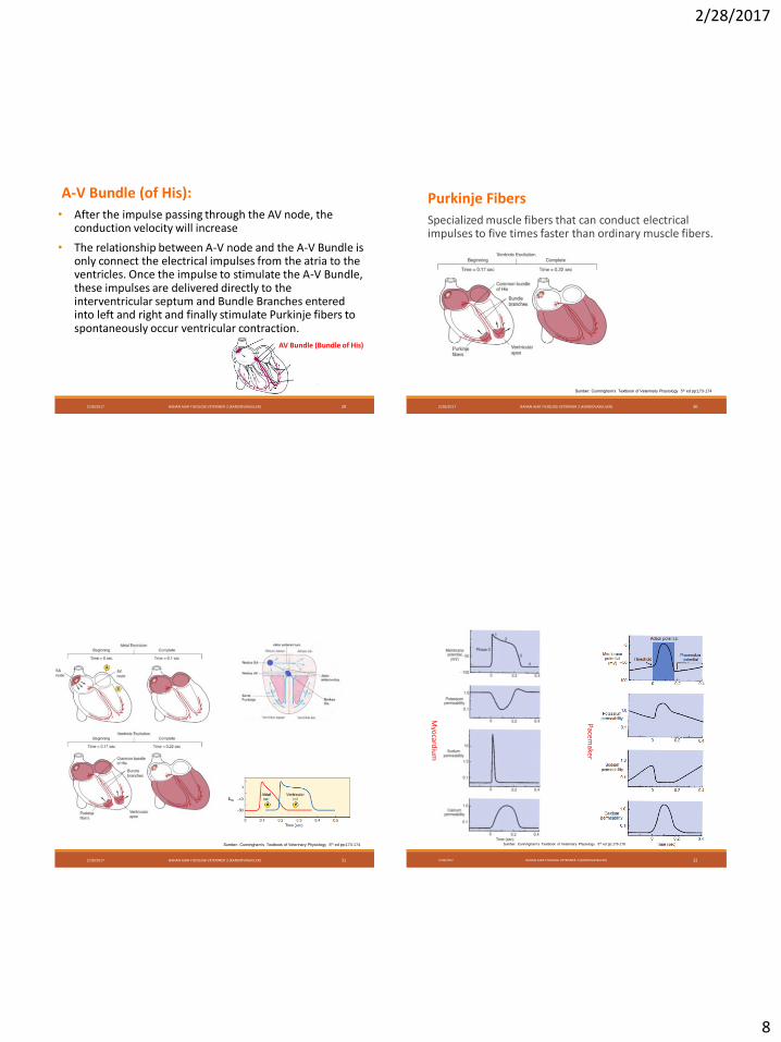
A-V Bundle (of His):
• After the impulse passing through the AV node, the conduction velocity will increase
• The relationship between A-V node and the A-V Bundle is only connect the electrical impulses from the atria to the ventricles. Once the impulse to stimulate the A-V Bundle, these impulses are delivered directly to the interventricular septum and Bundle Branches entered into left and right and finally stimulate Purkinje fibers to spontaneously occur ventricular contraction.
AV Bundle (Bundle of His)
2/28/2017 BAHAN AJAR FISIOLOGI VETERINER 2 (KARDIOVASKULER) 30
Purkinje Fibers
Specialized muscle fibers that can conduct electrical impulses to five times faster than ordinary muscle fibers.
Sumber: Cunningham’s Textbook of Veterinary Physiology 5th ed pp:173-174
2/28/2017 BAHAN AJAR FISIOLOGI VETERINER 2 (KARDIOVASKULER) 31
Sumber: Cunningham’s Textbook of Veterinary Physiology 5th ed pp:173-174
2/28/2017 BAHAN AJAR FISIOLOGI VETERINER 2 (KARDIOVASKULER) 32
Myo
cardiu
m
Pacemaker
Sumber: Cunningham’s Textbook of Veterinary Physiology 5th ed pp:176-178
2/28/2017
9
2/28/2017 BAHAN AJAR FISIOLOGI VETERINER 2 (KARDIOVASKULER) 33 2/28/2017 BAHAN AJAR FISIOLOGI VETERINER 2 (KARDIOVASKULER) 34
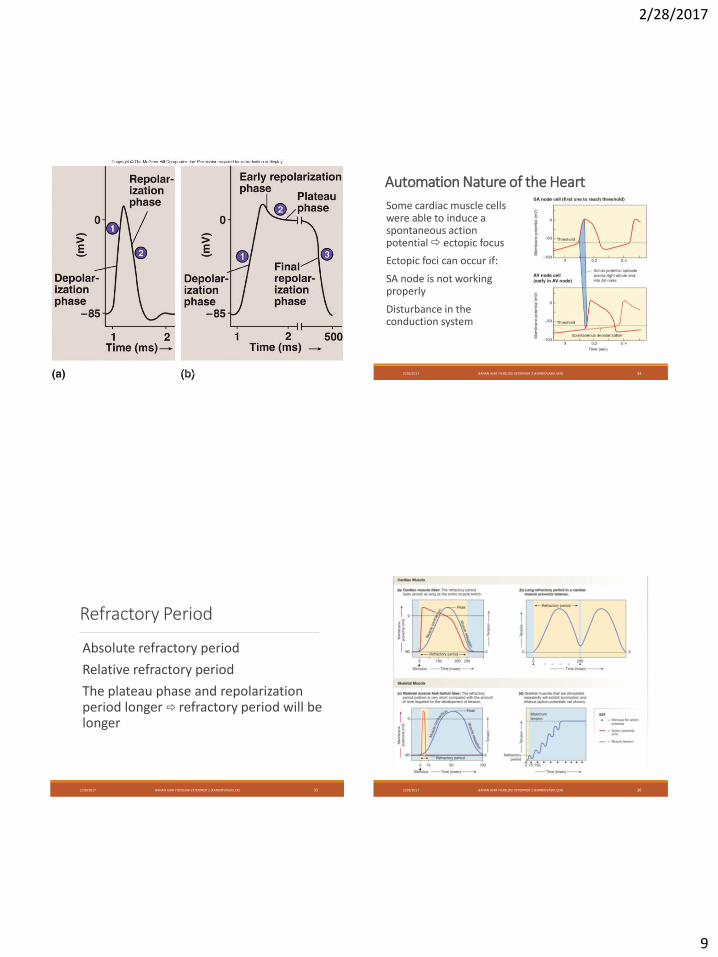
Automation Nature of the Heart
Some cardiac muscle cells were able to induce a spontaneous action potential ectopic focus
Ectopic foci can occur if:
SA node is not working properly
Disturbance in the conduction system
Refractory Period
Absolute refractory period
Relative refractory period
The plateau phase and repolarization period longer refractory period will be longer
2/28/2017 BAHAN AJAR FISIOLOGI VETERINER 2 (KARDIOVASKULER) 35 2/28/2017 BAHAN AJAR FISIOLOGI VETERINER 2 (KARDIOVASKULER) 36
2/28/2017
10
2/28/2017 BAHAN AJAR FISIOLOGI VETERINER 2 (KARDIOVASKULER) 37
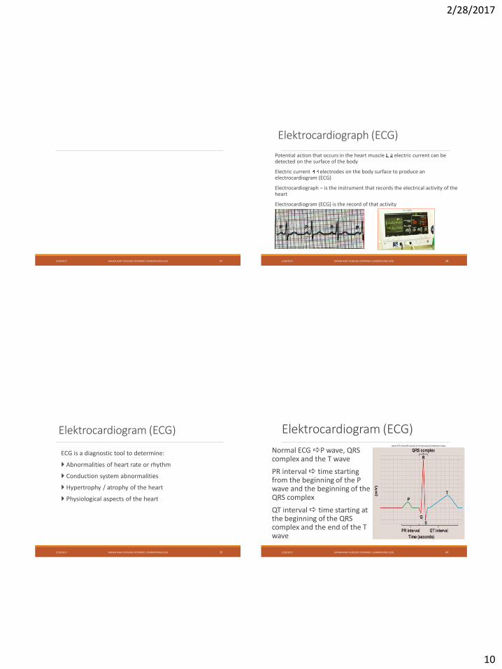
Elektrocardiograph (ECG)
Potential action that occurs in the heart muscle electric current can be detected on the surface of the body
Electric current electrodes on the body surface to produce an electrocardiogram (ECG)
Electrocardiograph – is the instrument that records the electrical activity of the heart
Electrocardiogram (ECG) is the record of that activity
2/28/2017 BAHAN AJAR FISIOLOGI VETERINER 2 (KARDIOVASKULER) 38
Elektrocardiogram (ECG)
ECG is a diagnostic tool to determine:
Abnormalities of heart rate or rhythm
Conduction system abnormalities
Hypertrophy / atrophy of the heart
Physiological aspects of the heart
2/28/2017 BAHAN AJAR FISIOLOGI VETERINER 2 (KARDIOVASKULER) 39
Elektrocardiogram (ECG)
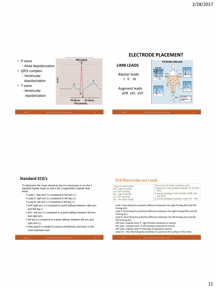
Normal ECG P wave, QRS complex and the T wave
PR interval time starting from the beginning of the P wave and the beginning of the QRS complex
QT interval time starting at the beginning of the QRS complex and the end of the T wave
2/28/2017 BAHAN AJAR FISIOLOGI VETERINER 2 (KARDIOVASKULER) 40
2/28/2017
11
• P wave
- Atrial depolarization
• QRS complex
- Ventricular
depolarization
• T wave
- Ventricular
repolarization
2/28/2017 BAHAN AJAR FISIOLOGI VETERINER 2 (KARDIOVASKULER) 41 2/28/2017 BAHAN AJAR FISIOLOGI VETERINER 2 (KARDIOVASKULER) 42
LIMB LEADS
Bipolar leadsI II III
Augment leadsaVR aVL aVF
2/28/2017 BAHAN AJAR FISIOLOGI VETERINER 2 (KARDIOVASKULER) 43
Standard ECG’s
• To determine the mean electrical axis it is necessary to run the 3
standard bipolar leads as well a the 3 augmented unipolar limb
leads.
• Lead I- right arm (+) compared to left arm (-).
• Lead II- right arm (-) compared to left leg (+),
• Lead III- left arm (-) compared to left leg (+)
• aVR-right arm (+) compared to point halfway between right arm
• and left leg (-)
• aVL- left arm (+) compared to a point halfway between left arm
• and right arm.
• left leg (+) compared to a point halfway between left arm and
• right arm (-).
• Only lead II is needed to assess arrhythmias and lead I is the
• most important lead
Type of electrodes:RA, right forelegLA, left forelegRL, right hind legLL, left hind legV1 - V6 chest leads
ECG Electrodes and Leads
There are 12 leads routinely used:3 pieces of raw sandapan bipolar (I, II and
III).3 pieces sandapan limb unipolar (aVR, aVL
and aVF).6 pieces sandapan unipolar chest (V1 - V6)
2/28/2017 BAHAN AJAR FISIOLOGI VETERINER 2 (KARDIOVASKULER) 44
Lead I: Describing the potential difference between the right foreleg (RA) and left foreleg (LA)Lead II: Describing the potential difference between the right foreleg (RA) and left hind leg (LL)Lead III: Describing the potential difference between the left foreleg (LA) and the left hind leg (LL)aVR lead :unipolar lead right foreleg compared to neutralaVL lead : unipolar lead left foreleg compared to neutralAVF lead: unipolar lead hind legs compared to neutralLead (V1 - V6): Recording the potential of a point on the surface of the chest.
2/28/2017
12
2/28/2017 BAHAN AJAR FISIOLOGI VETERINER 2 (KARDIOVASKULER) 45
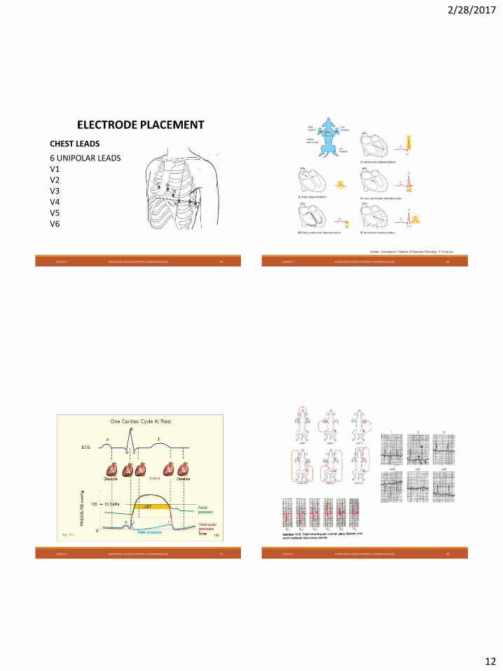
CHEST LEADS
6 UNIPOLAR LEADSV1V2 V3V4 V5V6
2/28/2017 BAHAN AJAR FISIOLOGI VETERINER 2 (KARDIOVASKULER) 46
Sumber: Cunningham’s Textbook of Veterinary Physiology 5th ed pp:191
2/28/2017 BAHAN AJAR FISIOLOGI VETERINER 2 (KARDIOVASKULER) 47 2/28/2017 BAHAN AJAR FISIOLOGI VETERINER 2 (KARDIOVASKULER) 48
2/28/2017
13
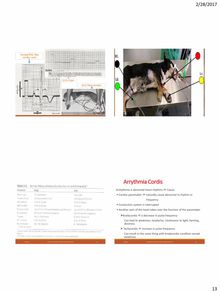
Normal ECG, One cardiac cycle
ECG Paper
ECG Measurement
2/28/2017 BAHAN AJAR FISIOLOGI VETERINER 2 (KARDIOVASKULER) 49
2/28/2017 BAHAN AJAR FISIOLOGI VETERINER 2 (KARDIOVASKULER) 51 2/28/2017 BAHAN AJAR FISIOLOGI VETERINER 2 (KARDIOVASKULER) 52
Arrythmia CordisArrhythmia is abnormal heart rhythms Cause:
Cardiac pacemaker naturally cause abnormal in rhythm or
frequency
Conduction system is interrupted
Another part of the heart takes over the function of the pacemaker
Bradycardia a decrease in pulse frequency
Can lead to weakness, headache, intolerance to light, fainting, dizziness
Tachycardia increase in pulse frequency
Can result in the same thing with bradycardia condition except weakness
2/28/2017
14
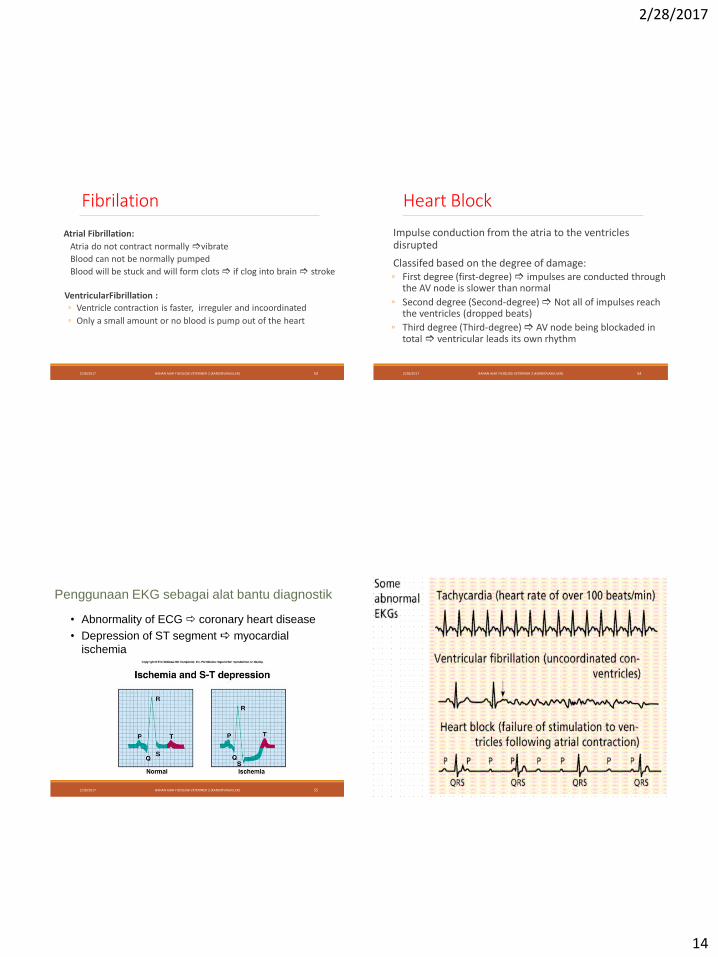
Fibrilation
Atrial Fibrillation:
Atria do not contract normally vibrate
Blood can not be normally pumped
Blood will be stuck and will form clots if clog into brain stroke
2/28/2017 BAHAN AJAR FISIOLOGI VETERINER 2 (KARDIOVASKULER) 53
VentricularFibrillation :
◦ Ventricle contraction is faster, irreguler and incoordinated
◦ Only a small amount or no blood is pump out of the heart
Heart Block
Impulse conduction from the atria to the ventricles disrupted
Classifed based on the degree of damage:◦ First degree (first-degree) impulses are conducted through
the AV node is slower than normal
◦ Second degree (Second-degree) Not all of impulses reach the ventricles (dropped beats)
◦ Third degree (Third-degree) AV node being blockaded in total ventricular leads its own rhythm
2/28/2017 BAHAN AJAR FISIOLOGI VETERINER 2 (KARDIOVASKULER) 54
Penggunaan EKG sebagai alat bantu diagnostik
• Abnormality of ECG coronary heart disease
• Depression of ST segment myocardial
ischemia
2/28/2017 BAHAN AJAR FISIOLOGI VETERINER 2 (KARDIOVASKULER) 55 2/28/2017 BAHAN AJAR FISIOLOGI VETERINER 2 (KARDIOVASKULER) 56
2/28/2017
15
2/28/2017 BAHAN AJAR FISIOLOGI VETERINER 2 (KARDIOVASKULER) 57 2/28/2017 BAHAN AJAR FISIOLOGI VETERINER 2 (KARDIOVASKULER) 58
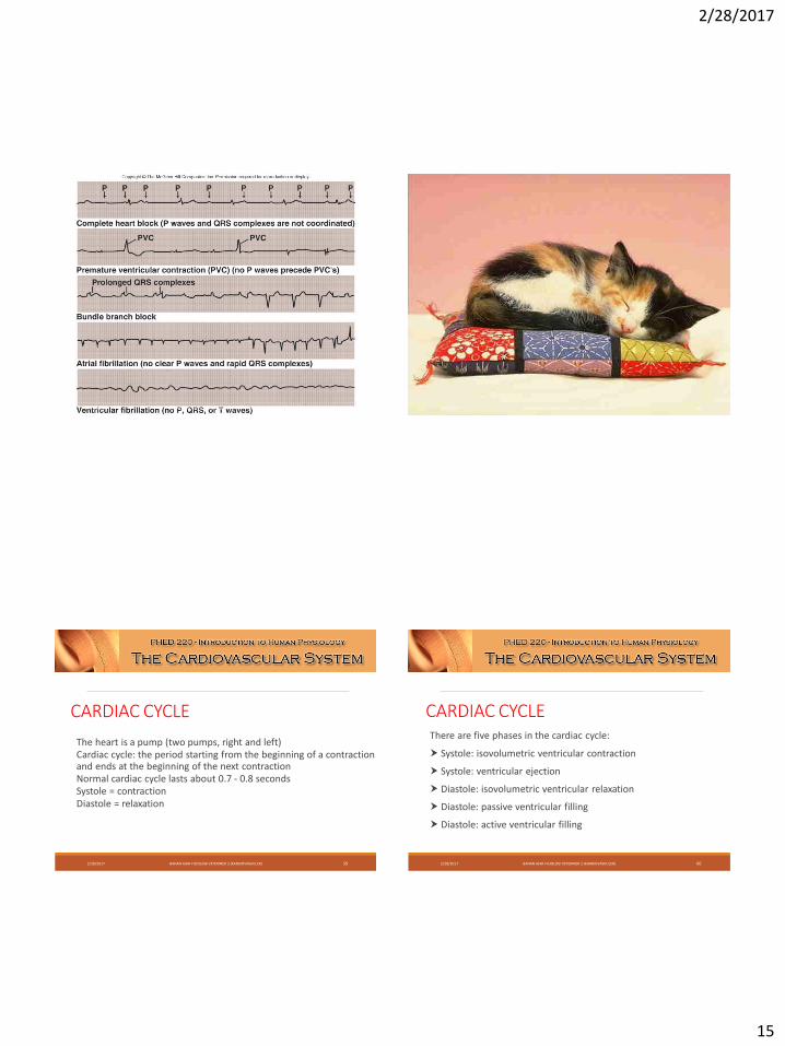
CARDIAC CYCLE
The heart is a pump (two pumps, right and left)Cardiac cycle: the period starting from the beginning of a contraction and ends at the beginning of the next contractionNormal cardiac cycle lasts about 0.7 - 0.8 secondsSystole = contractionDiastole = relaxation
2/28/2017 BAHAN AJAR FISIOLOGI VETERINER 2 (KARDIOVASKULER) 59
There are five phases in the cardiac cycle:
Systole: isovolumetric ventricular contraction
Systole: ventricular ejection
Diastole: isovolumetric ventricular relaxation
Diastole: passive ventricular filling
Diastole: active ventricular filling
2/28/2017 BAHAN AJAR FISIOLOGI VETERINER 2 (KARDIOVASKULER) 60
CARDIAC CYCLE
2/28/2017
16
Isovolumetric Ventricular Contraction
Ventricular contraction
Increased ventricular pressure rapidly
The entire valve remains closed no blood is pumped out of the heart
Ventricular volume remains constant
2/28/2017 BAHAN AJAR FISIOLOGI VETERINER 2 (KARDIOVASKULER) 61
Ventricular EjectionVentricular contraction continues
Pressure continues to rise
Ventricular pressure> pressure in the aorta and in the pulmonary artery
Aortic and pulmonary valves open
Blood pumping occurs
2/28/2017 BAHAN AJAR FISIOLOGI VETERINER 2 (KARDIOVASKULER) 62
Isovolumetric Ventricular Relaxation
Ventricular relaxation
Ventricular pressure decreased rapidly
Aortic and pulmonary valve closes
Ventricular volume remains constant
2/28/2017 BAHAN AJAR FISIOLOGI VETERINER 2 (KARDIOVASKULER) 63
Passive Ventricular FillingAtrial pressure is greater than the ventricular pressure
AV valve opens
Blood flows from the atria to the ventricles
Play in role in about 70% ventricular filling
The actual filling occurs during 1/3 of diastole
2/28/2017 BAHAN AJAR FISIOLOGI VETERINER 2 (KARDIOVASKULER) 64
2/28/2017
17
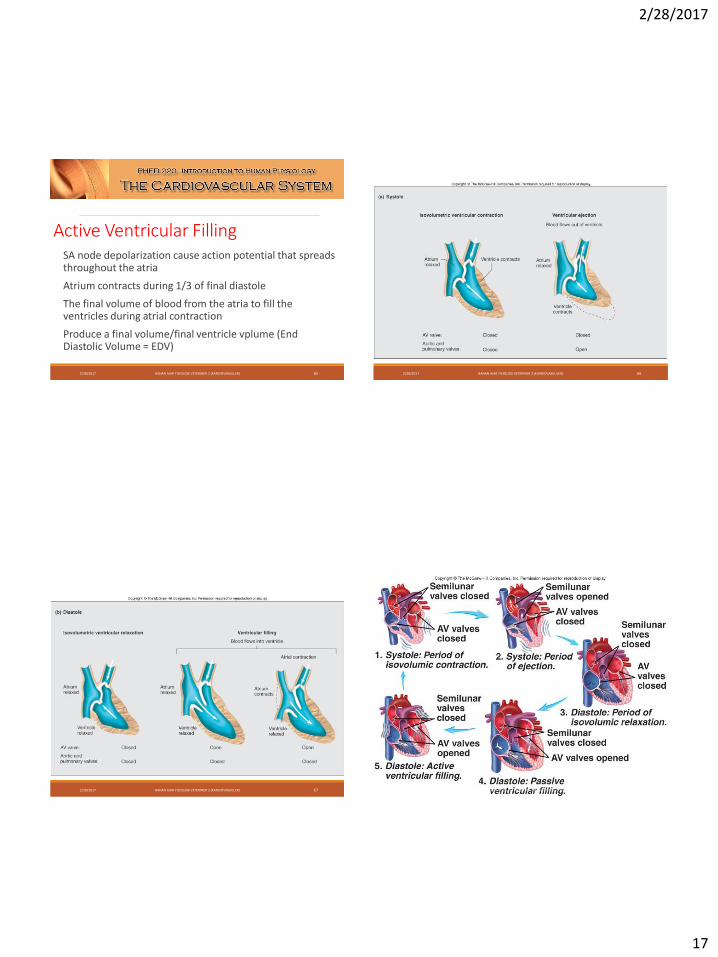
Active Ventricular FillingSA node depolarization cause action potential that spreads throughout the atria
Atrium contracts during 1/3 of final diastole
The final volume of blood from the atria to fill the ventricles during atrial contraction
Produce a final volume/final ventricle vplume (End Diastolic Volume = EDV)
2/28/2017 BAHAN AJAR FISIOLOGI VETERINER 2 (KARDIOVASKULER) 65 2/28/2017 BAHAN AJAR FISIOLOGI VETERINER 2 (KARDIOVASKULER) 66
2/28/2017 BAHAN AJAR FISIOLOGI VETERINER 2 (KARDIOVASKULER) 67 2/28/2017 BAHAN AJAR FISIOLOGI VETERINER 2 (KARDIOVASKULER) 68
2/28/2017
18
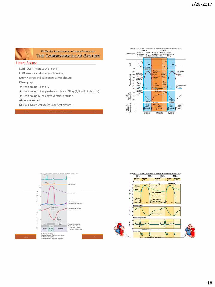
Heart SoundLUBB-DUPP (heart sound I dan II)
LUBB = AV valve closure (early systole).
DUPP = aortic and pulmonary valves closure
Phonograph
Heart sound III and IV
Heart sound III passive ventricular filling (1/3 end of diastole)
Heart sound IV active ventricular filling
Abnormal sound
Murmur (valve leakage or imperfect closure)
2/28/2017 BAHAN AJAR FISIOLOGI VETERINER 2 (KARDIOVASKULER) 69 2/28/2017 BAHAN AJAR FISIOLOGI VETERINER 2 (KARDIOVASKULER) 70
2/28/2017 BAHAN AJAR FISIOLOGI VETERINER 2 (KARDIOVASKULER) 71 2/28/2017 BAHAN AJAR FISIOLOGI VETERINER 2 (KARDIOVASKULER) 72
2/28/2017
19
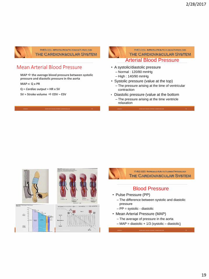
Mean Arterial Blood PressureMAP the average blood pressure between systolic pressure and diastolic pressure in the aorta
MAP Q x PR
Q = Cardiac output = HR x SV
SV = Stroke volume EDV – ESV
2/28/2017 BAHAN AJAR FISIOLOGI VETERINER 2 (KARDIOVASKULER) 73
Arterial Blood Pressure
• A systolic/diastolic pressure – Normal : 120/80 mmHg
– High : 140/90 mmHg
• Systolic pressure (value at the top)– The pressure arising at the time of ventricular
contraction
Diastolic pressure (value at the bottom
– The pressure arising at the time ventricle relaxation
2/28/2017 BAHAN AJAR FISIOLOGI VETERINER 2 (KARDIOVASKULER) 74
2/28/2017 BAHAN AJAR FISIOLOGI VETERINER 2 (KARDIOVASKULER) 75
Blood Pressure• Pulse Pressure (PP)
– The difference between systolic and diastolic
pressure
– PP = systolic - diastolic
• Mean Arterial Pressure (MAP)
– The average of pressure in the aorta
– MAP = diastolic + 1/3 (systolic – diastolic)
2/28/2017 BAHAN AJAR FISIOLOGI VETERINER 2 (KARDIOVASKULER) 76
2/28/2017
20
2/28/2017 BAHAN AJAR FISIOLOGI VETERINER 2 (KARDIOVASKULER) 77 2/28/2017 BAHAN AJAR FISIOLOGI VETERINER 2 (KARDIOVASKULER) 78
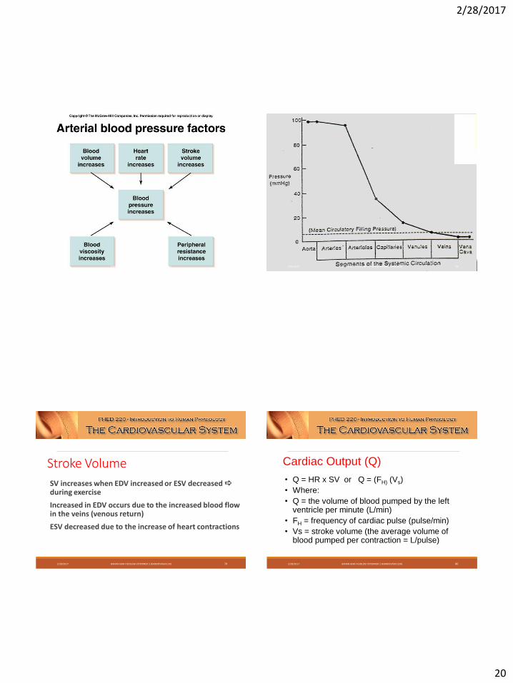
Stroke Volume
SV increases when EDV increased or ESV decreased during exercise
Increased in EDV occurs due to the increased blood flow in the veins (venous return)
ESV decreased due to the increase of heart contractions
2/28/2017 BAHAN AJAR FISIOLOGI VETERINER 2 (KARDIOVASKULER) 79
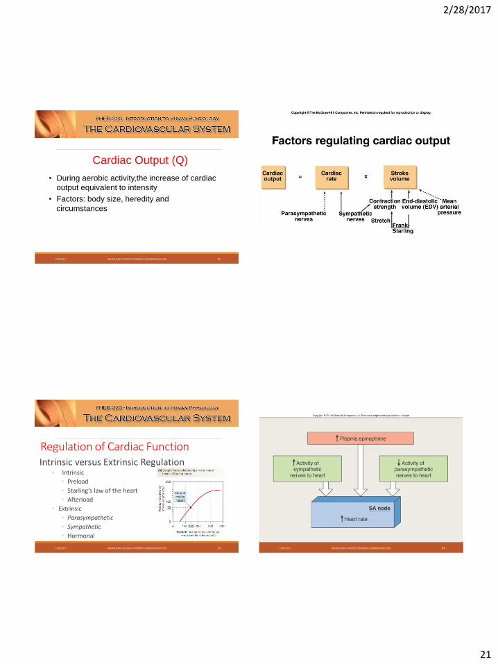
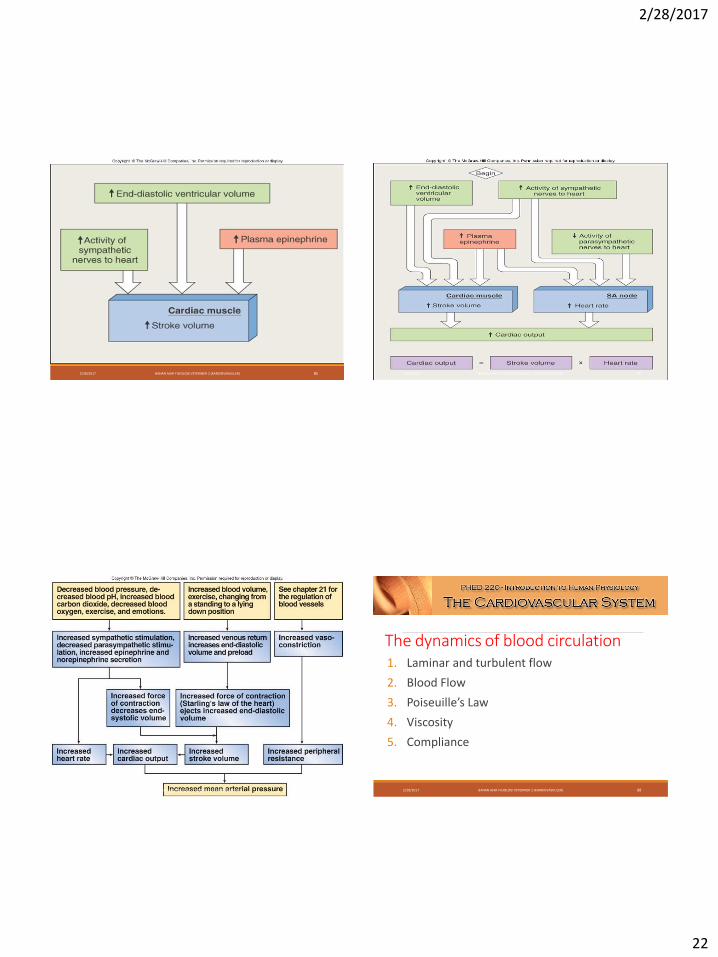
Cardiac Output (Q)
• Q = HR x SV or Q = (FH) (Vs)
• Where:
• Q = the volume of blood pumped by the left ventricle per minute (L/min)
• FH = frequency of cardiac pulse (pulse/min)
• Vs = stroke volume (the average volume of blood pumped per contraction = L/pulse)
2/28/2017 BAHAN AJAR FISIOLOGI VETERINER 2 (KARDIOVASKULER) 80
2/28/2017
21
Cardiac Output (Q)
• During aerobic activity,the increase of cardiac
output equivalent to intensity
• Factors: body size, heredity and
circumstances
2/28/2017 BAHAN AJAR FISIOLOGI VETERINER 2 (KARDIOVASKULER) 81 2/28/2017 BAHAN AJAR FISIOLOGI VETERINER 2 (KARDIOVASKULER) 82
Regulation of Cardiac FunctionIntrinsic versus Extrinsic Regulation
◦ Intrinsic
◦ Preload
◦ Starling’s law of the heart
◦ Afterload
◦ Extrinsic
◦ Parasympathetic
◦ Sympathetic
◦ Hormonal
2/28/2017 BAHAN AJAR FISIOLOGI VETERINER 2 (KARDIOVASKULER) 83 2/28/2017 BAHAN AJAR FISIOLOGI VETERINER 2 (KARDIOVASKULER) 84
2/28/2017
22
2/28/2017 BAHAN AJAR FISIOLOGI VETERINER 2 (KARDIOVASKULER) 85 2/28/2017 BAHAN AJAR FISIOLOGI VETERINER 2 (KARDIOVASKULER) 86
2/28/2017 BAHAN AJAR FISIOLOGI VETERINER 2 (KARDIOVASKULER) 87
The dynamics of blood circulation1. Laminar and turbulent flow
2. Blood Flow
3. Poiseuille’s Law
4. Viscosity
5. Compliance
2/28/2017 BAHAN AJAR FISIOLOGI VETERINER 2 (KARDIOVASKULER) 88
2/28/2017
23
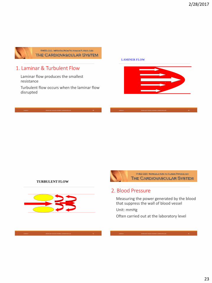
1. Laminar & Turbulent Flow
Laminar flow produces the smallest resistance
Turbulent flow occurs when the laminar flow disrupted
2/28/2017 BAHAN AJAR FISIOLOGI VETERINER 2 (KARDIOVASKULER) 89
LAMINER FLOW
2/28/2017 BAHAN AJAR FISIOLOGI VETERINER 2 (KARDIOVASKULER) 90
TURBULENT FLOW
2/28/2017 BAHAN AJAR FISIOLOGI VETERINER 2 (KARDIOVASKULER) 91
2. Blood Pressure
Measuring the power generated by the blood that suppress the wall of blood vessel
Unit: mmHg
Often carried out at the laboratory level
2/28/2017 BAHAN AJAR FISIOLOGI VETERINER 2 (KARDIOVASKULER) 92
2/28/2017
24
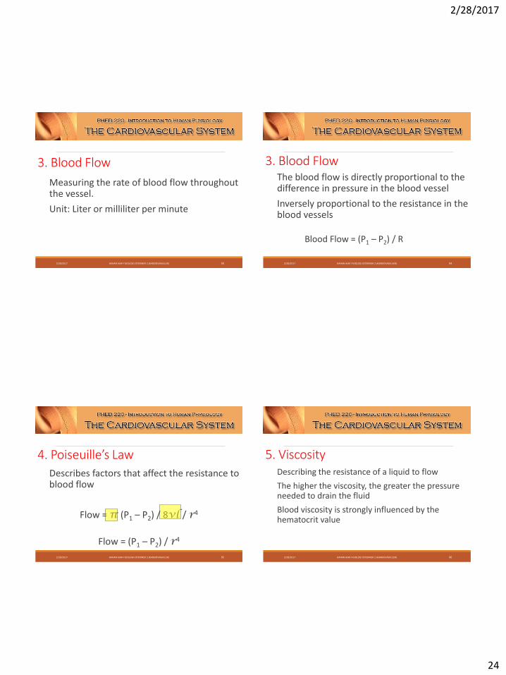
3. Blood Flow
Measuring the rate of blood flow throughout the vessel.
Unit: Liter or milliliter per minute
2/28/2017 BAHAN AJAR FISIOLOGI VETERINER 2 (KARDIOVASKULER) 93
3. Blood FlowThe blood flow is directly proportional to the difference in pressure in the blood vessel
Inversely proportional to the resistance in the blood vessels
Blood Flow = (P1 – P2) / R
2/28/2017 BAHAN AJAR FISIOLOGI VETERINER 2 (KARDIOVASKULER) 94
4. Poiseuille’s Law
Describes factors that affect the resistance to blood flow
Flow = π (P1 – P2) / 8vl / r4
Flow = (P1 – P2) / r4
2/28/2017 BAHAN AJAR FISIOLOGI VETERINER 2 (KARDIOVASKULER) 95
5. ViscosityDescribing the resistance of a liquid to flow
The higher the viscosity, the greater the pressure needed to drain the fluid
Blood viscosity is strongly influenced by the hematocrit value
2/28/2017 BAHAN AJAR FISIOLOGI VETERINER 2 (KARDIOVASKULER) 96
2/28/2017
25
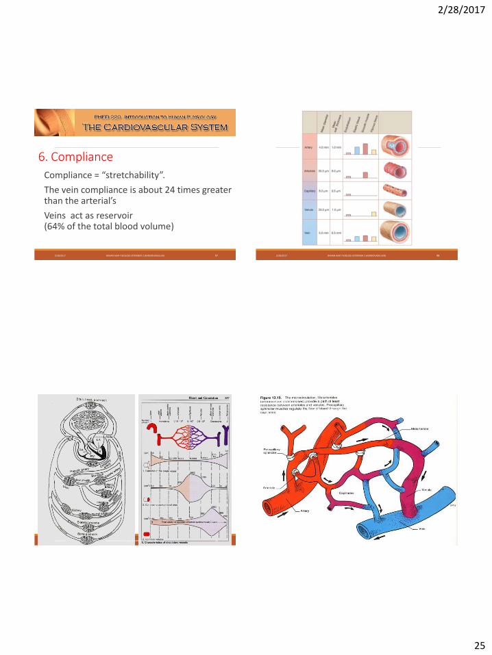
6. Compliance
Compliance = “stretchability”.
The vein compliance is about 24 times greaterthan the arterial’s
Veins act as reservoir (64% of the total blood volume)
2/28/2017 BAHAN AJAR FISIOLOGI VETERINER 2 (KARDIOVASKULER) 97 2/28/2017 BAHAN AJAR FISIOLOGI VETERINER 2 (KARDIOVASKULER) 98
2/28/2017 BAHAN AJAR FISIOLOGI VETERINER 2 (KARDIOVASKULER) 99 2/28/2017 BAHAN AJAR FISIOLOGI VETERINER 2 (KARDIOVASKULER) 100
2/28/2017
26
2/28/2017 BAHAN AJAR FISIOLOGI VETERINER 2 (KARDIOVASKULER) 101
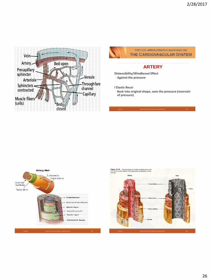
Distensibility/Windkessel Effect◦ Against the pressure
l Elastic Recoi◦ Back into original shape, save the pressure (reservoir
of pressure)
ARTERY
2/28/2017 BAHAN AJAR FISIOLOGI VETERINER 2 (KARDIOVASKULER) 102
2/28/2017 BAHAN AJAR FISIOLOGI VETERINER 2 (KARDIOVASKULER) 103 2/28/2017 BAHAN AJAR FISIOLOGI VETERINER 2 (KARDIOVASKULER) 104
2/28/2017
27
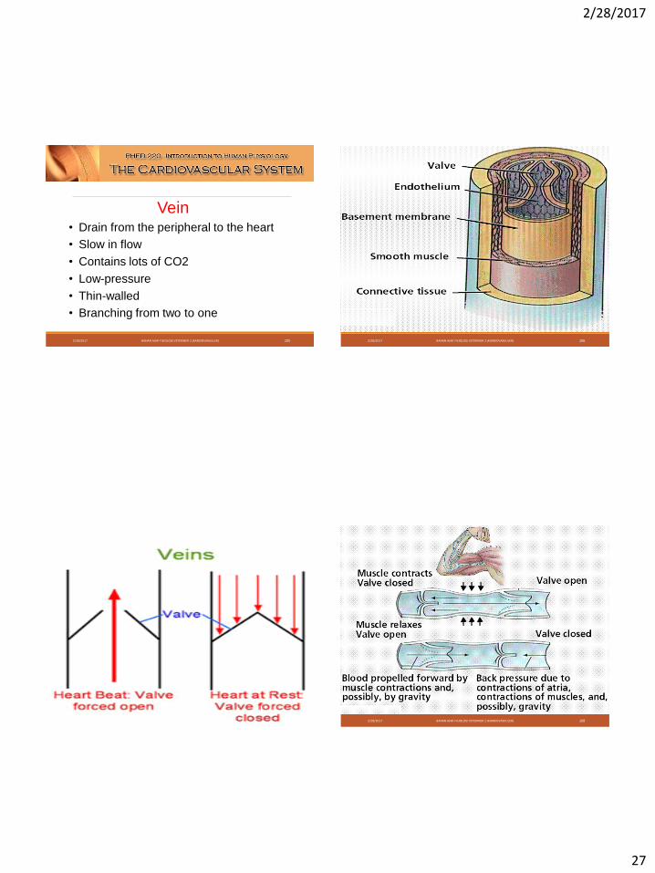
Vein• Drain from the peripheral to the heart
• Slow in flow
• Contains lots of CO2
• Low-pressure
• Thin-walled
• Branching from two to one
2/28/2017 BAHAN AJAR FISIOLOGI VETERINER 2 (KARDIOVASKULER) 105 2/28/2017 BAHAN AJAR FISIOLOGI VETERINER 2 (KARDIOVASKULER) 106
2/28/2017 BAHAN AJAR FISIOLOGI VETERINER 2 (KARDIOVASKULER) 107 2/28/2017 BAHAN AJAR FISIOLOGI VETERINER 2 (KARDIOVASKULER) 108
2/28/2017
28
2/28/2017 BAHAN AJAR FISIOLOGI VETERINER 2 (KARDIOVASKULER) 109 2/28/2017 BAHAN AJAR FISIOLOGI VETERINER 2 (KARDIOVASKULER) 110
2/28/2017 BAHAN AJAR FISIOLOGI VETERINER 2 (KARDIOVASKULER) 111
2/28/2017
29
Regulation of Blood Flow to the Tissue
In most tissues, blood flow is proportional to the metabolic needs
The blood flow is determined by dilatation of metarteriol and precapillary sphincter relaxation
The blood flow can be increased 7-8 times
2/28/2017 BAHAN AJAR FISIOLOGI VETERINER 2 (KARDIOVASKULER) 113
Regulation of Blood Flow to the Tissue
Vasodilator substances produced when the metabolism increases:
CO2
Lactic acid
Hydrogen ions
Etc2/28/2017 BAHAN AJAR FISIOLOGI VETERINER 2 (KARDIOVASKULER) 114
2/28/2017 BAHAN AJAR FISIOLOGI VETERINER 2 (KARDIOVASKULER) 115
Regulation of Blood Flow to the Tissue
Pengaturan Saraf & Hormonal untuk Sirkulasi Lokal
◦ Area di pons, otak tengah, dan diencephalon berperan dalam stimulasi dan inhibisi pusat vasomotor
2/28/2017 BAHAN AJAR FISIOLOGI VETERINER 2 (KARDIOVASKULER) 116
2/28/2017
30
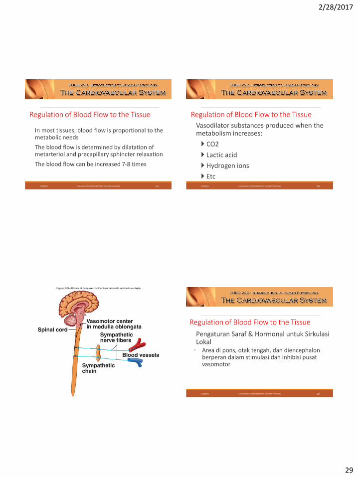
Regulation of Blood Flow to the Tissue Nerves & Hormonal control for the local circulation
Autonomous control functions quickly
Sympathetic motor fibers innervate all EXCEPT capillary blood vessels, pre-capillary sphincter and metarteriole
Center of the regulation is located at the vasomotor area in the lower pons and upper medulla oblongata.
2/28/2017 BAHAN AJAR FISIOLOGI VETERINER 2 (KARDIOVASKULER) 117
Pengaturan Aliran Darah ke Jaringan
Nerves & Hormonal control for the local circulation
◦ Neurotransmitter = norepinephrine
◦ Binding to the α-adrenergic receptor
vasoconstriction
2/28/2017 BAHAN AJAR FISIOLOGI VETERINER 2 (KARDIOVASKULER) 118
Nerves & Hormonal control for the local circulation
Epinephrine and norepinephrine from the adrenal medulla give the same effect
This hormone generally causes vasoconstriction, but in other tissues such as skeletal muscle, epinephrine binds to β-receptors and causes dilation of blood vessels
2/28/2017 BAHAN AJAR FISIOLOGI VETERINER 2 (KARDIOVASKULER) 119
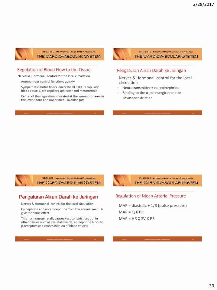
Regulation of Mean Arterial Pressure
MAP = diastolic + 1/3 (pulse pressure)
MAP = Q X PR
MAP = HR X SV X PR
2/28/2017 BAHAN AJAR FISIOLOGI VETERINER 2 (KARDIOVASKULER) 120
2/28/2017
31
2/28/2017 BAHAN AJAR FISIOLOGI VETERINER 2 (KARDIOVASKULER) 121
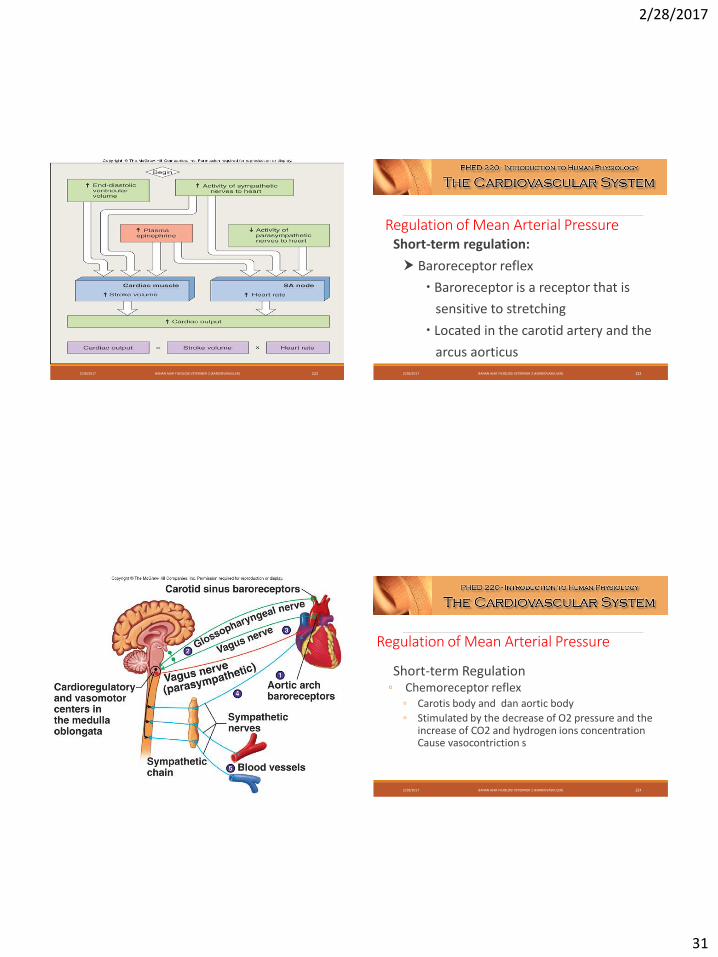
Regulation of Mean Arterial PressureShort-term regulation:
Baroreceptor reflex
Baroreceptor is a receptor that is
sensitive to stretching
Located in the carotid artery and the
arcus aorticus2/28/2017 BAHAN AJAR FISIOLOGI VETERINER 2 (KARDIOVASKULER) 122
2/28/2017 BAHAN AJAR FISIOLOGI VETERINER 2 (KARDIOVASKULER) 123
Regulation of Mean Arterial Pressure
Short-term Regulation◦ Chemoreceptor reflex
◦ Carotis body and dan aortic body
◦ Stimulated by the decrease of O2 pressure and the increase of CO2 and hydrogen ions concentrationCause vasocontriction s
2/28/2017 BAHAN AJAR FISIOLOGI VETERINER 2 (KARDIOVASKULER) 124
2/28/2017
32
2/28/2017 BAHAN AJAR FISIOLOGI VETERINER 2 (KARDIOVASKULER) 125 2/28/2017 BAHAN AJAR FISIOLOGI VETERINER 2 (KARDIOVASKULER) 126
Regulation of Mean Arterial Pressure
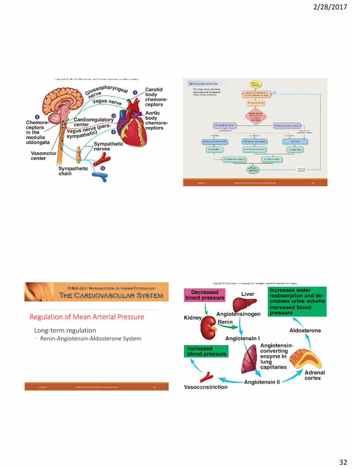
Long-term regulation ◦ Renin-Angiotensin-Aldosterone System
2/28/2017 BAHAN AJAR FISIOLOGI VETERINER 2 (KARDIOVASKULER) 127 2/28/2017 BAHAN AJAR FISIOLOGI VETERINER 2 (KARDIOVASKULER) 128
2/28/2017
33
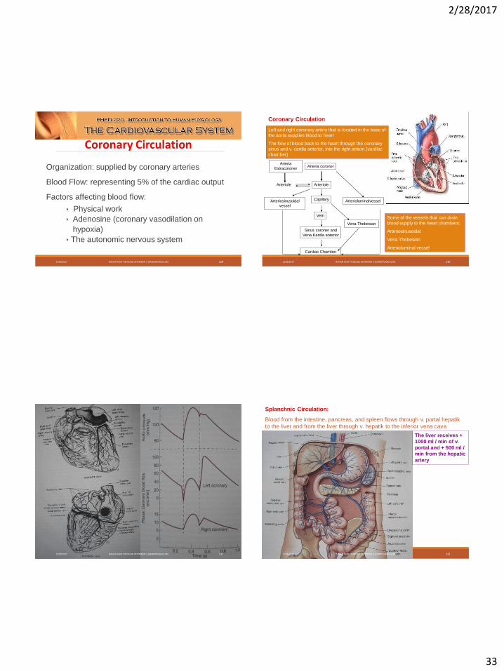
Organization: supplied by coronary arteries
Blood Flow: representing 5% of the cardiac output
Factors affecting blood flow:
Physical work
Adenosine (coronary vasodilation on
hypoxia)
The autonomic nervous system
Coronary Circulation
2/28/2017 BAHAN AJAR FISIOLOGI VETERINER 2 (KARDIOVASKULER) 129
Coronary Circulation
Arteria
ExtracoronerArteria coroner
ArterioleArteriole
Capillary
Vein
Sinus coroner and
Vena Kardia anterior
Cardiac Chamber
Arteriosinusoidal
vessel
Arterioluminalvessel
Vena Thebesian
Left and right coronary artery that is located in the base of
the aorta supplies blood to heart
The flow of blood back to the heart through the coronary
sinus and v. cardia anterior, into the right atrium (cardiac
chamber)
Some of the vessels that can drain
blood supply to the heart chambers:
Arteriosinusoidal
Vena Thebesian
Arterioluminal vessel
2/28/2017 BAHAN AJAR FISIOLOGI VETERINER 2 (KARDIOVASKULER) 130
2/28/2017 BAHAN AJAR FISIOLOGI VETERINER 2 (KARDIOVASKULER) 131
Splanchnic Circulation:
Blood from the intestine, pancreas, and spleen flows through v. portal hepatik
to the liver and from the liver through v. hepatik to the inferior vena cava
The liver receives +
1000 ml / min of v.
portal and + 500 ml /
min from the hepatic
artery
2/28/2017 BAHAN AJAR FISIOLOGI VETERINER 2 (KARDIOVASKULER) 132
2/28/2017
34
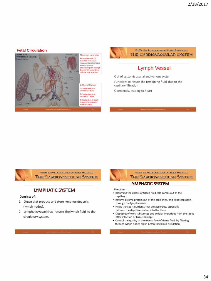
Fetal CirculationPlacenta = Lung fetus.
From maternal, O2
taken by fetal, CO2
released from the fetus
to the maternal
circulation pass-through
the villi villi resembling
cellular lung function
In Sheep / Human:
O2 saturation in v.
Umbilical = 80%
O2 saturation in a.
Umbilical = 55%
O2 saturation in adult
humans in systemic
arterial = 98%
2/28/2017 BAHAN AJAR FISIOLOGI VETERINER 2 (KARDIOVASKULER) 133
Out of systemic aterial and venous system
Function: to return the remaining fluid due to the capillary filtration
Open ends, leading to heart
Lymph Vessel
2/28/2017 BAHAN AJAR FISIOLOGI VETERINER 2 (KARDIOVASKULER) 134
Consists of:
1. Organ that produce and store lymphocytes cells
(lymph nodes).
2. Lymphatic vessel that returns the lymph fluid to the
circulatory system.
2/28/2017 BAHAN AJAR FISIOLOGI VETERINER 2 (KARDIOVASKULER) 135
Function::• Returning the excess of tissue fluid that comes out of the
capillary.• Returns plasma protein out of the capillaries, and reabsorp again
through the lymph vessels.• Helps transport nutrients that are absorbed, especially
fat from the digestive system into the blood.• Disposing of toxic substances and cellular impurities from the tissue
after infection or tissue damage. Control the quality of the excess flow of tissue fluid by filtering
through lymph nodes organ before back into circulation.
2/28/2017 BAHAN AJAR FISIOLOGI VETERINER 2 (KARDIOVASKULER) 136

2/28/2017
35
2/28/2017 BAHAN AJAR FISIOLOGI VETERINER 2 (KARDIOVASKULER) 137 2/28/2017 BAHAN AJAR FISIOLOGI VETERINER 2 (KARDIOVASKULER) 138
Water and substrate that are dissolved in the blood plasma (except blood cells and large protein molecules) an freely pass through the thin capillary walls (pores, D: 8 nm).
Every day an estimated of 20 liters of fluid is filtered and enter to the interstitial tissue (nonrenal). 18 l / day of it is reabsorped into the blood capillaries while 2 l / day returning to the bloodstream via the lymphatic system.
2/28/2017 BAHAN AJAR FISIOLOGI VETERINER 2 (KARDIOVASKULER) 139 Kontrol lokal atau Autoregulation Kontrol Neurohumoral2/28/2017 BAHAN AJAR FISIOLOGI VETERINER 2 (KARDIOVASKULER) 140
2/28/2017
36
2/28/2017 BAHAN AJAR FISIOLOGI VETERINER 2 (KARDIOVASKULER) 141