improving the outcome of psoriatic...
TRANSCRIPT
Improving the Outcome of Psoriatic Arthritis
by
Laura Claire Coates
Submitted in accordance with the requirements for the degree of
PhD
Leeds Institute of Molecular Medicine
Academic Section of Musculoskeletal Disease
School of Medicine
University of Leeds
November 2010
- ii -
The candidate confirms that the work submitted is her own, except where work
which has formed part of jointly-authored publications has been included. The
contribution of the candidate and the other authors to this work has been explicitly
indicated below. The candidate confirms that appropriate credit has been given
within the thesis where reference has been made to the work of others.
Chapter Four is based on work from a jointly authored publication by Dr
Coates and Dr Freeston. Dr Coates recruited the patients to the study and performed
the clinical assessments of enthesitis. Dr Freeston performed the ultrasound scans of
the patients. Both authors contributed jointly to the statistical analysis plan and the
writing of the paper.
The paper is as follows: JE Freeston, LC Coates, PS Helliwell, EMA Hensor,
RJ Wakefield, P Emery, PG Conaghan. Is there sub-clinical enthesitis in early
psoriatic arthritis? Rheumatology, submitted August 2010
This copy has been supplied on the understanding that it is copyright material
and that no quotation from the thesis may be published without proper
acknowledgement
© 2010 The University of Leeds and Laura Claire Coates

- iii -
Acknowledgments
The following people have all played a hugely significant part in the work that
has gone into this thesis, and I would like to thank them for all their help and effort.
In particular, I would like to thank my supervisors Dr Philip Helliwell, Professor
Philip Conaghan and Professor Paul Emery.
Study patients described in Chapter Three and Seven were recruited with the
help of the doctors at Chapel Allerton Hospital with organisational assistance from
David Pickles. Dr Jane Freeston kindly performed the ultrasound scans described in
Chapters Four and Seven and assisted with analysis and interpretation of the
ultrasound results. Statistical advice and SPSS support was provided by Dr Philip
Helliwell, Ms Robin Waxman and our departmental data analyst Dr Elizabeth
Hensor.
The work in Chapter Five could not have happened without the support and
advice of the Group for Research and Assessment of Psoriasis and Psoriatic Arthritis
(GRAPPA) Steering Committee and the co-operation of the GRAPPA members. Dr
Jaap Fransen provided valuable statistical advice to estimate required sample sizes.
Chapter Six utilised data from the observational University of Toronto
Psoriatic Arthritis Clinic supervised by Professor Dafna Gladman who was kind
enough to accept my application to work within her unit for one month. Statistical
expertise for this analysis was provided by Professor Richard Cook and Dr Ker-Ai
Lee. Data from the IMPACT and IMPACT2 clinical trials was kindly provided by
Centocor and the Principle Investigators of these studies, Drs Christian Antoni and
Arthur Kavanaugh.
My PhD was funded by the Arthritis Research Campaign (now Arthritis
Research UK) as a Clinical Research Fellowship. Arthritis Research UK has
provided funding through the Clinical Studies Group for the TICOPA study and its
expansion to a national multi-centre clinical trial. Funding for my month’s work at
the University of Toronto PsA Clinic was provided by a John Glyn bursary from the
Royal College of Physicians, UK.
Final thanks go to all of the friends and family who proof-read and checked
sections of this thesis, particularly my husband James for all of his support.

- iv -
List of publications/presentations arising from this thesis
Book chapters
Coates LC, Helliwell PS. Disease Measurement – enthesitis, skin, nails,
spine, dactylitis. In: Fitzgerald O, Maksymowych W, ed. Spondyloarthropathies.
Best Practice Research and Clinical Rheumatology.
Review articles
Coates LC, Helliwell PS, and members of the GRAPPA imaging
subcommittee. Clues to the pathogenesis of psoriasis and psoriatic arthritis from
imaging. A literature review. J Rheumatol 2008;35(7):1438-1442.
Coates LC, Helliwell PS. Classification and categorisation of psoriatic
arthritis. Clin Rheum 2008; 27:1211-1216.
Original articles
Coates LC, Fransen J, Helliwell PS. Defining minimal disease activity in
psoriatic arthritis: a proposed objective target for treatment. Ann Rheum Dis 2009;
69(1): 48-53.
Coates LC, Cook R, Lee KA, Chandran V, Gladman DD. Frequency,
predictors and prognosis of sustained minimal disease activity in an observational
psoriatic arthritis cohort. Arthritis Care Res 2010; 62(7): 970-6.
Coates LC, Helliwell PS. Validation of minimal disease activity criteria for
psoriatic arthritis using interventional trial data. Arthritis Care Res 2010; 62(7):
965-9.
Freeston JE*, Coates LC*, Helliwell PS, Hensor EMA, Wakefield RJ, Emery
P, Conaghan PG. Is there sub-clinical enthesitis in early psoriatic arthritis? A
clinical comparison with PD US. Rheumatology (submitted). (* denotes joint first
authorship)
Coates LC, Conaghan PG, Emery P, Green MJ, Ibrahim G, MakIver H,
Helliwell PS. Investigating the use of the CASPAR criteria in early psoriatic
arthritis. In preparation.
Oral presentations
- v -
Coates LC, Helliwell PS. Preliminary validation of the psoriatic arthritis
minimal disease activity criteria according to the OMERACT filter. Presented at the
European League Against Rheumatism Annual Meeting 2010, Rome, Italy.
Coates LC, Helliwell PS. Achieving minimal disease activity (MDA) criteria
with anti-TNF therapy in psoriatic arthritis can prevent progressive joint damage.
Presented at the British Society of Rheumatology Annual Meeting 2010,
Birmingham, UK.
Coates LC, Helliwell PS. Achieving minimal disease activity (MDA) criteria
with anti-TNF therapy in psoriatic arthritis can prevent progressive joint damage.
Presented at the American College of Rheumatology Annual Meeting 2009,
Philadelphia, USA.
Coates LC, Schentag CT, Lee K, Chandran V, Cook RJ, Gladman DD.
Achieving minimal disease activity criteria decreases progression of joint damage in
psoriatic arthritis. Presented at the IFPA World Psoriasis and Psoriatic Arthritis
Conference 2009, Stockholm, Sweden.
Coates LC, Schentag CT, Lee K, Chandran V, Cook RJ, Gladman DD.
Achieving minimal disease activity criteria decreases progression of joint damage in
psoriatic arthritis. Presented at the GRAPPA annual meeting 2009 – fellow’s
session, Stockholm, Sweden.
Coates LC, Schentag CT, Lee K, Chandran V, Cook RJ, Gladman DD.
Frequency and predictors of Minimal Disease Activity (MDA) in an observational
PsA cohort. Presented at European League Against Rheumatism Annual Meeting
2009, Copenhagen, Denmark
Coates LC, Fransen J, Helliwell PS. Defining minimal disease activity in
psoriatic arthritis – a proposed target for treatment. Presented at the American
College of Rheumatology Annual Meeting 2008, San Francisco, USA.
Coates LC, Fransen J, Helliwell PS. Defining minimal disease activity in
psoriatic arthritis – a proposed target for treatment. Presented at the York and
Northern Rheumatology Society Annual Meeting 2008, York, UK.
Coates LC, Emery P, Conaghan PG, Helliwell PS. Tight Control of Psoriatic
Arthritis (TICOPA). Presented at GRAPPA annual meeting 2008 – fellows session,
Leeds, UK.
Coates LC, Fransen J, Helliwell PS. Defining minimal disease activity in
psoriatic arthritis – a proposed target for treatment. Presented at OMERACT 9
conference 2008, Kananaskis, Canada.
- vi -
Poster presentations
Freeston JE, Coates LC, Helliwell PS, Hensor EMA, Wakefield RJ, Emery P,
Conaghan P. Does clinical examination overestimate the amount of active enthesitis
in early psoriatic arthritis? A comparison with power Doppler ultrasound. Arthritis
Rheum 2009; 60(10): S201
Freeston JE, Coates LC, Helliwell PS, Hensor EMA, Wakefield RJ, Emery P,
Conaghan P. Is there sub-clinical joint disease in early psoriatic arthritis? A clinical
comparison with power Doppler ultrasound. Arthritis Rheum 2009; 60(10): S757
Coates LC, Schentag CT, Lee K, Chandran V, Cook RJ, Gladman DD.
Achieving minimal disease activity criteria decreases progression of joint damage in
psoriatic arthritis. Annals Rheum Dis 2009; 68(3):137
Coates LC, Schentag CT, Lee K, Chandran V, Cook RJ, Gladman DD.
Frequency and predictors of Minimal Disease Activity (MDA) in an observational
PsA cohort. Arthritis Rheum 2008; 58(9): F52.
Coates LC, Emery P, Conaghan PG, Helliwell PS. Tight Control of Psoriatic
Arthritis (TICOPA). Presented at GRAPPA annual meeting 2008 – fellows session,
Leeds, UK.
Coates LC, Fransen J, Helliwell PS. Defining minimal disease activity in
psoriatic arthritis – a proposed target for treatment. OMERACT 9 conference 2008,
Kananaskis, Canada.

- vii -
Abstract
Psoriatic arthritis (PsA) is recognised to have a significant impact on
functional impairment, joint damage and quality of life. The aim of this thesis was
to investigate tools for early identification, to develop a clinical target for treatment
and to utilise these tools within a clinical trial.
The ClASssification of Psoriatic ARthritis (CASPAR) criteria, previously
developed in established PsA, were tested in patients with recent onset inflammatory
arthritis (PsA and controls) to test their discriminative ability in early arthritis. The
phenotype of early PsA was investigated further with clinical and ultrasound (US)
assessment. Clinical criteria for minimal disease activity (MDA) were developed
using a questionnaire. These were subsequently tested in an observational cohort
and interventional trial dataset. Finally, they are being utilised prospectively in an
RCT addressing the benefits of tight control in PsA.
The CASPAR criteria were found to have good sensitivity and specificity for
the diagnosis of recent onset PsA. No individual clinical parameters accurately
distinguished PsA from other types of inflammatory arthritis, but there was evidence
of more oligoarticular disease and enthesitis in PsA compared with rheumatoid
arthritis. US imaging showed a small burden of subclinical arthritis and enthesitis
but found good correlation between clinical and imaging assessment of disease
activity. Criteria for MDA were developed from expert consensus, covering all
aspects of psoriatic disease. They were evaluated against the OMERACT filter and
have supporting evidence for their validation in terms of truth, discrimination and
feasibility. Early unblinded results from the clinical trial indicated that 53%
achieved MDA at 12 months.
In summary, the CASPAR criteria can be used for early classification of PsA.
In addition, a new composite outcome measure has been developed and validated
and is now being utilised in a clinical trial.

- viii -
Contents
Improving the Outcome of Psoriatic Arthritis ........................................................ i
Acknowledgments .................................................................................................... iii
List of publications/presentations arising from this thesis ................................... iv
Abstract .................................................................................................................... vii
Contents .................................................................................................................. viii
List of Figures ......................................................................................................... xiii
List of Tables .......................................................................................................... xiv
List of Abbreviations ............................................................................................. xvi
1 Introduction ............................................................................................................ 1
2 Literature Review................................................................................................... 5
2.1 Epidemiology of PsA ........................................................................................ 5
2.1.1 Introduction .................................................................................... 5
2.1.2 Incidence and Prevalence .............................................................. 5
2.1.3 Clinical Features............................................................................. 6
2.1.4 Natural History .............................................................................. 7
2.1.5 Pathogenesis .................................................................................... 9
2.1.5.1 Immunology ..................................................................... 9
2.1.5.2 Genetics .......................................................................... 10
2.1.5.3 Environmental factors .................................................. 11
2.1.5.4 Imaging ........................................................................... 12
2.2 Diagnosis and Classification .......................................................................... 27
2.2.1 Diagnosis/classification ................................................................ 27
2.2.1.1 The importance of classification criteria in PsA ......... 28
2.2.1.2 Historic classification criteria ....................................... 28
2.2.1.3 The CASPAR criteria ................................................... 29
2.2.1.4 Investigations used in diagnosis of PsA ....................... 33
2.2.2 Subtypes of PsA ............................................................................ 33
2.2.2.1 Dividing axial and peripheral disease .......................... 34
2.2.2.2 Subgrouping peripheral disease ................................... 37

- ix -
2.2.2.3 Are subgroups clinically relevent? ............................... 38
2.3 Outcome measurement in PsA ...................................................................... 39
2.3.1 Clinical Outcome Measures ........................................................ 39
2.3.1.1 Peripheral Joint Disease................................................ 39
2.3.1.2 Skin and Nail Disease .................................................... 43
2.3.1.3 Enthesitis ........................................................................ 50
2.3.1.4 Dactylitis ......................................................................... 53
2.3.1.5 Axial Disease .................................................................. 56
2.3.1.6 Composite Outcome Measures ..................................... 61
2.3.1.7 Defining “state” in PsA ................................................. 64
2.3.2 Imaging ......................................................................................... 66
2.3.2.1 Conventional radiography ............................................ 66
2.3.2.2 Ultrasound ...................................................................... 74
2.3.2.3 MRI ................................................................................. 74
2.4 Treatment of PsA ........................................................................................... 78
2.4.1 Current treatment options .......................................................... 78
2.4.1.1 Non-steroidal anti-inflammatory drugs ...................... 78
2.4.1.2 Steroids – systemic ......................................................... 79
2.4.1.3 Steroids – local ............................................................... 79
2.4.1.4 Disease-modifying anti-rheumatic drugs .................... 80
2.4.1.5 Biologics .......................................................................... 85
2.4.2 Early treatment ............................................................................ 96
2.4.3 Tight control ................................................................................. 99
3 Validation of the CASPAR criteria in early disease ....................................... 101
3.1 Introduction .................................................................................................. 101
3.2 Methods ......................................................................................................... 101
3.2.1 Patient and control selection ..................................................... 101
3.2.2 Data collection ............................................................................ 102
3.2.3 Statistical methods ..................................................................... 102
3.2.3.1 Power calculation ........................................................ 102
3.2.3.2 Results Analysis ........................................................... 102
3.3 Results ........................................................................................................... 103
3.4 Discussion ...................................................................................................... 112
3.5 Limitations .................................................................................................... 112
3.6 Conclusion ..................................................................................................... 113
- x -
4 Imaging in Early Psoriatic Arthritis – the extent of sub-clinical disease ...... 115
4.1 Introduction .................................................................................................. 115
4.2 Methods ......................................................................................................... 117
4.2.1 Ultrasound .................................................................................. 117
4.2.2 Statistical analysis ...................................................................... 117
4.3 Results ........................................................................................................... 118
4.4 Discussion ...................................................................................................... 122
4.5 Limitations .................................................................................................... 123
4.6 Conclusion ..................................................................................................... 123
5 Defining Minimal Disease Activity in PsA – a proposed objective target ..... 124
5.1 Introduction .................................................................................................. 124
5.2 Methods ......................................................................................................... 124
5.2.1 Domains to be included .............................................................. 124
5.2.2 Materials ..................................................................................... 126
5.2.3 Statistical methods ..................................................................... 126
5.3 Results ........................................................................................................... 126
5.4 Discussion ...................................................................................................... 130
5.5 Limitations .................................................................................................... 131
5.6 Conclusion ..................................................................................................... 132
6 Validation of the Minimal Disease Activity Criteria ...................................... 133
6.1 Introduction .................................................................................................. 133
6.2 Methods ......................................................................................................... 134
6.2.1 Observational Cohort Data ....................................................... 134
6.2.1.1 Statistical Analysis ....................................................... 135
6.2.2 Randomised Controlled Trial Data .......................................... 135
6.2.2.1 Statistical analysis ........................................................ 136
6.2.3 Application of the OMERACT filter ........................................ 136
6.3 Results ........................................................................................................... 137
6.3.1 Truth ........................................................................................... 137
6.3.1.1 Face validity ................................................................. 137
6.3.1.2 Content validity ........................................................... 137
6.3.1.3 Criterion validity ......................................................... 138
6.3.1.4 Construct validity ........................................................ 140
6.3.2 Discrimination ............................................................................ 140
6.3.2.1 Classification ................................................................ 140

- xi -
6.3.2.2 Prognosis ...................................................................... 141
6.3.2.3 Responsiveness ............................................................. 143
6.3.2.4 Reliability ..................................................................... 143
6.3.3 Feasibility .................................................................................... 143
6.4 Discussion ...................................................................................................... 144
6.4.1 Truth ........................................................................................... 145
6.4.2 Discrimination ............................................................................ 145
6.4.3 Feasibility .................................................................................... 146
6.5 Limitations .................................................................................................... 146
6.6 Conclusion ..................................................................................................... 147
7 Tight Control of Psoriatic Arthritis.................................................................. 148
7.1 Introduction .................................................................................................. 148
7.2 Methods ......................................................................................................... 148
7.2.1 Patients ........................................................................................ 148
7.2.2 Treatment allocation and intervention..................................... 149
7.2.3 Assessment of end-points ........................................................... 151
7.2.4 Statistical analysis ...................................................................... 152
7.3 Results ........................................................................................................... 152
7.3.1 Baseline Demographics .............................................................. 154
7.3.2 Treatment during study period ................................................ 156
7.3.3 Response measures ..................................................................... 158
7.3.3.1 Minimal Disease Activity ............................................ 158
7.3.3.2 Outcome measures for individual domains of PsA ...................................................................................... 158
7.3.4 Patient reported outcomes ......................................................... 165
7.3.5 Relationship between MDA and other outcome measures ..... 168
7.4 Discussion ...................................................................................................... 171
7.5 Limitations .................................................................................................... 174
7.6 Conclusion ..................................................................................................... 174
8 Conclusions and Future Directions .................................................................. 175
8.1 Recent research developments relevant to thesis work ............................ 179
8.1.1 Novel diagnostic tools ................................................................. 179
8.1.2 Novel composite outcome measures in PsA ............................. 179
8.1.3 Novel treatment regimens in PsA ............................................. 180
8.2 Potential research questions arising from this thesis ................................ 181
8.2.1 Diagnosis and classification ....................................................... 181

- xii -
8.2.2 Assessment of disease activity in PsA ....................................... 182
8.2.3 Defining disease activity states .................................................. 182
8.2.4 Future directions for therapeutic studies ................................. 185
Bibliography .......................................................................................................... 189

- xiii -
List of Figures
Figure 1 – The CASPAR criteria ..................................................................... 31
Figure 2 – Scoring regions for the PsAMRIS scoring system ......................... 76
Figure 3 - ROC curve for CASPAR and Moll & Wright criteria predicting PsA
diagnosis .................................................................................................................. 106
Figure 4 - ROC curve for candidate definitions of minimal disease activity. 129
Figure 5 - Percentage of MDA & non-MDA patients achieving ACR responses
at wk 24 ................................................................................................................... 139
Figure 6 - Percentage of MDA & non-MDA patients achieving ACR responses
at wk 52 ................................................................................................................... 139
Figure 7 - Probability plot for change in total mS-vdH score for those in MDA
and those not. .......................................................................................................... 142
Figure 8 – Protocol for treatment in the TICOPA study ................................ 150
Figure 9 – Enrolment flow diagram for TICOPA study ................................ 153
Figure 10 – Change in median scores for enthesitis outcomes for those with
involvement at baseline ........................................................................................... 163
Figure 11 - Change in median score over time for dactylitis and nail outcomes
for those with involvement in that domain at baseline ........................................... 164
Figure 12 - changes in median VAS scores at key timepoints ....................... 166
Figure 13 – comparison of ACR responses in those achieving MDA and those
not ............................................................................................................................ 170

- xiv -
List of Tables
Table 1 – Proportion of patients with HLA associations in subgroups of PsA 36
Table 2 - The ASAS response criteria 2001 and 2004 ..................................... 58
Table 3 - The ASDAS draft formulae .............................................................. 60
Table 4 – Steinbrocker scores for joint radiographs ........................................ 68
Table 5 – Larsen’s scoring method as modified by Rau and Hehborn ............ 68
Table 6 – modified Sharp scoring system for PsA with inclusion of typical
features ...................................................................................................................... 70
Table 7 – modified Sharp-van der Heijde scoring method for PsA ................. 71
Table 8 – Ratingen scoring system for PsA ..................................................... 73
Table 9 – Summary of results from key anti-TNF therapy RCTs ................... 86
Table 10 – Results of ustekinumab study at week 12 ...................................... 91
Table 11 – Results of the abatacept in PsA trial at day 169 ............................. 93
Table 12 – 12 week results from the apremilast in PsA trial ........................... 95
Table 13 – Results of the RESPOND trial ....................................................... 98
Table 14 - baseline features of cases and controls recruited to the CASPAR
study ........................................................................................................................ 104
Table 15 - ROC analysis of Moll & Wright and CASPAR criteria ............... 105
Table 16 - Proportion of cases and control fulfilling each aspect of the
CASPAR criteria ..................................................................................................... 108
Table 17 – disease patterns in cases, controls and RA controls ..................... 109
Table 18 – Patterns of arthritis in cases, controls and RA controls ............... 111
Table 19 - Comparison of clinical and US assessment of disease activity by
individual joint (CE=clinical examination, US=ultrasound). ................................. 119
Table 20 – Comparison of clinical and US assessment of disease activity by
individual entheses .................................................................................................. 121

- xv -
Table 21 - Average value for each outcome measure for profiles designated as
MDA by different levels of agreement by respondents .......................................... 128
Table 22 - Possible cut points of each outcome measure for the different
potential definitions of MDA .................................................................................. 128
Table 23 – baseline characteristics of the TICOPA cohort ............................ 155
Table 24 – frequency of patients receiving synthetic and biologic DMARDs
................................................................................................................................. 157
Table 25 – frequency and average doses for DMARDs prescribed ............... 157
Table 26 – proportions of patients achieving MDA at key timepoints .......... 159
Table 27 – Composite arthritis outcomes at key timepoints .......................... 159
Table 28 – composite arthritis outcomes and MDA by joint disease subtype 159
Table 29 – PASI responses at key timepoints ................................................ 161
Table 30 – Quality of life and functional questionnaire scores at key timepoints
................................................................................................................................. 167
Table 31 – Comparison of patients achieving MDA and ACR outcome
measures .................................................................................................................. 169
- xvi -
List of Abbreviations
Ab antibodies
ACR American College of Rheumatology
ADEPT ADalimumab Effectiveness in Psoriatic arthritis Trial
AS ankylosing spondylitis
ASAS Assessment of SpondyloArthritis Society
ASDAS Ankylosing Spondylitis Disease Activity Score
AUC area under the curve
BASDAI Bath ankylosing spondylitis disease activity index
BASFI Bath ankylosing spondylitis functional index
BASMI Bath ankylosing spondylitis metrology index
BSA body surface area
CART classification and regression tree
CASPAR ClASsification of Psoriatic Arthritis
CCP anti-cyclic citrillinated peptide
CDAI clinical disease activity index
CI confidence interval
CoPSI Copenhagen Psoriasis Severity Index
CPDAI Composite PsA Disease Activity Index
CRP C-reactive protein
DAPSA Disease Activity in PSoriatic Arthritis
DAREA Disease Activity in Reactive Arthritis
DAS Disease Activity Score
DCE-MRI dynamic contrast enhanced MRI
DIP distal interphalangeal
DMARD disease-modifying anti-rheumatic drug
ESR erythrocyte sedimentation rate
ESSG European Spondyloarthropathy Study Group
EULAR European League Against Rheumatism
FT flexor tenosynovitis
- xvii -
GI gastrointestinal
GRACE GRAPPA Composite Exercise
GRAPPA Group for Research and Assessment of Psoriasis and Psoriatic Arthritis
GS grey-scale
HAQ-DI Health Assessment Questionnaire Disability Index
HIV human immunodeficiency virus
HLA human leukocyte antigen
HRQOL health-related quality of life
IA intra-articular
ICC intra-class coefficient
IL interleukin
IL2-R interleukin 2-receptor
IMPACT Infliximab Multinational Psoriatic Arthritis Controlled Trial
IMPART International Multicentre Psoriasis and Psoriatic Arthritis Reliability Trial
INSPIRE International Spondyloarthritis Interobserver Reliability Exercise
IP interphalangeal
IV intravenous
JSN joint space narrowing
LD laser Doppler
LDF laser Doppler flowmetry
LDI Leeds Dactylitis Instrument
LEI Leeds Enthesitis Index
LS-PGA Lattice System Physician’s Global Assessment
MASES Maastricht Ankylosing Spondylitis Entheses Score
MCP metocarpophalangeal
MDA minimal disease activity
MEI Mander Enthesitis Index
MHC major histocompatability complex
mNAPSI modified NAPSI
MRI magnetic resonance imaging
mSASSS modified Stoke Ankylosing Spondylitis Spinal Score
mS-vdH modified Sharp/van der Heijde score
MTP metatarsophalangeal
NADPH nicotinamide adenine dinucleotide phosphate
NAPSI Nail Psoriasis Severity Index

- xviii -
NOAR Norfolk Arthritis Register
NOR-
DMARD Norweigian DMARD
NPF National Psoriasis Foundation
NPF-PS NPF Psoriasis Score
NSAID non-steroidal anti-inflammatory drug
OA osteoarthritis
OASIS Outcome in Ankylosing Spondylitis International Study
OCT optical coherence tomography
OMERACT Outcome Measures in Rheumatology Clinical Trials
PABAK prevalence and bias adjusted Kappa
PAQ Psoriatic Arthritis Questionnaire
PASDAS Psoriatic Arthritis Disease Activity Score
PASE Psoriatic Arthritis Screening and Evaluation tool
PASI psoriasis area and severity index
PCA principle component analysis
PD power Doppler
PD4 phosphodiesterase-4
PEASI Psoriasis Exact Area and Severity Index
PEST Psoriasis Epidemiology Screening Tool
PIP proximal interphalangeal
PLASI Psoriasis Log-based Area and Severity Index
PNSS Psoriasis Nail Severity Score
PRESTA Psoriasis Randomised Etanercept STudy in subjects with Psoriatic Arthritis
PROMs patient reported outcome measures
PsA psoriatic arthritis
PsAMRIS PsA MRI score
PsARC PsA response criteria
PSORS1 psoriasis susceptibility 1
PsQOL Psoriatic arthritis Quality Of Life scale
PV plasma viscosity
RA rheumatoid arthritis
RAI Ritchie articular index
RAMRIS RA MRI score
RCT randomised controlled trial
- xix -
RESPOND REmicade Study in PsA patients Of methotrexate -Naïve Disease
RF rheumatoid factor
ROC receiver operator characteristic
SAPASI self-administered PASI
SAPHO synovitis/acne/pustulosis/hyperostosis/osteomyelitis
SD standard deviation
SDAI simplified disease activity index
SF-36 short form 36
SI sacroiliac
SIG special interest group
SJC swollen joint count
SpA spondyloarthritides
SPARCC Spondyloarthritis Research Consortium of Canada
SRM standardised response mean
STIR short-tau inversion recovery
SwePsA Swedish PsA register
TGF transforming growth factor
TICOPA TIght COntrol of Psoriatic Arthritis
TICORA TIght COntrol of Rheumatoid Arthritis
TJC tender joint count
TNF tumour necrosis factor
ToPAS Toronto Psoriatic Arthritis Screening tool
TOPAS Treatment of Psoriatic Arthritis Study
US ultrasound
UTE ultrashort echo time
VAS visual analogue scale
VAS GH VAS general health

- 1 -
1 Introduction
The aim of this thesis is to improve our understanding of early psoriatic
arthritis (PsA) and to investigate tools that can be used to optimise the care of
patients with PsA.
PsA is now recognised as a separate disease entity from other inflammatory
arthritides, although it is considered as part of the family of spondyloarthropathies.
For many years, PsA was dismissed as a mild disease with no long term sequelae
and treatment was therefore considered unnecessary. Many observational cohort
studies have confirmed a poor long-term outcome of PsA in terms of poor quality of
life, functional impairment and radiographic joint damage (Gladman et al. 1987;
Sokoll and Helliwell 2001; Bond et al. 2007). Research from the Toronto PsA
Clinic has shown that active arthritis (indicated by number of tender and swollen
joints) predicts future joint damage suggesting that inflammation in the joints is the
key to later damage and resultant disability (Bond et al. 2007). With this recognition
of PsA as a potentially damaging and destructive disease came an emphasis on
improving management of this disease to optimise outcome.
The first limiting factor in improving care in PsA is an accurate diagnosis early
in the course of the disease. PsA is a highly heterogenous disease with variable
clinical manifestations, and particularly in the early stages of disease it may be
difficult to identify. Typically patients develop skin disease prior to their joint
disease, but in up to 20% of early PsA cases, psoriasis may not be present (Kane and
Pathare 2005). Typical patterns of radiographic damage that are seen in PsA may be
absent at first presentation removing another potential identifying feature. Clearly it
would be beneficial if patients can be identified and treated before they develop
radiographic joint damage. Some newer, more sensitive imaging techniques such as
ultrasound (US) and magnetic resonance imaging (MRI) may be more useful in such
early patients. Unfortunately little is currently known about the extent of disease
seen on imaging in patients with early PsA.
For three decades, the gold standard classification criteria for PsA were the
Moll and Wright criteria (Moll and Wright 1973). Many modifications to these
were proposed to try to improve the specificity of the criteria but none have been
widely adopted. In 2006, new classification criteria called the ClASsification of
Psoriatic ARthritis (CASPAR) criteria were developed using patient derived data.
These include characteristic dermatological, clinical and radiological features and
have both high sensitivity and specificity for established PsA (Taylor et al. 2006).
However less than 10% of the patients had disease duration of less than 2 years, so
- 2 -
validation in early disease was not explored. Several groups have attempted to
assess the sensitivity of the criteria in identifying patients with recent onset PsA but
using only patients with a secure clinical diagnosis and no controls (Chandran et al.
2007b; D'Angelo et al. 2008; Lindqvist et al. 2008; D'Angelo et al. 2009). One
group highlighted the low prevalence of radiological criteria in early disease leading
to a lower sensitivity to identify PsA (D'Angelo et al. 2009). However, as yet, there
have been no properly powered studied addressing the sensitivity and specificity of
the CASPAR criteria in early disease and therefore they cannot currently be
recommended for use in studies of early PsA.
Once a diagnosis is made, the next issue is that of optimal treatment. The
majority of treatment is “borrowed” from rheumatoid arthritis (RA) using similar
disease-modifying agents, often without convincing evidence of efficacy in PsA.
Although methotrexate is widely used in clinical practice, there is very little
evidence to support the use of this treatment in PsA. Newer treatments, such as the
Tumour Necrosis Factor (TNF) blocking therapies, have proven efficacy in PsA
from large clinical trials but are not available to many patients in the UK (Kay and
Griffiths 2006), and are very rarely used as first-line therapy.
The strategy for treatment of RA has been revolutionised in recent years with
emphasis on early treatment to effectively control inflammation and prevent joint
damage. Empirically, this strategy seems ideal for PsA also given the evidence of
the link between inflammation and damage in peripheral joint disease in PsA.
However, there are no controlled studies of early treatment in PsA or investigating
the concept of “tight control” of inflammation. In the studies in RA, the Disease
Activity Score (DAS) definition of low disease activity or remission is routinely
used as a target for therapy to allow tight control of inflammation in clinical trials.
This is a clinical assessment of disease activity based principally on tender and
swollen joint counts. However in PsA there are no criteria defining low disease
activity or remission that can be utilised as an important clinical endpoint in such
studies.
The Outcome Measures in Rheumatology Clinical Trials (OMERACT) group
have agreed a conceptual definition of minimal disease activity which encompasses
remission and low disease activity. Criteria for RA have been developed based on
this concept but the same approach has not been tried in PsA. Due to the variable
phenotype of PsA, any proposed criteria would have to encompass all of the key
manifestations of the disease and would have to involve multiple outcome measures.
To maximise feasibility of use, it would be advantageous if any potential criteria
were based on clinical outcome measures with no specialist imaging or assessment
tools required. Investigating the prevalence of sub-clinical disease seen on imaging

- 3 -
will allow an evaluation of the accuracy of clinical outcome measures in this
population. Any criteria developed will then need validation in both research-
orientated interventional settings and real-life clinical observational cohorts. The
availability of such validated criteria in PsA would allow future research into tight
control of inflammation and would potentially provide an objective target for
treatment that could be used in interventional trials and routine clinical practice.
The focus of this thesis is to address the issues discussed above providing
insight into early diagnosis, better understanding of disease involvement in early
PsA, development of a target for treatment for clinical trials and finally investigating
the possible impact on tight control in early PsA. The structure of this thesis can be
summarised as follows:
Chapter 2 Review of the literature
This chapter provides an overview of PsA discussing epidemiology, diagnosis,
outcome measurement and treatment options. The key issues in the optimal
management of PsA, particularly difficulties in early diagnosis and limitations in
evidence-based treatment are highlighted.
Chapter 3 The validity of the CASPAR criteria in early PsA
This chapter evaluates the sensitivity and specificity of the CASPAR criteria
for PsA in an early inflammatory arthritis clinic setting.
Chapter 4 Imaging in early PsA – the extent of subclinical disease
This chapter investigates whether sub-clinical inflammatory disease (arthritis
and enthesitis) exists in patients presenting with a new diagnosis of PsA using grey
scale (GS) and power Doppler (PD) US techniques compared with clinical
examination.
Chapter 5 Defining Minimal Disease Activity in PsA
This chapter covers the development of new minimal disease activity (MDA)
criteria for PsA derived from expert opinion using real-life patient cases.
Chapter 6 Validation of the MDA criteria for PsA
This chapter assesses the validity of the new MDA criteria for PsA according
to the OMERACT filter using data from both an observational cohort and from two
large randomised interventional trials.
Chapter 7 Tight Control of PsA – preliminary analysis of a large Randomised
Controlled Trial (RCT)
- 4 -
This chapter presents a preliminary analysis of the first 40 patients enrolled
into the TIght COntrol of Psoriatic Arthritis (TICOPA) study, a randomised
controlled single-blind study comparing intensive management of disease with usual
clinical care. The analysis addresses the achievement of MDA in this unblinded
cohort 48 weeks after starting treatment.
Chapter 8 Conclusions and Future Directions
This chapter examines the conclusions drawn within each chapter and provides
a final summary of the work contained within this thesis. Recommendations for
improving the outcome of patients with PsA are given and future directions for
research are discussed.

- 5 -
2 Literature Review
2.1 Epidemiology of PsA
2.1.1 Introduction
Although inflammatory arthritis associated with psoriasis has been recognised
for many years, there was controversy about whether it represented a separate
disease entity, or simply the co-existence of RA and psoriasis. PsA was recognised
as a separate disease by the American Rheumatism Association (now the American
College of Rheumatology) in 1964 (O'Neill and Silman 1994), and it is recognised
as one of the forms of seronegative inflammatory spondyloarthritis. PsA was
initially defined by Moll and Wright as “an inflammatory arthritis in the presence of
psoriasis with a usual absence of rheumatoid factor” (Moll and Wright 1973).
2.1.2 Incidence and Prevalence
A number of studies have attempted to estimate the incidence and prevalence
of PsA with significant variation in results. A systematic review of papers published
between 1987 and 2006 revealed a median incidence rate of 6 per 100,000
population (range 0.1-23) and a median prevalence of 180 per 100,000 population
(range 1-420) (Alamanos et al. 2008). The majority of these studies were published
after 2000, and therefore relate to similar periods of time. However there are a few
studies published some decades before this (Alamanos et al. 2008), and a recent
study has directly compared the trends in epidemiology of PsA over the last 3
decades (Wilson et al. 2009b). This paper and a systematic review of previous
epidemiological studies have both shown an increase in the incidence and
prevalence of PsA (Alamanos et al. 2008; Wilson et al. 2009b). It is unclear what is
causing this increase, but several factors are likely to be at work: Firstly there is
likely to be an increased diagnosis of PsA as rheumatologists and dermatologists
recognise the condition; secondly there also seems to be an increasing prevalence of
psoriasis (Wilson et al. 2009a) which may account for an increase in PsA; and
finally there may be other reasons causing an increase in cases. Other causes of
increased incidence are likely to relate to environmental causes, as the increasing
prevalence over three decades cannot be explained by genetic modification. One
study has attempted to address this question by examining incident cases of PsA in a
cohort of patients newly diagnosed with psoriasis. They had confirmed the
increasing prevalence of psoriasis and PsA, but showed risk of PsA was similar
throughout the 30 year study period and was not associated with the year of
diagnosis (Wilson et al. 2009a).
- 6 -
Some of the variability between different studies seems to relate to the
population studied. Most of the studies were of European or Northern American
populations, but marked differences have been seen particularly in Japanese
populations. There is only one study of epidemiology of PsA in the Japanese
population, and this showed a very low rate of PsA with an annual incidence of 0.1
and a prevalence of 1 per 100,000 population. It has also been noted that Japan has
a very low incidence of ankylosing spondylitis (AS) and other spondyloarthritides
(SpA). There is a lower prevalence of human leukocyte antigen (HLA)-B27
positivity which may explain the reduction in occurrence of SpA given the strong
association between the two (Hukuda et al. 2001). However the association between
PsA and HLA-B27 is less clear and there may be other undiscovered genetic or
environmental factors that explain the low occurrence of PsA in Japan.
Prior to the publication of the CASPAR criteria (Taylor et al. 2006), there was
no consensus on suitable classification criteria, and most of these studies relied
either on the European Spondyloarthropathy Study Group (ESSG) criteria
(Dougados et al. 1991) or on the coexistence of psoriasis and arthritis. This
variation in case definition also increases the variability expected in the results.
The ESSG criteria have been shown to have a poor sensitivity in PsA (Taylor et al.
2006) and may underestimate the burden of disease in such studies. The use of
psoriasis + arthritis as a case definition has many potential pitfalls, particularly the
misdiagnosis of patients with true RA and psoriasis, and the omission of patients
with PsA sine psoriasis. A more recent large epidemiological study used the
CASPAR criteria to define cases to avoid such problems with misclassification
(Wilson et al. 2009b). Reassuringly, the incidence and prevalence reported is not
too dissimilar to the median rates reported in the review of previous studies
(Alamanos et al. 2008).
2.1.3 Clinical Features
PsA is considered part of the umbrella group of the seronegative SpA. This
concept of a group of related disorders was first introduced by Moll and Wright in
the 1970s (Helliwell 2004), who documented the common clinical presentations of
such disorders. The seronegative SpA include PsA, AS, reactive arthritis,
inflammatory bowel disease related arthritis and undifferentiated SpA. Typical
features of SpA include absence of rheumatoid factor (RF), asymmetric peripheral
oligoarthritis, dactylitis, sacroiliitis, enthesitis, anterior uveitis, psoriasis,
inflammatory bowel disease (Crohn’s or ulcerative colitis) and keratoderma
blenorrhagica. The identification of this group of disorders was strengthened by the
discovery of HLA-B27 (Helliwell 2004), which is linked with SpA, particularly AS.
AS is considered as the “prototype” SpA with typical features such as sacroiliitis, a
- 7 -
high prevalence of HLA-B27, and only minimal clinical variation. However PsA
shows significant clinical heterogeneity and can be harder to classify (see section
2.2.1). The existence of a separate diagnostic entity of PsA, rather than RA with co-
existent psoriasis, was a matter for debate until recently.
Although there is a relative paucity of research in this area, it has been
suggested by multiple studies that the prevalence of arthritis in patients with
psoriasis, particularly those with peripheral inflammatory arthritis is higher than
population controls (Fitzgerald and Dougados 2006). In the Norfolk Arthritis
Register (NOAR) cohort, the prevalence of psoriasis was 9.5% in patients with new
onset arthritis, higher than in the general population (Harrison et al. 1997).
Controversy remains concerning how to differentiate seronegative RA with psoriasis
and polyarticular PsA. The NOAR cohort showed no significant difference in
outcome of polyarthritis between patients with psoriasis and those without at one
(Harrison et al. 1997) and five years (Morgan et al. 2007). However analysis of the
CASPAR cohort, found that polyarticular PsA had more in common with
oligoarticular PsA than with RA. Polyarticular PsA patients had a similar
prevalence of RF and anti-cyclic citrillinated peptide (CCP) positivity as well as
enthesitis, spinal pain and stiffness. Dactylitis was also seen in a similar proportion
of both PsA groups (45-57%) and was uncommon in the RA group (Helliwell et al.
2007).
2.1.4 Natural History
The diagnosis of PsA, particularly in the early stages of disease, is difficult,
particularly due to the lack of validated classification criteria for early disease.
However a number of studies have attempted to address the initial presentation of
such a disease. The majority of patients develop psoriasis prior to the onset of
arthritis, even though it may not have been diagnosed by a physician previously.
However up to 20% of patients develop arthritis first and are often labelled as
undifferentiated SpA until the psoriasis becomes apparent (Kane and Pathare 2005).
Epidemiological studies of cohorts with psoriasis have attempted to identify
which patients are more likely to develop PsA. A cohort of 1,633 patients with
psoriasis were analysed and approximately 10% of patients were diagnosed with
PsA over a 30 year time period. Given the reliance on retrospective review of
medical records, the incidence of PsA is likely to be an underestimate. Clinical
features associated with the development of PsA were psoriatic nail changes, scalp
psoriasis and intergluteal/perianal psoriasis (Wilson et al. 2009a).
There are four main cohorts of early PsA with published data. The NOAR
cohort data split their patients according to presence of absence of psoriasis without
- 8 -
considering RF status. Although psoriasis patients were less likely to be RF
positive, they still had a relatively high prevalence of 13% when compared to the
CASPAR cohort (6% of polyarticular PsA were RF positive (Helliwell et al. 2007)).
Therefore, there is likely to be some misclassification of patients with RA and
coexistent psoriasis. Their clinical presentation was similar across the two groups,
although the psoriasis patients were more likely to be male and RF negative.
Outcome at one and five years found no obvious clinical differentiators, but the level
of radiographic joint damage in those with erosive disease was lower in those
patients with psoriasis (Harrison et al. 1997; Morgan et al. 2007)
A small Italian cohort of 66 patients, with PsA of less than one year duration,
was followed prospectively for two years. Patients were defined as elderly onset
(>60 years) or younger onset PsA, and this study showed a poorer outcome, with
more joint destruction in the elderly onset group. This was associated with higher
inflammatory markers and higher levels of key inflammatory cytokines. However it
must be noted that only 16 patients with elderly onset PsA were included and further
studies on this are warranted (Punzi et al. 1999).
A larger cohort of patients with PsA of less than two years duration were
followed prospectively in Dublin. Of 1018 patients referred to the early arthritis
clinic between 1994 and 2000, 129 (12.7%) were diagnosed with PsA and followed
for two years. The mean age at presentation was 41.2 and the mean age at psoriasis
onset was 29.8 years. At presentation, 60% of these had polyarticular disease and
40% had oligoarticular PsA. All patients had peripheral synovitis as this was a
criterion for referral to the clinic, but in addition to this, nearly 40% had
enthesopathy, 30% had dactylitis and 10% had inflammatory back pain. At
baseline, 27% of patients had evidence of erosive disease on radiographs, this had
increased to 47% of patients at two years (Kane et al. 2003b).
A further study using the Swedish PsA register (SwePsA) studied 183 patients
diagnosed with early PsA (<2 years duration) between 2000 and 2007. They
showed a similar proportion presented with oligo and polyarticular disease and
confirmed the evidence from a previous retrospective study (Jones et al. 1994)
which showed that the pattern of disease often changes over time (Lindqvist et al.
2008). The prevalence of radiographic damage at baseline (20%) and two year
follow up (32%) was lower than that seen in the Dublin cohort, possibly reflecting
more aggressive treatment in later years.
The long term prognosis for patients with PsA has been addressed in the most
detail by the Toronto PsA cohort. This cohort includes nearly 800 patients followed
prospectively for up to 30 years. An analysis of their first 220 patients identified
that polyarthritis was a more common presentation than oligoarthritis and was one of
- 9 -
the earliest papers to confirm the high rate of joint damage with 40% of patients
suffering with a deforming, erosive arthropathy (Gladman et al. 1987). Detailed
analyses of this cohort have identified key prognostic indicators that can be
identified at baseline and follow up. Poor prognostic factors for progressive joint
damage included high number of active joints, high number of previous medications
and high erythrocyte sedimentation rate (ESR) at presentation (Gladman et al.
1995). Analysis of subsequent visits and the progression of joint damage between
visits, identified that active joints, poor functional class and current joint damage
also predict ongoing damage at future visits (Gladman and Farewell 1999). When
looking at predictors for remission (defined as no active joints for >12 months) male
sex, fewer active joints and better functional class were independent predictors
(Gladman et al. 2001).
2.1.5 Pathogenesis
2.1.5.1 Immunology
Typically, PsA is thought of a seronegative disease and the majority of patients
do not have the RF or antibodies to CCP. However a significant minority of patients
are positive for one or both of these and this does not exclude a diagnosis of PsA.
Some studies have suggested a modification in phenotype seen in sero-positive
individuals.
Rheumatoid factor (RF)
Although PsA is typically considered to be sero-negative for RF, RF positivity
does occur in PsA. In the 588 patients with PsA recruited to develop the CASPAR
criteria, approximately 5% of patients had a positive RF compared to 76% in the RA
control group. Interestingly, patients with polyarticular PsA were no more likely to
be RF positive than those with oligoarthritis.
Antibodies to Cyclic Citrullinated Peptides (anti-CCP Ab)
The estimates for prevalence of anti-CCP Ab in PsA vary from 6 to 16% in
different cross-sectional studies (Bogliolo et al. 2005; Korendowych et al. 2005;
Alenius et al. 2006; Shibata et al. 2009). All of the studies with comparative groups
have found a trend towards a higher prevalence of CCP Ab in patients with PsA
when compared to healthy controls or patients with psoriasis, however the
prevalence is significantly lower than that seen in RA (Korendowych et al. 2005;
Alenius et al. 2006; Shibata et al. 2009). CCP Ab can also be detected in synovial
fluid of affected joints in PsA, but levels are significantly lower than those in RA
patients and similar to the levels found in osteoarthritis (OA) patients (Caspi et al.
2006; Spadaro et al. 2007). However the significance of CCP Ab in PsA is

- 10 -
uncertain. Nearly all of the studies have shown a significant association between
polyarthritis and CCP Ab (Bogliolo et al. 2005; Korendowych et al. 2005; Alenius et
al. 2006), but interestingly an analysis of a larger cohort from the CASPAR study
(n=588) showed CCP positivity in 7% of polyarticular PsA and 10% of
oligoarticular PsA or psoriatic spondylitis (Helliwell et al. 2007). Some have
identified an association with erosive disease (Bogliolo et al. 2005; Korendowych et
al. 2005) but others did not replicate the findings (Alenius et al. 2006).
2.1.5.2 Genetics
A genetic basis for disease has been investigated in many of the arthritides as
the heritability of RA and other autoimmune conditions has been recognised. PsA is
known to be a highly heritable disease from family studies with a greater heritability
than RA, Sjogrens and other autoimmune disorders. Interestingly the recurrence
risk (or ΛS) of PsA is estimated at 27 (Gladman et al. 2003) (risk to siblings/risk in
general population) which is also significantly higher than psoriasis (ΛS between 4
and 11) (Bhalerao and Bowcock 1998). A study in Iceland confirmed increased risk
ratios for development of PsA in first to fifth-degree relatives of those with PsA.
They showed risk ratios of 39, 12, 3.6, 2.3 and 1.2 respectively in first to fifth-
degree relatives. There was a significant difference between relatives and controls
in the first to fourth degree relatives (p<0.0001), with no significant difference
(p=0.236) seen in fifth-degree relatives (Karason et al. 2009).
The largest proportion of the genetic susceptibility to psoriasis is found in the
major histocompatability complex (MHC) class I region with strong associations
between the psoriasis susceptibility 1 (PSORS1) gene and HLA-Cw*0602 (Nograles
et al. 2009). The frequency of HLA-Cw*0602 is also increased in PsA patients
when compared to controls (Gladman et al. 1999). However further analysis has
suggested that it is only significantly associated with PsA patients with type I
psoriasis and not those with type II psoriasis (Ho et al. 2008). This suggests that its
major influence is on the age of onset of psoriasis not with susceptibility to PsA.
HLA-B27, typically associated with AS, is also found to be associated with
psoriatic spondylitis although the frequency of B27 positivity is much lower in PsA
patients (Queiro-Silva et al. 2004). HLA-B38 and HLA-B39 have been shown to be
associated with peripheral PsA, but this may be confounded by the correlation with
HLA-Cw*1203 (Nograles et al. 2009). The shared epitope (HLA-DRB1) seen in
RA, is also seen in patients with PsA and has been found to be associated with
erosive polyarticular disease (Korendowych et al. 2003).
There is a significant association seen with alleles of the interleukin (IL)12B
and IL23 receptor gene and susceptibility to both psoriasis (Nograles et al. 2009)

- 11 -
and PsA (Liu et al. 2008; Huffmeier et al. 2009) and this has been confirmed in
multiple studies (Nograles et al. 2009). Interestingly the same IL23R genes have
been found to be associated with inflammatory bowel disease and AS confirming a
further genetic link within the seronegative SpA (Nograles et al. 2009).
2.1.5.3 Environmental factors
Whilst the strong heritability seen in PsA suggests underlying genetic causes
of the disease, the other risk factors for developing PsA are less clear. The majority
of patients have psoriasis prior to developing the arthritis and the average duration
of psoriasis is 10 years at the time of diagnosis of PsA (Gladman et al. 2005).
Epidemiological studies have sought to answer why PsA develops in genetically
susceptible people at certain times, i.e. the triggers of disease.
The most reported trigger of PsA is trauma which seems particularly relevant
as psoriasis is also triggered by trauma in the form of the Koebner phenomenon.
Two studies suggested that acute physical trauma may be associated with PsA
(Scarpa et al. 1992; Punzi et al. 1997), and did not show the same association with
RA (Scarpa et al. 1992). A large case-control study looked at multiple potential risk
factors and again confirmed the strongest association with trauma, either physical
(injury, fracture) or psychological (moving house) (Pattison et al. 2008).
Infection may also be a significant trigger for PsA. In a Zambian clinic
population, PsA was seen almost exclusively in patients who were human
immunodeficiency virus (HIV) positive (Njobvu and McGill 2000). An association
has also been suggested with hepatitis C viral infection where one group found a
statistically significant increase in the prevalence of hepatitis C infection in patients
with PsA when compared to psoriasis, RA and general population controls (Taglione
et al. 1999), but subsequent reports have not shown a significant association (Palazzi
et al. 2005). There is a strong link between streptococcal infections of the
respiratory tract and skin psoriasis, particularly in its guttate form, probably caused
by cross-reactivity between streptococcal antigen and keratinocyte antigens.
However there is no evidence of a relationship between such infections and the
development of PsA.
Immunomodulatory effects have also been implicated in the triggering of PsA.
Thumboo et al showed that patients who developed PsA were more likely to have
been prescribed steroids prior to developing arthritis and were less likely to have
experienced a recent pregnancy when compared to controls with psoriasis (Thumboo
et al. 2002). There is a well documented risk of psoriasis flare on withdrawal of oral
- 12 -
corticosteroids (Griffiths 1997), and the authors postulated that a similar mechanism
may trigger PsA.
2.1.5.4 Imaging
Much work has been done in the past to determine the nature of PsA using
imaging studies. In contrast to the articular disease, imaging of the skin has been
relatively neglected, possibly because of the ready availability of the skin for biopsy
and histopathological studies. This section will review this literature on what
imaging the skin and joints can tell us about the pathology of PsA.
Conventional Radiography
Radiographic changes in PsA are seen more commonly in advancing disease.
Damage seen on radiographs is not as severe as patients with RA matched for
disease duration (Sokoll and Helliwell 2001) but even early on in the course of
disease, it can be significant. At presentation, around 27% have evidence of
erosions on x-ray and this increases to 47% at two years despite conventional
treatment with disease modifying anti-rheumatic drugs (DMARDs) (Kane et al.
2003b).
In the past literature the key radiographic features of PsA have been defined as
joint erosions, joint space narrowing (JSN), bony proliferation, osteolysis (including
pencil-in-cup deformity), ankylosis, and new bone formation at entheses, both
central and peripheral (Wassenberg et al. 2001). Erosive changes are marginal
(similar to RA) but become irregular with disease progression because of new bone
formation adjacent to the erosions (Ory et al. 2005). Severe erosions lead to the
pencil-in-cup deformity or osteolysis (Gold et al. 1988).
However the CASPAR study, comparing RA with PsA (where radiographs
were read blind to diagnosis) found a limited number of unique features. For
example, osteolysis at a peripheral joint was not found to discriminate between RA
and PsA. Osteolysis was only characteristic of PsA if it occurred at the DIP joint –
osteolysis at proximal inter-phalangeal joints and metacarpo-phalangeal joints was
seen equally in RA (Taylor et al. 2006). Further, the only distinguishing plain
radiographic features of PsA were irregular new bone formation adjacent to small
joints of the hand and foot and irregular new bone in the pelvis, particularly at sites
of attachment of inguinal ligament, sartorius and rectus femoris muscles to the ilium
(Helliwell and Porter 2007).
Axial disease in PsA was first reported by Wright et al who recognised the
frequent sacroiliac (SI) changes in patients with PsA compared to rheumatoid
controls (Wright 1961). This study of 99 patients with PsA and 90 RA controls
showed a significant increased incidence of erosion, sclerosis and ankylosis at the SI

- 13 -
joints. Spondylitis is seen in approximately 25% of PsA patients and
radiographically looks similar to AS with some important differences. Spinal
disease is more often unilateral or asymmetrical and the morphology of
syndesmophytes seen also differs from those in AS (McEwen et al. 1971).
Radioisotope Imaging
The majority of studies using bone scintigraphy were published prior to 2000,
since when the emphasis has shifted to MRI and US techniques. Most studies have
used bone seeking radioisotopes such as bisphosphonates which are ‘taken up’ at
sites of increased bone turnover. Scintigraphy thus lacks specificity but it has been
used as a surrogate marker of inflammation. Scintigraphy has been used to identify
abnormalities before the appearance of plain radiographic features. A study
comparing radiographs with standard bone scintigraphy showed that bone scans
mirror radiological changes with increased uptake in areas of bony damage
(O'Sullivan et al. 1988). It was also apparent that the bone scans were more
sensitive than x-ray in detecting clinically active disease.
Scintigraphic studies of patients with skin psoriasis but no clinical arthritis
have been particularly interesting in raising the prospect of sub-clinical disease in
those with psoriasis. Namey and Rosenthall scanned 12 psoriatic patients and 12
controls showing that all psoriasis patients had markedly abnormal scans with
symmetrically increased peri-articular uptake. In contrast, none of the controls had
similar findings (Namey and Rosenthall 1976). This is not evidence that all of these
patients will go on to develop clinical PsA but raises questions as to the pathological
correlate of these changes. Scintigraphic studies in PsA have also shown extra-
synovial abnormalities. Increased uptake has been shown in areas adjacent to or
even some distance from the synovial joint (Namey and Rosenthall 1976; Hahn et
al. 1980; Helliwell et al. 1991).
Scintigraphy has also been used to evaluate sacroiliitis. This allows
quantification of inflammation at the SI joints and can demonstrate differential
uptake in each side (Szanto and Ruden 1976), but again it is not specific to PsA and
therefore cannot aid diagnosis or understanding of pathogenesis.
MRI
The introduction of MRI scanning in the mid 1990s provided better anatomical
images of soft-tissue and started to give clues to pathogenesis, particularly in PsA.
Although synovitis in PsA and RA is indistinguishable on static or dynamic MRI
scanning (Cimmino et al. 2005), features of enthesitis, dactylitis and spondylitis are
in accordance with the appearances of the SpA group of disorders and can be used to
differentiate the two conditions (McQueen et al. 2006). Bone erosions do not have

- 14 -
disease specific appearances but probably progress at a slower rate in PsA than in
RA (Savnik et al. 2002). Interestingly, no proof exists to link bone oedema with
subsequent development of erosions as in RA (Savnik et al. 2001).
When considering extra-capsular anatomy, the features of PsA on MRI
imaging are significantly different to RA and more closely resemble changes seen in
other SpAs. Jevtic and co-workers (Jevtic et al. 1995) first described the extensive
extra-capsular inflammation seen in PsA. Half of their cases of PsA showed
changes similar to those seen in RA with predominantly synovial inflammation.
However, the other half of the patients showed inflammation also involving
neighbouring structures including thickened collateral ligaments and periarticular
soft tissue.
Bone oedema is commonly described as an MRI feature of PsA. This has
never been correlated with histopathological changes except in the SI joints of SpA
patients (Bollow et al. 2000), where bone oedema was found to correlate with
cellular inflammation. Bone oedema is by analogy widely accepted to represent
inflammation at other sites. Godfrin et al showed that bone marrow oedema at
entheses on MRI correlated with hot spots on radionuclide scanning (Godfrin et al.
2004). Bone oedema has been shown to respond to anti-TNF therapy (Marzo-
Ortega et al. 2001; Bongartz et al. 2005) also suggesting that bone oedema
represents tissue inflammation and that TNF is an important factor in this.
Giovagnoni and colleagues noted bone oedema associated with periarticular oedema
of soft tissues in 43% of their patients and described this as a possible “psoriatic
pattern” on MRI (Giovagnoni et al. 1995).
Inflammation in tendons and ligaments is viewed well on MRI showing
swelling and increased signal. In addition enthesitis is represented by increased
signal on STIR images at the tendon/ligament insertion and associated signal change
in the underlying bone. McGonagle et al studied enthesitis in knee arthritis
associated with new onset SpA (including PsA) and RA (McGonagle et al. 1998).
They found increased signal in the patellar tendon, iliotibial band and the posterior
capsule of the knee at their insertion into bone. Many of the SpA patients (6/10)
also showed bone marrow oedema which was maximal at the site of entheseal
insertions (McGonagle et al. 1998). The same group also imaged calcaneal
enthesopathy showing a similar increased peri-entheseal signal and bone marrow
oedema (McGonagle et al. 2002b).
MRI has also improved our ability to detect axial disease in PsA.
Traditionally, the diagnosis of sacroiliitis in all of the SpAs including PsA has relied
on radiological evidence of disease. However it can take between one and nine
years from the onset of inflammatory back pain for development of radiological

- 15 -
sacroiliitis (Braun et al. 2000). In the early 1990s, MRI was investigated as a tool to
detect sacroiliitis (Murphey et al. 1991; Docherty et al. 1992; Hanly et al. 1994). It
has been shown to demonstrate bone oedema and osteitis seen in the SI joints and in
the rest of the spine. Bone oedema adjacent to the SI joints has also been proven to
correlate with histopathological evidence of inflammatory disease (Bollow et al.
2000). MRI is now accepted as a diagnostic tool for axial disease in SpA including
PsA and has been used as an outcome measure to evaluate treatment with TNF
blockers (Marzo-Ortega et al. 2001).
The use of MRI in PsA has expanded rapidly with an explosion of publications
over the last 10 years. It’s ability to image soft tissue and bone accurately, and it’s
sensitivity for detecting inflammation in synovial tissue, entheses and bone has
meant that it has become the “gold standard” when considering imaging of
rheumatic conditions.
Further work is also underway exploring the use of MRI in assessing synovitis
and the vascularity seen in PsA. As noted above, there is the need to correlate
synovial immunohistochemical changes with MRI appearances to validate this
approach. The use of software to accurately quantify synovitis, and vascularity in
the form of dynamic contrast enhancement has already been explored (Rhodes et al.
2004) and is being further developed. This would then allow further research into
the pathology of PsA and the response to treatment such as TNF blockers.
The use of low-field or extremity MRI machines in rheumatology is now
expanding as they are practical for use in the outpatient department and are more
comfortable for patients. All of the validation studies investigating their use against
a gold standard of high field MRI have been in patients with RA. They have been
shown in these patients to be equivalent to high field MRI in sensitivity and
specificity of detecting bone erosions and synovitis (Ejbjerg et al. 2005). However
they are significantly less sensitive when identifying bone marrow oedema (Ejbjerg
et al. 2005) and this is of concern when considering their use in the imaging of
seronegative conditions.
The only study of low-field MRI scans in PsA was done by Scarpa et al. They
scanned 26 PsA patients to look at nail and DIP joint disease (Scarpa et al. 2007)
and found similar results to their previous study using high-field MRI (Scarpa et al.
2006). However there have been no studies directly comparing high field and low
field MR images to formally validate these machines.
New techniques using high field MRI scanners are also being developed.
Ultrashort echo time (UTE) imaging is a novel MR technique which allows the
detection of signal from tendons, fibrocartilage and cortical bone. These tissues can

- 16 -
be imaged directly, allowing them to be differentiated from each other. Structure
within the tendons and fibrocartilage can be appreciated with UTE. Changes which
lead to the high signal on conventional images should be seen at an earlier stage
using this technique. Using UTE sequences with intravenous (IV) contrast will
image tissue vascularity. Abnormalities in enhancement reflecting increased
vascularity due to inflammation are likely to precede structural changes. Thus, UTE
imaging may allow the detection of early changes at the enthesis, localisation of
changes to the fibrocartilage or tendon and visualisation of vascular, oedematous
and structural changes. Vascularity may be sensitive to changes in treatment, as it is
in the inflamed synovium of RA.
Dynamic contrast enhanced (DCE) MRI of areas of osteitis has the potential to
measure inflammatory activity better than conventional MRI of bone marrow
oedema. Not only is it more sensitive, it is inherently quantitative and provides
information about the vascularity of the inflamed bone. It shows more widespread
involvement and increased sensitivity to treatment compared to conventional
measures of bone marrow oedema in RA. DCE-MRI of bone marrow may therefore
allow early detection of osteitis and quantification of inflammatory activity at the
enthesis site.
Computed Tomography
Micro-computed tomography (µCT) has been used to further visualise the
bony changes in PsA and has provided additional information regarding the different
radiological features when compared to RA. Finzel et al scanned 30 patients with
PsA and 58 patients with RA looking in detail at both erosive disease and new bone
formation at the MCP joints. Although the number of erosions was similar in both
diseases, the extent of erosions was much greater in RA, particularly at the second
MCP joint. There was also a significant difference in the shape of erosions
identified. ‘U’-shaped lesions with a wide cortical break were typical for RA,
whereas ‘Ω’-shaped lesions with a small cortical break and a large erosion
underneath were typical for PsA. Osteophytes were found in all PsA patients, but
only 16% of RA patients and large osteophytes (>1mm in diameter) were almost
exclusively found in PsA patients. The authors concluded that different
pathophysiology may account for these differences, with an increased potential for
new bone formation, ie repair, in PsA explaining the different morphology and size
of erosions and osteophytes (Finzel et al. 2011).
Ultrasound
Musculoskeletal US has been used in PsA to investigate synovial disease,
enthesitis and sacroiliitis. US has helped to characterise the changes seen in PsA

- 17 -
and RA particularly in the finger joints. Fournie’s group showed that while the
synovial membrane of joints and tendon sheaths could be involved in both
conditions, extrasynovial abnormalities were only seen in PsA patients. These
included abnormalities denoting enthesitis, thickening of soft tissues and Doppler
signal from the base of the nail indicating periungual psoriatic involvement (Fournie
et al. 2006).
US is used also to investigate enthesitis. Oedema and increased vascularity at
the tendon are the characteristic findings (Lehtinen et al. 1994), but more recently,
PD has also been used to help distinguish between inflammatory and mechanical
enthesitis (D'Agostino et al. 2003).
Dactylitic digits have also been imaged using US which has shown conflicting
findings. Kane et al found subcutaneous soft tissue enlargement
(pseudotenosynovitis) in all affected digits. After that, flexor tenosynovitis (FT)
was the commonest finding (96% of cases) with some related synovitis in around
half of the digits (Kane et al. 1999). However Olivieri et al found that all dactylitic
digits showed fluid collections surrounding the tendons in keeping with FT but no
involvement of the peritendinous soft tissues or the synovial joints (Olivieri et al.
1996).
Enthesitis
Enthesitis has been most commonly imaged using US. Oedema and increased
vascularity at the tendon is the characteristic finding. Sonographic evaluation of
entheseal insertions has shown a high prevalence of enthesitis in patients with PsA
despite many being asymptomatic (Lehtinen et al. 1994).
Another study using PD techniques has shown that entheseal involvement on
US imaging affects 98% of patients with SpA including PsA but is far less common
in controls with mechanical back pain (44%) or RA (60%). The commonest sites of
involvement in PsA were Achilles tendon, patellar tendon, plantar fascia and greater
trochanter (D'Agostino et al. 2003). Falsetti et al looked specifically at the calcaneal
enthesis using US and plain radiography and compared patients with PsA, RA and
OA. There was no significant difference found between the PsA and RA groups but
there was a trend towards more Achilles enthesitis and plantar fasciitis in PsA and
more erosive disease in patients with RA (Falsetti et al. 2003).
Enthesitis as the Primary Pathology
McGonagle et al have hypothesised the primary role of enthesitis in PsA and a
secondary spread of inflammation to the synovium. Most studies involving
radionuclide scanning in PsA identified increased uptake adjacent to synovial joints

- 18 -
and in some cases, some distance away. No anatomical reason was given for this at
the time but it provides confirmation of inflammation distant from the synovium.
Jevtic and co-workers (Jevtic et al. 1995) first described the extensive extra-
capsular inflammation seen on MRI scans in PsA. Half of their cases of PsA
showed changes similar to those seen in RA with predominantly synovial
inflammation. However the other half of the patients showed inflammation also
involving neighbouring structures including thickened collateral ligaments and
periarticular soft tissue. This extra-capsular inflammation was much greater in the
soft tissue seen around dactylitic joints and there was no associated tenosynovitis to
explain the uniform swelling of the digit. In one joint, predominant extracapsular
inflammation was seen without significant associated synovitis thus raising the
possibility that the joint capsule and synovium is not always the primary target in
PsA (Jevtic et al. 1995). However not all of their patients showed evidence of
extracapsular inflammation. This research suggested that there may be
heterogeneity in PsA where some patients have a predominantly synovial disease as
in RA and some show a predominantly entheseal driven disease as in SpA. In the
latter respect it may be that the somewhat ambiguous criteria suggested by Moll and
Wright enabled the authors to include cases of sero-negative RA within their series.
McGonagle et al studied knee synovitis in patients with a recent onset of knee
effusions (McGonagle et al. 1998). Ten patients with RA and ten patients with SpA
(including three with PsA) were included. The aim of imaging early disease was to
try and establish a causal link between the enthesitis and synovitis seen in SpA
joints. Focal soft tissue oedema and bone marrow oedema adjacent to the entheseal
insertions was common in SpA, despite little clinical evidence of enthesitis. This
pattern of inflammation is similar to that seen in peripheral enthesitis and spondylitis
suggesting the possibility of a common pathogenesis.
However, as yet, the primary entheseal hypothesis awaits conclusive proof.
Enthesitis in PsA is not found universally in imaging studies but this does not
detract from the hypothesis – several other explanations might explain this including
stage and activity of disease and heterogeneity of disease phenotype.
Distal Interphalangeal Joint Disease
Distal interphalangeal (DIP) joint involvement, although not exclusive to PsA,
is certainly one of the characteristic features of this disorder. Nail involvement is
more common in PsA than uncomplicated psoriasis and DIP joint involvement is
almost never seen in the absence of nail disease. Any clue as to the pathogenesis of
this is likely to help us gain a better understanding of the disease. Imaging may have
helped in this respect. The changes seen on plain radiography include erosions of the
- 19 -
terminal tuft, periarticular new bone formation away from the joint margin, and
osteolysis leading to the so-called ‘pencil-in –cup’ deformity.
MRI studies have confirmed the intimate relationship between the nail bed, the
distal phalanx, the DIP joint and the insertion of the extensor tendon (Scarpa et al.
2004; Scarpa et al. 2006; Tan et al. 2006; Tan et al. 2007). Histological work has
shown that the extensor tendon attaches to the base of the terminal phalanx and then
extends distally to connect with the nail root. This can now been visualised on MRI
using high-resolution techniques.
Scarpa showed that nail thickening on MRI was extremely common in patients
with PsA, even when clinical evidence of onychopathy was lacking, and that all of
these patients also showed MRI involvement at the distal phalanx. In the majority
of patients without clinical onychopathy, the changes were confined to the nail and
distal phalanx with little evidence of DIP joint disease. However involvement of the
DIP joint was much more common in those with clinical onychopathy (58% of
cases) (Scarpa et al. 2006).
Both Scarpa and McGonagle’s work has raised the possibility of inflammation
being transmitted via the enthesis seen around the DIP joint and nail bed. The Leeds
group compared OA and PsA patients with DIP joint involvement. This showed that
PsA patients have significantly more entheseal and ligament enhancement,
extracapsular changes and diffuse bone oedema (Tan et al. 2006). This
inflammatory response seemed to be focused around the ligament origins/insertions
with almost normality of the ligaments up to the enthesis. Bone oedema was seen
diffusely throughout the distal phalanx in 80% of PsA digits but was seen maximally
at the insertion of the collateral ligament enthesis (Tan et al. 2006). The
inflammation seen was extensive enough in most cases to involve the nail bed, distal
phalanx and DIP joint (Tan et al. 2007) providing an explanation for the common
association of nail and DIP joint involvement. It seems likely that the inflammation
in this region is transmitted via the entheses which extend up to the nail bed and
envelop the DIP joint but there has been no way to identify where the primary site of
pathology lies.
Another study by Scarpa using radiographs showed that DIP joint involvement
was related to duration of onychopathy. He suggested that the involvement of the
DIP joint was secondary to that of the distal phalanx which was secondary to the
nail changes. However, there is no definite evidence to prove this due to the lack of
longitudinal studies. Given the usual scenario in this disease, where skin (and nails)
precedes joints then it seems more likely that the nail bed disease is primary. On the
other hand the same disease process may be responsible for both manifestations.
Longitudinal studies with high resolution MRI may help in this respect.

- 20 -
Dactylitis
Dactylitis, one of the hallmark clinical features of PsA, occurs in 16% to 48%
of reported cases. It is often painful, but a chronic, non-tender dactylitic swelling
can occur. Further, diffuse swelling of the upper limb has been described and may
be a similar manifestation. The pathogenesis of dactylitis is still not fully
understood and although FT is a key feature on imaging, other abnormalities are also
seen.
Jevtic and co-workers (Jevtic et al. 1995) first described the extensive extra-
capsular inflammation seen in dactylitic PsA on MRI. Many of the patients showed
inflammation also involving neighbouring structures including thickened collateral
ligaments and periarticular soft tissue. This extra-capsular inflammation was much
greater in the soft tissue seen around dactylitic joints and there was no associated
tenosynovitis to otherwise explain the uniform swelling of the digit.
In contrast, around the same time, Olivieri et al specifically imaged 12
dactylitic fingers using MRI and US and found contrasting results. This first study
by Olivieri showed that all dactylitic fingers had a moderate to severe FT but did not
see any peritendinous oedema. They initially concluded that dactylitis was due to
FT and that the peritendinous soft tissue was not involved (Olivieri et al. 1996).
However subsequent work by the same group showed that peritendinous
inflammation and oedema was present in a proportion of dactylitic digits (Olivieri et
al. 1997; Olivieri et al. 2002; Olivieri et al. 2003). Following this, they suggested
that the peritendinous oedema was probably caused by increased capillary
permeability secondary to FT.
McGonagle et al proposed the theory that enthesitis was the primary
abnormality in PsA but Olivieri’s group using MRI in eleven dactylitic fingers found
no evidence of bone oedema at the insertions of the extensor or flexor digitorum
tendons or in other sites in the phalanges (Olivieri et al. 2002). However, they again
found FT in all digits and subcutaneous peritendinous oedema in around half of the
cases.
A more recent study by Healy et al., imaging 19 dactylitic digits found soft-
tissue oedema and synovitis to be the most frequent abnormalities in 69% of digits.
A wide range of other abnormalities were frequently found including FT and bone
oedema, the latter in several patterns from discrete areas in a periarticular
distribution to abnormalities in the metaphysis of the phalanx (Healy et al. 2008).
Dactylitis has also been imaged using US with similar results seen in MRI
studies. US of dactylitic digits has shown subcutaneous soft tissue enlargement, FT
and some related synovitis. However studies have differed significantly in the
- 21 -
involvement of peritendinous soft tissues (also called pseudotenosynovitis) and
synovitis (Olivieri et al. 1996; Kane et al. 1999; Fournie et al. 2006). Fournie
suggested that pseudotenosynovitis may play a central role in it’s pathogenesis
(Fournie et al. 2006), explaining why dactylitis does not occur in RA where
inflammation is confined to the synovial membrane of the joint and tendon. The
early work of Olivieri showed that at least some of their patients with clinical
dactylitis did not have any evidence of soft tissue involvement but the authors have
conceded that this may have been due to limitations in the technology at that time
(Olivieri et al. and Olivieri personal communication).
Despite the controversy about the site of tissue inflammation in dactylitis it
seems clear that virtually all the tissues are involved in an affected digit. Thus MRI
scanning has shown us that in acute dactylitis there is synovitis and widespread
extra-synovial disease, including soft-tissue involvement (Healy et al. 2008). If
dactylitis is considered a paradigm for PsA what does it tell us about the pathology
of the disease? In contrast to RA there is inflammation outside the synovial cavity
but seemingly localised to certain affected digits.
It has been suggested that dactylitis is a form of the Koebner phenomenon,
occurring deep within the tissue of the digit. Given that the commonest affected
digits in dactylitis are, in the hand the 2nd and in the foot the 4th, there may be some
evidence to support this (Brockbank et al. 2005). It is also clear that repeated minor
physical insults to our digital joints and entheses are commonplace providing
adequate mechanical stimuli. The tissue response may therefore depend on other
factors such as immunogenetic profile and mechanoreceptor response in the target
tissues
Axial Disease
Wright et al initially noticed the asymmetry of spinal disease in PsA when they
found unilateral changes seen in 21% of those with PsA related sacroiliitis (Wright
1961).
Syndesmophytes are seen less commonly in PsA and are usually single or
asymmetrical (Jajic 1968). They are more frequently paramarginal and may not
appear in consecutive vertebrae (Ory et al. 2005). The most striking difference
between the SpA conditions in the study by McEwen et al was the significant
number of non-marginal syndesmophytes in the PsA and reactive arthritis group.
They also looked at progression of lesions relative to disease duration to suggest that
progression of spondylitis in PsA developed in a random fashion throughout the
spine rather than a gradual symmetrical extension of disease (McEwen et al. 1971).
A study attempting to replicate this work in 1998 did confirm the differences in

- 22 -
severity of involvement, symmetry and morphology of syndesmophytes (Helliwell
et al. 1998) although due to the study design they were unable to comment on
progression.
As more research has investigated the involvement of the spine in PsA, the
prevalence of asymptomatic spinal involvement has been identified. Around one-
third of patients have asymptomatic sacroiliitis (Gladman 1994; Gladman 1998;
Queiro et al. 2002) and it is more common in women (Queiro et al. 2002). HLA
B27 status does not appear to be an independent predictor of spinal involvement and
is certainly not a prerequisite for the development of this pathology (Queiro et al.
2002).
The asymmetrical nature of spinal disease in PsA has never been fully
explained. It is unclear whether this may represent an important difference in the
underlying pathology between axial disease in PsA and AS. Helliwell et al have
also raised the possibility that the spinal asymmetry found may be due to other
factors rather than a true difference in pathogenesis. They pointed out that
symmetry is a function of frequency of joint involvement in peripheral PsA joint
disease. Thus the asymmetry may be a function of the paucity of syndesmophytes in
the spine, rather than a true difference in pathology (Helliwell et al. 1998).
Suggestions have been made regarding the different morphology of spinal
syndesmophytes in PsA whilst compared with AS. The bulkier shape, first depicted
by Bywaters (Bywaters and Dixon 1965) and described by Bunim as “tear drop” or
“comma shaped”, show significant thickening of one portion (upper or lower) of the
syndesmophyte (McEwen et al. 1971). It is known that peripheral PsA is
characterised pathologically by intense osteoblastic activity and it may be that this
powerful activity produces bulkier syndesmophytes despite a similar initiating
pathogenesis (Helliwell et al. 1998). It is also possible that the cytokine profile
released in response to inflammation and/or stress in PsA differs from that found in
classical AS. Thus a predominance of transforming growth factor (TGF) beta and
bone morphogenic proteins may lead to a different pattern of bone deposition. An
alternative explanation is that PsA patients do not suffer from the same degree of
reduced spinal mobility caused by involvement of the apophyseal joints in AS.
Therefore greater mechanical stresses are felt anteriorly resulting in increased
inflammation/repair mechanism activation and additional bone formation (de Vlam
et al. 1996). Other characteristics of spondylitic PsA include atlantoaxial
subluxation, apophyseal joint ankylosis and ligamentous calcification (Salvarani et
al. 1992; Wassenberg et al. 2001; Laiho and Kauppi 2002).
Various studies have also investigated radiological changes in the cervical
spine and have estimated it to occur in up to 70-75% of patients with PsA (Blau and

- 23 -
Kaufman 1987; Salvarani et al. 1992) making it much more common than
sacroiliitis. Disease seen in the cervical spine is particularly interesting as it seems
that two distinct pathological types seem to occur. In 1964, Kaplan et al observed
that radiological changes in the cervical spine in PsA and skin psoriasis bore a closer
resemblance to AS than to RA (Kaplan et al. 1964). Blau and Kaufman went on to
describe two separate patterns of cervical spine disease: primarily ankylosing in
nature or a rheumatoid-like form of inflammatory cervical involvement (Blau and
Kaufman 1987).
This was confirmed in an Italian study some years later where further
investigation of these two groups was performed. Unfortunately, despite the
strikingly different radiological features, there was no difference found between the
two groups in terms of clinical symptoms. Multivariate analysis found that
rheumatoid-like disease was associated with B39 and DR4 antigens and with
evidence of radiocarpal erosions (Salvarani et al. 1992). Although, it is possible that
a small percentage of those patients may have had seronegative RA with co-existing
psoriasis, case reports have identified patients with severe erosive peripheral disease
inkeeping with arthritis mutilans with coexistant ankylosing-type cervical disease
(Ly et al. 2009). A further explanation may be reflected by the contrasting peripheral
pattern of disease in PsA with both new bone formation and erosive disease.
Synovitis in zygoapophyseal joints and within the atlanto-axial joint could be
associated with extensive extra-articular inflammation causing erosion and
instability in this region.
Skin and Nail Disease
US has been used to investigate skin psoriasis. Typically 20MHz scanners are
used because they offer visualisation of the layers of the skin with a reasonable
compromise between resolution and viewing depth. This has allowed assessment of
skin thickness within the different layers of the skin. Laser Doppler (LD) imaging
(or scanning LD) uses the same principle as Doppler US but uses low-level laser
light of a few milliwatts. This is directed onto the skin and penetrates a short
distance into the dermis, epidermis and beyond. The penetration depth is dependent
upon the tissue imaged and the wavelength of light used. Two methods of imaging
can be used – laser Doppler flowmetry (LDF) which measures flow at a single point,
or a newer technique of LD imaging which measures blood flow over an area of skin
(Murray et al. 2004b).
Imaging of the skin with US and LD imaging has improved our understanding
of skin psoriasis. US studies have been able to investigate the changes in psoriatic
plaques compared to normal skin. Initial studies found that the average skin
thickness is increased within the psoriatic plaque. These changes are due to a dense
- 24 -
layer of scaling, a subepidermal low-echogenic band and diffuse enlargement of
dermis itself. The thickened echopoor band seen on 20-40MHz US is not present in
normal skin. Some have suggested that this may correlate with the sum of
acanthosis and the upper dermis with inflammatory infiltrate (Murakami and Miki
1989; Fornage et al. 1993; Hoffmann et al. 1995; Gupta et al. 1996), while others
think it represents the papillary dermis (Di Nardo et al. 1992; Stiller et al. 1994;
Vaillant et al. 1994). US scanning with 100MHz probes has allowed improved
resolution. This meant that a similar thinner echo poor band could be identified in
normal skin which seemed to represent the viable epidermis together with the
papillary dermis. This band then thickened at the transition from normal to psoriatic
skin to around 500 micrometres. This band was also well correlated with
histometric thickness of this layer of the skin in related histological specimens. This
lends support to the theory that the change in psoriatic skin is that of increased
inflammatory infiltrate (El Gammal et al. 1999).
LDF and LD imaging have allowed quantification of the microvascular blood
flow in psoriatic plaques using a non-invasive technique and have confirmed the
increased blood flow in psoriatic plaques (Hull et al. 1989; Speight et al. 1993; Auer
et al. 1994; Hern et al. 1999; Davison et al. 2001). LD imaging has identified an
increase in blood flow in psoriatic plaques of around four times that of normal
(Speight et al. 1993; Murray et al. 2005) and can differentiate between the
advancing and non-advancing edges of the plaque (Hull et al. 1989; Speight et al.
1993; Davison et al. 2001; Murray et al. 2005). Interestingly there is also an
increased blood flow observed in clinically non-involved but adjacent skin. This is
again maximal near the advancing edge of the plaque and extends for around 4mm
beyond the clinically obvious plaque (Hull et al. 1989; Speight et al. 1993; Davison
et al. 2001). Auer et al used a 3D reconstruction of LD images to further investigate
the increased vascularity. They found that the volume of papillary vessels was twice
as large in psoriatic skin and suggested that this may be due to dilation of the blood
vessels and increased blood flow (Auer et al. 1994). Hern et al further investigated
the perfusion of psoriatic plaques and their response to altered vascular resistance to
try and identify the pathological cause of increased blood flow. This showed that
vasoconstriction of blood vessels within plaques was intact meaning that the
increased blood flow could only be due to more arterioles or a chronic structural
widening of the existing arterioles (Hern et al. 1999).
LDF and LD imaging have been used to identify leading edges of plaques to
allow directed biopsies to further investigate pathology (Hull et al. 1989; Speight et
al. 1993; Davison et al. 2001). Hull et al biopsied this hypervascular area just
beyond the advancing edge of the plaque, but found no epidermal changes of T-

- 25 -
lymphocyte infiltration when compared with the non-advancing edge (Hull et al.
1989). He therefore concluded that the LD had been able to identify the earliest
identifier of evolving plaques in the form of increased blood flow (Hull et al. 1989).
This fits with the observation that vascular abnormalities appear before a clinical
relapse.
More recently, technological advancements have meant that simultaneous
scanning with different wavelengths is possible (Murray et al. 2004b). In psoriasis,
this is done with red and green light which can differentiate between superficial and
deeper microvascular blood flow. It is believed that green wavelength LD images
small capillaries in the superficial dermis and red wavelength LD images larger
thermoregulatory vessels of the cutaneous plexus (Murray et al. 2004a), thus
allowing better understanding of precisely where the abnormal vascularity is found.
Using this dual wavelength LD imaging in psoriasis has shown that although blood
flow in both layers of the skin is increased, the increase is far more prominent in
deeper larger vessels than superficial capillaries (Murray et al. 2005).
As well as a recognising angiogenesis, Braverman and Yen showed on US that
lymphatic capillaries extended high into the papillary dermis (Braverman and Yen
1974). Unfortunately these are difficult to identify on skin biopsies and rely on
imaging for further investigation. Cliff et al used a novel method of fluorescence
microlymphography to further investigate dermal microlymphatics (Cliff et al.
1999). He found that the lymphatic density and maximum spread of fluorescin
tracer was decreased in plaque skin compared to perilesional skin.
After the recognition of possible increased lymphatic capillaries in the dermis
on US (Braverman and Yen 1974), it was postulated that abnormal proliferation of
lymph vessels occurred alongside blood vessel changes. Investigations with
microlymphography however found contrary results, showing fewer lymphatics in
the psoriatic skin and a poorer spread of the flourescin tracer (Cliff et al. 1999). The
authors suggested three possibilities for this:
1. A reduction in number of dermal lymphatics with abnormal extension
into the dermal papillae
2. Lymphangiogenesis does occur but the vessels are functionally
abnormal and cannot conduct the tracer used
3. Lymphatics are compressed by angiogenesis as in cancer models and
are collapsed and not seen.
The authors felt that there was most likely a structural or functional lymphatic
abnormality and this would be in keeping with Ryan’s hypothesis that the oedema

- 26 -
seen in the dermis of psoriatic plaques results in part from a failure of adequate
lymphatic drainage (Ryan 1980).
We are still unclear as to how psoriatic nail disease develops. In seems likely
from the work in DIP joint disease that there is a common link between
inflammation in the DIP joint, distal phalanx and nail bed and we have an
anatomical explanation for that. Nail disease in psoriasis has also been studied using
US. Wortsman et al observed a hyperechoic plaque at the ventral plate in psoriatic
nails (Wortsman et al. 2004). In severe disease, there was also the appearance of a
wavy dorsal plate with loss of definition of the ventral plate. They found no
significant differences between normal and psoriatic nail thickness overall but
showed an enlarged nail-bone distance in those with psoriatic onychopathy,
suggesting that the pathology is deeper than the nail plate itself. This was not seen
in patients with skin psoriasis but no clinical nail involvement. This contrasts with
MRI work by Scarpa et al which found a significantly increased nail thickness in
95.7% of all PsA patients (100% of those with clinical onychopathy) (Scarpa et al.
2006). However all of Scarpa’s patients had PsA, not just skin psoriasis and the vast
majority had MRI abnormalities in the distal phalanx.
Novel methods of imaging skin are also currently in development. Optical
coherence tomography (OCT) was initially developed for imaging the human eye
but is now being evaluated in skin disease. It uses infrared light instead of US to
produce two-dimensional images representing the optical inhomogeneities of the
tissue scanned (Welzel et al. 2003). This technique allows the visualisation of the
epidermis and dermis and provides much better resolution in comparison with US.
In psoriasis this has shown a pronounced entrance signal corresponding to the
scaling in psoriatic plaques and has visualised the thickening of the epidermis and
the ill-defined border between the enlarged papillary dermis and the epidermis
(Welzel et al. 2003). OCT is a promising technique that should become of greater
interest in dermatology as the resolution of these scans improves. This would allow
longitudinal, non-invasive, in-vivo assessment of skin lesions and may improve our
understanding of the pathology of skin psoriasis.
Another new method being developed is multi-photon fluorescence
microscopy. This allows visualisation of individual cells in the skin. Reflection of
light photons and autofluorescence due to naturally occurring fluorescent
biomolecules is used in this technique to perform optical imaging of the tissues
(Konig and Riemann 2003). It uses high intensity, near-infrared laser pulses that do
not damage tissues, but can penetrate the skin with maintenance of image resolution
beyond 200 µm (Masters et al. 1997). This allows clear visualisation of individual
cells with much better resolution than OCT. Further work may allow functional
- 27 -
imaging by autofluorescence detection of nicotinamide adenine dinucleotide
phosphate (NADPH) which may allow further investigation of the pathogenesis of
psoriasis.
Another novel technique is the use of photoacoustic microscopy which allows
visualisation of the microvasculature. This uses short pulses of laser but then detects
acoustic waves created by rapid thermoelastic expansion. These are detected and
reconstructed to create an image (Zemp et al. 2007). As blood has a significantly
larger absorption coefficient, blood vessels can be clearly imaged and this technique
has been used in an animal model (Zemp et al. 2007). It allows visualisation to a
larger depth than optical microscopy as it does not have the same problems with
multiply scattered light. Also, by using multiple optical wavelengths, it will enable
the assessment of microvascular oxygenation and angiogenesis (Maslov et al. 2006).
At present, there are no reports of its use in psoriasis but its ability to visualise
microvascular function and pathology means that it should be a promising future
area.
2.2 Diagnosis and Classification
2.2.1 Diagnosis/classification
Diagnosis or classification of PsA has always been a limiting factor in research
in this field. It is now recognised that we are researching a unique disease rather
than studying a condition where otherwise typical RA is modified by the presence of
psoriasis. However, there is no clear cut laboratory or imaging test that can make a
certain diagnosis, instead it relies on a combination of clinical, laboratory and
imaging features. Unlike RA and AS, which are strongly associated with different
antibodies to help clinch the diagnosis, no such tests are available for PsA. Most
rheumatologists now recognise typical cases of PsA easily, but there are a number of
grey areas which cause problems for diagnosis. In everyday rheumatology practice,
physicians without a specialty interest in PsA still have difficulty making the
diagnosis (Gorter et al. 2002) and there is even some doubt about the ability of
‘experts’ to agree on the diagnosis in these ‘grey’ cases (Symmons et al. 2006).
Common issues regarding the diagnosis of PsA include:
• The diagnosis of psoriasis is sometimes unclear. Patients are often
unaware of their psoriasis, particularly if it only affects a small amount
of body surface area or is only visible in the nails. The diagnosis of
scalp psoriasis is also often confused by seborrheic dermatitis.
- 28 -
• The patient may not have psoriasis. Up to 30% of cases of PsA
develop the arthritis prior to psoriasis and therefore there may be a
significant delay in diagnosis.
• Patients may present with polyarticular small joint arthritis and
psoriasis which may represent PsA or coexistent psoriasis and RA.
2.2.1.1 The importance of classification criteria in PsA
Classification criteria are vital in this disease, particularly for research, as
homogeneous populations are needed for clinical studies and therapeutic trials. The
ideal characteristics for classification criteria are usually defined according to the
planned use of the criteria. The use of the phrase “classification” rather than
“diagnostic” implies a certain use and related characteristics required. If criteria are
required for diagnosis the aim is to identify as many cases as possible, such as in
screening for a particular disease. In this case a high sensitivity (given a population
similar to that in which the criteria will be applied) is appropriate. If, however, cases
are required for observational research studies or therapeutic trials, then it is
necessary to derive criteria with a high specificity to ensure a homogeneous study
population. Therefore it is necessary to manipulate the sensitivity and specificity
according to the use to which the criteria are likely to be put.
2.2.1.2 Historic classification criteria
Virtually all published studies of PsA to date use the criteria suggested by Moll
and Wright in their classic paper published in 1973 (Moll and Wright 1973). The
criteria are simple to use and specify three conditions: the presence of psoriasis, an
inflammatory arthritis and the usual absence of a positive serological test for RF. In
addition to the Moll and Wright criteria several other criteria sets have been
proposed including Gladman et al (Gladman et al. 1987), McGonagle (McGonagle
et al. 1999), Bennett (Bennett 1979), Vasey & Espinoza (Vasey and Espinoza 1987),
ESSG (Dougados et al. 1991) and Fournie et al (Fournie et al. 1999). These other
criteria sets represent some modifications to the Moll and Wright criteria, and some
entirely new criteria. Until recently, none of these were validated and none of these
were founded on patient derived data.
The Moll and Wright criteria focused on sensitivity to detect PsA as diagnostic
criteria rather than classification criteria. Although the combination of psoriasis and
inflammatory arthritis has been shown subsequently to be relatively sensitive and
specific in the CASPAR cohort, they did not include any of the characteristic
clinical features of PsA, such as dactylitis and enthesitis, to aid diagnosis. There
still remains a difficulty in differentiating seronegative RA with coincidental
psoriasis from PsA.

- 29 -
It is likely that Moll and Wright were using some of the characteristic other
features of the disease to make their diagnosis rather than rigidly interpreting the
criteria. However since their publication, they have been used routinely in research
work with a strict interpretation of the criteria. As a result of this, the people
classified with PsA by subsequent authors differ significantly from those included
by Moll and Wright, particularly with reference to the number of involved joints.
The proportion of people with polyarthritis in the Moll and Wright series was 15%,
whereas in later series up to 65% had polyarthritis (Gladman et al. 1987; Biondi
Oriente et al. 1989; Helliwell et al. 1991; Torre Alonso et al. 1991; Jones et al.
1994).
Due to the interpretation of the third criteria (“usual absence of RF”) later
series also included patients who were RF positive, in up to 13% in some cases
(Gladman et al. 1987). This interpretation of the criteria raised the spectre of cases
of RA, even those sero-positive for RF but with coincidental psoriasis, being
classified as PsA. It seems from these studies, and from opinions of the authors
themselves, that Moll and Wright may have omitted the other defining features of
PsA from the published criteria in an effort to simplify them, while still using these
additional features in clinical practice.
All of the subsequent modifications to the Moll and Wright criteria and new
criteria developed for PsA were designed to add further specificity to the original
criteria, usually a feature which is enhanced at the cost of reduced sensitivity. They
aimed to produce classification criteria to allow identification of homogeneous
cohorts for research purposes. Recently, these criteria sets have been compared,
firstly in a retrospective cross-sectional study in two centres and secondly in a
prospective multi-centre design (Taylor et al. 2004; Taylor et al. 2006). The
specificity of all the criteria sets was found to be high (over 0.9) but the sensitivity
varied from 0.42 to 0.98. Further, not all cases could be classified due to missing
data. This was particularly true for the Fournie classification which included HLA
status among its criteria (Fournie et al. 1999).
2.2.1.3 The CASPAR criteria
The CASPAR study group was established to derive new data driven
classification criteria for PsA. Data were collected in 32 centres worldwide by
people with acknowledged expertise in this condition. On average each centre
contributed 20 cases and 20 controls, the controls being cases of other inflammatory
arthritis, with at least half of these having RA. Data was collected to a standard
format on consecutive clinic attendees with PsA (total included 588) and the next
case of inflammatory arthritis (total 536). Altogether over 100 clinical, radiological
and laboratory variables were collected. The new criteria were derived by logistic

- 30 -
regression and classification and regression tree (CART) analysis (as a cross check)
and the performance of the new criteria compared to the other existing criteria.
The new CASPAR criteria (figure 1) include characteristic dermatological,
clinical and radiological features and have both high sensitivity and very high
specificity. It is also interesting to note that, with these criteria, it is possible to be
RF positive and still have a diagnosis of PsA, providing other characteristic features
are present. People with the features described by Moll and Wright are still
classifiable as PsA. Dermatological features contribute more to the criteria than the
other features: the simple combination of psoriasis and an inflammatory arthritis
gave the very respectable figures of 0.96 and 0.97 for sensitivity and specificity
respectively (Taylor et al. 2006).
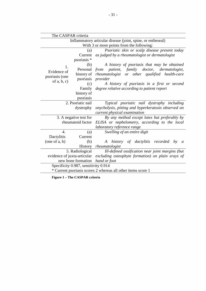
- 31 -
The CASPAR criteria Inflammatory articular disease (joint, spine, or entheseal)
With 3 or more points from the following:
1. Evidence of
psoriasis (one of a, b, c)
(a) Current
psoriasis *
Psoriatic skin or scalp disease present today as judged by a rheumatologist or dermatologist
(b) Personal
history of psoriasis
A history of psoriasis that may be obtained from patient, family doctor, dermatologist, rheumatologist or other qualified health-care provider
(c) Family
history of psoriasis
A history of psoriasis in a first or second degree relative according to patient report
2. Psoriatic nail dystrophy
Typical psoriatic nail dystrophy including onycholysis, pitting and hyperkeratosis observed on current physical examination
3. A negative test for rheumatoid factor
By any method except latex but preferably by ELISA or nephelometry, according to the local laboratory reference range
4. Dactylitis
(one of a, b)
(a) Current
Swelling of an entire digit
(b) History
A history of dactylitis recorded by a rheumatologist
5. Radiological evidence of juxta-articular
new bone formation
Ill-defined ossification near joint margins (but excluding osteophyte formation) on plain xrays of hand or foot
Specificity 0.987, sensitivity 0.914 * Current psoriasis scores 2 whereas all other items score 1
Figure 1 – The CASPAR criteria

- 32 -
The main limitation highlighted in the CASPAR paper concerns the
applicability of the criteria to early disease as the mean duration of disease of the
cases was 12.5 years. Recent research done by the Toronto group attempted to
address this issue and evaluated the use of the CASPAR criteria in early disease
within their clinic population (Chandran et al. 2007b). They performed a
retrospective analysis of patients enrolling into the specialist PsA clinic and found a
sensitivity of 99.1% in those patients with disease duration of less than 2.5 years,
and a sensitivity of 100% for those with disease duration of less than 12 months
(Chandran et al. 2007b). Although important, this study only included patients
referred to a specialist tertiary referral clinic and did not evaluate any control
population. It seems likely that only patients with secure clinical diagnoses are
referred and enrolled into this clinic, possibly leading to an overestimate of the
sensitivity of the criteria. As yet, the only evaluations of the CASPAR criteria in
new rheumatology referrals at a secondary care level have been studies in Sweden
and Italy. In the Swedish early PsA register, 134 of 183 patients fulfilled the
CASPAR criteria when compared to consultant diagnosis (Lindqvist et al. 2008). A
single centre study in Italy of 44 patients (D'Angelo et al. 2009), found that 34 of
the patients met the criteria on their first visit (77.3%) and the majority fulfilled the
criteria by having skin psoriasis and being RF negative. Only two patients satisfied
the radiological criteria as their disease duration was less than one year. They
concluded that the CASPAR criteria are less sensitive in early PsA, mainly because
patients are unlikely to have radiological evidence of new bone formation (D'Angelo
et al. 2009). There were no controls analysed in either of these studies, so only the
sensitivity could be calculated. There is early data from an Italian multicentre PsA
study which found a high sensitivity and specificity of 91% and 97.1% respectively
(D'Angelo et al. 2008). However, as yet, there have been no properly powered
studies addressing the sensitivity and specificity of the CASPAR criteria in early
disease and therefore they cannot currently be recommended for use in studies of
early PsA.
Perhaps the weakest aspect of the CASPAR criteria is the initial qualification
criterion: inflammatory arthritis including spinal, peripheral and entheseal disease.
As cases were physician diagnosed and without other stipulation in the selection
process, it was impossible to be more precise with this description. In fact the
majority of cases had a peripheral arthritis pattern, although 72 had a combined
axial/peripheral pattern and 21 did not have any peripheral joint involvement at all.
Of these 21, two had pure axial disease, eight dactylitis, 12 enthesitis, and 10
inflammatory spinal pain (in combinations) (Taylor et al. 2006).
- 33 -
A third concern with these criteria is located in the composition of the controls.
About 70% of the controls had RA but 13% had AS so the statistical analyses were
influenced against selecting spinal features as characteristic of PsA (Taylor et al.
2006). Although it has been suggested that the spondylitis of PsA is qualitatively
and quantitatively different from that seen in classical AS, these differences did not
appear as discriminating features. Of course, had the controls only consisted of RA
cases, it is possible that the spinal features would have appeared in the final criteria
set. The criteria derived, and the figures for sensitivity and specificity (and those for
post hoc calculations like the likelihood ratios) are therefore very dependent on the
control population (non- or alternative disease) used to derive them.
2.2.1.4 Investigations used in diagnosis of PsA
2.2.2 Subtypes of PsA
PsA is a heterogenous disease and there have been multiple attempts to
subgroup patients according to their clinical presentation. Wright originally
proposed three subgroups for PsA (Wright 1959). These were "DIP joint
predominant disease", "severely deforming arthritis" (which included patients with
axial disease), and "rheumatoid like disease". Later, Moll and Wright described the
five classic sub-groups (monoarthritis, oligoarthritis, DIP-predominant disease, RA-
like polyarticular disease and arthritis mutilans) (Moll and Wright 1973). Since then,
there have been problems highlighted with these subgroups and minor modifications
have been made to the Moll and Wright subgroups by a number of authors
(Gladman et al. 1987; Biondi Oriente et al. 1989; Helliwell et al. 1991; Torre Alonso
et al. 1991). Rather more drastic modifications have been proposed by Helliwell et
al, (with Wright as a senior co-author) and Veale et al (Helliwell et al. 1991; Veale
et al. 1994).
Gladman expanded the five subgroups to seven: distal disease (DIP joints only
affected), oligoarthritis (less than four joints), polyarthritis, spondylitis only, distal
plus spondylitis, oligoarthritis plus spondylitis, polyarthritis plus spondylitis
(Gladman et al. 1987). Arthritis mutilans was not seen sufficiently frequently to
require its own subgroup and was believed to be an indicator of severity, rather than
a distinct group. Torre Alonso removed the category of DIP joint arthritis as it can
occur in any subgroup, but retained the other four Moll and Wright categories (Torre
Alonso et al. 1991).
Later, Wright used scintigraphy to identify the distribution of both clinically
apparent disease and subclinical disease and then created an alternative three
subgroup classification: peripheral polyarthritis, spondarthritis, and
synovitis/acne/pustulosis/hyperostosis/osteomyelitis (SAPHO) (Helliwell et al.
- 34 -
1991). However, Veale et al felt that the peripheral arthritis group was too broad,
combining patients with a symmetrical polyarthritis and an asymmetrical
oligoarthritis who may differ significantly. They concluded with the following three
group classification: asymmetrical oligoarthritis, symmetrical polyarthritis and
predominant spondylitis (Veale et al. 1994).
2.2.2.1 Dividing axial and peripheral disease
The main difficulty in dividing axial and peripheral disease is in accurately
diagnosing and defining the axial subgroup. Axial involvement in PsA is particularly
hard to study as there is very little research into this subgroup. The majority of
evidence is “borrowed” from AS, but this may not be clinically valid. In particular,
the definitions used for AS – such as the modified New York criteria – may not be
applicable to PsA as spinal changes may develop in the absence of SI disease (Khan
et al. 2003; Taylor et al. 2004). Therefore, there is no clear definition of axial
involvement in PsA. The diagnosis of proven axial involvement still relies on
conventional radiography to detect typical changes in the SI joints and spine. This
can take a variable length of time to become apparent and at least partially accounts
for the increase in prevalence with longer disease duration.
Like many rheumatological conditions, research in AS is increasingly focusing
on the detection of early disease and lessons from this may help our ability to
diagnose axial PsA. MRI has been proposed as an ideal tool to demonstrate
inflammation in the spine and SI joints, but has its limitations. Research has shown
that MRI proven sacroiliitis is not always predictive of future AS. A study by
Bennett et al, followed a cohort of patients with a new presentation of early
inflammatory back pain and confirmed inflammation in the form of bone oedema
confirmed on MRI scan. Eight years after their initial presentation, only 33%
(n=13) had progressed to meet a radiographic diagnosis of AS. The key predictor of
progression to AS, as diagnosed by the modified New York criteria, was a
combination of HLA-B27 positivity and severe sacroiliitis on MRI (Bennett et al.
2008). This study highlights the need for caution when using MRI as a diagnostic
tool, particularly if the level of inflammation is low. Finally, axial involvement
may be apparent on conventional radiography or MRI when clinically absent
(Williamson et al. 2004b). It remains unclear why such changes occur in the
absence of symptoms and whether this represents clinically significant disease that
should be treated.
Data from genetics studies can also help us to identify axial disease in PsA.
Although PsA is classified as one of the spondyloarthropathies, the frequency of
HLA antigens varies within the different subgroups (see Table 1). A consistent

- 35 -
association between HLA-B27 and spinal involvement has been demonstrated,
especially with pure axial disease (Gladman et al. 1986; McHugh et al. 1987).

- 36 -
HLA Type Spondylitis Symmetrical polyarthritis resembling RA
DIP predominant disease
Oligoarthritis
B27 (%) 62 15 41 9 B38 (%) 0 12 0 5 CW6 (%) 50 15 18 23 DR4 (%) 25 - 40 61 53 18
Table 1 – Proportion of patients with HLA associations in subgroups of PsA
- 37 -
2.2.2.2 Subgrouping peripheral disease
To be useful from a clinical and prognostic point of view, subgroup
classification ought to be stable over time. Particularly when considering patterns of
peripheral joint involvement, this does not seem to be the case. Jones et al
retrospectively studied a cohort of patients diagnosed with PsA and recorded their
sub-group longitudinally. They found frequent changes in sub-group, particularly
progression from an oligoarticular pattern at onset to a polyarticular pattern at the
final assessment (Jones et al. 1994) and this was further corroborated by Marsal et al
(Marsal et al. 1999). Kane’s key paper investigating the features of early PsA also
found significant movement between sub-groups but in this study, they found
polyarthritis common at presentation but not at follow-up. This seemed to be related
to the proportion of patients treated with disease modifying therapy (Kane et al.
2003a). It seems that the change between groups is probably particularly marked at
the beginning of a disease course as the disease may still be evolving and treatment
is changed more frequently.
Subgroup definition may also be a function of the way joint involvement is
assessed. Clinical examination is a relatively insensitive way of identifying articular
involvement. The use of imaging, in particular colour Doppler US and MRI,
suggest that patterns of articular involvement are quantitative rather than qualitative
(Backhaus et al. 2002). US in patients with oligoarthritis has shown that many
patients would actually be re-classified as polyarthritis once subclinical disease is
identified on imaging (Wakefield et al. 2004). In this way, different methods of
evaluation will produce different patterns of joint and entheseal involvement.
One of the main reasons to form subgroups is that they might behave
differently over time either in terms of natural history or response to treatment. With
respect to the first of these points, data from the Toronto group originally found that
the number of inflamed joints at presentation was an important predictor variable for
long term damage (Gladman et al. 1995). Given the recent availability of highly
effective therapies for PsA, but the potential limitation due to the high cost of these
drugs, clinicians must target these therapies accurately. Those with polyarticular
disease should be targeted for early and intensive therapy to prevent a poor long
term outcome. Further work is required to test the efficacy of this practice given
that the polyarticular sub-group incorporates a wide range of severity. Since those
patients destined to develop arthritis mutilans will almost certainly be in this group,
it will be of interest to see if an aggressive treatment strategy will eliminate this
devastating condition.
- 38 -
In this respect, therefore, it is of importance to distinguish between oligo- and
polyarthritis at onset, even though people change subgroup with time and treatment.
With respect to treatment it may be possible to select subgroups by their response to
different drugs. It is clear that sulphasalazine and methotrexate work differently for
peripheral manifestations compared to axial manifestations in spondyloarthropathies
(Clegg et al. 1996), so it is reasonable to at least examine this possibility by
separately analysing people with axial disease and peripheral disease. However, the
recently introduced anti-TNF drugs seem to be beneficial for all aspects of the
disease, including SAPHO, suggesting that, at this level, there is no worth in
distinguishing between sub-groups (Mease et al. 2000; Antoni et al. 2005a).
Genetics studies have also helped to identify sub-groups of peripheral joint
involvement with a recent interest in class II HLA associations and the more severe
forms of polyarthritis. Gladman originally demonstrated an association between
DR4 and symmetrical polyarthritis resembling RA, and more recently the Bath
group have shown that this is probably a severity marker associated with the shared
epitope of DRB1, as in RA (Korendowych et al. 2003). In further studies, it has been
confirmed that the frequency of shared epitope is not increased in PsA compared to
controls (Ho et al. 2007). Interestingly, the presence of anti-CCP antibodies in PsA
is associated with the presence of the shared epitope, disease severity and activity
scores (Korendowych et al. 2005). The HLA data therefore support the concept of
different types of peripheral disease with the shared epitope acting as a marker for
severe, polyarticular disease.
2.2.2.3 Are subgroups clinically relevent?
There is a strong clinical argument for abandoning the concept of subgroups
all together in view of the difficulties in defining the groups and the high number of
patients who alter sub-group of peripheral disease (Taylor et al. 2005) This would
also be consistent with the ‘unifying’ hypothesis that the pathological basis of the
disease is enthesitis (McGonagle et al. 1999). However, treatment and HLA data
would support the distinction between at least two subgroups – axial and peripheral.
If the concept of subgroups is abandoned, as suggested by Taylor et al, classification
criteria become simplified but therapeutic trials remain problematic because of the
possibility of different treatment responses seen in, for example, axial and peripheral
groups.
- 39 -
2.3 Outcome measurement in PsA
2.3.1 Clinical Outcome Measures
Measuring outcomes in research is a key part of any study design. Clinical
outcome measures provide an assessment of disease activity, in a particular area of
disease, using only simple clinical interventions. Clinical outcome measures are
required for both research and clinical use to measure the impact of therapies and the
response of patients; both at a group and individual level.
Research in PsA has always lagged behind that in RA and it suffers significant
setbacks due to a lack of validated clinical outcome measures. PsA is a multi-
faceted disease with involvement in many areas including peripheral joints, skin and
nail disease, entheseal involvement, dactylitis and axial disease. There are many
different outcome measures available for each of these separate aspects of the
disease but most are borrowed from related diseases and only some of these have
been validated in PsA. At present, there are no composite outcome measures for
PsA that take all of these disease aspects into account.
2.3.1.1 Peripheral Joint Disease
The simplest assessments of peripheral joint activity are joint counts. Joint
counts used in PsA studies have included tender joint counts, swollen joint counts,
active joint counts and damaged joint counts. When assessing peripheral joints,
there are 78 recognised joints for assessment, however reduced joint counts are
usually used. It is widely accepted that 68 joint counts are acceptable, as it is
difficult clinically to distinguish the DIP joints of the toes. As it is not possible to
clinically evaluate the hip for swelling accurately, the hip is excluded from any
swollen joint counts, creating the 66/68 joint count.
Peripheral joint disease activity has been most thoroughly researched in RA as
patients with RA commonly only have disease in this domain. In addition to simple
joint counts, multiple peripheral joint disease activity measures have been proposed.
Most of these include results of a joint count, laboratory measures of disease activity
and a patient’s perspective of disease activity. The two most widely used measures
are the American College of Rheumatology (ACR) response criteria and the DAS
and its modifications. Some of these developed instruments are response criteria,
such as ACR response criteria, where improvement of a certain proportion from
baseline is calculated. Some are measures of state, such as the DAS and DAS28,
where a cross-sectional disease activity measure is obtained (van der Heijde et al.
1990; Prevoo et al. 1995). Subsequently, response criteria called the European
League Against Rheumatism (EULAR) response criteria were developed using the
DAS and DAS28 scores (van Gestel et al. 1996; van Gestel et al. 1998).
- 40 -
The ACR response criteria require measurement of the seven core domains in
RA which are tender joint count (TJC), swollen joint count (SJC), physician global
assessment visual analogue scale (VAS), patient global assessment VAS, patient
pain VAS, physical function measured by health assessment questionnaire disability
index (HAQ-DI) and laboratory acute phase response. The ACR response criteria
include the ACR20, 50 and 70. These require a 20%, 50% or 70% improvement in
both of the joint counts as well as the same percentage improvement in three of the
other five variables (Felson et al. 1995).
The DAS is calculated using the Ritchie articular index (RAI) based on 53
joints, an assessment of 44 joints for swelling, an ESR value and a patient reported
VAS (0-100mm) for general health (VAS GH); as follows (van der Heijde et al.
1990):
DAS= (0.54 x √RAI) + (0.065 x SJC) + (0.33 x ln[ESR]) + (0.007 x VAS GH)
DAS28 scores are based on the reduced 28 joint count for both tender and swollen
joints as follows (Prevoo et al. 1995):
DAS28= (0.56 x √TJC) + (0.28 x √SJC) + (0.7 x ln[ESR]) + (0.014 x VAS GH)
The EULAR response criteria define good response, moderate response and non-
response. A good response is defined as achieving a DAS ≤2.4 or DAS28 ≤3.2
together with an improvement in DAS of >1.2. Moderate response is defined as a
reduction of >0.6 with an end DAS of 2.4-3.7 or DAS28 of 3.2-5.1. Non-response is
defined as a reduction of ≤0.6 or a reduction of ≤1.2 with an end DAS of >3.7 or
DAS28 of >5.1 (van Gestel et al. 1996; van Gestel et al. 1998).
The ACR response criteria, DAS/DAS28 and EULAR responses have
undergone significant reliability testing in RA and are now reported widely in
clinical trials. Following the development of these composite measures, they were
then “borrowed” for use in therapeutic trials in PsA despite the fact that they were
not developed for such use, as there was little research into specific measures
developed for PsA.
The only specific outcome measure that was designed for use in PsA was the
PsA response criteria (PsARC) which is a response measure. The measures
included in the PsARC are the TJC, SJC, patient self assessment of disease activity
and physician assessment of disease activity (using 1-5 Likert scales). Treatment
response according to the PsARC requires improvement in at least two of the four
measures, one of which must be a joint count, and no worsening in the other
domains. Improvement/worsening are defined as a decrease/increase of 30% for
joint counts and a decrease/increase of one category for global assessments of
disease activity (Clegg et al. 1996). However the major drawback of the PsARC
- 41 -
criteria is that they were not developed based on patients’ data, but were simply
stated by a single researcher.
The majority of recent efficacy studies of new agents have used ACR or DAS
outcome measures despite a lack of validation. Therefore, subsequent to these trials,
retrospective analysis was undertaken using data derived from two phase II
randomised controlled clinical trials of TNF inhibitors (Fransen et al. 2006). The
aim of this was to analyse the responsiveness of such composite measures in PsA
trials and to compare this to the responsiveness of individual variables. The
combined data included measurements from 164 patients treated with active drug or
placebo. Analysis was reported using the standardised response mean (SRM=mean
change/standard deviation (SD) change), effect size (mean change/SD baseline) and
t-tests for difference in change between active drug and placebo. In both of the
trials, ACR20, EULAR response criteria and PsARC all showed a significant
difference between active drug and placebo (p<0.001). Importantly, all of the
disease activity composite measures demonstrated superior responsiveness to single
variables in terms of SRM and effect size (Fransen et al. 2006). This highlights the
importance of composite measures for use in clinical trials.
When comparing the different composite measures, it was noted that the
EULAR criteria performed slightly better than the ACR criteria, which in turn
performed better than the PsARC (Fransen et al. 2006). Most RCTs of TNF
blockers have used the ACR20 as a primary outcome measure because of its
reasonable responsiveness in early studies. It also shows a lower placebo response
rate than the PsARC making it a more stringent measure (Mease et al. 2005a). The
good performance of the EULAR criteria in the retrospective analysis was partially
due to the relatively high numbers of patients treated with active drug who achieved
the low disease activity criterion (Fransen et al. 2006). It is likely that in studies of
standard DMARDs, the EULAR and ACR criteria would appear more comparable.
Surprisingly the EULAR response based on DAS28 and the mean change in
DAS28 scores performed slightly better than the comparable responses based on full
DAS scores. The authors identified that the lower performance of the traditional
DAS score is due to the lower discriminitive ability of the RAI. They speculate that
this may be related to the relative inaccuracy of grading tenderness scores and the
scoring of multiple joints as one unit in the RAI (Fransen et al. 2006). There has
been a long-held concern that the DAS28 should not be used in PsA because of the
relatively high proportion of patients who have involvement in other joints, such as
the feet and DIP joints of the hands, which are not included in this measure.
Therefore, DAS28 potentially under represents current disease activity as these
joints are not included. Although the DAS28 outcome measures were shown to be
- 42 -
responsive in clinical trials there are some important caveats given the problems
introduced by the reduced joint count. Firstly, although the measures are responsive
at the group level, there is no validation of these in individual patients and the
potential underestimation of disease activity should preclude this. Secondly,
although the change in DAS28 or EULAR responses may be used, the numerical
scores of the DAS are not validated in PsA, therefore cut points such as low disease
activity or remission may be inappropriate. The risk of the underestimation of
disease is highlighted by the OMERACT PsA group who calculated that 25% of the
patients included in these clinical trials of TNF blockers would not have been
eligible if the DAS28 was used as an inclusion criteria (Gladman et al. 2007d)
despite the fact that 95% of these patients had more than five active joints at the time
of recruitment (Fransen et al. 2006).
The other concern is the accuracy of determining disease activity or response
in patients with other subtypes of PsA. Although the inclusion criteria for the major
TNF blocking studies all required a minimum of three tender and three swollen
joints, the mean joint count was far higher and the patients included almost
exclusively had a polyarticular joint pattern. These composite measures ‘borrowed’
from RA, have not been accurately evaluated in oligoarticular disease.
One further composite measure of arthritis has subsequently been investigated
for use in PsA. The disease activity in reactive arthritis (DAREA) score was
developed using patient data from 45 patients with reactive arthritis and is calculated
as a simple sum of the TJC, SJC, patient’s pain and global assessment VAS and a C-
reactive protein (CRP) (Eberl et al. 2000). Nell-Duxneuner and colleagues collected
data on key PsA outcomes in 105 patients and then performed a principle
component analysis (PCA) to identify which outcome measures best reflected
disease activity. They identified three principal components which reached
significance: patient’s pain and global disease activity assessment, tender and
swollen joint counts and CRP. A fourth component contained skin assessment but
this did not reach significance. After a review of existing measures, the DAREA
was thought to be the most relevant composite outcome that encompassed all of
these principal components. This group have now renamed the DAREA as the
disease activity in psoriatic arthritis (DAPSA) score (Nell-Duxneuner et al. 2010).
The advantage of this analysis in comparison to the validation of DAS and
ACR outcomes is that the analysis was based on PCA of a cross-section of PsA
patients, rather than being a retrospective analysis of RCTs. The same group have
also analysed its sensitivity to change using data from RCTs, providing some
support for its use (Schoels et al. 2010). However some similar limitations also
apply with this analysis, as for that with the ACR and EULAR outcome measures
- 43 -
developed for RA. The analyses are based principally on certain subsets of PsA and
may not be generalisable to the whole population. In particular, skin activity was
identified as the fourth domain in the PCA but was dropped as it did not quite meet
significance. This is likely to be related to the fact that the average psoriasis area
and severity index (PASI) in PsA patients identified from rheumatology clinics is
low (Helliwell 2009) and it therefore had a lower weighting. However for PsA
patients seen in dermatology or combined clinics, psoriasis may have a much more
significant impact on their overall disease activity. Although the DAPSA is a
composite measure, it is only truly a composite joint disease activity measure and
does not reflect changes in other aspects of the disease.
2.3.1.2 Skin and Nail Disease
Many measures have been used to assess disease activity in skin psoriasis.
The earliest measure to assess extent of skin involvement was the body surface area
(BSA) based on the ‘rule of nines’ used to assess the extent of burns (Wallace 1951).
This allows estimation of the extent of skin surface involved assuming that the head
represents approximately 10% of the surface area, upper limbs 20%, trunk 30% and
lower limbs 40% (Wallace 1951). This estimation of skin surface has been validated
in dermatology using volume of ointment required to cover an area as an estimation
of area. The areas were similar to those quoted above, with just the head
representing a slightly lower percentage of BSA due to the use of ointment only on
the face, neck and ears and not on the scalp (Long and Finlay 1991).
In practical assessment of the BSA, an estimation of area is commonly
performed using a handprint to represent 1% of BSA. This handprint or palm
includes the dorsal aspect of the palm, thumb and all four fingers and has been
shown to equate to between 0.51 and 0.91% of the BSA (Long et al. 1992; Rossiter
et al. 1996). In the majority of patients, the use of a handprint or palm to assess
BSA should therefore result in an overestimation of the extent of psoriasis. This has
been confirmed in studies where trained observers overestimate BSA compared to
image analysis of whole body photographs and schematic outlines (Ramsay and
Lawrence 1991; Tiling-Grosse and Rees 1993). This overestimation is particularly
marked when assessing mild psoriasis (Ramsay and Lawrence 1991). Assessing
skin psoriasis using BSA has a reasonable intra-observer reliability, but the inter-
observer reliability has been reported to show variability (Ramsay and Lawrence
1991; Tiling-Grosse and Rees 1993). In multi-centre studies, this has raised a
possible source of bias. However, this can easily be minimised by ensuring that the
same assessor measures BSA at each study site within a trial.
The PASI was developed in the 1970s within a clinical trial and is by far the
most commonly used outcome measure for psoriasis in clinical trials and studies.
- 44 -
This outcome measure combines assessment of BSA with assessment of disease
severity. Disease severity is based on assessments of the plaque’s erythema,
induration and desquamation. The head, upper limbs, trunk and legs are assessed
separately and then combined into the final score using weightings based on the
BSA (head=0.1, upper limbs=0.2, trunk=0.3, lower limbs=0.4). The degree of
erythema, induration and desquamation seen within the lesions in each area are
graded on a semi-quantitative scale from 0 (no involvement) to 4 (very marked
change) and these scores are summed to measure the disease severity. The area of
involvement of psoriasis in each body area is graded from 0-6 based on percentage
coverage of skin disease (0%=0, <10%=1, 10-29%=2, 30-49%=3, 50-69%=4, 70-
89%=5, 90-100%=6) and these body area scores are multiplied by the disease
severity score and the weighting for each body area. This gives a total score of
between 0-72 (Fredriksson and Pettersson 1978).
The target lesion score uses the same semi-quantitative scoring system for
erythema, induration and desquamation but just applies these to a ‘typical’ plaque of
psoriasis and does not account for BSA. Therefore the score for this target lesion
can vary between 0-12 (Feldman and Krueger 2005). This is ideal to measure
change in psoriasis at one site following localised therapy or can be combined with
other outcome measures such as a physicians global assessment and quality of life
measure to get a measure of the global impact of disease.
The PASI relies heavily on the BSA for components of the score and is
therefore subject to the same limitations introduced by the BSA, particularly
overestimation with small area plaque psoriasis. The use of subjective scores for
erythema, induration and desquamation has been validated comparing single
observer subjective scores to objective outcome measures (nitric oxide production,
reflectance erythema, US thickness and US entry echo) showing significant
correlations individually. When combined as a total score for severity as used in the
PASI, this has a higher power to detect change which was comparable to the
objective measurements (Ormerod et al. 1997). However, as there was only one
observer, the potential for inter-observer variation was not assessed and the
subjective scores were found to overestimate the treatment effect compared with the
objective measures (Ormerod et al. 1997).
In addition to considering the reliability of assessing BSA and psoriasis
severity scores, the validity and sensitivity to change of the PASI also have some
recognised shortcomings which must be considered. Criterion validity assesses the
ability of the PASI to match up to a “gold standard”. The comparison between
subjective and objective measures of the composites of the PASI goes some way to
address this, but the lack of a clear gold standard hampers further assessment of this.

- 45 -
To provide face and content validity, the PASI must assess the main aspects of
psoriasis and discriminate severity of disease. Clearly the PASI performs better than
the BSA in this aspect, as it also includes a subjective score for disease severity as
well as area. However it does not include assessment of the most important features
of severity as nominated by both dermatologists (systemic symptoms and pustular
change) and patients (embarrassment and quality of life) (Baughman and Sobel
1970).
The other major problem is the responsiveness of the scale. There is evidence
of general poor responsiveness and of problems at either end of the scale. Anecdotal
evidence and retrospective analysis of study data has suggested that the PASI
underestimates the degree of clinical improvement assessed by physicians (Carlin et
al. 2004) and patients (Jacobson and Kimball 2004). Although the theoretical range
of PASI score is 0-72, in practice it is rare to find a PASI score over 40, meaning
that nearly half of the score is unused. At the other end of the scale, the PASI has
poor sensitivity to change and responsiveness when skin psoriasis is mild. A
patient’s mild psoriasis may improve but still score the lowest score of 1 for
erythema and induration because it is still present, and when there is less than 10%
involvement in any body area, the area cannot reduce further from a score of 1 if
there is any psoriasis still present.
Several modifications to the PASI have been proposed to try to improve its
performance. These attempts have focused on the integration of BSA into the PASI.
The Psoriasis Log-based Area and Severity Index (PLASI) still scores area in six
categories, but the boundaries for each category are based on a log scale (0%, 1-2%,
3-5%, 6-10%, 11-21%, 22-46%, 47-100%). To allow greater weighting of BSA, the
area score is the upper limit of that category (Jacobson and Kimball 2004). These
alternative categories allow greater separation of patients with mild psoriasis. This
increases the weighting of the BSA within the PLASI as compared to the PASI and
improves sensitivity to change particularly among patients with mild disease.
However, conversely it reduces the sensitivity to change at the other end of the scale
as areas between 47 and 100% will score the same value (Jacobson and Kimball
2004). This is likely to be a less significant problem as only a minority of psoriasis
patients have such high BSA values.
The Psoriasis Exact Area and Severity Index (PEASI) was developed by the
same team, but uses actual BSA instead of an area score. This is then multiplied by
the severity score as in the PASI (Jacobson and Kimball 2004). Again this increases
the weighting of the BSA within the total score and improves sensitivity across the
scale. In addition, this use of true percentages rather than groups, avoids the
problem that a 1% change in BSA at the borders of the groups can significantly alter
- 46 -
the score (10% BSA weighted two times higher than 9% in original PASI)
(Jacobson and Kimball 2004)
Both the PLASI and the PEASI were analysed using trial data of 40 psoriasis
patients during their development. This analysis confirmed an increased percentage
improvement in patients using the PLASI and PEASI when compared to the PASI.
Both of the new scales also show a greater correlation with the patients’ self-
assessment of percentage improvement.
The self-administered PASI (SAPASI) has also been developed for patients to
assess their disease severity. This uses an anatomical sketch of a person which the
patient is asked to shade in to mark their psoriasis. The investigator then scores area
on a 0-6 scale as in the PASI. The patient is also asked to score the colour, thickness
and scaling of one typical lesion using three VAS. The severity score is calculated
by adding the three VAS scores, divided that by the maximum VAS score and
multiplying by four. The SAPASI is calculated using the product of the weighted
area scores and the severity score (Feldman et al. 1996). Investigations using the
SAPASI have shown that it has good intra and inter-rater reliability and correlates
well with the PASI (Feldman et al. 1996). The SAPASI was then administered in a
multi-centre drug trial involving 182 subjects and correlated with a physician-
equivalent PASI scoring erythema, induration, scale and BSA. This showed modest
but significant correlations between the two scores and their component parts
(Fleischer et al. 1999). The SAPASI was also demonstrated to identify changes in
disease severity within a clinical trial (Fleischer et al. 1999).
Comparison of the PASI derived scoring systems has shown good correlation
with each other (Henseler and Schmitt-Rau 2008). The SAPASI has very good
correlation with PASI and other PASI derived scoring methods, suggesting that it
would be valid for use in studies where an investigator is not available (Henseler and
Schmitt-Rau 2008). Overall, the analysis concluded that the PLASI is the optimal
tool to use in trials. This is due to the fact that it has high sensitivity to change and
differentiation, particularly at a lower BSA and it was shown to correlate best with
both patient and dermatologist’s clinical opinion on improvement (Henseler and
Schmitt-Rau 2008).
Due to an ongoing dissatisfaction with the PASI as an outcome measure, other
scoring systems have also been developed that are designed differently. However
these also use measurements of similar parameters including BSA, erythema,
induration and desquamation. The Lattice System Physician’s Global Assessment
(LS-PGA) requires an assessment of BSA to decide on a category of area involved
(0%, 1-3%, 4-9%, 10-20%, 21-29%, 30-50%, 51-100%) and then a factoring in of
thickness, erythema and scale from an average plaque. This results in a global
- 47 -
assessment from “clear” to “very severe” psoriasis (Langley and Ellis 2004). The
LS-PGA has shown good correlation with PASI and BSA which is not surprising as
they are based on similar observations. The inter and intra-rater reliability has been
shown to be reasonable. In the original study, the LS-PGA had higher intra and
inter-rater reliability than PASI and BSA (Langley and Ellis 2004), but in a
subsequent study, PASI showed higher reliability than the LS-PGA (Berth-Jones et
al. 2006). This scale is clearly much simpler to perform than the PASI, but does still
categorise BSA with the resultant limitations. Although the categories are designed
to be more responsive in milder disease, there are only three categories for
BSA<10% compared to four in the PLASI.
One of the latest instruments developed for use in psoriasis is the Copenhagen
Psoriasis Severity Index (CoPSI) developed by the Copenhagen Psoriasis Working
Group. This instrument does not involve estimation of BSA, but scores erythema,
thickness and scaling each on a scale of 0-3 at 10 sites of the body creating a
possible score of 0-90 (Berth-Jones et al. 2008). Testing using 16 patients and 14
raters found a substantial intra and inter-rater reliability (>80%) for the CoPSI and
the PASI, as well as good correlation between the two (Berth-Jones et al. 2008).
The exclusion of BSA removes the inherent inaccuracies with this evaluation.
However the number of sites involved provides some estimation of the extent of
disease. The arithmetic required to calculate this score is significantly easier than
the PASI and therefore should be less prone to error. The investigators also noted
that the range of CoPSI scores extended throughout the theoretical range unlike
PASI in which the upper half of the range was largely redundant. The CoPSI also
should provide better differentiation at the milder end of psoriasis cases (Berth-
Jones et al. 2008).
The National Psoriasis Foundation (NPF) developed an entirely new responder
index called the NPF Psoriasis Score (NPF-PS). This combines induration at two
target sites, ratio of current BSA to baseline, physician and patient global assessment
and patient assessment of itch all scored 0-5. These components are then summed to
create a scale from 0-30 (Krueger 1999). This has an advantage as it places more
weight on induration which is thought to be more relevant in psoriasis and is less
variable depending on temperature and emollient use. It also includes a patient
global assessment and assessment of itch which will capture some elements of the
impact of psoriasis on quality of life. An initial study of 17 patients compared the
NPF-PS to the PASI. The NPF-PS identified a significant change in psoriasis after
treatment indicating responsiveness. It also identified this significant improvement
at an earlier time point than the PASI, perhaps suggesting that it is more
discriminative (Carlin et al. 2003).

- 48 -
Finally, it must be noted that all of the measures discussed above to evaluate
psoriasis were designed for use and validated in plaque psoriasis and have not been
assessed in other forms of psoriasis.
Nail involvement is a common feature of both skin psoriasis and PsA. It has
been estimated to occur in up to 50% of patients with uncomplicated skin psoriasis
(Farber and Nall 1992) and is even more prevalent amongst patients with PsA. The
frequency of nail involvement in patients with PsA has been reported as between
63% and 83% (Cohen et al. 1999; Elkayam et al. 2000; Salomon et al. 2003;
Williamson et al. 2004a).
Psoriatic nail disease can be broadly divided into psoriasis affecting the nail
matrix and the nail bed. Involvement in the nail matrix most commonly results in
changes to the nail plate which can be seen as the nail grows out. The different
areas of the nail matrix are responsible for production of different layers of the nail,
with the proximal or dorsal portion of the matrix producing the most superficial
layer of the nail plate, and the distal or ventral portion producing the deepest layer of
the nail plate adjacent to the nail bed. The commonest change in nail psoriasis is
pitting (Tham et al. 1988; Zaias 1990), depressions in the surface of the nail plate
resulting from sloughing of abnormal cells laid down when there is involvement in
the proximal matrix. If the involvement is in the distal matrix, the abnormality is
seen deeper in the nail causing leukonychia. More extensive involvement of the
matrix can result in crumbling of the nail plate. Psoriasis affecting the matrix can be
viewed directly in the lunula which is a distal extension of the nail matrix.
Involvement here results in red spots in the lunula.
Involvement in the nail bed results in distinct patterns of nail disease.
Onycholysis and subungual hyperkeratosis are caused by psoriasis affecting the
hyponychium, the transitional area between nail bed and normal epidermis at the tip
of the digit. Involvement more proximal in the nail bed can be visualised through the
nail plate and can result in ‘oil drop change’, salmon spots or splinter haemorrhages.
Clearly assessment of nail disease in PsA must take all of these features into
account. Many studies of therapies for nail disease have used subjective measures
of nail involvement graded by the investigator as mild/moderate/severe or on a
VAS. Prior to the development of any systematic scoring system, therapeutic
studies used different grading measures of different features either for the fingernails
as a whole or for one target nail.
One of the earliest published nail assessments was developed by the Bath
group for use in their 1994 assessment of the outcome of PsA. Jones et al used a
predefined scoring system where each fingernail was scored for the presence of

- 49 -
pitting, onycholysis, hyperkeratosis and dystrophy. Each of these features scored
one point with a maximum score of 40 for the 10 fingernails (Jones et al. 1994).
This scoring system was then used and adapted by the Oxford group who used the
same scoring system for finger and toenails with a maximum score of 80. This
scoring system was labelled as the psoriasis nail severity score (PNSS) (Williamson
et al. 2004a).
In 2003, the Nail Psoriasis Severity Index (NAPSI) was devised and published
by dermatologists (Rich and Scher 2003). This scored nails by dividing each nail
into four quadrants and scoring one point for each quadrant which has any of the
features of nail bed psoriasis (onycholysis, oil drop, splinter haemorrhages, nail bed
hyperkeratosis) and one point for each quadrant which has any features of nail
matrix psoriasis (pitting, leukonychia, red spots in lunula, nail plate crumbling)
(Rich and Scher 2003). This results in a score of 0-8 per nail, 0-80 for all fingernails
or 0-160 if toenails are included. The advantage of this scoring system is that it does
not require scoring for each feature separately which would be more time-
consuming. However, it maintains differentiation between nail matrix and nail bed
involvement so that sub-analysis could reveal whether treatments affected psoriasis
equally in both areas of the nail. A small assessment of the NAPSI using pictures of
eight nails shown to 37 dermatologists revealed reasonable reproducibility but no
advanced statistical methods were used in the original paper (Rich and Scher 2003).
A subsequent study provided evidence of moderate to good interobserver reliability
between three dermatologists using the NAPSI scoring system with better reliability
in scoring for nail bed features (Aktan et al. 2007). Following on from this, the
NAPSI has been used as an outcome measure for both open-label and double-blind
trials of biological agents (Reich et al. 2005; Korver et al. 2006; Rigopoulos et al.
2008).
Subsequent to this, the modified NAPSI (mNAPSI) was proposed by
rheumatologists working in PsA. They began a study to validate the NAPSI score,
but with the aim of modifying the scoring system as required to “enhance its
performance characteristics” and create an ideal tool for clinical trials (Cassell et al.
2007). They collected digital photographs of all fingernails of 29 patients with PsA
and these were scored by six physicians using the original NAPSI instrument. There
was found to be a significant variability in scoring so two focus sessions were held
with all of the rating physicians to discuss problems with the scoring instrument.
The biggest obstacle found to scoring accurately was the use of quadrants of the
nail, which were difficult to quantify precisely. It was also felt that to improve the
sensitivity of the score, the individual features of nail matrix and nail bed disease
should be considered separately. The only exemption to this was oil drop

- 50 -
dyschromia which was considered with onycholysis as they represent the same
pathology (Omura 1985). To avoid the use of quadrants, a more quantitative scale
for different features of nail disease was introduced, scoring pitting, onycholysis and
crumbling as 0-3 depending on the area of the nail involved. The remaining features
(splinter haemorrhages, leukonychia, red spots in the lunula and nail bed
hyperkeratosis) were scored as either present or absent in each nail. Therefore the
possible range of scores is 0-14 per nail, or 0-140 for all fingernails (Cassell et al.
2007).
Regrading of the original photographs by five observers was then performed
using the new mNAPSI. Comparison of these mNAPSI scores showed excellent
inter-rater reliability and internal consistency as well as strong correlation with
physician VAS for nail severity (Cassell et al. 2007). This was confirmed by an
independent study of 20 patients with PsA who were assessed by ten
rheumatologists and nine dermatologists showing an excellent agreement for the
mNAPSI score regardless of speciality (Chandran et al. 2009).
2.3.1.3 Enthesitis
An enthesis is the site of attachment to bone of tendon, ligament or joint
capsule (Ball 1971). Enthesitis can cause pain, tenderness and swelling at entheseal
sites sometimes with resultant bony damage such as bone spurs at the calcaneum.
Enthesitis is recognised as a manifestation of active inflammatory disease in all of
the seronegative SpA including PsA, but the majority of outcome measures for
enthesitis have been developed and validated in patients with AS.
The first enthesitis index was developed by Mander et al. A list of all entheses
easily accessible to clinical examination was created and this was tested on six
outpatients with AS on two occasions, once on NSAIDs and once off all treatment.
Sites which scored zero in this pilot study were eliminated leaving 66 entheseal sites
to be examined for tenderness and graded on a semi-quantitative score from 0 to 3
(Mander et al. 1987). A further study with 19 AS patients showed correlation of the
Mander Enthesitis Index (MEI) with pain and stiffness VAS scales and a reduction
in the score with non-steroidal anti-inflammatory drug (NSAID) treatment. There
was some variability between different examiners performing the MEI, but intra-
observer variability was not formally tested (Mander et al. 1987).
Further validation was attempted using the longitudinal outcome in the
Outcome in Ankylosing Spondylitis International Study (OASIS) cohort designed to
investigate the long term outcome of AS. The authors provided evidence of a
correlation between the MEI and other disease activity measures in AS including the
Bath ankylosing spondylitis disease activity index (BASDAI). They also tried to

- 51 -
simplify the MEI and create a more feasible tool for use in both research and clinical
practice. The first step was to remove the tenderness scoring from 0-3 and replace it
with a dichotomous 0/1 score for tenderness. This is similar to the modifications
made to the RAI in the measurement of peripheral joints. The modified Mander
showed similar correlation with other disease activity measures. Then a process of
data reduction was performed using the data from patients who had an MEI>0 to try
and reduce the number of sites to be assessed. The most commonly involved
enthesis was identified and noted, then all patients who reported tenderness at this
site were excluded and the process was repeated. This step-wise reduction was
performed until 80% of all patients with a positive MEI score were identified. Data
was analysed from three timepoints and then all potential sites were combined.
After exclusion of entheses difficult to localise or near to other sites, there were 13
sites left for assessment in the Maastricht Ankylosing Spondylitis Enthesitis Score
(MASES) (Heuft-Dorenbosch et al. 2003). Correlation between all of the enthesitis
scoring scales and disease activity measures (BASDAI, physician VAS of disease
activity and patient VAS of disease activity) were similar with no marked reduction
in correlation seen with the shortened MASES (Heuft-Dorenbosch et al. 2003).
Other new modifications of the MEI (Gorman et al. 2002) and distinct scoring
systems (Braun et al. 2002) have been proposed as enthesitis indices but few have
been validated.
In particular, establishing criterion validity for these clinical outcome measures
has been difficult because of a lack of gold standard. Ideally a gold standard would
have associated evidence of tissue abnormality from histopathological studies.
However biopsy of tendons is neither safe nor easy, and there are limited research
data available. Histopathological studies in patients with SpA (mostly AS) have
shown inflammation in the subchondral bone with bone marrow oedema underlying
entheses (Ball 1971; Laloux et al. 2001). A further study identified changes in the
fibrous part of the enthesis with increased vascularity and cellular infiltrate
(McGonagle et al. 2002a). MRI has been shown to identify bone marrow oedema at
tendon insertions and abnormal signal around the enthesis (Marzo-Ortega et al.
2001). US scanning using GS and PD to identify increased vascularisation in and
around the enthesis can identify abnormal findings in symptomatic and
asymptomatic entheses (D'Agostino et al. 2003). Given that US identified PD
signal, and MRI bone marrow oedema have been correlated with tissue evidence of
inflammation in other rheumatic manifestations (Appel et al. 2006; Jimenez-Boj et
al. 2007; McQueen et al. 2007), it seems likely that imaging is the best “gold
standard” available at present.

- 52 -
More recently, the Spondyloarthritis Research Consortium of Canada
(SPARCC) created a new outcome measure for enthesitis in AS (Maksymowych et
al. 2009) using information from US and MRI studies to decide which entheseal
sites should be included. They picked 16 sites which had been identified as
commonly involved in imaging studies of SpA patients (D'Agostino et al. 2003;
Lambert et al. 2004) and could be clinically identified. Inter-observer reliability was
good following a brief specific training session in the scoring system and a
substantial correlation was seen between enthesitis score and other disease activity
measures. However the reduction in enthesitis score in a sub-group of patients
treated in a randomised controlled trial of anti-TNF therapy was not significant after
12 weeks of therapy (Maksymowych et al. 2009). Reduced versions of the
SPARCC enthesitis index using more commonly involved sites (SPARCC 8/16
score) and only sites which discriminated between treatment and placebo (SPARCC
6/16 score) showed larger effect sizes and SRMs (Maksymowych et al. 2009).
All of the entheseal outcome measures discussed previously were developed
and validated principally for patients with AS. The sites included in the SPARCC
score were chosen from studies using a full spectrum of SpA patients, but the
validation was only in patients with AS (Maksymowych et al. 2009). The Leeds
Enthesitis Index (LEI) is the only measure developed specifically for PsA. The
methodology for this study was identical to that of the development of the MASES
using dichotomous scoring for pain and then step-wise data reduction to identify and
retain only those sites with most common involvement. This resulted in an index
with six entheseal sites (bilateral lateral epicondyle, bilateral medial femoral condyle
and bilateral Achilles tendon insertion) (Healy and Helliwell 2008). This index was
then compared to other entheseal indices including the MEI, MASES and others, in
an open-label longitudinal study. The LEI showed closest correlation with other
disease activity measures, a large effect size and the smallest floor effect when
compared with the MEI. Although the LEI was developed using data from just 28
patients, it is the first tool developed using patients with PsA rather than AS. It has
a low floor effect, meaning that it can identify the majority of patients with
enthesitis using just six sites, making it far more feasible as an outcome tool (Healy
and Helliwell 2008). The reliability of the LEI, MASES, SPARCC and other
enthesitis scoring systems was investigated in a reliability study using both AS and
PsA patients. Nineteen patients with AS and PsA (n=9 and n=10 respectively) were
examined by ten experienced rheumatologists. There was excellent agreement for
all patients in all of the indices listed above. In patients with AS, the MASES and
SPARCC indices performed better, but in patients with PsA, the LEI and SPARCC
showed excellent agreement (Gladman et al. 2007a).

- 53 -
The use of clinical assessment tools for enthesitis has now become widespread
in clinical trials despite some of the debate about which particular scoring system is
optimal in each disease subtype. However, as alluded to above, there are some
crucial problems with the criterion validity of these clinical techniques. Imaging
studies have shown changes within tendons and around tendon sheaths at entheses
but the correlation with clinically appreciable tenderness or swelling is less
convincing (Ibrahim et al. 2009b). In most studies, US has identified more changes
at entheses than clinical examination alone (D'Agostino et al. 2003), but the question
of the clinical relevance of these subclinical changes has not been answered. In
addition to the soft tissue changes visualised with imaging, MRI scanning has
identified the involvement of bone with bone marrow oedema adjacent to the
enthesis. No studies have addressed whether this deep bony involvement can be
clinically identified and whether there is evidence of any correlation with clinical
enthesitis counts in these circumstances.
The other limitation of clinical enthesitis counts is the specificity of the
findings of tenderness in these areas. Obviously there is an issue regarding training
of assessors to ensure inter-observer reliability and there is some difficulty in the
anatomical location of some of these sites. However beyond that, there are doubts
about the specificity related to enthesitis and whether other pathologies seen in
arthritis could cause a false positive result. Many of the entheseal points are
relatively near to joints and it is well recognised that inflammation within joints can
cause pain in and around the joint. Also many of the entheseal points are located
near to accepted fibromyalgia points (Wolfe et al. 1990), raising the possibility that
misclassification of enthesitis could occur in patients without active inflammatory
lesions. The recognised clinical enthesitis outcome measures try to minimise the
possibilities of misclassification by advocating sites that are easily anatomically
identified and are distinct from sites of tenderness related to joint inflammation or
fibromyalgia. The key to the reliability of these tools in clinical practice is the
training provided to assessors in localising the correct points.
2.3.1.4 Dactylitis
Dactylitis describes a uniform swelling of a digit with inflammation causing a
“sausage digit” and is one of the key identifying features of PsA. Dactylitis is one
of the features used to make a diagnosis of PsA using the recently developed
CASPAR classification criteria for PsA (Taylor et al. 2006). Dactylitis can be
further characterised as acute/tender dactylitis where the digit is tender to touch and
often erythematous and warm to touch, or as chronic/sub-acute/non-tender dactylitis
where the digit is swollen but non-tender. It has been hypothesised that the chronic
form occurs following an episode of acute dactylitis in some patients but this has not

- 54 -
been confirmed in studies. Acute or tender dactylitis has been shown to be a
presenting feature of 33.5% of patients at their first visit to the clinic and to occur in
48% of patients at some point in their disease (Brockbank et al. 2005).
Our understanding of dactylitis, its pathogenesis and how to define it is
limited. Studies using imaging have confirmed that physical examination can
identify pathology in tender dactylitis. Olivieri et al scanned 12 tender dactylitic
fingers and their matching contralateral normal digits with both MRI and US. Both
scans identified distension of the flexor tendon sheaths and thickening of the flexor
tendons, with no involvement in the extensor tendons. Physical examination by one
physician identified flexor tendonitis in all of the involved digits with clinically
normal extensor tendons (Olivieri et al. 1996). Thus it seems that clinical
instruments for assessing dactylitis do have some criterion validity. However this
study only assessed 12 obviously swollen digits and the contralateral normal digits.
Therefore it seems likely that in normal clinical practice, there will be some
variation between observers resulting in lower agreement particularly in “grey”
cases, where digits may be slightly swollen or where matching digits on both sides
are involved when it is harder to classify as dactylitis. This idea was later confirmed
by a reliability study performed in Canada. This showed a moderate agreement
(kappa 0.57, 95%CI=0.34, 0.82) between 10 experienced observers for number of
digits with dactylitis (Gladman et al. 2004).
Clinical measures of dactylitis have been used as secondary outcome measures
in clinical trials but the majority have used non-validated measures. The simplest
measure used is a simple count of dactylitic digits, although some have counted only
acute/tender dactylitis (Salvarani et al. 2003) and some have included both tender
and non-tender digits (Clegg et al. 1996). Other large RCTs of new therapies have
attempted to grade dactylitis by physician-rated severity in addition to counting
affected digits (Kaltwasser et al. 2004; Antoni et al. 2005b) but none of these
measures were validated prior to their inclusion in studies.
The Leeds Dactylitis Instrument (LDI) was developed in response to this need
for a clinical, objective, validated outcome measure for dactylitis. This also
provided the first numerical definition of dactylitis as an increase in circumference
of the digit of more than 10% compared to the contralateral non-affected digit
(Helliwell et al. 2005). This was based on the evaluation of the median difference in
digital circumference between dactylitic digits and control digits.
The aim of the LDI is to provide a quantification of both the size of the
swollen digit and the tenderness so that the score can differentiate between tender
and non-tender dactylitis. The LDI tool is a circumferometer which is used to
measure the circumference of the affected digit as near to the web space as possible.

- 55 -
This is compared to the contralateral unaffected digit as a ratio. If the contralateral
digit is also dactylitic, then normative values based on population averages are
provided. The tenderness scoring can be based on the RAI with tenderness scored
from 0-3 (LDI scoring) or can be simplified to a dichotomous score of 0 for non-
tender and 1 for tender (LDI basic) (Helliwell et al. 2005).
The first study of this instrument tested its reliability, investigating inter- and
intraobserver agreement. A cross-sectional examination of seven patients by five
examiners using the tool was performed. This showed a relatively poor
interobserver reliability for identifying tender dactylitis and a poor agreement on
non-tender dactylitis. This was improved significantly by using the LDI scoring
system. Inter and intraobserver reliability for the LDI score was good, but was
increased further using the LDI basic, suggesting that some of the variability was
due to the inaccuracy of grading tenderness (Helliwell et al. 2005).
A longitudinal study was then performed to further investigate the use of this
clinical tool and to compare it to other methods used previously. This study
recruited 28 patients with dactylitis who were having a change in treatment for their
PsA. All five available measures for dactylitis (tender dactylitis count (Salvarani et
al. 2003), all dactylitis count (Clegg et al. 1996), IMPACT1 (Antoni et al. 2005b),
LDI (Helliwell et al. 2005), LDI basic (Helliwell et al. 2005)) were performed at
baseline and then regularly following the change in therapy. All measures showed a
change with treatment after three and six months with a reasonable effect size for the
variety of treatments used. The majority of these correlated with other clinical
disease activity measures such as joint counts and VAS for disease activity (Healy
and Helliwell 2007). Only the count of all dactylitic digits performed badly
probably due to the inclusion of non-tender dactylitis which may not be thought to
represent disease activity in patients’ and physicians’ opinions.
A subgroup of patients in the above study also had MRI scans performed at
baseline and six months to assess the inflammation in the dactylitic digits. Similar
to the Olivieri study (Olivieri et al. 1996), this showed that clinically tender
dactylitic digits had significant MRI abnormalities compared to non-involved digits
or non-tender dactylitis. However the correlation between the level of inflammation
on MRI and clinical evaluation was moderate at best (0.37 for LDI local score and
MRI score) (Healy et al. 2008). The identification of more inflammation on MRI
when compared to a clinical score is unsurprising given the poor sensitivity of
clinical examination compared to MRI as seen in other aspects of inflammatory
arthritis (Szkudlarek et al. 2004; Szkudlarek et al. 2006).
Although the LDI and LDI basic measures do take longer to perform,
particularly if multiple digits are involved, these measures perform better in terms of
- 56 -
both truth and discrimination when considering the tool in the context of the
OMERACT filter (Healy and Helliwell 2007). Thus, it is the most validated clinical
outcome measure available for dactylitis.
2.3.1.5 Axial Disease
Just as peripheral joint assessment tools have been borrowed from RA,
measures of axial disease activity have been used in PsA that were initially designed
for use in AS. The most commonly used outcomes in AS are the Bath measures
including the BASDAI, the Bath Ankylosing Spondylitis Functional Index (BASFI)
and the Bath Ankylosing Spondylitis Metrology Index (BASMI). The BASDAI is a
patient completed questionnaire with a series of six 100 mm VAS scales asking
about fatigue, pain and early morning stiffness (Garrett et al. 1994). This is well
validated in AS and has been used as the primary outcome in the vast majority of
clinical trials in AS. The BASDAI provides a static measure of disease activity and
cut points have been devised to define active disease. It is also routinely used as a
response measure. Responses can require a reduction of a certain number of units or
a reduction of a certain percentage such as a BASDAI50 which describes a
reduction in BASDAI of at least 50%.
The BASDAI has been evaluated in a cohort of PsA patients to assess its use in
this condition. This showed a high correlation between BASDAI scores and a self-
reported VAS of current overall arthritis activity, suggesting that it is a good
representation of patient’s perception of disease activity in PsA (Taylor and
Harrison 2004). However this correlation was very similar in those with axial
disease and those with peripheral disease suggesting that the BASDAI does not
differentiate between axial and peripheral disease activity (Taylor and Harrison
2004). No other assessment tools of axial disease activity have been evaluated in
PsA.
Related to the BASDAI is the BASFI, a measure of functional ability validated
and used in AS (Calin et al. 1994). Like the BASDAI, this is based on a series of 0-
100 mm VAS scales where patients rate their ability to perform everyday tasks from
easy to impossible. There are limited data concerning the use of the BASFI in PsA.
One study of the BASFI showed a correlation with other functional indices in PsA
patients but again it shows no difference in responses between patients with axial or
peripheral disease (Leung et al. 2008). Therefore there is no obvious benefit to
using the BASFI in preference over more established and validated tools such as the
HAQ-DI and SF-36.

- 57 -
The BASMI is a combination of five clinical measurements of axial mobility
developed from an assessment of twenty different measures in AS (Jenkinson et al.
1994). This shortened metrology index provides an assessment of spinal mobility
including the cervical and lumbar spine. These measures have been assessed in PsA
patients with axial disease using multiple observers. The International
Spondyloarthritis Interobserver Reliability Exercise (INSPIRE) study showed that
the intraclass correlation coefficients for patients with AS and axial PsA were
similar and were excellent (ICC 0.89) (Gladman et al. 2007b). A further study
tested the correlation between spinal mobility measures and radiographic evidence
of spinal damage in both AS and PsA. This only used 19 patients (taken from the
INSPIRE study) but did show a high correlation between the modified Stoke
Ankylosing Spondylitis Spinal Score (mSASSS) and measures of spinal mobility
(Chandran et al. 2007a). This provides some limited evidence of truth in the form of
criterion validity.
Increasingly, composite measures including some of the Bath scales are being
used to assess axial response in clinical trials in AS. The Assessment of
SpondyloArthritis Society (ASAS) response measures were developed in 2001 using
data from RCTs comparing NSAIDs versus placebo (Anderson et al. 2001). The
authors initially included the five core outcomes established by the ASAS group
(physical function, pain, spinal mobility, spinal stiffness/inflammation and the
patient’s global assessment), but spinal mobility was excluded as there was no
evidence of responsiveness to treatment. Multiple candidate definitions of
improvement were tested looking for a low placebo response (<25%) and a
significant difference between groups. The best criteria were the ASAS20 response
criteria (see table 2) (Anderson et al. 2001).
These were then revised in 2004, following the advent of new therapies. TNF-
inhibitors had proven efficacy in improving spinal mobility and acute phase
reactants, which had been excluded from the previous response measures. When the
original methodology was repeated using data from the RCTs of TNF inhibitors, the
new ASAS20 (5 of 6 criteria) and a modification of the original ASAS response
criteria using a 40% improvement were shown to be the best discriminators (see
table 2) (Brandt et al. 2004).
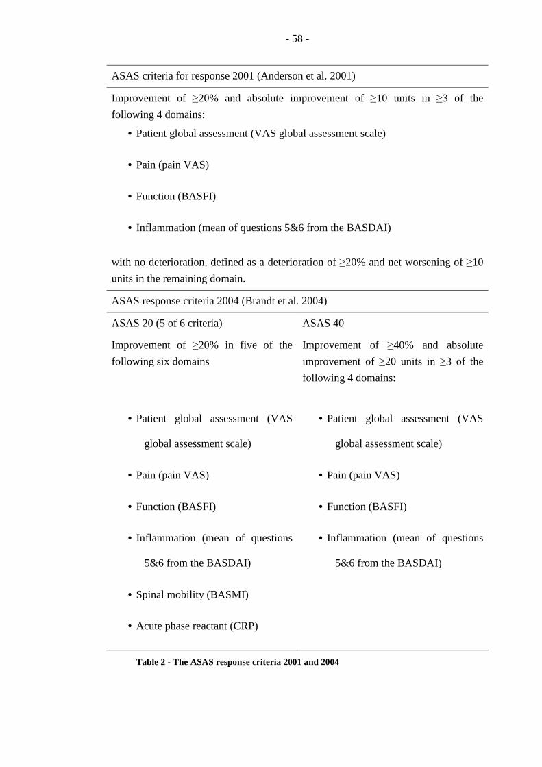
- 58 -
ASAS criteria for response 2001 (Anderson et al. 2001)
Improvement of ≥20% and absolute improvement of ≥10 units in ≥3 of the
following 4 domains:
• Patient global assessment (VAS global assessment scale)
• Pain (pain VAS)
• Function (BASFI)
• Inflammation (mean of questions 5&6 from the BASDAI)
with no deterioration, defined as a deterioration of ≥20% and net worsening of ≥10
units in the remaining domain.
ASAS response criteria 2004 (Brandt et al. 2004)
ASAS 20 (5 of 6 criteria)
Improvement of ≥20% in five of the
following six domains
ASAS 40
Improvement of ≥40% and absolute
improvement of ≥20 units in ≥3 of the
following 4 domains:
• Patient global assessment (VAS
global assessment scale)
• Pain (pain VAS)
• Function (BASFI)
• Inflammation (mean of questions
5&6 from the BASDAI)
• Spinal mobility (BASMI)
• Acute phase reactant (CRP)
• Patient global assessment (VAS
global assessment scale)
• Pain (pain VAS)
• Function (BASFI)
• Inflammation (mean of questions
5&6 from the BASDAI)
Table 2 - The ASAS response criteria 2001 and 2004
- 59 -
More recently, a new composite disease activity score has been developed by
the ASAS group to provide a composite measure that could reflect disease activity
in AS (Lukas et al. 2009). The Ankylosing Spondylitis Disease Activity Score
(ASDAS) was developed from 12 candidate variables selected by ASAS experts.
Data from 708 patients were used to develop the ASDAS. First, principal
component analysis was used to identify sets of correlated variables to remove
redundant outcome measures, then discriminant function analysis was used to
investigate the contribution of the different factors in the separation of high and low
disease activity. Finally, linear regression analysis was performed to provide a
weighting for each of the discriminant variables. This analysis was used to produce
four draft ASDAS formulae (see table 3) for future analysis. Cross validation was
performed in a second cohort confirming similar discriminatory ability. The
discriminatory ability, as measured by the standardised mean difference, was
considerably higher for all four ASDAS when compared with any of the single
variables (Lukas et al. 2009).
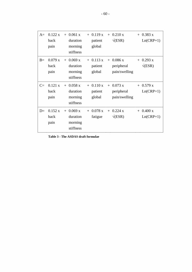
- 60 -
A= 0.122 x
back
pain
+ 0.061 x
duration
morning
stiffness
+ 0.119 x
patient
global
+ 0.210 x
√(ESR)
+ 0.383 x
Ln(CRP+1)
B= 0.079 x
back
pain
+ 0.069 x
duration
morning
stiffness
+ 0.113 x
patient
global
+ 0.086 x
peripheral
pain/swelling
+ 0.293 x
√(ESR)
C= 0.121 x
back
pain
+ 0.058 x
duration
morning
stiffness
+ 0.110 x
patient
global
+ 0.073 x
peripheral
pain/swelling
+ 0.579 x
Ln(CRP+1)
D= 0.152 x
back
pain
+ 0.069 x
duration
morning
stiffness
+ 0.078 x
fatigue
+ 0.224 x
√(ESR)
+ 0.400 x
Ln(CRP+1)
Table 3 - The ASDAS draft formulae
- 61 -
Further validation work has continued following this initial publication.
Analyses in two further independent cohorts again showed that all four draft
ASDAS were highly discriminatory (van der Heijde et al. 2009). Discussion within
the ASAS group was used to decide on the final ASDAS formula to be
recommended. It was decided that the ASDAS with CRP (ASDAS C in table 3)
should be the preferred ASDAS score with an alternative ASDAS including ESR
(ASDAS B in table 3) to be used if CRP is not available (van der Heijde et al. 2009).
A further external validation study showed responsiveness to change and concurrent
validity in patients with axial SpA treated with TNF blockers (Pedersen et al. 2010).
Preliminary cut-points have also been proposed for different levels of disease
activity and responses, although these need further investigation and validation
before they are proven.
Unfortunately these composite measures (both ASAS response measures and
the ASDAS) have not been assessed in PsA. Given the limitations of the BASDAI
in PsA, there are justifiable concerns about their use; however the inclusion of other
outcome measures may mean that these composite indices can be helpful in axial
PsA. Ideally, studies with newer effective therapies are required in axial PsA to
address this issue and to compare these different outcome measures.
2.3.1.6 Composite Outcome Measures
As discussed in previous sections, the majority of outcome measures used in
PsA research have been “borrowed” from other diseases and have only been
validated in subsets of disease. Recently, there have been increasing efforts in the
development of a validated composite outcome measure which reflects disease
activity in all of the domains in PsA and can be used in a variety of subsets of
disease.
In recent years, work has been underway on two key composite measure
initiatives, both led by members of Group for Research and Assessment of Psoriasis
and Psoriatic Arthritis (GRAPPA). Professor Fitzgerald and colleagues proposed a
composite outcome measure based on the GRAPPA treatment grid published by
Ritchlin and Kavanaugh (Ritchlin et al. 2009). The Composite PsA Disease Activity
Index (CPDAI) assigns a score of 0-3 to each of the five domains of PsA based on
disease activity and impact of disease for this domain. These scores can then be
added together to give a total score of 0-15 with an overall assessment of disease
severity based on this total score (Mumtaz et al. 2010). One concern raised during
the development of this measure was that patients with severe disease activity in
only one domain may be disadvantaged by a relatively low total score. Two
- 62 -
solutions have been proposed for this. The first is that anyone with a single domain
scored as severe would also be classified as severe overall. The second solution was
the proposal of a modified CPDAI, with the total score being divided by the number
of domains involved to give a mean CPDAI score.
Both of these CPDAI scores have undergone preliminary validation using data
from observational cohorts and datasets from RCTs. Data from patients followed in
rheumatology clinics in both Dublin and Leeds were used to relate CPDAI scores to
patient and physician global disease activity VAS and to treatment decisions. The
CPDAI score showed an excellent correlation with both patient and physician VAS
and in multiple analyses, higher scores were found to be associated with treatment
changes (Mumtaz et al. 2010).
A second initiative was started by GRAPPA following their annual meeting in
2008. In an attempt to develop an inclusive composite outcome measure based on
real patient data, the GRAPPA Composite Exercise (GRACE) project was
developed (Coates et al. 2010c). Based on the development of other composite
outcome measures for both RA and AS (the DAS and ASDAS), observational
longitudinal data are being collected on a large cohort of PsA patients across the
world. Individual outcomes assessing disease activity in all of the domains of PsA,
as well as patient reported outcome measures (PROMs) are being collected. Patients
are classified by their treating physician in two groups: those requiring a treatment
change (i.e. active disease) and those not (assumed to have a low disease activity).
In this way, the two groups can be compared to see where significant differences
exist between the two groups and which individual outcome measures account for
this difference. To date, cross-sectional data has been collected on over 400 patients
and these patients continue in follow up.
At the OMERACT10 meeting in 2010, the PsA working group (composed of
key members of the GRAPPA group) presented a summary of the existing
composite scores for PsA and preliminary work from the GRACE initiative.
Professor FitzGerald presented data from the psoriasis randomised study in subjects
with psoriatic arthritis (Psoriasis Randomised Etanercept STudy in subjects with
Psoriatic Arthritis or PRESTA study) investigating the responsiveness of the CPDAI
and other proposed indices including the DAREA or DAPSA. This study is
particularly interesting as it is the only large RCT to include only patients with both
significant joint disease activity and skin disease activity. The study compared
standard dose etanercept (50 mg once weekly) with a higher dose of 50mg twice
weekly to evaluate if higher doses were more effective in PsA. There was a
significant difference seen between the treatment groups in terms of skin disease,
but similar improvements were seen in joint disease activity (Sterry et al. 2010).

- 63 -
This differential response makes this database ideal for analysing proposed
composite measures. The analysis showed that all measures performed well at
discriminating between baseline and the primary endpoint, but none found a
significant difference between the treatment groups related to the differential
response in skin disease. Interestingly, the mean CPDAI score did show a trend
between the two groups, possibly influenced by the skin disease activity but this did
not reach significance.
Finally at OMERACT10, preliminary data were presented from the GRACE
analysis. When considering individual domains of PsA, there was a clear
differentiation between the two groups identified in the GRACE data collection for
all outcome measures except those measuring nail psoriasis (mNAPSI), enthesitis
(MASES), ESR and BASMI. The lack of a significant difference for these
parameters is likely to be related to different factors. In some cases, particularly the
nail disease, there is a relatively low prevalence of these features in the patient data.
This lack of difference may also suggest that these features are less important to
physicians when considering disease activity and potential changes in therapy. A
PCA was performed for all variables included in disease activity measures with log
transformation for PASI, LEI, dactylitis, joint counts and CRP, to identify which
outcome measures best identified the variability in the data. The PCA identified five
significant factors that were best represented by
1. Global disease activity VAS
2. Skin disease activity (BSA and PASI)
3. TJC and enthesitis count
4. SJC and dactylitis count
5. CRP
Regression analysis was then applied showing that nearly 80% of the
variability (adjusted R2) was contributed by the global disease activity VAS, and
over 90% of the variance can be summarised with just three VAS scores (patient
global, patient skin, and physician global). Therefore the PsA disease activity score
(PASDAS) was proposed as a composite of three VAS scales (Coates et al. 2010c).
A preliminary analysis of a modular approach to disease assessment rating
each domain (peripheral arthritis, skin, dactylitis, enthesitis and spine) in terms of an
intention to treat was also presented at the OMERACT 10 conference. Each domain
would be rated from A to E on a five point scale where E represents no involvement
of that domain and A severe involvement requiring DMARD therapy.

- 64 -
This method of using PCA and regression analysis was used in the
development of both the RA DAS and the ASDAS. However, precisely because of
the nature of PsA, this methodology has some disadvantages. The most significant
issue is that any potential formula is based on a particular sample of patients’ data,
and their disease pattern influences the resulting formula. For example, the majority
of the GRACE participants were recruited from rheumatology clinics where skin
disease is usually mild. Therefore skin disease may have less of an impact in these
patients on their total disease activity. However patients seen in dermatology clinics
are likely to have greater skin disease activity, and the disease activity score may not
accurately reflect their pattern of disease. In a more homogenous condition, this is
less of an issue as the sample is likely to be representative of most of the patients it
will be utilised in. However in PsA, basing disease activity scores on “average”
rheumatology patients may severely disadvantage those with other patterns of
disease.
2.3.1.7 Defining “state” in PsA
There are no definitions of high or low disease activity states available in PsA.
In RA, there are continuous composite outcome measures such as the DAS, for
which cutpoints for different disease severities ranging from remission to high
disease activity have been defined. The availability of these definitions in RA has
provided a different outcome measure for clinical trials. They allow identification
of patients who achieve a desired state, rather than a significant reduction in disease
activity that may still represent a considerable burden of disease. Despite validation
of the DAS scoring system in PsA which showed that it could discriminate well
between active treatment and placebo, there are no validated cutpoints for different
disease severities of peripheral joint activity in PsA.
The availability of these cutpoints for disease has also meant that they can be
used in clinical trials to guide therapy. In particular, the TIght COntrol of
Rheumatoid Arthritis (TICORA) study demonstrated that tight control of disease
utilising pre-defined activity levels to guide therapeutic changes (escalation of
treatment if DAS >2.4), resulted in significantly better clinical and radiographic
outcomes compared to routine care with no formal therapeutic protocol (Grigor et al.
2004). Many recent RA studies have used similar objective measures (usually the
DAS) to guide therapy (Goekoop-Ruiterman et al. 2005) and this is now beginning
to be translated into routine clinical practice.
Remission is clearly the ultimate goal of therapy in PsA as in other
inflammatory arthritides, and a definition proposed by Kavanaugh et al suggested
- 65 -
that remission in PsA should be characterised by “a complete absence of disease
activity, with no signs or symptoms of active disease” (Kavanaugh and Fransen
2006). However, this paper also recognised that remission was not only difficult to
achieve and maintain, but that, in some patients, mild disease activity in one domain
can be acceptable. Given this, they concluded that “near remission” or “low disease
activity” could be an appropriate goal for individual patient’s treatment (Kavanaugh
and Fransen 2006).
The need for a definition of MDA was first recognised in the RA research
community and was developed within OMERACT. It was recognised that the most
important target was achieving a desired state rather than simply a reduction in
disease activity that may leave patients with significant ongoing disease. Remission
or an absence of disease activity was felt to be an unrealistic goal in many patients,
therefore MDA aimed to encompass both remission and “near remission” or “low
disease activity” as acceptable targets for therapy. The development of a concept of
MDA was done through an OMERACT working group with involvement of
rheumatologists, methodologists and patients (Wells et al. 2003).
It was discussed that for physicians, low disease activity state was linked to
treatment decisions and to prognosis. For patients, low disease activity state was
linked to satisfaction with current symptoms and also to adaptation since a ‘cure’ is
not currently possible. The concept of a usual target of treatment was developed
based on the example set by the group who developed the RA DAS. They reasoned
that a disease activity score should reflect real-life clinical practice and related low
and high disease activity to the decision of a physician to initiate or escalate
treatment. It was also noted that any definition should be limited in time as
treatment options and expectations of both patients and physicians are likely to
evolve over time (Boers et al. 2003a).
The conceptual definition of MDA was agreed at the OMERACT 6 conference
as “that state of disease activity deemed a useful target of treatment by both the
patient and physician, given current treatment possibilities and limitations” (Boers et
al. 2003b). This definition is designed to reflect clinical practice and for the
rheumatologist is linked to treatment decisions. For the patient, this should be
linked to a satisfactory symptomatic state. Following on from the agreement of this
conceptual definition, the OMERACT working group went on to create a set of
MDA criteria for RA (Wells et al. 2005) based on the ACR core set and the DAS
scoring system. Researchers in other rheumatic diseases are now starting to develop
disease specific criteria for MDA (Magni-Manzoni et al. 2008).
Discussion regarding remission in PsA has also occurred and this has been
investigated in a number of cohort studies. However all of these definitions are

- 66 -
different and were not based on patient-derived data (Gladman et al. 2001; Cantini et
al. 2008; Lindqvist et al. 2008). Interesting, the majority only assess peripheral joint
activity and do not take other aspects of the disease into account (Gladman et al.
2001; Lindqvist et al. 2008).
Any criteria for disease activity in PsA must assess many aspects of this
complex disease and a core set of measures for future PsA research was agreed at
OMERACT 8 in 2006. The six core measures agreed were peripheral joint activity,
skin activity, pain, patient global assessment, physical function and health-related
quality of life (HRQOL) (Gladman et al. 2007d). Other important domains included
spinal disease, dactylitis, enthesitis, fatigue, nail disease, radiography, physician
global assessment and acute-phase reactants (Gladman et al. 2007d). If states of
disease activity are to be defined, these core and important measures must be taken
into account to ensure that a holistic view of the patient is considered.
2.3.2 Imaging
2.3.2.1 Conventional radiography
Conventional radiography is considered the “gold standard” in measuring
progression of joint damage. The importance of assessing radiographic evidence of
joint damage in clinical trials is becoming apparent with the advent of newer
biological treatments that can impact on the natural progression of disease.
Initially, research into scoring systems for plain radiographs concentrated on
RA, and many of the scoring systems developed for PsA are adaptations of these.
The majority of radiographic scoring systems for RA assess JSN and erosions.
While these features are seen in PsA, additional typical features must be considered.
In PsA, particularly in the mutilans subtype, extensive erosions may be seen
extending to osteolysis of both joints and phalanges. One of the typical features of
PsA, the “pencil-in-cup” deformity, results from marked erosive changes on the
proximal side of a joint with new bone formation around the distal side of the joint.
This demonstrates the co-existence of bone erosion and new bone formation in PsA.
This bone formation can occur adjacent to the joints and the shafts of the bones and
may even extend to ankylosis of a joint. Most adaptations of radiographic scoring
methods attempt to identify both bone destruction and bone formation in key joints.
At the Toronto PsA Clinic, a modification of the Steinbrocker technique has
been used to evaluate the radiographic outcome over time. The original
Steinbrocker technique was developed for use in RA in 1949 and scored a patient
according to their worst joint on a scale of 0-4 (table 4) (Steinbrocker et al. 1949).
There was a concern that just scoring one joint would not be sensitive to change and

- 67 -
therefore a modified Steinbrocker is used for monitoring in the clinic. The
modification scores 42 joints according to that scale including all joints on the hands
(wrist = 1 joint), all metatarsophalangeal (MTP) joints and the interphalangeal (IP)
joint of the big toes (Rahman et al. 1998b). This modification has been validated
within the Toronto cohort and has shown good inter and intra-observer reliability as
well as confirming sensitivity to change over time (Rahman et al. 1998b).
The Toronto validation study also analysed the use of the Larsen score in PsA.
This is a similar method where each joint is given a global semi-quantitative score
and was also originally developed for use in RA (Larsen et al. 1977). The Toronto
group used Larsen’s method as modified by Rau and Hehborn (table 5) (Rau and
Herborn 1995) and adapted the score to include the same joints as their modified
Steinbroker scoring system (Rahman et al. 1998b). This study confirmed similar
validity of the Larsen scoring system in PsA with similar inter and intra-observer
variability and similar sensitivity to change to the modified Steinbrocker score used
routinely in the clinic (Rahman et al. 1998b).
Although both the Larsen and Steinbrocker techniques have been validated in
PsA, they both simply assess each joint with a global score. There is no
differentiation between different pathologies, and no identification of typical PsA
features such as the pencil-in-cup deformity. Three more detailed scoring systems
have also been outlined for use in PsA, two of these are based on the Sharp method
for scoring RA radiographs and the final method is a score specifically developed
for PsA.
The most widely used detailed scoring system for radiographic damage in RA
is the Sharp method (Sharp et al. 1971). A modification to this was made in 1989 by
van der Heijde to include joints in the feet in addition to those in the hands (van der
Heijde et al. 1989). Although more time consuming than the scores which utilise a
global score for each joint, such as the Steinbrocker and Larsen scores, the Sharp
scores do provide a higher reliability and sensitivity to change due to the detail
included in the assessment.
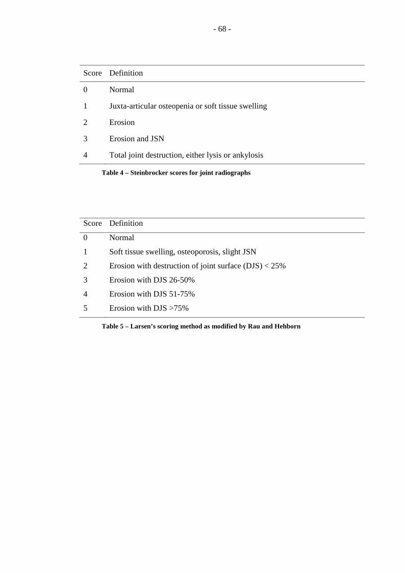
- 68 -
Score Definition
0 Normal
1 Juxta-articular osteopenia or soft tissue swelling
2 Erosion
3 Erosion and JSN
4 Total joint destruction, either lysis or ankylosis
Table 4 – Steinbrocker scores for joint radiographs
Score Definition
0 Normal
1 Soft tissue swelling, osteoporosis, slight JSN
2 Erosion with destruction of joint surface (DJS) < 25%
3 Erosion with DJS 26-50%
4 Erosion with DJS 51-75%
5 Erosion with DJS >75%
Table 5 – Larsen’s scoring method as modified by Rau and Hehborn

- 69 -
The first modification of the Sharp scoring method was developed for use in
trials of anti-TNF therapies. A similar range of joints were assessed as in the
standard Sharp RA method (Sharp et al. 1985), except that the DIP joints were
assessed in addition for both erosions and JSN. Erosions and JSN are scored using a
similar scoring system to that in RA (Sharp et al. 1985), but with additional scores
for specific PsA features (table 6). The presence or absence of pencil-in-cup
deformity is scored for all lesions of grade 6 or 7 and these additional specific PsA
features (grade 6 erosion, grade 7 erosion, pencil-in-cup deformity, grade 5
widening) are not added in the final score but are kept separate to it. Other
radiographic features of PsA (including periostitis, tuft resorption) were also scored
separately, when this system was first trialled. However once this data was
analysed, the system showed sensitivity to change based on the erosion and JSN
scores (Mease et al. 2004) and scoring of additional features did not provide any
additional improvement in differentiation between treatment groups (van der Heijde
et al. 2005a).
A second modification based on the Sharp-van der Heijde method, was
developed later for further RCTs of biologic therapies. Again, a similar range of
joints is scored with addition of the DIPs (van der Heijde et al. 2005b). Joints are
scored for erosions, JSN, subluxation and ankylosis using the table below (table 7).
The maximum score for erosions is 5 in the joints of the hand and 10 in the joints of
the feet. Gross osteolysis and pencil-in-cup are scored separately; and any joints
found to have these abnormalities are given the maximum score for both erosions
and JSN.

- 70 -
Erosion scores
Score Definition
0 No erosion
1 One discrete erosion of involvement of <21% of the joint area by
erosion
2 Two discrete erosions or involvement of 21-40% of the joint
3 Three discrete erosions or involvement of 41-60% fo the joint
4 Four discrete erosions or involvement of 61-80% of the joint
5 Extensive destruction involving more than 80% of the joint
6 More extensive osteolysis
7 Gross osteolysis
Pencil-in-cup Present or absent
Joint space narrowing scores
Score Definition
0 Normal joint
1 Asymmetrical and or minimal narrowing
2 Definite narrowing with loss of up to 50% of the normal space
3 Definite narrowing with loss of 51-99% of the normal space
4 Absence of a joint space, presumptive evidence of ankylosis
5 Widening
Table 6 – modified Sharp scoring system for PsA with inclusion of typical features
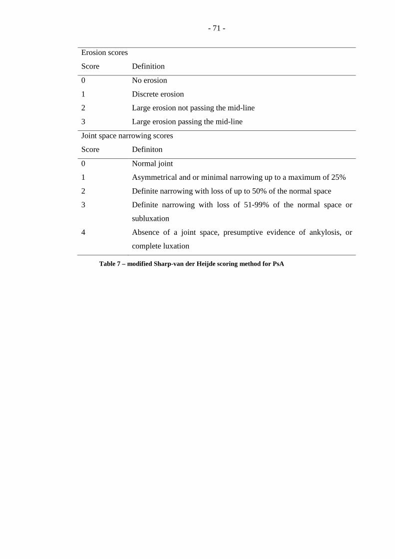
- 71 -
Erosion scores
Score Definition
0 No erosion
1 Discrete erosion
2 Large erosion not passing the mid-line
3 Large erosion passing the mid-line
Joint space narrowing scores
Score Definiton
0 Normal joint
1 Asymmetrical and or minimal narrowing up to a maximum of 25%
2 Definite narrowing with loss of up to 50% of the normal space
3 Definite narrowing with loss of 51-99% of the normal space or
subluxation
4 Absence of a joint space, presumptive evidence of ankylosis, or
complete luxation
Table 7 – modified Sharp-van der Heijde scoring method for PsA
- 72 -
This scoring system was utilised in the IMPACT and IMPACT2 studies of
infliximab therapy in PsA. These analyses found relatively high kappa scores
showing good agreement between readers. It also identified a reduction in joint
damage progression with infliximab therapy, using the total modified Sharp/van der
Heijde (mS-vdH) score, the erosion score and the JSN score. The additional PsA
specific features of pencil-in-cup and gross osteolysis occurred infrequently and did
not show any change over time (Kavanaugh et al. 2006; van der Heijde et al. 2007).
Therefore, it was suggested that these may not be needed, and that the RA scoring
system can be utilised with the addition of the DIP joints only.
The Ratingen score is the only radiographic scoring system developed
specifically for PsA. The authors recognised the co-existence of bone destruction
and bone proliferation in PsA and devised a score for each of these aspects
individually (table 8) (Wassenberg et al. 2001). A similar number of joints are
scored to that of the modified Steinbrocker, except that the 1st MTP joints are
excluded leaving a joint count of 40. The validity of this method was assessed using
20 pairs of radiographs from patients with PsA taken three years apart. Inter and
intra-reader reliability and sensitivity to change were found to be comparable to
results in RA studies using the Larsen, Ratingen and mS-vdH scoring methods
(Wassenberg et al. 2001). Interestingly, although both bone destruction and bone
proliferation progressed over the study period, there was little correlation between
the amount of progression in bone proliferation and bone destruction scores
(Wassenberg et al. 2001). This confirms the independence of these two pathological
processes and highlights the importance of such a scoring system where each feature
is assessed independently.

- 73 -
Bone Destruction Score (DS)
Score Definition
0 Normal
1 One or more definite erosions with an interruption of the cortical plate of
>1mm but destruction of less than 10% of the total joint surface
2 Destruction of 11-25%
3 Destruction of 26-50%
4 Destruction of 51-75%
5 Destruction of >75% of joint surface
Bone Proliferation Score (PS)
Score Definition
0 Normal
1 Bony proliferation measured from the original bone surface of 1-2mm, or, if
the margins of the proliferation cannot be distinguished from the original
bone surface, clearly identifiable bone growth not exceeding 25% of the
original diameter of the bone
2 Bony proliferation of 2-3mm or bone growth between 25-50%
3 Bone proliferation of >3mm or bone growth >50%
4 Bony ankylosis
Table 8 – Ratingen scoring system for PsA
- 74 -
2.3.2.2 Ultrasound
US is an expanding area of interest in rheumatology clinical trials. The use of
US as an objective quantitative outcome measure has been expanding, particularly in
RA clinical trials. US allows real time image acquisition of multiple joints to assess
both bony changes (erosion) and inflammation. Definitions of pathologies (bone
erosion, synovial fluid, synovial hypertrophy, tenosynovitis and enthesopathy) seen
on US have been agreed by the US special interest group (SIG) at OMERACT 7 for
use in future studies. The majority of studies using US have utilised these
definitions and a semi-quantitative 0-3 scoring system where 0=normal, 1=mild,
2=moderate and 3=severe pathology. Erosions can be scored using a binary
present/absent system or a 0-3 semi-quantitative score (Szkudlarek et al. 2003).
One of the key questions raised by the use of US as an outcome measure is the
question of how many joints should be scanned. Obviously scanning many joints
provides additional information, but US is still relatively time-consuming when
multiple joints must be assessed, and feasibility is a key issue. Early work has
begun in RA to identify reduced joint combinations that can still accurately identify
pathology and both seven and 12 joint scores have been proposed (Naredo et al.
2008; Backhaus et al. 2009). Similar work in PsA has yet to be done, and this is
likely to be more challenging given the heterogeneity of clinical presentation in PsA.
2.3.2.3 MRI
While MRI has been used since its development as a research tool to
investigate pathogenesis in arthritis, it has less commonly been used as an outcome
measure for disease activity and joint damage. However increasingly, studies are
looking for objective methods of measuring disease activity and MRI is able to
visualise all of the structures in and around the joint. MRI also has major
advantages over conventional radiography in terms of its sensitivity to change when
investigating progression of joint damage.
The OMERACT MRI in inflammatory arthritis group decided to develop a
scoring system for PsA, following on from the development of the RA MRI score
(RAMRIS). The first step was a literature review of PsA MRI studies to identify
typical pathologies that would need inclusion into a scoring system (McQueen et al.
2006).
Synovitis, bone oedema and bone erosions have been identified using MRI in
both RA and PsA. Despite the fact that synovial tissue samples have suggested a
difference in the histopathology of PsA and RA tissue (Reece et al. 1999), and MRI
studies have shown more extensive synovitis in RA compared to PsA (Marzo-

- 75 -
Ortega et al. 2009), the synovitis is indistinguishable, when matched for disease
activity (Cimmino et al. 2005). Similarly, although bone erosions are seen at
different sites in the two diseases, the appearance of the erosions is non-specific. In
both diseases, a break in the bone surface can be visualised on MRI (Ostergaard et
al. 2003). This may be filled with inflammatory tissue which will enhance with
gadolinium or with fibrous tissue which is non-enhancing. Similarly, the
appearance of bone oedema across the arthritides is similar on MRI. Images show
an increased signal within the bone on short-tau inversion recovery (STIR) imaging,
T2 weighted films with fat saturation and post contrast T1 weighted images
(Ostergaard et al. 2003). Similar to the situation regarding bone erosion, the
differences seen between RA and PsA relate to the sites of bone oedema and the
severity of disease.
The possibility of a different pattern of involvement in PsA was first raised in
1995 in two papers investigating MRI in PsA (Giovagnoni et al. 1995; Jevtic et al.
1995). Both of these papers showed extensive inflammation in the periarticular
tissue extending beyond the joint capsule and into the surrounding subcutaneous
tissue in patients with PsA whereas in patients with RA, the inflammation was
confined to the joint capsule in most cases. Where periarticular oedema was seen in
RA patients, it did not extend to the subcutaneous tissue. These papers highlight the
additional features seen on MR imaging in PsA and it was recognised that these
must be included in a potential scoring system.
The PsA MRI score (PsAMRIS) system has been developed for scoring hand
MRIs in PsA. The RAMRIS scores the wrist joints and the metocarpophalangeal
(MCP) joints of fingers 2-5. There was a concern about excluding the proximal
interphalangeal (PIP) and DIP joints in a potential PsA scoring system given the
increased involvement typically in the DIP joints. It was also felt that there was less
involvement seen in the wrists in patients with PsA. Therefore the joints scored in
the PsAMRIS are MCP, PIP and DIP of fingers 2-5. These joint regions were
divided by the midpoints of the phalangeal bones and were then subdivided at the
joint space line to give three joint regions and six sub-regions (see figure 2)
(Ostergaard et al. 2009).
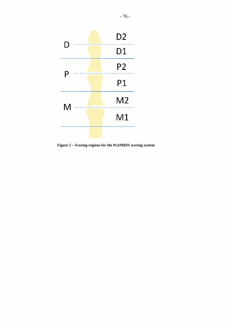
Figure 2 – Scoring regions for the PsAMRIS scoring system
- 76 -
Scoring regions for the PsAMRIS scoring system

- 77 -
Given that synovitis, bone erosions and bone oedema appear similar to that of
RA, the semi-quantitative scoring system used in the RAMRIS was adopted
(Ostergaard et al. 2009). Following this system, synovitis is scored in each joint
region from 0-3, bone erosions are scored at each articular surface on a scale of 0-10
dependent on the percentage of bone loss and bone oedema is scored from 0-3 for
each sub-region. In addition to this, other key features were defined and added to
the scoring system. Tenosynovitis is assessed in each joint region in each of the four
flexor tendons on a scale of 0-3 depending on the thickness of enhancing or bright
signal within the tendon. Periarticular inflammation is scored as present or absent
adjacent to each joint region on the dorsal and palmar aspect of the finger. Bone
proliferation, defined as “abnormal bone formation in the periarticular region, such
as at the entheses (enthesophytes) and across the joint (ankylosis)” (Ostergaard et al.
2009), is also scored as present or absent in each joint region.
Following on from the development of the scoring system, it has been tested in
multiple validation exercises. The first exercise was cross-sectional and involved
four experienced RAMRIS readers scoring 10 MRI scans of patients with PsA using
the proposed PsAMRIS. This showed good to moderate inter-reader reliability for
bone erosion and bone oedema, but low inter-reader reliability for the other features
(McQueen et al. 2009). Difficulty assessing PIP and DIP joints was reported by the
readers as the image resolution was poor at the extremes of the field of view. Also
at this stage, readers were asked to record patterns of bone oedema as
“subchondral”, “entheseal” or “diaphyseal”. This was felt to be too difficult to
distinguish and was removed from later versions of the scoring system. Also,
following on from this exercise, further definitions were created to improve inter-
reader reliability in scoring (Ostergaard et al. 2009).
A second cross-sectional validation exercise utilised a further 10 MRI scans of
patients with PsA and 2 healthy controls. These were scored by eight readers and
found an improved intra-class coefficient (ICC) between readers of 0.84-0.91 except
for the scoring of periarticular inflammation which was low (McQueen et al. 2009).
This provides evidence for an increased reliability following clear definitions of
pathologies and a removal of some features.
A longitudinal validation exercise was then performed by eight readers. They
scored paired scans taken prior to treatment and at six weeks following treatment
with anti-TNF therapy (adalimumab). This confirmed good reliability and
sensitivity to change in synovitis, tenosynovitis and periarticular inflammation
(McQueen et al. 2009). However given that the scans were only six weeks apart,
there was not enough change in the bony features (bone erosion, bone oedema) to
accurately assess the validity of scoring these features in a longitudinal study.
- 78 -
Further longitudinal validation exercises are required with a longer time interval
between scans.
2.4 Treatment of PsA
2.4.1 Current treatment options
2.4.1.1 Non-steroidal anti-inflammatory drugs
The use of NSAIDs to treat PsA has been recommended for many years, and in
mild cases, they are often recommended as the only therapy (Cuellar et al. 1994).
However there is a lack of randomised trials to support their use. Only one RCT has
compared a standard NSAID (nimesulide) to placebo showing reduce pain, stiffness
and active joint counts (Sarzi-Puttini et al. 2001). However this drug has caused
many cases of hepatotoxicity and is not licensed for use in most countries (Soriano
and McHugh 2006). There have been four RCTs comparing different NSAIDs
(Lassus 1976; Lonauer and Wirth 1980; Leatham et al. 1982; Hopkins et al. 1985),
but none of them have shown a clear superiority of a particular drug (Soriano and
McHugh 2006).
In addition to the standard concerns about gastrointestinal (GI) toxicity caused
by NSAIDs and their risks in renal impairment, there is a concern about NSAIDs
worsening skin psoriasis. The potential for this flare of skin lesions was highlighted
by dermatologists in case reports (Griffiths 1997). However in the five RCTs
discussed above, there was no evidence of an effect on psoriasis (Soriano and
McHugh 2006).
More recently, one RCT comparing celecoxib to placebo in 608 patients with
PsA has been published. This study evaluated the use of celecoxib in PsA flare,
therefore all patients had their NSAID and analgesic therapy withdrawn at screening
and had to demonstrate a flare in their disease in the ensuing week. Celecoxib
treatment resulted in a significant number of patients achieving an improvement in
disease activity (ACR20 response) at week two, but further improvements in the
placebo arm by week 12 meant that there was no significant difference between the
groups at the end of the 12 week study. There was no significant difference in the
proportions of patients achieving ACR50 or 70 responses. Importantly celecoxib
was well tolerated with less than 3% of patients stopping therapy due to GI side
effects. Only two patients experienced a worsening of their skin psoriasis and
stopped therapy, while change in PASI scores showed no difference between
treatment or placebo (Kivitz et al. 2007).

- 79 -
2.4.1.2 Steroids – systemic
The use of systemic steroids (either given orally or intramuscularly) in PsA has
always been controversial. There are no studies assessing the efficacy of
corticosteroids in PsA, but they have been widely used in the past and continue to be
used today. A multicentre study of 10 rheumatology centres in Italy between 1990
and 1992 revealed that 24% of PsA patients were treated with average daily doses of
5mg methylprednisolone. Interestingly there was a wide range between different
centres from none to 55% of patients reflecting a large variation in treatment (Grassi
et al. 1998). This variation is likely to represent the uncertainty over the best
treatment for patients with PsA and the lack of evidence for standard treatments at
that time.
Obviously, the long term side effects of steroids are well recognised and
therefore prolonged use is not recommended. In patients with PsA, there may be
additional concerns. The concern from colleagues in dermatology is that systemic
corticosteroids can cause a flare of skin psoriasis when they are withdrawn.
Dermatologists have argued that systemic corticosteroids should not be used in PsA,
and have blamed their use for an increase in the incidence of treatment-resistant skin
psoriasis and pustular psoriasis (Griffiths 1997). However, there is a lack of
prospective studies in PsA assessing the use of corticosteroids and further research is
required to evaluate the risk/benefit ratio in PsA.
2.4.1.3 Steroids – local
Despite widespread use, there is little data to support the use of intra-articular
(IA) steroids in PsA. Expert opinion is that IA glucocorticoid injections may be
used in persistent mono or oligoarthritis with good clinical results (Soriano and
McHugh 2006). Evidence from studies in a range of oligoarthritic patients have
shown a benefit with IA steroid treatment but only a small proportion of the
populations had PsA (Green et al. 2001; Marzo-Ortega et al. 2007).
An analysis of the observational Toronto PsA cohort has also attempted to
address the issue of efficacy of IA steroids. They reviewed the notes of 220 patients
who received 579 IA steroid injections, particularly to finger, knee and wrist joints.
They found that the probability of remission in that particular joint at three months
follow up was 41%, and a third of these relapsed within 12 months (Eder et al.
2010). Unfortunately, as this data was collected from the clinic database
retrospectively, most patients only have data recorded at their regular clinic visits
every 3-6 months. This study was unable to address the response rate in the shorter
term and cannot differentiate how many of the 60% who did not achieve remission
had no response to steroids, and how many had responded and relapsed quickly.

- 80 -
While these data do suggest a role for IA steroids, they also highlight the need for
additional therapy to treat active arthritis and prevent relapse.
2.4.1.4 Disease-modifying anti-rheumatic drugs
Methotrexate
Interest in methotrexate for the treatment of PsA evolved following its use in
RA. However, the evidence from RCTs has always been lacking. Only two RCTs
have ever been published on this topic and they have evaluated entirely different
regimes (Black et al. 1964; Willkens et al. 1984). The first, published in 1964,
investigated the use of IV pulsed methotrexate. Despite an improvement seen with
treatment versus placebo, the side effects of this high dose IV regime were high. In
particular, one patient died of marrow aplasia and several other adverse events were
reported (Black et al. 1964).
The second study evaluated low dose oral methotrexate with a weekly dose of
7.5-15mg spread over three consecutive days. This did show a superior response
with methotrexate in terms of a physicians assessment of arthritis activity and the
BSA of psoriasis (Willkens et al. 1984). A few uncontrolled studies have shown
improvement in a variety of outcomes. Espinoza et al described the use of oral
methotrexate in 40 patients for a mean period of 34 months. The majority (n=38)
were reported to have shown an excellent or good response, with just two non-
responders. Despite significant numbers of patients developing liver test
abnormalities, the incidence of cirrhosis or liver inflammation was low (Espinoza et
al. 1992). A later Cochrane review calculated a moderate effect size of 0.65 based
on both patients and physicians global assessments but the confidence intervals did
just overlap zero (Jones et al. 2000). This systematic review also highlighted the
improvement seen in the placebo groups of all RCTs in PsA. There is a similar
improvement seen in all placebo groups over the time of the study, and the
magnitude of improvement is similar to that seen in RA (Jones et al. 2000). They
therefore guard against the use of evidence from uncontrolled trials in PsA.
Later studies have compared methotrexate to other DMARDs. A controlled
trial of methotrexate vs cyclosporin A showed comparable efficacy in most outcome
measures, and a superior effect of methotrexate on ESR. They also saw a significant
increase in liver enzymes with methotrexate, but persistence of methotrexate therapy
was better than cyclosporin (72.2% vs 58.8% at one year) (Spadaro et al. 1995). A
retrospective case note review comparing IM gold and methotrexate also showed
superior effects with methotrexate therapy. Patients were nearly nine times more
likely to respond to methotrexate than gold (response defined as ≥50% reduction in
active joint count) and were five times more likely to persevere with methotrexate

- 81 -
therapy (Lacaille et al. 2000). Patients with a longer disease duration were less
likely to respond to either drug, suggesting a possible benefit in early treatment.
A case-control study utilising the Toronto PsA database in 1995, compared 38
patients treated with methotrexate and their matched controls. The analysis revealed
no significant benefit in terms of clinical response (45% showed improvement of
≥40% in active joint counts) or radiographic damage compared to controls, even
after two years of therapy (Abu-Shakra et al. 1995). A further analysis was
undertaken using the same cohort, but including patients treated with methotrexate
between 1994 and 2004. Comparison to the earlier 1995 study showed that patients
were being treated with methotrexate earlier in the course of their disease and using
higher doses. The clinical response rate (using the same definition as above) was
68% and there was a trend towards a reduced rate of radiographic joint damage
progression. However the differences between the cohorts were not found to be
significant, probably due to the small numbers in the original study (Chandran et al.
2008).
The increasing emphasis on earlier treatment, led to a trial of immediate vs
delayed methotrexate in early psoriatic oligoarthritis. Patients were randomised to
methotrexate for six months or to NSAID therapy for three months, followed by
methotrexate therapy from month three to six. Although both groups showed a
significant improvement in disease activity from baseline, the improvement in both
tender and swollen joint count was significantly better at three months in those
treated with methotrexate compared to NSAID. There was a similar improvement in
other measures of disease activity, such as inflammatory blood tests and pain VAS
when comparing NSAID to methotrexate at three months. The authors suggest that
this may be due to an incomplete response to methotrexate with partial control of
disease (Scarpa et al. 2008b).
To assess effectiveness of methotrexate and anti-TNF therapies, an analysis of
the Norwegian DMARD (NOR-DMARD) register was carried out. They identified
526 cases treated with methotrexate (n=380) or anti-TNF +/- methotrexate (n=146).
Due to selection bias, patients treated with anti-TNF therapies had a generally more
active and severe disease compared to patients in the methotrexate group. Although
the adjusted changes in the group receiving anti-TNF therapies were significantly
higher than those in the methotrexate group for inflammatory blood results, patients
derived outcomes and quality of life, those patients in the methotrexate group did
show a similar improvement in both tender and swollen joint counts. The drug
adherence rates at six months were similar in both groups, with adverse event being
the commonest reason for withdrawal in both groups (Heiberg et al. 2007).
- 82 -
Sulfasalazine
Sulfasalazine is probably the most studied standard DMARD in PsA with
multiple RCTs. A meta-analysis performed as part of a Cochrane review assessed
six RCTs (Farr et al. 1990; Fraser et al. 1993; Dougados et al. 1995; Gupta et al.
1995; Clegg et al. 1996; Combe et al. 1996), and found a significant benefit of
sulfasalazine over placebo (Jones et al. 2000) although effect sizes were small
(Soriano and McHugh 2006). Five of these studies were specific to PsA, but one
included patients with a mixture of spondyloarthropathies of whom 136 had PsA
(Dougados et al. 1995). They found that sulfasalazine appeared to be more effective
in patients with PsA than in those with reactive arthritis (Dougados et al. 1995).
Later studies have been open-label in design and have looked at efficacy in
daily practice. A more randomised controlled trial used three arms to compare
sulfasalazine, cyclosporin and standard treatment (NSAIDs, analgesics and/or low
dose prednisolone). This study did not show any significant differences associated
with sulfasalazine therapy when compared to standard treatment with regard to
arthritis activity or global assessments (Salvarani et al. 2001). However the study
only included a total of 99 patients and may have been underpowered.
Data from the Toronto observational database has also been used to investigate
the use of sulfasalazine. They identified 36 patients treated with sulfasalazine, but
only 20 of them continued treatment for over three months. Fourteen of the 16 who
discontinued therapy did so due to adverse events. The 20 patients treated with
sulfasalazine for over three months were compared to 20 matched controls who were
not treated with sulfasalazine. There was no significant difference in disease
activity or radiographic score between the two groups (Rahman et al. 1998a). This
is the only study that has attempted to assess the impact of sulfasalazine on
radiographic damage, therefore there is no evidence that sulfasalazine slows or stops
the progression of radiographic joint damage.
Leflunomide
Leflunomide was approved for use in RA in 2000 and interest grew in whether
this could be an effective treatment for PsA. An early open label study with just 12
PsA patients using leflunomide either alone or in combination with other DMARDs,
suggested a role for leflunomide and the need for further trials. Despite the small
numbers there were significant improvements in TJC, grip strengths and patient’s
global assessments. Other outcomes showed a trend towards improvement but
without statistical significance (Liang and Barr 2001). A case report in 2002 (Reich
et al. 2002) highlighted the use of leflunomide in a patient with PsA and pustular
psoriasis. Following addition of leflunomide, there was a striking improvement seen
- 83 -
in her arthritis activity and in the pustular skin lesions. This patient had stable liver
function throughout treatment despite previous hepatic toxicity with methotrexate
(Reich et al. 2002).
A further small open label study in psoriasis patients (n=10) confirmed a mild-
moderate improvement in joint swelling, pain and physical activity in patients with
PsA (n=3) but did not show any significant improvement in plaque or pustular
psoriasis (Thami and Garg 2004).
Following on from this selection of positive case reports and small open-label
studies, a large multinational randomised trial was conducted comparing
leflunomide and placebo (Treatment of Psoriatic Arthritis Study or TOPAS). One
hundred and ninety patients with active psoriasis and PsA were randomised to either
leflunomide (100mg/day loading dose for three days, then 20mg daily) or placebo
for 24 weeks. There was a significant benefit in arthritis activity (PsARC and
ACR20 responses), skin disease activity (PASI50), function and quality of life seen
with leflunomide treatment (Kaltwasser et al. 2004). However the effect sizes in
most of these outcomes were relatively small (Soriano and McHugh 2006),
indicating that although leflunomide is significantly better than placebo, the benefit
seen with treatment is not that large.
A high number of patients withdrew from the study early (leflunomide n=38,
placebo n=51) and the majority withdrew due to lack of efficacy. Unsurprisingly
more of patients withdrew due to lack of efficacy in the placebo group and more
patients withdrew due to adverse events in the treatment group (Kaltwasser et al.
2004). The commonest adverse event reported was diarrhoea (23/92 patients on
leflunomide), which is a recognised side effect of leflunomide, particularly
precipitated by the initial three day loading regime. An increase in liver
transaminases was seen in 12 of the leflunomide treated patients compared to five of
the placebo treated patients. All five of the patients on placebo had only mild
elevations of ALT. In the leflunomide group, three patients were withdrawn due to
raised transaminases, and all of these elevations were found to be reversible. There
were no cases of severe liver toxicity (Kaltwasser et al. 2004).
A further 24 week open label study was completed following this to further
evaluate the efficacy and safety of leflunomide in daily clinical practice (Behrens et
al. 2007). The treatment regime was identical to that of the TOPAS study
(Kaltwasser et al. 2004) with a three day loading dose initially. This study
confirmed a significant PsARC response rate of 78.1%, higher than the double-blind
RCT. They also showed significant differences between pre and post-treatment
measures of pain, dactylitis, skin disease and fatigue although many non-validated

- 84 -
outcome measures were used in these analyses. Again, the commonest adverse
event was diarrhoea, with a very low rate of hepatic toxicity (Behrens et al. 2007).
One of the major limitations of the TOPAS study is that there was no
assessment of radiographic outcomes to analyse the ability of leflunomide to impact
on joint damage progression (Kaltwasser et al. 2004). A case report of a patient
with PsA treated with leflunomide suggested that leflunomide may have an impact
on structural damage progression (Cuchacovich and Soto 2002). This particular
patient had evidence of erosive disease on plain radiographs of the feet prior to
treatment with leflunomide. After one year of treatment, there were no new erosions
and partial filling of a subchondral cyst seen on repeat radiographs. Clearly
although this suggests a role for leflunomide in radiographic progression, this does
not provide substantial evidence that leflunomide can reduce erosive progression.
Further investigations, preferably in controlled trials are required to answer this
question.
Cyclosporin
At the time of analysis (2004), there were no studies to be included in the
Cochrane review and there have been no RCTs comparing cyclosporin to placebo
(Jones et al. 2000). However a number of studies have compared cyclosporin with
other DMARDs either alone or in combination.
In 1995, the effectiveness and toxicity of cyclosporin was compared with
methotrexate in a small RCT of 35 patients. Both cyclosporin (3-5mg/kg/day) and
methotrexate (7.5-15mg/week) showed a significant improvement in measurements
of disease activity. The changes in laboratory tests were similar in both groups,
except for a significant increase in liver enzymes in the methotrexate group. After
one year of therapy, 41% of patients had stopped cyclosporin with a trend towards
better persistence in the methotrexate group (Spadaro et al. 1995). Although this
study showed a benefit associated with cyclosporin, the study was not placebo
controlled and the numbers are too small to assess which treatment is better.
An open-label comparison of cyclosporin, sulfasalazine and standard therapy
was undertaken in 99 PsA patients. The cyclosporin arm showed a significant
improvement in joint counts, pain scores, global assessments and CRP when
compared with the standard therapy arm, and improvement in pain scores was also
significantly better than sulfasalazine. In this study, cyclosporin was well tolerated
despite development of a mild, reversible kidney dysfunction in a minority of
patients (Salvarani et al. 2001).
The final RCT using cyclosporin assessed the benefit of adding cyclosporin
into the treatment regime of methotrexate partial responders. Patients continued on
- 85 -
a stable dose of methotrexate and were randomised to receive cyclosporin or placebo
in addition to this. In addition to clinical outcomes, a sub-group of patients were
also assessed using high resolution US to look for synovitis. Although both groups
showed a significant reduction in joint tenderness, the methotrexate/cyclosporin
group also showed a significant reduction in SJC, CRP and PASI. When comparing
the two groups, significant differences were noted in the PASI score and US
detected synovitis, in favour of combination treatment. Although the patients
treated with cyclosporin had a very slight increase in blood pressure and serum urea,
there were no significant differences between the two groups in these measures. In
keeping with results from the previous studies, a number of patients discontinued
therapy due to adverse events (13/38 in the cyclosporin arm) (Fraser et al. 2005).
None of the RCTs discussed above included radiographic measures of joint
damage. One small open-label study (n=15) did assess the change in radiographic
scores over a two year treatment period, but there was no control group. Macchioni
et al found an increase in the number of joints with radiographic erosions over the
time period, but some patients developed far less joint damage than others
(Macchioni et al. 1998). Nine patients developed ≤1 new eroded joint while the
remaining six patients had five or more new eroded joints. The progression of
erosion was correlated with levels of serum soluble interleukin 2-receptor (IL2-R)
which is inhibited by cyclosporin. This study therefore suggested that cyclosporin
could partially control the progression of joint damage based on these results
(Macchioni et al. 1998). However the sample size is small with no control group. It
is well known that PsA is a heterogenous disease and there is great variation
between natural disease progression in different cases. There is certainly no
significant evidence to confirm that cyclosporin is truly disease-modifying.
2.4.1.5 Biologics
Tumour Necrosis Factor Inhibitors
TNF is a key pro-inflammatory cytokine, and blockade of TNF in-vitro causes
a down regulation of multiple pro-inflammatory cytokines. TNF and TNF receptor
were shown to be present in joint tissue in RA using immunohistochemistry, and
studies using TNF blockade were initially performed in mouse models (Feldmann et
al. 2005). Early clinical trials in RA showed impressive clinical benefit with TNF
blocking agents (Elliott et al. 1993) and led on to large randomised clinical trials.
Research in PsA found high levels of TNF in both psoriatic plaques in the skin and
in synovial fluid taken from active arthritic joints (Partsch et al. 1997), providing a
rationale for treatment of PsA with this class of drugs. A summary of the results
from the key trials of TNF blockers is shown in table 9.

Table 9 – Summar
- 86 -
Summary of results from key anti-TNF therapy RCTs

- 87 -
Etanercept
Etanercept was the first anti-TNF therapy to be tested in a randomised trial in
patients with PsA. The first study was published in 2000 and included 60 patients
with active PsA and psoriasis who were randomised to receive etanercept (25mg
subcutaneous injections twice a week) or placebo. This showed an impressive
response to treatment with 87% of etanercept-treated patients achieving a PsARC
response compared with 23% of placebo-treated patients. There was also a
significant improvement in patient-reported disability (as measured by the HAQ-DI)
following treatment with etanercept and those patients with substantial skin psoriasis
also showed a significant improvement in PASI scores. The safety profile seemed
acceptable with no significant increase in adverse events (Mease et al. 2000). The
major issue with this publication was the lack of a clear case definition for PsA.
Patients all had psoriasis, but the high number of active joints at baseline (mean TJC
20, mean SJC 14) raised the concern that some of these patients may have RA with
coexistant psoriasis (Marzo-Ortega et al. 2000; Taylor et al. 2000). Mease et al later
stated that all recruiting doctors were aware of the Moll and Wright criteria (Moll
and Wright 1973) and the Gladman modification (Gladman et al. 1987) to these
(Marzo-Ortega et al. 2000; Taylor et al. 2000), but it has not been clearly stated that
all patients fulfilled these criteria at study entry.
Following on from this phase II study, a larger phase III study involving 205
patients was published in 2004. In addition to confirming the articular and
cutaneous response to etanercept, this study also evaluated its effect on joint
damage. Radiographic disease progression at 12 months was halted by etanercept
treatment (mean annualized rate of change in mTSS was -0.03 compared to +1.00 in
the placebo group). Again etanercept was well tolerated with similar low rates of
adverse events in both treatment and placebo groups (Mease et al. 2004). A follow-
up of these patients, with an open label treatment extension, also confirmed the long
term inhibition of joint damage. Patients treated with etanercept throughout the
study showed no damage progression over the 2 year period. Patients initially
treated with placebo developed progressive joint damage over the first year, but this
damage was halted when they switched to open label etanercept (Mease et al. 2006).
Infliximab
Following on from successful trials of infliximab in the treatment of psoriasis,
a phase II study of infliximab in PsA (Infliximab Multinational Psoriatic Arthritis
Controlled Trial or IMPACT) was published in 2005. This recruited 104 patients
with PsA who had double-blind placebo-controlled treatment for 14 weeks followed
by an open label extension up to one year. The outcomes at 14 weeks showed a
significant benefit with infliximab therapy in terms of ACR and PsARC responses,
- 88 -
HAQ-DI, PASI scores, enthesitis and dactylitis (Antoni et al. 2005b). Again the
case definition for PsA was not clarified (“established diagnosis of PsA of six
months duration or longer”), but all patients were required to be RF negative. The
outcome measures used for dactylitis and enthesitis were not stated in the paper and
are not validated scoring systems.
Further analysis on this trial examined the effect of infliximab on radiographic
progression. This showed a halt in radiographic progression with infliximab therapy
over the 12 month treatment time, but there was no direct comparison with a placebo
group as placebo patients had the option of switching to open label therapy at from
16 weeks. They estimated a projected annual rate of progression of 5.8 mS-vdH
points per year by dividing the baseline xray score by the disease duration
(Kavanaugh et al. 2006). This method may not be an accurate representation of the
potential damage, without treatment, as there is no evidence that damage
progression is linear in PsA.
Following on from the success of the IMPACT study, a larger phase III study
(IMPACT 2) was performed. This recruited 200 patients with active PsA. Although
specific classification criteria were not stated, all patients had to have “active plaque
psoriasis” and “a negative test for RF in their serum”, therefore they all would have
met the Moll and Wright criteria (Moll and Wright 1973) and the later CASPAR
criteria (Taylor et al. 2006). This study confirmed the efficacy of infliximab in
terms of improving peripheral joint disease, skin psoriasis, dactylitis, enthesitis and
physical and mental function (as measured by the short form 36 or SF-36) compared
to placebo (Antoni et al. 2005a). Again the outcome measures for dactylitis and
enthesitis were non-validated, using a simple count of dactylitic digits and an
assessment of enthesitis only in the feet. This larger group also allowed a more
thorough assessment of safety. The overall incidence of adverse events was similar
in both groups, although a higher rate of liver function test abnormalities was seen in
the infliximab group (Antoni et al. 2005a).
The analysis of joint damage for the IMPACT 2 study was more robust than
the previous IMPACT study and used analysis of radiographs at baseline, week 24
(prior to cross-over) and week 54. These showed a significant reduction in
radiographic damage in the infliximab group compared to placebo between baseline
and week 24. This is despite the fact that nearly half of the placebo-treated patients
actually entered an “early escape” arm at week 16 and will have received eight
weeks treatment with infliximab at this timepoint (Antoni et al. 2005a). Repeat
radiographs at week 54 showed continued inhibition of joint damage in the
infliximab/infliximab group and no further damage in the placebo/infliximab
crossover patients (van der Heijde et al. 2007).
- 89 -
Adalimumab
Adalimumab has also been tested in PsA following the publication of the
above trials. The first study was a placebo-controlled RCT of 100 patients with
active PsA. This study only included patients who had failed DMARD therapy, in
comparison with other anti-TNF trials which usually include NSAID failures. The
study did show a significant difference in favour of adalimumab, but the proportions
of patients achieving PsARC and ACR outcomes was lower than in other anti-TNF
trials (Genovese et al. 2007). The ADalimumab Effectiveness in Psoriatic arthritis
Trial (ADEPT) was a 24 week double-blind placebo-controlled study of 315 patients
with active PsA with an open label extension phase beyond week 24. Patients were
required to have either active skin psoriasis or a documented history of psoriasis. A
positive RF was not included in the exclusion criteria for the study, and around 10%
of the patients did have a positive RF at baseline. However this paper did report the
sub-type of PsA within their patient population, and around 25% of patients did have
a typical asymmetrical oligoarthritis. A significant improvement with adalimumab
was noted in peripheral joint disease, skin disease, HAQ-DI, SF-36 and fatigue
scores. They also confirmed the inhibition of joint damage seen with anti-TNF
treatment, showing a halt in radiographic progression as measured by the mS-vdH
score. The safety profile of adalimumab was also similar to other anti-TNF
therapies with similar numbers of adverse events in both groups, but a small increase
in liver enzymes seen with adalimumab-treated patients (Mease et al. 2005b).
Subsequent analyses, after week 48 and 144 of the open-label extension study,
showed continued inhibition of disease activity and joint damage as well as
sustained improvements in assessments of disability (Gladman et al. 2007c; Mease
et al. 2009c).
Golimumab
Golimumab is a newer anti-TNF agent recently licensed for PsA. It is a human
monoclonal antibody which can be given by IV infusion or by four weekly
subcutaneous injection. A large phase III study (called GO-REVEAL) recruited 405
patients with active PsA to receive placebo or golimumab in doses of 50 or 100mg.
All patients fulfilled the Moll and Wright and the CASPAR criteria as they had to
have active skin psoriasis and a negative RF. Patients had active disease and were
either NSAID or DMARD non-responders. Around half of the population were on
concomitant methotrexate. At week 14, 48% of golimumab treated patients
achieved an ACR20 compared to 9% of placebo treated patients (p<0.001). There
was no statistical difference dependent on whether patients were taking methotrexate
and there was no marked difference between the two golimumab doses. In addition
to improvement in function and quality of life, there was also an improvement in
- 90 -
enthesitis (measured at the MASES sites plus bilateral plantar fascia), the dactylitis
score (measured by the IMPACT semi-quantitative scoring system), and the NAPSI
score for nail psoriasis (Kavanaugh et al. 2009b).
Ongoing analysis from this study has shown impressive clinical responses up
to week 104 (Kavanaugh et al. 2009c) and has confirmed a reduction in radiographic
progression in the one year data for golimumab (Kavanaugh et al. 2009d).
Additional analyses have investigated the effect of golimumab treatment in health
economic terms showing benefit even in the short placebo-controlled phase of the
GO-REVEAL study (Kavanaugh et al. 2009a).
Ustekinumab
Ustekinumab is a human monoclonal antibody that binds to the p40 subunit
present on both human interleukin 12 and 23. It therefore prevents binding of these
interleukins to the interleukin 12Rβ1 receptor blocking this signalling pathway.
Studies in skin psoriasis have shown a significant response to ustekinumab (Papp et
al. 2008), and therefore trials in PsA were conducted.
The phase II trial of ustekinumab in PsA recruited 146 patients to a double-
blind crossover design randomised trial where they were randomised into two
groups. Patients in the group one received weekly infusions of ustekinumab for four
weeks only (weeks 0, 1, 2 and 3) and were then monitored for response. At week
12, patients “crossed over” to the other arm and group one then received placebo
infusions. Group two were given four placebo infusions at weeks 0, 1, 2, and 3, but
were then treated with two infusions of ustekinumab, given at week 12 and week 16
(Gottlieb et al. 2009).
At week 12, a greater proportion of patients in group one (who had received
ustekinumab) achieved the ACR20, 50 and 70 response measures (table 10).
Although all patients had to have skin psoriasis to enter the study, PASI responses
were analysed for only those patients who had a BSA of at least 3%. PASI
responses were also significant for both PASI75 and PASI90. The proportion
meeting these response criteria peaked at weeks 12-16 and then showed a very
gradual decline to week 36. At week 24, after the cross-over, patients in group two
showed similar ACR20 responses following 2 doses of ustekinumab to those in
group one.
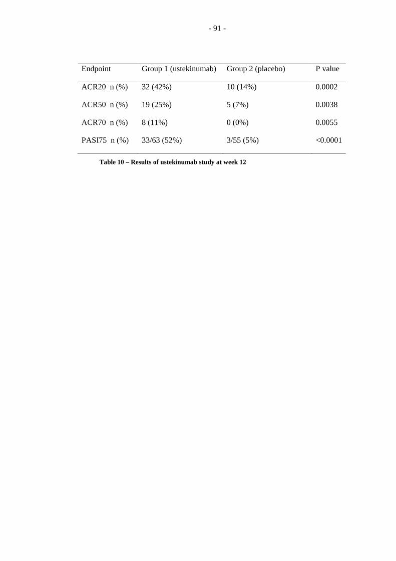
- 91 -
Endpoint Group 1 (ustekinumab) Group 2 (placebo) P value
ACR20 n (%) 32 (42%) 10 (14%) 0.0002
ACR50 n (%) 19 (25%) 5 (7%) 0.0038
ACR70 n (%) 8 (11%) 0 (0%) 0.0055
PASI75 n (%) 33/63 (52%) 3/55 (5%) <0.0001
Table 10 – Results of ustekinumab study at week 12
- 92 -
In addition to these responses, improvement was also seen in patients with
enthesitis (measured at the plantar fascia and Achilles tendons) and dactylitis
(measured with a non-validated semi-quantitative 0-3 score). Significant
improvement in function, as measured by the HAQ-DI, was also seen following
treatment with ustekinumab. Ustekinumab also seemed to be well tolerated in this
short study. Rates of adverse events and infections were similar in both groups,
although it was noteworthy that three patients developed abnormal fasting blood
glucose levels during the study (Gottlieb et al. 2009). This may be a reflection of
the high rate of metabolic syndrome seen in patients with psoriasis and PsA but it
does require further evaluation in larger prospective studies.
Phase III trials of ustekinumab are planned to further evaluate optimal dosing
regimens and safety in PsA. This is the first biological drug to show efficacy in PsA
that is not a TNF blocker. Although TNF blocking agents are remarkably effective
in PsA, there is a subgroup of patients who seem to be resistant to these drugs or
who lose efficacy over time. Newer therapies such as ustekinumab with different
modes of action may help us to treat these patients with resistant disease. However
as newer modes of action become available, research must help clinicians to
evaluate which therapies should be used first-line and subsequently. From this
study, it seems possible that ustekinumab may be less effective in treating PsA than
the TNF blockers, however this is a small study and the populations in all of the
individual RCTs differ significantly. At present, no head to head trials exist
comparing biological therapies in PsA meaning that decisions about which therapies
to use have a poor evidence base.
Abatacept
Abatacept (CTLA4Ig) is a selective T-cell co-stimulation modulator. A phase
II study investigating its use in PsA has recently been presented. One-hundred and
seventy patients with PsA and active skin psoriasis were recruited and randomised
1:1:1:1 to placebo, abatacept 3mg/kg, abatacept 10mg/kg or abatacept 30/10mg/kg.
The final group received two loading doses of 30mg/kg abatacept followed by
subsequent 10mg/kg doses for the rest of the study. The drug was well tolerated
with only 17 discontinuations for inefficacy (n=7) or adverse events (n=10) by the
six month timepoint. At six months, there was a significant difference in the
proportion of patients achieving the ACR20 response in the two groups receiving
10mg/kg when compared to placebo (table 11). A lower response rate was seen in
the 3mg group and this was not significantly higher than the placebo rate. When
considering skin responses, both 3mg and 10mg/kg doses significantly improved
psoriasis target lesion scores (Mease et al. 2009a).

- 93 -
Outcome Placebo Abatacept
3mg/kg 10mg/kg 30/10mg/kg
ACR20 (%)
All patients (%) 19 33 48 42
No prev TNF (%) 20 35 56 48
Prev TNF (%) 17 31 31 36
Target lesion 50 (%) 17 36 33 30
Target lesion 75 (%) 10 29 10 16
Improvement in HAQ-
DI (%)
19 36 45 35
Table 11 – Results of the abatacept in PsA trial at day 169

- 94 -
During sub-analysis, it was noted that patients who had previously been
exposed to anti-TNF therapy had poorer response rates. This is clearly
demonstrated in table 11 where ACR20 responses appear lower for this group.
Investigators also used this as a reason to explain the relatively poorer responses in
the 30/10mg/kg group as this group had a higher rate of prior TNF exposure (51%
vs 29-36%) (Mease et al. 2009a). Although this is perhaps not surprising, it is
disappointing, as it was hoped that drugs with a different mode of action would
work in TNF resistant patients.
At present this trial has only been presented in abstract form and the full paper
is not available. It is disappointing that the results given in the table above (taken
from the published abstract) only include ACR20 as an arthritis response measures
and do not include any results for higher response rates (ACR50 and 70). It appears,
like ustekinumab, that the response rates seem lower than expected when compared
to studies of TNF blockers. Further data from this study and larger future trials are
necessary to characterise the response rate in more detail.
Apremilast
Apremilast is a new phosphodiesterase-4 (PD4) inhibitor which is orally
active. The inhibition of PD4 causes suppression of multiple inflammatory
cytokines including TNF. Studies in psoriasis showed moderate activity, similar in
response rate to ciclosporin (Gottlieb et al. 2008). Phase II trials in PsA have now
been completed. Although the response rates are significantly lower than those seen
in a trial of anti-TNF therapy, it does show efficacy in arthritis (see table 12). There
were significant differences in the proportions of people achieving ACR20 and
ACR50 response rates when compared with placebo (Schett et al. 2009).

- 95 -
Treatment Groups
Placebo Apremilast 20mg
BD
Apremilast 40mg
OD
ACR20 (%) 11.8 43.5 35.8
ACR50 (%) 2.9 17.4 13.4
ACR70 (%) 1.5 5.8 7.5
Table 12 – 12 week results from the apremilast in PsA trial
- 96 -
2.4.2 Early treatment
For around 15 years, the concept of early treatment for RA has been
considered and has become the established management plan for those newly
diagnosed with RA. Multiple studies have shown that aggressive early treatment
can improve outcome in the medium to long term. A meta-analysis, including six
follow-up studies and six cohort studies, showed that early treatment with DMARDs
resulted in a significant 33% reduction in long term radiographic damage when
compared with a later initiation of treatment (Finckh et al. 2006). This has informed
the concept of the “therapeutic window of opportunity” where treatment is more
effective if given early in the course of disease. Following on from the data using
traditional DMARDs, TNF blocking therapies have also been used in early
interventional studies. These studies have shown a remarkable benefit with high
rates of remission (Emery et al. 2008).
This concept has not previously been well investigated in PsA. The only
equivalent paper concerning PsA, is a small unblinded study which was published
by Scarpa et al in 2008. They enrolled a small cohort of 35 patients with a recent
onset (<12 weeks) psoriatic oligoarthritis. Patients were randomised to either daily
NSAIDs for three months followed by methotrexate treatment, or to a combination
of NSAIDs and methotrexate from baseline. At three months, both groups showed a
significant improvement compared to baseline, but the combination therapy group
had a significantly greater improvement in their tender and swollen joint counts
(p<0.05). Other parameters including CRP and ESR did not differentiate the two
treatment groups. At six months, there was no significant difference between the
groups in terms of joint counts, blood results or physician/patient global disease
activity VAS. The authors concluded that methotrexate was only providing partial
disease control, and recommended treatment with combination TNF blocking
therapy and methotrexate (Scarpa et al. 2008b). However the dose of methotrexate
given is not specified and the study was only of six months duration. Long term
outcome in this small cohort is unknown.
An observational cohort study comparing RA and PsA found a surprisingly
high frequency of remission using a modification of the ACR remission criteria for
RA. These patients were treated with methotrexate or a methotrexate/CyA
combination in the case of resistant disease. From 2003, infliximab and etanercept
were also used in the case of non-response to methotrexate or multiple DMARDs.
They found that 57/236 patients (24%) with PsA achieved remission (defined as low
VAS scores, no active joints, CRP<0.5mg/dl, no dactylitis/enthesitis/inflammatory
spinal pain). Although the frequency of remission was higher in those treated with

- 97 -
TNF blocking therapy (79.5% vs 20.4%, p<0.001), the duration of remission did not
differ depending on treatment (Cantini et al. 2008).
To date, only one study, the RESPOND (REmicade Study in PsA patients Of
methotrexate-Naïve Disease) trial, has investigated the use of early TNF blocking
therapy in PsA. A cohort of 110 DMARD-naïve patients with polyarticular PsA (>5
active joints) of less than two years duration, were randomised to receive either
methotrexate or a combination of methotrexate and infliximab. There was a
significant improvement in all outcomes in favour of infliximab with an ACR20
response of 86% and an ACR70 response of 50% (see table 13). This study has also
provided evidence for the benefit of methotrexate in early PsA with an ACR20 of
66.7% (Nasonov et al. 2009).
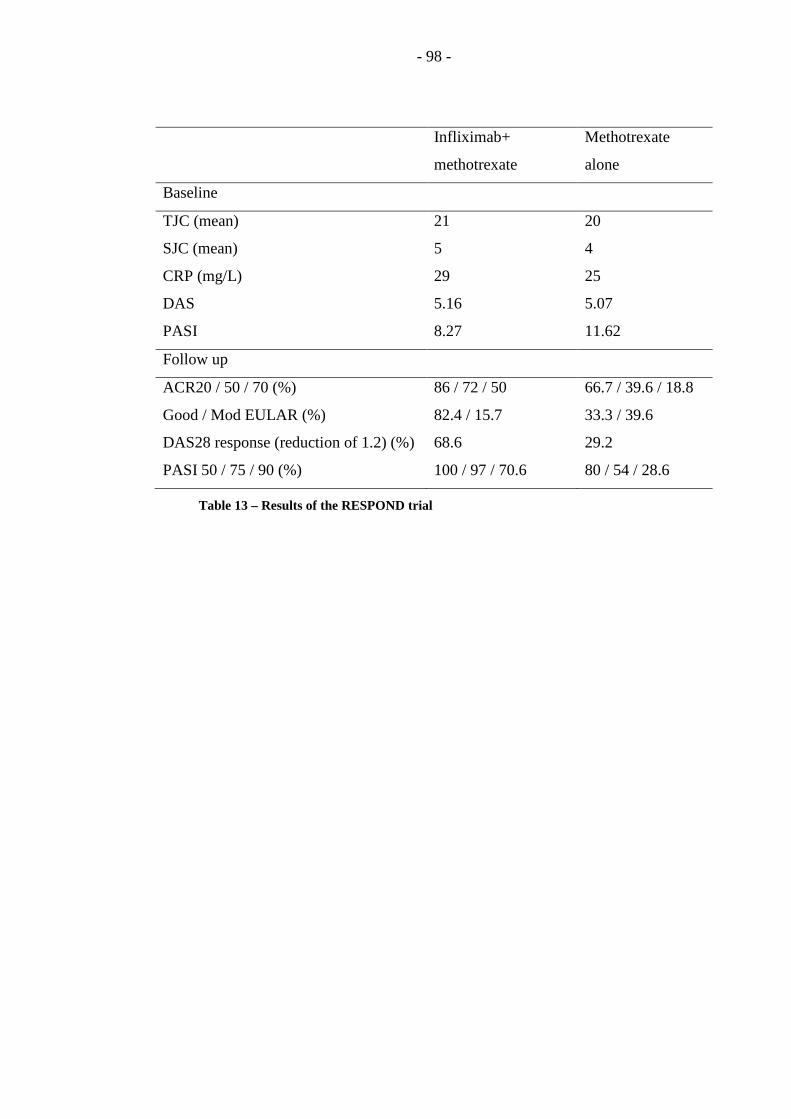
- 98 -
Infliximab+
methotrexate
Methotrexate
alone
Baseline
TJC (mean) 21 20
SJC (mean) 5 4
CRP (mg/L) 29 25
DAS 5.16 5.07
PASI 8.27 11.62
Follow up
ACR20 / 50 / 70 (%) 86 / 72 / 50 66.7 / 39.6 / 18.8
Good / Mod EULAR (%) 82.4 / 15.7 33.3 / 39.6
DAS28 response (reduction of 1.2) (%) 68.6 29.2
PASI 50 / 75 / 90 (%) 100 / 97 / 70.6 80 / 54 / 28.6
Table 13 – Results of the RESPOND trial
- 99 -
2.4.3 Tight control
In addition to early treatment with disease-modifying therapies, the concept of
“tight control” has also revolutionised care in RA. Multiple studies have confirmed
that sustained inflammation causes joint damage, and the aim of disease-modifying
therapy in RA is to control inflammation and prevent such ongoing damage. The
concept of tight control was introduced to RA by the TICORA study. This study
randomised 111 patients with active RA to either standard care or intensive
management. Intensive management involved monthly assessments and a protocol
led treatment algorithm where therapy was escalated if the patient had a DAS score
of >2.4 (moderate disease activity). Despite only using conventional DMARDs in
established disease, the study was able to demonstrate a significant benefit in terms
of reduction in clinical disease activity and radiographic progression for those
treated in the intensive management arm. However, even in the intensive
management group, significant progression in joint damage was seen over the 18
month study period (increase in total Sharp score 4.5) (Grigor et al. 2004).
Since this demonstration of “tight control”, large numbers of studies in RA
have adopted the use of DAS within a treatment protocol. The BeSt study, which
compared four different treatment strategies in early RA also used a DAS44 of <2.4
as a target for treatment. Treatment adjustments were made every three months if
this low level of disease activity was not achieved (Goekoop-Ruiterman et al. 2005).
Observational data in PsA has indicated that there is a similar link between
inflammation and damage. Cohort studies have shown that number of active joints
at baseline and follow up visits predicts long term joint damage in terms of both
clinically assessed and radiographic joint damage (Gladman et al. 1987; Gladman
and Farewell 1999; Bond et al. 2007). As in other inflammatory arthritides,
remission is the ultimate goal of therapy in PsA and a definition proposed by
Kavanaugh et al suggested that remission in PsA should be characterised by “a
complete absence of disease activity, with no signs or symptoms of active disease”
(Kavanaugh and Fransen 2006). However, this paper also recognised that remission
was not only difficult to achieve and maintain, but that, in some patients, mild
disease activity in one domain may be acceptable. Given this, they concluded that
“near remission” or “low disease activity” could be an appropriate goal for
individual patient’s treatment (Kavanaugh and Fransen 2006).
The concept of “tight control” has never previously been investigated in PsA.
The obvious limitation in designing such a study is that there is no measure of an
acceptable disease state available for PsA that could be utilised instead of the DAS

- 100 -
low disease activity state. Whilst DAS responses have been partially validated in
polyarticular PsA (Fransen et al. 2006), the cut points for disease activity have never
been investigated. Until such a measure exists, there is no objective target for
treatment in such a study.

- 101 -
3 Validation of the CASPAR criteria in early disease
3.1 Introduction
The CASPAR criteria were developed as new classification criteria for PsA
using a large cohort of patients with PsA and controls with other inflammatory
arthritides. They include characteristic features of PsA and have been shown to
have a high sensitivity and specificity in a population with established disease
(Taylor et al. 2006).
The main limitation of the CASPAR criteria is that they have not been
validated in early disease. Research done by the Toronto group found that the
CASPAR criteria had a high sensitivity for identifying early disease within their PsA
clinic population (Chandran et al. 2007b). However, this study only included
patients referred to a specialist tertiary referral clinic; therefore there is a significant
risk of overestimating the sensitivity of the criteria. Studies in Sweden and Italy
have also investigated the sensitivity of the criteria in early PsA (D'Angelo et al.
2008; Lindqvist et al. 2008; D'Angelo et al. 2009), but as yet no studies have been
completed with a control population to address the sensitivity and specificity of the
CASPAR criteria in early disease. At present, they cannot currently be
recommended for use in studies of early PsA for this reason. This study aimed to
emulate the methodology of the original CASPAR study and to recruit both early
PsA cases and controls with other inflammatory arthritides to allow calculation of
the sensitivity and specificity of the criteria in this group.
The other key issue raised with the CASPAR criteria is the entry criteria that
state that the patients must have “Inflammatory articular disease (joint, spine, or
entheseal)”. Particularly in early PsA, the pattern of disease may be evolving and
may be different from that of established disease. For that reason, an assessment of
arthritis, enthesitis and axial involvement was included to investigate the pattern of
disease in treatment-naive early arthritis patients.
3.2 Methods
3.2.1 Patient and control selection
Patients and controls were recruited from Early Arthritis Clinics and from new
referrals to rheumatology clinics in four hospitals based in the Yorkshire region.
Clinic attendees with a new diagnosis of PsA were enrolled in the study. For each
case enrolled, the consecutive patient with new onset inflammatory arthritis was

- 102 -
enrolled also. All cases and controls were required to have disease duration of less
than 2 years (from onset of symptoms) and to be DMARD naive.
3.2.2 Data collection
Data were recorded on standardised proformas including demographics, date
of onset of arthritis, duration of early morning stiffness, activity of arthritis,
enthesitis and axial disease, and application of the CASPAR criteria. Activity of
arthritis and enthesitis was assessed using clinical examination. All patients
underwent a 66/68 swollen/tender joint count and assessment of entheses including
those entheseal sites required for the LEI (Healy and Helliwell 2008), the MASES
(Heuft-Dorenbosch et al. 2003) and sites used in the IMPACT and IMPACT2
clinical trials (bilateral Achilles tendon and plantar fascia) (Antoni et al. 2005a;
Antoni et al. 2005b). Results of any relevant investigations were also recorded
including presence or absence of periosteal new bone formation on radiographs and
evidence of axial involvement identified by plain radiography or MRI.
3.2.3 Statistical methods
3.2.3.1 Power calculation
Figures for sensitivity and specificity for the CASPAR criteria in established
disease are 0.91 and 0.99 respectively. Prior to the development of the CASPAR
criteria, the Moll and Wright criteria have traditionally been used in both early and
late disease and the figures for these criteria are 0.99 and 0.92 respectively.
Assuming that in early case definition the emphasis is on specificity the null
hypothesis is that the specificity for the CASPAR criteria will be equivalent to that
for the Moll and Wright (HA = HO). The alternative hypothesis is that the specificity
of CASPAR differs from that of Moll and Wright (HA > HO). From Obuchowski
(Obuchowski 1998) the number of patients required to test the alternative (two
tailed) hypothesis, with an alpha value of 0.05 and a beta of 0.20 is 111. Therefore
111 cases and 111 consecutive controls are required.
3.2.3.2 Results Analysis
Sensitivity and specificity of the CASPAR and Moll and Wright criteria were
compared using McNemar’s tests for paired variables. Receiver operator
characteristic (ROC) curves were used to estimate the area under the curve (AUC)
for both criteria and the optimal cut-point for the CASPAR criteria for the diagnosis
of early PsA. Univariate and multivariate forward step-wise binary logistic
regression was used to identify which features included in the CASPAR criteria
were independently associated with PsA.

- 103 -
When considering patterns of arthritis, oligoarthritis was defined as less than 5
active joints and polyarthritis as 5 or more joints. Small joints were defined as
temperomandibular joints, sternoclavicular joints, acromioclavicular joints, carpo-
meta-carpal joints, MCP joints, PIP and DIP joints, MTP joints and IP joints of the
toes (total 58 joints). Large joints were defined as the glenohumeral joints, elbows,
hips, knees and ankle joints (total 10 joints). The symmetry number was calculated
as the number of joints as symmetric pairs/total number of joints involved. Each
joint pair (n=34) was assessed for symmetry individually. Symmetry was defined as
a symmetry number of ≥0.5. Significance testing was performed using McNemar’s
tests for categorical variables and Mann-Whitney U tests for continuous variables.
3.3 Results
In total, 111 cases of early PsA and 111 controls with other forms of
inflammatory arthritis were recruited to the study. The baseline features of these
patients are shown in table 14.
The sensitivity of the CASPAR criteria (score ≥3) in classifying early PsA was
87.4% compared to 80.2% for the Moll and Wright criteria (p=0.008). The
specificity for both criteria was 99.1%, with only 1 control patient fulfilling both
criteria for PsA.
Using ROC analysis, the AUC for the CASPAR criteria was 0.991 compared
to 0.896 for the Moll & Wright criteria. When considering different cut-points for
the CASPAR criteria, the best cut-point for classification remained a score of ≥3 as
in the original CASPAR analysis (table 15 and figure 3). Considering a score of ≥2
gave a higher sensitivity but resulted in a drop in specificity that would not be ideal
for classification criteria.
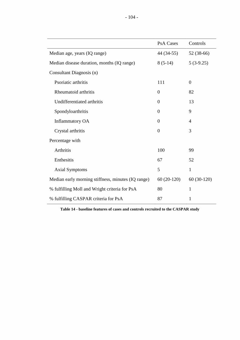
- 104 -
PsA Cases Controls
Median age, years (IQ range) 44 (34-55) 52 (38-66)
Median disease duration, months (IQ range) 8 (5-14) 5 (3-9.25)
Consultant Diagnosis (n)
111
0
0
0
0
0
0
82
13
9
4
3
Psoriatic arthritis
Rheumatoid arthritis
Undifferentiated arthritis
Spondyloarthritis
Inflammatory OA
Crystal arthritis
Percentage with
100
67
5
99
52
1
Arthritis
Enthesitis
Axial Symptoms
Median early morning stiffness, minutes (IQ range) 60 (20-120) 60 (30-120)
% fulfilling Moll and Wright criteria for PsA 80 1
% fulfilling CASPAR criteria for PsA 87 1
Table 14 - baseline features of cases and controls recruited to the CASPAR study
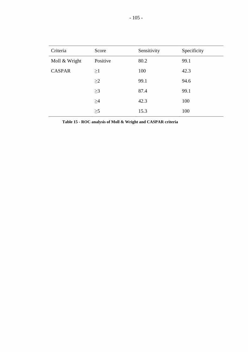
- 105 -
Criteria Score Sensitivity Specificity
Moll & Wright Positive 80.2 99.1
CASPAR ≥1 100 42.3
≥2 99.1 94.6
≥3 87.4 99.1
≥4 42.3 100
≥5 15.3 100
Table 15 - ROC analysis of Moll & Wright and CASPAR criteria
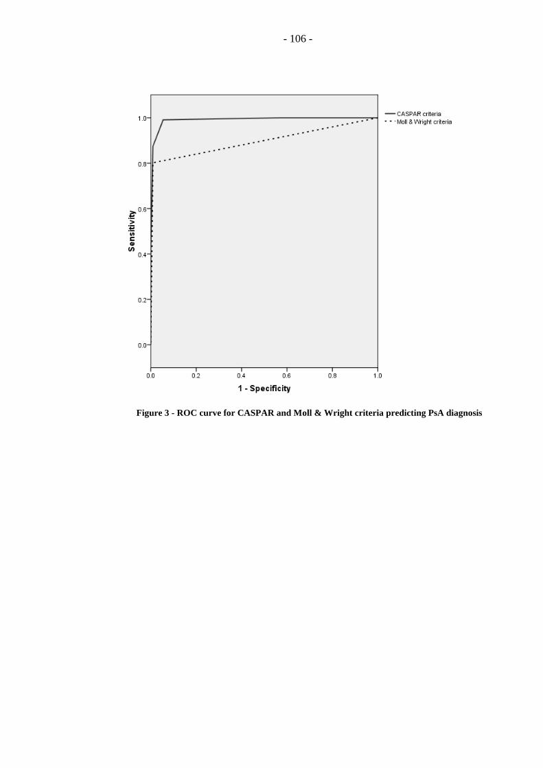
- 106 -
Figure 3 - ROC curve for CASPAR and Moll & Wright criteria predicting PsA diagnosis

- 107 -
When considering the individual components of the CASPAR criteria, 96.4%
of cases had current, previous or a family history of psoriasis with 84% having
current psoriasis. Dactylitis and nail psoriasis were highly discriminatory as only
one control patient had dactylitis and another one had nail psoriasis. Univariate
binary logistic regression analysis identified that all of the features of the CASPAR
criteria, with the exception of radiographic new bone formation were significant in
differentiating between PsA and controls. Multivariate forward step-wise logistic
regression identified that current psoriasis, negative RF, nail psoriasis, family
history of psoriasis and dactylitis were all associated with PsA. Table 16 shows in
more detail what proportion of the patients fulfilled each individual aspect of the
CASPAR criteria.
When considering the diagnosis of inflammatory articular disease (joint, spine
or entheseal) required for the first aspect of the CASPAR criteria, there were some
minor differences between the cases and controls. Nearly all patients had arthritis.
Presence of enthesitis was significantly more frequent in the PsA patients compared
to controls (67% vs 52%, p=0.029). There was a trend towards higher enthesitis
counts in PsA cases compared to control patients but this was not significant (see
table 17). Spondylitis appeared more frequent in the PsA patients but the difference
was not significant due to small numbers (5% vs 1%, p=0.055).

- 108 -
PsA Cases (%) Controls (%)
Any psoriasis 96 12
Current psoriasis
Previous psoriasis
Family history of psoriasis
84
6
18
4
1
8
Nail psoriasis 38 1
Negative RF 96 47
Current or previous dactylitis 28 1
Xray evidence of new bone formation 2 0
Table 16 - Proportion of cases and control fulfilling each aspect of the CASPAR criteria

- 109 -
Cases
(n=111)
Controls
(n=111)
RA controls
(n=82)
Proportion with arthritis (%) 100 99 100
Oligoarthritis 30 24 15
Polyarthritis 70 76 85
Median TJC (IQ range) 7 (13) 8 (15) 12 (16)
Median SJC (IQ range) 5 (7) 5 (9) 7 (11)
Median active joint count (IQ range) 9 (13) 10 (15) 12.5 (16.25)
Proportion with enthesitis (%) 67 52 52
Median enthesitis count (IQ range) 2 (4) 1 (3) 1 (4)
Proportion with spondylitis (%) 5 1 0
Table 17 – disease patterns in cases, controls and RA controls

- 110 -
Further analysis examined the pattern of arthritis seen in PsA and other
inflammatory arthritides (Table 18). The average tender and swollen joint counts
and the proportions of people with oligoarticular and polyarticular disease did not
differ significantly between cases and controls. When comparing PsA cases to only
controls with RA (n=82), PsA cases had significantly lower joint counts (p<0.008).
The higher number of joints involved in RA when compared to PsA was
principally due to an increased number of small joints involved. In PsA cases, there
were a similar proportion of patients with predominantly large and predominantly
small joint disease. However in RA controls, over 60% had predominantly small
joint disease. There was no significant difference seen in large vs small joint
arthritis when comparing PsA cases to all controls, or when comparing PsA patients
with RA controls. Specifically in small joint arthritis, DIP joint involvement was
significantly more common in PsA cases than controls (32% vs 16%, p=0.007).
A symmetry number of ≥0.5 defines symmetrical disease. The symmetry
number was significantly higher in controls than PsA cases (p=0.003) and this
difference was even more pronounced when comparing RA controls with PsA
(p<0.0001). The remaining control patients had a lower median symmetry number
but this was not significant to PsA cases (0.27 vs 0.5, p=0.58). Analysing this in
another way, when comparing the proportion of patients with symmetrical disease,
the controls, particularly those with RA, had a higher proportion of symmetrical
disease when compared to PsA. Comparing PsA to all controls, there was no
significant difference in the proportion with symmetrical disease (p=0.11), however
when comparing PsA to RA controls, there was a significant difference (p=0.001).
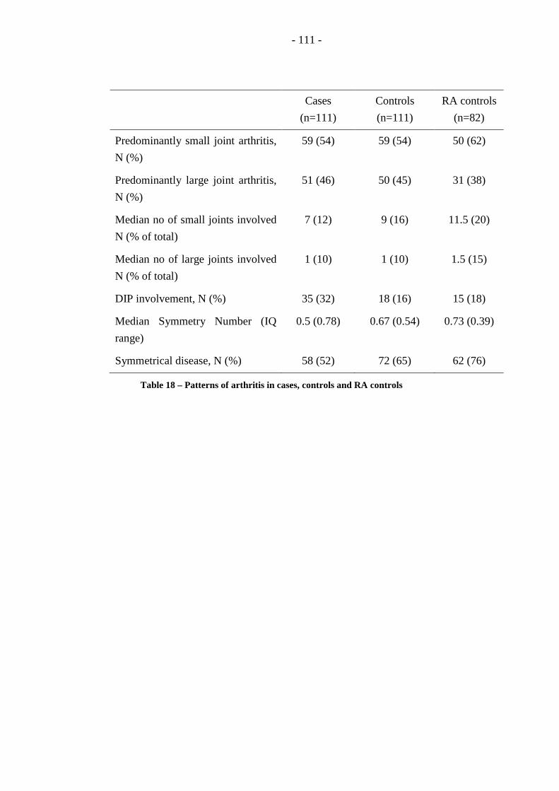
- 111 -
Cases
(n=111)
Controls
(n=111)
RA controls
(n=82)
Predominantly small joint arthritis,
N (%)
59 (54) 59 (54) 50 (62)
Predominantly large joint arthritis,
N (%)
51 (46) 50 (45) 31 (38)
Median no of small joints involved
N (% of total)
7 (12) 9 (16) 11.5 (20)
Median no of large joints involved
N (% of total)
1 (10) 1 (10) 1.5 (15)
DIP involvement, N (%) 35 (32) 18 (16) 15 (18)
Median Symmetry Number (IQ
range)
0.5 (0.78) 0.67 (0.54) 0.73 (0.39)
Symmetrical disease, N (%) 58 (52) 72 (65) 62 (76)
Table 18 – Patterns of arthritis in cases, controls and RA controls

- 112 -
3.4 Discussion
These data confirm that the CASPAR criteria have greater sensitivity
compared to the Moll and Wright criteria in the identification of early PsA. The
most important difference between the two criteria is that the CASPAR criteria
allow a diagnosis of PsA to be made in patients without skin psoriasis. These
criteria utilise additional features such as dactylitis, nail psoriasis and positive family
history to increases the sensitivity of the CASPAR criteria. This is particularly
relevant in early disease when not all features of PsA may have manifest. In
particular, 20% of people presenting with PsA do not have skin psoriasis, therefore
excluding them from a diagnosis using the Moll and Wright criteria. All patients
who fulfil the Moll and Wright criteria with a negative RF will also fulfil CASPAR
so there is significant overlap between the two criteria.
In comparison with established disease, sensitivity remains slightly lower. A
short duration of disease means that many patients will not yet have experienced all
of the typical features of PsA that they may develop during the course of their
disease. In particular, there is a much lower incidence of typical radiographic
changes that can be identified in early patients. The vast majority of patients with
new onset of inflammatory arthritis have normal radiographic appearances at
presentation. This problem has been identified in previous early PsA cohorts where
little new bone formation could be identified (D'Angelo et al. 2009).
When considering the pattern of inflammatory articular disease, there was a
higher proportion of patients with active enthesitis in PsA cases compared to
controls. There were also more patients in the PsA group who had axial disease,
although this difference was not significant due to small numbers affected in both
groups. Patterns of arthritis showed that PsA patients have a lower total number of
joints involved compared to RA controls and this difference was principally
explained by a higher number of small joints involved in RA patients, confirming
the identification of RA as a predominantly small joint polyarthropathy.
Symmetrical joint disease was less common in PsA patients compared RA controls,
and there was a significant difference in symmetry number between the patient
groups.
3.5 Limitations
The most significant limitation of this study relates to the selection of the
cases. Cases were recruited only if they had a consultant diagnosis of PsA and few
- 113 -
patients presenting with undifferentiated arthritis were included in this study. This
may be less important when considering the use of the CASPAR criteria as
classification criteria, as originally intended. The results of this study support their
use as inclusion criteria for future research studies with recent onset PsA. However
these data will result in an overestimate of the sensitivity of the criteria if
considering their use as a diagnostic tool in a general early arthritis clinic
population.
In this circumstance, the CASPAR criteria remain superior to the Moll and
Wright criteria because they allow other characteristic features of the disease to
identify PsA in patients without psoriasis. However they still do not account for
patients who initially present as undifferentiated arthritis and later evolve into a
typical psoriatic phenotype. Patients who are unaware of their family history of
psoriasis or who may have had dactylitis, but not had this confirmed by a specialist
may be left undiagnosed using these criteria.
The other limitation is the identification of ideal controls. Again, controls are
more likely to have a certain diagnosis, such as RA, and undifferentiated arthritis
patients were not seen in significant numbers. There is also a difficulty in
identifying controls with axial SpA who were eligible for this study, as patients were
required to have a disease duration of less than two years. A significant number of
cases with axial disease present to rheumatology with over two years symptom
duration, thus making them ineligible.
Future research ideally should apply these criteria routinely to all new
rheumatology referrals within an early arthritis or similar clinic. They should be
recorded at presentation and then long term follow up must be completed to ensure
that patients are accurately classified as PsA or controls. This is particularly crucial
in the undifferentiated arthritis cohort where additional diagnostic tools are required
to predict what type of arthritis may develop.
3.6 Conclusion
This work supports the use of the CASPAR criteria as classification criteria for
future research in early PsA. Although the sensitivity is slightly lower than that in
established disease, the specificity is reassuring. Their use as diagnostic criteria in
cases where diagnosis is unclear cannot be recommended based on this study, but
future work evaluating their use in an unselected early arthritis clinic population
would address this issue.
Different clinical phenotypes cannot be relied upon in isolation to identify
early PsA, but different patterns are seen in PsA patients compared to other

- 114 -
inflammatory arthritides. Enthesitis and DIP joint involvement are more frequent in
early PsA compared to other forms of inflammatory arthritis, however a significant
proportion of controls (including a significant proportion of patients with RA) also
have enthesitis. Spondylitis is likely to be more frequent in PsA than in RA,
however it cannot be relied upon to differentiate from other forms of inflammatory
arthritis. Patterns of arthritis confirm the typical view that RA is a symmetrical
small joint predominant polyarthropathy, whereas patients with PsA generally
present with fewer joints involved, particularly small joints, and, as a consequence,
with less symmetrical disease (Helliwell et al. 2000).

- 115 -
4 Imaging in Early Psoriatic Arthritis – the extent of sub-clinical disease
4.1 Introduction
Since the seminal works of Moll and Wright (Moll and Wright 1973), it has
been customary to divide patients with PsA into those with polyarticular and
oligoarticular disease. Research has shown that the number of active joints at
presentation influences the long term outcome of these patients (Gladman et al.
1987), and therefore this subdivision may have prognostic value. Traditionally,
assessment of joint involvement has been done clinically using tender and swollen
joint counts. However, research in mixed oligoarthritis cohorts has shown that a
significant proportion of patients have subclinical joint involvement, identifiable on
US, that would result in their re-classification from oligoarthritis to polyarthritis
(Wakefield et al. 2004).
US has been validated in PsA as a method for assessing joint inflammation.
Two studies have compared US with other imaging modalities in PsA. The first
imaged 13 patients with established PsA using US, MRI, radiography and
scintigraphy in addition to clinical assessments (Weiner et al. 2008). This showed a
sensitivity of 40% and specificity of 91% for joint erosions when compared to
conventional radiography. When assessing joint inflammation, MRI was used as the
gold standard. US showed a sensitivity of 48% and a specificity of 91% for the
detection of joint inflammation. Unfortunately, this study used only GS or B-mode
US and did not utilise PD.
A second study imaged 15 PsA patients in addition to 5 RA patients and 5
healthy controls for comparison. This study used both GS and PD US giving an
optimal assessment of this technique. US and MRI showed good agreement in the
assessment of bony changes (absolute agreement 85-100%) and synovitis (absolute
agreement 73-100%) but with variable kappa statistics. When looking at sensitivity
and specificity in this study, MRI was used as the gold standard for both bone
changes and inflammatory changes. US showed good sensitivity and specificity for
synovitis (sensitivity 40-70%, specificity 87-88%) as well as high specificity for
bone erosions (88-99%). However there was lower sensitivity of US for erosions (0-
57%) (Wiell et al. 2007).
Little research is available in PsA comparing clinical and imaging findings. Of
the 80 patients imaged in the previous paper investigating sub-clinical arthritis in
oligoarthritis, only 6 had a clinical diagnosis of PsA (Wakefield et al. 2004). One of
- 116 -
the validation studies discussed above compared imaging and clinical examination
but only in 13 cases. They found that 17% of joints showed US abnormalities
despite being clinically normal (Weiner et al. 2008). However this study did not use
PD which may have affected the specificity of the US findings. Further data in
larger cohorts is required to improve our understanding of the relationship between
clinical examination and US in early PsA.
No longitudinal studies exist in PsA or oligoarthritis to establish the
significance of this sub-clinical synovitis. However studies following patients with
RA who were considered to be in remission (i.e. with low numbers of active joints)
have shown that sub-clinical US evidence of PD inflammation correlates
significantly with progressive radiographic damage after 12 months of follow up
(Brown et al. 2006).
In addition to evidence of sub-clinical arthritis, there are many papers
identifying the presence of significant levels of sub-clinical enthesitis in patients
with established PsA and other SpA which is detectable by US (D'Agostino et al.
2003). Some theories of pathogenesis in PsA have postulated that inflammation
starts at the enthesis in early SpA and PsA patients before spreading to the joint
(McGonagle et al. 1999). This suggests that there is an entheseal initiation of
disease, meaning that scanning patients with early disease could be crucial in
identifying early sub-clinical inflammation that is yet to spread to the joints to create
further synovitis. Unfortunately the majority of previous studies with PsA and SpA
patients have recruited patients with established disease.
One study has looked at enthesitis using US and bone scintigraphy imaging of
patients with early PsA (Scarpa et al. 2008a). It confirmed the existence of both
sub-clinical arthritis and enthesitis on bone scintigraphy and then used US in these
areas to confirm their findings. All patients had ultrasonography on the joints and
entheses that showed increased tracer uptake on scintigraphy (both those that were
clinically involved and those that were not clinically involved) and on two control
joints with normal clinical examination and normal tracer uptake. It reported that
US showed some abnormal changes in all articular and entheseal sites identified on
scintigraphy. Only 2 of the 94 control joints that were examined with US showed
any degree of joint involvement. In 71% of the clinically involved joints there was
evidence of PD but in the “sub-clinically” involved joints, they did not find any
evidence of PD in the areas of tracer uptake. Thus there is some doubt cast over
whether this truly represents “sub-clinical inflammation” where both GS and PD US
signal would be expected, or whether some of these changes could be either
mechanical or within normal physiological limits. Other studies comparing
scintigraphy and US showed a similar lack of agreement on the presence of

- 117 -
synovitis where tracer uptake can be found at a site with normal US assessment and
where joints with no tracer uptake had evidence of synovitis on US (Weiner et al.
2008).
4.2 Methods
Patients with new onset PsA, according to the CASPAR criteria (Taylor et al.
2006), were recruited. They were all required to be DMARD naïve and have a
disease duration of less than 2 years. All patients had active disease (defined as ≥1
active joint, active entheseal site or BASDAI >4 for axial disease). All patients had
a clinical assessment including a 66/68 swollen/tender joint count and assessment of
entheses including those entheseal sites required for the LEI (Healy and Helliwell
2008), the MASES (Heuft-Dorenbosch et al. 2003) and sites used in the IMPACT
and IMPACT2 clinical trials (bilateral Achilles tendon and plantar fascia) (Antoni et
al. 2005a; Antoni et al. 2005b). In addition to this, dactylitis was assessed using a
simple dactylitic digit count and the LDI (Helliwell et al. 2005).
4.2.1 Ultrasound
US was performed using a Philips HDI 5000 machine with a 12-5 or 13-7
MHz probe as appropriate. For PD scanning a medium wall filter was applied. All
scans were performed by a rheumatologist experienced in musculoskeletal US (Dr J
E Freeston). Gel was applied to the skin to provide an acoustic interface.
All patients underwent GS and PD US of the most symptomatic hand and wrist
(11 joints per patient) and additional target joints that the patients identified as
symptomatic. The digits of the 4 fingers on this hand were also scanned for FT
(defined as presence of GS and/or PD). GS and PD were scored separately on a 0-3
semi-quantitative scale for each joint imaged. A GS score of ≥2 and/or a PD score
>0 were used to identify US active joints.
All patients underwent GS and PD US of bilateral lateral epicondyles of the
elbow, Achilles tendons and plantar fascia. GS and PD were scored separately on a
0-3 semi-quantitative scale for each enthesis imaged. A GS score of ≥2 and/or a PD
score >0 were used to identify active US enthesitis.
4.2.2 Statistical analysis
Joints, entheses and digits were all considered individually to compare clinical
and imaging assessment of arthritis, enthesitis and dactylitis. A prevalence and bias
adjusted Kappa (PABAK) was used to compare agreement for clinical and US
assessment of active disease. This statistic adjusts the raw kappa value for
- 118 -
differences in prevalence and bias. This is particularly relevant when the
distributions of positive and negative results are not equally split in the population.
4.3 Results
40 patients (18 male, 22 female) with new onset, DMARD naive PsA were
recruited. Their average age was 47 years (range 19-71 years) and they had a
median disease duration of 10 months (IQ range 5-15 months). All of the patients
were negative for RF and anti-CCP tests. On clinical examination, they were found
to have a mean of 10.7 tender joints and 6.2 swollen joints. Nineteen patients were
classified as having oligoarthritis (<5 active joints on clinical examination) and 21
were classified as having polyarticular disease.
When considering joint inflammation at the patient level, only one of the 19
oligoarthritic patients had 5 or more involved joints on US and was therefore re-
classified from oligoarticular to polyarticular.
When considering each joint individually, a total of 431 joints were assessed
by both clinical and US examination. Of these joints, 68.9% had imaging findings
consistent with their clinical examination (table 19). A substantial agreement
(Kappa =0.36, PABAK =0.38) was identified between clinical assessment of activity
(tender and/or swollen) and imaging assessment of activity. The agreement over
active joints was 60.1% whereas the agreement over inactive joints was 74.5%.
Clinical examination rarely over-estimated activity, particularly the SJC where
95/100 swollen joints show imaging activity. However, subclinical disease was
present with 93 clinically inactive joints scanned showing significant imaging
findings.
Dactylitis was less common with only 10 patients having dactylitis in the
fingers or toes. Those who had dactylitis had an average of two digits involved. A
total of 160 digits (fingers only) were examined for clinical dactylitis and FT on US.
Of these, 88% had consistent findings (two digits were clinically dactylitic with FT
on US). FT was found in an additional 20 digits (13%) that were not clinically
considered to be dactylitic.

- 119 -
US active US inactive Total
CE active 101 (23%) 41 (10%) 142
CE inactive 93 (22%) 196 (45%) 289
Total 194 237 431
Table 19 - Comparison of clinical and US assessment of disease activity by individual
joint (CE=clinical examination, US=ultrasound).
- 120 -
Clinically, enthesitis was common with 31/40 patients having clinical evidence
of tender enthesitis using any of the clinical tools discussed above, and an average of
three sites involved per patient. Considering the bilateral lateral epicondyles of the
elbow, Achilles tendons and plantar fascia which were examined by both clinical
examination and US, 34/202 entheses were found to be tender, but none were
clinically swollen. Using combined GS and PD, 14/202 had active enthesitis on US.
Of all entheses examined, 85% had imaging findings consistent with clinical
examination. A substantial correlation (PABAK = 0.70) was identified between
clinical assessment of activity (tender enthesis) and imaging assessment of activity
(see table 20).
In this early cohort, there was little evidence of sub-clinical enthesitis with
only 2% (5/202) of non-tender entheses showing significant US changes. All of the
5 non-tender, US active entheses were in the lower limb, and there was no sub-
clinical disease seen at the lateral epicondyles. However, clinical examination over-
estimated activity in 12% of entheses. Of the 25 tender but US inactive entheses, 7
were adjacent to clinically tender joints. Bony erosion was only present in 8 of 202
entheses.

- 121 -
Type of assessment US active US inactive Total
Tender 9 (4%) 25 (12%) 34
Non-tender 5 (2%) 163 (81%) 168
Total 14 188 202
Table 20 – Comparison of clinical and US assessment of disease activity by individual
entheses

- 122 -
4.4 Discussion
When comparing US and clinical examination of arthritis at the patient level in
this cohort, there was only one patient identified who was clinically diagnosed as
having oligoarthritis which was reclassified as polyarthritis following US
assessment. The other nineteen patients with oligoarthritis did not have evidence of
significant sub-clinical arthritis that would result in their reclassification. This is in
contrast to previous research in oligoarthritis cohorts (Wakefield et al. 2004). This
may be due to the fact that previous research has looked at undifferentiated
oligoarthritis rather than concentrating on one particular subtype. These previous
cohorts of undifferentiated oligoarthritis may have evolved to other forms of
arthritis, including even RA which was undiagnosed at the time. There is also the
issue of clinical skill effect. All of these patients underwent clinical examination as
part of a research procedure by two examiners experienced in PsA. In this setting, it
is possible that more involvement is identified clinically when enough time is
available for assessment and examiners are experienced in performing full joint
counts.
Considering arthritis at the joint level, there was reasonable correlation
between US and clinical examination particularly when compared to previous
research in other inflammatory arthritides. However, subclinical arthritis was
identified using PD US, particularly in patients with multiple joints involved
clinically. This is in keeping with previous research showing a higher sensitivity of
US to detect clinical synovitis.
When considering enthesitis, there was surprisingly little evidence of enthesitis
in this early PsA cohort. Considering stated hypotheses that PsA originates in the
enthesis and then spreads to the joint, it is surprising that only 9 of 202 entheses
assessed had evidence of US enthesitis. This is certainly a very different picture to
that seen in established disease where significant US abnormalities are identified in
PsA and SpA cohorts (D'Agostino et al. 2003). This may reflect the difference
between early and established disease patterns and it may also be explained partially
by an increase in entheseal abnormalities with increasing age. All of the non-tender
entheses with US changes were in the lower limb where mechanical factors are
likely to be a larger issue. No subclinical enthesitis was identified at the elbow. The
data seem to suggest an overestimate of enthesitis using clinical examination with a
number of tender entheses showing no US abnormality. This may represent disease
not identified by US (such as bone oedema) or the tenderness identified may be
related to other pathologies such as fibromyalgia.
- 123 -
4.5 Limitations
This study represents a step forward in assessing a more homogeneous cohort
of patients, all with PsA and all with less than 2 years disease duration. This allows
us to look in detail at the presentation of these patients. However due to time
limitations, US examination was not complete for all joints and entheses. It
therefore raises the possibility that some subclinical disease could have been missed.
Ideally all patients should have had US assessment of all of their joints and entheses,
but this would be extremely time-consuming for both ultrasonographer and patient.
All patients were required to have active disease and be DMARD naive. A
selection bias therefore exists in this cohort where patients with borderline or very
little disease activity were not included. The characteristics of this cohort also
define how this data can be extrapolated. All of these patients were off treatment
with active disease. Therefore such data cannot be extrapolated to patients on
treatment. Ideally, a similar study needs to be repeated, to scan patients on
treatment, particularly those whose disease control is thought to be adequate to
identify if subclinical disease is an important issue in such a cohort. If significant
subclinical disease is identified, then longitudinal data will be required to assess the
long term implications of such findings.
4.6 Conclusion
In early PsA there is a significant correlation between clinical examination and
US of joints and entheses. Around one-fifth of joints have subclinical synovitis
which can be detected on US using PD and this is more common in those with
clinically-identified polyarticular joint involvement. Enthesitis was not identified in
the majority of patients; indeed clinical examination seemed to overestimate the
involvement of these structures. Further research is required to address whether this
sub-clinical synovitis persists in patients once they are started on therapy and
whether this has any prognostic implications.

- 124 -
5 Defining Minimal Disease Activity in PsA – a proposed objective target
5.1 Introduction
The conceptual definition of MDA was agreed at the OMERACT 6 conference
as “that state of disease activity deemed a useful target of treatment by both the
patient and physician, given current treatment possibilities and limitations” (Wells et
al. 2005). MDA encompasses both remission and “low disease activity” as
acceptable targets for therapy.
Any criteria for disease activity in PsA must assess many aspects of this
complex disease and a core set of measures for future PsA research was agreed at
OMERACT 8 in 2006. The six core measures agreed were peripheral joint activity,
skin activity, patient’s assessment of pain, patient’s assessment of global disease
activity, physical function and HRQOL (Gladman et al. 2007d). Other important
domains included spinal disease, dactylitis, enthesitis, fatigue, nail disease,
radiography, physician global assessment and acute-phase reactants (Gladman et al.
2007d). Using these core and important measures, this study was designed to
develop criteria for MDA in PsA, using expert opinion on real-life cases.
5.2 Methods
5.2.1 Domains to be included
Firstly, a decision was taken on which aspects of the PsA disease spectrum
needed to be included to create the MDA definition. Ideally, any criteria need to
contain the minimum of measurements (to allow feasibility) but must incorporate
measures of all of the important aspects of the disease. The starting point was the
OMERACT agreed core set of domains for PsA (as above). Analysis of PsA cases
with a low disease activity (defined as physician global disease activity VAS of <3)
showed large variation in HRQOL (measured by the Psoriatic arthritis Quality Of
Life scale or PsQOL). HRQOL was excluded because of concerns that it may be
unrelated to disease activity and unduly influenced by external factors.
As recommended by OMERACT, peripheral joint activity was measured using
the 68 tender/66 swollen joint count. Skin activity was initially planned to be
represented by a PASI score as this is the most commonly used outcome measure for
psoriasis. However, concern was raised by key dermatologists within GRAPPA that
the PASI was not representative in patients with low psoriasis disease activity, in

- 125 -
particular with a PASI of <10. Therefore both PASI and BSA were included. The
aim of this was to allow physicians to use either PASI or BSA depending on their
personal preference, and to provide a maximum cut-off level for each. Pain and
patient’s global assessment were measured using 100mm VAS scales, and the HAQ-
DI was used as a measure of physical function.
Next, the five domains of PsA, peripheral joint disease, skin, enthesitis,
dactylitis and axial disease, were considered to see how they would be represented
by these core measures. Peripheral joint and skin activity were included, but there
was no specific measure of dactylitis, enthesitis or spinal disease. The concern was
that activity in any of these domains would not be recognised by the MDA criteria,
allowing a person to be incorrectly classified as being in MDA. Minimising the
likelihood of false positives is particularly important in this exercise to ensure that
patients with active disease are not undertreated. Dactylitis is identified as part of a
peripheral joint count, where dactylitic digits will result in a quantifiable tender
and/or swollen joint count. Therefore further measurement was not felt necessary.
Enthesitis was felt to be an important measure that may not be accurately
identified using any of the other core measures. Therefore a decision was made to
include an enthesitis count. Multiple enthesitis outcome measures have been
proposed for clinical research, some “borrowed” from AS and other SpA research,
but there is no clear optimal measure for use in PsA. Therefore, the design of the
MDA criteria did not specify a certain enthesitis outcome measure but was designed
to be used with any of the commonly used enthesitis scores in PsA. As such, a raw
enthesitis count was included with a maximum value of 13 representing the
maximum score of these commonly used measures (IMPACT – 4, LEI – 6, MASES
– 13). This allows the criteria to be used with any of the enthesitis outcome
measures.
Spinal disease is particularly under-researched in PsA and no validated disease
activity measures are available. The BASDAI has been evaluated in PsA and was
not shown to correlate with doctor’s perception of disease activity or with treatment
decisions (Taylor and Harrison 2004). Interestingly, it was found to correlate highly
with the patient’s global disease activity VAS in both axial and peripheral joint
disease, which is a core set measure in PsA and was already planned to be included
in the profiles. Given the lack of outcome measures available, no specific
measurement of axial disease activity was included. It was felt that active axial
disease would be identified by the patient’s assessment of pain, global disease
activity and physical function.
- 126 -
5.2.2 Materials
A questionnaire was compiled consisting of 40 PsA patient profiles, using
measures agreed above (TJC: 0-68, SJC: 0-66, enthesitis count: 0-13, PASI: 0-72,
BSA: 0-100, patient VAS pain: 0-100, patient VAS global disease activity: 0-100
and HAQ-DI: 0-3). The profiles were created using data from real patients with PsA
selected from a prospective longitudinal database. The profiles included a full range
of disease activity but with additional patients with low disease activity (based on
physician’s assessment). The questionnaire included the OMERACT definition of
MDA and the instructions stated “this patient comes to see you in clinic. He/she has
been on a stable dose of disease modifying anti-rheumatic drug (DMARD) therapy
for over 6 months.” They were then asked “Do you consider this patient to be in a
‘minimal disease activity’ state?”. This was then circulated to rheumatologists and
dermatologists with an interest in PsA, identified through the GRAPPA
membership.
5.2.3 Statistical methods
Summary statistics were calculated for different levels of consensus (>90%,
>80% and >70%). Providing there were no major differences between these values,
the >70% agreement would be used to maximise the number of cases for further
analysis. Assuming this is the case, if ≥70% of respondents classified the profile as
MDA, then it was taken to correspond to a patient in MDA, the other profiles were
not. Summary statistics for each outcome measure, using the profiles agreed to be in
MDA, were calculated including mean, rounded mean, upper 95% confidence
interval (CI), rounded upper 95% CI and maximum. These were investigated as
potential cutpoints to define a maximum value for each of the outcome measures.
Then the number of outcome measure results that must not exceed this cutpoint for
the patient to be considered as in a MDA state was investigated. There were 7
measures creating 7 variations on the number of cutpoints that must be met. This
created 35 possible definitions for MDA when each of the five possible cutpoints
were combined with the 7 variations in the number of cutpoints to be met. For each
definition, sensitivity and specificity was calculated using receiver operating curves.
From these, the best definition was selected.
5.3 Results
A total of 60 respondents (82% rheumatology, 18% dermatology background)
completed the questionnaire. Nearly all were physicians, but one was a
rheumatology specialist nurse with an interest in PsA. There was substantial
consensus between the experts who completed the questionnaire on the profiles that

- 127 -
represented MDA. There was a ≥90%, ≥80% and ≥70% agreement that 7, 10 and 13
profiles, respectively, were in MDA. Summary statistics are shown for each of these
levels of agreement and show little variation (table 21).
In view of this, the 70% consensus was used to maximise the number of cases.
Therefore 13 cases were taken as being in MDA, and the other 27 cases were
considered not to be in MDA. Using these 13 cases, 5 cutpoints were calculated for
each core measure (table 22). Application of these cutpoints with 7 variations
depending on the number of cutpoints achieved yielded 35 definitions which were
tested using receiver operating curves. ROC curves showed high sensitivity and
specificity and an AUC ≥0.895 for all definitions (figure 4).
The maximum value, rounded mean and rounded upper 95% CI were felt to be
more practical for use in a criteria as they were integers and were therefore
considered preferentially. The definition based on achieving 7/7 of the maximum
tolerated values had the best combination of sensitivity and specificity (100% for
both) and the highest area under the curve (AUC =1). However these maximums
included outlying values (in particular, patient global disease activity VAS 87 and
enthesitis 6) that were felt to be unrepresentative of minimal disease activity. The
best criteria using the rounded mean cutpoint (AUC 0.946) was with 5/7 criteria met
(sensitivity 92%, specificity 89%, AUC 0.946). Two possible criteria using the
rounded 95% CI (AUC 0.974) were identified with 5/7 criteria met (sensitivity
100%, specificity 82%) and 6/7 criteria met (sensitivity 85%, specificity 96%).
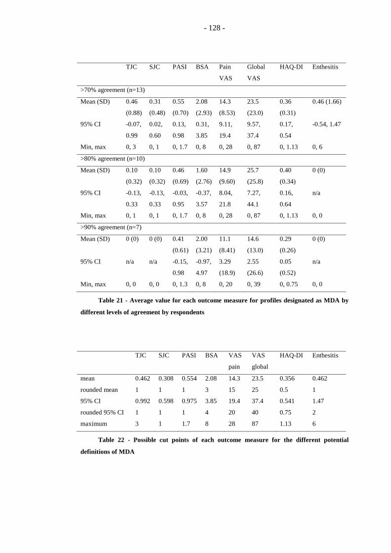
- 128 -
TJC SJC PASI BSA Pain
VAS
Global
VAS
HAQ-DI Enthesitis
>70% agreement (n=13)
Mean (SD) 0.46
(0.88)
0.31
(0.48)
0.55
(0.70)
2.08
(2.93)
14.3
(8.53)
23.5
(23.0)
0.36
(0.31)
0.46 (1.66)
95% CI -0.07,
0.99
0.02,
0.60
0.13,
0.98
0.31,
3.85
9.11,
19.4
9.57,
37.4
0.17,
0.54
-0.54, 1.47
Min, max 0, 3 0, 1 0, 1.7 0, 8 0, 28 0, 87 0, 1.13 0, 6
>80% agreement (n=10)
Mean (SD) 0.10
(0.32)
0.10
(0.32)
0.46
(0.69)
1.60
(2.76)
14.9
(9.60)
25.7
(25.8)
0.40
(0.34)
0 (0)
95% CI -0.13,
0.33
-0.13,
0.33
-0.03,
0.95
-0.37,
3.57
8.04,
21.8
7.27,
44.1
0.16,
0.64
n/a
Min, max 0, 1 0, 1 0, 1.7 0, 8 0, 28 0, 87 0, 1.13 0, 0
>90% agreement (n=7)
Mean (SD) 0 (0) 0 (0) 0.41
(0.61)
2.00
(3.21)
11.1
(8.41)
14.6
(13.0)
0.29
(0.26)
0 (0)
95% CI n/a n/a -0.15,
0.98
-0.97,
4.97
3.29
(18.9)
2.55
(26.6)
0.05
(0.52)
n/a
Min, max 0, 0 0, 0 0, 1.3 0, 8 0, 20 0, 39 0, 0.75 0, 0
Table 21 - Average value for each outcome measure for profiles designated as MDA by
different levels of agreement by respondents
TJC SJC PASI BSA VAS
pain
VAS
global
HAQ-DI Enthesitis
mean 0.462 0.308 0.554 2.08 14.3 23.5 0.356 0.462
rounded mean 1 1 1 3 15 25 0.5 1
95% CI 0.992 0.598 0.975 3.85 19.4 37.4 0.541 1.47
rounded 95% CI 1 1 1 4 20 40 0.75 2
maximum 3 1 1.7 8 28 87 1.13 6
Table 22 - Possible cut points of each outcome measure for the different potential
definitions of MDA

- 129 -
Figure 4 - ROC curve for candidate definitions of minimal disease activity
0
20
40
60
80
100
0 20 40 60 80 100
sen
siti
vity
1-specificity
Mean
Rounded mean
95% CI
Rounded 95% CI
Maximum

- 130 -
When considering these three candidate definitions, thought was given to the
ideal characteristics of the criteria. When balancing sensitivity and specificity, it
was felt that specificity was of more importance. This is because it would be
preferable to miss a few patients who were truly in MDA, than to misclassify
patients with significant disease activity into an MDA group where they may
potentially be undertreated. Therefore the definition based on meeting 5/7 of the
cutpoints based on the upper 95% CI was discarded as it had a lower specificity.
The final decision was between using the rounded mean cutpoint with 5/7
criteria met (sensitivity 92%, specificity 89%) or the rounded 95% CI with 6/7
criteria met (sensitivity 85%, specificity 96%). Again, there was concern that one of
the rounded 95% CI cutpoints may be too high to truly represent MDA (global
disease activity VAS 40) and so the final definition selected used the rounded mean
values.
Therefore, a core set definition places patients with PsA in MDA when they
meet 5 of 7 of the following criteria
• TJC ≤1
• SJC ≤1
• PASI ≤1 or BSA ≤3
• patient pain VAS ≤15
• patient global activity VAS ≤20
• HAQ-DI ≤0.5
• tender entheseal points ≤1
5.4 Discussion
The aim of this study was to produce clinical criteria for MDA in PsA, and it
provides the first measure of state in PsA as a composite outcome. Such measures
of state defining remission, low and high disease activity have been available in RA
for some time and have been shown to be important for use in research trials and in
daily clinical practice.
Research in RA supports the link between inflammation and subsequent joint
damage. It has been established in both clinical and imaging studies that
inflammation in the joints, in the form of synovitis, results in subsequent bony
damage (Conaghan et al. 2003; Forslind et al. 2004). The concept of “tight control”
was originally introduced by the TICORA study. This demonstrated that tight
control of disease utilising pre-defined, objective activity levels to guide therapeutic

- 131 -
changes resulted in significantly better clinical and radiographic outcomes compared
to routine care with no formal therapeutic protocol (Grigor et al. 2004). Following
on from this research, many studies in RA have adopted the use of an objective
measure of response to guide therapeutic decisions (Goekoop-Ruiterman et al.
2005).
Observational data in PsA has also suggested a link between inflammation and
damage as it has been shown that clinical and radiological joint damage is related to
the number of actively inflamed joints as a time-varying indicator (Gladman et al.
1995; Gladman and Farewell 1999),. Therapeutic trials of highly effective
biological therapies such as TNF blockers in PsA have shown a reduction in disease
activity and a corresponding reduction in radiological progression (Mease et al.
2004; Mease et al. 2005b). However the lack of a criteria or cut-off for remission or
low disease activity in PsA has meant that the concept of ‘tight control’ of disease
activity using objective measures has never been evaluated.
5.5 Limitations
This study is based on a questionnaire using profiles of patients sent to experts
in PsA including both rheumatologists and dermatologists. This raises some
limitations in the research. In RA, research has shown that there is a high
correlation between judgments made on “paper patients” and those based on real
patients seen in clinical practice (Kirwan et al. 1983). However it is not known in
PsA whether judgments about these profiles or “paper patients” are truly reflective
of clinical assessments. Our profiles also contained more information than that
given in the validation study in RA (Kirwan et al. 1983) to counteract the problem of
a more complex and heterogeneous disease. This variability of disease patterns with
different aspects of the disease involved may make it harder to make judgments
about “paper patients”.
A link to the questionnaire was sent to all members of GRAPPA with a request
for their participation. A follow up email was also sent two weeks later when the
response rate had started to decrease. This recruited 60 respondents and the
response rate is similar to that of previous GRAPPA membership surveys. The
online tool used to complete the questionnaires is familiar to many GRAPPA
members and allowed respondents to link to the questionnaire on repeated occasions
if they did not have time to complete it in one sitting. It also ensured that
respondents could not answer the questionnaire more than once. A lower proportion
of dermatologists completed the questionnaire, probably partially because
dermatologists constitute a minority of the experts who were contacted. However
- 132 -
analysis has not shown a significant difference in their responses. Interestingly the
possible cutpoints for skin disease are identical in both groups’ assessments.
The number of profiles included is limited in these questionnaire based
exercises by the number of profiles that physicians can be expected to rate reliably.
All profiles contained 7 variables resulting in thousands of possible combinations in
different profiles. Using random sampling of possible profiles (according to Hahn
and Shapiro) across a range of disease activity reduced the minimum number of
profiles to 32 and a compromise at 40 was reached. The results of this study are
based on a particular sample of 40 patients and these results need validating on
larger and distinct samples.
Importantly, this analysis of expert opinion represents current views and may
change in the future. The OMERACT definition of MDA is “that state of disease
activity deemed a useful target of treatment by both the patient and physician, given
current treatment possibilities and limitations”. This relationship to current
treatment possibilities means that acceptable targets of treatment are likely to have
changed rapidly in the last few decades as new therapies have become available.
Studies in RA have shown a lowering target for remission and low disease activity
based on expert opinion over time, and it is suggested that this is related to the
increasing potential to achieve these states with newer therapies (Aletaha et al.
2005). Similarly, in future, the acceptable levels of disease activity may drop further
as the quest for remission becomes increasingly important.
5.6 Conclusion
In summary, this study provides the first definition of a “state” of disease
activity in PsA and defines a target for treatment. It is based on current expert
opinion and uses a composite of key outcome measures in PsA to encompass all of
the domains of the disease. Given the new therapeutic options in PsA, patients with
PsA are likely to benefit if these disease activity states are targeted, rather than
simply looking for a clinical response.

- 133 -
6 Validation of the Minimal Disease Activity Criteria
6.1 Introduction
Before a new outcome measure can be recommended for routine use, it must
prove to be valid in assessment. The OMERACT filter domains of truth,
discrimination and feasibility provide a framework to assess new tools for use in
research trials and clinical practice. These three domains can be summarised as
follows. Truth aims to assess whether the measure is relevant and truthful and can
measure what is intended without bias. Discrimination assesses the ability of the
measure to discriminate between states at one time or different times as well as
assessing reliability and sensitivity to change. Feasibility assesses whether the
measure is easy to use, quick and inexpensive (Boers et al. 1998).
The truth domain includes face, content, criterion (or concurrent) and construct
validity. Face validity provides evidence that the criteria are sensible to
practitioners who will use them. Content validity asks if the criteria include the
most relevant characteristics of the condition and if each variable contributes
something distinct and important to facilitate classification of the patient. Criterion
validity asks if the criteria produce the same or similar results as does a gold
standard criterion. If no gold standard is available, concurrent validity can be
assessed to ask if the criteria agree with other criteria designed to measures the same
or similar constructs. Overall do the criteria adequately measure the underlying
construct? This is based on an accumulation of knowledge about the criteria and
their relationship to other tests.
Discrimination asks if the criteria are able to distinguish between states of
interest and can be further sub-divided into classification, prognosis, responsiveness
and reliability. Classification represents the division of patients according to the
criteria into two or more groups, in this case MDA or not. Prognosis questions
whether the states defined by the criteria have clinical relevance. This is clearly key
to their usefulness in research and clinical practice. Responsiveness is the ability of
subjects to change disease states over time. The proposed criteria must allow
patients to shift from one disease state to another. Reliability measures how
concordant different assessors are when classifying patients with the same clinical
picture into the same disease state category.
Feasibility asks three questions: can it be done quickly, easily and cheaply?
This is a key component of the filter as unfeasible outcome measures will never be
routinely adopted into practice.

- 134 -
The new MDA criteria developed for PsA are a composite measure. Patients
must fulfil 5 or more of the 7 following criteria: TJC ≤1; SJC ≤1; PASI ≤1 or BSA
≤3; patient pain VAS ≤15; patient global disease activity VAS ≤20; HAQ-DI ≤0.5;
tender entheseal points ≤1 (Coates et al. 2010b) to be classified as in MDA. The
work in this chapter aims to provide preliminary validation work for the criteria
using the OMERACT filter framework.
6.2 Methods
6.2.1 Observational Cohort Data
The observational cohort study was conducted at the University of Toronto
PsA clinic. Patients were evaluated using a standard protocol every 6-12 months.
Clinical assessments include a 68 tender/66 swollen joint count, the SPARCC
enthesitis instrument and dactylitis using the LDI (Gladman et al. 2007a; Healy and
Helliwell 2007). Skin assessment includes both the BSA and the PASI (Fredriksson
and Pettersson 1978). A clinically damaged joint count is recorded in addition to
tender and swollen joint counts at each visit. The number of damaged joints
includes those that have a reduced range of motion >20% of the range that cannot be
explained by joint effusion, joints showing deformity, subluxation, loosening or
ankylosis or that have undergone surgery. The reliability of this measure in PsA has
been demonstrated both in the Toronto clinic and across Canada (Gladman et al.
1990; Gladman et al. 2004). A physician global assessment is completed and
patients complete self reported questionnaires including the HAQ-DI and patient
global assessments routinely. Blood results including ESR are recorded at each
visit. ESR levels are recorded as a continuous variable and also as normal or raised
(>15 for male patients, >20 for female patients).
As the MDA criteria require data on joint counts, enthesitis counts, skin
activity, pain, patient’s assessment of global disease activity and HAQ-DI, the
analysis was restricted to those visits occurring after 2003 when all information was
available. Patients were classified as achieving MDA if they achieved the criteria at
consecutive visits for a minimum of 12 months. These patients were compared to
controls who had persistently active disease or achieved the criteria for less than 12
months. The change in the clinically damaged joint count was measured from the
first visit in which patients achieved MDA to the last visit or from the prior visit
closest to the median time of achieving MDA for the controls. Progression of
clinical damage was said to have occurred if any joint changed from being ‘non-
damaged’ to ‘damaged’.
- 135 -
6.2.1.1 Statistical Analysis
Patients achieving MDA and the control group (non-MDA) were compared
using descriptive statistics. Factors affecting the likelihood of patients achieving
MDA were investigated using logistic regression models. Three outcomes were
defined based on the change in damaged joint count. Regression models were fitted
for the raw change in damaged joint count, the change standardized by the patient-
years at risk, and a binary response indicating whether one or more joints became
damaged. Linear regression was used for the first two outcomes and logistic
regression for the latter. The independent variables investigated were MDA status,
age at onset of PsA, and ESR, duration of PsA, and an indicator of having at least
one damaged joint as measured at the first visit following January 1, 2003. In
addition, we examined the role of anti-TNF agents both before and after the time of
MDA for those achieving MDA or the median time of MDA for controls.
Univariate and multivariate regression models were fitted, with backwards
elimination carried out to identify variables that were independently predictive of
joint damage.
6.2.2 Randomised Controlled Trial Data
The trial data available for this analysis included most patients in the
previously published phase II and III randomised placebo-controlled trials of
infliximab in PsA (IMPACT 1 and 2). In both studies, patients with active PsA
were treated with infliximab 5mg/kg or placebo, followed by open label treatment.
The data from a randomly selected 80% of patients from both studies were available
for analysis.
The IMPACT1 study recruited 104 patients with established PsA who had
active arthritis (≥5 tender and swollen joints and raised inflammatory markers or
significant early morning stiffness) and had failed at least one DMARD. Patients
received infliximab or placebo infusions at weeks 0, 2, 6, and 14 before switching to
open label infliximab therapy up to week 46.
The IMPACT2 study recruited 200 patients with very similar inclusion criteria
except that these patients had to have failed either ≥1 DMARD or ≥1 NSAID,
meaning that some of the patients were DMARD naïve. Patients in the IMPACT2
study received infliximab or placebo infusions at weeks 0, 2, 6, 14, and 22, but were
eligible for “early escape” to open label infliximab at week 16 if they had <10%
improvement in both tender and swollen joint counts.
A comparison was made of the proportion of patients achieving MDA in
treatment and placebo groups at the primary endpoint (IMPACT 1- week 16,
IMPACT 2 – week 24) using intention-to-treat analysis. Long term outcome was
- 136 -
also analysed by assessing the proportion of all patients achieving MDA at the end
of one year in both studies.
Radiological progression was assessed using mS-vdH scores for the hands and
feet. Radiographs were performed at baseline and weeks 52 and 104 (IMPACT1) or
weeks 24 and 54 (IMPACT2). For this report, baseline, one and two year
radiographs were analysed where available.
6.2.2.1 Statistical analysis
All analysis was on an intention-to-treat basis, including the analysis of those
patients who entered the “early escape” arm of the IMPACT2 study. Radiological
progression was defined as an increase in the mS-vdH score of ≥0. Significance
testing was performed using Chi-squared tests for categorical variables and Mann-
Whitney U tests for continuous variables. All analysis was performed using SPSS
12.
6.2.3 Application of the OMERACT filter
Face and content validity were assessed when the criteria were designed.
Criterion validity is difficult to assess as this is the first such measure of minimal
disease activity in PsA, and therefore there is no formal outcome measure that can
be used as a gold standard. The gold standard in this case is physician opinion. The
criteria were created using the gold standard as a comparison but need further
evaluation against physician opinions in other cohorts. Using longitudinal
observational or trial datasets provides an opportunity to test concurrent validity. In
both of these datasets, the criteria were tested for concordance with other prognostic
measures and with other response measures. ACR responses have been shown to be
useful in PsA clinical trials (Fransen et al. 2006) and correlation of achieving MDA
with achieving high ACR responses can be used to assess concurrent validity.
Construct validity represents a broad view of the total validity of the criteria. It can
be summarised by investigating convergent and divergent validity in relationship to
other criteria developed to measure similar or different constructs. In PsA, this
remains difficult as no other validated measures of disease state have been
developed to date. A literature review was performed to identify criteria used in
previous publications for various disease activity states such as remission, and
convergent validity with these other definitions was assessed.
During the development of the criteria, their ability to classify patients
correctly was assessed using the sensitivity and specificity of the criteria compared
to the physician’s opinion. Further confirmation was performed using the
observational dataset investigating the characteristics of those patients who were
classified as being in MDA.

- 137 -
Whether the criteria have prognostic ability (related to joint damage
progression) was tested using the observational and trial dataset. In the
observational cohort there is an element of concern that this is testing only
discrimination between different groups of patients, and not changes within patients
related to time or therapy. Patients with less severe PsA are more likely to achieve
MDA and have less joint damage but this may not be related to treatment, just their
natural disease course. To investigate the discrimination within patients, the clinical
trial dataset was used. In this case, all patients have high levels of disease activity at
entry and usually have severe disease according to traditional prognostic indicators,
such as polyarticular disease and high inflammatory markers. The responsiveness
was also analysed using data from the observational database and the trial datasets.
Analysis of observational data investigated whether patients move in and out of
MDA as their disease and treatment varies. RCT data investigated whether a
significantly higher proportion of patients with active disease who start a therapeutic
trial in the active drug arm can achieve MDA when compared to those on placebo.
The reliability of this criteria set relies upon the reliability of all of its
component outcome measures. A literature review was performed to assess the
reliability of the outcome measures included in the criteria. The assessment of
feasibility is made during development of the criteria and with ongoing use.
Feasible outcome measures should be quick, easy and cheap.
6.3 Results
6.3.1 Truth
6.3.1.1 Face validity
Face validity provides evidence that the criteria are sensible to practitioners
who will use them. The MDA criteria were developed following extensive
discussion with members of the GRAPPA executive and steering committee, a panel
of international experts in the field of PsA (Helliwell 2007). This discussion
informed the choice of which domains should be included in the consensus exercise
to ensure that all aspects of the disease were assessed. The creation of the criteria
was then performed using expert consensus on a selection of “paper cases”
representing real-life patients with PsA.
6.3.1.2 Content validity
Content validity is a measure of whether the criteria include the most relevant
characteristics of the condition and if each variable contributes something distinct
and important to facilitate classification of the patient.

- 138 -
The criteria were developed using commonly used outcome measures in a
composite definition. The outcome measures to be included and excluded from the
criteria were based on international experts’ opinion as discussed at the OMERACT
PsA group (Gladman et al. 2007d) and by careful review of other important
domains. Discussion was facilitated through the GRAPPA group of international
experts and the outcome measures used cover all of the relevant core domains plus
one other important domain (enthesitis). This aimed to cover all aspects of psoriatic
disease (peripheral joints, skin, enthesitis, dactylitis, pain, function and a global
measure). They also represent the minimum number of measures that could do this.
The only aspect of disease not specifically measured is spinal disease, as no
validated disease activity measures are available. The BASDAI has been evaluated
in PsA and was not shown to correlate with doctor’s perception of disease activity or
with treatment decisions (Taylor and Harrison 2004). Interestingly, it was found to
correlate highly with the patient’s global disease activity VAS, which is included in
the profiles. The group felt that active axial disease would be identified by the
patient’s assessment of pain, global disease activity and physical function.
6.3.1.3 Criterion validity
As this is the first such measure of minimal disease activity in PsA, there is no
formal outcome measure that can be used as a gold standard. Concurrent validity is
therefore tested as an alternative to criterion validity.
Those patients who achieved MDA were significantly more likely to achieve
all of the ACR outcome measures (ACR20, ACR50 and ACR70 - p<0.001 – see
figure 5 and 6).
In addition to the relationship with composite measures, there was an
association seen between achievement of MDA and the levels of systemic
inflammatory response. There was a significant difference between the levels of
CRP in patients who achieved the MDA criteria and those who did not (median (IQ
range) CRP 0.4mg/L (0.4, 0.6) vs 0.5mg/L (0.4, 1.3) respectively, p=0.019). There
was also an association seen with the physicians’ assessment of disease where there
was a significant difference in the physicians’ global assessment of disease in
patients who achieved the MDA criteria and those who did not (median (IQ range)
VAS 0.5 (0.1, 1.0) vs 2.4 (0.8, 5.4) respectively, p<0.001).
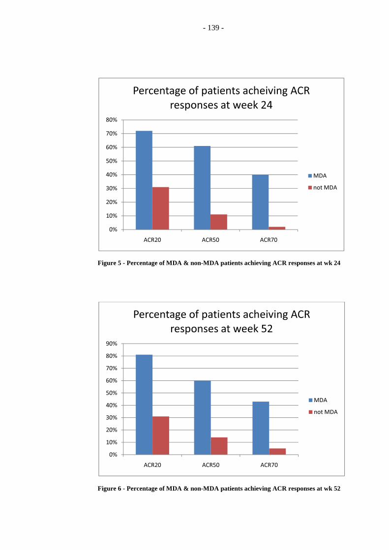
- 139 -
Figure 5 - Percentage of MDA & non-MDA patients achieving ACR responses at wk 24
Figure 6 - Percentage of MDA & non-MDA patients achieving ACR responses at wk 52
0%
10%
20%
30%
40%
50%
60%
70%
80%
ACR20 ACR50 ACR70
Percentage of patients acheiving ACR
responses at week 24
MDA
not MDA
0%
10%
20%
30%
40%
50%
60%
70%
80%
90%
ACR20 ACR50 ACR70
Percentage of patients acheiving ACR
responses at week 52
MDA
not MDA

- 140 -
6.3.1.4 Construct validity
Three previous studies investigating the frequency of remission in PsA were
identified in the literature review. There are no publications investigating minimal
or low disease activity. Two of the published studies assessed remission in the
peripheral joints and did not take into account any of the other aspects of the disease
(Gladman et al. 2001; Lindqvist et al. 2008). In a previous Toronto study, it was
shown that 18% of patients achieved remission in their peripheral joint activity (no
actively inflamed joints) for at least 12 months but 52% of these patients relapsed
during follow up (Gladman et al. 2001). In the Swedish early PsA cohort, 17% were
found to be in remission at 2 years (no tender or swollen joints and normal
inflammatory markers) (Lindqvist et al. 2008). A further study of remission in early
disease used more stringent criteria requiring no enthesitis or dactylitis disease
activity, in addition to peripheral joint disease, but did not include skin disease
(Cantini et al. 2008). This showed a higher frequency of remission, 24%, which
may partially represent the changing availability of treatments over the last decade.
These rates of remission compare to a rate of 34% of patients achieving MDA
in the Toronto observational study. This higher rate can be accounted for as the
MDA criteria should encompass both remission and low disease activity. The
remission criteria used for peripheral disease activity and enthesitis are lower than
the domain cut-point used in the MDA criteria, in keeping with the fact that the
remission criteria should be more stringent. Patients classified as remission would
achieve the cut points for the joint and entheses domains in the MDA criteria.
However it is impossible to say if they would achieve the full MDA criteria as there
was no inclusion of PROMs.
6.3.2 Discrimination
6.3.2.1 Classification
Although the sensitivity and specificity of the criteria whilst in development
were high, there is always a risk of misclassification of patients, particularly those
who are “borderline” on the criteria. The most crucial part of classification of these
criteria is their specificity, to ensure that patients with active disease are not falsely
classified as MDA which may result in a suboptimal treatment. Analysis of data
from the observational cohort has examined the characteristics of those patients who
met the MDA criteria. This showed that of the 208 patients who achieved MDA at
one or more visit, 110 patients (37%) met five of the seven criteria, 114 (38%) met
six criteria and 76 (25%) fulfilled all seven criteria. Of those who did not fulfil all
seven criteria, the most common active domain was skin psoriasis (n=44) followed
- 141 -
by patient global VAS (n=17). None of the patients had a SJC of >1. Of the
patients maintaining MDA, only 15 patients (12.9%) had escalation of their drug
therapy made during their time in MDA.
6.3.2.2 Prognosis
Firstly, the prognostic ability of the criteria was analysed in the observational
cohort. Patients in sustained MDA were compared to 200 controls who did not
achieve the MDA criteria for >12 months. The average follow up time to assess
progression of disease was matched at 34 months for both groups. The mean change
in damaged joint counts over the study period was 0.93 (range 0 to 12) in the MDA
group and 2.25 (range 0 to 17) in the controls (p<0.001). Of the MDA patients, 69%
showed no progression of joint damage, compared to 51% in the control group.
Reduced multivariate regression models looking at predictors for changes in
damaged joint counts identified that those patients who achieved the MDA state had
a lower rate of progression of clinical damage (p=0.03).
Prognosis was addressed in the trial data using radiographic evidence of
progressive joint damage. In the IMPACT1 study, 96% of those patients who
achieved MDA showed no progression of radiological disease at week 52 (increase
in mS-vdH score of ≥0), compared to 67% of those who did not achieve MDA
(p=0.012). At week 104 numbers were greatly reduced and 12/37 (30%) were in
MDA. All patients who achieved MDA at week 104 showed no progression of
radiological disease, compared to 58% of those who did not achieve MDA (p=0.03).
In the IMPACT2 study, 78% of those patients who achieved MDA showed no
progression of radiological disease at week 52, compared to 57% of those who did
not achieve MDA (p=0.009). There was a trend towards a reduced progression in
total mS-vdH score and the erosion score at week 52 in patients who achieved MDA
at this time (p=0.052, p=0.053 respectively) but no significant difference in
progression of JSN. There was no significant difference seen at week 24 related to
MDA.
Cumulative probability plots of changes in mS-vdH score through week 54 are
shown (figure 7). The curve for patients consistently in MDA (at week 24 and 52)
lies to the right of the control curve indicating less radiographic progression and a
smaller amount of radiographic progression per patient in MDA patients.
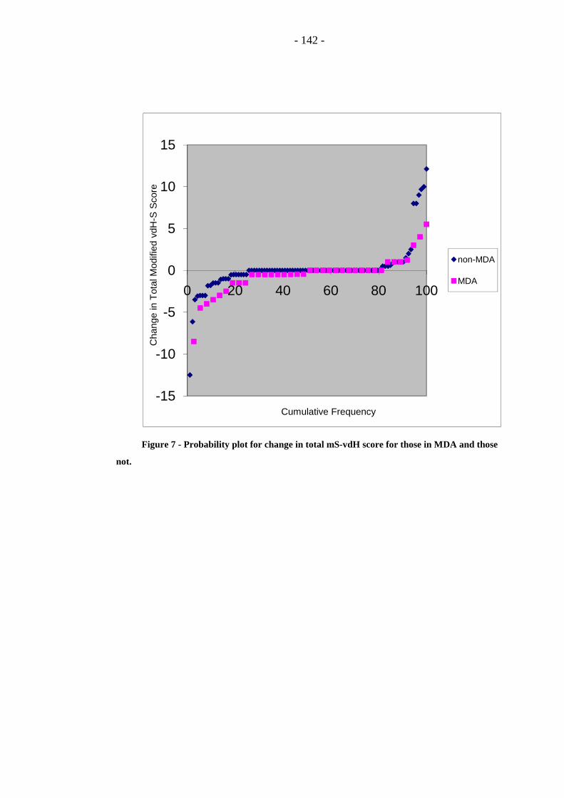
- 142 -
Figure 7 - Probability plot for change in total mS-vdH score for those in MDA and those
not.
-15
-10
-5
0
5
10
15
0 20 40 60 80 100
Cha
nge
in T
otal
Mod
ified
vdH
-S S
core
Cumulative Frequency
non-MDA
MDA
- 143 -
6.3.2.3 Responsiveness
Firstly, the number of patients achieving the MDA criteria in the observational
cohort was studied. Of the 344 patients, 208 (61%) achieved MDA at one or more
visit, and 116 (34%) achieved MDA on consecutive visits for a minimum duration
of 12 months. The median duration of MDA was 28 months (range 12 to 48). The
characteristics of the patients who met and sustained the MDA criteria are shown in
Table 1. After the minimum 12 month period required for inclusion, twelve patients
(10%) experienced a flare of their disease and ceased to fulfil the MDA criteria after
an average of 34 months in MDA. The remainder were still in MDA at their most
recent follow-up.
When considering the trial data, the responsiveness of the criteria was
confirmed by comparing the proportion of patients achieving MDA in the placebo
and treatment groups. In the IMPACT1 study data were available for 63 patients.
Of those receiving infliximab, 48% (15/31) achieved MDA at week 16 compared to
3% (1/32) on placebo (p<0.0001). At week 52, when all patients were treated with
infliximab, 42% were in MDA. In the IMPACT2 study data were available on 157
patients. At the primary endpoint (week 24), 52% (40/77) of the patients
randomised to receiving infliximab achieved MDA compared to 21% (17/80)
randomised to placebo (p<0.001). At week 52, when all patients were on
infliximab, 40% were in MDA.
6.3.2.4 Reliability
The literature review identified previous evidence of reliability for each of the
outcome measures included in the criteria. This had been assessed in previous
studies including INSPIRE and IMPART (International Multicentre Psoriasis and
Psoriatic Arthritis Reliability Trial) which analysed the reliability of TJC, SJC,
PASI, BSA and a variety of enthesitis measures (Gladman et al. 2007a; Chandran et
al. 2009). The HAQ-DI is self-administered by the patient and has been shown to be
reliable in PsA without modification (Leung et al. 2008). The patient reported
outcomes such as the VAS scales are again self-administered by the patients and
they have been shown to be reliable in PsA (Cauli et al. 2007). Further reliability
data are therefore not required.
6.3.3 Feasibility
All of these outcome measures included within the criteria are considered
routine to researchers in this field. Three of the seven outcome measures are
completed by the patient and can be done in the waiting room prior to their
appointment. As part of the assessment, the assessor must complete a joint count,
- 144 -
PASI or BSA and enthesitis count all of which can be completed within 5-10
minutes in the clinic setting. Where doubt exists about the best outcome measure to
apply, the criteria have been left open so that researchers can use the outcome
measure that they are familiar with (eg PASI vs BSA and different enthesitis scores).
None of the outcome measures included require any special equipment to perform,
so the only significant cost to the assessor is of time. As stated above, the time
needed to complete these measures is minimal.
6.4 Discussion
Until recently, there were no composite disease activity measures available to
measure a “state” of disease in PsA. The proposed criteria for minimal disease
activity in PsA encompass different aspects of this heterogeneous disease, and could
provide a new outcome measure for future clinical trials in PsA (Coates et al.
2010b). Preliminary validation work, highlighted here, provides support for their
further investigation and use.
The data in this study show that sustained MDA occurs in approximately one-
third of the PsA population. The higher prevalence is not surprising as the criteria
are less strict than those used for remission in previous studies. Low levels of
disease activity are allowed in all domains and only five of the seven cut points must
be met. As seen in the previous remission studies, flare of disease occurred after a
significant duration of time in MDA in around 10% of the patients demonstrating
the variable activity of disease both on and off therapy. The vast majority of
patients in MDA stayed on stable therapy, but 15 did have an escalation of their
treatment. This may represent specific treatment for a certain high domain (e.g.
active skin psoriasis only) or an increasing focus by the PsA clinic on the target of
remission.
Although there is a marked reduction in progression of joint damage, there is
still evidence of some progression despite patients consistently achieving MDA. It
must be noted that these criteria are not remission criteria but minimal disease
activity and therefore a small amount of disease activity may be present. The
patients achieving the MDA criteria can have active disease in one or two domains
whilst still fulfilling the criteria and even in the other domains, low disease activity
is considered acceptable by the criteria (e.g. 1 tender joint). This may account for
the lower rate of joint damage. We also know that clinical examination does not
identify all inflamed joints and it is particularly insensitive to low levels of
inflammation (subclinical disease) (Wakefield et al. 2004). Even in patients with
RA who are in remission (as defined by DAS), joint damage progression has been

- 145 -
shown to occur and is related to subclinical inflammation seen on imaging (Brown
et al. 2006).
6.4.1 Truth
Face and content validity were carefully considered during the design and
development phase of the MDA criteria. International consensus from members of
GRAPPA provides support of this. Assessment of criterion validity in trial data has
shown evidence of concurrent validity with other measures used to identify disease
activity including composite disease activity measures such as ACR outcomes,
laboratory tests such as CRP and physician assessments of disease activity. Ideally
convergent and divergent validity should be assessed by comparing the new criteria
to criteria measuring similar disease states (such as low disease activity/remission)
and those measuring different disease states (such as active disease). The only
criteria available for comparison are previous definitions of remission used in
publications. Patients in remission should be encompassed within MDA, but the
MDA criteria should also include patients who have some low disease activity.
There is some evidence for convergent and divergent validity with remission criteria
in PsA but direct comparisons with these cannot be used as they use distinct
conceptual definitions. Therefore there is very limited evidence for construct
validity at this time.
6.4.2 Discrimination
The ability of the criteria to classify patients as MDA seemed high in the initial
development of the criteria when compared to physician opinion. However
misclassification is always a risk. As stated previously, the risk to the patient of
misclassification is particularly important when considering patients with active
disease who are misclassified as MDA and potentially denied additional therapy.
Observational cohort data has been reassuring in this regard as it has shown that
none of the MDA patients had swollen joints and that very few had an escalation of
therapy providing some reassurance that there was a low rate of misclassification in
this cohort. Ideally further validation should be undertaken in a prospective cohort
with borderline patients.
The impact of MDA on prognosis in terms of joint damage has been confirmed
in both the observational (with regard to clinically damaged joint counts) and trial
cohort data (with radiographic outcomes). It is clearly shown that patients can
change states over time both in the observational cohort and in a randomised trial
where drug and placebo groups are compared. There is a significant difference in
the achievement of MDA between patients treated with anti-TNF therapy and
- 146 -
placebo, suggesting that this could be used as a trial outcome measure for new
therapies.
Reliability of the individual outcome measures included in the criteria has been
confirmed in a literature review. In this situation, the reliability of the MDA criteria
are validated by the reliability of their constituent parts.
6.4.3 Feasibility
All of these outcome measures included within the criteria are quick and easy
to perform with no equipment required, and three of the seven outcome measures are
completed by the patient. The criteria are being used in further ongoing studies and
have proved easy to integrate into a standard clinic visit.
6.5 Limitations
Although provisional data are available to address each of the constituents of
the OMERACT filter, further data are required before they can be classified as valid.
Ongoing use in prospective studies should provide further evidence towards their
suitability as an outcome measure. Specifically, further evidence is required of their
classification ability, construct and criterion validity. There is no confirmed gold
standard of MDA limiting the assessment of criterion validity. Ideally a further
study should be performed in an independent population comparing the criteria
against physician and patient opinion. The assessments of convergent and divergent
validity are also limited at present as no other criteria for minimal disease activity or
low disease activity at present. If other criteria are developed, further testing could
be done.
There are some limitations introduced by the type of datasets used to assess
validity. Some of these are overcome by the use of two different data cohorts. The
advantage of using observational data from a cohort such as the PsA program in
Toronto is that it provides relevant information on real-life patients all treated
according to their clinical need rather than any set trial protocol. However exactly
because of this, there are some limitations on the conclusions that can be drawn
from the data.
There is a significant potential for confounding in data from observational
cohorts. PsA is known to be a heterogeneous disorder and the progression of joint
damage is highly variable between individuals. Patients presenting with active
disease and joint damage are highly likely to be prescribed biological therapies
given their active disease, but also have a more severe disease phenotype and a
higher potential for damage progression. Although we have shown an improved
prognosis in those patients achieving a state of MDA, we cannot be sure what
- 147 -
proportion of this is due to the natural history of the disease and what impact
successful treatment has on outcome. Also, in the observational study, a clinically
damaged joint count was used, rather than assessment of joint damage on
conventional radiography. This outcome measures has been validated against
conventional radiography (Gladman et al. 1990; Gladman et al. 2004), but is
probably less sensitive to early joint damage and to minor changes than radiography.
Therefore the progression in both groups may be an underestimate of the
progression of damage in PsA.
Analysis of interventional therapeutic trials can remove much of the potential
for confounding and can provide stronger evidence for the prognostic ability of the
MDA criteria. Data for prognosis in this study used radiological outcomes with a
validated scoring system providing a more robust assessment of joint damage.
However, the assessment of joint damage is only one aspect of prognosis. Further
long term research is required to look at other measures of impact on patients’ long
term outcome, such as functional ability, quality of life and work status.
Finally, the development of the criteria based on expert opinion represents
current views and these may change in the future. The OMERACT definition of
MDA is “that state of disease activity deemed a useful target of treatment by both
the patient and physician, given current treatment possibilities and limitations”.
This relationship to current treatment possibilities means that acceptable targets of
treatment may drop further as additional therapeutic options are available.
6.6 Conclusion
Data from both observational and controlled clinical trial cohorts have
provided initial evidence supporting the validity of the MDA criteria as an outcome
measure. Although further work is required in some areas, initial assessments of the
truth, discrimination and feasibility of the PsA MDA criteria are positive and the
MDA criteria can be recommended for use in future research. Further use of these
criteria in prospective studies will provide further validation evidence.

- 148 -
7 Tight Control of Psoriatic Arthritis
7.1 Introduction
Since the TICORA study highlighted the benefit of tight control in RA (Grigor
et al. 2004), it has become standard of care in the majority of RA clinical trials and
is gradually becoming adopted in clinical practice. The use of an objective target to
guide treatment decisions has been shown to be associated with improved clinical
and radiographic outcomes in RA.
Until recently, there were no objective targets designed specifically for PsA.
With the development and validation of the MDA criteria (Coates et al. 2010a;
Coates et al. 2010b; Coates and Helliwell 2010), a disease specific target for
treatment has been established in PsA and it is possible to investigate the use of tight
control in PsA. The aim of this study was to utilise the MDA criteria in a tight
control protocol to treat newly diagnosed PsA.
7.2 Methods
The TICOPA study is a multicentre national trial currently running at 6 sites.
It is designed as a randomised, controlled, parallel group, open label, clinical trial of
206 patients with recent onset PsA. Patients are randomised on a 1:1 basis to
receive either standard care or tight control for a period of 48 weeks. The study was
approved by national and local ethics committees and all patients gave written
consent before inclusion.
7.2.1 Patients
Recruitment started in May 2008 and inclusion criteria are PsA of less than
two years symptom duration with active arthritis or enthesitis. As the CASPAR
criteria were unvalidated in short disease duration PsA at the time of study design, a
consultant diagnosis of PsA was used as the inclusion criteria. Exclusion criteria
included age less than 18 years, previous treatment for arthritis with DMARDs,
recent or concomitant use of an experimental drug, wish to conceive during the
study period or within 6 months of the end of the study or inadequate contraception.
Patients are also required to have an adequate baseline full blood count
(haemoglobin > 8.5 g/dL, white cell count > 3.5 x 10⁹/L, neutrophils > 1.5 x 10⁹/L,
platelets > 100 x 109/L) and hepatobiliary function (aspartate aminotransferase or
alanine aminotransferase (ALT) <3 times the upper limit of normal).
- 149 -
7.2.2 Treatment allocation and intervention
Patients are randomised 1:1 using dynamic allocation incorporating a random
element, via a computer-generated programme, to ensure treatment groups are well-
balanced for randomising centre and pattern of arthritis (oligoarticular vs
polyarticular). The trial uses a dynamic allocation process to allocate the
interventions and thus minimise imbalance between the two trial arms.
Patients assigned to the intensive group are seen every 4 weeks by the study
physician at each centre. A strict treatment protocol (see figure 8) is followed for
treatment. At each visit, from three months onwards, the MDA criteria (Coates et al.
2010b) are assessed. These criteria include assessment of 66/68 joint count, PASI,
enthesitis count, patient global and pain VAS and HAQ-DI. Treatment with
DMARDs is escalated to the maximum dose according to the protocol in figure 1 if
patients do not achieve the MDA criteria. In the case of intolerance, that drug is
discontinued and the next step on the protocol is initiated. Any patient who cannot
tolerate the maximum dose specified in the protocol due to toxicity or intolerance, is
permitted to continue on the highest tolerable dose and then progress to the next step
in the protocol if required. IA and intra-muscular steroids are also permitted in the
intensive group arm to control disease. Patients are offered local joint injections to
active joints and/or intramuscular steroid by the physicians if considered
appropriate.

- 150 -
Figure 8 – Protocol for treatment in the TICOPA study
- 151 -
Patients randomised to the standard therapy group are treated in a general
rheumatology outpatient clinic supervised by a consultant rheumatologist and
including trainee rheumatologists working under supervision. These participants are
reviewed as directed by the physician but generally every 12 weeks, with no formal
measures of disease activity used in clinical decision making. There is no
requirement or restriction on prescribing within this arm of the study.
7.2.3 Assessment of end-points
Every 12 weeks, clinical assessments are performed by a research nurse or
metrologist blinded to the allocated treatment group. These include 66/68 joint
count, PASI (Fredriksson and Pettersson 1978), nail psoriasis scoring using the
mNAPSI (Cassell et al. 2007) and a comprehensive assessment of enthesitis
including entheseal sites required for the LEI (Healy and Helliwell 2008), the
MASES (Heuft-Dorenbosch et al. 2003) and sites used in the IMPACT and
IMPACT2 clinical trials (bilateral Achilles tendon and plantar fascia) (Antoni et al.
2005a; Antoni et al. 2005b) and dactylitis assessment using the LDI (Helliwell et al.
2005).
At baseline and 48 weeks, plain radiographs of the hands and feet and an US
scan of the most symptomatic or dominant hand were performed. Radiographs were
scored using the mS-vdH scoring system for PsA (van der Heijde et al. 2005b).
They were scored as paired images but assessors were blinded to patient identity and
timepoint. US was performed using a Philips HDI 5000 machine with a 12-5 or 13-
7 MHz probe as appropriate. For PD scanning a medium wall filter was applied.
All scans were performed by a rheumatologist experienced in musculoskeletal US
(Dr J E Freeston). Gel was applied to the skin to provide an acoustic interface. All
patients underwent GS and PD US of the most symptomatic hand and wrist (wrist,
MCP2-5 and PIP2-5 joints - 11 joints per patient). GS and PD were scored
separately on a 0-3 semi-quantitative scale for each joint imaged. A GS score of ≥2
and/or a PD score >0 were used to identify US active joints
The primary endpoint of the study is achievement of ACR20 response at 48
weeks. This was chosen as research has shown that it is responsive in PsA and has a
lower placebo response than is seen with the PsARC (Fransen et al. 2006). This
analysis also identified the EULAR responses based on the DAS as being responsive
in PsA, but there was concern about using an outcome measure based on a 28-joint
count in oligoarticular disease. Secondary endpoints at week 48 include ACR50,

- 152 -
ACR70, achievement of MDA criteria and radiographic joint damage according to
change in mS-vdH score.
7.2.4 Statistical analysis
The primary outcome in this study is the proportion of patients who achieve an
ACR 20 response at 48 weeks from baseline. Previous data for patients with PsA
(Healy and Helliwell 2007) starting on DMARDs (predominantly methotrexate) has
shown ACR 20 at 3 and 6 months to be 50% and 43% respectively. We have
therefore assumed that the proportion of patients achieving ACR20 in the standard
care arm will be approximately 50% at 48 weeks. An absolute difference in ACR20
rates of 20% has been deemed to be a clinically significant difference. Therefore,
with 80% power and based on a chi-squared test without continuity at the 2-sided
5% significance level, 93 patients per arm (186 patients in total) are required to
detect and increase on ACR 20 rates of 20%. To allow for a 10% drop-out rate, 206
patients will be recruited in total.
This large RCT is currently ongoing and therefore analysis for this thesis has
been restricted to blinded data. To date 40 patients have been randomised into the
study and have completed their 48 week follow up. Data regarding treatment is
provided in summary form for the cohort, but detailed analysis comparing treatment
and response has been limited to ensure that treatment groups remain unblinded.
Significance testing was performed using Chi-squared tests for categorical
variables, Mann-Whitney U tests for independent continuous variables and
Wilcoxon signed rank tests for paired continuous variables. Univariate forward
step-wise binary logistic regression was used to identify which baseline features
were associated with achievement of MDA at the 48 week timepoint. All analysis
was performed using SPSS 16.
7.3 Results
To date, in the main RCT, 96 patients have been recruited. A flowchart
detailing their recruitment and follow up is shown in figure 9. The remainder of this
chapter includes the analysis of the first 40 patients to be randomised and to
complete follow-up.
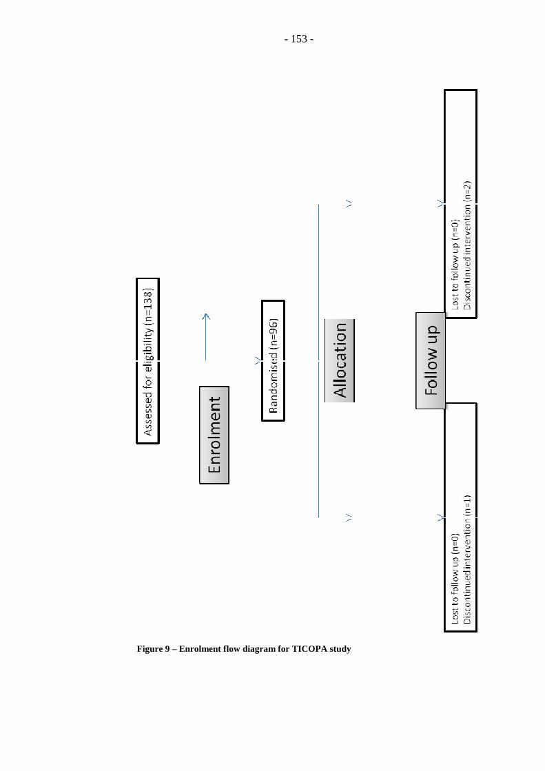
Figure 9 – Enrolment flow diagram for TICOPA study
- 153 -
Enrolment flow diagram for TICOPA study

- 154 -
7.3.1 Baseline Demographics
Of the 40 patients completing TICOPA to date, all had a consultant diagnosis
of PsA, but only 34 met the CASPAR criteria. The remaining 6 patients all scored 2
points in the CASPAR criteria. These patients did not have current psoriasis, but all
had 2 indicators of PsA from the other features included in the CASPAR criteria.
Baseline characteristics of the cohort are shown in table 23.
Considering imaging at baseline, plain radiographs of the hands and feet were
available for 29 patients and US of the most affected or dominant hand was
available for 38 patients. At baseline, the majority of patients had little or no
evidence of radiographic damage (median mS-vdH score 1, IQ range 0-4.5). JSN
was seen in around half of the cohort (n=15) and the median score for those with
JSN was 4 (IQ range 1 to 5). Erosions were identified in only 5 patients (17%) with
a median score of 4 (IQ range 1 to 6.5). US at baseline identified relatively low
levels of disease in the hand and wrist with predominant GS synovitis and low levels
of PD. The median total GS score was 11.5 (IQ range 8 to 18.25) with a median PD
score of 0 (IQ range 0 to 3.25). When the number of US active joints (GS score of
≥2 and/or a PD score >0) was considered, only 16 patients had active disease in the
one hand that was scanned at baseline, and these patients had a median active US
joint count of 2.
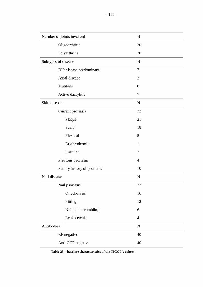
- 155 -
Number of joints involved N
Oligoarthritis 20
Polyarthritis 20
Subtypes of disease N
DIP disease predominant 2
Axial disease 2
Mutilans 0
Active dactylitis 7
Skin disease N
Current psoriasis 32
Plaque 21
Scalp 18
Flexural 5
Erythrodermic 1
Pustular 2
Previous psoriasis 4
Family history of psoriasis 10
Nail disease N
Nail psoriasis 22
Onycholysis 16
Pitting 12
Nail plate crumbling 6
Leukonychia 4
Antibodies N
RF negative 40
Anti-CCP negative 40
Table 23 – baseline characteristics of the TICOPA cohort
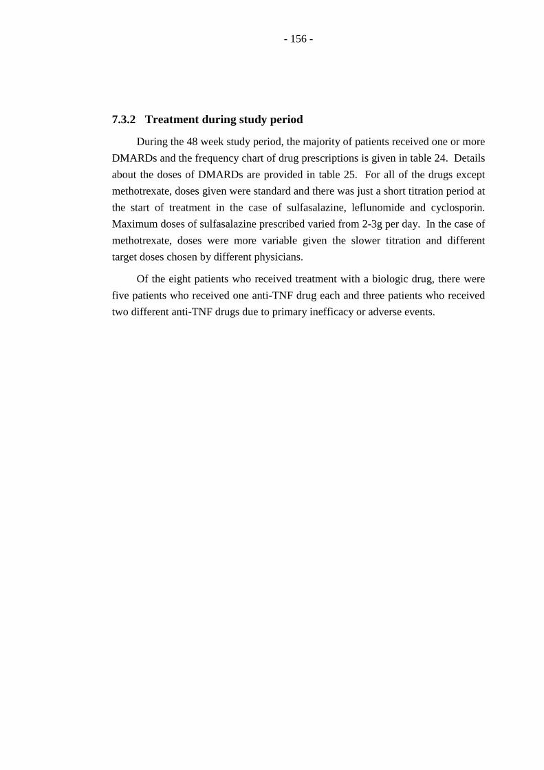
- 156 -
7.3.2 Treatment during study period
During the 48 week study period, the majority of patients received one or more
DMARDs and the frequency chart of drug prescriptions is given in table 24. Details
about the doses of DMARDs are provided in table 25. For all of the drugs except
methotrexate, doses given were standard and there was just a short titration period at
the start of treatment in the case of sulfasalazine, leflunomide and cyclosporin.
Maximum doses of sulfasalazine prescribed varied from 2-3g per day. In the case of
methotrexate, doses were more variable given the slower titration and different
target doses chosen by different physicians.
Of the eight patients who received treatment with a biologic drug, there were
five patients who received one anti-TNF drug each and three patients who received
two different anti-TNF drugs due to primary inefficacy or adverse events.
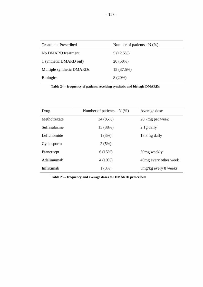
- 157 -
Treatment Prescribed Number of patients - N (%)
No DMARD treatment 5 (12.5%)
1 synthetic DMARD only 20 (50%)
Multiple synthetic DMARDs 15 (37.5%)
Biologics 8 (20%)
Table 24 – frequency of patients receiving synthetic and biologic DMARDs
Drug Number of patients – N (%) Average dose
Methotrexate 34 (85%) 20.7mg per week
Sulfasalazine 15 (38%) 2.1g daily
Leflunomide 1 (3%) 18.3mg daily
Cyclosporin 2 (5%)
Etanercept 6 (15%) 50mg weekly
Adalimumab 4 (10%) 40mg every other week
Infliximab 1 (3%) 5mg/kg every 8 weeks
Table 25 – frequency and average doses for DMARDs prescribed

- 158 -
7.3.3 Response measures
7.3.3.1 Minimal Disease Activity
The proportion of patients achieving the MDA state increased steadily over the
48 week study period (table 26) to a maximum of 53%. Univariate logistic
regression analysis identified that a low patient’s global disease activity VAS at
baseline (OR=0.96; 95% CI = 0.93, 0.99; p=0.020) and the absence of enthesitis at
baseline (OR=10.46; 95% CI = 1.16, 94.48; p=0.037) were associated with a
increased chance of achieving MDA at 48 weeks. Other factors investigated were
not significant including joint counts, pattern of arthritis (oligoarthritis or
polyarthritis), presence of dactylitis, baseline physicians global disease activity
VAS, quality of life (PsQOL), function (HAQ-DI) or baseline CRP levels.
7.3.3.2 Outcome measures for individual domains of PsA
All patients had arthritis at study entry and so all were assessed for the
composite ACR and EULAR joint response measures. Table 27 shows the
proportion of patients achieving both the ACR response measures and the EULAR
response criteria based on the DAS28 score. The response rates improved over the
course of the 48 week study period.
Both the ACR and EULAR outcomes have been validated in polyarticular
PsA, but less is known about their responsiveness in oligoarticular disease. The 20
patients with oligoarticular disease at baseline were compared to those presenting
with polyarticular disease, to identify any differential response in these outcome
measures at 48 weeks (table 28). For all of the responder indices (ACR and EULAR
outcomes), a slightly higher proportion of patients with polyarticular disease
achieved the outcomes, with a difference that was more pronounced for ACR70 in
comparison to ACR20. In contrast, the MDA criteria were more commonly
achieved by patients with oligoarthritis at baseline.

- 159 -
Week 12 Week 24 Week 36 Week 48
Number (%) acheiving MDA 10 (25) 12 (30) 15 (38) 21 (53)
Table 26 – proportions of patients achieving MDA at key timepoints
Number (%) achieving outcome
Timepoint ACR20 ACR50 ACR70 Mod
EULAR
response
Good
EULAR
response
Week 12 22(55) 8 (20) 1 (3) 9 (23) 3 (8)
Week 24 17 (43) 12 (30) 6 (15) 12 (30) 10 (25)
Week 36 18 (45) 11 (28) 7 (18) 12 (30) 10 (25)
Week 48 23 (58) 18 (45) 15 (38) 16 (40) 16 (40)
Table 27 – Composite arthritis outcomes at key timepoints
Number (%) achieving outcome at 48 weeks
Subtype ACR20 ACR50 ACR70 Mod/Good
EULAR
response
MDA
Oligoarthritis 11 (55) 8 (40) 6 (30) 6 (30) 12 (63)
Polyarthritis 12 (60) 10 (50) 9 (45) 10 (50) 9 (45)
Table 28 – composite arthritis outcomes and MDA by joint disease subtype

- 160 -
In addition to clinical outcome measures for articular disease, data are
available for imaging with plain radiographs and US. Radiographic data identified
that the majority of patients had no change in mS-vdH score during the 48 week
period. There was no significant difference found between baseline and week 48
scores for erosions, JSN or total score (p>0.83). At week 48, the median mS-vdH
score was 2 (IQ range 0-4) with an erosion score of 0 and JSN of 2. Only 3 patients
changed their status from non-erosive to erosive disease during the time period and
their erosion scores remained low (median 1, range 1-2).
US data at baseline and 48 weeks were available for 38 of the 40 patients. US
showed a significant reduction in imaging activity both in terms of GS synovitis and
PD score (p<0.002). When the number of US active joints (GS score of ≥2 and/or a
PD score >0) was considered, only 16 patients had active disease in the hand at
baseline. Again there was a significant reduction in US activity seen in these
patients with a reduction in the median joint count from 2 to 0.5 (p=0.008).
Whilst peripheral joint outcome measures detailed above were calculated using
all patients, the outcomes for the other domains of PsA (skin, nails, enthesitis and
dactylitis) have been restricted to those with involvement of that particular domain
at baseline. Skin psoriasis was documented in 32 patients at baseline and PASI
scores at baseline were low (median 2.3) with only 14 patients with PASI ≥ 3 and
only one patient who had a baseline PASI score of greater than 10. PASI responses
for both of these groups (all patients with psoriasis and those with baseline PASI ≥
3) are given in table 29.
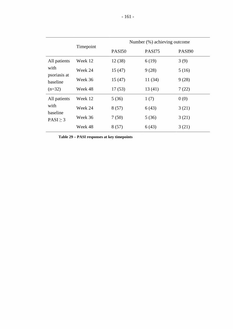
- 161 -
Timepoint
Number (%) achieving outcome
PASI50 PASI75 PASI90
All patients
with
psoriasis at
baseline
(n=32)
Week 12 12 (38) 6 (19) 3 (9)
Week 24 15 (47) 9 (28) 5 (16)
Week 36 15 (47) 11 (34) 9 (28)
Week 48 17 (53) 13 (41) 7 (22)
All patients
with
baseline
PASI ≥ 3
Week 12 5 (36) 1 (7) 0 (0)
Week 24 8 (57) 6 (43) 3 (21)
Week 36 7 (50) 5 (36) 3 (21)
Week 48 8 (57) 6 (43) 3 (21)
Table 29 – PASI responses at key timepoints
- 162 -
Enthesitis, dactylitis and nail disease were less prevalent in the cohort affecting
31, 12 and 20 patients respectively. Enthesitis score shown in figure 10 relates to a
total sum of all enthesitis sites examined (n=19). Individual results are also shown
for the MASES, LEI and IMPACT enthesitis indices. The LEI and MASES scores
all showed a reduction in enthesitis activity over the 48 week study period, but the
IMPACT index did not identify significant amounts of enthesitis.
Results for count of dactylitic digits and LDI score are shown in figure 11. A
significant improvement in dactylitis outcomes was noted within the first 12 weeks
(p=0.002 for dactylitic digit count and LDI score) with median scores of 0 after this
timepoint. The decrease in mNAPSI scores for nail psoriasis was rapid and
sustained to the 6 month timepoint when a slowing of improvement was noted
(figure 11).

- 163 -
Figure 10 – Change in median scores for enthesitis outcomes for those with involvement
at baseline
0
0.5
1
1.5
2
2.5
3
3.5
baseline week 12 week 24 week 36 week 48
Total enthesitis count
LEI
MASES
IMPACT
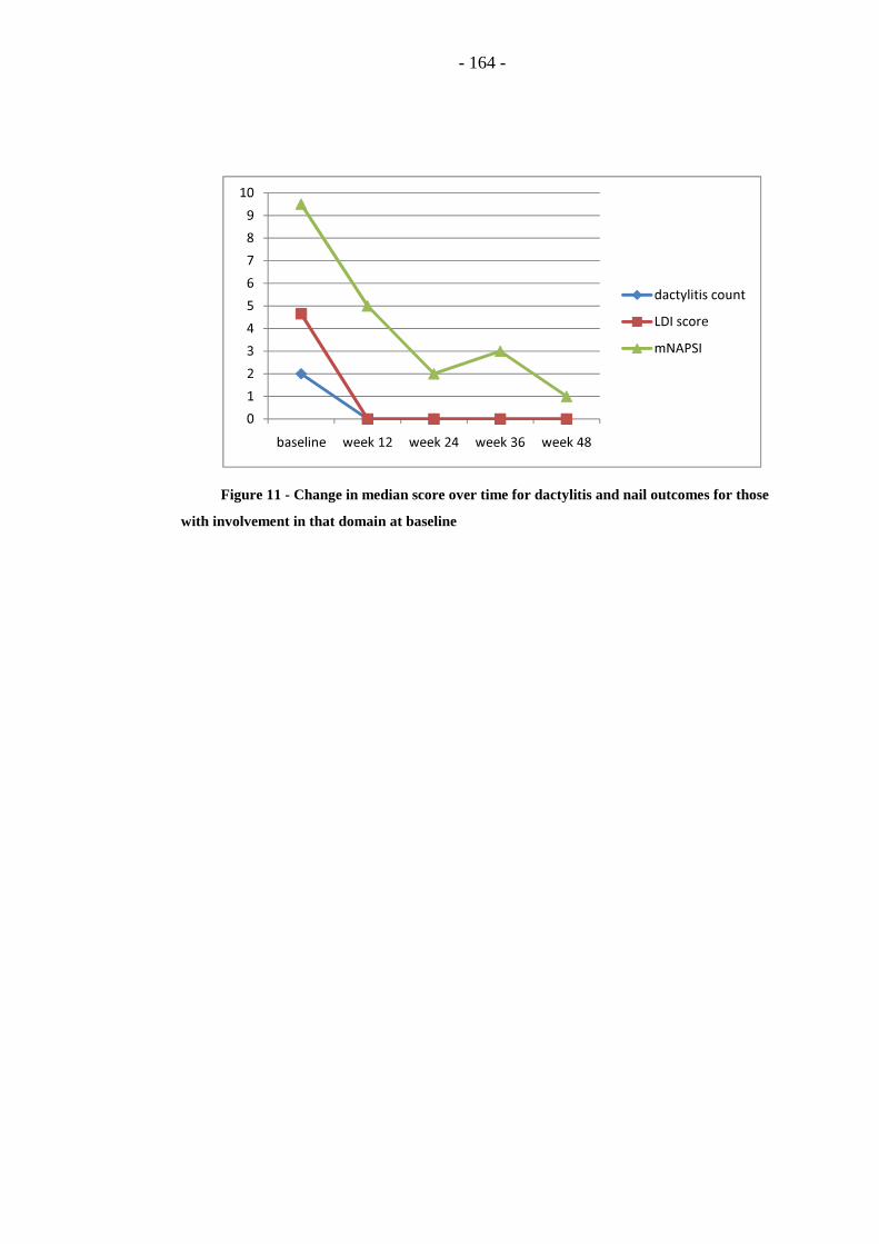
- 164 -
Figure 11 - Change in median score over time for dactylitis and nail outcomes for those
with involvement in that domain at baseline
0
1
2
3
4
5
6
7
8
9
10
baseline week 12 week 24 week 36 week 48
dactylitis count
LDI score
mNAPSI

- 165 -
7.3.4 Patient reported outcomes
All patients completed PROMs at 12 weekly intervals. Over the study period,
there was a reduction in VAS scores for pain, global disease activity, fatigue and
global health, all indicating an improvement in health status (figure 12). Quality of
life, as measured by the PsQOL, and functional ability, measured by the HAQ-DI,
also showed an improvement over the study period (table 30).

- 166 -
Figure 12 - changes in median VAS scores at key timepoints
0
10
20
30
40
50
60
baseline week 12 week 24 week 36 week 48
Disease activity
Global health
Pain
Fatigue

- 167 -
Baseline Week 12 Week 24 Week 36 Week 48
Median PsQOL 6 4 2 3 2
Median HAQ-DI 0.75 0.44 0.13 0.38 0.13
Table 30 – Quality of life and functional questionnaire scores at key timepoints
- 168 -
7.3.5 Relationship between MDA and other outcome measures
There was a strong association seen between achieving the MDA disease state
and the ACR outcome response rate from week 24 onwards (p<0.004 for all except
ACR20 at week 24, see figure 13). This was most marked at week 48. However,
even at week 48, there was not total agreement between ACR outcome measures and
the MDA state. When considering the ACR70 response rate, there was one patient
who achieved this response but was still not in MDA. Conversely there were seven
patients in MDA who did not achieve a 70% reduction in the ACR outcome (table
31). The majority of patients (6 of 7) who achieved MDA but did not achieve an
ACR70 response were patients with oligoarthritis. Some patients had shown an
improvement in joint counts but not a 70% reduction (eg from three tender joints to
one tender joint), and some fulfilled the MDA criteria despite activity in the TJC
which prevented them from achieving the ACR outcome.
When comparing blood levels of inflammatory markers in patients with
different MDA outcomes, the association was less clear. There was no significant
difference between CRP, plasma viscosity (PV) and ESR results for those in MDA
and those who were not at any of the key timepoints. However, there were a number
of patients in the cohort who did not demonstrate raised inflammatory markers, even
at baseline. Analyses were repeated, limiting the patients to those with
inflammatory markers greater than the normal reference range (CRP≥10, ESR≥20,
PV≥1.72) at baseline. CRP levels were significantly lower at week 48 for those
patients in MDA (p=0.03), although a significant difference was not found at other
timepoints. ESR levels were significantly different at week 24 (p<0.03) but not at
other timepoints. There were no significant relationships between PV and MDA
found at any of the key timepoints.
US data was also used to compare to different MDA outcomes. At week 48
there was a trend towards lower US scores for those in MDA (median GS score 9.5
vs 11, p=0.48, median PD score 0 vs 1.5, p=0.10) but the difference did not reach
significance. Similarly, there was a trend towards a lower number of US active
joints at week 48 in those with MDA compared to those who were not (median no 0
vs 1, p=0.23) but this was not significant, probably due to the low numbers in this
subanalysis (n=15).

- 169 -
Table 31 – Comparison of patients achieving MDA and ACR outcome measures
MDA Non-MDA
ACR20 Yes 19 4
No 2 14
ACR50 Yes 16 2
No 5 16
ACR70 Yes 14 1
No 7 17

- 170 -
Figure 13 – comparison of ACR responses in those achieving MDA and those not
0
2
4
6
8
10
12
14
16
18
20
ACR20
ACR50
ACR70
ACR20
ACR50
ACR70
ACR20
ACR50
ACR70
ACR20
ACR50
ACR70
week 12 week 24 week 36 week 48
MDA
not MDA
- 171 -
7.4 Discussion
The TICOPA study is a large RCT designed to investigate the benefits of early
and tight control in PsA. There are few studies examining such an early PsA cohort
particularly of the planned recruitment size (n=206). The patients all had a disease
duration of less than 2 years and the majority fulfilled the CASPAR criteria. Those
who did not, had other typical features of PsA and all were found to be seronegative
for both RF and anti-CCP antibodies. The vast majority had normal or near-normal
radiographs at baseline compatible with early disease.
As the study is currently ongoing, and recruitment is planned to continue until
April 2011, unblinded data are not available for analysis. However, of interest, is
the fact that this study is the first to prospectively use the newly developed MDA
criteria and the blinded data can be used to further evaluate the validity of these
criteria.
In this small cohort (n=40), over 50% of the patients combined from the
intensive and standard arm achieved MDA over the 48 week study period.
Interestingly this exceeds the number seen in the previous validation studies (both
observational cohort data (Coates et al. 2010a) and results of the anti-TNF drug trial
(Coates and Helliwell 2010)). There are two likely reasons that may contribute to
this increased proportion in MDA. Firstly, the patients in TICOPA had early disease
and were DMARD naïve prior to starting treatment in the study. They therefore
represent a different population from those seen in the established disease cohort in
Toronto and the patients recruited to the IMPACT and IMPACT2 studies who had
established polyarticular disease and many of whom had previously failed synthetic
DMARDs. The other key difference is the use of MDA as a target for treatment for
those patients treated in the intensive arm of the TICOPA study. Half of the patients
in this cohort had their treatment escalated until they achieved MDA, so the rate of
MDA at the end of the study is expected to be higher.
When considering all of the individual aspects of PsA, there were good
responses seen in all of these areas. Composite joint scores showed an impressive
improvement in arthritis over the study period. In particular, the proportion of
patients achieving good responses (such as the ACR70 and EULAR good response)
were higher than expected when compared to other published data (Nasonov et al.
2009). There was no significant change identified in measures of radiographic
damage over the study period. There are two likely reasons for this. Firstly, in a
cohort of PsA patients, even those with established disease, the rate of radiographic
progression is not particularly rapid with an average increase of around 1 in the mS-
vdH score in one year (Ravindran et al. 2010). Secondly, in contrast to the
- 172 -
population of patients recruited to many drug trials previously reported in PsA, this
study recruited all patients with any active disease and did not select for
polyarticular disease or raised inflammatory markers. Therefore the likelihood of
radiographic progression over 48 weeks is lower than that seen in previous studies.
Unlike radiographs, US has the ability to measure active inflammation and was
able to identify a change in imaging assessment of disease activity in the study
period. To maintain feasibility within the study, only one hand was scanned using
US at both baseline and follow up. Due to the heterogeneity of PsA phenotype, only
16 patients had involvement seen in the hand at baseline, but despite the low
numbers a significant change in US disease activity was seen over the 48 weeks.
There were good proportions achieving PASI responses despite low levels of
skin psoriasis involvement at baseline. Interestingly similar PASI response rates
were seen in those with baseline PASI>3 and in the total cohort with skin disease.
Although PASI is recognised to be less responsive at the lower end of the scale, it
can clearly identify responses in a proportion of patients.
When considering the responses in other domains, a rapid and significant
improvement in both dactylitis and nail psoriasis was seen within the first 12 weeks
of the study period. During this time, patients had only received synthetic
DMARDs, with no prescriptions given for anti-TNF therapy at this stage. The vast
majority of patients were treated only with methotrexate which has not been
recognised as a treatment for dactylitis or nail disease previously (Cassell and
Kavanaugh 2006; Helliwell 2006). The improvement in enthesitis was slower,
showing some improvement within the first 12 weeks, but ongoing improvement up
to the 48 week visit. Although these data relate to open label treatment in a small
number of patients, they suggest efficacy of DMARDs, particularly methotrexate, in
many domains of PsA.
PROMs showed a steady improvement over the course of the study period
with the largest change seen in the first 12 weeks. There was a significant
improvement in patients’ VAS scales for global disease activity, pain and global
health in addition to questionnaires assessing quality of life and function.
In further investigation of the MDA criteria as an outcome measure, response
rates of other outcome measures were compared for those in MDA and those not
achieving the target. There is a clear link between MDA and composite joint
outcomes such as the ACR outcomes confirming previous validation work. Not
surprisingly, the correlation appears strongest for the higher level of responses such
as ACR70 where the majority of patients achieving such a significant improvement,
also achieve MDA. However there were some discrepancies seen with one patient

- 173 -
achieving the ACR70 response but not in MDA and seven patients in MDA who did
not achieve a 70% reduction in the ACR outcome.
When considering the different outcome measures in detail, this is not
particularly surprising. Firstly, the MDA criteria encompass other aspects of disease
such as enthesitis and skin disease that are not taken into account in the composite
arthritis outcomes. This may account for some of the discrepancies. Secondly, the
ACR outcome measures are response measures in comparison to MDA which is a
measure of a certain disease state. Patients with particularly active disease can show
an improvement of 70% in their arthritis outcome measures, but still have residual
active disease that prevents them from being classified as in MDA. Conversely
patients with oligoarticular disease at baseline may be able to achieve MDA but do
not have such a reduction of 70% in their joint counts. This differential response
between oligoarticular and polyarticular disease subtypes is highlighted by
comparing the proportions achieving each of the outcomes. For all of the response
measures, a higher proportion of polyarticular patients met these outcomes
compared to those with oligoarticular disease. In contrast, the MDA criteria, which
measure an absolute level of joint involvement, were more likely to be met by those
with oligoarticular disease. This has important implications for choice of outcome
measures in psoriatic disease. Arthritis responder indices are likely to be less
sensitive to change in patients with oligoarticular disease at baseline.
Relationships between MDA and other measures of disease activity including
systemic markers of inflammation and US evidence of synovitis showed some
trends but few significant differences. In this small cohort, these analyses are
limited due to the small numbers of patients included. Many patients with PsA do
not have a significant acute phase response despite active disease. US scans at
baseline and follow up were only available for the joints of one hand. The
heterogeneity of PsA means that many patients have little or no disease in the small
joints of the hand and therefore in a small group, significant associations are
unlikely to be demonstrated. Baseline predictors of MDA were briefly addressed
using univariate analysis but due to the small numbers in the cohort, multivariate
analysis could not be attempted. There was some evidence that patient global
disease activity VAS and presence of enthesitis at baseline were predictors of MDA,
but this is difficult to interpret as both of these measures are included in the
assessment of MDA. Given the small numbers and the link between predictive
baseline factors and the outcome measures included in the MDA criteria, it seems
that no clear conclusions can be drawn regarding this. Data from the full TICOPA
study with 206 patients will allow a future multivariate analysis of multiple factors
and this may provide information on predictors of MDA.

- 174 -
7.5 Limitations
The principle limitations of this analysis are the small numbers and the
restriction to blinded data to preserve the blinding of the main study. The small
numbers analysed mean that predictors of MDA could not be accurately
investigated. Only univariate regression could be performed and with small
numbers, subtle influences will not have been identified. As discussed above, the
associations identified are not truly independent of the assessment of the MDA
criteria so they should be interpreted with caution. Detailed analysis of medications
used are not available as this would lead to an “unblinding” of the data, so only
summaries of treatment data are available to be presented.
However this is the first study using MDA prospectively and offers some
insight into outcomes in early PsA treated with synthetic and biologic DMARDs as
required. The large range of outcome measures collected covering all domains of
psoriatic disease allow assessment of response in all areas of the disease.
7.6 Conclusion
This interim analysis of the larger TICOPA RCT provides insight into the
outcomes seen in multiple domains of early PsA. A high proportion of patients
achieve the MDA state within the 48 week study period and good responses are seen
in all domains of disease and in PROMs. Further validation of the MDA criteria is
provided by their concurrent validity with the ACR outcome measures as seen in
previous validation work. Due to the small size of the sample, it is not possible, at
this stage, to properly investigated predictors of response. However in future this
should be possible with the full TICOPA dataset.
- 175 -
8 Conclusions and Future Directions
This thesis is concerned with developing clinical tools and methodology to
improve the outcome of PsA. This final chapter will present an overview of the
results of the thesis, looking at all of the studies performed and analysing the
findings in the context of current literature. The potential research questions arising
from the work in this thesis will then be considered.
PsA is now recognised as a potentially damaging and destructive disease and
the aim of this research was to provide tools to improve disease management and
optimise outcome. The first priority is to identify patients early in the course of their
disease so that assessment and therapy can be initiated. However it was identified
that there was a lack of validated classification criteria to aid identification of
patients with early PsA for both clinical interventions and future research. The
recently developed CASPAR criteria have both high sensitivity and specificity in
established disease (Taylor et al. 2006), but it is stated in the paper that less than 5%
of the original study population had a disease duration of less than 12 months. A
few small studies have suggested that the CASPAR criteria may be useful in early
disease but no properly powered studies have addressed their validity in early PsA.
Once identified, the next challenge was to optimise management for patients
with early PsA. In RA, management has focused on early intervention in the course
of disease with “tight control” of inflammatory disease to improve prognosis.
However, this concept of “tight control” has not been investigated in PsA. In the
RA studies, validated outcome measures such as DAS, clinical disease activity index
(CDAI) and simplified disease activity index (SDAI) give definitions of low disease
activity or remission that can be used as a target for therapy. One of the major
issues preventing research in this area, perhaps reflecting a lack of research in PsA,
was the lack of validated criteria to define an “acceptable” disease state. The
concept of minimal disease activity had been defined by OMERACT but validated
criteria had only been developed for RA using this framework.
This thesis therefore concentrated on these two major challenges to the optimal
management of early PsA, and aimed to provide feasible clinical solutions to the
early identification of PsA and management using optimal treatment targets.
Chapter Three The validity of the CASPAR criteria in early PsA
In Chapter Three, the CASPAR criteria were found to be sensitive and specific
in identifying early PsA. Although their sensitivity is slightly lower than that found
- 176 -
in established disease, they performed better than the previous gold standard
classification criteria developed by Moll & Wright. Detailed assessment of the
patients and controls included in this study highlighted some of the clinical
phenotypical differences between early PsA and other form of early inflammatory
arthritis. However, there were no individual features that could be relied upon to
differentiate diagnosis between PsA and other forms of inflammatory arthritis.
Chapter Four Imaging in early PsA – the extent of sub-clinical disease
Modern imaging is more sensitive than clinical examination for detecting a
variety of important joint pathologies. A detailed assessment of a sub-group of
patients with early PsA investigated the US evidence for arthritis and enthesitis.
There were low levels of sub-clinical arthritis and particularly low levels of US-
identified enthesitis. Similarly, the correlation between clinical and US assessment
was high suggesting that, in most cases of early PsA, clinical examination provides a
reasonable assessment of disease activity for both synovitis and enthesitis.
Chapter Five Defining MDA in PsA
MDA criteria were developed for use in PsA using a web-based questionnaire
of international experts. The criteria include clinical measures of arthritis, enthesitis,
skin disease and PROMs to ensure that all aspects of psoriatic disease are
encompassed.
Chapter Six Validation of the MDA criteria for PsA
Following on from the development of the MDA criteria in the previous
chapter, validation work was completed following the methodology of the
OMERACT filter. This analysis provided evidence within the domains of truth,
discrimination and feasibility proving the performance metrics of the PsA MDA
criteria. A large observational cohort and two large datasets from interventional
randomised controlled trials were utilised in order to assess their validity in routine
clinical practice and as a potential outcome measure for future interventional trials.
Chapter Seven Tight Control of PsA – preliminary analysis of a large RCT
The TICOPA study is the first prospective study to utilise the MDA criteria as
a target for treatment in an intensive treatment regime. The study is ongoing and
therefore data are limited within this thesis chapter. The preliminary analysis
identified significant clinical improvements seen with treatment of early PsA in all
domains of the disease. It also confirmed the concurrent validity of the MDA
criteria with significant agreement seen between these and other composite outcome
measures used in PsA that were designed to measure similar constructs.

- 177 -
In summary, the work in this thesis provides a sensitive and specific tool for
early identification of PsA patients and a validated target for treatment that can be
utilised in clinical practice, while also presenting preliminary data from a study of a
tight-control treatment regimen.
Discussion and limitations of current work
Research addressing the use of the CASPAR criteria in early disease in
Chapter Three provides evidence for their use as classification criteria in early
disease. Early diagnosis of PsA is recognised to be difficult due to the heterogeneity
in clinical phenotype, the potential for patients to present “sine psoriasis” and the
lack of confirmatory blood tests. Prior studies developing and validating the
CASPAR criteria have concentrated on established disease. The few studies
addressing their use in short duration disease have only included a small number of
patients and the majority have lacked controls, meaning that only sensitivity and not
specificity can be calculated. The sensitivity for the CASPAR criteria was found to
be slightly lower in early disease than that in established PsA. Previous authors
have predicted this finding as there is little chance of patients fulfilling the
radiological element of the CASPAR criteria at an early stage in disease (D'Angelo
et al. 2009). However, the criteria do have better sensitivity than the Moll and
Wright criteria as they allow a diagnosis of PsA without a requirement to have both
arthritis and current evidence of psoriasis. The use of other typical features of PsA
to make a diagnosis using the CASPAR criteria means that these criteria are more
inclusive when classifying PsA.
The main limitation of the work in Chapter Three is that it does not address the
need for diagnostic criteria in an unselected early arthritis population. The ideal
characteristics for such criteria are defined according to their planned use.
Classification is a separate issue to clinical diagnosis, although following the
publication of classification criteria, clinicians frequently misuse them for diagnosis.
Classification criteria, used in research, should have a greater emphasis on
specificity to ensure a homogeneous population for therapeutic trials and laboratory
and genetic studies. Diagnostic criteria should be designed with a higher sensitivity
to ‘screen’ for a particular disease. In diagnosis the impact of misclassification for
an individual may have a great impact on their future care and is therefore
significant. However in classification, the criteria are applied to a group recruited
for research where misclassification of one individual will have less impact.
There are therefore two issues which prevent the work in Chapter Three from
addressing this need for diagnostic criteria. Firstly, the CASPAR criteria were
developed as classification criteria rather than diagnostic criteria and have also been
tested as such in this cohort. The high specificity found in this study means that the
- 178 -
criteria are ideal for the classification of patients for future clinical trials. However,
their lower sensitivity means that their use as diagnostic criteria is limited.
Secondly the selection of patients used to test the criteria creates limitations.
Patients were assigned to either the case or control cohort if the consultant felt they
had a diagnosis of PsA or an alternative inflammatory arthritis. Therefore there will
have been some patients excluded from the recruitment where the diagnosis was
more uncertain. This results in a higher sensitivity and specificity when testing the
criteria in this cohort, compared to their true validity in a mixed undifferentiated
arthritis population.
The development, subsequent validation and prospective routine use of the
MDA criteria has provided a tool for future research and clinical assessment of PsA
patients. There is evidence provided in Chapter Four that clinical assessment shows
good correlation with US for both arthritis and enthesitis activity suggesting that
clinical assessment is reliable in PsA. The concept of MDA was developed by
OMERACT, firstly in the field of RA (Wells et al. 2005), and has now been utilised
in other forms of arthritis (Magni-Manzoni et al. 2008). The conceptual definition
was agreed by a large international group of experts within the OMERACT
framework. The development of the MDA criteria for PsA was done with the
assistance and expertise of the GRAPPA steering committee and wider membership.
The criteria are based on “paper cases” of real clinical patients and were created
using expert consensus of a worldwide sample of rheumatology and dermatology
experts in psoriasis and PsA.
In Chapter Six the validity of the MDA criteria was tested in both
observational cohorts and interventional trial datasets. Using both of these settings
allows evaluation of multiple aspects of the OMERACT filter and provides evidence
of their utility both as an outcome measure for future research and as a tool for
clinical practice. Importantly, their prognostic ability was identified in both cohorts
providing encouraging evidence that treating to an objective target can impact on
long term outcome. Some aspects of the OMERACT filter have not been assessed
or could benefit from further work. The assessment of criterion validity is not
possible as there is no recognised gold standard. In these situations, concurrent
validity is assessed as an alternative. However this is also difficult as it requires
comparing the MDA criteria against other criteria measuring similar constructs. The
level of agreement between certain response criteria (such as the ACR measures)
and other measures of disease activity has been assessed in the IMPACT data in
Chapter Six as well as in the preliminary analysis of the TICOPA data in Chapter
Seven. However, at present, there are few composite disease activity measures that
encompass all aspects of psoriatic disease and these are still in development.
- 179 -
8.1 Recent research developments relevant to thesis work
8.1.1 Novel diagnostic tools
During the course of this PhD, additional studies have confirmed a high
sensitivity and specificity of the CASPAR criteria in established PsA in a variety of
populations (Congi and Roussou 2010; Leung et al. 2010). However, no further
studies evaluating the use of classification or diagnostic criteria, such as the
CASPAR criteria, have been published in early disease. No other potential
diagnostic or classification criteria have been suggested. Work in the current
literature aiming to identify PsA early has concentrated on the use of screening
tools, particularly aimed at dermatology clinics.
There are four PsA screening tools available; the Psoriatic Arthritis
Questionnaire (PAQ) (Alenius et al. 2002), the Psoriatic Arthritis Screening and
Evaluation tool (PASE) (Husni et al. 2007), the Toronto Psoriatic Arthritis
Screening tool (ToPAS) (Gladman et al. 2009) and the Psoriasis Epidemiology
Screening Tool (PEST) (Ibrahim et al. 2009a). All are paper-based questionnaires
with 5-15 questions designed for completion by the patient prior to their
dermatology appointment. All have been tested in dermatology clinics and found to
be sensitive and specific as screening tools for arthritis. Some of the questionnaires
have also been used in other settings such as UK general practice populations and
US family medicine clinics. These screening tools can then be used as a brief
intervention by dermatologists or primary care physicians to identify which patients
should be referred to a rheumatologist. A head-to-head study comparing three of
these questionnaires (PASE, ToPAS and PEST) in dermatology clinics is currently
underway in the UK with the aim of identifying which tool performs the best
(Helliwell 2010). Patients in the study will then be assessed by a physician for the
presence of PsA and will also have the CASPAR criteria applied as classification
criteria. This will allow further validation of the CASPAR criteria compared to
physician diagnosis in an independent national cohort of newly diagnosed PsA.
8.1.2 Novel composite outcome measures in PsA
The MDA criteria developed as part of this PhD have also been assessed in the
current literature by other groups. In particular, analyses of adalimumab clinical
trials have looked at the proportion of patients achieving MDA in the ADEPT study
(Mease et al. 2009b). The authors reported a significant difference in the proportion
of patients achieving MDA after treatment with adalimumab when compared to
placebo (p<0.001) in keeping with the results of the analysis of the IMPACT and
IMPACT2 studies shown in Chapter Six. Interestingly, the authors of the ADEPT
study have also proposed a modification to use an alternative psoriasis measure.

- 180 -
The original MDA criteria were developed for use with either BSA or PASI score.
The researchers from the ADEPT study have suggested the use of the physicians
global assessment (PGA) of psoriasis score of “clear” or “nearly clear” as a cut point
for MDA in the skin domain and have analysed this again in the ADEPT data
(Mease et al. 2010). This may be particularly useful in patients with little skin
psoriasis where PASI in particular can be misleading. It also makes the tool more
feasible for routine clinical use as the PGA is quick to complete.
Over the duration of this thesis, there has been significant work in the
development of composite disease activity measures for psoriatic arthritis. The
CPDAI uses a grid system to assess disease activity in each of the five domains of
PsA (arthritis, skin disease, enthesitis, dactylitis, axial disease) and this can be
summed to a disease activity score (Mumtaz et al. 2010). The DAPSA (originally
the DAREA) is a composite measure of peripheral arthritis activity validated in PsA
following its original development in reactive arthritis (Nell-Duxneuner et al. 2010).
Both of these are summarised in section 2.3.1.6. Ongoing work under the umbrella
of the OMERACT PsA and GRAPPA groups is providing more evidence comparing
these existing measures of disease activity. There is data to support the use of the
CPDAI and DAPSA within the GRACE dataset and from retrospective analyses of
clinical trials. Both perform well but the DAPSA is recognised as a peripheral
arthritis measure only; there is no attempt to quantify disease activity in the other
domains of PsA. There is also work developing a novel composite index using data
from the GRACE initiative (Coates et al. 2010c).
The advantages of the CPDAI, DAPSA and future GRACE composite
measures in comparison to that of MDA, is that they allow assessment of a full
range of disease activity rather than one particular state. In the future, potential cut
offs could be defined for these measures to define remission, low disease activity
and high disease activity. However, at present, there are no other proposed
definitions of disease states and therefore nothing that could be used as a target for
treatment.
8.1.3 Novel treatment regimens in PsA
Investigation of treatment algorithms for PsA is still in its infancy. To date,
there have been few studies comparing different treatment algorithms, with the
majority of studies concentrating on proving the efficacy of individual drugs against
placebo. A small open label study attempted to address the issue of immediate vs
delayed prescription of DMARDs. This showed a significant difference at 12 weeks
after the differing treatment regimens but did not show any prolonged benefit once
the delayed group had commenced on methotrexate (Scarpa et al. 2008b). One
study, the RESPOND study, has compared methotrexate vs infliximab for early PsA
- 181 -
(Nasonov et al. 2009) but this study was open label and is likely to be affected by
bias introduced by this study design.
There is growing interest in the investigation of optimal treatment for patients
with PsA, and the TICOPA study should provide significant insight regarding
treatment outcomes with tight control of disease activity. The study is recruiting at
six UK sites. It is estimated that recruitment will be complete by July 2011 with
results in available in 2012. The comprehensive outcome measures included in the
blinded assessments may also shed some light onto the efficacy of synthetic
DMARDs in PsA, particularly in less researched domains such as enthesitis and
dactylitis.
8.2 Potential research questions arising from this thesis
8.2.1 Diagnosis and classification
Some of the future research questions arising from the work in this thesis have
been highlighted in the preceding chapters. There is now evidence that the
CASPAR criteria are valid as classification criteria for use in future early PsA
research. However, the selection of cases and controls may mean that their
sensitivity may have been overestimated. Ideally the criteria should be tested on all
consecutive patients attending an early arthritis clinic with prospective follow up to
ensure accurate diagnosis and to identify potential evolution of disease.
The CASPAR criteria were designed for use as classification criteria rather
than as diagnostic criteria. However, given the lack of diagnostic criteria or
algorithms for PsA, there is a risk that they will be used to aid diagnosis. The recent
development of screening questionnaires, discussed earlier, will aid appropriate
referrals to rheumatology, but validated diagnostic criteria for use in rheumatology
clinics could then support an accurate diagnosis of PsA. Although different
performance characteristics are required for classification and diagnostic functions,
it is likely that similar ‘typical’ features of PsA would be used for diagnostic criteria.
The ROC curve analysis of the CASPAR criteria in Chapter Three identified that a
lower CASPAR score of 2 or more was associated with a high sensitivity (99%) and
slightly lower specificity (95%) than the originally proposed score of 3. As stated
above, these are likely to be overestimates due to the selection of cases and controls
and the possible exclusion of grey cases. However a future prospective study with
longitudinal follow up of early arthritis patients would be able to investigate the use
of the CASPAR criteria with a lower required score as diagnostic criteria for early
PsA
- 182 -
8.2.2 Assessment of disease activity in PsA
In the future, work on composite measures within the GRACE dataset will
allow a powerful comparison of many different disease activity measures including
the CPDAI, DAPSA and the eventual GRAPPA-developed composite disease
activity measure. As the aim of the GRACE study is to address the validation of
proposed composite disease activity measures and develop new measures, there may
also be opportunities to compare the MDA criteria with future proposed measures.
At present, there are no other definitions of disease state related to these proposed
composite indices. However, any proposed definitions could be compared against
the MDA criteria to provide evidence of their criterion and concurrent validity
against a previously validated measure.
At present there is little longitudinal data regarding the use of US and MRI as
outcome measures in PsA. Multiple imaging outcomes will be available in the
TICOPA study to compare these different imaging modalities in early PsA.
Definitions and semi-quantitative scoring systems developed in RA will be utilised
for scoring US as an outcome measure in the study. As seen in Chapter Seven, only
16 of the 38 patients had active disease identified by US of one hand (wrist, MCP
and PIP joints), potentially limiting the sensitivity of this measure. It will be
interesting to investigate the sensitivity to change of this measure at the group level
in a heterogeneous disease where involvement in the small joints of the hand is less
universal than in RA.
In recent years, the OMERACT MRI in inflammatory arthritis group has
developed and provided initial validation data for the PsAMRIS (McQueen et al.
2009; Ostergaard et al. 2009). Again this is based on RA scoring methods but has
been modified to include typical MRI features of PsA. The TICOPA study will
provide a large cohort to investigate the use of this scoring system using both high
field (3 tesla) and low field MRI (0.2 tesla) scans. The PsAMRIS system scores
disease activity in one hand (MCP, PIP and DIP joints) and therefore similar
limitations in its sensitivity may exist in PsA given the low proportion of patients
with hand involvement seen in Chapter Seven.
8.2.3 Defining disease activity states
Although development and preliminary validation of the MDA criteria has
been completed within this PhD, further studies would provide further validation
with additional supporting data. There is a plan for further validation of the MDA
criteria within the GRACE dataset. To provide additional data on the classification
aspect of the OMERACT filter, the MDA criteria will be compared to both

- 183 -
physicians’ and patients’ opinion of disease activity. When the criteria were
developed, they were based on physician’s opinions of disease activity state, but no
subsequent testing against physician’s opinion has been conducted in an independent
cohort. Correlation with patient’s opinion of disease activity has not been
investigated previously. This is crucial as the conceptual definition of MDA states
that it should be an acceptable disease state for both patient and physician.
As part of the questionnaire sent to GRAPPA members to develop the MDA
criteria, the respondents were also asked their opinion of whether or not the patients
presented were in remission. The concept of remission is distinct from that of MDA
which aims to combine remission and low disease activity. Other research groups
have suggested remission criteria (Gladman et al. 2001; Cantini et al. 2008;
Lindqvist et al. 2008), but all of these still require validation. Experts in PsA have
suggested previously that remission should be defined as an absence of disease
activity in any of the domains of PsA including arthritis, enthesitis, dactylitis, axial
disease, skin and nail disease (Kavanaugh and Fransen 2006). The data on
remission as part of the questionnaires have not yet been analysed. However, the
data are available to perform a similar exercise as in Chapter Five to define clinical
criteria for remission.
The current concern preventing progress with this project is related to the
conceptual definition of remission and whether clinical criteria can accurately reflect
this. Remission has been a commonly used concept for years in RA, related to
certain DAS, CDAI or SDAI scores. However, studies in RA have shown that these
clinical definitions have significant limitations, given that residual disease activity,
identified with imaging, can be present (Brown et al. 2006). For this reason, clinical
disease activity measures developed more recently, such as the ASDAS in AS, have
specifically avoided the term remission when defining disease states. Although the
burden of subclinical disease in recent onset PsA appears far lower than that in RA
from the work in Chapter Four, there is evidence for some subclinical disease in
active treatment-naive PsA. The prevalence of subclinical disease in patients with
PsA in remission, and its prognostic significance, is still unknown and this may
present similar limitations in clinical assessment. Applying a similar approach with
imaging in patients who have achieved MDA at the end of the TICOPA study will
allow the extent of subclinical arthritis and enthesitis to be assessed in such a cohort.
Assuming that subclinical disease is identified, any criteria for PsA remission based
on clinical assessments of disease activity should be clearly labelled as clinical
remission criteria to recognise this limitation.
Developing criteria that more accurately represent the concept of disease
remission is likely to require imaging techniques that can accurately assess
- 184 -
inflammation. At present, ultrasound imaging is likely to be the best modality as it
allows a relatively quick assessment of synovitis and enthesitis at multiple
anatomical sites using GS and PD. However it does have its limitations. As the
sound waves are unable to penetrate the bone cortex, bone marrow oedema (BMO)
cannot be assessed, meaning that BMO would not be identified using this technique.
The other limitations in the development of ultrasound remission criteria relate
to feasibility. Ultrasound is also both machine-dependent and operator-dependent
meaning that significant training is required and standardisation across different
clinical locations would be difficult. Current new developments in ultrasound
techniques, particularly the advent of automated quantification of volumes and 3D
scanning, may allow easier standardisation of techniques. Time is also a concern as
scanning multiple joints to assess disease activity would result in a total time for US
assessment that may limit its practical use. US research groups have developed a
reduced 12 and 7 joint ultrasound counts for RA (Naredo et al. 2008; Backhaus et al.
2009) that show excellent correlation with both clinical disease activity measures
and with a full US assessment of 60 joints. This provides a feasible method for
longitudinal assessment of disease activity with US in RA. It still requires further
testing in patients in clinical remission to ensure that these reduced joint counts
identify all subclinical disease. Similar methodology could be applied in PsA to
attempt to define a feasible US joint assessment. However, the heterogeneity of
joint and entheseal involvement in PsA is likely to mean that more sites need to be
assessed to ensure an accurate reflection of disease activity. This will be particularly
true in attempting to define remission to ensure that all disease activity is excluded
MRI allows 3D assessment of inflammatory disease and has the advantage of
visualising changes within bone such as BMO. This is therefore an attractive
imaging option for the assessment of remission when all aspects of disease should
be considered. However, at present MRI scans concentrate on one anatomical area
to allow adequate resolution of images. It is also relatively time consuming, taking
between 30 and 45 minutes to scan one particular anatomical area. In parallel with
US, research in MRI has investigated which joint areas must be scanned in RA to
identify disease activity and progression. Longitudinal studies frequently image one
hand (wrist, MCP and PIP joints) to assess disease activity and progression in RA
(Olech et al. 2008). As for US in the discussion above, similar research in PsA may
identify key sites for assessment. However, the heterogeneity of joint and entheseal
involvement in PsA means that it is likely that a number of anatomical areas would
have to be scanned to ensure that there was no disease activity, limiting the
feasibility of MRI in assessing remission. New techniques have the greatest
potential to alter this, particularly with the advent of full body MRI scanning. This
- 185 -
technique is still in development with little validation at present, but potentially
means that a full body MRI scan to be performed in 45-60 minutes allowing
assessment of multiple joint and entheseal sites.
8.2.4 Future directions for therapeutic studies
The TICOPA study is ongoing and follow-up is expected to be completed in
2012. The analysis of this study will address the issue of tight control and the
potential benefits of protocol-led treatment in PsA as its primary outcome with
multiple clinical and imaging outcome measures. In addition, the study should
provide valuable data to assess the use of non-biologic DMARDs in PsA. There are
very few placebo-controlled studies of these medications and therefore the open-
label data from TICOPA will provide important clues to the effectiveness of these
drugs in the different domains of psoriatic disease. Given the comprehensive nature
of the outcome measures collected, future outcome measures currently in
development, such as the future GRAPPA composite measure, will be tested within
this dataset.
Further research will be needed beyond TICOPA on optimal treatment regimes
for PsA using DMARDs and anti-TNF therapies that are currently available. Other
algorithms for treatment will need investigating to establish the future direction of
clinical care in PsA. Following on from the example in RA, it may be that earlier
treatment with combination therapy or more potent drugs may be beneficial in
comparison to the step-up approach in treatment utilised in TICOPA. The TICOPA
treatment protocol was deliberately designed to follow current National Institute for
Clinical Excellence (NICE) guidance (National Institute for Clinical Excellence
2010) for the use of biologics in PsA with the failure of two synthetic DMARDs
prior to their use. This ensures that patients in TICOPA who respond well to these
drugs can continue on them in the long term. However the early use of biologics has
been shown to be beneficial in PsA compared to methotrexate in an open label study
(Nasonov et al. 2009) and further research into this is warranted.
In the future, as newer therapies become available their role will also require
further investigation. Drugs such as abatacept, apremilast and ustekinumab have all
been tested in PsA in phase II clinical trials and further studies are underway. Other
drugs, including tociluzimab (anti-IL6) and drugs targeting IL17, are also being
trialled but no results are available at present. Biologic drugs directed against
targets other than TNF have not shown results comparative to anti-TNF therapies to
date, so it seems likely that anti-TNF drugs will remain the first line choice for
biologic therapies in PsA. Often the response rate is particularly low in those
patients who have previously failed treatment with TNF. This is particularly
disappointing as options are limited for this resistant group of patients. Switching to

- 186 -
an alternative anti-TNF therapy is the only option currently available and this is
likely to remain the treatment of choice until another drug proves efficacy in this
subgroup of patients.
The next question raised is whether a rigid treatment algorithm is optimal in a
heterogeneous disease such as PsA. The aim of the TICOPA study is to assess the
benefit of objective disease assessment linked to a prescriptive treatment protocol.
Although objective assessment has logical conceptual benefits, particularly in a
disease which can involve many domains, a single prescriptive treatment protocol
may not be optimal for all patients with different phenotypes of PsA. The TICOPA
protocol has attempted to address this partially by leaving some choices in therapy
to the discretion of the treating physician as long as treatment is changed and
escalated for those not achieving MDA. However the potential for differential
treatment responses means that future studies may have to develop different
treatment regimes for different subtypes of PsA. This idea of individualised medical
care is has obvious benefits but means that much more research is required to find
optimal regimes for different subtypes of disease.
The first potential way of addressing the idea of individualised care in PsA is
to use prognostic features as a guide to a treatment algorithm. Research in
observational cohorts has identified clinical features that can be used to stratify
patients into different prognostic categories. It is recognised that patients with
polyarthritis and high systemic levels of inflammation have a poorer prognosis with
progressive joint damage and disability. Patients in this category are more likely to
benefit from intensive therapy with multiple DMARDs or early use of biologics in a
“step-down” approach. However, those with a more benign disease subtype could
be treated with a gradual “step-up” approach trialling local therapy with
corticosteroids and escalating to DMARDs as required.
The second possible approach would be to investigate optimal treatment
regimens separately for different clinical subtypes of PsA. This has some overlap
with the use of prognostic features discussed above. In routine practice, it is likely
that clinicians use this methodology already to guide their therapy. It is likely that
most rheumatologists would use different therapies for asymmetrical oligoarthritis,
symmetrical polyarthritis and psoriatic spondyloarthritis in routine clinical practice.
However, this idea also raises further issues with definitions of disease subtype
which would have to be addressed prior to further research being conducted.
Although there have been many different subtypes of PsA proposed and described,
there are no clear definitions to classify such populations. Another potential
complication is that previous observational studies have also shown that patients can
move between different subtypes over time, related either to treatment or differing

- 187 -
natural progression of the disease. This may be particularly important in early
disease when a patient’s disease phenotype may still be evolving.
Beyond the use of clinical features and subtypes to guide therapy, newer
techniques such as genetics and translational basic science research may open up far
more sophisticated methods for identifying personalised therapies. Very little
research has addressed the issue of identifying which patients will respond to
different therapies in PsA. To date, little is known about predictors of response to
synthetic DMARDs in PsA, although a model combining clinical features and
pharmacogenetic markers has been shown to be useful in RA (Wessels et al. 2007).
When investigating the response to anti-TNF therapy, genetics research has
identified a polymorphism at position -308 in the promoter of the TNF gene which is
associated with a differential response in both RA and SpA patients (Seitz et al.
2007). Specific antibodies for IL6, IL17, IL12/23 and other cytokines are available
and undergoing testing in PsA. A number of these studies are collecting tissue
samples within the clinical trial to investigate genetic and cytokine markers and their
correlation with clinical response. With the advent of these newer cytokine-specific
targeted therapies, there is great potential for biochemical markers and
pharmacogenetics to provide a personalised choice of medication.
Assuming that the prognosis for patients can be improved by the investigation
of personalised treatment regimens, the next question raised will be that of treatment
continuation. In RA, the concept of remission induction has been established, where
initial use of intensive therapy with biologics or combination DMARDs to achieve
remission or low disease activity has then been followed by a successful reduction
or withdrawal of treatment. Potentially this has benefits for the patients who require
less dramatic long-term immune-suppression and other risks related to ongoing
therapy. It also has obvious positive cost implications due to reductions in ongoing
drug prescribing. In the UK, due to the relatively high costs, the use of biologic
drugs is restricted in PsA to those patients who have failed two standard DMARD
therapies. However, assuming a positive response, patients are then permitted to
stay on lifelong therapy with anti-TNF treatment with a significant cost of around
£10,000 per year. If remission induction could also be achieved in PsA, the earlier
use of anti-TNF therapy for a limited time period (such as 12 months or after six
months in a pre-defined low disease activity state) may be more cost-effective in the
longer term. It has been shown that health costs in PsA are closely related to
functional ability (as measured by the HAQ-DI) (Poole et al. 2010), so prevention of
deteriorating function will significantly reduce the overall cost burden of PsA.
Dose reduction and withdrawal of therapy have not been investigated in PsA.
In some patients, PsA is characterised by a relapsing and remitting course where
- 188 -
uninterrupted long term therapy may not be required. Therefore, the natural history
of PsA in some patients may point to a successful outcome. Many patients ask
about withdrawing therapy if they have felt well for a period of time, but their
questions regarding the outcome of this have not been answered. Understandably,
patients want to know how likely it is that their disease will flare given treatment
withdrawal or reduction. There are no studies looking at the likelihood of flare
given treatment withdrawal in a significant cohort. The other crucial data currently
outstanding would address whether there any baseline patient characteristics that can
be used to predict disease flare. Information regarding this in a large cohort would
allow a more specific prediction of risks for individual patients rather than for a
large cohort of patients with different phenotypes of disease. Finally, the other issue
that must be investigated is whether re-introduction of prior therapy can regain
control of disease activity so that if PsA becomes active again, it can be successfully
treated.
Summary
In conclusion, the future clinical research agenda in PsA needs to continue to
address the issues of early diagnosis and optimal treatment algorithms. There is a
need to develop diagnostic algorithms for use in clinical practice, in addition to the
classification criteria now validated. Current work on composite disease activity
measures must be completed with subsequent validation to create a definitive
measure for use in future clinical trials. Finally, investigation of different treatment
algorithms, with potential withdrawal of treatment in selected individuals, will allow
optimal, evidence-based clinical care of patients with PsA in the future.

- 189 -
Bibliography
Abu-Shakra, M., D. D. Gladman, et al. (1995). "Longterm methotrexate therapy in psoriatic arthritis: clinical and radiological outcome." J Rheumatol 22(2): 241-245.
Aktan, S., T. Ilknur, et al. (2007). "Interobserver reliability of the Nail Psoriasis Severity Index." Clin Exp Dermatol 32(2): 141-144.
Alamanos, Y., P. V. Voulgari, et al. (2008). "Incidence and prevalence of psoriatic arthritis: a systematic review." J Rheumatol 35(7): 1354-1358.
Alenius, G. M., E. Berglin, et al. (2006). "Antibodies against cyclic citrullinated peptide (CCP) in psoriatic patients with or without joint inflammation." Ann Rheum Dis 65(3): 398-400.
Alenius, G. M., B. Stenberg, et al. (2002). "Inflammatory joint manifestations are prevalent in psoriasis: prevalence study of joint and axial involvement in psoriatic patients, and evaluation of a psoriatic and arthritic questionnaire." Journal of Rheumatology 29(12): 2577-2582.
Aletaha, D., M. M. Ward, et al. (2005). "Remission and active disease in rheumatoid arthritis: defining criteria for disease activity states." Arthritis Rheum 52(9): 2625-2636.
Anderson, J. J., G. Baron, et al. (2001). "Ankylosing spondylitis assessment group preliminary definition of short-term improvement in ankylosing spondylitis.[see comment]." Arthritis & Rheumatism 44(8): 1876-1886.
Antoni, C., G. G. Krueger, et al. (2005a). "Infliximab improves signs and symptoms of psoriatic arthritis: results of the IMPACT 2 trial." Annals of the Rheumatic Diseases 64(8): 1150-1157.
Antoni, C. E., A. Kavanaugh, et al. (2005b). "Sustained benefits of infliximab therapy for dermatologic and articular manifestations of psoriatic arthritis: results from the infliximab multinational psoriatic arthritis controlled trial (IMPACT).[erratum appears in Arthritis Rheum. 2005 Sep;52(9):2951]." Arthritis & Rheumatism 52(4): 1227-1236.
Appel, H., C. Loddenkemper, et al. (2006). "Correlation of histopathological findings and magnetic resonance imaging in the spine of patients with ankylosing spondylitis." Arthritis Res Ther 8(5): R143.
Auer, T., M. Bacharach-Buhles, et al. (1994). "The hyperperfusion of the psoriatic plaque correlates histologically with dilatation of vessels." Acta Derm Venereol Suppl (Stockh) 186: 30-32.
Backhaus, M., G. R. Burmester, et al. (2002). "Prospective two year follow up study comparing novel and conventional imaging procedures in patients with arthritic finger joints." Ann Rheum Dis 61(10): 895-904.
Backhaus, M., S. Ohrndorf, et al. (2009). "Evaluation of a novel 7-joint ultrasound score in daily rheumatologic practice: a pilot project." Arthritis & Rheumatism 61(9): 1194-1201.
Ball, J. (1971). "Enthesopathy of rheumatoid and ankylosing spondylitis." Ann Rheum Dis 30(3): 213-223.

- 190 -
Baughman, R. D. and R. Sobel (1970). "Psoriasis. A measure of severity." Arch Dermatol 101(4): 390-395.
Behrens, F., L. Meier, et al. (2007). "Efficacy of leflunomide in psoriatic joint and skin disease: results from a large German prospective observational study of psoriatic arthritis treated with leflunomide (OSPAL)." Annals of the Rheumatic Diseases 66 (SII): 99 (abstract).
Bennett, A. N., D. McGonagle, et al. (2008). "Severity of baseline magnetic resonance imaging-evident sacroiliitis and HLA-B27 status in early inflammatory back pain predict radiographically evident ankylosing spondylitis at eight years." Arthritis Rheum 58(11): 3413-3418.
Bennett, R. M. (1979). Psoriatic arthritis. Philadelphia, Lea & Febiger. Berth-Jones, J., K. Grotzinger, et al. (2006). "A study examining inter- and intrarater
reliability of three scales for measuring severity of psoriasis: Psoriasis Area and Severity Index, Physician's Global Assessment and Lattice System Physician's Global Assessment." Br J Dermatol 155(4): 707-713.
Berth-Jones, J., J. Thompson, et al. (2008). "A study examining inter-rater and intrarater reliability of a novel instrument for assessment of psoriasis: the Copenhagen Psoriasis Severity Index." Br J Dermatol 159(2): 407-412.
Bhalerao, J. and A. M. Bowcock (1998). "The genetics of psoriasis: a complex disorder of the skin and immune system." Hum Mol Genet 7(10): 1537-1545.
Biondi Oriente, C., R. Scarpa, et al. (1989). "Psoriasis and psoriatic arthritis. Dermatological and rheumatological co-operative clinical report." Acta Derm Venereol Suppl (Stockh) 146: 69-71.
Black, R. L., W. M. O'Brien, et al. (1964). "Methotrexate Therapy in Psoriatic Arthritis; Double-Blind Study on 21 Patients." Jama 189: 743-747.
Blau, R. H. and R. L. Kaufman (1987). "Erosive and subluxing cervical spine disease in patients with psoriatic arthritis." J Rheumatol 14(1): 111-117.
Boers, M., J. J. Anderson, et al. (2003a). "Deriving an operational definition of low disease activity state in rheumatoid arthritis." Journal of Rheumatology 30(5): 1112-1114.
Boers, M., J. J. Anderson, et al. (2003b). "Deriving an operational definition of low disease activity state in rheumatoid arthritis." J Rheumatol 30(5): 1112-1114.
Boers, M., P. Brooks, et al. (1998). "The OMERACT filter for Outcome Measures in Rheumatology." J Rheumatol 25(2): 198-199.
Bogliolo, L., C. Alpini, et al. (2005). "Antibodies to cyclic citrullinated peptides in psoriatic arthritis." J Rheumatol 32(3): 511-515.
Bollow, M., T. Fischer, et al. (2000). "Quantitative analyses of sacroiliac biopsies in spondyloarthropathies: T cells and macrophages predominate in early and active sacroiliitis- cellularity correlates with the degree of enhancement detected by magnetic resonance imaging." Ann Rheum Dis 59(2): 135-140.
Bond, S. J., V. T. Farewell, et al. (2007). "Predictors for radiological damage in psoriatic arthritis: results from a single centre." Annals of the Rheumatic Diseases 66(3): 370-376.
Bongartz, T., P. Harle, et al. (2005). "Successful treatment of psoriatic onycho-pachydermo periostitis (POPP) with adalimumab." Arthritis Rheum 52(1): 280-282.
Brandt, J., J. Listing, et al. (2004). "Development and preselection of criteria for short term improvement after anti-TNF alpha treatment in ankylosing spondylitis." Ann Rheum Dis 63(11): 1438-1444.
Braun, J., J. Brandt, et al. (2002). "Treatment of active ankylosing spondylitis with infliximab: a randomised controlled multicentre trial." Lancet 359(9313): 1187-1193.
Braun, J., J. Sieper, et al. (2000). "Imaging of sacroiliitis." Clin Rheumatol 19(1): 51-57.

- 191 -
Braverman, I. M. and A. Yen (1974). "Microcirculation in psoriatic skin." Journal of Investigative Dermatology 62(5): 493-502.
Brockbank, J. E., M. Stein, et al. (2005). "Dactylitis in psoriatic arthritis: a marker for disease severity?" Ann Rheum Dis 64(2): 188-190.
Brown, A. K., M. A. Quinn, et al. (2006). "Presence of significant synovitis in rheumatoid arthritis patients with disease-modifying antirheumatic drug-induced clinical remission: evidence from an imaging study may explain structural progression." Arthritis Rheum 54(12): 3761-3773.
Bywaters, E. G. and A. S. Dixon (1965). "Paravertebral ossification in psoriatic arthritis." Ann Rheum Dis 24(4): 313-331.
Calin, A., S. Garrett, et al. (1994). "A new approach to defining functional ability in ankylosing spondylitis: the development of the Bath Ankylosing Spondylitis Functional Index." J Rheumatol 21(12): 2281-2285.
Cantini, F., L. Niccoli, et al. (2008). "Frequency and duration of clinical remission in patients with peripheral psoriatic arthritis requiring second-line drugs." Rheumatology (Oxford) 47(6): 872-876.
Carlin, C. S., K. P. Callis, et al. (2003). "Efficacy of acitretin and commercial tanning bed therapy for psoriasis." Arch Dermatol 139(4): 436-442.
Carlin, C. S., S. R. Feldman, et al. (2004). "A 50% reduction in the Psoriasis Area and Severity Index (PASI 50) is a clinically significant endpoint in the assessment of psoriasis." J Am Acad Dermatol 50(6): 859-866.
Caspi, D., M. Anouk, et al. (2006). "Synovial fluid levels of anti-cyclic citrullinated peptide antibodies and IgA rheumatoid factor in rheumatoid arthritis, psoriatic arthritis, and osteoarthritis." Arthritis Rheum 55(1): 53-56.
Cassell, S. and A. F. Kavanaugh (2006). "Therapies for psoriatic nail disease. A systematic review." Journal of Rheumatology 33(7): 1452-1456.
Cassell, S. E., J. D. Bieber, et al. (2007). "The modified Nail Psoriasis Severity Index: validation of an instrument to assess psoriatic nail involvement in patients with psoriatic arthritis." J Rheumatol 34(1): 123-129.
Cauli, A., D. Gladman, et al. (2007). "Patient and physician perception of disease in psoriatic arthritis (PsA). A Multicentre GRAPPA and OMERACT study." Arthritis & Rheumatism 56 (9S): 610 (abstract).
Chandran, V., A. Gottlieb, et al. (2009). "International multicenter psoriasis and psoriatic arthritis reliability trial for the assessment of skin, joints, nails, and dactylitis." Arthritis & Rheumatism 61(9): 1235-1242.
Chandran, V., F. D. O'Shea, et al. (2007a). "Relationship between spinal mobility and radiographic damage in ankylosing spondylitis and psoriatic spondylitis: a comparative analysis." J Rheumatol 34(12): 2463-2465.
Chandran, V., C. T. Schentag, et al. (2007b). "Sensitivity of the classification of psoriatic arthritis criteria in early psoriatic arthritis." Arthritis Rheum 57(8): 1560-1563.
Chandran, V., C. T. Schentag, et al. (2008). "Reappraisal of the effectiveness of methotrexate in psoriatic arthritis: results from a longitudinal observational cohort." J Rheumatol 35(3): 469-471.
Cimmino, M. A., M. Parodi, et al. (2005). "Dynamic magnetic resonance of the wrist in psoriatic arthritis reveals imaging patterns similar to those of rheumatoid arthritis." Arthritis Res Ther 7(4): R725-731.
Clegg, D. O., D. J. Reda, et al. (1996). "Comparison of sulfasalazine and placebo in the treatment of psoriatic arthritis. A Department of Veterans Affairs Cooperative Study." Arthritis & Rheumatism 39(12): 2013-2020.
Cliff, S., A. J. Bedlow, et al. (1999). "An in vivo study of the microlymphatics in psoriasis using fluorescence microlymphography." Br J Dermatol 140(1): 61-66.
Coates, L. C., R. Cook, et al. (2010a). "Frequency, predictors, and prognosis of sustained minimal disease activity in an observational psoriatic arthritis cohort." Arthritis Care Res (Hoboken) 62(7): 970-976.

- 192 -
Coates, L. C., J. Fransen, et al. (2010b). "Defining minimal disease activity in psoriatic arthritis: a proposed objective target for treatment." Annals of the Rheumatic Diseases 69(1): 48-53.
Coates, L. C. and P. S. Helliwell (2010). "Validation of minimal disease activity for psoriatic arthritis using interventional trial data." Arthritis Care and Research 62(2): 965-969.
Coates, L. C., A. Mumtaz, et al. (2010c). "Development of a Disease Severity and Responder Index for psoriatic arthritis – report of the psoriatic arthritis special interest group at OMERACT10 " Journal of Rheumatology submitted.
Cohen, M. R., D. J. Reda, et al. (1999). "Baseline relationships between psoriasis and psoriatic arthritis: analysis of 221 patients with active psoriatic arthritis. Department of Veterans Affairs Cooperative Study Group on Seronegative Spondyloarthropathies." J Rheumatol 26(8): 1752-1756.
Combe, B., P. Goupille, et al. (1996). "Sulphasalazine in psoriatic arthritis: a randomized, multicentre, placebo-controlled study." Br J Rheumatol 35(7): 664-668.
Conaghan, P. G., P. O'Connor, et al. (2003). "Elucidation of the relationship between synovitis and bone damage: a randomized magnetic resonance imaging study of individual joints in patients with early rheumatoid arthritis." Arthritis & Rheumatism 48(1): 64-71.
Congi, L. and E. Roussou (2010). "Clinical application of the CASPAR criteria for psoriatic arthritis compared to other existing criteria." Clin Exp Rheumatol 28(3): 304-310.
Cuchacovich, M. and L. Soto (2002). "Leflunomide decreases joint erosions and induces reparative changes in a patient with psoriatic arthritis." Ann Rheum Dis 61(10): 942-943.
Cuellar, M. L., G. Citera, et al. (1994). "Treatment of psoriatic arthritis." Baillieres Clin Rheumatol 8(2): 483-498.
D'Agostino, M. A., R. Said-Nahal, et al. (2003). "Assessment of peripheral enthesitis in the spondylarthropathies by ultrasonography combined with power Doppler: a cross-sectional study." Arthritis Rheum 48(2): 523-533.
D'Angelo, S., M. C. Ferrante, et al. (2008). "The performance of CASPAR criteria in early psoriatic arthritis: perliminary results from an italian prospective multicentre study." Annals of the Rheumatic Diseases 67 (SII): 525 (abstract).
D'Angelo, S., G. A. Mennillo, et al. (2009). "Sensitivity of the classification of psoriatic arthritis criteria in early psoriatic arthritis." J Rheumatol 36(2): 368-370.
Davison, S. C., A. Ballsdon, et al. (2001). "Early migration of cutaneous lymphocyte-associated antigen (CLA) positive T cells into evolving psoriatic plaques." Exp Dermatol 10(4): 280-285.
de Vlam, K., H. Mielants, et al. (1996). "Association between ankylosing spondylitis and diffuse idiopathic skeletal hyperostosis: reality or fiction?" Clin Exp Rheumatol 14(1): 5-8.
Di Nardo, A., S. Seidenari, et al. (1992). "B-scanning evaluation with image analysis of psoriatic skin." Exp Dermatol 1(3): 121-125.
Docherty, P., M. J. Mitchell, et al. (1992). "Magnetic resonance imaging in the detection of sacroiliitis." J Rheumatol 19(3): 393-401.
Dougados, M., S. van der Linden, et al. (1991). "The European Spondylarthropathy Study Group preliminary criteria for the classification of spondylarthropathy.[see comment]." Arthritis & Rheumatism 34(10): 1218-1227.
Dougados, M., S. van der Linden, et al. (1995). "Sulfasalazine in the treatment of spondyloarthropathy." Arthritis & Rheumatism 38(5): 618-627.
- 193 -
Eberl, G., A. Studnicka-Benke, et al. (2000). "Development of a disease activity index for the assessment of reactive arthritis (DAREA)." Rheumatology 39(2): 148-155.
Eder, L., V. Chandran, et al. (2010). "Predictors of response to intra-articular steroid injection in psoriatic arthritis." Rheumatology 49(7): 1367-1373.
Ejbjerg, B. J., E. Narvestad, et al. (2005). "Optimised, low cost, low field dedicated extremity MRI is highly specific and sensitive for synovitis and bone erosions in rheumatoid arthritis wrist and finger joints: comparison with conventional high field MRI and radiography." Annals of the Rheumatic Diseases 64(9): 1280-1287.
El Gammal, S., C. El Gammal, et al. (1999). "Sonography of the skin at 100 MHz enables in vivo visualization of stratum corneum and viable epidermis in palmar skin and psoriatic plaques." J Invest Dermatol 113(5): 821-829.
Elkayam, O., J. Ophir, et al. (2000). "Psoriatic arthritis: interrelationships between skin and joint manifestations related to onset, course and distribution." Clin Rheumatol 19(4): 301-305.
Elliott, M. J., R. N. Maini, et al. (1993). "Treatment of rheumatoid arthritis with chimeric monoclonal antibodies to tumor necrosis factor alpha." Arthritis & Rheumatism 36(12): 1681-1690.
Emery, P., F. C. Breedveld, et al. (2008). "Comparison of methotrexate monotherapy with a combination of methotrexate and etanercept in active, early, moderate to severe rheumatoid arthritis (COMET): a randomised, double-blind, parallel treatment trial." Lancet 372(9636): 375-382.
Espinoza, L. R., L. Zakraoui, et al. (1992). "Psoriatic arthritis: clinical response and side effects to methotrexate therapy." J Rheumatol 19(6): 872-877.
Falsetti, P., B. Frediani, et al. (2003). "Sonographic study of calcaneal entheses in erosive osteoarthritis, nodal osteoarthritis, rheumatoid arthritis and psoriatic arthritis." Scand J Rheumatol 32(4): 229-234.
Farber, E. M. and L. Nall (1992). "Nail psoriasis." Cutis 50(3): 174-178. Farr, M., G. D. Kitas, et al. (1990). "Sulphasalazine in psoriatic arthritis: a double-
blind placebo-controlled study." Br J Rheumatol 29(1): 46-49. Feldman, S. R., A. B. Fleischer, Jr., et al. (1996). "The self-administered psoriasis
area and severity index is valid and reliable." J Invest Dermatol 106(1): 183-186.
Feldman, S. R. and G. G. Krueger (2005). "Psoriasis assessment tools in clinical trials." Annals of the Rheumatic Diseases 64 Suppl 2: ii65-68; discussion ii69-73.
Feldmann, M., F. M. Brennan, et al. (2005). "Anti-TNF therapy: where have we got to in 2005?" J Autoimmun 25 Suppl: 26-28.
Felson, D. T., J. J. Anderson, et al. (1995). "American College of Rheumatology. Preliminary definition of improvement in rheumatoid arthritis." Arthritis Rheum 38(6): 727-735.
Finckh, A., M. H. Liang, et al. (2006). "Long-term impact of early treatment on radiographic progression in rheumatoid arthritis: A meta-analysis." Arthritis Rheum 55(6): 864-872.
Finzel, S., M. Englbrecht, et al. (2011). "A comparative study of periarticular bone lesions in rheumatoid arthritis and psoriatic arthritis." Annals of the Rheumatic Diseases 70(1): 122-127.
Fitzgerald, O. and M. Dougados (2006). "Psoriatic arthritis: one or more diseases?" Best Practice & Research in Clinical Rheumatology 20(3): 435-450.
Fleischer, A. B., Jr., S. R. Feldman, et al. (1999). "The SAPASI is valid and responsive to psoriasis disease severity changes in a multi-center clinical trial." J Dermatol 26(4): 210-215.
Fornage, B. D., M. H. McGavran, et al. (1993). "Imaging of the skin with 20-MHz US." Radiology 189(1): 69-76.

- 194 -
Forslind, K., M. Ahlmen, et al. (2004). "Prediction of radiological outcome in early rheumatoid arthritis in clinical practice: role of antibodies to citrullinated peptides (anti-CCP)." Ann Rheum Dis 63(9): 1090-1095.
Fournie, B., L. Crognier, et al. (1999). "Proposed classification criteria of psoriatic arthritis. A preliminary study in 260 patients." Revue du Rhumatisme (English Edition) 66(10): 446-456.
Fournie, B., N. Margarit-Coll, et al. (2006). "Extrasynovial ultrasound abnormalities in the psoriatic finger. Prospective comparative power-doppler study versus rheumatoid arthritis." Joint Bone Spine 73(5): 527-531.
Fransen, J., C. Antoni, et al. (2006). "Performance of response criteria for assessing peripheral arthritis in patients with psoriatic arthritis: analysis of data from randomised controlled trials of two tumour necrosis factor inhibitors." Ann Rheum Dis 65(10): 1373-1378.
Fraser, A. D., A. W. van Kuijk, et al. (2005). "A randomised, double blind, placebo controlled, multicentre trial of combination therapy with methotrexate plus ciclosporin in patients with active psoriatic arthritis." Ann Rheum Dis 64(6): 859-864.
Fraser, S. M., R. Hopkins, et al. (1993). "Sulphasalazine in the management of psoriatic arthritis." Br J Rheumatol 32(10): 923-925.
Fredriksson, T. and U. Pettersson (1978). "Severe psoriasis--oral therapy with a new retinoid." Dermatologica 157(4): 238-244.
Garrett, S., T. Jenkinson, et al. (1994). "A new approach to defining disease status in ankylosing spondylitis: the Bath Ankylosing Spondylitis Disease Activity Index." Journal of Rheumatology 21(12): 2286-2291.
Genovese, M. C., P. J. Mease, et al. (2007). "Safety and efficacy of adalimumab in treatment of patients with psoriatic arthritis who had failed disease modifying antirheumatic drug therapy." Journal of Rheumatology 34(5): 1040-1050.
Giovagnoni, A., W. Grassi, et al. (1995). "MRI of the hand in psoriatic and rheumatical arthritis." Eur Radiol 5: 590-595.
Gladman, D. D. (1994). "Natural history of psoriatic arthritis." Baillieres Clin Rheumatol 8(2): 379-394.
Gladman, D. D. (1998). "Psoriatic arthritis." Rheum Dis Clin North Am 24(4): 829-844, x.
Gladman, D. D., K. A. Anhorn, et al. (1986). "HLA antigens in psoriatic arthritis." J Rheumatol 13(3): 586-592.
Gladman, D. D., C. Antoni, et al. (2005). "Psoriatic arthritis: epidemiology, clinical features, course, and outcome." Annals of the Rheumatic Diseases 64 Suppl 2: ii14-17.
Gladman, D. D., C. Cheung, et al. (1999). "HLA-C locus alleles in patients with psoriatic arthritis (PsA)." Hum Immunol 60(3): 259-261.
Gladman, D. D., R. J. Cook, et al. (2004). "The clinical assessment of patients with psoriatic arthritis: results of a reliability study of the spondyloarthritis research consortium of Canada." J Rheumatol 31(6): 1126-1131.
Gladman, D. D., V. Farewell, et al. (1990). "Reliability of measurements of active and damaged joints in psoriatic arthritis." J Rheumatol 17(1): 62-64.
Gladman, D. D. and V. T. Farewell (1999). "Progression in psoriatic arthritis: role of time varying clinical indicators." Journal of Rheumatology 26(11): 2409-2413.
Gladman, D. D., V. T. Farewell, et al. (1995). "Clinical indicators of progression in psoriatic arthritis: multivariate relative risk model." Journal of Rheumatology 22(4): 675-679.
Gladman, D. D., V. T. Farewell, et al. (2003). "HLA is a candidate region for psoriatic arthritis. evidence for excessive HLA sharing in sibling pairs." Hum Immunol 64(9): 887-889.
Gladman, D. D., E. N. Hing, et al. (2001). "Remission in psoriatic arthritis." J Rheumatol 28(5): 1045-1048.

- 195 -
Gladman, D. D., R. D. Inman, et al. (2007a). "International spondyloarthritis interobserver reliability exercise--the INSPIRE study: II. Assessment of peripheral joints, enthesitis, and dactylitis." J Rheumatol 34(8): 1740-1745.
Gladman, D. D., R. D. Inman, et al. (2007b). "International spondyloarthritis interobserver reliability exercise--the INSPIRE study: I. Assessment of spinal measures." J Rheumatol 34(8): 1733-1739.
Gladman, D. D., P. J. Mease, et al. (2007c). "Adalimumab for long-term treatment of psoriatic arthritis: forty-eight week data from the adalimumab effectiveness in psoriatic arthritis trial." Arthritis Rheum 56(2): 476-488.
Gladman, D. D., P. J. Mease, et al. (2007d). "Consensus on a core set of domains for psoriatic arthritis." J Rheumatol 34(5): 1167-1170.
Gladman, D. D., C. T. Schentag, et al. (2009). "Development and initial validation of a screening questionnaire for psoriatic arthritis: the Toronto Psoriatic Arthritis Screen (ToPAS)." Annals of the Rheumatic Diseases 68(4): 497-501.
Gladman, D. D., R. Shuckett, et al. (1987). "Psoriatic arthritis (PSA)--an analysis of 220 patients." Quarterly Journal of Medicine 62(238): 127-141.
Godfrin, B., L. Zabraniecki, et al. (2004). "Spondyloarthropathy with entheseal pain. A prospective study in 33 patients." Joint Bone Spine 71(6): 557-562.
Goekoop-Ruiterman, Y. P., J. K. de Vries-Bouwstra, et al. (2005). "Clinical and radiographic outcomes of four different treatment strategies in patients with early rheumatoid arthritis (the BeSt study): a randomized, controlled trial." Arthritis Rheum 52(11): 3381-3390.
Gold, R. H., L. W. Bassett, et al. (1988). "The other arthritides. Roentgenologic features of osteoarthritis, erosive osteoarthritis, ankylosing spondylitis, psoriatic arthritis, Reiter's disease, multicentric reticulohistiocytosis, and progressive systemic sclerosis." Radiol Clin North Am 26(6): 1195-1212.
Gorman, J. D., K. E. Sack, et al. (2002). "Treatment of ankylosing spondylitis by inhibition of tumor necrosis factor alpha.[see comment]." New England Journal of Medicine 346(18): 1349-1356.
Gorter, S., D. M. van der Heijde, et al. (2002). "Psoriatic arthritis: performance of rheumatologists in daily practice." Ann Rheum Dis 61(3): 219-224.
Gottlieb, A., A. Menter, et al. (2009). "Ustekinumab, a human interleukin 12/23 monoclonal antibody, for psoriatic arthritis: randomised, double-blind, placebo-controlled, crossover trial." Lancet 373(9664): 633-640.
Gottlieb, A. B., B. Strober, et al. (2008). "An open-label, single-arm pilot study in patients with severe plaque-type psoriasis treated with an oral anti-inflammatory agent, apremilast." Curr Med Res Opin 24(5): 1529-1538.
Grassi, W., R. De Angelis, et al. (1998). "Corticosteroid prescribing in rheumatoid arthritis and psoriatic arthritis." Clin Rheumatol 17(3): 223-226.
Green, M., H. Marzo-Ortega, et al. (2001). "Predictors of outcome in patients with oligoarthritis: results of a protocol of intraarticular corticosteroids to all clinically active joints." Arthritis Rheum 44(5): 1177-1183.
Griffiths, C. E. (1997). "Therapy for psoriatic arthritis: sometimes a conflict for psoriasis." Br J Rheumatol 36(4): 409-410.
Grigor, C., H. Capell, et al. (2004). "Effect of a treatment strategy of tight control for rheumatoid arthritis (the TICORA study): a single-blind randomised controlled trial." Lancet 364(9430): 263-269.
Gupta, A. K., J. S. Grober, et al. (1995). "Sulfasalazine therapy for psoriatic arthritis: a double blind, placebo controlled trial." J Rheumatol 22(5): 894-898.
Gupta, A. K., D. H. Turnbull, et al. (1996). "The use of high-frequency ultrasound as a method of assessing the severity of a plaque of psoriasis." Arch Dermatol 132(6): 658-662.
Hahn, K., G. Thiers, et al. (1980). "[Bone scintigraphy in psoriasis (author's transl)]." Nuklearmedizin 19(4): 178-186.

- 196 -
Hanly, J. G., M. J. Mitchell, et al. (1994). "Early recognition of sacroiliitis by magnetic resonance imaging and single photon emission computed tomography." J Rheumatol 21(11): 2088-2095.
Harrison, B. J., A. J. Silman, et al. (1997). "Presence of psoriasis does not influence the presentation or short-term outcome of patients with early inflammatory polyarthritis." Journal of Rheumatology 24(9): 1744-1749.
Healy, P., C. Groves, et al. (2008). "MRI changes in psoriatic dactylitis--extent of pathology, relationship to tenderness and correlation with clinical indices." Rheumatology 47(1): 92-95.
Healy, P. J. and P. S. Helliwell (2007). "Measuring dactylitis in clinical trials: which is the best instrument to use?" J Rheumatol 34(6): 1302-1306.
Healy, P. J. and P. S. Helliwell (2008). "Measuring clinical enthesitis in psoriatic arthritis: assessment of existing measures and development of an instrument specific to psoriatic arthritis." Arthritis Rheum 59(5): 686-691.
Heiberg, M. S., C. Kaufmann, et al. (2007). "The comparative effectiveness of anti-TNF therapy and methotrexate in patients with psoriatic arthritis: 6 month results from a longitudinal, observational, multicentre study." Ann Rheum Dis 66(8): 1038-1042.
Helliwell, P., A. Marchesoni, et al. (1991). "A re-evaluation of the osteoarticular manifestations of psoriasis." Br J Rheumatol 30(5): 339-345.
Helliwell, P. S. (2004). "Relationship of psoriatic arthritis with the other spondyloarthropathies." Current Opinion in Rheumatology 16(4): 344-349.
Helliwell, P. S. (2006). "Therapies for dactylitis in psoriatic arthritis. A systematic review." Journal of Rheumatology 33(7): 1439-1441.
Helliwell, P. S. (2007). "GRAPPA--Group for Research and Assessment of Psoriasis and Psoriatic Arthritis, Stockholm, May/June 2006." J Rheumatol 34(1): 214-219.
Helliwell, P. S. (2009). "Established psoriatic arthritis: clinical aspects." J Rheumatol Suppl 83: 21-23.
Helliwell, P. S. (2010). "Psoriasis Epidemiology Screening Tool (PEST): A Report from the GRAPPA 2009 Annual Meeting." Journal of Rheumatology submitted.
Helliwell, P. S., J. Firth, et al. (2005). "Development of an assessment tool for dactylitis in patients with psoriatic arthritis." Journal of Rheumatology 32(9): 1745-1750.
Helliwell, P. S., J. Hetthen, et al. (2000). "Joint symmetry in early and late rheumatoid and psoriatic arthritis: comparison with a mathematical model." Arthritis & Rheumatism 43(4): 865-871.
Helliwell, P. S., P. Hickling, et al. (1998). "Do the radiological changes of classic ankylosing spondylitis differ from the changes found in the spondylitis associated with inflammatory bowel disease, psoriasis, and reactive arthritis?" Ann Rheum Dis 57(3): 135-140.
Helliwell, P. S. and G. Porter (2007). "Sensitivity and specificity of plain radiographic features of peripheral enthesopathy at major sites in psoriatic arthritis." Skeletal Radiol 36(11): 1061-1066.
Helliwell, P. S., G. Porter, et al. (2007). "Polyarticular psoriatic arthritis is more like oligoarticular psoriatic arthritis, than rheumatoid arthritis." Ann Rheum Dis 66(1): 113-117.
Henseler, T. and K. Schmitt-Rau (2008). "A comparison between BSA, PASI, PLASI and SAPASI as measures of disease severity and improvement by therapy in patients with psoriasis." Int J Dermatol 47(10): 1019-1023.
Hern, S., A. W. Stanton, et al. (1999). "Control of cutaneous blood vessels in psoriatic plaques." J Invest Dermatol 113(1): 127-132.
Heuft-Dorenbosch, L., A. Spoorenberg, et al. (2003). "Assessment of enthesitis in ankylosing spondylitis." Annals of the Rheumatic Diseases 62(2): 127-132.

- 197 -
Ho, P. Y., A. Barton, et al. (2008). "Investigating the role of the HLA-Cw*06 and HLA-DRB1 genes in susceptibility to psoriatic arthritis: comparison with psoriasis and undifferentiated inflammatory arthritis." Ann Rheum Dis 67(5): 677-682.
Ho, P. Y., A. Barton, et al. (2007). "HLA-Cw6 and HLA-DRB1*07 together are associated with less severe joint disease in psoriatic arthritis." Ann Rheum Dis 66(6): 807-811.
Hoffmann, K., T. Dirschka, et al. (1995). "20 MHz sonography, colorimetry and image analysis in the evaluation of psoriasis vulgaris." J Dermatol Sci 9(2): 103-110.
Hopkins, R., H. A. Bird, et al. (1985). "A double-blind controlled trial of etretinate (Tigason) and ibuprofen in psoriatic arthritis." Ann Rheum Dis 44(3): 189-193.
Huffmeier, U., J. Lascorz, et al. (2009). "Genetic variants of the IL-23R pathway: association with psoriatic arthritis and psoriasis vulgaris, but no specific risk factor for arthritis." J Invest Dermatol 129(2): 355-358.
Hukuda, S., M. Minami, et al. (2001). "Spondyloarthropathies in Japan: nationwide questionnaire survey performed by the Japan Ankylosing Spondylitis Society." J Rheumatol 28(3): 554-559.
Hull, S. M., M. Goodfield, et al. (1989). "Active and inactive edges of psoriatic plaques: identification by tracing and investigation by laser--Doppler flowmetry and immunocytochemical techniques." J Invest Dermatol 92(6): 782-785.
Husni, M. E., K. H. Meyer, et al. (2007). "The PASE questionnaire: pilot-testing a psoriatic arthritis screening and evaluation tool." J Am Acad Dermatol 57(4): 581-587.
Ibrahim, G. H., M. H. Buch, et al. (2009a). "Evaluation of an existing screening tool for psoriatic arthritis in people with psoriasis and the development of a new instrument: the Psoriasis Epidemiology Screening Tool (PEST) questionnaire." Clin Exp Rheumatol 27(3): 469-474.
Ibrahim, G. H., C. Groves, et al. (2009b). "Ultrasound validation of the Leeds Enthesitis Index in psoriatic arthritis." Annals of the Rheumatic Diseases submitted.
Jacobson, C. C. and A. B. Kimball (2004). "Rethinking the Psoriasis Area and Severity Index: the impact of area should be increased." Br J Dermatol 151(2): 381-387.
Jajic, I. (1968). "Radiological changes in the sacro-iliac joints and spine of patients with psoriatic arthritis and psoriasis." Ann Rheum Dis 27(1): 1-6.
Jenkinson, T. R., P. A. Mallorie, et al. (1994). "Defining spinal mobility in ankylosing spondylitis (AS). The Bath AS Metrology Index." J Rheumatol 21(9): 1694-1698.
Jevtic, V., I. Watt, et al. (1995). "Distinctive radiological features of small hand joints in rheumatoid arthritis and seronegative spondyloarthritis demonstrated by contrast-enhanced (Gd-DTPA) magnetic resonance imaging." Skeletal Radiol 24(5): 351-355.
Jimenez-Boj, E., I. Nobauer-Huhmann, et al. (2007). "Bone erosions and bone marrow edema as defined by magnetic resonance imaging reflect true bone marrow inflammation in rheumatoid arthritis." Arthritis Rheum 56(4): 1118-1124.
Jones, G., M. Crotty, et al. (2000). "Interventions for psoriatic arthritis." Cochrane Database Syst Rev(3): CD000212.
Jones, S. M., J. B. Armas, et al. (1994). "Psoriatic arthritis: outcome of disease subsets and relationship of joint disease to nail and skin disease." British Journal of Rheumatology 33(9): 834-839.
Kaltwasser, J. P., P. Nash, et al. (2004). "Efficacy and safety of leflunomide in the treatment of psoriatic arthritis and psoriasis: a multinational, double-blind,

- 198 -
randomized, placebo-controlled clinical trial." Arthritis Rheum 50(6): 1939-1950.
Kane, D., T. Greaney, et al. (1999). "Ultrasonography in the diagnosis and management of psoriatic dactylitis." J Rheumatol 26(8): 1746-1751.
Kane, D. and S. Pathare (2005). "Early psoriatic arthritis." Rheumatic Diseases Clinics of North America 31(4): 641-657.
Kane, D., L. Stafford, et al. (2003a). "A classification study of clinical subsets in an inception cohort of early psoriatic peripheral arthritis--'DIP or not DIP revisited'." Rheumatology (Oxford) 42(12): 1469-1476.
Kane, D., L. Stafford, et al. (2003b). "A prospective, clinical and radiological study of early psoriatic arthritis: an early synovitis clinic experience." Rheumatology 42(12): 1460-1468.
Kaplan, D., C. M. Plotz, et al. (1964). "Cervical Spine in Psoriasis and in Psoriatic Arthritis." Ann Rheum Dis 23: 50-56.
Karason, A., T. J. Love, et al. (2009). "A strong heritability of psoriatic arthritis over four generations--the Reykjavik Psoriatic Arthritis Study." Rheumatology 48(11): 1424-1428.
Kavanaugh, A., C. E. Antoni, et al. (2006). "The Infliximab Multinational Psoriatic Arthritis Controlled Trial (IMPACT): results of radiographic analyses after 1 year." Ann Rheum Dis 65(8): 1038-1043.
Kavanaugh, A. and J. Fransen (2006). "Defining remission in psoriatic arthritis." Clin Exp Rheumatol 24(6 Suppl 43): S-83-87.
Kavanaugh, A., D. Gladman, et al. (2009a). "Golimumab administered subcutaneously every four weeks in psoriatic arthritis patients: 52-week health-related quality of life, physical function and health economic results of the randomized placebo-controlled GO-REVEAL study." Arthritis & Rheumatism 60 (10): S1261.
Kavanaugh, A., I. McInnes, et al. (2009b). "Golimumab, a new human tumor necrosis factor alpha antibody, administered every four weeks as a subcutaneous injection in psoriatic arthritis: Twenty-four-week efficacy and safety results of a randomized, placebo-controlled study." Arthritis & Rheumatism 60(4): 976-986.
Kavanaugh, A., P. Mease, et al. (2009c). "Golimumab, a new, human, TNF alpha antibody, administered every 4 weeks in psoriatic arthritis patients: 104-week efficacy and safety results of the randomized, placebo-controlled GO-REVEAL study." Arthritis & Rheumatism 60 (10): S512.
Kavanaugh, A., D. Van der Heidje, et al. (2009d). "Golimumab inhibits progression of radiographic damage in patients with psoriatic arthritis: 52 week results from the GO-REVEAL study." Arthritis & Rheumatism 60 (10): SLB5.
Kay, L. J. and I. D. Griffiths (2006). "UK consultant rheumatologists' access to biological agents and views on the BSR Biologics Register." Rheumatology 45(11): 1376-1379.
Khan, M., C. Schentag, et al. (2003). "Clinical and radiological changes during psoriatic arthritis disease progression." J Rheumatol 30(5): 1022-1026.
Kirwan, J. R., D. M. Chaput de Saintonge, et al. (1983). "Clinical judgment in rheumatoid arthritis. I. Rheumatologists' opinions and the development of 'paper patients'." Ann Rheum Dis 42(6): 644-647.
Kivitz, A. J., L. R. Espinoza, et al. (2007). "A comparison of the efficacy and safety of celecoxib 200 mg and celecoxib 400 mg once daily in treating the signs and symptoms of psoriatic arthritis." Semin Arthritis Rheum 37(3): 164-173.
Konig, K. and I. Riemann (2003). "High-resolution multiphoton tomography of human skin with subcellular spatial resolution and picosecond time resolution." J Biomed Opt 8(3): 432-439.
Korendowych, E., J. Dixey, et al. (2003). "The Influence of the HLA-DRB1 rheumatoid arthritis shared epitope on the clinical characteristics and radiological outcome of psoriatic arthritis." J Rheumatol 30(1): 96-101.

- 199 -
Korendowych, E., P. Owen, et al. (2005). "The clinical and genetic associations of anti-cyclic citrullinated peptide antibodies in psoriatic arthritis." Rheumatology (Oxford) 44(8): 1056-1060.
Korver, J. E., A. M. Langewouters, et al. (2006). "Therapeutic effects of a 12-week course of alefacept on nail psoriasis." J Eur Acad Dermatol Venereol 20(10): 1252-1255.
Krueger, G. G. (1999). "New method being developed for assessing psoriasis." National Psoriasis Foundation Forum 5: 4-5.
Lacaille, D., H. B. Stein, et al. (2000). "Longterm therapy of psoriatic arthritis: intramuscular gold or methotrexate?" J Rheumatol 27(8): 1922-1927.
Laiho, K. and M. Kauppi (2002). "The cervical spine in patients with psoriatic arthritis." Ann Rheum Dis 61(7): 650-652.
Laloux, L., M. C. Voisin, et al. (2001). "Immunohistological study of entheses in spondyloarthropathies: comparison in rheumatoid arthritis and osteoarthritis." Ann Rheum Dis 60(4): 316-321.
Lambert, R. G., S. S. Dhillon, et al. (2004). "High prevalence of symptomatic enthesopathy of the shoulder in ankylosing spondylitis: deltoid origin involvement constitutes a hallmark of disease." Arthritis Rheum 51(5): 681-690.
Langley, R. G. and C. N. Ellis (2004). "Evaluating psoriasis with Psoriasis Area and Severity Index, Psoriasis Global Assessment, and Lattice System Physician's Global Assessment." J Am Acad Dermatol 51(4): 563-569.
Larsen, A., K. Dale, et al. (1977). "Radiographic evaluation of rheumatoid arthritis and related conditions by standard reference films." Acta Radiol Diagn (Stockh) 18(4): 481-491.
Lassus, A. (1976). "A comparative pilot study of azapropazone and indomethacin in the treatment of psoriatic arthritis and Reiter's disease." Curr Med Res Opin 4(1): 65-69.
Leatham, P. A., H. A. Bird, et al. (1982). "The run-in period in trial design: a comparison of two non-steroidal anti-inflammatory agents in psoriatic arthropathy." Agents Actions 12(1-2): 221-224.
Lehtinen, A., M. Taavitsainen, et al. (1994). "Sonographic analysis of enthesopathy in the lower extremities of patients with spondylarthropathy." Clin Exp Rheumatol 12(2): 143-148.
Leung, Y. Y., L. S. Tam, et al. (2010). "Evaluation of the CASPAR criteria for psoriatic arthritis in the Chinese population." Rheumatology 49(1): 112-115.
Leung, Y. Y., L. S. Tam, et al. (2008). "Comparison of 4 functional indexes in psoriatic arthritis with axial or peripheral disease subgroups using Rasch analyses." J Rheumatol 35(8): 1613-1621.
Liang, G. C. and W. G. Barr (2001). "Open trial of leflunomide for refractory psoriasis and psoriatic arthritis." J Clin Rheumatol 7(6): 366-370.
Lindqvist, U. R., G. M. Alenius, et al. (2008). "The Swedish early psoriatic arthritis register-- 2-year followup: a comparison with early rheumatoid arthritis." J Rheumatol 35(4): 668-673.
Liu, Y., C. Helms, et al. (2008). "A genome-wide association study of psoriasis and psoriatic arthritis identifies new disease loci." PLoS Genet 4(3): e1000041.
Lonauer, G. and W. Wirth (1980). "[Controlled double blind study on the effectiveness and adverse effects of acemetacin and indomethacin in the treatment of psoriatic arthritis]." Arzneimittelforschung 30(8A): 1440-1444.
Long, C. C. and A. Y. Finlay (1991). "The finger-tip unit--a new practical measure." Clin Exp Dermatol 16(6): 444-447.
Long, C. C., A. Y. Finlay, et al. (1992). "The rule of hand: 4 hand areas = 2 FTU = 1 g." Arch Dermatol 128(8): 1129-1130.
Lukas, C., R. Landewe, et al. (2009). "Development of an ASAS-endorsed disease activity score (ASDAS) in patients with ankylosing spondylitis." Annals of the Rheumatic Diseases 68(1): 18-24.

- 200 -
Ly, J., C. Pinto, et al. (2009). "Axial bone proliferation causing cervical myelopathy in the mutilans form of psoriatic arthritis despite peripheral bone erosion." Ann Rheum Dis 68(3): 443-444.
Macchioni, P., L. Boiardi, et al. (1998). "The relationship between serum-soluble interleukin-2 receptor and radiological evolution in psoriatic arthritis patients treated with cyclosporin-A." Rheumatol Int 18(1): 27-33.
Magni-Manzoni, S., N. Ruperto, et al. (2008). "Development and validation of a preliminary definition of minimal disease activity in patients with juvenile idiopathic arthritis." Arthritis & Rheumatism 59(8): 1120-1127.
Maksymowych, W. P., C. Mallon, et al. (2009). "Development and validation of the Spondyloarthritis Research Consortium of Canada (SPARCC) Enthesitis Index." Annals of the Rheumatic Diseases 68(6): 948-953.
Mander, M., J. M. Simpson, et al. (1987). "Studies with an enthesis index as a method of clinical assessment in ankylosing spondylitis." Ann Rheum Dis 46(3): 197-202.
Marsal, S., L. Armadans-Gil, et al. (1999). "Clinical, radiographic and HLA associations as markers for different patterns of psoriatic arthritis." Rheumatology (Oxford) 38(4): 332-337.
Marzo-Ortega, H., M. J. Green, et al. (2007). "A randomized controlled trial of early intervention with intraarticular corticosteroids followed by sulfasalazine versus conservative treatment in early oligoarthritis." Arthritis Rheum 57(1): 154-160.
Marzo-Ortega, H., D. McGonagle, et al. (2001). "Efficacy of etanercept in the treatment of the entheseal pathology in resistant spondylarthropathy: a clinical and magnetic resonance imaging study.[see comment]." Arthritis & Rheumatism 44(9): 2112-2117.
Marzo-Ortega, H., D. McGonagle, et al. (2000). "Case definition of psoriatic arthritis." Lancet 356(9247): 2095; author reply 2096.
Marzo-Ortega, H., S. F. Tanner, et al. (2009). "Magnetic resonance imaging in the assessment of metacarpophalangeal joint disease in early psoriatic and rheumatoid arthritis." Scand J Rheumatol: 1-5.
Maslov, K., M. Sivaramakrishnan, et al. (2006). "Technical considerations in quantitative blood oxygenation measurement using photoacoustic microscopy in vivo." Progress in Biomedical Optics and Imaging 7(9): R1-11.
Masters, B. R., P. T. So, et al. (1997). "Multiphoton excitation fluorescence microscopy and spectroscopy of in vivo human skin." Biophys J 72(6): 2405-2412.
McEwen, C., D. DiTata, et al. (1971). "Ankylosing spondylitis and spondylitis accompanying ulcerative colitis, regional enteritis, psoriasis and Reiter's disease. A comparative study." Arthritis Rheum 14(3): 291-318.
McGonagle, D., P. G. Conaghan, et al. (1999). "Psoriatic arthritis: a unified concept twenty years on.[see comment][erratum appears in Arthritis Rheum 1999 Sep;42(9):1997]." Arthritis & Rheumatism 42(6): 1080-1086.
McGonagle, D., W. Gibbon, et al. (1998). "Characteristic magnetic resonance imaging entheseal changes of knee synovitis in spondylarthropathy." Arthritis Rheum 41(4): 694-700.
McGonagle, D., H. Marzo-Ortega, et al. (2002a). "Histological assessment of the early enthesitis lesion in spondyloarthropathy." Ann Rheum Dis 61(6): 534-537.
McGonagle, D., H. Marzo-Ortega, et al. (2002b). "The role of biomechanical factors and HLA-B27 in magnetic resonance imaging-determined bone changes in plantar fascia enthesopathy." Arthritis Rheum 46(2): 489-493.
McHugh, N. J., M. R. Laurent, et al. (1987). "Psoriatic arthritis: clinical subgroups and histocompatibility antigens." Ann Rheum Dis 46(3): 184-188.
- 201 -
McQueen, F., M. Lassere, et al. (2009). "Testing an OMERACT MRI scoring system for peripheral psoriatic arthritis in cross-sectional and longitudinal settings." Journal of Rheumatology 36(8): 1811-1815.
McQueen, F., M. Lassere, et al. (2006). "Magnetic resonance imaging in psoriatic arthritis: a review of the literature." Arthritis Res Ther 8(2): 207.
McQueen, F. M., A. Gao, et al. (2007). "High-grade MRI bone oedema is common within the surgical field in rheumatoid arthritis patients undergoing joint replacement and is associated with osteitis in subchondral bone." Annals of the Rheumatic Diseases 66: 1581-1587.
Mease, P., M. Genovese, et al. (2009a). "Abatacept in psoriatic arthritis: results of a phase II study." Arthritis & Rheumatism 60 (10): S1260.
Mease, P., M. Olds, et al. (2010). "Modification of minimal disease activity score by replacement of PASI with PGA for patients with psoriatic arthritis treated with adalimumab." Clinical and Experimental Rheumatology 28(4): 626.
Mease, P., R. J. Perdok, et al. (2009b). "Application of a new composite measure of minimal disease activity in patients with psoriatic arthritis treated with adalimumab: subanalysis of the ADEPT trial." Arthritis & Rheumatism(60 (9)): S537.
Mease, P. J., C. E. Antoni, et al. (2005a). "Psoriatic arthritis assessment tools in clinical trials." Ann Rheum Dis 64 Suppl 2: ii49-54.
Mease, P. J., D. D. Gladman, et al. (2005b). "Adalimumab for the treatment of patients with moderately to severely active psoriatic arthritis: results of a double-blind, randomized, placebo-controlled trial." Arthritis & Rheumatism 52(10): 3279-3289.
Mease, P. J., B. S. Goffe, et al. (2000). "Etanercept in the treatment of psoriatic arthritis and psoriasis: a randomised trial.[see comment]." Lancet 356(9227): 385-390.
Mease, P. J., A. J. Kivitz, et al. (2006). "Continued inhibition of radiographic progression in patients with psoriatic arthritis following 2 years of treatment with etanercept." J Rheumatol 33(4): 712-721.
Mease, P. J., A. J. Kivitz, et al. (2004). "Etanercept treatment of psoriatic arthritis: safety, efficacy, and effect on disease progression." Arthritis & Rheumatism 50(7): 2264-2272.
Mease, P. J., P. Ory, et al. (2009c). "Adalimumab for long-term treatment of psoriatic arthritis: 2-year data from the Adalimumab Effectiveness in Psoriatic Arthritis Trial (ADEPT)." Annals of the Rheumatic Diseases 68(5): 702-709.
Moll, J. M. and V. Wright (1973). "Psoriatic arthritis." Seminars in Arthritis & Rheumatism 3(1): 55-78.
Morgan, C., M. Lunt, et al. (2007). "Five-year outcome of a primary-care-based inception cohort of patients with inflammatory polyarthritis plus psoriasis." Rheumatology (Oxford) 46(12): 1819-1823.
Mumtaz, A., P. Gallagher, et al. (2010). "Development of a Composite Disease Activity Index (CPDAI) in Psoriatic Arthritis." Annals of the Rheumatic Diseases accepted.
Murakami, S. and Y. Miki (1989). "Human skin histology using high-resolution echography." J Clin Ultrasound 17(2): 77-82.
Murphey, M. D., L. H. Wetzel, et al. (1991). "Sacroiliitis: MR imaging findings." Radiology 180(1): 239-244.
Murray, A. K., R. E. Gorodkin, et al. (2004a). "Comparison of red and green laser doppler imaging of blood flow." Lasers Surg Med 35(3): 191-200.
Murray, A. K., A. L. Herrick, et al. (2004b). "Laser Doppler imaging: a developing technique for application in the rheumatic diseases." Rheumatology (Oxford) 43(10): 1210-1218.
Murray, A. K., A. L. Herrick, et al. (2005). "Dual wavelength (532 and 633 nm) laser Doppler imaging of plaque psoriasis." Br J Dermatol 152(6): 1182-1186.
- 202 -
Namey, T. C. and L. Rosenthall (1976). "Periarticular uptake of 99mtechnetium diphosphonate in psoriatics: correlation with cutaneous activity." Arthritis Rheum 19(3): 607-612.
Naredo, E., M. Rodriguez, et al. (2008). "Validity, reproducibility, and responsiveness of a twelve-joint simplified power doppler ultrasonographic assessment of joint inflammation in rheumatoid arthritis." Arthritis Rheum 59(4): 515-522.
Nasonov, E., N. Kungurov, et al. (2009). "Infliximab plus methotrexate significantly improves synovitis and psoriatic lesions in methotrexate naive psoriatic arthritis (PsA) patients: Results of the RESPOND trial." Annals of the Rheumatic Diseases 68 (S3): 137 (abstract).
National Institute for Clinical Excellence (2010). Psoriatic arthritis - etanercept, infliximab and adalimumab.
Nell-Duxneuner, V. P., T. A. Stamm, et al. (2010). "Evaluation of the appropriateness of composite disease activity measures for assessment of psoriatic arthritis." Annals of the Rheumatic Diseases 69(3): 546-549.
Njobvu, P. and P. McGill (2000). "Psoriatic arthritis and human immunodeficiency virus infection in Zambia." J Rheumatol 27(7): 1699-1702.
Nograles, K. E., R. D. Brasington, et al. (2009). "New insights into the pathogenesis and genetics of psoriatic arthritis." Nat Clin Pract Rheumatol 5(2): 83-91.
O'Neill, T. and A. J. Silman (1994). "Psoriatic arthritis. Historical background and epidemiology." Baillieres Clin Rheumatol 8(2): 245-261.
O'Sullivan, M. M., N. Powell, et al. (1988). "Inflammatory joint disease: a comparison of liposome scanning, bone scanning, and radiography." Ann Rheum Dis 47(6): 485-491.
Obuchowski, N. A. (1998). "Sample size calculations in studies of test accuracy." Stat Methods Med Res 7(4): 371-392.
Olech, E., J. E. Freeston, et al. (2008). "Using extremity magnetic resonance imaging to assess and monitor early rheumatoid arthritis: the optimal joint combination to be scanned in clinical practice." Journal of Rheumatology 35(4): 580-583.
Olivieri, I., L. Barozzi, et al. (1996). "Dactylitis in patients with seronegative spondylarthropathy. Assessment by ultrasonography and magnetic resonance imaging." Arthritis Rheum 39(9): 1524-1528.
Olivieri, I., L. Barozzi, et al. (1997). "Toe dactylitis in patients with spondyloarthropathy: assessment by magnetic resonance imaging." J Rheumatol 24(5): 926-930.
Olivieri, I., C. Salvarani, et al. (2002). "Fast spin echo-T2-weighted sequences with fat saturation in dactylitis of spondylarthritis. No evidence of entheseal involvement of the flexor digitorum tendons." Arthritis Rheum 46(11): 2964-2967.
Olivieri, I., E. Scarano, et al. (2003). "Dactylitis involving most of the fingers." Clin Exp Rheumatol 21(3): 406.
Omura, E. F. (1985). "Histopathology of the nail." Dermatol Clin 3(3): 531-541. Ormerod, A. D., C. M. Dwyer, et al. (1997). "A comparison of subjective and
objective measures of reduction of psoriasis with the use of ultrasound, reflectance colorimetry, computerized video image analysis, and nitric oxide production." J Am Acad Dermatol 37(1): 51-57.
Ory, P. A., D. D. Gladman, et al. (2005). "Psoriatic arthritis and imaging." Ann Rheum Dis 64 Suppl 2: ii55-57.
Ostergaard, M., F. McQueen, et al. (2009). "The OMERACT psoriatic arthritis magnetic resonance imaging scoring system (PsAMRIS): definitions of key pathologies, suggested MRI sequences, and preliminary scoring system for PsA Hands." Journal of Rheumatology 36(8): 1816-1824.
Ostergaard, M., C. Peterfy, et al. (2003). "OMERACT Rheumatoid Arthritis Magnetic Resonance Imaging Studies. Core set of MRI acquisitions, joint
- 203 -
pathology definitions, and the OMERACT RA-MRI scoring system." J Rheumatol 30(6): 1385-1386.
Palazzi, C., I. Olivieri, et al. (2005). "Hepatitis C virus infection in psoriatic arthritis." Arthritis Rheum 53(2): 223-225.
Papp, K. A., R. G. Langley, et al. (2008). "Efficacy and safety of ustekinumab, a human interleukin-12/23 monoclonal antibody, in patients with psoriasis: 52-week results from a randomised, double-blind, placebo-controlled trial (PHOENIX 2)." Lancet 371(9625): 1675-1684.
Partsch, G., G. Steiner, et al. (1997). "Highly increased levels of tumor necrosis factor-alpha and other proinflammatory cytokines in psoriatic arthritis synovial fluid." Journal of Rheumatology 24(3): 518-523.
Pattison, E., B. J. Harrison, et al. (2008). "Environmental risk factors for the development of psoriatic arthritis: results from a case-control study." Ann Rheum Dis 67(5): 672-676.
Pedersen, S. J., I. J. Sorensen, et al. (2010). "Responsiveness of the Ankylosing Spondylitis Disease Activity Score (ASDAS) and clinical and MRI measures of disease activity in a 1-year follow-up study of patients with axial spondyloarthritis treated with tumour necrosis factor alpha inhibitors." Annals of the Rheumatic Diseases 69(6): 1065-1071.
Poole, C. D., M. Lebmeier, et al. (2010). "Estimation of health care costs as a function of disease severity in people with psoriatic arthritis in the UK." Rheumatology 49(10): 1949-1956.
Prevoo, M. L., M. A. van 't Hof, et al. (1995). "Modified disease activity scores that include twenty-eight-joint counts. Development and validation in a prospective longitudinal study of patients with rheumatoid arthritis." Arthritis Rheum 38(1): 44-48.
Punzi, L., M. Pianon, et al. (1997). "[Prevalence of post-traumatic psoriatic rheumatism]." Presse Med 26(9): 420.
Punzi, L., M. Pianon, et al. (1999). "Clinical and laboratory manifestations of elderly onset psoriatic arthritis: a comparison with younger onset disease." Ann Rheum Dis 58(4): 226-229.
Queiro-Silva, R., J. C. Torre-Alonso, et al. (2004). "The effect of HLA-DR antigens on the susceptibility to, and clinical expression of psoriatic arthritis." Scand J Rheumatol 33(5): 318-322.
Queiro, R., J. Belzunegui, et al. (2002). "Clinically asymptomatic axial disease in psoriatic spondyloarthropathy. A retrospective study." Clin Rheumatol 21(1): 10-13.
Rahman, P., D. D. Gladman, et al. (1998a). "The use of sulfasalazine in psoriatic arthritis: a clinic experience." J Rheumatol 25(10): 1957-1961.
Rahman, P., D. D. Gladman, et al. (1998b). "Radiological assessment in psoriatic arthritis." Br J Rheumatol 37(7): 760-765.
Ramsay, B. and C. M. Lawrence (1991). "Measurement of involved surface area in patients with psoriasis." Br J Dermatol 124(6): 565-570.
Rau, R. and G. Herborn (1995). "A modified version of Larsen's scoring method to assess radiologic changes in rheumatoid arthritis." J Rheumatol 22(10): 1976-1982.
Ravindran, J., C. Cavill, et al. (2010). "A modified Sharp score demonstrates disease progression in established psoriatic arthritis." Arthritis Care Res (Hoboken) 62(1): 86-91.
Reece, R. J., J. D. Canete, et al. (1999). "Distinct vascular patterns of early synovitis in psoriatic, reactive, and rheumatoid arthritis." Arthritis & Rheumatism 42(7): 1481-1484.
Reich, K., K. M. Hummel, et al. (2002). "Treatment of severe psoriasis and psoriatic arthritis with leflunomide." Br J Dermatol 146(2): 335-336.

- 204 -
Reich, K., F. O. Nestle, et al. (2005). "Infliximab induction and maintenance therapy for moderate-to-severe psoriasis: a phase III, multicentre, double-blind trial." Lancet 366(9494): 1367-1374.
Rhodes, L. A., A. L. Tan, et al. (2004). "Regional variation and differential response to therapy for knee synovitis adjacent to the cartilage-pannus junction and suprapatellar pouch in inflammatory arthritis: implications for pathogenesis and treatment." Arthritis Rheum 50(8): 2428-2432.
Rich, P. and R. K. Scher (2003). "Nail Psoriasis Severity Index: a useful tool for evaluation of nail psoriasis.[see comment]." Journal of the American Academy of Dermatology 49(2): 206-212.
Rigopoulos, D., S. Gregoriou, et al. (2008). "Evaluation of the efficacy and safety of infliximab on psoriatic nails: an unblinded, nonrandomized, open-label study." Br J Dermatol 159(2): 453-456.
Ritchlin, C. T., A. Kavanaugh, et al. (2009). "Treatment recommendations for psoriatic arthritis." Annals of the Rheumatic Diseases 68(9): 1387-1394.
Rossiter, N. D., P. Chapman, et al. (1996). "How big is a hand?" Burns 22(3): 230-231.
Ryan, T. J. (1980). "Microcirculation in psoriasis: blood vessels, lymphatics and tissue fluid." Pharmacol Ther 10(1): 27-64.
Salomon, J., J. C. Szepietowski, et al. (2003). "Psoriatic nails: a prospective clinical study." J Cutan Med Surg 7(4): 317-321.
Salvarani, C., F. Cantini, et al. (2003). "Efficacy of infliximab in resistant psoriatic arthritis." Arthritis Rheum 49(4): 541-545.
Salvarani, C., P. Macchioni, et al. (1992). "The cervical spine in patients with psoriatic arthritis: a clinical, radiological and immunogenetic study." Ann Rheum Dis 51(1): 73-77.
Salvarani, C., P. Macchioni, et al. (2001). "A comparison of cyclosporine, sulfasalazine, and symptomatic therapy in the treatment of psoriatic arthritis." J Rheumatol 28(10): 2274-2282.
Sarzi-Puttini, P., S. Santandrea, et al. (2001). "The role of NSAIDs in psoriatic arthritis: evidence from a controlled study with nimesulide." Clin Exp Rheumatol 19(1 Suppl 22): S17-20.
Savnik, A., H. Malmskov, et al. (2001). "Magnetic resonance imaging of the wrist and finger joints in patients with inflammatory joint diseases." J Rheumatol 28(10): 2193-2200.
Savnik, A., H. Malmskov, et al. (2002). "MRI of the wrist and finger joints in inflammatory joint diseases at 1-year interval: MRI features to predict bone erosions." Eur Radiol 12(5): 1203-1210.
Scarpa, R., A. Cuocolo, et al. (2008a). "Early psoriatic arthritis: the clinical spectrum." J Rheumatol 35(1): 137-141.
Scarpa, R., A. Del Puente, et al. (1992). "Interplay between environmental factors, articular involvement, and HLA-B27 in patients with psoriatic arthritis." Ann Rheum Dis 51(1): 78-79.
Scarpa, R., F. Manguso, et al. (2004). "Is the involvement of the distal interphalangeal joint in psoriatic patients related to nail psoriasis?" Clin Rheumatol 23(1): 27-30.
Scarpa, R., R. Peluso, et al. (2008b). "The effectiveness of a traditional therapeutical approach in early psoriatic arthritis: results of a pilot randomised 6-month trial with methotrexate." Clin Rheumatol 27(7): 823-826.
Scarpa, R., E. Soscia, et al. (2006). "Nail and distal interphalangeal joint in psoriatic arthritis." J Rheumatol 33(7): 1315-1319.
Scarpa, R., E. Soscia, et al. (2007). "Diagnostic reliabilty of low-field magnetic resonance imaging (MRI) for the study of nail and distal interphalangeal (DIP) joint in psoriatic arthritis (PsA)." Rheumatology 46 S1: i50 (abstract).
- 205 -
Schett, G., J. Wollenhaupt, et al. (2009). "Apremilast is active in the treatment fo psoriatic arthritis (PsA)." Arthritis & Rheumatism 60 (10): S1258 (abstract).
Schoels, M., D. Aletaha, et al. (2010). "Application of the DAREA/DAPSA score for assessment of disease activity in psoriatic arthritis." Annals of the Rheumatic Diseases 69(8): 1441-1447.
Seitz, M., U. Wirthmuller, et al. (2007). "The -308 tumour necrosis factor-alpha gene polymorphism predicts therapeutic response to TNFalpha-blockers in rheumatoid arthritis and spondyloarthritis patients." Rheumatology 46(1): 93-96.
Sharp, J. T., G. B. Bluhm, et al. (1985). "Reproducibility of multiple-observer scoring of radiologic abnormalities in the hands and wrists of patients with rheumatoid arthritis." Arthritis Rheum 28(1): 16-24.
Sharp, J. T., M. D. Lidsky, et al. (1971). "Methods of scoring the progression of radiologic changes in rheumatoid arthritis. Correlation of radiologic, clinical and laboratory abnormalities." Arthritis Rheum 14(6): 706-720.
Shibata, S., Y. Tada, et al. (2009). "Anti-cyclic citrullinated peptide antibodies and IL-23p19 in psoriatic arthritis." J Dermatol Sci 53(1): 34-39.
Sokoll, K. B. and P. S. Helliwell (2001). "Comparison of Disability and Quality of Life in Rheumatoid and Psoriatic Arthritis." Journal of Rheumatology 28(8): 1842-1846.
Soriano, E. R. and N. J. McHugh (2006). "Therapies for peripheral joint disease in psoriatic arthritis. A systematic review." J Rheumatol 33(7): 1422-1430.
Spadaro, A., V. Riccieri, et al. (2007). "Anti-cyclic citrullinated peptide antibody determination in synovial fluid of psoriatic arthritis." Clin Exp Rheumatol 25(4): 599-604.
Spadaro, A., V. Riccieri, et al. (1995). "Comparison of cyclosporin A and methotrexate in the treatment of psoriatic arthritis: a one-year prospective study." Clin Exp Rheumatol 13(5): 589-593.
Speight, E. L., T. J. Essex, et al. (1993). "The study of plaques of psoriasis using a scanning laser-Doppler velocimeter." Br J Dermatol 128(5): 519-524.
Steinbrocker, O., C. H. Traeger, et al. (1949). "Therapeutic criteria in rheumatoid arthritis." J Am Med Assoc 140(8): 659-662.
Sterry, W., J. P. Ortonne, et al. (2010). "Comparison of two etanercept regimens for treatment of psoriasis and psoriatic arthritis: PRESTA randomised double blind multicentre trial." BMJ 340: c147.
Stiller, M. J., C. A. Gropper, et al. (1994). "Diagnostic ultrasound in dermatology: current uses and future potential." Cutis 53(1): 44-48.
Symmons, D. P., M. Lunt, et al. (2006). "Developing classification criteria for peripheral joint psoriatic arthritis. Step I. Establishing whether the rheumatologist's opinion on the diagnosis can be used as the "gold standard"." J Rheumatol 33(3): 552-557.
Szanto, E. and B. Ruden (1976). "99mTc in evaluation of sacro-iliac arthritis." Scand J Rheumatol 5(1): 11-15.
Szkudlarek, M., M. Court-Payen, et al. (2003). "Interobserver agreement in ultrasonography of the finger and toe joints in rheumatoid arthritis." Arthritis Rheum 48(4): 955-962.
Szkudlarek, M., M. Klarlund, et al. (2006). "Ultrasonography of the metacarpophalangeal and proximal interphalangeal joints in rheumatoid arthritis: a comparison with magnetic resonance imaging, conventional radiography and clinical examination." Arthritis Res Ther 8(2): R52.
Szkudlarek, M., E. Narvestad, et al. (2004). "Ultrasonography of the metatarsophalangeal joints in rheumatoid arthritis: comparison with magnetic resonance imaging, conventional radiography, and clinical examination." Arthritis & Rheumatism 50(7): 2103-2112.
Taglione, E., M. L. Vatteroni, et al. (1999). "Hepatitis C virus infection: prevalence in psoriasis and psoriatic arthritis." J Rheumatol 26(2): 370-372.

- 206 -
Tan, A. L., M. Benjamin, et al. (2007). "The relationship between the extensor tendon enthesis and the nail in distal interphalangeal joint disease in psoriatic arthritis--a high-resolution MRI and histological study." Rheumatology (Oxford) 46(2): 253-256.
Tan, A. L., A. J. Grainger, et al. (2006). "A high-resolution magnetic resonance imaging study of distal interphalangeal joint arthropathy in psoriatic arthritis and osteoarthritis: are they the same?" Arthritis Rheum 54(4): 1328-1333.
Taylor, W., D. Gladman, et al. (2006). "Classification criteria for psoriatic arthritis: development of new criteria from a large international study." Arthritis Rheum 54(8): 2665-2673.
Taylor, W. J., D. E. Fellow, et al. (2000). "Case definition of psoriatic arthritis." Lancet 356(9247): 2095; author reply 2096.
Taylor, W. J. and A. A. Harrison (2004). "Could the Bath Ankylosing Spondylitis Disease Activity Index (BASDAI) be a valid measure of disease activity in patients with psoriatic arthritis?" Arthritis Rheum 51(3): 311-315.
Taylor, W. J., A. Marchesoni, et al. (2004). "A comparison of the performance characteristics of classification criteria for the diagnosis of psoriatic arthritis." Semin Arthritis Rheum 34(3): 575-584.
Taylor, W. J., H. G. Zmierczak, et al. (2005). "Problems with the definition of axial and peripheral disease patterns in psoriatic arthritis." J Rheumatol 32(6): 974-977.
Tham, S. N., J. J. Lim, et al. (1988). "Clinical observations on nail changes in psoriasis." Ann Acad Med Singapore 17(4): 482-485.
Thami, G. P. and G. Garg (2004). "Leflunomide in psoriasis and psoriatic arthritis: a preliminary study." Arch Dermatol 140(10): 1288-1289.
Thumboo, J., K. Uramoto, et al. (2002). "Risk factors for the development of psoriatic arthritis: a population based nested case control study." J Rheumatol 29(4): 757-762.
Tiling-Grosse, S. and J. Rees (1993). "Assessment of area of involvement in skin disease: a study using schematic figure outlines." Br J Dermatol 128(1): 69-74.
Torre Alonso, J. C., A. Rodriguez Perez, et al. (1991). "Psoriatic arthritis (PA): a clinical, immunological and radiological study of 180 patients." Br J Rheumatol 30(4): 245-250.
Vaillant, L., M. Berson, et al. (1994). "Ultrasound imaging of psoriatic skin: a noninvasive technique to evaluate treatment of psoriasis." Int J Dermatol 33(11): 786-790.
van der Heijde, D., B. Dijkmans, et al. (2005a). "Efficacy and safety of infliximab in patients with ankylosing spondylitis: results of a randomized, placebo-controlled trial (ASSERT)." Arthritis & Rheumatism 52(2): 582-591.
van der Heijde, D., A. Kavanaugh, et al. (2007). "Infliximab inhibits progression of radiographic damage in patients with active psoriatic arthritis through one year of treatment: Results from the induction and maintenance psoriatic arthritis clinical trial 2." Arthritis Rheum 56(8): 2698-2707.
van der Heijde, D., E. Lie, et al. (2009). "ASDAS, a highly discriminatory ASAS-endorsed disease activity score in patients with ankylosing spondylitis." Annals of the Rheumatic Diseases 68(12): 1811-1818.
van der Heijde, D., J. Sharp, et al. (2005b). "Psoriatic arthritis imaging: a review of scoring methods." Annals of the Rheumatic Diseases 64 Suppl 2: ii61-64.
van der Heijde, D. M., M. A. van 't Hof, et al. (1990). "Judging disease activity in clinical practice in rheumatoid arthritis: first step in the development of a disease activity score." Ann Rheum Dis 49(11): 916-920.
van der Heijde, D. M., P. L. van Riel, et al. (1989). "Effects of hydroxychloroquine and sulphasalazine on progression of joint damage in rheumatoid arthritis." Lancet 1(8646): 1036-1038.
- 207 -
van Gestel, A. M., C. J. Haagsma, et al. (1998). "Validation of rheumatoid arthritis improvement criteria that include simplified joint counts." Arthritis Rheum 41(10): 1845-1850.
van Gestel, A. M., M. L. Prevoo, et al. (1996). "Development and validation of the European League Against Rheumatism response criteria for rheumatoid arthritis. Comparison with the preliminary American College of Rheumatology and the World Health Organization/International League Against Rheumatism Criteria." Arthritis Rheum 39(1): 34-40.
Vasey, F. and L. R. Espinoza (1987). Psoriatic arthropathy. Orlando, Grune & Stratton.
Veale, D., S. Rogers, et al. (1994). "Classification of clinical subsets in psoriatic arthritis.[see comment]." British Journal of Rheumatology 33(2): 133-138.
Wakefield, R. J., M. J. Green, et al. (2004). "Should oligoarthritis be reclassified? Ultrasound reveals a high prevalence of subclinical disease." Ann Rheum Dis 63(4): 382-385.
Wallace, A. B. (1951). "The exposure treatment of burns." Lancet 1(6653): 501-504. Wassenberg, S., V. Fischer-Kahle, et al. (2001). "A method to score radiographic
change in psoriatic arthritis." Z Rheumatol 60(3): 156-166. Weiner, S. M., S. Jurenz, et al. (2008). "Ultrasonography in the assessment of
peripheral joint involvement in psoriatic arthritis : a comparison with radiography, MRI and scintigraphy." Clin Rheumatol 27(8): 983-989.
Wells, G., M. Boers, et al. (2003). "MCID/Low Disease Activity State Workshop: low disease activity state in rheumatoid arthritis." Journal of Rheumatology 30(5): 1110-1111.
Wells, G. A., M. Boers, et al. (2005). "Minimal disease activity for rheumatoid arthritis: a preliminary definition." J Rheumatol 32(10): 2016-2024.
Welzel, J., M. Bruhns, et al. (2003). "Optical coherence tomography in contact dermatitis and psoriasis." Arch Dermatol Res 295(2): 50-55.
Wessels, J. A., S. M. van der Kooij, et al. (2007). "A clinical pharmacogenetic model to predict the efficacy of methotrexate monotherapy in recent-onset rheumatoid arthritis." Arthritis & Rheumatism 56(6): 1765-1775.
Wiell, C., M. Szkudlarek, et al. (2007). "Ultrasonography, magnetic resonance imaging, radiography, and clinical assessment of inflammatory and destructive changes in fingers and toes of patients with psoriatic arthritis." Arthritis Res Ther 9(6): R119.
Williamson, L., N. Dalbeth, et al. (2004a). "Extended report: nail disease in psoriatic arthritis--clinically important, potentially treatable and often overlooked." Rheumatology (Oxford) 43(6): 790-794.
Williamson, L., J. L. Dockerty, et al. (2004b). "Clinical assessment of sacroiliitis and HLA-B27 are poor predictors of sacroiliitis diagnosed by magnetic resonance imaging in psoriatic arthritis." Rheumatology (Oxford) 43(1): 85-88.
Willkens, R. F., H. J. Williams, et al. (1984). "Randomized, double-blind, placebo controlled trial of low-dose pulse methotrexate in psoriatic arthritis." Arthritis Rheum 27(4): 376-381.
Wilson, F. C., M. Icen, et al. (2009a). "Incidence and clinical predictors of psoriatic arthritis in patients with psoriasis: A population-based study." Arthritis Rheum 61(2): 233-239.
Wilson, F. C., M. Icen, et al. (2009b). "Time Trends in Epidemiology and Characteristics of Psoriatic Arthritis Over 3 Decades: A Population-based Study." J Rheumatol 36(2): 361-367.
Wolfe, F., H. A. Smythe, et al. (1990). "The American College of Rheumatology 1990 Criteria for the Classification of Fibromyalgia. Report of the Multicenter Criteria Committee." Arthritis Rheum 33(2): 160-172.
Wortsman, X. C., E. A. Holm, et al. (2004). "Real-time spatial compound ultrasound imaging of skin." Skin Res Technol 10(1): 23-31.

- 208 -
Wright, V. (1959). "Rheumatism and psoriasis: a re-evaluation." Am J Med 27: 454-462.
Wright, V. (1961). "Psoriatic arthritis. A comparative radiographic study of rheumatoid arthritis and arthritis associated with psoriasis." Ann Rheum Dis 20: 123-132.
Zaias, N. (1990). The nail in health and disease. Norwalk, Appleton and Lange. Zemp, R. J., R. Bitton, et al. (2007). "Photoacoustic imaging of the microvasculature
with a high-frequency ultrasound array transducer." J Biomed Opt 12(1): 010501.