high-throughput reclassification of scn5a variants
TRANSCRIPT

ARTICLE
High-Throughput Reclassificationof SCN5A Variants
Andrew M. Glazer,1 Yuko Wada,1 Bian Li,2 Ayesha Muhammad,1 Olivia R. Kalash,1 Matthew J. O’Neill,1
Tiffany Shields,1 Lynn Hall,1 Laura Short,1 Marcia A. Blair,1 Brett M. Kroncke,1,3 John A. Capra,2
and Dan M. Roden1,3,4,*
Partial or complete loss-of-function variants in SCN5A are themost common genetic cause of the arrhythmia disorder Brugada syndrome
(BrS1). However, the pathogenicity of SCN5A variants is often unknown or disputed; 80% of the 1,390 SCN5A missense variants
observed in at least one individual to date are variants of uncertain significance (VUSs). The designation of VUS is a barrier to the use
of sequence data in clinical care. We selected 83 variants: 10 previously studied control variants, 10 suspected benign variants, and
63 suspected Brugada syndrome-associated variants, selected on the basis of their frequency in the general population and in individuals
with Brugada syndrome. We used high-throughput automated patch clamping to study the function of the 83 variants, with the goal of
reclassifying variants with functional data. The ten previously studied controls had functional properties concordant with published
manual patch clamp data. All 10 suspected benign variants had wild-type-like function. 22 suspected BrS variants had loss of channel
function (<10% normalized peak current) and 22 variants had partial loss of function (10%–50% normalized peak current). The previ-
ously unstudied variants were initially classified as likely benign (n ¼ 2), likely pathogenic (n ¼ 10), or VUSs (n ¼ 61). After the patch
clamp studies, 16 variants were benign/likely benign, 45 were pathogenic/likely pathogenic, and only 12 were still VUSs. Structural
modeling identified likely mechanisms for loss of function including altered thermostability and disruptions to alpha helices, disulfide
bonds, or the permeation pore. High-throughput patch clamping enabled reclassification of the majority of tested VUSs in SCN5A.
Introduction
Variants in the cardiac sodium channel gene SCN5A
(which encodes the protein NaV1.5) have been linked to
many arrhythmia and cardiac conditions, including Bru-
gada syndrome (BrS [MIM: 601144]),1 long QT syndrome
(LQT [MIM: 603830]),2 dilated cardiomyopathy (MIM:
601154),3 cardiac conduction disease (MIM: 113900),4
and sick sinus syndrome (MIM: 608567).5 BrS is diagnosed
by a characteristic ECG pattern (ST-segment elevation in
the right precordial leads) at baseline or upon drug chal-
lenge and is associated with an increased risk for sudden
cardiac death.6 SCN5A loss-of-function variants are the
most common (and in fact, the only ClinGen-validated)7
genetic cause of BrS, and are present in approximately
30% of case subjects (BrS1).1,8,9 Variants that destabilize
fast inactivation of the channel and generate a persistent
(‘‘late’’) current are associated with LQT.10,11 The risk of
sudden cardiac death in individuals with Brugada syn-
drome can be partially prevented, for example with
implantable defibrillators. Therefore, incidentally discov-
ered SCN5A variants that are pathogenic or likely patho-
genic for BrS are considered actionable and reportable.12
A multi-center study of 2,111 unrelated individuals with
Brugada syndrome discovered 293 distinct SCN5A variants,
including 225 variants present in only one individual.1
This result indicates that Brugada syndrome risk is distrib-
uted among many different, rare SCN5A variants. A large
1Vanderbilt Center for Arrhythmia Research and Therapeutics, Division of Clin
Center, Nashville, TN 37232, USA; 2Department of Biological Sciences, Center
sity, Nashville, TN 37235, USA; 3Department of Pharmacology, Vanderbilt Uni
ical Informatics, Vanderbilt University Medical Center, Nashville, TN 37232, U
*Correspondence: [email protected]
https://doi.org/10.1016/j.ajhg.2020.05.015.
The Am
� 2020 American Society of Human Genetics.
body of work reporting phenotypes of SCN5A variant car-
riers and studying these variants by in vitro patch clamping
has established the link between partial or total loss of
NaV1.5 function and Brugada syndrome.1,13–16 In a recent
literature curation, we identified 1,712 SCN5A variants that
have been observed to date in at least one individual in the
literature or in the genome aggregation database (gno-
mAD).10 The strongest predictor of a variant’s association
with Brugada syndrome was a partial or total reduction
in peak current recorded during voltage clamp experi-
ments in heterologous expression systems (mainly
HEK293 cells).10 Additionally, a weaker association was
observed between a variant’s Brugada syndrome associa-
tion and its V1/2 activation, the voltage that elicits half-
maximal channel activation.
Because SCN5A variants linked to Brugada syndrome
have incomplete penetrance, assessment of variant risk us-
ing phenotypic data in single variant carriers can be mini-
mally informative.17 Nevertheless, an important challenge
is to accurately classify variants, especially when only
limited phenotype data are available.18 The American Col-
lege of Medical Genetics and Genomics (ACMG) classifica-
tion scheme uses 28 criteria to classify variants into 5 cat-
egories: pathogenic, likely pathogenic, benign, likely
benign, or variant of uncertain significance (VUS).19 Two
‘‘strong evidence’’ ACMG criteria rely on in vitro functional
data: pathogenic strong criterion 3 (PS3, well-established
functional variant studies show a deleterious effect) and
ical Pharmacology, Department of Medicine, Vanderbilt University Medical
for Structural Biology, and Vanderbilt Genetics Institute, Vanderbilt Univer-
versity Medical Center, Nashville, TN 37232, USA; 4Department of Biomed-
SA
erican Journal of Human Genetics 107, 111–123, July 2, 2020 111
benign strong criterion 3 (BS3, well-established functional
variant studies show no deleterious effect).
Conventionally, variants in ion channels such as SCN5A
have been assessed by time-intensive manual patch clamp-
ing of variants in heterologous expression. Recently, auto-
mated high-throughput patch clamp devices have allowed
the rapid evaluation of dozens of variants in ion channel
genes, including the arrhythmia-associated genes KCNQ1
and KCNH2.20–22 In this study, we use automated patch
clamping to study 83 SCN5A variants. We identify 44
new partial or total loss-of-function variants and reclassify
49/61 variants of uncertain significance. We further use
structural modeling to identify likely mechanisms of
NaV1.5 loss of function, including altered thermostability
and disruptions to critical protein features.
Material and Methods
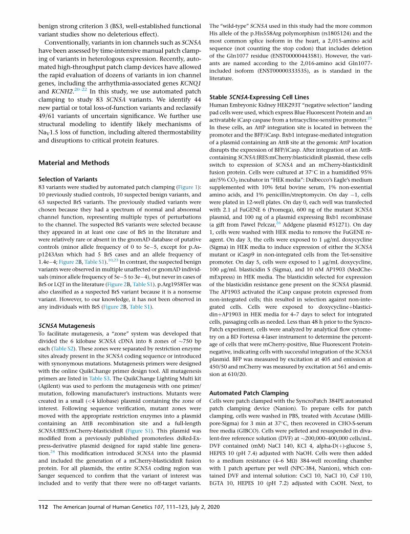
Selection of Variants83 variants were studied by automated patch clamping (Figure 1):
10 previously studied controls, 10 suspected benign variants, and
63 suspected BrS variants. The previously studied variants were
chosen because they had a spectrum of normal and abnormal
channel function, representing multiple types of perturbations
to the channel. The suspected BrS variants were selected because
they appeared in at least one case of BrS in the literature and
were relatively rare or absent in the gnomAD database of putative
controls (minor allele frequency of 0 to 5e�5, except for p.As-
p1243Asn which had 5 BrS cases and an allele frequency of
1.4e�4; Figure 2B, Table S1).10,23 In contrast, the suspected benign
variants were observed inmultiple unaffected or gnomAD individ-
uals (minor allele frequency of 5e�5 to 3e�4), but never in cases of
BrS or LQT in the literature (Figure 2B, Table S1). p.Arg1958Ter was
also classified as a suspected BrS variant because it is a nonsense
variant. However, to our knowledge, it has not been observed in
any individuals with BrS (Figure 2B, Table S1).
SCN5A MutagenesisTo facilitate mutagenesis, a ‘‘zone’’ system was developed that
divided the 6 kilobase SCN5A cDNA into 8 zones of �750 bp
each (Table S2). These zones were separated by restriction enzyme
sites already present in the SCN5A coding sequence or introduced
with synonymous mutations. Mutagenesis primers were designed
with the online QuikChange primer design tool. All mutagenesis
primers are listed in Table S3. The QuikChange Lighting Multi kit
(Agilent) was used to perform the mutagenesis with one primer/
mutation, following manufacturer’s instructions. Mutants were
created in a small (<4 kilobase) plasmid containing the zone of
interest. Following sequence verification, mutant zones were
moved with the appropriate restriction enzymes into a plasmid
containing an AttB recombination site and a full-length
SCN5A:IRES:mCherry-blasticidinR (Figure S1). This plasmid was
modified from a previously published promoterless dsRed-Ex-
press-derivative plasmid designed for rapid stable line genera-
tion.24 This modification introduced SCN5A into the plasmid
and included the generation of a mCherry-blasticidinR fusion
protein. For all plasmids, the entire SCN5A coding region was
Sanger sequenced to confirm that the variant of interest was
included and to verify that there were no off-target variants.
112 The American Journal of Human Genetics 107, 111–123, July 2, 2
The ‘‘wild-type’’ SCN5A used in this study had the more common
His allele of the p.His558Arg polymorphism (rs1805124) and the
most common splice isoform in the heart, a 2,015-amino acid
sequence (not counting the stop codon) that includes deletion
of the Gln1077 residue (ENST00000443581). However, the vari-
ants are named according to the 2,016-amino acid Gln1077-
included isoform (ENST00000333535), as is standard in the
literature.
Stable SCN5A-Expressing Cell LinesHuman Embryonic Kidney HEK293T ‘‘negative selection’’ landing
pad cells were used, which express Blue Fluorescent Protein and an
activatable iCasp caspase from a tetracycline-sensitive promoter.25
In these cells, an AttP integration site is located in between the
promoter and the BFP/iCasp. Bxb1 integrase-mediated integration
of a plasmid containing an AttB site at the genomic AttP location
disrupts the expression of BFP/iCasp. After integration of an AttB-
containing SCN5A:IRES:mCherry:blasticidinR plasmid, these cells
switch to expression of SCN5A and an mCherry-blasticidinR
fusion protein. Cells were cultured at 37�C in a humidified 95%
air/5%CO2 incubator in ‘‘HEKmedia’’: Dulbecco’s Eagle’s medium
supplemented with 10% fetal bovine serum, 1% non-essential
amino acids, and 1% penicillin/streptomycin. On day �1, cells
were plated in 12-well plates. On day 0, each well was transfected
with 2.1 ml FuGENE 6 (Promega), 600 ng of the mutant SCN5A
plasmid, and 100 ng of a plasmid expressing Bxb1 recombinase
(a gift from Pawel Pelczar,26 Addgene plasmid #51271). On day
1, cells were washed with HEK media to remove the FuGENE re-
agent. On day 3, the cells were exposed to 1 mg/mL doxycycline
(Sigma) in HEK media to induce expression of either the SCN5A
mutant or iCasp9 in non-integrated cells from the Tet-sensitive
promoter. On day 5, cells were exposed to 1 mg/mL doxycycline,
100 mg/mL blasticidin S (Sigma), and 10 nM AP1903 (MedChe-
mExpress) in HEK media. The blasticidin selected for expression
of the blasticidin resistance gene present on the SCN5A plasmid.
The AP1903 activated the iCasp caspase protein expressed from
non-integrated cells; this resulted in selection against non-inte-
grated cells. Cells were exposed to doxycyclineþblastici-
dinþAP1903 in HEK media for 4–7 days to select for integrated
cells, passaging cells as needed. Less than 48 h prior to the Syncro-
Patch experiment, cells were analyzed by analytical flow cytome-
try on a BD Fortessa 4-laser instrument to determine the percent-
age of cells that were mCherry-positive, Blue Fluorescent Protein-
negative, indicating cells with successful integration of the SCN5A
plasmid. BFP was measured by excitation at 405 and emission at
450/50 andmCherry was measured by excitation at 561 and emis-
sion at 610/20.
Automated Patch ClampingCells were patch clamped with the SyncroPatch 384PE automated
patch clamping device (Nanion). To prepare cells for patch
clamping, cells were washed in PBS, treated with Accutase (Milli-
pore-Sigma) for 3 min at 37�C, then recovered in CHO-S-serum
free media (GIBCO). Cells were pelleted and resuspended in diva-
lent-free reference solution (DVF) at �200,000–400,000 cells/mL.
DVF contained (mM) NaCl 140, KCl 4, alpha-D(þ)-glucose 5,
HEPES 10 (pH 7.4) adjusted with NaOH. Cells were then added
to a medium resistance (4–6 MU) 384-well recording chamber
with 1 patch aperture per well (NPC-384, Nanion), which con-
tained DVF and internal solution: CsCl 10, NaCl 10, CsF 110,
EGTA 10, HEPES 10 (pH 7.2) adjusted with CsOH. Next, to
020

Figure 1. Molecular and Functional Expression of SCN5A(A) Flow cytometry plot of representative wild-type stable cell line. Histogram of mCherry signal is shown. 89.8% of cells expressed ahigh-level of mCherry, indicating successful plasmid integration.(B) Boxplot of percentage of wild-type wells with NaV1.5-like current across eight independent transfections. Only wells with a cell pass-ing seal resistance and capacitance criteria (see Material and Methods) were considered.(C) Wild-type current density-voltage plot, averaged across 405 wild-type cells passing quality control (seal resistance, capacitance, involtage control, and minimum peak current criteria, see Material and Methods). The predicted reversal potential given the internaland external solutions used in this study is 45.3 mV. Error bars indicate SEM.(D) Example SyncroPatch experiment. A typical experiment studied 5 variants and wild-type on a single 384-well chip. In this experi-ment all 5 variants had wild-type-like currents. Green wells indicate a cell with seal resistance>0.5 GU, blue wells indicate a cell with sealresistance 0.2–0.5 GU (not included in analysis), and gray wells indicate no cell present or a cell with seal resistance <0.2 GU (notincluded in analysis). Randomly selected cells with seals >0.5GU are highlighted with a black square and displayed on the right.
enhance seal resistance, 50% of the DVF was exchanged with a
calcium-containing seal enhancing solution: NaCl 80, NMDG
60, KCl 4, MgCl2 1, CaCl2 10, alpha-D(þ)-glucose 5, HEPES 10
(pH 7.4) adjusted with HCl. The cells were washed three times
in external recording solution: NaCl 80, NMDG 60, KCl 4,
MgCl2 1, CaCl2 2, alpha-D(þ)-glucose 5, HEPES 10 (pH 7.4)
adjusted with HCl. Currents elicited in response to activation,
inactivation, and recovery from inactivation protocols were
then recorded (Figure S2). A late current measurement was
The Am
captured every 5 s. After 1 min, 50% of the external solution
was exchanged with external solution containing 200 mM tetra-
caine hydrochloride (Sigma; effective concentration 100 mM
tetracaine). After tetracaine addition, late current measurements
were obtained every 5 s for 1 additional minute. At least 10 cells
expressing wild-type SCN5A were included for comparison in
each SyncroPatch experiment (Figure 1), and at least 2 indepen-
dent transfections and at least 10 replicate cells were studied
per mutant. Recordings were performed at room temperature.
erican Journal of Human Genetics 107, 111–123, July 2, 2020 113
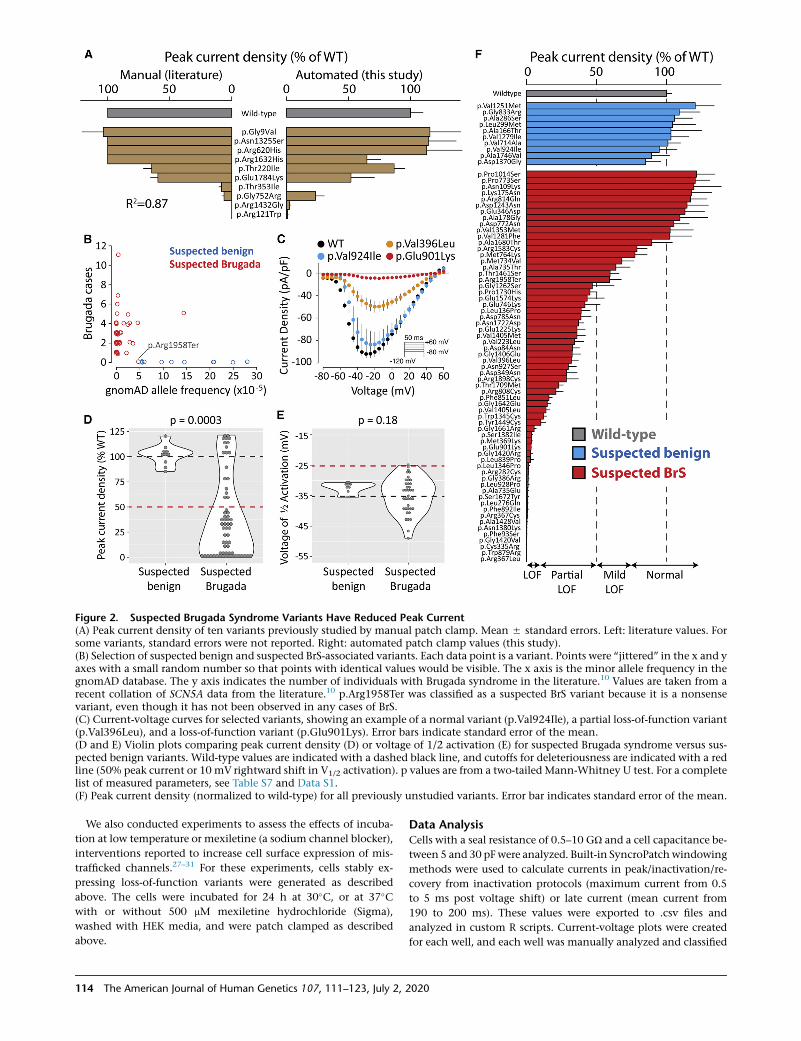
Figure 2. Suspected Brugada Syndrome Variants Have Reduced Peak Current(A) Peak current density of ten variants previously studied by manual patch clamp. Mean 5 standard errors. Left: literature values. Forsome variants, standard errors were not reported. Right: automated patch clamp values (this study).(B) Selection of suspected benign and suspected BrS-associated variants. Each data point is a variant. Points were ‘‘jittered’’ in the x and yaxes with a small random number so that points with identical values would be visible. The x axis is the minor allele frequency in thegnomAD database. The y axis indicates the number of individuals with Brugada syndrome in the literature.10 Values are taken from arecent collation of SCN5A data from the literature.10 p.Arg1958Ter was classified as a suspected BrS variant because it is a nonsensevariant, even though it has not been observed in any cases of BrS.(C) Current-voltage curves for selected variants, showing an example of a normal variant (p.Val924Ile), a partial loss-of-function variant(p.Val396Leu), and a loss-of-function variant (p.Glu901Lys). Error bars indicate standard error of the mean.(D and E) Violin plots comparing peak current density (D) or voltage of 1/2 activation (E) for suspected Brugada syndrome versus sus-pected benign variants. Wild-type values are indicated with a dashed black line, and cutoffs for deleteriousness are indicated with a redline (50% peak current or 10 mV rightward shift in V1/2 activation). p values are from a two-tailed Mann-Whitney U test. For a completelist of measured parameters, see Table S7 and Data S1.(F) Peak current density (normalized to wild-type) for all previously unstudied variants. Error bar indicates standard error of the mean.
We also conducted experiments to assess the effects of incuba-
tion at low temperature or mexiletine (a sodium channel blocker),
interventions reported to increase cell surface expression of mis-
trafficked channels.27–31 For these experiments, cells stably ex-
pressing loss-of-function variants were generated as described
above. The cells were incubated for 24 h at 30�C, or at 37�Cwith or without 500 mM mexiletine hydrochloride (Sigma),
washed with HEK media, and were patch clamped as described
above.
114 The American Journal of Human Genetics 107, 111–123, July 2, 2
Data AnalysisCells with a seal resistance of 0.5–10 GU and a cell capacitance be-
tween 5 and 30 pF were analyzed. Built-in SyncroPatchwindowing
methods were used to calculate currents in peak/inactivation/re-
covery from inactivation protocols (maximum current from 0.5
to 5 ms post voltage shift) or late current (mean current from
190 to 200 ms). These values were exported to .csv files and
analyzed in custom R scripts. Current-voltage plots were created
for each well, and each well was manually analyzed and classified
020
as normal/in voltage control (voltage-dependent current with
peak current near�20 mV), out of voltage control (voltage-depen-
dent current that rapidly jumped around �60 mV), or having no
current (<25 pA peak current). Example current-voltage curves
of these three classes are shown in Figure S3.
Only wells that were in voltage control (Figure S3) with peak cur-
rents between 100 and 2,000 pAwere used to assess additional fea-
tures, such as the voltage dependence of activation, voltage depen-
dence of inactivation, inactivation time, or recovery from
inactivation (Table S4). Additional details on peak current aver-
aging are presented in the Supplemental Methods. Only wells
with a peak current above 500 pA and wells where the seal resis-
tance and capacitance did not change by more than 10% during
addition of tetracaine (see below) were used to measure late cur-
rent. Activation and inactivation best-fit curves were calculated
for each well by fitting Boltzmann equations using the R function
nls (nonlinear least-squares). Recovery from inactivation data and
inactivation time data were fitted with exponential curves with
the nls function.Wells with high noise for which the best-fit curve
did not fit the data well (data points had >10% average deviation
from the best-fit line) were removed from the analysis. Tetracaine-
sensitive late current was calculated as the mean of 5 post-tetra-
caine raw late current values subtracted from the mean of 5 pre-
tetracaine values and was normalized to tetracaine-sensitive peak
current. Outlier values exceeding 3 standard deviations from the
mean were excluded (1.5% of all values, Table S4). Per-well V1/2
activation (voltage at which half the channels are activated), V1/
2 inactivation (voltage at which half the channels are inactivated),
time of 50% recovery from inactivation, inactivation time con-
stant, and late current parameters were averaged across all wells
meeting the above inclusion criteria. All measured parameters
with at least 5 cells meeting inclusion criteria are reported. For
many severe loss-of-function variants, there were fewer than 5
qualifying wells to accurately quantify channel parameters other
than peak current density. All comparisons of variant parameters
between groups were made in R with two-tailed t tests (t.test) or
with two-tailedMann-Whitney U tests (wilcox.testwith the param-
eter paired ¼ FALSE) when the distributions were non-normally
distributed. Differences in dispersion between groups were tested
with Levene’s Test (levene.test, car package). Violin plots were
made with geom_violin (ggplot2).
Variants were classified according to American College of Medi-
cal Genetics and Genomics (ACMG) criteria (Figure S4).19 A
custom R script was used to implement these criteria. Variant clas-
sifications pre- and post-functional data are presented in Table S1.
A cutoff of 6/�250,000 alleles in gnomAD v.2.123 was used to
determine criteria BS1 and PM2, following a previous recommen-
ded cutoff for Brugada syndrome.32 BP4 and PP3 were determined
from the consensus of PROVEAN33 and PolyPhen234 classifica-
tions. PS4 was interpreted to mean at least 5 observed individuals
with Brugada syndrome and an estimated Brugada syndrome
penetrance from literature reports whose 95% confidence interval
excluded 0.10 Variants with peak current densities between 75%
and 125% of wild-type, <10 mV shifts in activation or inactiva-
tion, <2-fold shifts in recovery from inactivation, and <1% late
current (% of peak) were considered to have normal in vitro func-
tional data (BS3). Variants with peak current densities <50% of
wild-type or a >10 mV rightward shift in V1/2 activation were
considered to have abnormal loss-of-function functional data
(PS3). Variants with >75% peak current and a late current >1%
(normalized to peak current) were considered to have abnormal
gain-of-function functional data (PS3). These cutoffs were deter-
The Am
mined from a previous analysis of the correlation between func-
tional parameters and Brugada syndrome and long QT syndrome
risk.10 These cutoffs reflect the observation that variants that are
not linked to disease often have mild perturbations to their patch
clamp parameters. ClinVar classifications35 were used to deter-
mine criteria BP6 and PP5. All literature and gnomAD case/control
counts, peak current densities, and classifications pre- and post-
patch clamp data are presented in Table S1, and ACMG criteria
used for variant classification are presented in Table S5. All variants
were considered to meet PP2 (missense variant in gene with low
rate of benignmissense variants and pathogenic missense variants
common) except p.Arg1958Ter, which was considered to meet
PM4 (protein length changing variant). For gnomAD counts, liter-
ature counts, and classifications, variants with the same outcome
on the protein sequence (due to the redundant genetic code) were
grouped together. Therefore, the PS1 criterion (same amino acid
change as established pathogenic variant) was not used. No vari-
ants satisfied the PP1 criterion (statistical co-segregation with dis-
ease in multiple affected family members) due to the low numbers
of carriers in the literature and lack of large published pedigrees for
these variants. We performed an initial round of classification
without using PM5 (missense variant at a position where a
different variant is pathogenic/likely pathogenic). Then, for each
variant, we determined the PM5 criterion by searching for variants
at the same amino acid position that were initially classified as
pathogenic/likely pathogenic. Finally, classifications were recalcu-
lated including PM5.
Homology Model and Structural CalculationsAll computational modeling was conducted in parallel to and
blinded from the experimental characterizations. Structural
models of human SCN5A (UniProtKB: Q14524-1, modeled resi-
dues: 30–440, 685–957, 1174–1887) bound with SCN1B (Uni-
ProtKB: Q07699-1, modeled residues: 20–192) were generated by
homology modeling using the protein structure prediction soft-
ware Rosetta (v.3.10).36 The cryo-EM structure of human SCN9A
bound with SCN1B and the Ig domain of SCN2B resolved to
3.2 A (PDB: 6J8H)37 was used as the primary template while the
cryo-EM structure of NavPaS from American Cockroach resolved
to 2.6 A (PDB: 6A95)38 was used as a secondary template. The
percent identity between the aligned positions of SCN9A and
SCN5A sequences was 76.7%. While the percent identity between
NavPaS and SCN5A was only moderate (45.6%), the N-terminal
and C-terminal domains in the NavPaS structure were partially
resolved, providing coordinates for modeling the corresponding
domains of SCN5A. The final model (Figures 4 and S5) covers 70
of the 73 functionally characterized variants. Three variants—
p.Pro1014Ser, p.Arg1898Cys, and p.Arg1958Ter—fall outside of
the set of modeled residues, and are therefore not covered. Addi-
tional information about the homology model and DDG (thermo-
stability) calculations are presented in the Supplemental Methods
and Table S6.
Results
Automated Patch Clamping of NaV1.5 Variants
Across multiple transfections and variants, a high percent-
age (85.4% 5 12.3% SEM) of HEK293 cells were mCherry
positive, indicating successful integration of an SCN5A
expression plasmid (Figures 1A and S1). Wild-type and
erican Journal of Human Genetics 107, 111–123, July 2, 2020 115
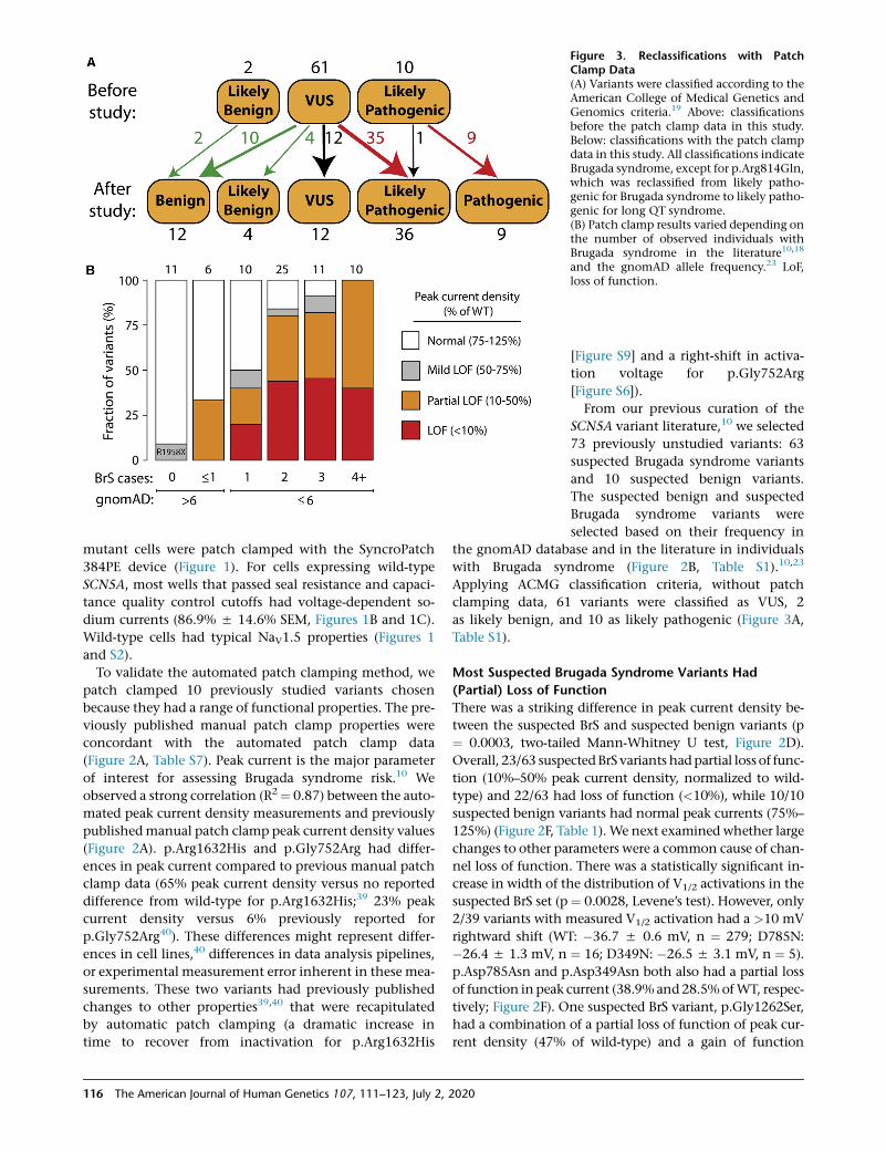
Figure 3. Reclassifications with PatchClamp Data(A) Variants were classified according to theAmerican College of Medical Genetics andGenomics criteria.19 Above: classificationsbefore the patch clamp data in this study.Below: classifications with the patch clampdata in this study. All classifications indicateBrugada syndrome, except for p.Arg814Gln,which was reclassified from likely patho-genic for Brugada syndrome to likely patho-genic for long QT syndrome.(B) Patch clamp results varied depending onthe number of observed individuals withBrugada syndrome in the literature10,18
and the gnomAD allele frequency.23 LoF,loss of function.
mutant cells were patch clamped with the SyncroPatch
384PE device (Figure 1). For cells expressing wild-type
SCN5A, most wells that passed seal resistance and capaci-
tance quality control cutoffs had voltage-dependent so-
dium currents (86.9% 5 14.6% SEM, Figures 1B and 1C).
Wild-type cells had typical NaV1.5 properties (Figures 1
and S2).
To validate the automated patch clamping method, we
patch clamped 10 previously studied variants chosen
because they had a range of functional properties. The pre-
viously published manual patch clamp properties were
concordant with the automated patch clamp data
(Figure 2A, Table S7). Peak current is the major parameter
of interest for assessing Brugada syndrome risk.10 We
observed a strong correlation (R2¼ 0.87) between the auto-
mated peak current density measurements and previously
publishedmanual patch clamp peak current density values
(Figure 2A). p.Arg1632His and p.Gly752Arg had differ-
ences in peak current compared to previous manual patch
clamp data (65% peak current density versus no reported
difference from wild-type for p.Arg1632His;39 23% peak
current density versus 6% previously reported for
p.Gly752Arg40). These differences might represent differ-
ences in cell lines,40 differences in data analysis pipelines,
or experimental measurement error inherent in these mea-
surements. These two variants had previously published
changes to other properties39,40 that were recapitulated
by automatic patch clamping (a dramatic increase in
time to recover from inactivation for p.Arg1632His
116 The American Journal of Human Genetics 107, 111–123, July 2, 2020
[Figure S9] and a right-shift in activa-
tion voltage for p.Gly752Arg
[Figure S6]).
From our previous curation of the
SCN5A variant literature,10 we selected
73 previously unstudied variants: 63
suspected Brugada syndrome variants
and 10 suspected benign variants.
The suspected benign and suspected
Brugada syndrome variants were
selected based on their frequency in
the gnomAD database and in the literature in individuals
with Brugada syndrome (Figure 2B, Table S1).10,23
Applying ACMG classification criteria, without patch
clamping data, 61 variants were classified as VUS, 2
as likely benign, and 10 as likely pathogenic (Figure 3A,
Table S1).
Most Suspected Brugada Syndrome Variants Had
(Partial) Loss of Function
There was a striking difference in peak current density be-
tween the suspected BrS and suspected benign variants (p
¼ 0.0003, two-tailed Mann-Whitney U test, Figure 2D).
Overall, 23/63 suspectedBrSvariants hadpartial loss of func-
tion (10%–50% peak current density, normalized to wild-
type) and 22/63 had loss of function (<10%), while 10/10
suspected benign variants had normal peak currents (75%–
125%) (Figure 2F, Table 1).We next examined whether large
changes to other parameters were a common cause of chan-
nel loss of function. There was a statistically significant in-
crease in width of the distribution of V1/2 activations in the
suspected BrS set (p ¼ 0.0028, Levene’s test). However, only
2/39 variants with measured V1/2 activation had a >10 mV
rightward shift (WT: �36.7 5 0.6 mV, n ¼ 279; D785N:
�26.4 5 1.3 mV, n ¼ 16; D349N: �26.5 5 3.1 mV, n ¼ 5).
p.Asp785Asn and p.Asp349Asn both also had a partial loss
of function in peak current (38.9%and28.5%ofWT, respec-
tively; Figure 2F). One suspected BrS variant, p.Gly1262Ser,
had a combination of a partial loss of function of peak cur-
rent density (47% of wild-type) and a gain of function

Table 1. Functional Class of Suspected Benign and Brugada Syndrome Variants
Functional Class Peak Current Density Suspected Benign Suspected BrS
Normal 75%–125% 10 (100%) 14 (22%)
Mild loss of function 50%–75% 0 (0%) 4 (6%)
Partial loss of function 10%–50% 0 (0%) 23 (37%)
Loss of function <10% 0 (0%) 22 (35%)
Total * 10 63
leftward shift in V1/2 activation (�12.2mV; Figure S6). Over-
all, there was no significant difference in V1/2 activation be-
tween suspected BrS and suspected benign variants (p ¼0.18,Mann-WhitneyU test, Figure 2E). Therefore, in this da-
taset, large shifts in activation gating were not a common
cause of Brugada syndrome. Surprisingly, one suspected
BrS-associated variant, p.Arg814Gln, had near wild-type-
like peak current density (117% of wild-type) and increased
late current (1.4%of peak). In addition toBrugada syndrome
cases, p.Arg814Gln has been observed in two cases of long
QTsyndrome,41 consistentwith its gain-of-function late cur-
rent phenotype. Besides these variants, nomajor differences
between suspected BrS and suspected benign variants were
observed for V1/2 inactivation, inactivation time, recovery
from inactivation, or late current (Figures S6–10, Table S7).
Despite being a nonsense variant, p.Arg1958Ter generated
substantial current (peak current density of 59.3% of wild-
type), likelydue to the fact that the stop codon is in the distal
C terminus of the protein (Figure 2F).
Some suspected BrS variants have only been observed in
only a single individual with BrS, whereas other variants
have been observed in multiple individuals. In this study,
the patch clamp phenotypes varied according to the
strength of the phenotypic evidence for Brugada syndrome
(Figure 3B). For example, 4/10 variants that have been
observed in exactly 1 case of Brugada syndrome and %6
in gnomAD had partial or complete loss of function (0%–
50% peak current). In contrast, 10/10 variants that have
been observed in at least 4 BrS1 cases and %6 in gnomAD
had partial or complete loss of function. 2/6 variants seen
in R1 cases of Brugada syndrome but also in >6 counts in
gnomAD had partial loss-of-function defects. Therefore,
variants that were more commonly observed in Brugada
syndrome cases and less frequently observed in the popu-
lation were more likely to have loss of channel function.
Reclassification of Variants with Functional Data
Whenwe implemented theACMGclassification criteria19 to
classify all studied variants without the automated patch
clamp data (Figure 3A), 60/73 variants were classified as
VUS. Based on our previous literature curation study, which
showed an elevated BrS risk for variants with peak current
density <50% of wild-type,10 we defined cutoffs for ACMG
functional criteria PS3 and BS3. Variants with <50% peak
current were considered to meet criterion PS3 (well-estab-
lished functional assays show a change), and variants with
The Am
75%–125% peak current were considered to meet criterion
BS3 (well-established functional assays show no change).
Since the phenotypic consequences of mild LoF variants
(50%–75% peak current) to BrS risk is unclear,10 these vari-
antswere not considered to satisfy either PS3 or BS3. Because
of its elevated late current and normal peak current,
p.Arg814Gln was considered to meet the PS3 criterion for
abnormal gain of function, and was reclassified from likely
pathogenic for Brugada syndrome to likely pathogenic for
long QT syndrome. p.Gly1262Ser, which had peak current
density of 47% of wild-type and a gain of function 12.2
mV leftward shift in V1/2 activation, was considered to
meet neither the PS3 nor the BS3 criteria because of the un-
certain impact of these features on BrS risk. After patch
clamping data were incorporated, 36/61 VUSs were reclassi-
fied as likely pathogenic and 14 were reclassified as likely
benign or benign. Overall, classifications were changed for
49/61 VUSs (80%) and 61/73 (84%) previously unstudied
variants. Therefore, for this set of variants, functional data
led to reclassification of the great majority of VUSs.
Partial Rescue of Some Loss-of-Function Variants
Previous studies have shown that pre-incubation of cells at
a lower temperature or with a sodium channel blocker can
partially rescue the function of some loss-of-function
SCN5A variants, typically by improving protein folding
and trafficking to the cell surface.27,29–31 Therefore, we
tested whether pre-incubating cells at 30�C or with the so-
dium channel blocker mexiletine could partially rescue so-
dium current for the 22 loss-of-function variants (<10%
normalized peak current density). Cells expressing these
variants were cultured for 24 h in usual conditions at 37�
with no added drug, at 30�C, or with 500 mM mexiletine.
Compared to usual conditions, 8/22 variants had signifi-
cantly increased peak current at 30�C and 2/22 variants
had significantly increased peak current when treated
with mexiletine (Figure S11). p.Phe892Ile and
p.Met369Lys had the largest responses to 30�C incubation,
with normalized peak current density increasing from
8.7% at 37�C to 34.8% at 30�C for p.Phe892Ile (p ¼0.006, two-tailed t test) and from 3.9% to 42.5% for
p.Met369Lys (p ¼ 0.0003; Figure S11).
Structural Basis of Loss of Function
In this study, 22 previously unstudied variants had <10%
peak current and an additional 23 variants had 10%–50%
erican Journal of Human Genetics 107, 111–123, July 2, 2020 117

Figure 4. Structural Basis of SCN5A Loss-of-Function Variants(A) Two-dimensional schematic of NaV1.5structure. All previously unstudied variantsare shown and color-coded based on peakcurrent density (white 75%–125%, gray50%–75%, orange 10%–50%, red < 10%).(B and C) Three-dimensional homologymodel of NaV1.5. Variants are colored as in(A).(D) Top-down view of WT (top) andp.Glu901Lys (bottom), as modeled usingRosetta. The lysine residue projects intothe pore, likely disrupting sodium passage.(E) View of WT and p.Cys335Arg, asmodeled using Rosetta. The WT proteinhas a disulfide bond between Cys335 (left)and Cys280 (right), which was inferredfrom the spatial proximity of these two res-idues and the fact that the correspondingresidues in the template structures alsoform a disulfide bond; this bond is disruptedby p.Cys335Arg. The disulfide bond is indi-cated with an asterisk (*).(F) Four leucine -> proline variants in thisstudy. p.Leu136Pro is a partial loss-of-func-tion variant and p.Leu839P, p.Leu928Pro,and p.Leu1340Pro are loss of function. Thestructures of these four variants were notmodeled because modeling drastic struc-tural changes involving prolines that arepart of a helix usually cannot be reliablymodeled in Rosetta. However, these variantslikely cause loss of protein function bycausing a kink in the alpha helix and pro-tein misfolding.
current. We explored the structural basis of these variants’
decreased channel function. Although the suspected BrS
variants were selected for study independent of their posi-
tion in the protein, 42/45 (partial) loss-of-function variants
were located in the four structured transmembrane do-
mains, as opposed to unstructured linker regions or the
N and C termini (Figures 4A–4C). 33/45 (73%) loss/partial
loss-of-function variants were located in the pore-forming
or pore-adjacent S5, S5-S6 linker, or S6 regions, areas that
we have previously identified as a hotspot for Brugada var-
iants.10,17 Indeed, variant distance from the pore in the
protein structure was strongly correlated with normalized
peak current density (Pearson’s r ¼ 0.54, p ¼ 1.3e–6,
Figure S12).
Many disease-causing variants cause amino acid substi-
tutions that lead to a significant perturbation to native
thermostability (|DDG|) of protein structure.42,43 To inves-
tigate the extent to which the perturbation to the native
thermostability of NaV1.5 is correlated with molecular
function, we evaluated the impact of each variant on esti-
mated thermostability relative to the wild-type structure
with a Rosetta DDG protocol.44 Normalized peak currents
118 The American Journal of Human Genetics 107, 111–123, July 2, 2
and estimated |DDG| values were significantly negatively
correlated (Pearson’s r ¼ �0.31, p ¼ 0.0092, Figure S13).
The estimated |DDG| of variants with functional effects
on peak current (peak current density < 50%, n ¼ 44, me-
dian |DDG| ¼ 2.00 kcal/mol) was significantly larger than
normal/mild loss-of-function variants (peak current den-
sity R 50%, n ¼ 26, median |DDG| ¼ 0.88 kcal/mol,
Mann-Whitney U test, p¼ 0.0031, Figure S13B). This result
suggests that variant-induced disruption of native thermo-
stability of NaV1.5 may be a major factor contributing to
compromised function.
Variants can cause complete or partial loss of function
through mechanisms other than affecting thermostability,
such as disrupting the native topology or electrostatic envi-
ronment of the pore. Using a homology model of NaV1.5,
several variants had plausible mechanisms that could
explain their loss-of-function phenotype (Figures 4D–4F).
Seven pore-lining residues in this study, p.Asp349Asn,
p.Arg367Cys, p.Arg367Leu, p.Trp879Arg, p.Phe892Ile,
p.Glu901Lys, and p.Thr1709Met, caused either complete
or partial loss of function while inducing negligible or
only minor perturbations to native thermostability
020
(Figures 4C, 4D, and S14). In particular, the residue Glu901
lines the channel pore, and in the homology model, the
loss-of-function variant p.Glu901Lys projects into the
pore, likely disrupting sodium permeation (Figure 4D).
p.Cys335Arg disrupts a disulfide bond, likely destabilizing
the tertiary structure of the protein and explaining its loss-
of-function phenotype (Figure 4E). Three loss-of-function
leucine/proline variants (p.Leu839Pro, p.Leu928Pro,
and p.Leu1346Pro) and a fourth partial loss-of-function
variant (p.Leu136Pro) likely disrupt alpha helices, as is
seen in other proteins with proline variants (Figure 4F).45
In contrast, p.Ala166Thr, a suspected benign variant
located distally from the pore and not predicted to alter
protein structure, had wild-type-like electrophysiological
properties (Figures S5B and S5C, Table S1). These data indi-
cate that structural features can help predict or explain
channel loss of function in SCN5A variants.
Discussion
Reclassification of Brugada Syndrome Variants with
Patch Clamping Data
This study identified 44 novel (partial) loss-of-function var-
iants by high-throughput, automated patch clamping. In
addition, this nearly doubles (from 24 to 46) the number
of known loss-of-function missense SCN5A variants with
<10% peak current (Table S8). As a result of the patch
clamp data, 35 novel pathogenic/likely pathogenic vari-
ants and 14 novel benign/likely benign variants were iden-
tified. Overall, 61/73 variants, including 49/61 VUSs, were
reclassified with our patch clamp data. A nonsense variant,
p.Arg1958Ter, has been observed in 13 individuals in the
gnomAD database and never in a published case of Bru-
gada syndrome. p.Arg1958Ter generated substantial cur-
rent (peak current density of 59.3% of wild-type) and likely
escapes nonsense-mediated decay because it located is in
the last exon of SCN5A. p.Arg1958Ter was not considered
to meet the PVS1 criterion (null variant) and was classified
as a VUS both pre- and post-study due to its mild loss-of-
function in vitro phenotype. Eight other nonsense and
frameshift variants in the C terminus of SCN5A are present
in the gnomAD database;23 these variants may also
generate sodium current and not have complete loss of
channel function.
A recent guide from the ClinGen consortium recom-
mended that in vitro assays that are well validated with
clearly pathogenic and benign controls and predict disease
risk well be used as PS3/BS3 at the strong level in the
ACMG classification scheme.46 Since patch clamping is
the gold standard method to assay ion channel function
and has been performed for hundreds of SCN5A variants
including (for the most part) correctly predicting the
phenotypic impact of dozens of clearly benign and patho-
genic variants, we implemented the PS3 and BS3 criteria at
the strong level for variants between 0%–50% peak current
density (PS3) and 75%–125% (BS3). These ranges were
chosen based on the correlation between peak current
The Am
and variant-specific BrS risk.10 Because of their uncertain
impact on Brugada syndrome risk, mild loss-of-function
variants (50%–75% peak current) were not considered to
meet BS3 or PS3. As a result, the classification of these var-
iants was not changed due to this study.
While each variant is rare, the collective allele frequency
of the reclassified variants in this study is 0.2% in gnomAD
(�0.4% of individuals). Although themajority of the 1,390
observed SCN5A missense variants remain VUSs,10 this
study suggests that variant properties can be used to iden-
tify a subset of SCN5A variants that are highly enriched for
altered in vitro properties and disease association. In this
study, two properties predict a high rate of in vitro loss of
function: (1) the observation of the variant in at least
one individual with BrS and (2) ultra-rare frequency in
the population (%6 in gnomAD—concordant with a previ-
ous computational estimate32). Including this study, 135
of the 276 variants with these properties have been now
studied in vitro. The remaining 141 variants are excellent
candidates for future high-throughput functional charac-
terization and possible reclassification. An important
open question is the rate of deleterious in vitro phenotypes
in the 742 ultra-rare variants (%6 in gnomAD) that have
not been observed in any BrS cases to date. Some of these
variants might not have appeared in published BrS cases
despite a true BrS risk, or carriers may present with other
SCN5A-associated arrhythmia phenotypes.47
It is important to note that SCN5A variant classification
does not completely predict Brugada syndrome risk in indi-
vidual patients. BrS is an incompletely penetrant disease,
and its presentation is influenced by demographic factors
such as age and sex,48 as well as common genetic vari-
ants—including multiple noncoding haplotypes near
SCN5A.49 Individuals with loss-of-function SCN5A variants
can present with other arrhythmias besides Brugada syn-
drome, including sick sinus syndrome,5 atrial standstill,50
or other conduction disease.51 Therefore, carriers of the
loss-of-function variants identified in this study may pre-
sent with those conditions instead of Brugada syndrome.
In addition, some ‘‘overlap’’ SCN5A variants can have
increased risk of both Brugada syndrome and long QT syn-
drome.52 One suspected BrS-associated variant in this
study, p.Arg814Gln, had late current above our cutoff
(>1% of peak current) and has appeared in the literature
in both BrS and LQT cases.41,53 Carriers of incidental path-
ogenic or likely pathogenic SCN5A variants should have
follow-up ECG screening and a clinical and family history
taken to determine each individual’s phenotype and sud-
den cardiac death risk. A common variant in SCN5A, p.Hi-
s558Arg, is present at a minor allele frequency of 22.3% in
the gnomAD database.23 This variant has been shown to
modulate the effect of some SCN5A variants, although
there is still a strong correlation between variant properties
in the His558 and Arg558 backgrounds.54 Future work will
investigate the role of the Arg558 background on the vari-
ants in this study, which may enable more precise predic-
tions of disease risk from SCN5A genotype.
erican Journal of Human Genetics 107, 111–123, July 2, 2020 119

Mechanisms of NaV1.5 Loss of Function
In this set of variants, the most common cause of channel
loss of function was a partial or total reduction in peak cur-
rent. This is consistent with previous clamp studies13–15
and our previous literature analysis which found that
SCN5A peak current was the largest single predictor of Bru-
gada syndrome risk.10 Two variants, p.Asp785Asn and
p.Asp349Asn, had a >10 mV rightward shift in V1/2 activa-
tion; however, these two variants also had <50% peak cur-
rent. Thus, while variants with large gating defects have
been previously described (e.g., p.Arg1632His39), the ma-
jor mechanism of NaV1.5 loss of function is a reduction
in peak current. Previous studies have found that channel
misfolding and a resulting cell-surface trafficking defi-
ciency is a common mechanism of loss-of-function vari-
ants in SCN5A5,27 and other ion channel genes.55 We
observed a negative correlation between peak current den-
sity and computationally predicted |DDG|, an estimate of
variant-induced perturbations to native thermostability,
consistent with other studies that showed that thermosta-
bility perturbations are a major cause of altered protein
function.56–59 In addition, structural modeling identified
several probable modes of channel dysfunction, including
pore-lining variants that likely disrupt sodium permeation,
removal of a disulfide bond, or creation of prolines that
likely disrupt alpha helices. Although these structural ana-
lyses were enlightening, structural features do not yet fully
predict channel function, highlighting the complemen-
tarity of structural modeling and empirical electrophysio-
logical measurements. Consistent with previous studies
of SCN5A variants,27–31 some but not all (8/22) SCN5A
loss-of-function variants were partially rescued by 30�
treatment or mexiletine. Although these treatments are
not practical in the clinic for treatment of Brugada syn-
drome, this result suggests that other drugs or interven-
tions may help rescue some SCN5A loss-of-function
variants.
High-Throughput Approaches to Variant Classification
High-throughput patch clamping is a promising method
for reclassifying the thousands of variants of uncertain sig-
nificance in Mendelian arrhythmia genes. Previously, Va-
noye et al.22 reclassified 23/35 ‘‘uninformative’’ KCNQ1
variants (variants of uncertain significance, variants with
conflicting interpretations, or no available data). Ng
et al.21 reclassified 13 uninformative variants in KCNH2
with patch clamping and cell surface abundance assays.
This work combines high-throughput patch clamping
and structural modeling, and explicitly incorporates
ACMG classification criteria to reclassify 49/61 SCN5A
VUSs, 35 to likely pathogenic, and 14 to benign/likely
benign. Our approach may be extended to study addi-
tional SCN5A variants, including gain-of-function long
QT-associated variants or additional variants observed in
population sequencing efforts. Accurate classification of
the large number of variants in arrhythmia-associated
genes will require integrating data from multiple different
120 The American Journal of Human Genetics 107, 111–123, July 2, 2
model systems, such as patch clamping,21,22 induced
pluripotent stem cell-derived cardiomyocytes,60–62 struc-
tural and computational models,17,63,64 and ultra-high-
throughput multiplexed assays.65–67
Limitations
This study assays variants in a heterologous expression sys-
tem. While peak current as measured in this system is the
strongest available in vitro predictor of Brugada syndrome
risk,10 it is possible for some variants to show different
properties in HEK293T cells compared to cardiomyocytes,
e.g., because of other proteins that interact with or modify
NaV1.5.68–70 This study examined only the most common
haplotype (H558) and themost common cardiac splice iso-
form of SCN5A; for some variants, it has been shown that
alternate haplotypes/isoforms canmodulate channel prop-
erties.54
Conclusion
This study used automated patch clamping to study 73 pre-
viously unstudied SCN5A variants, resulting in the reclassi-
fication of 49/61 variants of uncertain significance. This
approach can help reclassify variants in this important dis-
ease gene and improve the accuracy and scope of genetic
medicine.
Data and Code Availability
Additional data and code from this study is available upon
reasonable request from the corresponding author.
Supplemental Data
Supplemental Data can be found online at https://doi.org/10.
1016/j.ajhg.2020.05.015.
Acknowledgments
We thank Kenneth Matreyek and Douglas Fowler for sharing
HEK293 cell lines and Tim Strassmaier and Carlos Vanoye for help-
ful advice. TheNanion SyncroPatch 384PE is housed andmanaged
within the Vanderbilt High-Throughput Screening center, an
institutionally supported core, and was funded by NIH Shared
Instrumentation Grant 1S10OD025281. This research was funded
by NIH grants K99 HG010904 (A.M.G.), K99 HL135442 (B.M.K.),
R01 HL149826 (D.M.R.), and P50 GM115305 (D.M.R.) and Amer-
ican Heart Association fellowships 20POST35220002 (B.L.) and
20PRE35180088 (A.M.).
Declaration of Interests
The authors declare no competing interests.
Received: December 27, 2019
Accepted: May 19, 2020
Published: June 12, 2020
Web Resources
RCSB Protein Data Bank, http://www.rcsb.org/pdb/home/home.do
020
References
1. Kapplinger, J.D., Tester, D.J., Alders, M., Benito, B., Berthet, M.,
Brugada, J., Brugada, P., Fressart, V., Guerchicoff, A., Harris-
Kerr, C., et al. (2010). An international compendium of muta-
tions in the SCN5A-encoded cardiac sodium channel in pa-
tients referred for Brugada syndrome genetic testing. Heart
Rhythm 7, 33–46.
2. Kapplinger, J.D., Tester, D.J., Salisbury, B.A., Carr, J.L., Harris-
Kerr, C., Pollevick, G.D., Wilde, A.A.M., and Ackerman, M.J.
(2009). Spectrum and prevalence of mutations from the first
2,500 consecutive unrelated patients referred for the FAMI-
LION long QT syndrome genetic test. Heart Rhythm 6,
1297–1303.
3. Moreau, A., Gosselin-Badaroudine, P., Delemotte, L., Klein,
M.L., and Chahine,M. (2015). Gating pore currents are defects
in common with two Nav1.5 mutations in patients with
mixed arrhythmias and dilated cardiomyopathy. J. Gen. Phys-
iol. 145, 93–106.
4. Bezzina, C.R., Rook, M.B., Groenewegen, W.A., Herfst, L.J.,
van der Wal, A.C., Lam, J., Jongsma, H.J., Wilde, A.A., and
Mannens, M.M. (2003). Compound heterozygosity for muta-
tions (W156X and R225W) in SCN5A associated with severe
cardiac conduction disturbances and degenerative changes
in the conduction system. Circ. Res. 92, 159–168.
5. Gui, J., Wang, T., Jones, R.P., Trump, D., Zimmer, T., and Lei,
M. (2010). Multiple loss-of-function mechanisms contribute
to SCN5A-related familial sick sinus syndrome. PLoS ONE 5,
e10985.
6. Brugada, J., Campuzano, O., Arbelo, E., Sarquella-Brugada, G.,
and Brugada, R. (2018). Present Status of Brugada Syndrome:
JACC State-of-the-Art Review. J. Am. Coll. Cardiol. 72, 1046–
1059.
7. Hosseini, S.M., Kim, R., Udupa, S., Costain, G., Jobling, R., Lis-
ton, E., Jamal, S.M., Szybowska, M., Morel, C.F., Bowdin, S.,
et al.; National Institutes of Health Clinical Genome Resource
Consortium (2018). Reappraisal of Reported Genes for Sudden
Arrhythmic Death: Evidence-Based Evaluation of Gene Valid-
ity for Brugada Syndrome. Circulation 138, 1195–1205.
8. Brugada, P., and Brugada, J. (1992). Right bundle branch block,
persistent ST segment elevation and sudden cardiac death: a
distinct clinical and electrocardiographic syndrome. A multi-
center report. J. Am. Coll. Cardiol. 20, 1391–1396.
9. Milman, A., Andorin, A., Postema, P.G., Gourraud, J.B., Sacher,
F., Mabo, P., Kim, S.H., Maeda, S., Takahashi, Y., Kamakura, T.,
et al. (2019). Ethnic differences in patients with Brugada syn-
drome and arrhythmic events: New insights from Survey on
Arrhythmic Events in Brugada Syndrome. Heart Rhythm 16,
1468–1474.
10. Kroncke, B.M., Glazer, A.M., Smith, D.K., Blume, J.D., and Ro-
den, D.M. (2018). SCN5A (NaV1.5) Variant Functional Pertur-
bation and Clinical Presentation: Variants of a Certain Signif-
icance. Circ Genom Precis Med 11, e002095.
11. Wang, D.W., Yazawa, K., George, A.L., Jr., and Bennett, P.B.
(1996). Characterization of human cardiac Naþ channel mu-
tations in the congenital long QT syndrome. Proc. Natl. Acad.
Sci. USA 93, 13200–13205.
12. Kalia, S.S., Adelman, K., Bale, S.J., Chung, W.K., Eng, C.,
Evans, J.P., Herman, G.E., Hufnagel, S.B., Klein, T.E., Korf,
B.R., et al. (2017). Recommendations for reporting of second-
ary findings in clinical exome and genome sequencing, 2016
update (ACMG SF v2.0): a policy statement of the American
The Am
College of Medical Genetics and Genomics. Genet. Med. 19,
249–255.
13. Chen, Q., Kirsch, G.E., Zhang, D., Brugada, R., Brugada, J., Bru-
gada, P., Potenza, D., Moya, A., Borggrefe, M., Breithardt, G.,
et al. (1998). Genetic basis and molecular mechanism for idio-
pathic ventricular fibrillation. Nature 392, 293–296.
14. Deschenes, I., Baroudi, G., Berthet, M., Barde, I., Chalvidan, T.,
Denjoy, I., Guicheney, P., and Chahine, M. (2000). Electro-
physiological characterization of SCN5A mutations causing
long QT (E1784K) and Brugada (R1512W and R1432G) syn-
dromes. Cardiovasc. Res. 46, 55–65.
15. Kapplinger, J.D., Giudicessi, J.R., Ye, D., Tester, D.J., Callis, T.E.,
Valdivia, C.R., Makielski, J.C.,Wilde, A.A., and Ackerman,M.J.
(2015). Enhanced Classification of Brugada Syndrome-Associ-
ated and Long-QT Syndrome-Associated Genetic Variants in
the SCN5A-Encoded Na(v)1.5 Cardiac Sodium Channel. Circ
Cardiovasc Genet 8, 582–595.
16. Priori, S.G., Napolitano, C., Gasparini, M., Pappone, C., Della
Bella, P., Giordano, U., Bloise, R., Giustetto, C., De Nardis, R.,
Grillo, M., et al. (2002). Natural history of Brugada syndrome:
insights for risk stratification and management. Circulation
105, 1342–1347.
17. Kroncke, B.M., Mendenhall, J., Smith, D.K., Sanders, C.R.,
Capra, J.A., George, A.L., Blume, J.D., Meiler, J., and Roden,
D.M. (2019). Protein structure aids predicting functional
perturbation of missense variants in SCN5A and KCNQ1.
Comput. Struct. Biotechnol. J. 17, 206–214.
18. Kroncke, B.M., Smith, D.K., Glazer, A.M., Roden, D.M., and
Blume, J.D. (2019). A Bayesianmethod using sparse data to es-
timate penetrance of disease-associated genetic variants. bio-
Rxiv. https://doi.org/10.1101/571158.
19. Richards, S., Aziz, N., Bale, S., Bick, D., Das, S., Gastier-Foster,
J., Grody, W.W., Hegde, M., Lyon, E., Spector, E., et al.; ACMG
Laboratory Quality Assurance Committee (2015). Standards
and guidelines for the interpretation of sequence variants: a
joint consensus recommendation of the American College
of Medical Genetics and Genomics and the Association for
Molecular Pathology. Genet. Med. 17, 405–424.
20. Kang, S.K., Vanoye, C.G., Misra, S.N., Echevarria, D.M., Cal-
houn, J.D., O’Connor, J.B., Fabre, K.L., McKnight, D., Dem-
mer, L., Goldenberg, P., et al. (2019). Spectrum of KV2.1
dysfunction in KCNB1-associated neurodevelopmental disor-
ders. Ann. Neurol. 86, 899–912.
21. Ng, C.A., Perry, M.D., Liang,W., Smith, N.J., Foo, B., Shrier, A.,
Lukacs, G.L., Hill, A.P., and Vandenberg, J.I. (2020). High-
throughput phenotyping of heteromeric human ether-a-go-
go-related gene potassium channel variants can discriminate
pathogenic from rare benign variants. Heart Rhythm 17,
492–500.
22. Vanoye, C.G., Desai, R.R., Fabre, K.L., Gallagher, S.L., Potet, F.,
DeKeyser, J.M., Macaya, D., Meiler, J., Sanders, C.R., and
George, A.L., Jr. (2018). High-Throughput Functional Evalua-
tion of KCNQ1 Decrypts Variants of Unknown Significance.
Circ Genom Precis Med 11, e002345.
23. Lek, M., Karczewski, K.J., Minikel, E.V., Samocha, K.E., Banks,
E., Fennell, T., O’Donnell-Luria, A.H., Ware, J.S., Hill, A.J.,
Cummings, B.B., et al.; Exome Aggregation Consortium
(2016). Analysis of protein-coding genetic variation in
60,706 humans. Nature 536, 285–291.
24. Matreyek, K.A., Stephany, J.J., and Fowler, D.M. (2017). A plat-
form for functional assessment of large variant libraries in
mammalian cells. Nucleic Acids Res. 45, e102.
erican Journal of Human Genetics 107, 111–123, July 2, 2020 121

25. Matreyek, K.A., Stephany, J.J., Chiasson, M.A., Hasle, N., and
Fowler, D.M. (2020). An Improved Platform for Functional
Assessment of Large Protein Libraries in Mammalian Cells.
Nucleic Acids Res. 48, e1.
26. Hermann, M., Stillhard, P., Wildner, H., Seruggia, D., Kapp, V.,
Sanchez-Iranzo, H., Mercader, N., Montoliu, L., Zeilhofer,
H.U., and Pelczar, P. (2014). Binary recombinase systems for
high-resolution conditional mutagenesis. Nucleic Acids Res.
42, 3894–3907.
27. Clatot, J., Ziyadeh-Isleem, A., Maugenre, S., Denjoy, I., Liu, H.,
Dilanian, G., Hatem, S.N., Deschenes, I., Coulombe, A., Gui-
cheney, P., and Neyroud, N. (2012). Dominant-negative effect
of SCN5A N-terminal mutations through the interaction of
Na(v)1.5 a-subunits. Cardiovasc. Res. 96, 53–63.
28. Makiyama, T., Akao, M., Tsuji, K., Doi, T., Ohno, S., Takenaka,
K., Kobori, A., Ninomiya, T., Yoshida, H., Takano, M., et al.
(2005). High risk for bradyarrhythmic complications in pa-
tients with Brugada syndrome caused by SCN5A gene muta-
tions. J. Am. Coll. Cardiol. 46, 2100–2106.
29. Pfahnl, A.E., Viswanathan, P.C., Weiss, R., Shang, L.L., Sanyal,
S., Shusterman, V., Kornblit, C., London, B., and Dudley, S.C.,
Jr. (2007). A sodium channel pore mutation causing Brugada
syndrome. Heart Rhythm 4, 46–53.
30. Valdivia, C.R., Ackerman, M.J., Tester, D.J., Wada, T., McCor-
mack, J., Ye, B., and Makielski, J.C. (2002). A novel SCN5A
arrhythmia mutation, M1766L, with expression defect
rescued by mexiletine. Cardiovasc. Res. 55, 279–289.
31. Valdivia, C.R., Tester, D.J., Rok, B.A., Porter, C.B., Munger,
T.M., Jahangir, A., Makielski, J.C., and Ackerman, M.J.
(2004). A trafficking defective, Brugada syndrome-causing
SCN5A mutation rescued by drugs. Cardiovasc. Res. 62, 53–
62.
32. Whiffin, N., Minikel, E., Walsh, R., O’Donnell-Luria, A.H.,
Karczewski, K., Ing, A.Y., Barton, P.J.R., Funke, B., Cook, S.A.,
MacArthur, D., and Ware, J.S. (2017). Using high-resolution
variant frequencies to empower clinical genome interpreta-
tion. Genet. Med. 19, 1151–1158.
33. Choi, Y., and Chan, A.P. (2015). PROVEAN web server: a tool
to predict the functional effect of amino acid substitutions
and indels. Bioinformatics 31, 2745–2747.
34. Adzhubei, I.A., Schmidt, S., Peshkin, L., Ramensky, V.E., Gera-
simova, A., Bork, P., Kondrashov, A.S., and Sunyaev, S.R.
(2010). A method and server for predicting damaging
missense mutations. Nat. Methods 7, 248–249.
35. Landrum, M.J., Lee, J.M., Benson, M., Brown, G., Chao, C.,
Chitipiralla, S., Gu, B., Hart, J., Hoffman, D., Hoover, J.,
et al. (2016). ClinVar: public archive of interpretations of clin-
ically relevant variants. Nucleic Acids Res. 44 (D1), D862–
D868.
36. Leaver-Fay, A., Tyka, M., Lewis, S.M., Lange, O.F., Thompson,
J., Jacak, R., Kaufman, K., Renfrew, P.D., Smith, C.A., Sheffler,
W., et al. (2011). ROSETTA3: an object-oriented software suite
for the simulation and design of macromolecules. Methods
Enzymol. 487, 545–574.
37. Shen, H., Liu, D., Wu, K., Lei, J., and Yan, N. (2019). Structures
of human Nav1.7 channel in complex with auxiliary subunits
and animal toxins. Science 363, 1303–1308.
38. Shen, H., Li, Z., Jiang, Y., Pan, X., Wu, J., Cristofori-Armstrong,
B., Smith, J.J., Chin, Y.K.Y., Lei, J., Zhou, Q., et al. (2018). Struc-
tural basis for the modulation of voltage-gated sodium chan-
nels by animal toxins. Science 362, 362.
122 The American Journal of Human Genetics 107, 111–123, July 2, 2
39. Benson, D.W., Wang, D.W., Dyment, M., Knilans, T.K., Fish,
F.A., Strieper, M.J., Rhodes, T.H., and George, A.L., Jr. (2003).
Congenital sick sinus syndrome caused by recessivemutations
in the cardiac sodium channel gene (SCN5A). J. Clin. Invest.
112, 1019–1028.
40. Potet, F., Mabo, P., Le Coq, G., Probst, V., Schott, J.J., Airaud, F.,
Guihard, G., Daubert, J.C., Escande, D., and Le Marec, H.
(2003). Novel brugada SCN5Amutation leading to ST segment
elevation in the inferior or the right precordial leads.
J. Cardiovasc. Electrophysiol. 14, 200–203.
41. Itoh, H., Berthet, M., Fressart, V., Denjoy, I., Maugenre, S.,
Klug, D., Mizusawa, Y., Makiyama, T., Hofman, N., Stallmeyer,
B., et al. (2016). Asymmetry of parental origin in long QT syn-
drome: preferential maternal transmission of KCNQ1 variants
linked to channel dysfunction. Eur. J. Hum. Genet. 24, 1160–
1166.
42. Yue, P., Li, Z., and Moult, J. (2005). Loss of protein structure
stability as a major causative factor in monogenic disease.
J. Mol. Biol. 353, 459–473.
43. Stein, A., Fowler, D.M., Hartmann-Petersen, R., and Lindorff-
Larsen, K. (2019). Biophysical and Mechanistic Models for
Disease-Causing Protein Variants. Trends Biochem. Sci. 44,
575–588.
44. Park, H., Bradley, P., Greisen, P., Jr., Liu, Y., Mulligan, V.K., Kim,
D.E., Baker, D., and DiMaio, F. (2016). Simultaneous Optimi-
zation of Biomolecular Energy Functions on Features from
Small Molecules and Macromolecules. J. Chem. Theory Com-
put. 12, 6201–6212.
45. Kim,M.K., and Kang, Y.K. (1999). Positional preference of pro-
line in alpha-helices. Protein Sci. 8, 1492–1499.
46. Brnich, S.E., Abou Tayoun, A.N., Couch, F.J., Cutting, G.R.,
Greenblatt, M.S., Heinen, C.D., Kanavy, D.M., Luo, X.,
McNulty, S.M., Starita, L.M., et al.; Clinical Genome Resource
Sequence Variant Interpretation Working Group (2019). Rec-
ommendations for application of the functional evidence
PS3/BS3 criterion using the ACMG/AMP sequence variant
interpretation framework. Genome Med. 12, 3.
47. Wilde, A.A.M., and Amin, A.S. (2018). Clinical Spectrum of
SCN5A Mutations: Long QT Syndrome, Brugada Syndrome,
and Cardiomyopathy. JACC Clin. Electrophysiol. 4, 569–579.
48. Milman, A., Gourraud, J.B., Andorin, A., Postema, P.G., Sacher,
F., Mabo, P., Conte, G., Giustetto, C., Sarquella-Brugada, G.,
Hochstadt, A., et al. (2018). Gender differences in patients
with Brugada syndrome and arrhythmic events: Data from a
survey on arrhythmic events in 678 patients. Heart Rhythm
15, 1457–1465.
49. Bezzina, C.R., Barc, J., Mizusawa, Y., Remme, C.A., Gourraud,
J.B., Simonet, F., Verkerk, A.O., Schwartz, P.J., Crotti, L., Dag-
radi, F., et al. (2013). Common variants at SCN5A-SCN10A
and HEY2 are associated with Brugada syndrome, a rare dis-
ease with high risk of sudden cardiac death. Nat. Genet. 45,
1044–1049.
50. Takehara, N., Makita, N., Kawabe, J., Sato, N., Kawamura, Y.,
Kitabatake, A., and Kikuchi, K. (2004). A cardiac sodium chan-
nel mutation identified in Brugada syndrome associated with
atrial standstill. J. Intern. Med. 255, 137–142.
51. Probst, V., Allouis, M., Sacher, F., Pattier, S., Babuty, D., Mabo,
P., Mansourati, J., Victor, J., Nguyen, J.M., Schott, J.J., et al.
(2006). Progressive cardiac conduction defect is the prevailing
phenotype in carriers of a Brugada syndrome SCN5A muta-
tion. J. Cardiovasc. Electrophysiol. 17, 270–275.
020
52. Remme, C.A., Wilde, A.A., and Bezzina, C.R. (2008). Cardiac
sodium channel overlap syndromes: different faces of
SCN5A mutations. Trends Cardiovasc. Med. 18, 78–87.
53. Frigo, G., Rampazzo, A., Bauce, B., Pilichou, K., Beffagna, G.,
Danieli, G.A., Nava, A., and Martini, B. (2007). Homozygous
SCN5A mutation in Brugada syndrome with monomorphic
ventricular tachycardia and structural heart abnormalities.
Europace 9, 391–397.
54. Makielski, J.C., Ye, B., Valdivia, C.R., Pagel, M.D., Pu, J., Tester,
D.J., and Ackerman, M.J. (2003). A ubiquitous splice variant
and a common polymorphism affect heterologous expression
of recombinant human SCN5A heart sodium channels. Circ.
Res. 93, 821–828.
55. Anderson, C.L., Delisle, B.P., Anson, B.D., Kilby, J.A., Will,
M.L., Tester, D.J., Gong, Q., Zhou, Z., Ackerman, M.J., and
January, C.T. (2006). Most LQT2 mutations reduce Kv11.1
(hERG) current by a class 2 (trafficking-deficient) mechanism.
Circulation 113, 365–373.
56. Casadio, R., Vassura, M., Tiwari, S., Fariselli, P., and Luigi Mar-
telli, P. (2011). Correlating disease-related mutations to their
effect on protein stability: a large-scale analysis of the human
proteome. Hum. Mutat. 32, 1161–1170.
57. DePristo, M.A., Weinreich, D.M., and Hartl, D.L. (2005).
Missense meanderings in sequence space: a biophysical view
of protein evolution. Nat. Rev. Genet. 6, 678–687.
58. Tokuriki, N., and Tawfik, D.S. (2009). Stability effects of muta-
tions and protein evolvability. Curr. Opin. Struct. Biol. 19,
596–604.
59. Zhou, Y., and Bowie, J.U. (2000). Building a thermostable
membrane protein. J. Biol. Chem. 275, 6975–6979.
60. Chavali, N.V., Kryshtal, D.O., Parikh, S.S., Wang, L., Glazer,
A.M., Blackwell, D.J., Kroncke, B.M., Shoemaker, M.B., and
Knollmann, B.C. (2019). Patient-independent human
induced pluripotent stem cell model: A new tool for rapid
determination of genetic variant pathogenicity in long QT
syndrome. Heart Rhythm 16, 1686–1695.
61. Fatima, A., Kaifeng, S., Dittmann, S., Xu, G., Gupta, M.K.,
Linke, M., Zechner, U., Nguemo, F., Milting, H., Farr, M.,
et al. (2013). The disease-specific phenotype in cardiomyo-
cytes derived from induced pluripotent stem cells of two
long QT syndrome type 3 patients. PLoS ONE 8, e83005.
62. Selga, E., Sendfeld, F., Martinez-Moreno, R., Medine, C.N.,
Tura-Ceide, O., Wilmut, S.I., Perez, G.J., Scornik, F.S., Brugada,
The Am
R., and Mills, N.L. (2018). Sodium channel current loss of
function in induced pluripotent stem cell-derived cardiomyo-
cytes from a Brugada syndrome patient. J. Mol. Cell. Cardiol.
114, 10–19.
63. Heyne, H.O., Baez-Nieto, D., Iqbal, S., Palmer, D., Brunklaus,
A., the Epi25 Collaborative, Johannesen, K.M., Lauxmann, S.,
Lemke, J.R., Moller, R.S., et al. (2019). Predicting functional
effects of missense variants in the voltage-gated sodium
and calcium channels. bioRxiv. https://doi.org/10.1101/
671453.
64. Li, B., Mendenhall, J.L., Kroncke, B.M., Taylor, K.C., Huang,
H., Smith, D.K., Vanoye, C.G., Blume, J.D., George, A.L., Jr.,
Sanders, C.R., and Meiler, J. (2017). Predicting the Functional
Impact of KCNQ1 Variants of Unknown Significance. Circ
Cardiovasc Genet 10, 10.
65. Findlay, G.M., Daza, R.M., Martin, B., Zhang, M.D., Leith, A.P.,
Gasperini, M., Janizek, J.D., Huang, X., Starita, L.M., and
Shendure, J. (2018). Accurate classification of BRCA1 variants
with saturation genome editing. Nature 562, 217–222.
66. Glazer, A.M., Kroncke, B.M., Matreyek, K.A., Yang, T., Wada,
Y., Shields, T., Salem, J.E., Fowler, D.M., and Roden, D.M.
(2020). Deep Mutational Scan of an SCN5A voltage sensor.
Circ Genom Precis Med 13, e002786.
67. Matreyek, K.A., Starita, L.M., Stephany, J.J., Martin, B., Chias-
son, M.A., Gray, V.E., Kircher, M., Khechaduri, A., Dines, J.N.,
Hause, R.J., et al. (2018). Multiplex assessment of protein
variant abundance by massively parallel sequencing. Nat.
Genet. 50, 874–882.
68. Aiba, T., Farinelli, F., Kostecki, G., Hesketh, G.G., Edwards, D.,
Biswas, S., Tung, L., and Tomaselli, G.F. (2014). A mutation
causing Brugada syndrome identifies a mechanism for altered
autonomic and oxidant regulation of cardiac sodium currents.
Circ Cardiovasc Genet 7, 249–256.
69. Casini, S., Albesa, M.,Wang, Z., Portero, V., Ross-Kaschitza, D.,
Rougier, J.S., Marchal, G.A., Chung, W.K., Bezzina, C.R.,
Abriel, H., and Remme, C.A. (2019). Functional Consequences
of the SCN5A-p.Y1977N Mutation within the PY Ubiquityla-
tion Motif: Discrepancy between HEK293 Cells and Trans-
genic Mice. Int. J. Mol. Sci. 20, 11.
70. Clatot, J., Hoshi, M., Wan, X., Liu, H., Jain, A., Shinlapawit-
tayatorn, K., Marionneau, C., Ficker, E., Ha, T., and Deschenes,
I. (2017). Voltage-gated sodium channels assemble and gate as
dimers. Nat. Commun. 8, 2077.
erican Journal of Human Genetics 107, 111–123, July 2, 2020 123