high resolution zone-doubled fresnel zone … vila comamala.pdf · high resolution zone-doubled...

1
HIGH RESOLUTION ZONE-DOUBLED FRESNEL ZONE PLATES FOR THE MULTI-keV REGIME J. Vila-Comamala 1,2 , A. Diaz 1 , M. Guizar-Sicairos 1 , R. Barrett 3 , S. Gorelick 4 , C. M. Kewish 5 V. A. Guzenko 1 , P. Karvinen 1 , E. Färm 6 , M. Ritala 6 and C. David 1 1 Paul Scherrer Institut, CH-5232 Villigen PSI, Switzerland 2 X-ray Science Division, Argonne National Laboratory, Argonne IL 60439, USA 3 ESRF, F-38043 Grenoble Cedex 9, France 4 VTT Technical Research Centre of Finland, P.O. Box 1000, FI-02044 VTT, Finland 5 Synchrotron SOLEIL, F-91192 Gif-sur-Yvette, France 6 Department of Chemistry, University of Helsinki, FI-00014 Helsinki, Finland *[email protected] X-ray microscopy based on Fresnel zone plates is a powerful technique for sub-100 nm resolution imaging of biological and inorganic materials. Here, we report on the modeling, fabrication and characterization of zone- doubled Fresnel zone plates for the multi-keV regime (412 keV). We demonstrate unprecedented spatial resolution by resolving 15 nm lines and spaces in scanning transmission X-ray microscopy, and focusing diffraction efficiencies of 7.5 % at 6.2 keV photon energy. These developments represent a significant step towards 10 nm spatial resolution for hard X-ray energies of up to 12 keV. In addition to conventional characterization techniques, we have used ptychographic scanning coherent diffractive imaging to obtain a 3D reconstruction of the hard X-ray beam focus of a high resolution Fresnel zone plate with an outermost zone width of 20 nm, with high spatial resolution and dynamic range. Figure. Scanning electron micrographs of a zone-doubled FZP made of HSQ resist and iridium (100 μm diameter, outermost zone width of w = Δr = 20 nm and height of h ~ 550 nm). (a) and (b) show overviews of the diffractive X-ray lens. (c) High magnification top view of the outermost region consisting of 20 nm-wide lines made of HSQ resist in 80 nm-period before the iridium coating. (d) High magnification top view of the zone-doubled structure after the iridium coating by atomic layer deposition. Tilted views of the edge of the zone- doubled FZP (e) before and (f) after the iridium deposition (tilt angle of 50º). The final structure contains an effective iridium grating made of 20 nm lines and spaces with an aspect ratio of h/w > 27.5.

Upload: duonganh

Post on 20-Feb-2018

221 views

Category:

Documents


2 download

TRANSCRIPT

HIGH RESOLUTION ZONE-DOUBLED FRESNEL ZONE PLATES FOR THE MULTI-keV REGIME

J. Vila-Comamala1,2, A. Diaz1, M. Guizar-Sicairos1, R. Barrett3, S. Gorelick4, C. M. Kewish5

V. A. Guzenko1, P. Karvinen1, E. Färm6, M. Ritala6 and C. David1

1Paul Scherrer Institut, CH-5232 Villigen PSI, Switzerland 2X-ray Science Division, Argonne National Laboratory, Argonne IL 60439, USA

3ESRF, F-38043 Grenoble Cedex 9, France 4VTT Technical Research Centre of Finland, P.O. Box 1000, FI-02044 VTT, Finland

5Synchrotron SOLEIL, F-91192 Gif-sur-Yvette, France 6Department of Chemistry, University of Helsinki, FI-00014 Helsinki, Finland

*[email protected]

X-ray microscopy based on Fresnel zone plates is a powerful technique for sub-100 nm resolution imaging of biological and inorganic materials. Here, we report on the modeling, fabrication and characterization of zone-doubled Fresnel zone plates for the multi-keV regime (4―12 keV). We demonstrate unprecedented spatial resolution by resolving 15 nm lines and spaces in scanning transmission X-ray microscopy, and focusing diffraction efficiencies of 7.5 % at 6.2 keV photon energy. These developments represent a significant step towards 10 nm spatial resolution for hard X-ray energies of up to 12 keV. In addition to conventional characterization techniques, we have used ptychographic scanning coherent diffractive imaging to obtain a 3D reconstruction of the hard X-ray beam focus of a high resolution Fresnel zone plate with an outermost zone width of 20 nm, with high spatial resolution and dynamic range.

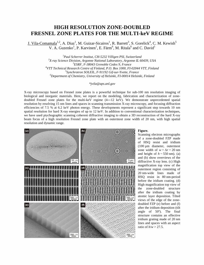
Figure. Scanning electron micrographs of a zone-doubled FZP made of HSQ resist and iridium (100 µm diameter, outermost zone width of w = ∆r = 20 nm and height of h ~ 550 nm). (a) and (b) show overviews of the diffractive X-ray lens. (c) High magnification top view of the outermost region consisting of 20 nm-wide lines made of HSQ resist in 80 nm-period before the iridium coating. (d) High magnification top view of the zone-doubled structure after the iridium coating by atomic layer deposition. Tilted views of the edge of the zone-doubled FZP (e) before and (f) after the iridium deposition (tilt angle of 50º). The final structure contains an effective iridium grating made of 20 nm lines and spaces with an aspect ratio of h/w > 27.5.