health effects support document for perfluorooctane...
TRANSCRIPT

Health Effects Support Document for
Perfluorooctane Sulfonate (PFOS)
United States Environmental Protection Agency
Office of Water Mail Code 4304T
EPA 822-R-16-002 May 2016
Perfluorooctane sulfonate (PFOS) –May 2016 i

Health Effects Support Document
for
Perfluorooctane Sulfonate (PFOS)
U.S. Environmental Protection Agency Office of Water (4304T)
Health and Ecological Criteria Division Washington, DC 20460
http://www.epa.gov/dwstandardsregulations/drinking-water-contaminant-human-health-effects-
information.
EPA Document Number: 822-R-16-002 May 2016
Perfluorooctane sulfonate (PFOS) –May 2016 ii

BACKGROUND
The Safe Drinking Water Act (SDWA), as amended in 1996, requires the Administrator of the U.S. Environmental Protection Agency (EPA) to establish a list of unregulated microbiological and chemical contaminants known or anticipated to occur in public water systems and that might require control in the future through national primary drinking water regulations. The SDWA also requires the Agency to make regulatory determinations on at least five contaminants on the Contaminant Candidate List (CCL) every 5 years. For each contaminant on the CCL, before EPA makes a regulatory determination, the Agency needs to obtain sufficient data to conduct analyses on the extent to which the contaminant occurs and the risk it poses to populations via drinking water. Ultimately, this information will assist the Agency in determining the most appropriate course of action in relation to the contaminant (e.g., developing a regulation to control it in drinking water, developing guidance, or deciding not to regulate it).
The PFOS health assessment was initiated by the Office of Water, Office of Science and Technology in 2009. The draft Health Effects Support Document for Perfluorooctane Sulfonate Acid (PFOS) was completed in 2013 and released for public comment in February 2014. An external peer-review panel meeting was held on August 21 and 22, 2014. The final document reflects input from the panel as well as public comments received on the draft document. Both the peer-reviewed draft and this document include only the sections of a health effects support document (HESD) that cover the toxicokinetics and health effects of PFOS. If a decision is made to regulate the contaminant, this document will be expanded.
One of the challenges inherent in conducting this assessment was the wealth of experimental data published before and during its development. This section provides a synopsis of the approach used in identifying and selecting the publications reflected in the final assessment.
Data were identified through the following:
Monthly/bimonthly literature searches conducted by EPA library staff (2009–2015) and New Jersey Department of Environmental Protection library staff (2012–2015).
• Papers identified by EPA internal and external peer reviewers. • Papers identified through public comments on the draft assessments. • Papers submitted to EPA by the public.
In mid-2013, the EPA library searches were expanded to cover other members of the perfluorocarboxylic acids (C-4 to C-12) and sulfonate families (C-4, C-6, C-8). Appendix A describes the literature search strategy used by the libraries. Through the literature search, documents were identified for retrieval, review, and inclusion in the HESD using the following criteria:
• The study examines a toxicity endpoint or population not examined by studies already included in the draft document.
• Aspects of the study design such as the size of the population exposed or quantification approach make it superior to key studies already included in the draft document.
• The data contribute substantially to the weight of evidence for any of the toxicity endpoints covered by the draft document.
• Elements of the study design merit its inclusion in the draft document based on its contribution to the mode of action or the quantification approach.
Perfluorooctane sulfonate (PFOS) –May 2016 iii

• The study elucidates the mode of action for any toxicity endpoint or toxicokinetic property associated with PFOS exposure.
• The effects observed differ from those in other studies with comparable protocols.
In addition to each publication being evaluated against the criteria above, the relevance of the study to drinking water exposures and to the U.S. population also were considered.
The studies included in the final draft were determined to provide the most current and comprehensive description of the toxicological properties of PFOS and the risk it poses to humans exposed to it in their drinking water. Appendix B summarizes the studies evaluated for inclusion in the HESD following the August 2014 peer review and identifies those selected for inclusion in the final assessment. Appendix B includes epidemiology data that provide a high-level summary of the outcomes across the studies evaluated.
Development of the hazard identification and dose-response assessment for PFOS has followed the general guidelines for risk assessment forth by the National Research Council (1983) and EPA’s Framework for Human Health Risk Assessment to Inform Decision Making (USEPA 2014a). Other EPA guidelines used in the development of this assessment include the following:
• Guidelines for the Health Risk Assessment of Chemical Mixtures (USEPA 1986a) • Guidelines for Mutagenicity Risk Assessment (USEPA 1986b) • Recommendations for and Documentation of Biological Values for Use in Risk
Assessment (USEPA 1988) • Guidelines for Developmental Toxicity Risk Assessment (USEPA 1991) • Interim Policy for Particle Size and Limit Concentration Issues in Inhalation Toxicity
Studies (USEPA 1994a) • Methods for Derivation of Inhalation Reference Concentrations and Application of
Inhalation Dosimetry (USEPA 1994b) • Use of the Benchmark Dose Approach in Health Risk Assessment (USEPA 1995) • Guidelines for Reproductive Toxicity Risk Assessment (USEPA 1996) • Guidelines for Neurotoxicity Risk Assessment (USEPA 1998) • Science Policy Council Handbook: Peer Review (2nd edition) (USEPA 2000a) • Supplemental Guidance for Conducting Health Risk Assessment of Chemical Mixtures
(USEPA 2000b) • A Review of the Reference Dose and Reference Concentration Processes (USEPA 2002) • Guidelines for Carcinogen Risk Assessment (USEPA 2005a) • Supplemental Guidance for Assessing Susceptibility from Early-Life Exposure to
Carcinogens (USEPA 2005b) • Science Policy Council Handbook: Peer Review (USEPA 2006a) • A Framework for Assessing Health Risks of Environmental Exposures to Children
(USEPA 2006b) • Highlights of the Exposure Factors Handbook (USEPA 2011) • Benchmark Dose Technical Guidance Document (USEPA 2012) • Child-Specific Exposure Scenarios Examples (USEPA 2014b)
Perfluorooctane sulfonate (PFOS) –May 2016 iv
AUTHORS, CONTRIBUTORS, AND REVIEWERS
Joyce Morrissey Donohue, Ph.D. (Chemical Manager) Office of Water U.S. Environmental Protection Agency, Washington, D.C.
Amal Mahfouz, Ph.D. (Chemical Manager, pre-retirement). Office of Water U.S. Environmental Protection Agency, Washington, D.C.
Tina Moore Duke, M.S. (previously with Office of Water, U.S. Environmental Protection Agency)
John Wambaugh, Ph.D. Office of Research and Development U.S. Environmental Protection Agency, Research Triangle Park, NC
The following contractor authors supported the development of this document:
Dana F. Glass-Mattie, D.V.M. Environmental Sciences Division Oak Ridge National Laboratory, Oak Ridge, TN
Carol S. Wood, Ph.D., D.A.B.T. Environmental Sciences Division Oak Ridge National Laboratory, Oak Ridge, TN
This document was prepared under the U.S. EPA Contract No. DW-8992342701, Work Assignment No. 2011-001 with Oak Ridge National Laboratory. The Lead U.S. EPA Scientist is Joyce Morrissey Donohue, Ph.D., Health and Ecological Criteria Division, Office of Science and Technology, Office of Water.
The Oak Ridge National Laboratory is managed and operated by UT-Battelle, LLC., for the U.S. Department of Energy under Contract No. DE-AC05-00OR22725.
CONTRIBUTORS AND REVIEWERS
Internal Contributors and Reviewers
Office of Water, U.S. Environmental Protection Agency Elizabeth Doyle, Ph.D. (retired) Edward Hackett
Office of Research and Development, U.S. Environmental Protection Agency Glinda Cooper, Ph.D. Barbara Glenn, Ph.D. Erin Hines, Ph.D. Christopher Lau, Ph.D. Matthew Lorber, Ph.D. Jaqueline Moya Linda Phillips, Ph.D.
Perfluorooctane sulfonate (PFOS) –May 2016 v

Paul White, Ph.D. Michael Wright, Sc.D.
Office of Chemical Safety and Pollution Prevention, U.S. Environmental Protection Agency E. Laurence Libelo Andrea Pfehales-Hutchens, Ph.D. Tracy Williamson David Lai, Ph.D. (retired) Jennifer Seed, Ph.D. (retired)
Office of Children’s Health Protection, U.S. Environmental Protection Agency Gregory Miller
Office of Land and Emergency Management, U.S. Environmental Protection Agency
External Reviewers
James Bruckner, Ph.D. Department of Pharmacology and Toxicology University of Georgia, Athens, GA
Deborah Cory-Slechta, Ph.D. Department of Environmental Medicine University of Rochester Medical Center, Rochester, NY
Jamie DeWitt, Ph.D. Pharmacology and Toxicology East Carolina University, Greenville, NC
Jeffrey Fisher, Ph.D. Biochemical Toxicology, National Center for Toxicological Research U.S. Food and Drug Administration, Jefferson, AK
William Hayton, Ph.D. College of Pharmacy (Emeritus) The Ohio State University, Columbus, OH
Matthew Longnecker, M.D., Sc.D. Biomarker-based Epidemiology Group National Institute of Environmental Health Sciences, Research Triangle Park, NC
Angela Slitt, Ph.D. Biomedical and Pharmaceutical Sciences University of Rhode Island, Kingston, RI
Perfluorooctane sulfonate (PFOS) –May 2016 vi

CONTENTS
BACKGROUND ........................................................................................................................... iii
ABBREVIATIONS AND ACRONYMS .................................................................................... xiii
EXECUTIVE SUMMARY .......................................................................................................ES-1
1. IDENTITY: CHEMICAL AND PHYSICAL PROPERTIES .............................................. 1-1
2. TOXICOKINETICS ............................................................................................................. 2-1
2.1 Absorption.................................................................................................................... 2-1 2.1.1 Oral Exposure ........................................................................................................ 2-1 2.1.2 Inhalation Exposure ............................................................................................... 2-2 2.1.3 Dermal Exposure ................................................................................................... 2-2
2.2 Distribution .................................................................................................................. 2-2 2.2.1 Oral Exposure ........................................................................................................ 2-4 2.2.2 Inhalation and Dermal Exposure ......................................................................... 2-15 2.2.3 Other Routes of Exposure .................................................................................... 2-15
2.3 Metabolism ................................................................................................................ 2-16
2.4 Excretion .................................................................................................................... 2-17 2.4.1 Oral Exposure ...................................................................................................... 2-17 2.4.2 Inhalation Exposure ............................................................................................. 2-19
2.5 Pharmacokinetic Considerations ................................................................................ 2-20 2.5.1 Pharmacokinetic models ...................................................................................... 2-20 2.5.2 Half-life data ........................................................................................................ 2-30 2.5.3 Volume of Distribution Data ............................................................................... 2-34
2.6 Toxicokinetic Summary ............................................................................................. 2-36
3. HAZARD IDENTIFICATION ............................................................................................ 3-1
3.1 Human Effects ............................................................................................................. 3-1 3.1.1 Long-Term Noncancer Epidemiological Studies ................................................... 3-2
3.1.1.1 Serum Lipids and Cardiovascular Diseases ...................................................... 3-2 3.1.1.2 Liver Enzymes and Liver Disease .................................................................. 3-10 3.1.1.3 Biomarkers of Kidney Function and Kidney Disease ..................................... 3-11 3.1.1.4 Reproductive Hormones and Reproductive/Developmental Studies .............. 3-13 3.1.1.5 Thyroid Effect Studies .................................................................................... 3-30 3.1.1.6 Immunotoxicity ............................................................................................... 3-36 3.1.1.7 Other Effects ................................................................................................... 3-41 3.1.1.8 Summary and conclusions from the human epidemiology studies ................. 3-41
3.1.2 Carcinogenicity Studies ....................................................................................... 3-44 3.1.2.1 Summary and Conclusions from the Human Cancer Epidemiology Studies ... 3-49
3.2 Animal Studies ........................................................................................................... 3-49 3.2.1 Acute Toxicity ..................................................................................................... 3-50 3.2.2 Short-Term Studies .............................................................................................. 3-51 3.2.3 Subchronic Studies............................................................................................... 3-56 3.2.4 Neurotoxicity ....................................................................................................... 3-60
Perfluorooctane sulfonate (PFOS) –May 2016 vii

3.2.5 Developmental/Reproductive Toxicity ................................................................ 3-62 3.2.6 Specialized Developmental/Reproductive Studies .............................................. 3-73 3.2.7 Chronic Toxicity .................................................................................................. 3-78 3.2.8 Carcinogenicity .................................................................................................... 3-79
3.3 Other Key Data .......................................................................................................... 3-81 3.3.1 Mutagenicity and Genotoxicity ............................................................................ 3-81 3.3.2 Protein binding ..................................................................................................... 3-82 3.3.3 Immunotoxicity .................................................................................................... 3-83 3.3.4 Physiological or Mechanistic Studies of Noncancer Effects ............................... 3-89 3.3.5 Structure-Activity Relationship ......................................................................... 3-102 3.3.6 ToxCast Assays .................................................................................................. 3-102
3.4 Hazard Characterization ........................................................................................... 3-104 3.4.1 Synthesis and Evaluation of Major Noncancer Effects ..................................... 3-104
3.4.1.1 Liver Effects, Cholesterol, and Uric Acid .................................................... 3-104 3.4.1.2 Developmental/Reproductive Toxicity ......................................................... 3-107 3.4.1.3 Immunotoxicity ............................................................................................. 3-109 3.4.1.4 Neurotoxicity ................................................................................................ 3-110 3.4.1.5 Thyroid Effects ............................................................................................. 3-111
3.4.2 Synthesis and Evaluation of Carcinogenic Effects ............................................ 3-113 3.4.3 Mode of Action and Implications in Cancer Assessment .................................. 3-114 3.4.4 Weight of Evidence Evaluation for Carcinogenicity ......................................... 3-114 3.4.5 Potentially Sensitive Populations ....................................................................... 3-115
4. DOSE-RESPONSE ASSESSMENT .................................................................................... 4-1
4.1 Dose-Response for Noncancer Effects ........................................................................ 4-1 4.1.1 RfD Determination ................................................................................................. 4-1
4.1.1.1 Pharmacokinetic Model .................................................................................... 4-7 4.1.1.2 RfD Quantification .......................................................................................... 4-14
4.1.2 RfC Determination ............................................................................................... 4-17
4.2 Dose-Response for Cancer Effects ............................................................................ 4-17
5. REFERENCES ..................................................................................................................... 5-1
Appendix A: Literature Search Strategy Developing the Search ............................................... A-1
Appendix B: Studies Evaluated Since August 2014 ....................................................................B-1
Appendix C: Summary of Data ....................................................................................................C-1
Perfluorooctane sulfonate (PFOS) –May 2016 viii

TABLES
Table 1-1. Chemical and Physical Properties of PFOS ............................................................... 1-2
Table 2-1. Mean % (± SE) of 14C-K+PFOS in Rats After a Single Dose of 4.2 mg/kg .............. 2-2
Table 2-2. Percent (%) Binding of PFOS to Human Plasma Protein Fractions .......................... 2-2
Table 2-3. Average PFOS Level (µg/mL or ppm) in Serum Of Monkeys .................................. 2-6
Table 2-4. Levels of PFOS in Serum and Bile of Rats Treated for 5 Days ................................. 2-7
Table 2-5. Mean (± SD) Daily PFOS Consumption and Tissue Residue Levels in Rats Treated for 28 Days ................................................................................................... 2-7
Table 2-6. Concentrations of PFOS in Male Rats’ Whole Blood (µg/mL) and Various Tissues (µg/g) After 28 Days ..................................................................................... 2-8
Table 2-7. PFOS Levels in the Serum and Liver of Rats ............................................................. 2-8
Table 2-8. Mean Concentration of PFOS (± SD) in Various Tissues of Mice ............................ 2-9
Table 2-9. Levels of PFOS (Means ± SE) in Mouse Serum Following Treatment for 10 Days ......................................................................................................................... 2-10
Table 2-10. PFOS Concentrations (Mean ± Standard Deviation [SD]) in Samples from Pregnant Dams and Fetuses (GD 21 only) in µg/mL (ppm) for Serum and Urine and µg/g for Liver and Feces ................................................................................... 2-11
Table 2-11. Mean PFOS (± Standard Error) Concentrations in Serum, Liver, snd Brain Tissue in Dams and Offspring ................................................................................. 2-12
Table 2-12. PFOS Contents in Serum, Hippocampus, and Cortex of Offspring (n = 6) ........... 2-13
Table 2-13. Mean PFOS Content in Serum and Lungs of Rat Offspring (n = 6) ...................... 2-14
Table 2-14. Ratios (Means ± SD) Between the Concentrations Of 35S-Labeled PFOS in Various Organs and Blood of Mouse Dams, Fetuses, and Pups Versus the Average Concentration in Maternal Blood .............................................................. 2-14
Table 2-15. Percent Distribution (%) of PFOS in Mice After a 50 mg/kg Subcutaneous Injection ................................................................................................................... 2-16
Table 2-16. Pharmacokinetic Parameters from Wambaugh et al. (2013) Meta-Analysis of Literature Data ......................................................................................................... 2-30
Table 2-17. PFOS Pharmacokinetic Data Summary for Monkeys ............................................ 2-32
Table 2-18. PFOS Pharmacokinetic Data Summary for Rats .................................................... 2-33
Table 2-19. PFOS Pharmacokinetic Data Summary for Mice ................................................... 2-33
Table 2-20. Summary of Half-Life Data .................................................................................... 2-34
Table 3-1. Association of Serum PFOS with Serum Lipids ........................................................ 3-6
Table 3-2. Summary of Epidemiology Studies of PFOS and Liver Enzymes ........................... 3-10
Table 3-3. Summary of Epidemiology Studies of PFOS and Measures of Kidney Function ... 3-12
Table 3-4. Summary of Epidemiology Studies of PFOS and Pregnancy Outcomes ................. 3-14
Table 3-5. Summary of Epidemiology Studies of PFOS and Fetal Growth .............................. 3-16
Perfluorooctane sulfonate (PFOS) –May 2016 ix

Table 3-6. Summary of Epidemiology Studies of PFOS and Thyroid Effects .......................... 3-34
Table 3-7. Summary of Epidemiology Studies of PFOS and Immune Suppression (Infectious Disease and Vaccine Response) ............................................................ 3-38
Table 3-8. Summary of PFOS Epidemiology Studies of Cancer ............................................... 3-47
Table 3-9. Mean (± SD) Values for Select Parameters in Rats Treated for 4 Weeks ................ 3-52
Table 3-10. Mean (± SD) Values for Select Parameters in Rats Treated for 28 Days .............. 3-53
Table 3-11. Mean (± SD) Values for Select Parameters in Monkeys Treated for 182 Days ..... 3-58
Table 3-12. Mean (± SD) Values for Select Parameters in Rats Treated for 14 Weeks ............ 3-60
Table 3-13. Fertility and Litter Observations in Dams Administered 0 to 2.0 mg PFOS/kg/day ............................................................................................................ 3-66
Table 3-14. Effects Observed in the Mice Administered PFOS from GD 0 to GD 17/18 ........ 3-72
Table 3-15. Incidence of Nonneoplastic Liver Lesions in Rats (Number Affected/Total Number) ................................................................................................................... 3-79
Table 3-16. Tumor Incidence (%) .............................................................................................. 3-80
Table 3-17. Genotoxicity of PFOS in vitro ................................................................................ 3-82
Table 3-18. Genotoxicity of PFOS in vivo ................................................................................. 3-82
Table 3-19. Summary of SRBC and NK Cell Findings in Mice after PFOS Exposure ............ 3-89
Table 3-20. Thyroid Hormone Levels in PFOS Treated Rats ................................................... 3-91
Table 3-21. Summary of PFAS Transactivation of Mouse and Human PPARα, β/δ, and γ ..... 3-94
Table 4-1. NOAEL/LOAEL and Effects for Longer-Term Duration Studies of PFOS .............. 4-4
Table 4-2. NOAEL/LOAEL Data for Short-Term Oral Studies of PFOS ................................... 4-6
Table 4-3. Predicted Final Serum Concentration and Time Integrated Serum Concentration (AUC) for Different Treatments of Rat ..................................................................... 4-9
Table 4-4. Predicted Final Serum Concentration and Time Integrated Serum Concentration (AUC) for the Mouse ................................................................................................. 4-9
Table 4-5. Predicted Final Serum Concentration and Time Integrated Serum Concentration (AUC) for the Monkey ............................................................................................. 4-10
Table 4-6. Average serum Concentrations for the Duration of Dosing ..................................... 4-10
Table 4-7. Comparison of Average Serum Concentration and Steady-State Concentration ..... 4-12
Table 4-8. Human Equivalent Doses Derived from the Modeled Animal Average Serum Values ........................................................................................................... 4-14
Table 4-9. POD Outcomes for the HEDs from the Pharmacokinetic Model Average Serum Values ...................................................................................................................... 4-15
Table B-1. PFOS Epi Papers—Post Peer Review (Retrieved and Reviewed) ............................B-1
Table B-2. PFOA Post Peer Review Animal Toxicity Studies ....................................................B-3
Table B-3. Toxicokinetics: Post Peer Review .............................................................................B-5
Perfluorooctane sulfonate (PFOS) –May 2016 x

Table B-4. Association of Serum PFOS with Serum Lipids and Uric Acid ................................B-6
Table B-5. Association of Serum PFOS with Reproductive and Developmental Outcomes ......B-8
Table B-6. Association of PFOS Level with the Prevalence of Thyroid Disease and Thyroid Hormone Levels .......................................................................................................B-11
Table B-7. Association of Serum PFOS with Markers of Immunotoxicity ...............................B-12
Table C-1. PFOS Toxicokinetic Information ...............................................................................C-2
Table C-2. Summary of Animal Studies with Exposure to PFOS ...............................................C-7
Perfluorooctane sulfonate (PFOS) –May 2016 xi

FIGURES
Figure 1-1. Chemical Structure of PFOS ..................................................................................... 1-1
Figure 2-1. Distribution of Radiolabeled PFOS in Dams and in Fetuses/Pups in the Liver, Lung, Kidney, and Brain (Figure from Borg et al. 2010) ............................. 2-15
Figure 2-2. PFOS Contents in Urine, Feces, and Overall Excretion in Male Rats Treated for 28 Days ............................................................................................................... 2-19
Figure 2-3. Schematic for a Physiologically-Motivated Renal Resorption Pharmacokinetic Model ....................................................................................................................... 2-21
Figure 2-4. Structure of Model for PFOS in Rats and Monkeys ............................................... 2-22
Figure 2-5. Structure of the PFOS PBPK Model in Monkeys and Humans .............................. 2-22
Figure 2-6. Structure of the PBPK Model for PFOS in the Adult Sprague-Dawley Rat ........... 2-25
Figure 2-7. Predicted Daily Average Concentration of PFOS in Maternal (Black Line) and Fetal (Gray Line) Plasma at External Doses to the Dam ......................................... 2-26
Figure 2-8. PBPK Model Structure for Simulating PFOA and PFOS Exposure During Pregnancy in Humans (Maternal, Left; Fetal, Right) .............................................. 2-27
Figure 3-1. Functional Categories of Genes Modified by PFOS in Wild-Type and Null Mice ......................................................................................................................... 3-98
Figure 3-2. Function Distribution and Category Enrichment Analysis of the Differential Proteins .................................................................................................................... 3-99
Perfluorooctane sulfonate (PFOS) –May 2016 xii

ABBREVIATIONS AND ACRONYMS
ACh acetylcholine ADHD attention deficit hyperactivity disorder ALP alkaline phosphatase ALT alanine transaminase ANOVA analysis of variance AP activation protein AST aspartate aminotransferase AUC area under the curve BMD benchmark dose BMDL benchmark dose – lower 95th percentile confidence bound BMI body mass index BUN blood urea nitrogen °C degrees Celsius CAR constitutive androstane receptor CAS Chemical Abstracts Service CASRN Chemical Abstracts Service Registry Number CCL Chemical Contaminants List CD circular dichroism CFSE 6-carboxyfluorescein succinimidyl ester CHMS Canadian Health Measures Survey CI confidence interval CL clearance CoA coenzyme A Conc. Concentration Css Steady-state concentration CSF cerebrospinal fluid CSM cholestyramine Cte acyl CoA thioesterase d day DA dansylamide DAUDA 11-(5-dimethylaminoapthalenesulphonyl)-undecanoic acid DCDQ Developmental Coordination Disorder Questionnaire DIO1 type 1 deiodinase DIO3 type 3 deiodinase dL deciliter DMEM Dulbecco’s Minimal Essential Medium DMSO dimethyl sulfoxide DNA deoxyribonucleic acid DNBC Danish National Birth Cohort DP dansyl-L-proline DR dose rate E estradiol EAA excitatory amino acid EC50 half maximal effective concentration ECF Electro-Chemical Fluorination ED equilibrium dialysis
Perfluorooctane sulfonate (PFOS) –May 2016 xiii

eGFR estimated glomerular filtration rate EMM Estimated Marginal Mean EPA U.S. Environmental Protection Agency FABP fatty acid binding proteins FAI free androgen index FR fecundability ratio FSH Follicle-stimulating hormone FT free testosterone FT3 free triiodothyronine FT4 free thyroxin g gram GABA gamma-aminobutyric acid GAP-43 growth-associated protein-43 GD gestation day GFAP glial fibrillary acidic protein GFR glomerular filtration rate GGT gamma-glutamyl transpeptidase GI gastrointestinal GJIC gap junction intercellular communication GLP good laboratory practice Glu glutamate GS glutamine synthetase HDL high density lipoprotein HED human equivalent dose HESD Health Effects Support Document HL-60 human promyelocytic leukemia cell line HMG-CoA 3-hydroxy-3-methylglutaryl coenzyme A HOMA homeostatic model assessment HPT hypothalamic-pituitary-thyroid HPLC/MS/MS High-performance liquid chromatography/tandem mass spectrometry h hour HSDB Hazardous Substances Database HSI hepatosomatic index ICa inward calcium currents IC50 half-maximal Inhibiting Concentration ICR imprinting control region IgE Immunoglobulin E IL interleukin INUENDO Biopersistent Organochlorines in Diet and Human Fertility study IQR interquartile range IRR incidence rate ratio IU international unit IV intravenous Kow octanol-water partition coefficient Kt affinity constant kg kilogram KO knockout L liter LBW low birth weight
Perfluorooctane sulfonate (PFOS) –May 2016 xiv

LC50 Lethal concentration for 50% (statistical median) of animals LC/MS/MS liquid chromatography/tandem mass spectrometry LD lactation day LD50 Lethal dose for 50% (statistical median) of animals LDL low density lipoprotein L-FABP liver fatty acid binding protein LI labeling index LIFE Longitudinal Investigation of Fertility and the Environment LLOQ lower limit of quantitation LOAEL lowest observed adverse effect level LOEC lowest observed effect concentration LOQ Limit of quantitation LPS Lipopolysaccharide m meter MDA malondialdehyde ME malic enzyme µg microgram mg milligram min minute mL milliliter mmol millimole µmol micromole MOA mode of action mol mole MRP multidrug resistance-associated protein NA not applicable NCEH1 Neutral Cholesterol Ester Hydrolase 1 ND not detected or not determined ng nanogram NHANES National Health and Nutrition Examination Survey NIS sodium iodide symporter NJDEP New Jersey Department of Environmental Protection NK natural killer nmol nanomole NMRI Naval Medical Research Institute NOAEL no observed adverse effect level NOEC no observed effect concentration NR1H3 Nuclear Receptor Subfamily 1, Group H, Member 3 NS no sample NSP newborn screening program NT not tested OAT organic anion transporter OATp organic anion transporting peptide OR odds ratio p probability PB phenobarbital PBDE polybrominated diphenyl ether PBMC peripheral blood mononuclear cells PBPK physiologically-based pharmacokinetic
Perfluorooctane sulfonate (PFOS) –May 2016 xv

PCB polychlorinated biphenyl PCNA proliferating cell nuclear antigen PCoAO palmitoyl CoA oxidase PFAS perfluoroalkyl substance PFBA perfluorobutyric acid PFBS perfluorobutane sulfonate PFHxS Perfluorohexanesulfonic acid PFNA perfluorononanoic acid PFOA Perfluorooctanoic acid PFOS perfluoroocatane sulfonate PFOSA perfluorooctane sulfamide pg picogram PI proliferation index PK pharmacokinetic pKa acid dissociation constant pmol picomole PND postnatal day POD point of departure POSF perfluorooctanesulfonyl fluoride PPAR peroxisome proliferator activated receptor ppb parts per billion ppm parts per million mPSC miniature post-synaptic current mRNA messenger ribonucleic acid PTU propylthiouracil PUFA polyunsaturated fatty acid PXR pregnane X receptor Q flow in and out of tissues RBC red blood cell RfC reference concentration RfD reference dose RIA radio immunoassay RNA ribonucleic acid RR rate ratio RSI renal-somatic index SD standard deviation SDQ Strengths and Difficulties Questionnaire SDWA Safe Drinking Water Act SHBG sex hormone-binding globulin SIR standardized incidence ratio SMR standardized mortality ratio SOD superoxide dismutase SPC saponin compound SRBC sheep red blood cells Syn 1 synapsin 1 Syp synaptophysin T total testosterone T-AOC total antioxidation capability T3 triiodothyronine
Perfluorooctane sulfonate (PFOS) –May 2016 xvi
T4 thyroxine t1/2 chemical half-life T1/2 elimination half-time Tm transport maximum TBG thyroxine-binding globulin TC total cholesterol TG triglycerides TH thyroid hormone TNF-α tumor necrosis factor-α TNP trinitrophenol TPO thyroid peroxidase TPOAb thyroid peroxidase antibody TRH thyrotropin releasing hormone TSH thyroid stimulating hormone TSHR thyroid stimulating hormone receptor TT3 total triiodothyronine TT4 total thyroxin TTP time to pregnancy TTR thyroid hormone transport protein, transthyretin TUNEL Terminal deoxynucleotidyl transferase dUTP nick end labeling UCB umbilical cord blood UF uncertainty factor UGT uridine diphosphoglucuronosyl transferase UK United Kingdom U.S. United States Vd volume of distribution VLDL very low density lipoprotein WHO World Health Organization
Perfluorooctane sulfonate (PFOS) –May 2016 xvii

EXECUTIVE SUMMARY
Perfluorooctane sulfonate (PFOS) is a fluorinated organic compound with an eight-carbon backbone and a sulfonate functional group. PFOS-related chemicals are used in a variety of products, including surface treatments for soil/stain resistance; surface treatments of textiles, paper, and metals; and in specialized applications such as firefighting foams. Because of strong carbon-fluorine bonds, PFOS is stable to metabolic and environmental degradation and is resistant to biotransformation. Data in humans and animals demonstrate ready absorption of PFOS and distribution of the chemical throughout the body by noncovalent binding to serum albumin and other plasma proteins. Both experimental data and pharmacokinetic models show higher levels of PFOS in fetal serum and brain compared with the maternal compartments. PFOS is not readily eliminated from humans as evidenced by the estimated average half-life values of 4.1–8.67 years. In contrast, half-life values for the monkey, rat, and mouse are 121 days, 48 days, and 37 days, respectively. The long half-lives appear to be the result of saturable resorption from the kidney. In other words, after initial PFOS removal from blood by the kidney, a substantial fraction of what would normally be eliminated in urine is resorbed from the renal tubules and returned to the blood. A number of published toxicokinetic models use saturable resorption as a basis for predicting serum values in animals and humans, including one developed by the U.S. Environmental Protection Agency (EPA) to support this assessment.
Peroxisome proliferation as a result of binding to and activation of peroxisome proliferator-activated receptor-alpha (PPARα), is usually associated with hepatic lesions in the rat, but some uncertainties exist as to whether this is true for liver effects induced by PFOS. Increased hepatic lipid content in the absence of a strong PPARα response is a characteristic of exposure to PFOS. In two studies, mice administered PFOS showed differential expression of proteins mainly involved in lipid metabolism, fatty acid uptake, transport, biosynthetic processes, and response to stimulus. Many of the genes activated by PFOS are associated with nuclear receptors other than PPARα.
Numerous epidemiology studies have examined occupational populations at large-scale PFOS production plants in the United States and a residential population living near a PFOA production facility in an attempt to determine the relationship between serum PFOS concentration and various health outcomes. Epidemiology data report associations between PFOS exposure and high cholesterol and reproductive and developmental parameters. The strongest associations are related to serum lipids with increased total cholesterol and high density lipoproteins (HDLs). Data also suggest a correlation between higher PFOS levels and decreases in female fecundity and fertility, in addition to decreased body weights in offspring, and other measures of postnatal growth. Several human epidemiology studies evaluated the association between PFOS and cancers including bladder, colon, and prostate, but these data present a small number or cases and some are cofounded by failure to adjust for smoking. The associations for most epidemiology endpoints are mixed. While mean serum values are presented in the human studies, actual estimates of PFOS exposure (i.e., doses/duration) are not currently available. Thus, the serum level at which the effects were first manifest and whether the serum had achieved steady state at the point the effect occurred cannot be determined. It is likely that some of the human exposures that contribute to serum PFOS values come from PFOS derivatives or precursors that break down metabolically to PFOS. These compounds may originate from PFOS in diet and materials used in the home, thus, there is potential for confounding. Additionally, most of the subjects of the epidemiology studies have many perfluoroalkyl substances (PFAS), other contaminants, or both in their blood. Taken together, the weight of evidence for human
Perfluorooctane sulfonate (PFOS) – May 2016 ES-1

studies supports the conclusion that PFOS exposure is a human health hazard. At this time, EPA concludes that the human studies are adequate for use qualitatively in the identification hazard and are supportive of the findings in laboratory animals.
Short-term and chronic exposure studies in animals demonstrate increases in liver weight consistently at doses generally ≥ 0.5 milligrams per kilogram per day (mg/kg/day). Co-occurring effects in these studies include decreased cholesterol, hepatic steatosis, lower body weight, and liver histopathology.
One and two generation toxicity studies also show decreased pup survival and body weights. Additionally, developmental neurotoxicity studies show increased motor activity and decreased habituation and increased escape latency in the water maze test following in utero and lactational exposure to PFOS. Gestational and lactational exposures were also associated with higher serum glucose levels and evidence of insulin resistance in adult offspring. Limited evidence suggests immunological effects in mice.
EPA derived a reference dose (RfD) for PFOS of 0.00002 mg/kg/day based on decreased neonatal rat body weight from the two-generation study by Luebker et al. (2005b). A pharmacokinetic model was used to predict an area under the curve (AUC) for the no observed adverse effect level (NOAEL) and used to calculate a human equivalent dose (HED)NOAEL. The total uncertainty factor (UF) applied to the HEDNOAEL from the rat study was 30, which included a UF of 10 for intrahuman variability and a UF of 3 to account for toxicodynamic differences between animals and humans. The HED for effects on pup body weight in the two generation study is supported by comparable values derived from the lowest observed adverse effect level for the same effect in the one-generation study and the NOAEL for effects seen in a developmental neurotoxicity study.
Applying the U.S. EPA Guidelines for Carcinogen Risk Assessment, there is suggestive evidence of carcinogenic potential for PFOS (USEPA 2005a). In a chronic oral toxicity and carcinogenicity study of PFOS in rats, liver, thyroid, and mammary fibroadenomas were identified. The biological significance of the mammary fibroadenomas and thyroid tumors was questionable as a linear response to dose was not observed. The liver tumors also showed a slight, but statistically-significant increase only in high-dose males and females. The liver tumors most found were adenomas (7/60 and 5/60 in high-dose males and females, respectively, versus none in the controls of either sex). Only one hepatocellular carcinoma was found in a high-dose female. The genotoxicity data are uniformly negative. Human epidemiology studies did not find a direct correlation between PFOS exposure and the incidence of carcinogenicity in worker-based populations. Although one worker cohort found an increase in bladder cancer, smoking was a major confounding factor, and the standardized incidence ratios were not significantly different from the general population. Other worker and general population studies found no statistically-significant trends for any cancer type. Thus, the weight of evidence for the carcinogenic potential to humans was judged to be too limited to support a quantitative cancer assessment.
Perfluorooctane sulfonate (PFOS) – May 2016 ES-2

1. IDENTITY: CHEMICAL AND PHYSICAL PROPERTIES
Perfluorooctane sulfonate, commonly known as PFOS, and its salts are fluorinated organic compounds and are part of the group of chemicals called perfluoroalkyl substances (PFAS). The two most widely known PFAS have an eight-carbon backbone with either a sulfonate (PFOS) or carboxylate (perfluorooctanoic acid, PFOA) attached (Lau et al. 2007). PFOS-related chemicals are used in a variety of products including surface treatments for soil/stain resistance, coating of paper as a part of a sizing agent formulation, and in specialized applications such as firefighting foams. PFOS is produced commercially from perfluorooctanesulfonyl fluoride (POSF), which is primarily used as an intermediate to synthesize other fluorochemicals.
POSF is manufactured through a process called Simons Electro-Chemical Fluorination (ECF) in which an electric current is passed through a solution of anhydrous hydrogen fluoride and an organic feedback of 1-octanesulfonyl fluoride, causing the carbon-hydrogen bonds on molecules to be replaced with carbon-fluorine bonds (OECD 2002). This process yields a mixture of linear and branched chain isomers (Beesoon and Martin 2015). The isomer ratio is about 70% linear and 30% branched chain. Yu et al. (2015) measured the isomer profiles of drinking water samples collected from 10 locations in China and found that the levels of the branched isomers accounted for 31.8% to 44.6% of the PFOS present using limits of quantification (LOQ) that ranged from 0.04 to 0.06 nanograms per liter (ng/L). Some systems had 1-methyl and 6-methyl isomers that were > 2% of the total. Levels of the other isomers were lower. Isomer concentrations are important because half-life decreases as the percentage of branched isomers increases.
A second process for preparing PFOS is called telomerization. It produces linear chains and was the favored process in the United States until the time 3M voluntarily ceased production in 2002 (Beesoon et al. 2011). PFOS can also be formed in the environment by the degradation of other POSF-derived fluorochemicals such as N-methyl or N-ethyl perfluorooctane sulfonamides (PFOSAs) often referred to as precursors.
Because of strong carbon-fluorine bonds, PFOS is stable to metabolic and environmental degradation. It is a solid at room temperature and has a low vapor pressure. Because of the surface-active properties of PFOS, it forms three layers in octanol/water making determination of an n-octanol/water partition coefficient (Kow) impossible. No direct measurement of the acid dissociation constant (pKa) of the acid has been located; however, the chemical is considered to have a low pKa and exist as a highly dissociated anion. The chemical structure is provided in Figure 1-1, and the physical properties for PFOS are provided in Table 1-1.
Source: Environment Canada (2006)
Figure 1-1. Chemical Structure of PFOS
The branched chain isomers have a 7 carbon linear chain with methyl groups located on carbons 1, 3, 4, 5, or 6 (Beesoon and Martin 2015).
Perfluorooctane sulfonate (PFOS) – May 2016 1-1

Table 1-1. Chemical and Physical Properties of PFOS
Property PFOS, acidic form* Source Chemical Abstracts Service Registry Number (CASRN)
1763-23-1
Chemical Abstracts Index Name
1,1,2,2,3,3,4,4,5,5,6,6,7,7,8,8,8-heptadecafluoro-1-octanesulfonic acid
Synonyms Perfluorooctane sulfonic acid; heptadecafluoro-1-octane sulfonic acid; PFOS acid
Chemical Formula C8HF17O3S Molecular Weight (grams per mole [g/mol])
500.13 Lewis (2004); Hazardous Substances Database (HSDB) (2012); SRC (2016)
Color/Physical State White powder (potassium salt)
OECD (2002)
Boiling Point 258–260 oC SRC (2016) Melting Point No data Vapor Pressure 2.0 x 10⁻³ milligrams Mercury (mm Hg)
at 25 oC (estimate) HSDB (2012)
Henry’s Law Constant Not measureable ATSDR (2015) Kow Not measurable EFSA (2008); ATSDR (2015) organic carbon water partitioning coefficient (Koc)
2.57 Higgins and Luthy (2006)
Solubility in Water 680 mg/L OECD (2002) Half-life in Water Stable UNEP (2006) Half-life in Air Stable UNEP (2006)
Notes: *PFOS is commonly produced as a potassium salt (CASRN 2795-39-3). Properties specific to the salt are not included. This CASRN given are for linear PFOS, respectively, but the toxicity studies are based on a mixture of linear and branched, and thus the RfD applies to the total linear and branched.
Perfluorooctane sulfonate (PFOS) – May 2016 1-2

2. TOXICOKINETICS
Because of strong carbon-fluorine bonds, PFOS is stable to metabolic and environmental degradation. It is not readily eliminated and can have a long half-life in humans and animals. However, the toxicokinetic profile and the underlying mechanism for the chemical’s long half-life are not completely understood. In the case of another perfluorinated compound (PFAS), PFOA, membrane transporter families appear to play an important role in absorption, distribution, and excretion. The transporter families identified for PFOA include organic anion transporters (OATs), organic anion transporting peptides (OATps), multidrug resistance-associated proteins (MRPs), and urate transporters. Transporters play a critical role in gastrointestinal absorption, uptake by the tissues, and excretion via the kidney. Limited data are available regarding the transporters and PFOS, however the toxicokinetic properties of PFOS suggest facilitated transport functions in tissue uptake and renal resorption. Hepatic OATp1, OATp2, and MRP2 messenger ribonucleic acid (mRNA) respond to PFOA exposure in a dose-related manner. Some inhibition studies suggest that PFOS with its similar chain length, renal excretion properties and liver accumulation could involve the same transporters. However, transporter-specific data related to PFOS are minimal.
Animal studies indicate that PFOS is well-absorbed orally and distributes primarily to the blood and liver. While PFOS can form as a metabolite from other perfluorinated compounds, PFOS itself does not undergo further metabolism after absorption takes place. PFAS are known to activate peroxisome proliferator activated receptor (PPAR) pathways by increasing transcription of mitochondrial and peroxisomal lipid metabolism, as well as sterol and bile acid biosynthesis based on transcriptional activation of many genes in PPARα-null mice, the effects of PFAS involve more than activation of PPAR receptors (Andersen et al. 2008). A summary of toxicokinetic data are provided in Appendix C, Table C-1.
2.1 Absorption
The absorption process requires transport across the tissue interface with the external environment. PFOS displays both hydrophobic and oleophobic properties, indicating that movement across the membrane surface is likely to be associated with transporters rather than simple diffusion. Unfortunately no information on the interaction of PFOS with intestinal, lung, or skin transporters in mammals was identified.
While there are no absorption studies available that quantify absorption in humans, extensive data on serum PFOS demonstrate uptake from the environment but not the exposure route. Studies that provide the basis for human half-life estimates rely on changes in serum levels over time. Section 2.5.2 of this document provides serum levels measured in humans.
2.1.1 Oral Exposure
Chang et al. (2012) administered a single dose of 4.2 milligrams per kilogram (mg/kg) of PFOS-14C in solution to 3 male rats. At 48 hours after dosing, 3.32% of the total dose was found in the digestive tract and 3.24% in the feces, indicating that most of the dose had been absorbed with some of the unabsorbed material excreted in fecal matter (Table 2-1).
Perfluorooctane sulfonate (PFOS) – May 2016 2-1
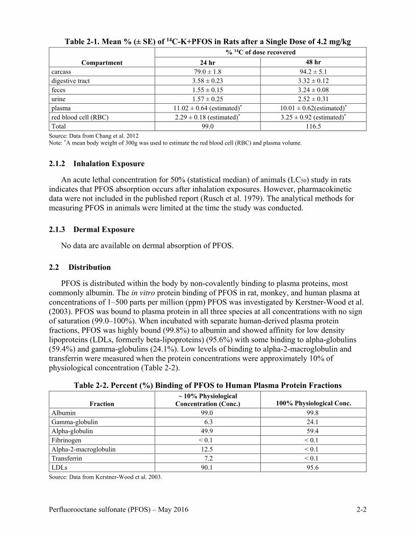
Table 2-1. Mean % (± SE) of 14C-K+PFOS in Rats after a Single Dose of 4.2 mg/kg
Compartment % 14C of dose recovered
24 hr 48 hr carcass 79.0 ± 1.8 94.2 ± 5.1 digestive tract 3.58 ± 0.23 3.32 ± 0.12 feces 1.55 ± 0.15 3.24 ± 0.08 urine 1.57 ± 0.25 2.52 ± 0.31 plasma 11.02 ± 0.64 (estimated)* 10.01 ± 0.62(estimated)* red blood cell (RBC) 2.29 ± 0.18 (estimated)* 3.25 ± 0.92 (estimated)* Total 99.0 116.5
Source: Data from Chang et al. 2012 Note: *A mean body weight of 300g was used to estimate the red blood cell (RBC) and plasma volume.
2.1.2 Inhalation Exposure
An acute lethal concentration for 50% (statistical median) of animals (LC50) study in rats indicates that PFOS absorption occurs after inhalation exposures. However, pharmacokinetic data were not included in the published report (Rusch et al. 1979). The analytical methods for measuring PFOS in animals were limited at the time the study was conducted.
2.1.3 Dermal Exposure
No data are available on dermal absorption of PFOS.
2.2 Distribution
PFOS is distributed within the body by non-covalently binding to plasma proteins, most commonly albumin. The in vitro protein binding of PFOS in rat, monkey, and human plasma at concentrations of 1–500 parts per million (ppm) PFOS was investigated by Kerstner-Wood et al. (2003). PFOS was bound to plasma protein in all three species at all concentrations with no sign of saturation (99.0–100%). When incubated with separate human-derived plasma protein fractions, PFOS was highly bound (99.8%) to albumin and showed affinity for low density lipoproteins (LDLs, formerly beta-lipoproteins) (95.6%) with some binding to alpha-globulins (59.4%) and gamma-globulins (24.1%). Low levels of binding to alpha-2-macroglobulin and transferrin were measured when the protein concentrations were approximately 10% of physiological concentration (Table 2-2).
Table 2-2. Percent (%) Binding of PFOS to Human Plasma Protein Fractions
Fraction ~ 10% Physiological
Concentration (Conc.) 100% Physiological Conc. Albumin 99.0 99.8 Gamma-globulin 6.3 24.1 Alpha-globulin 49.9 59.4 Fibrinogen < 0.1 < 0.1 Alpha-2-macroglobulin 12.5 < 0.1 Transferrin 7.2 < 0.1 LDLs 90.1 95.6
Source: Data from Kerstner-Wood et al. 2003.
Perfluorooctane sulfonate (PFOS) – May 2016 2-2

Zhang et al. (2009) used equilibrium dialysis, fluorophotometry, isothermal titration
calorimetry and circular dichroism (CD) to characterize interactions between PFOS and serum albumin and deoxyribonucleic acid (DNA). Solutions containing known amounts of serum albumin or DNA were placed in dialysis tubing and suspended in solutions with varying concentrations of PFOS. The solutions were allowed to equilibrate while measuring the change in the PFOS concentration in the dialysis solution. During dialysis, the PFOS concentration in the solution decreased reflecting its ability to cross the dialysis membrane and bind to the biopolymer within the dialysis bag. Based on the data, the serum albumin could bind up to 45 moles of PFOS per mole of protein and 0.36 moles per base pair of DNA. The binding ratio increased with increasing PFOS concentrations and decreasing solution pH (i.e., capable of promoting protein and DNA denaturation), thus providing an increased number of binding sites. It is important to remember that these studies were conducted in vitro and may not reflect in vivo situations.
The authors concluded that the interactions between serum albumin and PFOS were the results of surface electrostatic interactions between the sulfonate functional group and the positively charged side chains of lysine and arginine. Hydrogen binding interactions between the negative dipoles (fluorine) of the PFOS carbon-fluorine bonds could also play a role in the non-covalent bonding of PFOS with serum albumin. Intrinsic fluorescence analysis of tryptophan residues in serum albumin suggested a potential interaction of PFOS with tryptophan, an amino acid likely to be found in a hydrophobic portion of the albumin. In the case of DNA, the authors postulated that the interaction with PFOS occurred along the major or minor grooves of the double helix and was stabilized by the hydrogen bonding and van der Waals interactions.
Serum albumin plays an important role in the transport of a number of endogenous and exogenous compounds, such as fatty acids, bile acids, some medications and pesticides (Zhang et al. 2009). Accordingly, changes in conformation could change its transporting activity. CD spectrometry was used to determine if PFOS changed the conformation of the albumin or DNA in solution. The results of both analyses indicated conformational changes as a result of PFOS binding. However, the CD results did not demonstrate whether there was a change in transport function as a result of the conformational change.
Binding of five perfluoroalkyl acids, including PFOS, to human serum albumin was investigated by using site-specific fluorescence (Chen and Guo, 2009). Intrinsic fluorescence of trytophan-214 in human serum albumin was monitored upon addition of the perfluoroalkyl acids. PFOS induced fluorescence quenching indicative of binding. A binding constant of 2.2 x 10⁴ M-1 and a binding ratio of PFOS to human albumin of 14 moles PFOS/mole albumin were calculated.
Human serum albumin has two high-affinity drug binding sites which are known as Sudlow’s drug Site I and Site II. Past experiments have shown that two fluorescence probes, dansylamide (DA) and dansyl-L-proline (DP), are specific for the two drug binding sites on human serum albumin. Alone these two probes emit negligible fluorescence; after binding with albumin, fluorescence increases. The titration of PFOS into human serum albumin pretreated with DA (site I), showed that at low concentrations of PFOS (0.07 mmol), DA emission increased as the PFOS concentration increased until it was at 140% the original intensity. At the higher PFOS concentrations (0.7–4 mmol), however, the fluorescence dropped. The author speculated that the rise in fluorescence was induced by the conformational changes of the protein after PFOS binds to it at a site different from Site I, and the decrease at higher concentrations was from displacement of DA by PFOS. For Site II, PFOS caused a fluorescence reduction that was quick at first, but then became more gradual suggesting the possibility that PFOS was binding to this
Perfluorooctane sulfonate (PFOS) – May 2016 2-3

site with two different affinities. The binding constant calculated at Site II was 7.6 x 10⁶ M-1. These findings indicate PFOS has binding sites that are similar to those identified for fatty acids.
Structure and the energy of PFOS binding sites were determined for human serum albumin using molecular modeling (Salvalaglio et al. 2010). Calculations were based on a compound approach docking, molecular dynamics simulations, and estimating free binding energies by adopting the weighted histogram analysis method umbrella sampling and semiempirical methodology. The binding sites impacted were ones identified as human serum albumin fatty acid binding sites. The PFOS binding site with the highest energy (−8.8 kilocalories per mole [kcal/mol]) was located near the tip of the tryptophan-214 binding site, and the maximum number of ligands that could bind to human serum albumin for PFOS was 11. The most populated albumin binding site for PFOS was dominated by van der Waals interactions. The author indicated that eleven PFOS molecules were adsorbed on the surface of the albumin.
PFOS binding to bovine serum albumin was evaluated using electrospray ionization mass spectrometry by D’Alessandro et al. (2013). Using this approach, the estimate for the maximum number of PFOS binding sites was also 11, but the data on collision-induced PFOS removal was more consistent with 7 binding sites. Two of the potential binding sites (Sudlow’s sites I and II) are binding sites for a number of pharmaceuticals.
D’Alessandro et al. (2013) also examined whether PFOS could prevent binding of ibuprofen to its Sudlow II site and whether it was also able to displace bound ibuprofen. The study showed that PFOS competes with ibuprofen for its site when the PFOS:ibuprofen ratio is ≥ 0.5 moles:1 mole. In addition, when the binding site is occupied by PFOS, ibuprofen is unable to bind. Zhang et al. (2009) conducted a similar study of the impact of PFOS on the ability of serum albumin to bind vitamin B2 (riboflavin). The study found that at normal physiological conditions, 1.2 mmol/L of PFOS decreased the binding ratio of serum albumin for riboflavin in vitro by > 30%. These data suggest that PFOS can alter the pharmacokinetics and pharmacodynamics of medicinal and natural substances that share a common site on albumin.
Beesoon and Martin (2015) examined differences in the binding of the linear and branched chain isomers to serum albumin and human serum proteins. The linear PFOS molecule was found to bind more strongly to calf serum albumin than the branched chain isomers. When arranged in order of increasing binding the order was 3m < 4m < 1m < 5m < 6m (iso) <linear. In the isomer-specific binding to spiked total human serum protein, the 1m appeared to bind most strongly and the 4m the least. Binding was estimated based on the concentrations in the ultrafiltrate after spiking with 5 to 60 mg/L technical PFOS. The human serum was diluted ten-fold before spiking.
2.2.1 Oral Exposure
PFOS entry from serum into tissues appears to be controlled by several families of membrane transporters based on PFOA studies. Yu et al. (2011) administered PFOS to rats and extracted the mRNAs for OATp1, OATp2, and MRP2 from the liver to determine if they were involved in hepatic uptake. Approximately six female Wistar rats per group were administered vehicle (0.5% Tween 20), or PFOS at 0.2, 1.0, or 3.0 mg/kg in Tween 20 once daily by gavage for 5 consecutive days. Blood, bile, and liver tissue were collected 24 hours after the last dose. Exposure to 3.0 mg/kg of PFOS increased hepatic OATp2 mRNA expression (1.43 times control) while MRP2 was increased approximately 1.80 and 1.69 times that of controls in the
Perfluorooctane sulfonate (PFOS) – May 2016 2-4

1.0 and 3.0 mg/kg groups, respectively. No effect with treatment was observed on OATp1. No additional information on PFOS tissue transport was identified.
Humans. In humans, PFOS distributes mostly to the liver and blood. Olsen et al. (2003a) sampled both liver and serum from cadavers for PFOS. There was a good correlation between samples from the same subject. There was no difference in the PFOS concentrations identified in males and females or between age groups. Kärrman et al. (2010) identified PFOS in postmortem liver samples (n = 12; 6 males and 6 females 27–79 years old) with a mean concentration of 26.6 ng/g tissue.
Pérez et al. (2013) collected tissue samples from 20 adult subjects (aged 28–83) who had been living in Catalonia, Spain for 10 years and died of a variety of causes. Autopsies and tissue collection (liver, kidney, brain lung, and bone) were carried out in the first 24 hours after death. The tissues were analyzed for 21 perfluorinated compounds. PFOS was present in 90% of the samples but could be quantified in only 20% (median 1.9 ng/g). PFOS accumulated primarily in the liver (104 ng/g), kidney (75.6 ng/g), and lung (29.1 ng/g), and it was low in brain (4.9 ng/g) and bone (not detected) based on the mean wet weight tissue concentration. Detection levels varied with the tissue evaluated.
Stein et al. (2012) compared PFAS levels in maternal serum and amniotic fluid paired samples from 28 females in their second trimester of pregnancy. PFOS (0.0036–0.0287 µg/mL) was detected in all serum samples and in nine amniotic fluid samples (0.0002–0.0018 µg/mL). The Spearman correlation coefficient between the serum and amniotic fluid levels was 0.76 and is significant (p = 0.01), indicating a direct relationship between the levels in blood and amniotic fluid. The median ratio of maternal serum:amniotic fluid concentration was 25.5:1. Based on a simple regression between the levels in each compartment, PFOS was rarely detected in amniotic fluid unless the serum concentration was ≥ 0.0055 µg/mL.
Harada et al. (2007) obtained cerebrospinal fluid (CSF) from seven patients (6 males and 1 female; aged 56–80) to evaluate the partitioning of PFOS between serum and the CSF. The median concentration of PFOS in the serum was 0.0184 µg/mL, compared to the concentration in the CSF (0.00010 µg/mL). The CSF to serum ratio was 9.1 x 10⁻³. The levels identified indicate that PFOS does not easily cross the adult blood-brain barrier.
PFOS has been detected in both umbilical cord blood and breast milk indicating that maternal transfer occurs (Apelberg et al. 2007; Von Ehrenstein et al. 2009; Völkel et al. 2008). Kärrman et al. (2010) identified PFOS in breast milk samples from healthy females (n = 10; females 30–39 years old). The levels in milk (mean 0.12 ng/mL) were low compared to liver levels.
Animals
Monkey. Seacat et al. (2002) administered 0, 0.03, 0.15, or 0.75 mg/kg/day potassium PFOS orally in a capsule by intragastric intubation to six young-adult to adult cynomolgus monkeys/sex/group, except for the 0.03 mg/kg/day group which was 4/sex, daily for 26 weeks (182 days). Serum and tissues were collected at the time of sacrifice. The dosing was followed by a 52-week recovery period in 2 animals in the control, 0.15 and 0.75 mg/kg/day groups. Levels of PFOS were recorded in the serum and liver. Serum PFOS measurements demonstrate a linear increase with dosing duration in the 0.03 and 0.15 mg/kg/day groups and a non-linear increase in the 0.75 mg/kg/day group. Levels in the high-dose group appeared to plateau after about 100 days (14 weeks). Serum levels of PFOS decreased with recovery in the two highest
Perfluorooctane sulfonate (PFOS) – May 2016 2-5

dosed groups. The average percent of the cumulative dose of PFOS in the liver at the end of treatment ranged from 4.4% to 8.7% with no difference by dose group or gender. The concentration of PFOS in the liver decreased during the recovery period. Serum levels are provided in Table 2-3.
Table 2-3. Average PFOS Level (µg/mL or ppm) in Serum of Monkeys
Time (weeks)
Group 1 0.0 milligram
(mg)/kilogram (kg)/day Group 2
0.03 mg/kg/day Group 3
0.15 mg/kg/day Group 4
0.75 mg/kg/day Males Females Males Females Males Females Males Females
1 < LOQ < LOQ 0.869 ± 0.147
0.947 ± 0.110
4.60 ± 0782
3.71 ± 0.455
21.0 ± 1.57
20.4 ± 2.71
4 < LOQ < LOQ 3.20 ± 0.577
3.40 ± 0.291
17.8 ± 1.68
16.5 ± 1.87
95.3 ± 70.4
92.7 ± 39.6
16 0.04 ± 0.01
0.04 ± 0.008
11.2 ± 2.44
10.5 ± 1.90
56.2 ± 5.84
42.1 ± 4.04
189 ± 15.9
162 ± 19.3
27 0.05 ± 0.01
0.04 ± 0.01
15.9 ± 5.54
11.1 ± 1.52
68.1 ± 5.75
58.5 ± 4.67
194 ± 8.93
160 ± 23.9
35 0.05 ± 0.003
0.07 ± 0.004
Not Determined
Not Determined
84.5 ± 12.0
74.7 ± 9.53
181 ± 19.5
171 ± 10.1
57 0.03 ± 0005
0.0445 ± 0.00385
Not Determined
Not Determined
30.2 ± 2.36
32.3 ± 1.34
78.0 ± 16.3
106 ± 3.84
79 0.02 ± 0.003
0.02 ± 0.003
Not Determined
Not Determined
19.1 ± 0.805
21.4 ± 2.01
41.1 ± 25.9
41.4 ± 1.15
Source: Data from p. 304 in OECD 2002. Note: LOQ = limit of quantitation (value not stated)
At the two low doses, serum levels were comparable in the males and females, whereas at the high dose, the levels were higher in the males than females. Only for the highest dose group did the animals appear to reach serum steady state (week 16 for both males and females). In the lower dose groups, the serum levels continued to increase with dose across the dosing period. Once dosing ceased serum levels declined in all animals monitored.
Rat. Martin et al. (2007) administered 10 mg PFOS/kg to adult male Sprague-Dawley rats (n = 5) for 1, 3, or 5 days by gavage and determined the liver and serum levels. Blood was collected via cardiac puncture and PFOS concentration was determined by high-performance liquid chromatography-electrospray tandem mass spectrometry. The mean liver PFOS concentration was 83 ± 5, 229 ± 10, and 401 ± 21 μg/g after 1, 3, or 5 daily doses, respectively. The mean serum concentration was 23 ± 2.8 and 87.7 ± 4.1 μg/mL, after 1 and 3 days of dosing, respectively. Serum PFOS concentration was not determined after 5 days of dosing due to sample unavailability (not further explained by the authors).
Yu et al. (2011) administered the doses of 1, 0.2, 1.0, or 3.0 mg PFOS/kg dissolved in 0.5% Tween 20 as the vehicle to 6 female Wistar rats/group once daily by gavage for 5 consecutive days as part of a study of the effects of PFOS on the thyroid. Blood and bile were collected 24 hours after the last dose (Table 2-4). The data demonstrate a dose-related distribution to both serum and bile.
Perfluorooctane sulfonate (PFOS) – May 2016 2-6

Table 2-4. Levels of PFOS in Serum and Bile of Rats Treated for 5 Days
PFOS (mg/kg bw) Serum PFOS (microgram
[µg]/milliliter [mL]) Bile PFOS (µg/mL) 0.0 < LOQ < LOQ 0.2 1.09 ± 0.12 1.51 ± 0.42 1.0 8.20 ± 0.13 3.58 ± 0.66 3.0 33.5 ± 1.79 6.51 ± 0.67
Source: Data from Table 2 in Yu et al. 2011. Note: LOQ = limit of quantification, 0.5 µg/L
Groups of 15 Sprague-Dawley rats/sex/group were administered 0, 20, 50 or 100 mg PFOS/kg diet (Curran et al. 2008). Tissues were analyzed for PFOS residue by liquid chromatography negative electrospray tandem mass spectrometry. Distribution of PFOS is provided in Table 2-5 and indicates that the highest levels were distributed to the liver and spleen. There were no consistent differences between sexes for the liver tissues, however levels in the spleen and heart tended to be higher in females (F) than males (M) at all doses. The levels in the liver were considerably higher than those in the heart and spleen in both sexes for all doses.
Table 2-5. Mean (± SD) Daily PFOS Consumption and Tissue Residue Levels in Rats Treated for 28 Days
Parameter 0 mg/kg diet 2 mg/kg diet 20 mg/kg diet 50 mg/kg diet 100 mg/kg diet M F M F M F M F M F
PFOS consumption (mg/kg bw/day)
0 0 0.14 ± 0.02
0.15 ± 0.02
1.33 ± 0.24
1.43 ± 0.24
3.21 ± 0.57
3.73 ± 0.57
6.34 ± 1.35
7.58 ± 0.68
Serum (µg PFOS/g serum)
0.47 ± 0.27
0.95 ± 0.51
0.95 ± 0.13
1.50 ± 0.23
13.45 ± 1.48
15.40 ± 1.56
20.93 ± 2.36
31.93 ± 3.59
29.88 ± 3.53
43.20 ± 3.95
Liver (µg PFOS/gram (g) liver)
0.79 ± 0.49
0.89 ± 0.44
48.28 ± 5.81
43.44 ± 6.79
560.23 ±
104.43
716.55 ± 59.15
856.90 ±
353.83
596.75 ±
158.01
1030.40 ±
162.80
1008.59 ± 49.41
Ratio liver:serum PFOS
2.04 ± 1.39
1.30 ± 1.32
51.34 ± 9.20
29.99 ± 8.11
42.10 ± 9.20
46.81 ± 5.26
41.42 ± 16.95
20.23 ± 7.50
35.23 ± 8.50
23.48 ± 1.98
Spleen (µg PFOS/g spleen)
0.27 ± 0.36
2.08 ± 4.17
6.07 ± 1.85
7.94 ± 3.76
45.27 ± 2.16
70.03 ± 36.66
122.51 ± 7.83
139.45 ± 15.44
230.73 ± 11.47
294.96 ± 26.66
Heart (µg PFOS/g heart)
0.10 ± 0.14
1.42 ± 2.91
4.67 ± 1.73
6.54 ± 3.07
33.00 ± 3.44
54.65 ± 30.89
90.28 ± 4.95
107.53 ± 6.24
154.13 ± 11.78
214.45 ± 17.58
Source: Data from Table 1 on in Curran et al. 2008 Note: M = male; F = female; SD = standard deviation
Ten three-month old male Sprague-Dawley rats/group were administered 0 (Milli-Q water only), 5, or 20 mg/kg/day of PFOS by oral gavage for 28 days (Cui et al. 2009). Rats were sacrificed after the exposure and blood and tissue samples obtained. Concentrations identified in rat whole blood and various tissues at the end of the exposure are provided in Table 2-6. The study indicated that the highest levels of PFOS were identified in the liver after 28 days of exposure.
Perfluorooctane sulfonate (PFOS) – May 2016 2-7
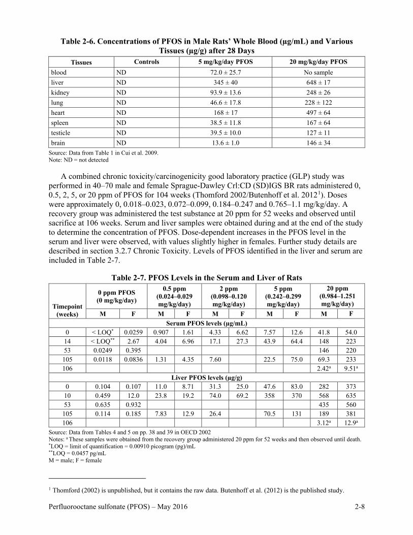
Table 2-6. Concentrations of PFOS in Male Rats’ Whole Blood (µg/mL) and Various
Tissues (µg/g) after 28 Days Tissues Controls 5 mg/kg/day PFOS 20 mg/kg/day PFOS
blood ND 72.0 ± 25.7 No sample liver ND 345 ± 40 648 ± 17 kidney ND 93.9 ± 13.6 248 ± 26 lung ND 46.6 ± 17.8 228 ± 122 heart ND 168 ± 17 497 ± 64 spleen ND 38.5 ± 11.8 167 ± 64 testicle ND 39.5 ± 10.0 127 ± 11 brain ND 13.6 ± 1.0 146 ± 34
Source: Data from Table 1 in Cui et al. 2009. Note: ND = not detected
A combined chronic toxicity/carcinogenicity good laboratory practice (GLP) study was performed in 40–70 male and female Sprague-Dawley Crl:CD (SD)IGS BR rats administered 0, 0.5, 2, 5, or 20 ppm of PFOS for 104 weeks (Thomford 2002/Butenhoff et al. 20121). Doses were approximately 0, 0.018–0.023, 0.072–0.099, 0.184–0.247 and 0.765–1.1 mg/kg/day. A recovery group was administered the test substance at 20 ppm for 52 weeks and observed until sacrifice at 106 weeks. Serum and liver samples were obtained during and at the end of the study to determine the concentration of PFOS. Dose-dependent increases in the PFOS level in the serum and liver were observed, with values slightly higher in females. Further study details are described in section 3.2.7 Chronic Toxicity. Levels of PFOS identified in the liver and serum are included in Table 2-7.
Table 2-7. PFOS Levels in the Serum and Liver of Rats
Timepoint (weeks)
0 ppm PFOS (0 mg/kg/day)
0.5 ppm (0.024–0.029 mg/kg/day)
2 ppm (0.098–0.120 mg/kg/day)
5 ppm (0.242–0.299 mg/kg/day)
20 ppm (0.984–1.251 mg/kg/day)
M F M F M F M F M F Serum PFOS levels (µg/mL)
0 < LOQ* 0.0259 0.907 1.61 4.33 6.62 7.57 12.6 41.8 54.0 14 < LOQ** 2.67 4.04 6.96 17.1 27.3 43.9 64.4 148 223 53 0.0249 0.395 146 220 105 0.0118 0.0836 1.31 4.35 7.60 22.5 75.0 69.3 233 106 2.42a 9.51a
Liver PFOS levels (µg/g) 0 0.104 0.107 11.0 8.71 31.3 25.0 47.6 83.0 282 373
10 0.459 12.0 23.8 19.2 74.0 69.2 358 370 568 635 53 0.635 0.932 435 560 105 0.114 0.185 7.83 12.9 26.4 70.5 131 189 381 106 3.12a 12.9a
Source: Data from Tables 4 and 5 on pp. 38 and 39 in OECD 2002 Notes: a These samples were obtained from the recovery group administered 20 ppm for 52 weeks and then observed until death. *LOQ = limit of quantification = 0.00910 picogram (pg)/mL **LOQ = 0.0457 pg/mL M = male; F = female
1 Thomford (2002) is unpublished, but it contains the raw data. Butenhoff et al. (2012) is the published study.
Perfluorooctane sulfonate (PFOS) – May 2016 2-8
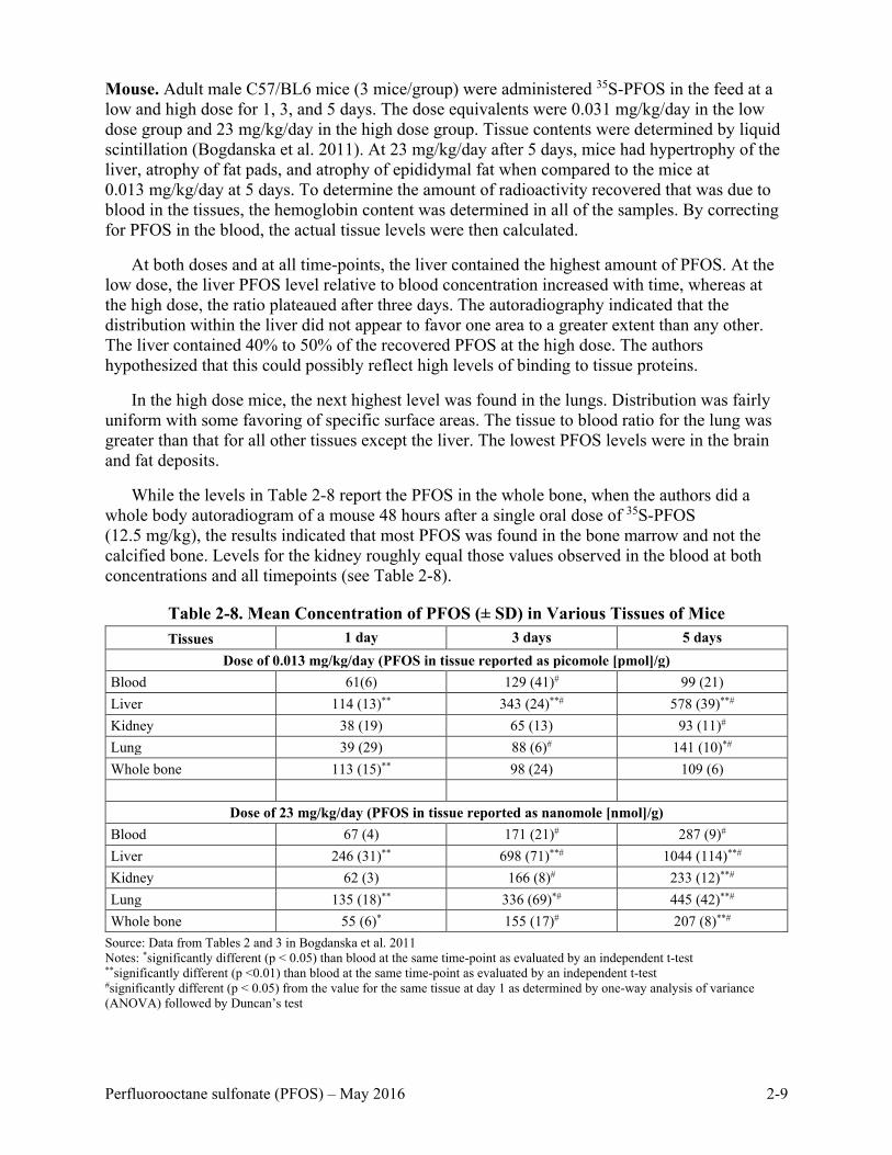
Mouse. Adult male C57/BL6 mice (3 mice/group) were administered 35S-PFOS in the feed at a low and high dose for 1, 3, and 5 days. The dose equivalents were 0.031 mg/kg/day in the low dose group and 23 mg/kg/day in the high dose group. Tissue contents were determined by liquid scintillation (Bogdanska et al. 2011). At 23 mg/kg/day after 5 days, mice had hypertrophy of the liver, atrophy of fat pads, and atrophy of epididymal fat when compared to the mice at 0.013 mg/kg/day at 5 days. To determine the amount of radioactivity recovered that was due to blood in the tissues, the hemoglobin content was determined in all of the samples. By correcting for PFOS in the blood, the actual tissue levels were then calculated.
At both doses and at all time-points, the liver contained the highest amount of PFOS. At the low dose, the liver PFOS level relative to blood concentration increased with time, whereas at the high dose, the ratio plateaued after three days. The autoradiography indicated that the distribution within the liver did not appear to favor one area to a greater extent than any other. The liver contained 40% to 50% of the recovered PFOS at the high dose. The authors hypothesized that this could possibly reflect high levels of binding to tissue proteins.
In the high dose mice, the next highest level was found in the lungs. Distribution was fairly uniform with some favoring of specific surface areas. The tissue to blood ratio for the lung was greater than that for all other tissues except the liver. The lowest PFOS levels were in the brain and fat deposits.
While the levels in Table 2-8 report the PFOS in the whole bone, when the authors did a whole body autoradiogram of a mouse 48 hours after a single oral dose of 35S-PFOS (12.5 mg/kg), the results indicated that most PFOS was found in the bone marrow and not the calcified bone. Levels for the kidney roughly equal those values observed in the blood at both concentrations and all timepoints (see Table 2-8).
Table 2-8. Mean Concentration of PFOS (± SD) in Various Tissues of Mice Tissues 1 day 3 days 5 days
Dose of 0.013 mg/kg/day (PFOS in tissue reported as picomole [pmol]/g) Blood 61(6) 129 (41)# 99 (21) Liver 114 (13)** 343 (24)**# 578 (39)**# Kidney 38 (19) 65 (13) 93 (11)# Lung 39 (29) 88 (6)# 141 (10)*# Whole bone 113 (15)** 98 (24) 109 (6)
Dose of 23 mg/kg/day (PFOS in tissue reported as nanomole [nmol]/g) Blood 67 (4) 171 (21)# 287 (9)# Liver 246 (31)** 698 (71)**# 1044 (114)**# Kidney 62 (3) 166 (8)# 233 (12)**# Lung 135 (18)** 336 (69)*# 445 (42)**# Whole bone 55 (6)* 155 (17)# 207 (8)**#
Source: Data from Tables 2 and 3 in Bogdanska et al. 2011 Notes: *significantly different (p < 0.05) than blood at the same time-point as evaluated by an independent t-test **significantly different (p <0.01) than blood at the same time-point as evaluated by an independent t-test #significantly different (p < 0.05) from the value for the same tissue at day 1 as determined by one-way analysis of variance (ANOVA) followed by Duncan’s test
Perfluorooctane sulfonate (PFOS) – May 2016 2-9

In an immunotoxicity study, four to six C57BL/6 male mice/group were administered diets
with 0% to 0.02% PFOS for 10 days. Levels in the serum increased as the concentration increased (Table 2-9) (Qazi et al. 2009a).
Table 2-9. Levels of PFOS (Means ± SE) in Mouse Serum Following Treatment for 10 Days Dietary level (% w/w) Number of mice ppm
PFOS (0) 4 0.0287 ± 0.01 PFOS (0.001%) 4 50.8 ± 2.5 PFOS (0.005%) 4 96.7 ± 5.2 PFOS (0.02%) 4 340 ± 16
Source: Data from study report by Qazi et al. 2009a
Distribution during Reproduction and Development
The availability of distribution data from pregnant females plus animal pups and neonates is a strength of the PFOS pharmacokinetic database, because it helps to identify those tissues receiving the highest concentration of PFOS during development. For this reason the information on tissue levels during reproduction and development are presented separately from those that are representative of other life stages.
Humans. T. Zhang et al. (2013) recruited pregnant females for a study to examine the distribution of PFOS between maternal blood, cord blood, the placenta, and amniotic fluid. Thirty two females from Tianjin, China volunteered to take part in the study. Samples were collected at time of delivery. Maternal ages ranged from 21 to 39 years, and periods ranged from 35 to 37 weeks. It was the first child for 26 of the females and the second child for 6. The study yielded 31 maternal whole blood samples, 30 cord blood samples, 29 amniotic fluid samples, and 29 placentas. The maternal blood contained variable levels of 10 PFAS, 8 acids and 2 sulfonates. The mean maternal blood concentration was highest for PFOS (14.6 ng/mL), followed by PFOA (3.35 ng/mL). In both cases, the mean was greater than the median, indicating a distribution skewed toward the higher concentrations.
PFOS was found in all fluids/tissues sampled. It was transferred to the amniotic fluid to a lesser extent than PFOA based on their relative proportions in the maternal blood and cord blood (21% versus 58%, respectively). Compared to the mean PFOS value in maternal blood, the mean levels in the cord blood, placenta, and amniotic fluid were 21%, 56%, and 0.14% of the mean levels in the mother’s blood, respectively. The correlation coefficients between the maternal PFOS blood levels and placenta, cord blood, and amniotic fluid levels were good (0.7 to 0.9), and the relationships were statistically-significant (p < 0.001).
Rat. To determine the dose-response curve for neonatal mortality in rat pups born to PFOS exposed dams and to investigate associated biochemical and pharmacokinetic parameters, 5 groups of 16 female Sprague-Dawley Crl:CD(SD)IGS VAF/Plus rats each were administered 0, 0.1, 0.4, 1.6, or 3.2 mg PFOS/kg bw/day by oral gavage beginning 42 days prior to cohabitation and continuing through gestation day (GD) 14 or 20 (Luebker et al. 2005b). Eight rats from each group were randomly chosen and sacrificed on GD 15, followed by Caesarean removal of the pups. All remaining animals were sacrificed and C-sectioned on GD 21. Urine and feces were collected overnight from dams on the eve of cohabitation day 1 and during GDs 6–7, 14–15, and 20–21. Serum samples were collected just prior to cohabitation and on GD 7, GD 15, and GD 21. Fetal liver and blood samples were obtained on GD 21 and pooled by litter.
Perfluorooctane sulfonate (PFOS) – May 2016 2-10

The urine, feces, and liver of the control animals all contained PFOS at small concentrations.
In treated rats, the highest concentration of PFOS was in the liver. Serum levels in the dams for each dose were consistent between GD 1 and GD 15, indicating achievement of steady state prior to conception (Table 2-10). The GD 21 levels in the dams had dropped below those observed earlier in the pregnancy. Serum levels in the GD,21 fetuses were higher than those in the dams. In contrast, the liver levels in the dams on GD 21 were about three times higher than in the fetuses. Fecal excretion was greater than urinary excretion by the dams.
Table 2-10. PFOS Concentrations (Mean ± Standard Deviation [SD]) in Samples from Pregnant Dams and Fetuses (GD 21 Only) in µg/mL (ppm) for Serum and Urine and µg/g
for Liver and Feces
Parameter Dose
(mg/kg/day) GD 1 GD 7 GD 15 GD 21
Dams Fetuses Serum 0.1 8.90 ± 1.10 7.83 ± 1.11 8.81 ± 1.47 4.52 ± 1.15 9.08 0.4 40.7 ± 4.46 40.9 ± 5.89 41.4 ± 4.80 26.2 ± 16.1 34.3 1.6 160 ± 12.5 154 ±14.0 156 ± 25.9 136 ± 86.5 101 3.2 318 ± 21.1 306 ± 32.1 275 ± 26.7 155 ± 39.3 164 Liver 0.1 - - - 29.2 ± 10.5 7.92 0.4 - - - 107 ± 22.7 30.6 1.6 - - - 388 ± 167 86.5 3.2 - - - 610 ± 142 230 Urine 0.1 0.05 ± 0.02 0.06 ± 0.03 0.07± 0.04 0.06 ± 0.01 - 0.4 0.28 ± 0.19 0.31 ± 0.20 0.53 ± 0.23 0.55 ± 0.16 - 1.6 0.96 ± 0.39 1.10 ± 0.57 0.36 ± 0.35 2.71 ± 2.07 - 3.2 1.53 ± 0.87 1.60 ± 0.97 0.52 ± 0.28 1.61 ± 0.53 - Feces 0.1 0.50 ± 0.14 0.49 ± 0.11 0.66 ± 0.10 0.42 ± 0.10 - 0.4 2.42 ± 0.49 2.16 ± 0.43 2.93 ±0.62 2.39 ± 1.21 - 1.6 10.3 ± 3.01 9.20 ± 2.68 11.1 ± 3.28 9.94 ± 4.51 - 3.2 23.9 ± 4.16 33.0 ± 10.0 29.5 ± 8.92 20.1 ± 4.21 -
Source: Data from Luebker et al. 2005b Note: - = no sample obtained
This same study also included a subset of dams allowed to litter naturally and dosed through lactation day (LD) 4. Liver and serum samples were collected from dams and pups on LD 5. In this sampling, serum PFOS levels were similar between the dam and offspring, but the liver values were now higher in the neonates than in their dams.
Twenty five female Sprague-Dawley rats/group were administered 0, 0.1, 0.3, or 1.0 mg/kg/day potassium PFOS by gavage from GD 0 through postnatal day (PND) 20. An additional 10 mated females served as satellite rats to each of the four groups and were used to collect additional blood and tissue samples. Further details from this study are provided in section 3.2.6 as reported in Butenhoff et al. (2009). Samples were taken from the dams, fetuses, and pups for serum and tissue PFOS concentrations and the results reported by Chang et al. (2009). The blood and tissue sampling results are provided in Table 2-11.
Perfluorooctane sulfonate (PFOS) – May 2016 2-11

Table 2-11. Mean PFOS (± Standard Error) Concentrations in Serum, Liver, and Brain
Tissue in Dams and Offspring
Time Dose
(mg/kg) Serum PFOS (µg/mL) Liver PFOS (µg/g) Brain PFOS (µg/g)
Dam Offspring Dam Offspring Dam Offspring GD 20a Control < LLOQ 0.009 ± 0.001 < LLOQ < LLOQ < LLOQ < LLOQ 0.1 1.722 ±
0.068 3.906 ± 0.096 8.349 ±
0.344 3.205 ± 0.217 0.151 ±
0.012 1.233 ± 0.067
0.3 6.245 ± 0.901
10.446 ± 0.291 21.725 ± 0.721
5.814 ± 0.245 0.368 ± 0.043
3.126 ± 0.238
1.0 26.630 ± 3.943
31.463 ± 1.032 48.875 ± 72.733
20.025 ± 2.021 0.999 ± 0.083
12.984 ± 1.122
PND 4a Control 0.008 ± 0.000
< LLOQ NS < LLOQ NS < LLOQ
0.1 3.307 ± 0.080
2.236 ± 0.070 NS 9.463 ± 0.512 NS 0.680 ± 0.033
0.3 10.449 ± 0.234
6.960 ± 0.163 NS 20.130 ± 0.963 NS 1.910 ± 0.074
1.0 34.320 ± 31.154
22.440 ± 0.723 NS 50.180 ± 1.124 NS 6.683 ± 0.428
PND 21 Control 0.007 ± 0.000
< LLOQ – m/f NS < LLOQ – m/f NS < LLOQ – m/f
0.1 3.159 ± 0.081
1.729 ± 0.079 (M) 1.771 ± 0.076 (F)
NS 5.980 ± 0.614 (M) 5.278 ± 0.174 (F)
NS 0.220 ± 0.014 (M) 0.229 ± 0.011 (F)
0.3 8.981 ± 0.275
5.048 ± 0.108 (M) 5.246 ± 0.138 (F)
NS 14.780 ± 0.832 (M) 13.550 ± 0.298 (F)
NS 0.649 ± 0.053 (M) 0.735 ± 0.039 (F)
1.0 30.480 ± 1.294
18.611 ± 1.011 (M) 18.010 ± 0.744 (F)
NS 44.890 ± 2.637 (M) 41.230 ± 2.295 (F)
NS 2.619 ± 0.165 (M) 2.700 ± 0.187 (F)
PND 72 Control NA < LLOQ – m/f NA < LLOQ – m/f NA NS – M/F 0.1 NA 0.042 ± 0.004 (M)
0.207 ± 0.042 (F) NA 0.981± 0.091 (M)
0.801 ± 0.082 (F) NA NS – M/F
0.3 NA 0.120 ± 0.009 (M) 0.556 ± 0.062 (F)
NA 2.464 ± 0.073 (M) 2.252 ± 0.095 (F)
NA NS – M/F
1.0 NA 0.560 ± 0.105 (M) 1.993 ± 0.293 (F)
NA 7.170 ± 0.382 (M) 7.204 ± 0.414 (F)
NA NS – M/F
Source: Data from Table 2 in Chang et al. 2009 Notes: a Data are from samples pooled by litters in the fetuses/pups < LLOQ = sample less than lower limit of quantitation, serum = 0.01 µg/mL; liver = 0.05 µg/g; brain = 0.025 µg/g NS = no sample obtained NA = not applicable; all dams sacrificed on PND 21 m = male; f = female
On GD 20, PFOS concentration in maternal serum, liver, and brain correlated with the daily doses administered. Maternal liver-to-serum PFOS ratios ranged from 1.8 to 4.9, while the maternal brain-to-serum ratios were 0.04 to 0.09 (Chang et al. 2009). The concentrations in the brains of fetuses was about ten times higher than in their dams for all doses.
Perfluorooctane sulfonate (PFOS) – May 2016 2-12

Based on the maternal and offspring data on GD 20, there is placental transfer of PFOS from
rat dams to developing fetuses. Serum values were approximately 1–2 times greater in the fetuses than in the dams at GD 20. The concentration of PFOS in fetal liver was less than that of dams, and the brain values were much higher; this is possibly due to the lack of development of the blood-brain barrier at this stage of offspring development. PFOS serum concentrations in the offspring were lower than those for the dams on PND 4 and continued to drop through PND 72. However, based on the concentrations still present in the neonate serum, lactational transfer of PFOS was occurring. At PND 72, the males appeared to be eliminating PFOS more quickly as the serum values were lower than those in the females; this difference was not observed at earlier time-points. In the liver, PFOS was the greatest in the offspring at PND 4 and decreased significantly by PND 72. Liver values were similar at all time-points between males and females. On GD 20, the brain levels for the pups were ten-fold higher than those for the dam. The levels in pup brain gradually declined between PND 4 and PND 21.
In a study by Zeng et al. (2011) ten pregnant Sprague-Dawley rats/group were administered 0, 0.1, 0.6, or 2.0 mg/kg/day of PFOS by oral gavage in 0.5% Tween 80 from GD 2 to GD 21. On GD 21, dams were monitored for parturition, and the day of delivery was designated PND 0. On PND 0, five pups/litter were sacrificed and the trunk blood, cortex, and hippocampus were collected for examination. The other pups were randomly redistributed to dams within the dosage groups and allowed to nurse until PND 21, when they were sacrificed with the same tissues collected as previously described. PFOS concentration in the hippocampus, cortex, and serum increased in a dose-dependent manner but overall was lower in all tissues on PND 21 when compared to PND 0. Levels of PFOS are included in Table 2-12.
Table 2-12. PFOS Contents in Serum, Hippocampus, and Cortex of Offspring (n = 6) Time Dose group (mg/kg/day) Serum (µg/mL) Hippocampus (µg/g) Cortex (µg/g)
PND 0 Control ND ND ND 0.1 1.50 ± 0.43* 0.63 ± 0.19* 0.39 ± 0.09* 0.6 24.60 ± 3.02** 7.43 ± 1.62* 5.23 ± 1.58** 2.0 45.69 ± 4.77** 17.44 ± 4.12* 13.43 ± 3.89** PND 21 Control ND ND ND 0.1 0.37 ± 1.12* 0.25 ± 0.14* 0.06 ± 0.04* 0.6 1.86 ± 0.35** 1.59 ± 0.78** 1.03 ± 0.59** 2.0 4.26 ± 1.73*** 6.09 ± 1.30*** 3.69 ± 0.95***
Source: Data from Table 2 in Zeng et al. 2011 Notes: ND = not detected * p < 0.05 compared with control in the same day ** p < 0.05 compared with 0.1 mg/kg group in the same day *** p < 0.05 compared with 0.6 mg/kg group in the same day
Sprague-Dawley rats were administered PFOS in 0.05% Tween (in deionized water) once daily by gavage from GD 1 to GD 21 at 0, 0.1, or 2.0 mg/kg/day. There was a postnatal decline in the serum and brain PFOS levels between PND 0 and PND 21. PFOS concentrations were higher in the serum when compared to the lung in offspring on both PND 0 and 21 (Chen et al. 2012) (see Table 2-13).
Perfluorooctane sulfonate (PFOS) – May 2016 2-13

Table 2-13. Mean PFOS Content in Serum and Lungs of Rat Offspring (n = 6) Age Treatment PFOS in serum (µg/mL) PFOS in lung (µg/g)
PND 0 0 mg/kg/day ND ND 0.1 mg/kg/day 1.7 ± 0.35* 0.92 ± 0.04* 2.0 mg/kg/day 47.52 ± 3.72* 22.4 ± 1.03* PND 21 0 mg/kg/day ND ND 0.1 mg/kg/day 0.41 ± 0.11* 0.21 ± 0.04* 2.0 mg/kg/day 4.46 ± 1.82** 3.16 ± 0.11**
Source: Data from Table 2 in Chen et al. 2012 Notes: ND = not detected * p < 0.05 compared with control ** p < 0.01 compared with control
Mouse. Borg et al. (2010) administered a single dose of 12.5 mg/kg 35S-PFOS by intravenous injection (n = 1) or gavage (n = 5) on GD 16 to C57Bl/6 dams. Using whole-body autoradiography and liquid scintillation, counting distribution of PFOS was determined for the dams/fetuses (GD 18 and 20) and the neonates on PND 1. Distribution in the dams was similar regardless of the route of exposure, with the hepatic level being approximately four times greater than the serum. Maternal PFOS levels were highest in the liver and lungs at all timepoints. In dams, the concentration of PFOS in the liver was approximately 4 times and in the lung was approximately 2 times the blood concentrations, respectively. The distribution of PFOS in the kidneys was similar and the amount in the brain was lower than that of the blood. In the fetuses, the highest concentrations of PFOS were found in the kidneys and liver. In the fetuses on GD 18, values in the lungs were similar to the maternal lungs, and this value increased by GD 20. In the kidneys, the highest concentration of PFOS was observed in the fetuses on GD 18 (3 times higher than maternal levels)
In the offspring at all timepoints, PFOS was homogeneously distributed in the liver at a level 2.5 times higher than maternal blood and 1.7 times lower than maternal liver. In pups on PND 1, PFOS was mostly concentrated in the lungs and liver. Pups on PND 1 had PFOS levels that were 3 times higher in the lungs, compared to maternal blood with a heterogeneous distribution. In the kidneys, the levels in pups on PND 1 were similar to their dams despite being higher on GD 18. Levels in the brain were similar at all timepoints in the offspring and higher than in the maternal brain, likely due to an immature brain-blood barrier. Select data are provided in Table 2-14 and Figure 2-1.
Table 2-14. Ratios (Means ± SD) Between the Concentrations of 35S-Labeled PFOS in Various Organs and Blood of Mouse Dams, Fetuses, and Pups versus the Average
Concentration in Maternal Blood [35S-PFOS]organ/[35S-PFOS]maternal blood
Subject Liver
(n = 6–8) Lungs
(n = 5–6) Kidneys (n = 3–6)
Brain (n = 6–9)
Blood (n = 1–6)
Dams 4.2** ± 0.7 2.0* ± 0.4 0.9 ± 0.1 0.2** ± 0.05 1.0 Fetus on GD 18 2.6** ± 0.8 2.1* ± 0.6 2.8** ± 0.3 1.2 ± 0.3 2.3 Fetus on GD 20 2.4** ± 0.5 2.5** ± 0.4 1.4 ± 0.2 0.9 ± 0.1 1.1 ± 0.04 Pups on PND 1 2.4* ± 0.4 3.0** ± 0.5 1.0 ± 0.5 0.9 ± 0.2 1.7** ± 0.3
Source: Data from Table 1 in Borg et al. 2010 Notes: *Statistically-significant (p ≤ 0.01) in comparison to maternal blood **Statistically-significant (p ≤ 0.001) in comparison to maternal blood
Perfluorooctane sulfonate (PFOS) – May 2016 2-14

Filled symbols are representative after oral exposure; open after intravenous exposure. A = p ≤ 0.001 and a = p ≤ 0.01, compared to maternal blood B = p ≤ 0.001 and b = p ≤ 0.05, compared to maternal tissue C = p ≤ 0.001 and c = p ≤ 0.05, comparing between fetuses/pups on GD 20/PND 1 with corresponding value on GD 18;
Figure 2-1. Distribution of Radiolabeled PFOS in Dams and in Fetuses/Pups in the Liver, Lung, Kidney, and Brain
(Figure from Borg et al. 2010)
2.2.2 Inhalation and Dermal Exposure
No data on distribution following inhalation or dermal exposures were identified.
2.2.3 Other Routes of Exposure
Male and female mice were administered PFOS by subcutaneous injection one time on PNDs 7, 14, 21, 28, or 35 at concentrations of 0 or 50 mg/kg bodyweight (bw) (Liu et al. 2009). Animals were killed 24 hours after treatment and the PFOS concentration levels obtained. The percent distribution found in the blood, brain, and liver are provided in Table 2-15. The distribution shows that beyond PND 14 the levels in the liver are approximately two to four times greater than those found on PND 7.
Perfluorooctane sulfonate (PFOS) – May 2016 2-15
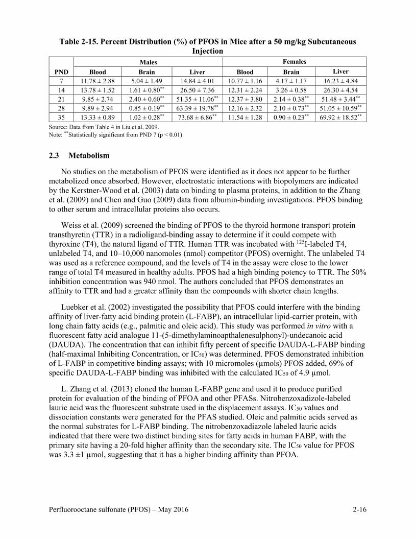
Table 2-15. Percent Distribution (%) of PFOS in Mice after a 50 mg/kg Subcutaneous
Injection
PND Males Females
Blood Brain Liver Blood Brain Liver 7 11.78 ± 2.88 5.04 ± 1.49 14.84 ± 4.01 10.77 ± 1.16 4.17 ± 1.17 16.23 ± 4.84
14 13.78 ± 1.52 1.61 ± 0.80** 26.50 ± 7.36 12.31 ± 2.24 3.26 ± 0.58 26.30 ± 4.54 21 9.85 ± 2.74 2.40 ± 0.60** 51.35 ± 11.06** 12.37 ± 3.80 2.14 ± 0.38** 51.48 ± 3.44** 28 9.89 ± 2.94 0.85 ± 0.19** 63.39 ± 19.78** 12.16 ± 2.32 2.10 ± 0.73** 51.05 ± 10.59** 35 13.33 ± 0.89 1.02 ± 0.28** 73.68 ± 6.86** 11.54 ± 1.28 0.90 ± 0.23** 69.92 ± 18.52**
Source: Data from Table 4 in Liu et al. 2009. Note: **Statistically significant from PND 7 (p < 0.01)
2.3 Metabolism
No studies on the metabolism of PFOS were identified as it does not appear to be further metabolized once absorbed. However, electrostatic interactions with biopolymers are indicated by the Kerstner-Wood et al. (2003) data on binding to plasma proteins, in addition to the Zhang et al. (2009) and Chen and Guo (2009) data from albumin-binding investigations. PFOS binding to other serum and intracellular proteins also occurs.
Weiss et al. (2009) screened the binding of PFOS to the thyroid hormone transport protein transthyretin (TTR) in a radioligand-binding assay to determine if it could compete with thyroxine (T4), the natural ligand of TTR. Human TTR was incubated with 125I-labeled T4, unlabeled T4, and 10–10,000 nanomoles (nmol) competitor (PFOS) overnight. The unlabeled T4 was used as a reference compound, and the levels of T4 in the assay were close to the lower range of total T4 measured in healthy adults. PFOS had a high binding potency to TTR. The 50% inhibition concentration was 940 nmol. The authors concluded that PFOS demonstrates an affinity to TTR and had a greater affinity than the compounds with shorter chain lengths.
Luebker et al. (2002) investigated the possibility that PFOS could interfere with the binding affinity of liver-fatty acid binding protein (L-FABP), an intracellular lipid-carrier protein, with long chain fatty acids (e.g., palmitic and oleic acid). This study was performed in vitro with a fluorescent fatty acid analogue 11-(5-dimethylaminoapthalenesulphonyl)-undecanoic acid (DAUDA). The concentration that can inhibit fifty percent of specific DAUDA-L-FABP binding (half-maximal Inhibiting Concentration, or IC50) was determined. PFOS demonstrated inhibition of L-FABP in competitive binding assays; with 10 micromoles (µmols) PFOS added, 69% of specific DAUDA-L-FABP binding was inhibited with the calculated IC50 of 4.9 µmol.
L. Zhang et al. (2013) cloned the human L-FABP gene and used it to produce purified protein for evaluation of the binding of PFOA and other PFASs. Nitrobenzoxadizole-labeled lauric acid was the fluorescent substrate used in the displacement assays. IC50 values and dissociation constants were generated for the PFAS studied. Oleic and palmitic acids served as the normal substrates for L-FABP binding. The nitrobenzoxadiazole labeled lauric acids indicated that there were two distinct binding sites for fatty acids in human FABP, with the primary site having a 20-fold higher affinity than the secondary site. The IC50 value for PFOS was 3.3 ±1 µmol, suggesting that it has a higher binding affinity than PFOA.
Perfluorooctane sulfonate (PFOS) – May 2016 2-16

2.4 Excretion
2.4.1 Oral Exposure
Humans. Urinary excretion of PFOS in humans is impacted by the isomeric composition of the mixture present in blood and the gender/age of the individuals. The half-lives of the branched chain PFOS isomers are shorter than those for the linear molecule, an indication that renal resorption is less likely with the branched chains.
Y. Zhang et al. (2013) determined half-lives for PFOA isomers based on paired serum samples and early morning urine samples collected from healthy volunteers in two large Chinese cities. Half-lives were determined using a one compartment model and an assumption of first order clearance. The volume of distribution (Vd) applied in the analysis as determined by Thompson et al. (2010) was 170 mL/kg. Clearance was estimated from the concentration in urine normalized for creatinine and assuming excretion of 1.2 and 1.4 L/day of urine and 0.9 and 1.1 mg creatinine/day for males and females, respectively. The mean half-life for the sum of all PFOS isomers in younger females (n = 20) was 6.2 years (range: 5.0–10 years), while that for all males and older females (n = 66) was 27 (range: 14–90 years); the medians were 6.0 and 18 years, respectively.
The mean half-life values for the six branched chain isomers of PFOS were lower than the value for the linear chain with the exception of the 1-methyl heptane sulfonate, suggesting that resorption transporters may favor uptake of the linear chain and 1-methyl branched chain over the other isomers. Older females and males have longer half-lives than young females, suggesting the importance of monthly menstruation as a pathway for excretion (Y. Zhang et al. 2013). The mean half-life for the 1-methylheptane sulfonate in the males and older females (n = 43) was considerably greater than that for the sum of all isomers (90 years versus 27 years). For males and older females there were considerable inter-individuals differences, with 100-fold differences between the minimum and maximum values among the males and older females compared to < 10-fold differences for the younger females.
T. Zhang et al. (2014) derived estimates for PFOA’s urinary excretion rate using paired urine and blood samples from 54 adults (29 male, 25 female) in the general population and 27 pregnant females in Tainjin, China The age range for the general population was 22–62 and that for the pregnant females was 21–39. Urinary excretion was calculated based on the concentration in the urine times volume of urine, wherein a urinary volume of 1200 mL/day was applied to all females and 1600 mL/day applied to all males. Urine samples were first draw morning samples. Total daily intakes for PFOS were calculated from the concentration in blood using first order assumptions, a half-life of 5.4 years (Olsen et al. 2007) and a volume of distribution of 170 mL/kg (Thompson et al. 2010; Egeghy and Lorber 2011). Urinary elimination rate was then calculated from the urine levels and the modeled total daily intake. Total daily intake, and thus the urinary elimination rate, was not calculated for pregnant females due to the highly variable blood levels of PFOS during pregnancy. PFOS was detected in the blood samples for all participants but only for 48% of the urine samples from the general population and 11% of samples from the pregnant females. Unfortunately the urinary PFOS was below detection for most of the females in the study.
The calculated geometric mean total daily intake for PFOS was 89.2 ng/day for the adult general population, resulting in a daily urinary excretion rate of 16% of the estimated intake; there was no significant difference between males and females. From the limited number of urine
Perfluorooctane sulfonate (PFOS) – May 2016 2-17

samples available, the urine:blood ratio was lower for pregnant females than nonpregnant females (0.0004 versus 0.0013) suggesting other removal pathways such as placenta and cord blood. There was a difference between the younger menstruating females (21–50 years versus 51–61 years), with a higher ratio for the younger females (0.0018 versus 0.0006). There is no indication that data were collected from the participants relative to menstruation status on the day of blood and urine collection. There was a significant difference between PFOS urinary excretion in older adults compared to younger adults (p = 0.015), with a higher elimination rate in the younger adults compared to the older age group.
Wong et al. (2014) looked at the role of menstrual blood as an excretory pathway to explain the shorter half-life of PFOS in females than males. They fit a population-based pharmacokinetic model to six cross-sectional National Health and Nutrition Examination Survey (NHANES) data sets (1999–2012) for males and females. They concluded that menstruation could account for about 30% of the elimination half-life difference between females and males. Verner and Longnecker (2015) suggested a need to consider the nonblood portion of the menstrual fluid and its albumin content in the Wong et al. (2014) estimate for the menstrual fluid volume. A yearly estimate for serum loss of 868 mL/year by Verner and Longnecker (2015) compared to the 432 mL/year estimate of Wong et al. (2014) suggests that the menstrual fluid loss can account for > 30% of the difference in the elimination half-life between females and males.
Harada et al. (2007) obtained serum and bile samples from patients (2 male and 2 female; aged 63–76) undergoing gallstone surgery to determine the bile to serum ratio and biliary resorption rate. The median concentration for PFOS in the serum was 23.2 ng/mL (0.023 ppm), compared to the bile, 27.9 ng/mL (0.028 ppm). The fact that the levels in bile concentrations are higher than in serum is supportive of bile as a route of excretion. The biliary resorption rate was 0.97, which could contribute to the long half-life in humans. Method of exposure to PFOS was unknown.
Biliary excretion in humans and the potential for resorption from bile discharged to the gastrointestinal (GI) tract is supported by the Genuis et al. (2010) self-study of the potential for cholestyramine to lower the levels of PFAS in blood. Ingestion of 4 g/day cholestyramine (a bile acid sequestrant) in three doses for 20 weeks decreased the PFOS serum levels from 23 ng/g serum to 14.4 ng/g serum.
Animals. In a study by Chang et al. (2012), three Sprague-Dawley rats/sex/timepoint were administered 14C-PFOS as the potassium salt, one time by oral gavage at a dose of 4.2 mg/kg. Urine and feces were collected after 24 and 48 hours. The amounts recovered in urine and feces were approximately equivalent at each time point: 1.57% and 1.55%, respectively, at 24 hours and 2.52% and 3.24%, respectively, at 48 hours.
Ten male Sprague-Dawley rats (~ 9 weeks old)/group were administered 0, 5, or 20 mg/kg/day of either PFOA or PFOS by gavage once daily, 7 days a week for 4 weeks (Cui et al. 2010). The dose groups were identified as the following: Group (G) 0 = ultrapure water; G1 = 5 mg/kg/day PFOA; G2 = 20 mg/kg/day PFOA; G3 = 5 mg/kg/day PFOS; and G4 = 20 mg/kg/day PFOS. Urine and fecal samples were obtained after the daily gavage by placing the rats in metabolism cages for 24 hour intervals on the following days: prior to treatment (day 0), day 1, and days 3, 5, 7, 10, 14, 18, 21, 24, and 28. Urine was collected three times daily, and the volume of the urine sample and weight of the fecal sample were recorded. Samples were stored at −40 degrees Celsius (°C) prior to analyzing. Target analytes were
Perfluorooctane sulfonate (PFOS) – May 2016 2-18

determined by using a high-performance liquid chromatography-electrospray tandem mass spectrometry system with separation of PFOS and PFOA achieved by the analytical column.
An upward trend of increased excretion was observed in the rats administered 5 mg/kg/day PFOS during the study and a similar trend was observed in the rats administered 20 mg/kg/day PFOS. However, in the third week, mortalities occurred. By study day 24, there were only 2 out of 10 rats in the 20 mg/kg/day group surviving. The range of PFOS excreted in urine by rats treated with 20 mg/kg/day was 0.080 mg on day 1 to 0.673 mg on day 14. In the feces, the lowest amount of PFOS was at 5 mg/kg/day on day 1 (0.0015 mg) and the highest on day 28 (0.355 mg). A similar trend in feces was observed in the rats treated with 20 mg/kg/day until the deaths occurred; however, the fecal excretion reached a steady state after a maximum on day 18 (0.519 mg). This steady state could have been the result of lower feces volume because the rats had decreased food intake as well. The mean fecal excretion rates of PFOS between the two dose groups was comparable as 1.2% and 1.3% of the oral doses were eliminated by fecal excretion in the 5 mg/kg/day and 20 mg/kg/day groups on day 1, respectively, indicating a majority of the dose was absorbed. Overall, more PFOS was eliminated in the urine rather than the feces, but there was not a notable difference in total excretion between the two PFOS dose groups. When the average elimination rates (urinary, fecal, and overall) of PFOA versus PFOS were compared, the amount of PFOA being eliminated was higher than PFOS, especially on the first day. The elimination rates on the first day were 2.6% and 2.8% in rats at 5 mg PFOS/kg/day and 20 mg PFOS/kg/day, respectively (see Figure 2.2).
Notes: No urine was available after day 18 in the 20 mg/kg/day group due to high mortality in this group. *Statistically-significant at p < 0.05**Statistically-significant at p < 0.01
Figure 2-2. PFOS Contents in Urine, Feces, and Overall Excretion in Male Rats Treated for 28 Days
Five groups of 16 female Crl:CD(SD)IGS VAF/Plus rats each were administered 0, 0.1, 0.4, 1.6, or 3.2 mg PFOS/kg bw/day by oral gavage beginning 42 days prior to cohabitation and continuing through GD 14 or 20 (Luebker et al. 2005b). Urine and feces were collected overnight from dams on the eve of cohabitation day 1 and during GDs 6–7, 14–15, and 20–21. The concentrations in the feces were consistently about 5 times greater than in the urine (see Table 2-10).
2.4.2 Inhalation Exposure
In a case report, a 51-year old asymptomatic male researcher lived in a home with carpet flooring that had been treated intermittently with soil/dirt repellants. The carpeting also had an
Perfluorooctane sulfonate (PFOS) – May 2016 2-19

in-floor heating system under the carpets (Genuis et al. 2010). Because of his work, the man knew that he had an unusually high amount of PFASs in his serum, primarily perfluorohexanesulfonic acid (PFHxS), PFOS, and PFOA. The level of PFOS in his serum was 26 ng/g, the level in his urine was < 0.50 ng/mL, and it was < 0.50 ng/g in sweat and stool samples. The man began treatment with two bile acid sequestrants, cholestyramine (CSM) and saponin compounds (SPCs) to see if they would lower the serum PFAS levels. Stool samples were evaluated for PFOS levels after administration of each compound. The concentration of PFOS was increased after CSM treatment, suggesting that it may help with removing PFOS that gains access to the GI tract with bile. The first stool sample after approximately 20 weeks of CSM treatment showed PFOS levels of 9.06 ng/g and the second, 7.94 ng/g. The treatment with SPCs did not increase the PFOS found in the stool. Serum levels of PFOS decreased to 15.6 ng/g after 12 weeks of treatment with CSM and to 14.4 ng/g after 20 weeks of treatment even though the man’s exposure at his home had not changed.
2.5 Pharmacokinetic Considerations
2.5.1 Pharmacokinetic models
Toxicokinetic models that can accommodate half-life values that are longer than would be predicted based on standard absorption, distribution, metabolism, and excretion concepts have been published as tools to estimate internal doses for humans, monkeys, and rats. The underlying assumption for all of the models is saturable resorption from the kidney filtrate, which consistently returns a portion of the excreted dose to the systemic circulation and prolongs both clearance from the body (e.g., extends half-life) and the time needed to reach steady state.
One of the earliest physiologically-based pharmacokinetic (PBPK) models (Andersen et al. 2006) was developed for PFOS using two dosing situations in cynomolgus monkeys. In the first, three male and three female monkeys received a single intravenous dose of potassium PFOS at 2 mg/kg (Noker and Gorman 2003). For oral dosing, groups of four to six male and female monkeys were administered daily oral doses of 0, 0.03, 0.15, or 0.75 mg/kg PFOS for 26 weeks (Seacat et al. 2002).
This model was based on the hypothesis that saturable resorption capacity in the kidney would account for the unique half-life properties of PFOS across species. The model structure (Figure 2-3; Andersen et al. 2006) was derived from a published model for glucose resorption from the glomerular filtrate via transporters on the apical surface of renal tubule epithelial cells.
The model was parameterized using the body weight and urine output for cynomolgus monkeys (Butenhoff et al. 2002, 2004) and a cardiac output of 15 liters (L)/hour (h)-kg from the literature (Corley et al. 1990). Other parameters were assumed or optimized to fit the best for monkeys. In the intravenous time course data, some time and/or dose-dependent changes occurred in distribution of PFOS between the blood and tissue compartments, and these changes were less noticeable in the females, therefore, only the female data were used. The simulation captured the overall time course scenario but did not provide good correspondence with the initial rapid loss from plasma and the apparent rise in plasma concentrations over the first 20 days. For the oral dosing, the 0.15 mg/kg dose simulation was uniformly lower, and the 0.75 mg/kg dose simulation was higher than the data. When compared to PFOA, PFOS had a longer terminal half-life and more rapid approach to steady-state with repeated oral administration.
Perfluorooctane sulfonate (PFOS) – May 2016 2-20

Figure 2-3. Schematic for a Physiologically-Motivated Renal Resorption
Pharmacokinetic Model
Tan et al. (2008) developed a physiologically-based pharmacokinetic model by modifying the model by Andersen et al. (2006). The new model included time-dependent descriptions and a liver compartment for rats and monkeys to simulate the data on plasma and urine concentrations of PFOS in male and female cynomolgus monkeys after a single intravenous (IV) injection of 2 mg PFOS/kg bw (Noker and Gorman, 2003), and to simulate the time course data on plasma concentrations of PFOS in rats after single oral dosing (see Figure 2-4 below). Only one time-dependent function (protein binding) was needed to fit the plasma data from male monkeys exposed to PFOS, while two functions (protein binding and volume of distribution) were needed to fit the male rat data. The PFOS retention in the liver appeared to occur only in male rats but not in male monkeys because of the higher liver:blood partition coefficient and additional binding in the rat liver. The liver:blood partition coefficient was 1 in the monkey and 6.51 in the rat. Comparing the renal resorption parameters, the transport maximum (Tm) was about 1,500 times higher in the monkey than the rat. Comparing PFOA and PFOS, the model suggested that PFOS was retained in the tissues longer by the higher liver:blood partition coefficient and renal filtration. The author stated that development of a human model was feasible.
Loccisano et al. (2011) developed a PFOS PBPK model for monkeys based on the Andersen et al. (2006) and Tan et al. (2008) models, and they extrapolated it for use in humans (Figure 2-5). The model reflects saturable renal absorption of urinary PFOS by the proximal tubule of the kidney. This is represented in Figure 2-5 by the interactions between the plasma and kidney, plus the interaction of the filtrate compartment with both plasma and kidney. A second route for PFOS resorption is represented by the gut plasma interaction that allows for resorption of PFOS from bile secreted into the gastrointestinal tract.
The fraction of PFOS free in plasma and available for glomerular filtration was based on data fit and was considered to decrease over time. Lacking primary data on transporter resorption kinetics, the rate was based on the best fit to the plasma/urine data. Binding to serum albumin allowed for less than a tenth of the plasma concentration to be available for glomerular filtration. A storage compartment was added to the model between the filtrate compartment and urine because PFOS appears in the urine at a slower rate than it disappears from the plasma.
Perfluorooctane sulfonate (PFOS) – May 2016 2-21

Notes: Tm = transport maximum, Kt = affinity constant, and Q = flow in and out of tissues
Figure 2-4. Structure of Model for PFOS in Rats and Monkeys
Notes: Tm = transport maximum, Kt = affinity constant, and Q = flow in and out of tissues
Figure 2-5. Structure of the PFOS PBPK Model in Monkeys and Humans
Perfluorooctane sulfonate (PFOS) – May 2016 2-22
Existing data sets for the cynomolgus monkey were used to develop the monkey model. The
IV data came from monkeys administered a single dose of 2 mg/kg, and the concentrations in plasma and urine were monitored for up to 161 days after dosing (Noker and Gorman 2003). The repeat-dose oral data were from Seacat et al. (2002) with exposures to 0, 0.03, 0.15, or 0.75 mg/kg by capsule for 26 weeks with follow-up monitoring of plasma levels in two monkeys per group at the two highest doses for a year after the cessation of dosing. Both data sets show that the plasma and liver are the primary target tissues for PFOS. The model projections for the repeat dose oral study were in good agreement with the Seacat et al. (2002) data for the 0.15 mg/kg dose, but overestimated the plasma values for the 0.75 mg/kg/day dose. The model projected a sharper rise in plasma levels with achievement of steady state more rapidly than indicated by the experimental results.
Human data for PFOS are limited, although serum concentrations were collected from retired workers (Olsen et al. 2007) and from residents (n = 25) in Little Hocking, Ohio. The structure of the human model was similar to that used for the monkeys (Loccisano et al. 2011). The fact that the serum data applied to measurements made following uncertain exposure routes and uncertain exposure durations presented a challenge in the assessment of model fit. The human half-life used for the model (5.4 years) came from an occupational study (Olsen et al. 2007, see section 2.5.2). No measures of PFOS concentration were available for the drinking water at Little Hocking, so the authors estimated the value that could account for the average population serum concentration. The value for the drinking water was estimated to be 0.34 parts per billion (ppb). The model results can be characterized as good when compared to the reported average serum measurements. The average daily exposure, consistent with the serum value, was estimated as 0.003 µg/kg/day during the period from 1999 to 2000, and about 0.002 µg/kg/day for the 2003 to 2009 time period. The authors concluded that in order to refine the human model more data are needed on the kinetics of renal transporters and intrahuman variability, as well as definitive information on exposures.
Additional projections of human exposures consistent with measured average serum levels from selected human populations have also been published (Egeghy and Lorber 2011; Thompson et al. 2010). Both papers used a first-order, one-compartment model to assess PFOS exposure from both an intake and body burden perspective using the following equations to determine clearance (CL) with information on Vd and chemical half-life (t½).
CL = Vd x (ln2 ÷ t½)
Human dose = average serum concentration x CL
Egeghy and Lorber (2011) estimated PFOS exposures from both intake and serum measurements for both typical and contaminated scenarios for adults and children, using available data from peer-reviewed publications. A range of intakes was estimated from the PFOS serum concentrations reported by NHANES, as well as published concentrations in various media including dust, air, water, and foods. In the absence of human data, high and low bounding estimates of 3 L/kg and 0.2 L/kg were used for volume of distribution. Total PFOS intakes over all pathways were estimated to be 160 and 2,200 ng/day for adults and 50 and 640 ng/day for children in typical and contaminated scenarios, respectively, with food ingestion being the main exposure source in adults and food and dust ingestion being the two main sources in children. Based on the model predictions, the range of intake of PFOS consistent with the serum levels was 1.6 to 24.2 ng/kg-bw/day for adults, assuming a 70 kg body weight.
Perfluorooctane sulfonate (PFOS) – May 2016 2-23

Thompson et al. (2010) predicted PFOS concentration in blood serum as a function of dose,
elimination rate, and volume of distribution. The volume of distribution in this study, 0.23 L/kg bw, was adjusted by 35% from the calibrated data for PFOA in accordance with the differences in PFOA and PFOS volumes of distribution calculated by Andersen et al. (2006). The volume of distribution from PFOA was obtained by calibrating human serum and exposure data collected from two communities in the Little Hocking, Ohio area (see section 2.5.3). Applying the volume of distribution and elimination rate values for PFOS calculated from the Little Hocking population to serum data collected from members of the Australian population, the predicted intake by the Australian population was calculated to be 1.7 to 3.6 ng/kg bw/day.
Fàbrega et al. (2014) adapted the Loccisano et al. (2011) model to include compartments for the brain and lung and remove the skin. They applied the adjusted model to humans by using intake and body burden data from residents in Tarragona County, Spain. Food and drinking water were the major vehicles of exposure. Body burden information came from blood samples from 48 residents, and tissue burdens came from 99 samples of autopsy tissues. The adjusted model over-predicted serum levels by a factor of about two for PFOS but under-predicted the levels in both liver (slightly) and kidney (by a factor of about 4).
The authors also looked at the value of using partition coefficients from human tissues in place of the Loccisano et al. (2011) rat data. The PFOS simulation values were closer to the human experimental data when using the human partition coefficients values for liver, brain, and kidney but not for the lung PFOS results. However, the Loccisano et al. (2011) model demonstrated better performance overall. The authors suggested that both saturable resorption and variations in protein binding are important parameters for pharmacokinetic models. With the exception of serum albumin, the existing models have not considered protein binding constants within tissues. Even though the use of human partition coefficients improved the steady state predictions overall for tissues there were still considerable differences between the experimental values and the predictions for both models.
Loccisano et al. (2012a) utilized the saturable resorption hypothesis and pharmacokinetic data from Chang et al. (2012), 3M Environmental Laboratory (2009), and Seacat et al. (2003) for adult Sprague-Dawley rats to develop the model depicted in Figure 2-6. The structure of the model is similar to that for the monkey/human model depicted in Figure 2-5 but lacks the fat and skin compartments and includes a storage compartment to accommodate fecal loss of unabsorbed dietary PFOS as well as that from biliary secretions. Partition coefficients for liver:plasma, kidney:plasma, and rest of the body:plasma were derived from unpublished data on mice by DePierre (2009) through personal communication to authors (Loccisano et al. 2012a); most of the other kinetic parameters were based on values providing the best fit to the experimental data. The free fraction in plasma was allowed to decrease with time suggesting a strong binding to serum proteins.
The agreement between the experimental data and the model output was good but requires additional data from experimental studies on plasma binding and renal tubular transporters to support further refinement of the parameters derived from model fit. In general, liver and plasma concentrations after daily dosing were overestimated by a factor of about two. Male and female rats did not differ significantly in their ability to move PFOS from tissues to urine or in resorption capability. PFOS appeared to have a greater capacity to bind to sites in the liver than PFOA.
Perfluorooctane sulfonate (PFOS) – May 2016 2-24

Figure 2-6. Structure of the PBPK Model for PFOS in the Adult Sprague-Dawley Rat
Loccisano et al. (2012b) expanded their adult Sprague-Dawley rat model described above to cover gestational and lactational exposure to the fetus and pups. The data from Thibodeaux et al. (2003) and Chang et al. (2012) for GDs 0 to 20 were used in model development. Both studies used multiple dose levels in addition to data on serum and selected tissue concentrations (liver, brain) from the dams and fetuses at one or more time points. The gestational model structure for the dams is similar to Figure 2-6. The model was expanded to include the fetuses linked to the dams by way of the placenta. Uptake from the placenta was described by simple diffusion; the fetal plasma compartment was separate from the dams as was distribution to fetal tissues and amniotic fluid. The model allowed for saturable binding of PFOS within the liver and to serum proteins. Model performance was judged by its ability to predict 24-hour area under the curve (AUC) for plasma, liver, and brain for both the fetus and dam. Brain data were only available from the Chang et al. (2012) study.
According to the model, liver concentrations for the dam are six to seven times greater than those for the fetus, and the brain levels for the fetus about eight times greater than those for the dam. Model performance in comparison to the experimental data was judged to be good. The model was used to project the maternal and fetal plasma levels expected at the doses employed in the Butenhoff et al. (2009), Luebker et al. (2005a, 2005b), Yu et al. (2009a), and Lau et al. (2003) studies as depicted in Figure 2-7.
Perfluorooctane sulfonate (PFOS) – May 2016 2-25

Figure 2-7. Predicted Daily Average Concentration of PFOS in Maternal (Black Line) and
Fetal (Gray Line) Plasma at External Doses to the Dam
The lactational component of the Loccisano et al. (2012b) model allowed for PFOS transport to neonates via mammary-tissue secretion and consequent ingestion by the pups. Pup tissues included in the lactational model included the gut, liver, kidney, and the remainder of the body. A renal filtrate compartment linked to plasma and the kidney allowed for neonate PFOS resorption. PFOS transfer to milk via the mammary gland was assumed to be controlled by simple diffusion. Pup urine returned PFOS from the kidney filtrate to the dam.
Loccisano et al. (2013) extended their model development to cover humans during pregnancy and lactation, building on the work done with rodents and recognizing the limitations of the human data available for evaluating the model predictions. Figure 2-8 illustrates the structure of the model used. The basic structure was derived from the rat model discussed above. Some of the key features of the model are summarized below:
• The fetus is exposed via the placenta through simple bidirectional diffusion. • Transfer rates to the fetus from the amniotic fluid are governed by bidirectional diffusion. • Transfer from the fetal plasma to tissues is flow-limited. • Maternal plasma is directly linked to the milk compartment and considered to be flow
limited; only the free fraction in plasma is transferred to maternal milk. • The neonate is exposed to PFOS only via maternal milk for the first 6 months
postpartum. • The infant in the model is treated as one compartment with a volume of distribution.
Perfluorooctane sulfonate (PFOS) – May 2016 2-26

Figure 2-8. PBPK Model Structure for Simulating PFOA and PFOS Exposure During
Pregnancy in Humans (Maternal, Left; Fetal, Right)
Limitations to the model are acknowledged and attributed primarily to lack of data to support a more mechanistic approach. Physiological parameters applicable to a pregnant or lactating woman, the fetus, and the nursing infant were obtained from a variety of referenced publications.
In order to obtain a plasma value at the time of conception, the model was run until it reached a prepregnancy steady state concentration. The model predicted 30 years as the exposure necessary to reach steady state (1 x 10⁻⁴ to 2 x 10⁻³ µg/kg bw/day) for the general female population. The model performance simulations for PFOS were run using an exposure of 1.35 × 10⁻³ µg/kg bw/day. Projections were developed for maternal plasma, fetal plasma, infant plasma, and maternal milk. Agreement between the observed concentrations (µg/L) and the predicted values was considered satisfactory if the predicted value was within 1% of the observed value. Model output was compared to maternal and fetal plasma values at delivery or at specific time points, and for the infant plasma and milk data where available. Predicted maternal:fetal plasma (cord blood) concentration ratios were more variable for PFOS than PFOA in the comparisons to the published data. The projections for fetal internal dose were reasonable, and there was good agreement between the model and the available human lactation data. When modeled, the maternal plasma was 14 µg/L at conception, slowly decreased across the gestation period, and increased slightly at delivery. For the most part the modeled results fell within ± 1 SD of the observed data.
Perfluorooctane sulfonate (PFOS) – May 2016 2-27

During lactation there was a gradual, very-slight, decline in maternal plasma across the six
months of lactation. Thereafter, plasma values slowly increased and stabilized at about 12.5 µg/L at six months postpartum. The fetal plasma was about 6.5 µg/L at the start of gestation, and declined to about 5.5 µg/L at the time of delivery. Maternal plasma values are about twice those for the fetus. During the lactation period, the infant plasma increased in a linear fashion to a terminal value of about 13 µg/L. Milk concentrations declined very slightly across the lactation period with an initial concentration of 0.16 µg/L and a final value of 0.15 µg/L. These concentrations were estimated from the graphic data presentation.
The projections for PFOS differed from those for PFOA in several respects. Most importantly maternal and fetal plasma values were similar for PFOA but for PFOS, maternal levels were approximately two-fold greater than fetal levels. Compared with PFOS, there was a much greater decline in maternal PFOA plasma values during lactation accompanied by a comparable decline in the PFOA concentration in milk. The increase in infant plasma across the lactation period was comparable for PFOA and PFOS with the concentration at 6 months postpartum about 2.5 times higher than that at 1 month.
The authors compared the human pregnancy lactation model results to published data, and they identified several important research needs as follows:
• Are there differences in the transporter preferences and transfer rates for the individuals PFASs? Do those differences correlate with half-life differences?
• Are there qualitative or quantitative differences between the transporters favored by PFOS compared to PFOA?
• What physiological factors influence clearance for the mother, the fetus, and the infant during gestation and lactation?
• Are placental transport processes active, facilitated, or passive?
These research needs are more pronounced for PFOS than PFOA, because the information supporting renal resorption and tissue uptake via membrane transporters for PFOS is very limited. Most models infer that PFOS and PFOA are similar based on their half-lives rather than on published research on transporters.
The authors acknowledged the lack of primary experimental data on PFOS transport and potential transporters. Similarity to PFOA was assumed in model development, and PFOS was transparently described as lacking supporting transporter data. The authors concluded that additional research on PFOS binding to serum proteins and liver tissues, its biliary excretion and resorption, and information on renal resorption transporters in dams and pups are needed to accomplish further refinements to the published model (Loccisano et al. 2012b, 2013).
Building on the work of other researchers, Wambaugh et al. (2013) developed and published a pharmacokinetic model to support the development of an EPA reference dose for PFOS. The model was applied to data from studies conducted in monkeys, rats, or mice that demonstrated an assortment of systemic, developmental, reproductive, and immunological effects. A saturable renal resorption pharmacokinetic (PK) model was again used. This concept has played a fundamental role in the design of all of the published PFOS models summarized in this section. In this case, an oral dosing version of the original model introduced by Andersen et al. (2006) and summarized early in section 2.5.3 was selected for having the fewest number of parameters that would need to be estimated. A unique feature of the Wambaugh et al. (2013) approach was to use a single model for all species in the toxicological studies in order to examine the
Perfluorooctane sulfonate (PFOS) – May 2016 2-28
consistency in the average serum values associated with effects and with no effects from 13 animal studies of PFOS. The model structure is that depicted in Figure 2-3 with minor modifications.
Wambaugh et al. (2013) placed bounds on the estimated values for some parameters of the Andersen et al. (2006) model to support the assumption that serum carries a significant portion of the total PFOS body load. The Andersen et al. (2006) model is a modified two-compartment model in which a primary compartment describes the serum and a secondary deep tissue compartment acts as a specified tissue reservoir. Wambaugh et al. (2013) constrained the total volume of distribution to a value of not > 100 times that in the serum. As a result, the ratio of the two volumes (serum versus total) was estimated in place of establishing a rate of transfer from the tissue to serum.
A nonhierarchical model for parameter values was assumed. Under this assumption a single numeric value represents all individuals of the same species, gender, and strain. The gender assumption was applied to the monkeys and mice, while male and female rats were treated separately because of the established gender toxicokinetic differences. Body weight, the number of doses, and magnitude of the doses were the only parameters to vary. In place of external doses, serum concentrations as measured at the time of euthanasia were used as the metric for dose magnitude. Measurement errors were assumed to be log-normally distributed. Table 2-16 provides the estimated and assumed PK parameters applied in the Wambaugh et al. (2013) model for each of the species evaluated.
The PK data that supported the analysis were derived from two PFOS PK in vivo studies. The monkey PK data were derived from Seacat et al. (2002) and Chang et al. (2012). Data for the rats (male/females) and mice were both from Chang et al. (2012). The data were analyzed within a Bayesian framework using a Markov Chain Monte Carlo sampler implemented as an R package developed by EPA to allow predictions across species, strains, and genders and identify serum levels associated with the no observed adverse effect level (NOAEL) and lowest observed adverse effect level (LOAEL) external doses. The model chose vague, bounded prior distributions on the parameters being estimated allowing them to be significantly informed by the data. The values were assumed to be log normally distributed constraining each parameter to a positive value.
The model predictions were evaluated by comparing each predicted final serum concentration to the serum value in the supporting animal studies. The predictions were generally similar to the experimental values. There were no systematic differences between the experimental data and the model predictions across species, strain, or gender, and median model outputs uniformly appeared to be biologically plausible despite the uncertainty reflected in some of the 95th percentile credible intervals. The application of the model outputs in the derivation of a human RfD is the focus of section 4 of this document.
Perfluorooctane sulfonate (PFOS) – May 2016 2-29

Table 2-16. Pharmacokinetic Parameters from Wambaugh et al. (2013) Meta-Analysis of
Literature Data
Parameter Units CD1 Mouse (F)a CD1 Mouse
(M)a Sprague-
Dawley Rat (F)a
Sprague-Dawley Rat
(M)a
Cynomolgus Monkey (M/F)a
Bodyweightb kg 0.02 0.02 0.203 0.222 3.42 Cardiac Outputc
L/h/kg0.74 8.68 8.68 12.39 12.39 19.8
ka L/h 1.16 (0.617–42,400)
433.4 (0.51–803.8)
4.65 (3.02–1,980)
0.836 (0.522–1.51)
132 (0.225–72,100)
Vcc L/kg 0.264 (0.24–0.286)
0.292 (0.268–0.317)
0.535 (0.49–0.581)
0.637 (0.593–0.68)
0.303 (0.289–0.314)
k12 L/h 0.0093 (2.63 x e-10 – 38,900)
2,976 (2.8 x e-10
–4.2 x e4) 0.0124 (3.1 x e-10 –46,800)
0.00524 (2.86 x e-10–43,200)
0.00292 (2.59 x e-10–34,500)
RV2:V1 Unitless 1.01 (0.251–4.06)
1.29 (0.24–4.09) 0.957 (0.238–3.62)
1.04 (0.256–4.01)
1.03 (0.256–4.05)
Tmaxc µmol/h 57.9 (0.671–32,000)
1.1 x e4 (2.1–7.9 x e4)
1,930 (4.11–83,400)
1.34 x e-6 (1.65 x e-10–44)
15.5 (0.764–4,680)
KT µmol 0.0109 (1.44 x e-5
–1.45) 381 (2.6 x e-5–
2.9 x e3) 9.49 (0.00626–
11,100) 2.45 (4.88 x e-10
–60,300) 0.00594 (2.34 x
e-5–0.0941) Free Unitless 0.00963
(0.00238–0.0372)
0.012 (0.0024–0.038)
0.00807 (0.00203–0.0291)
0.00193 (0.000954–
0.00249)
0.0101 (0.00265–0.04)
Qfilc Unitless 0.439 (0.0125–307)
27.59 (0.012–283)
0.0666 (0.0107–8.95)
0.0122 (0.0101–0.025)
0.198 (0.012–50.5)
Vfilc L/kg 0.00142 (4.4 x e-10 –6.2)
0.51 (3.5 x e-10
–6.09) 0.0185 (8.2 x e-7
–7.34) 0.000194 (1.48 x
e-9–5.51) 0.0534 (1.1 x
e-7–8.52) Notes: Means and 95% confidence interval (in parentheses) from Bayesian analysis are reported. For some parameters the distributions are quite wide, indicating uncertainty in that parameter (i.e., the predictions match the data equally well for a wide range of values). a Data sets modeled for the mouse and rat were from Chang et al. 2012 and for the monkey from Seacat et al. 2002 and Chang et al. 2012 b Average bodyweight for species:individual-specific bodyweights c Cardiac outputs obtained from Davies and Morriss 1993 Qfilc = median fraction of blood flow to the filtrate Tmax = time of maximum plasma concentration M = male; F = female
2.5.2 Half-life data
Differences between species were observed in studies determining the elimination half-life (T1/2) of PFOS in rats, mice, monkeys, and humans. Gender differences in rats do not appear to be as dramatic for PFOS as they are for PFOA (Loccisano et al. 2012a, 2012b).
Humans
Occupational Population. Blood sampling was performed on retirees from the 3M plant in Decatur, Alabama where PFOS was produced. These samples were taken approximately every 6 months over a 5-year period to predict the half-life of PFOS. Results ranged from approximately 4 years to 8.67 years (3M Company 2000; Burris et al. 2002). Both of these studies exhibited some deficiencies in sample collection and methods.
Perfluorooctane sulfonate (PFOS) – May 2016 2-30

More recently, Olsen et al. (2007) obtained samples from 26 retired fluorochemical
production workers (24 males and 2 females) from the 3M plant in Decatur, Alabama to determine the half-life of PFOS. Periodic serum samples (total of 7–8 samples per person) were collected over a period of 5 years, stored at −80 °C, and at the end of the study, High-performance liquid chromatography/mass spectrometry was used to analyze the samples. The study took place from 1998 to 2004. The mean number of years worked at the plant was 31 years (range: 20–36 years), the mean age of the participants at the initial blood sampling was 61 years (range: 55–75 years), and the average number of years retired was 2.6 years (range: 0.4–11.5 years). The initial arithmetic mean serum concentration of PFOS was 0.799 µg/mL (range: 0.145–3.490 µg/mL), and when samples were taken at the end of the study the mean serum concentration was 0.403 µg/mL (range: 0.037–1.740 µg/mL). Semi-log graphs of concentration versus time for each of the 26 individuals were created, and individual serum elimination half-lives were determined using first-order elimination. The arithmetic and geometric mean serum elimination half-lives of PFOS were 5.4 years (95% confidence interval [CI]: 3.9–6.9 years) and 4.8 years (95% CI: 4.1–5.4 years), respectively.
General Population. No data on the half-life of PFOS in the general population were identified.
Infants. Newborn Screening Programs (NSPs) collect whole blood as dried spots on filter paper from almost all infants born in the United States. One hundred and ten of the NSPs collected in the state of New York from infants born between 1997 and 2007 were analyzed for PFOS (Spliethoff et al. 2008). The analytical methods were validated by using freshly drawn blood from healthy adult volunteers. The mean whole blood concentration for PFOS ranged from 0.00081 to 0.00241 µg/mL. The study grouped the blood spots by two different time-points; those collected in 1999–2000 and in 2003–2004, which corresponded to the intervals reported by NHANES. The PFOS concentrations decreased with a mean value of 0.00243 µg/mL reported in 1999–2000 and 0.00174 µg/mL in 2003–2004. The study authors determined the half-life of PFOS using the regression slopes for natural log blood concentrations versus the year 2000 and after. The calculated half-life for PFOS was 4.1 years.
Animal Data
A series of studies was performed to determine the pharmacokinetic parameters of PFOS in rats, mice, and monkeys following administration of single doses (Chang et al. 2012). Another study provided half-life information from monkeys administered PFOS for 26 weeks (Seacat et al. 2002). Minimal gender-related differences were observed in the species examined.
Monkeys. In the study by Chang et al. (2012), three male and three female monkeys were administered a single IV dose of PFOS of 2 mg/kg and followed for 161 days. All monkeys were observed twice daily for clinical signs, and body weights were obtained weekly. Urine and serum samples were taken throughout the study. There was no indication that elimination was different from males versus females. Serum elimination half-lives ranged 122–146 days in male monkeys and 88–138 days in females. Mean values are shown in Table 2-17. The Vd values suggest that distribution was predominately extracellular.
Perfluorooctane sulfonate (PFOS) – May 2016 2-31

Table 2-17. PFOS Pharmacokinetic Data Summary for Monkeys
Species
Time evaluated
after last dose Route Sex
Amount K+PFOS (mg/kg)
Mean serum T1/2 by sex
(days)
Mean serum T1/2 by species (days)
Mean serum Vd
by sex (mL/kg)
Cynomolgus monkeys 23 weeks IV
M 2 132.0 ± 7 120.8
202
F 2 110.0 ± 15 274 Source: Data from Chang et al. 2012 M = male; F = female
Seacat et al. (2002) administered 0, 0.03, 0.15, or 0.75 mg/kg/day potassium PFOS orally in a capsule by intragastric intubation to 6 young-adult to adult cynomolgus monkeys/sex/group, except for the 0.03 mg/kg/day group which had 4/sex, daily for 26 weeks (182 days) in a GLP study. Two monkeys/sex/group in the control, 0.15, and 0.75 mg/kg/day groups were monitored for 1 year after the end of the treatment period for reversible or delayed toxicity effects. The elimination half-life for potassium PFOS in monkeys was estimated from the elimination curves as approximately 200 days. This value is consistent with that reported by Chang et al. (2012) above.
Rats. Chang et al. (2012) conducted a series of pharmacokinetic studies in rats (Table 2-18). First, a single oral dose of 4.2 mg 14C-K+PFOS/kg was administered to male Sprague-Dawley rats (3/timepoint). Urine and fecal samples were collected for 24 and 48 hours. Interim sacrifices to obtain plasma samples were obtained at 1, 2, 6, 12, 24, 48, 96, and 144 hours post-dosing. In the next study, 3 rats/sex were administered 2.2 mg PFOS/kg once by oral gavage or IV administration. The rats had a jugular cannula in place and serum samples from it were obtained at 0.25, 0.5, 1, 2, 4, 8, 18, and 24 hours post-dosing. The T1/2 values should be viewed with caution because the blood samples were limited to a 24-hour post-dose observation period in contrast to the 144-hour (6-day) period from the first study.
In a third study, serum uptake and elimination of PFOS were evaluated at two dose levels: 2 mg/kg and 15 mg/kg. PFOS was administered as a single oral dose in a 0.5% Tween 20 vehicle to 3 rats/sex or 5/sex at the low and high dose, respectively. Periodic serum, urine, and fecal samples were taken for up to 10 weeks. Liver concentrations were evaluated at termination. Half-life estimates (Table 2-18) did not differ significantly with dose, but there was a difference by sex, with values for the males about half those for the females. There were also gender related differences in the volume of distribution values. PFOS concentrations in the liver exceeded those for paired serum concentrations.
The studies by Chang et al. (2012) described above are limited in that they each reflect pharmacokinetic features associated with a single dose. In an unpublished study by 3M (Butenhoff and Chang 2007), 5 rats/sex were administered 1 mg/kg/day of PFOS orally for 28 days. Interim blood, urine, and feces were obtained for up to 10 weeks. There was no effect on body weight, and PFOS elimination was more prominent in the urine than the feces. The elimination of PFOS in this study approximated first order kinetics with a ‘stair-stepping’ pattern. Using nonlinear, noncompartmental software for computation, the half-lives for males ranged 35–53 days and that for females ranged 33–55 days.
Perfluorooctane sulfonate (PFOS) – May 2016 2-32

Table 2-18. PFOS Pharmacokinetic Data Summary for Rats
Species
Time evaluated after last
dose Route Sex
Amount K+PFOS (mg/kg)
Mean serum T1/2
by dose (days)
Mean serum T1/2 by
sex (days)
Mean serum T1/2
by species (days)
Mean serum Vd by dose
(mL/kg) SD rats 144 hours Oral M 4.2 8.2 ± 1.5 275
SD rats 24 hours Oral
M
2.2
3.1ab
Not determined due to study design.
765 a F 1.9b 521
IV M 8.0b 649 F 5.6a 586 a
SD rats 10 weeks Oral M 1 x 28 days 35–53 48.2
47.6 -
F 1 x 28 days 33–55 46.9 -
SD rats 10 weeks Oral
M 2 38.3 ± 2.3
39.8
53.3
1,228 15 41.2 ± 2.0 666
F 2 62.3 ± 2.1
66.7 484
15 71.1 ± 11.3 468 Source: Data from Chang et al. 2012 and Butenhoff and Chang 2007 Notes: a Data reflected a single value derived from one rat only b Within limits of the study design and a follow-up duration of only 24 hours NA= not available M = male; F = female
Mice. CD-1 male and female mice were administered PFOS as a single oral dose of 1 or 20 mg/kg (Chang et al. 2012). At designated times (2, 4, 8 hours and 1, 8, 15, 22, 36, 50, 64, and 141 days) post-dosing, four mice/sex were sacrificed and blood, kidneys, and liver samples were obtained. Urine and feces were collected for each 24-hour period up until sacrifice. At the end of the observation period, the daily urinary and fecal excretion was < 0.1% of the administered dose. Results are shown in Table 2-19. Serum elimination values were similar for males and females, independent of dose administered (distribution appeared to be mostly extracellular).
Table 2-19. PFOS Pharmacokinetic Data Summary for Mice
Species
Time evaluated after last
dose Route Sex
Amount K+PFOS (mg/kg)
Mean serum T1/2 by dose
(days)
Mean serum T1/2 by
sex (days)
Mean serum T1/2 by species (days)
Mean serum Vd by dose (mL/kg)
CD-1 mice 20 weeks Oral M
1 42.8 39.6
36.9
290.0 20 36.4 263.0
F 1 37.8
34.2 258.0
20 30.5 261.0 Source: Data from Chang et al. 2012 M = male; F = female
Table 2-20 summarizes the half-life data from the studies discussed above. Despite the limitation that the half-life values from most animal studies were derived from administration of only one dose (Chang et al. 2012), consistency was found in the half-lives for males and females for the monkeys, rats, and mice. In rats, this is in contrast to the results observed for PFOA, where there is a much longer half-life in males than in females. However, similar to PFOA, the half-life of PFOS in humans is much greater than that in laboratory animals. A measure of PFOS
Perfluorooctane sulfonate (PFOS) – May 2016 2-33

half-life in a retired worker population is 5.4 years (Olsen et al. 2007), compared with several months in the laboratory animals.
Table 2-20. Summary of Half-Life Data Source Human Monkey Rat Mouse Strain
Spliethoff et al. 2008 4.1 years ND ND ND Infants 3M Company 2000 4–8.67 years ND ND ND Occupational Olsen et al. 2007 5.4 years ND ND ND Occupational Butenhoff and Chang 2007 ND ND 48.2 days (M)
46.9 days (F) ND SD; 28 days oral
Chang et al. 2012 ND ND 39.8 days (M) 66.7 days (F)
ND SD; single oral dose
ND ND ND 39.6 days (M) 34.2 days (F)
CD-1; single oral dose
ND 132 days (M) 110 days (F)
ND ND Cynomolgus; single IV dose
Seacat et al. 2002 ND 200 days (M/F) ND ND Cynomolgus; oral, 182 days
Note: ND = No Data M = male; F = female
The animal data summarized in Table 2-20 show fairly consistent half-life values following single and multiple dosing regimens in both the rat and monkey, probably due to the relatively long follow-up in both species after the last dosing was given. In the rat, half-lives for males and females were nearly identical at 48.2 and 46.9 days, respectively, after 28 days of dosing and 10 weeks of follow-up (Butenhoff and Chang 2007). These results for rats were more consistent between sexes than those half-life values calculated after a single oral dose (Chang et al. 2012). In male and female monkeys, half-life values were similar for either a single intravenous dose (Chang et al. 2012) or repeated oral dosing for 182 days (Seacat et al. 2002). Half-life values for male and female monkeys from Chang et al. (2012) were calculated from the serum concentrations measured over 23 weeks, while the value from Seacat et al. (2002) was estimated from the elimination curves.
2.5.3 Volume of Distribution Data
Humans. None of the available studies provide data for calibration of volume of distribution of PFOS in humans. However, several researchers have attempted to characterize PFOS exposure and intake in humans (Thompson et al. 2010; Egeghy and Lorber 2011) through pharmacokinetic modeling. In the models discussed below, volume of distribution was defined as the total amount of PFOS in the body divided by the blood or serum concentration.
Both research groups defined a volume of distribution for humans using a simple, first-order, one-compartment pharmacokinetic model (Thompson et al. 2010; Egeghy and Lorber 2011). The models developed were designed to estimate intakes of PFOS by young children and adults (Egeghy and Lorber 2011) and the general population of urban areas on the east coast of Australia (Thompson et al. 2010). In both models, the volume of distribution was calibrated using human serum concentration and exposure data from NHANES, and it was assumed that most PFOS intake was from contaminated drinking water. Thus, the value for volume of distribution was calibrated so that model prediction of elevated blood levels of PFOS matched those seen in the study population.
Perfluorooctane sulfonate (PFOS) – May 2016 2-34

Thompson et al. (2010) used a first-order, one-compartment pharmacokinetic model, as
described previously, to predict PFOS concentration in blood serum as a function of dose, elimination rate, and volume of distribution. The volume of distribution was first obtained for PFOA by calibrating human serum and exposure data. The volume of distribution for PFOS (230 mL/kg) was adjusted from the calibrated PFOA data by 35% in accordance with the differences in PFOA and PFOS volumes of distribution calculated by Andersen et al. (2006). The original Andersen et al. (2006) model was developed from oral data in monkeys and optimized a volume of distribution of 220 mL/kg for PFOS and 140 mL/kg for PFOA. Thus, the volume of distribution in monkeys for PFOS was approximately 35% greater than that for PFOA in the optimized models. Therefore, Thompson et al. (2010) used a volume of distribution of 230 mL/kg for humans in their model.
Egeghy and Lorber (2011) used high and low bounding estimates of 3,000 mL/kg and 200 mL/kg for volume of distribution since data in humans were not available. The two separate estimates of volume of distribution were used in a first-order, one-compartment model to estimate a range of intakes of PFOA. They concluded that the volume of distribution was likely closer to the lower value based on a comparison of predicted modeled intake with estimates of intakes based on exposure pathway analyses. Use of the lower value gave a modeled intake prediction similar to that obtained by a forward-modeled median intake based on an exposure assessment. The authors concluded that the lower value of 200 mL/kg was appropriate for their analysis.
Both of the models described above used a volume of distribution calibrated from actual human data on serum measurements and intake estimates. A calibration parameter obtained from human studies, where constant intake was assumed and blood levels were measured, is considered a more robust estimate for volume of distribution than that optimized within a model developed from animal data.
Animals. The Chang et al. (2012) series of pharmacokinetic studies on rats, mice, and monkeys described above, included volume of distribution calculations. Values for all species were calculated following a single oral or IV dose of PFOS. As discussed below, the volume of distribution values reported for male and female monkeys, female rats, and male and female mice were reasonably similar.
The volume of distribution was 202 and 274 mL/kg, for male and female cynomolgus monkeys, respectively (Table 2-17), following a single IV dose of 2 mg/kg (Chang et al. 2012). Animals were evaluated up to 23 weeks after dosing, and the resulting volumes of distribution are similar to the 230 mL/kg calibrated from human data by Thompson et al. (2010) described above.
The Chang et al. (2012) volume of distribution findings for rats are in Table 2-18. Those values calculated from a follow-up duration of only 24 hours are not considered reliable. In studies with a longer follow-up after dosing, the values for male rats were 275, 666, and 1228 mL/kg and, for female rats, values were 468 and 484 mL/kg. The volume of distribution was notably greater for male rats than that of female rats or other species including humans, with the exception of one value. The authors could not explain the higher value for the male rat but concluded that the volume of distribution for monkeys, rats, and mice is likely in the range of 200–300 mL/kg.
Perfluorooctane sulfonate (PFOS) – May 2016 2-35

Data for mice (Chang et al. 2012) are shown in Table 2-19. For males and females the
volume of distribution was 263–290 mL/kg and 258–261 mL/kg, respectively, following a single oral dose.
Pharmacokinetic models based on animal data described previously in this section generally optimized the value for volume of distribution based on model output. The original Andersen et al. (2006) model was developed using data from Seacat et al. (2002) on serum PFOS concentrations in cynomolgus monkeys following oral dosing. The volume of distribution in this model was 220 mL/kg.
2.6 Toxicokinetic Summary
Uptake and egress of PFOS from cells is largely regulated by transporters in cell membranes based on data collected for PFOA, a structurally similar chemical. On the basis of the tissue concentrations found in the pharmacokinetic studies (Cui et al. 2009; Curran et al. 2008), PFOS is absorbed from the gastrointestinal tract, as indicated by the serum measurements in treated animals, and distributed to the tissues. The highest tissue concentrations are usually those in the liver. Post mortem tissues samples collected from 20 adults in Spain found PFOS in liver, kidney, and lung (Pérez et al. 2013). The levels in brain and bone were low. In serum, PFOS is electrostatically bound to albumin occupying up to eleven sites (Weiss et al. 2009). Linear PFOS chains display stronger binding than branched chains (Beesoon and Martin 2015). Binding causes a change in the conformation of serum albumin (Weiss et al. 2009) thereby changing its affinity for the endogenous compounds also transported by serum albumin. PFOS binds to other serum proteins including immunoglobulins and transferrin (Kerstner-Wood et al. 2003). It is not metabolized, thus any effects observed in toxicological studies are not the effects of metabolites.
Electrostatic interactions with proteins are an important toxicokinetic feature of PFOS. Studies demonstrate binding or interactions with nuclear receptors (e.g., PPARα), transport proteins (e.g., transthyretin [TTR]), FABP), and enzymes (Luebker et al. 2002; Ren et al. 2015; L. Wang et al. 2014; Weiss et al. 2009; Wolf et al. 2008; L. Zhang et al. 2013, 2014). Saturable renal resorption of PFOS from the glomerular filtrate via transporters in the kidney tubules is believed to be a major contributor to the long half-life of this compound. No studies were identified on specific renal tubular transporters for PFOS, but many are available for PFOA. All toxicokinetic models for PFOS and PFOA are built on the concept of saturable renal resorption first proposed by Anderson et al. (2006). Some PFOS is removed from the body with bile (Chang et al. 2012; Harada et al. 2007), a process that is also transporter-dependent. Accordingly, the levels in fecal matter represent both unabsorbed material and that discharged with bile.
The arithmetic mean half-life in humans for occupationally exposed workers (Olsen et al. 2007) was 5.4 years (95% CI: 3.9–6.9 years). Half-lives from animals include 120.8 days for monkeys, 33–35 days for male and female Sprague-Dawley rats, and 36.9 days for male and female CD1 mice (Chang et al. 2012). The half-life differences between male and female rats observed for PFOA were not observed with PFOS. This indicates a lack of sex-related differences in renal excretion in rats and implies that the renal excretion and/or resorption transporters for PFOS differ from those for PFOA. No comprehensive studies of PFOS transporters in humans or laboratory animals were identified.
Perfluorooctane sulfonate (PFOS) – May 2016 2-36
3. HAZARD IDENTIFICATION
The Hazard Identification section provides a summary and synthesis of the data from a large number of human epidemiology studies accompanied by studies in laboratory animals designed to identify both the dose-response and critical effects that result from exposures to PFOS and to examine the mode of action leading to toxicity.
3.1 Human Effects
There is a substantial body of research on the adverse effects of PFOS in both humans and animals. The human database lacks data on acute effects and short term exposures, but it includes many epidemiology studies. The database of human studies is large, in part, due to the extensive research program conducted by the C8 Science Panel on residents of communities in Ohio and West Virginia that were impacted by PFOA discharges from the DuPont Washington Works plant in Parkersburg, West Virginia. The purpose of the C8 Health Project is to assess if there are any probable links between PFOA (and PFOS) exposure and disease. During the period August 2005–July 2006, about 69,000 study participants were identified. Eligible participants included those who had consumed drinking water for at least one year up to and including December 4, 2004 from the (1) Lubeck and Mason County water districts in West Virginia; (2) the Belpre, Little Hocking, Tuppers Plains-Chester, and Pomeroy water districts in Ohio; or (3) private water source within the geographical boundaries of the public water sources. The participants (n = 69,030; 33,242 males, 35,788 females; aged < 10 to 70 years and older) donated a blood sample, filled out an extensive questionnaire, and received $400 in compensation. Although the project was designed to examine the impact of PFOA on health effects among residents of the impacted community, the serum was analyzed for other perfluorochemicals, including PFOS. Medical records were used to validate diseases reported by participants. The C8 Science panel studies were funded by DuPont under a consent decree. Some of the studies evaluated the impact of PFOS (or PFOA) on outcome.
Commercial use of PFOS and other PFASs began over 60 years ago, resulting in global release of this family of compounds. As a result, population monitoring of serum is widespread and has supported multiple epidemiological investigations of the general population within the United States and abroad. Occupational epidemiology studies are available from 3M, a U.S. manufacturer of PFOS. Studies investigating the association between PFOS levels and health effects in the U.S. general population have also been conducted using the NHANES data set. The NHANES examined representative members of the U.S. population through their surveys focusing on different health topics. These studies consist of an interview (demographic, socioeconomic, dietary, and medical questions) and examination (medical including blood and urine collection, dental, and physiological parameters).
A study by Jain (2014) examined the influence of diet and other factors on the levels of serum PFOS and other PFASs using the NHANES 2003–2004, 2005–2006, and 2007–2008 data. Significantly higher serum PFOS levels were found in males (0.020 µg/mL) compared to females (0.014 µg/mL). There was a significant decreasing trend in serum PFOS concentration between 2003 and 2008. There was a positive association of PFOS with increases in serum cholesterol (p < 0.01) and serum albumin (p < 0.01) in the 5,591 records used for the assessment. Intakes of meat and fish were positively associated with serum PFOA (p < 0.01).
Perfluorooctane sulfonate (PFOS) – May 2016 3-1
3.1.1 Long-Term Noncancer Epidemiological Studies
3.1.1.1 Serum Lipids and Cardiovascular Diseases
Occupational studies. Cross-sectional, as well as a longitudinal analyses of medical surveillance data from the 3M Decatur, Alabama and Antwerp, Belgium plants were conducted to evaluate possible associations between PFOS levels and hematology, clinical chemistry, and hormonal parameters (Olsen et. al 2001a, 2001b, 2003b). In the cross-sectional study, male (n = 215) and female (n = 48) volunteers working at the Decatur plant and male (n = 206) and female (n = 49) volunteers working at the Antwerp plant underwent clinical chemistry tests to evaluate hepatic enzyme activity, renal function, thyroid activity, and cholesterol levels. Data on employees from both plants appeared to be combined for the regression analyses; however, it was not clear whether females were included or whether the analyses only included males. The mean PFOS level in all employees from the Decatur and Antwerp plants was 1.40 µg/mL (range: 0.11–10.06 µg/mL) and 0.96 µg/mL (range: 0.04–6.24 µg/mL), respectively. Positive significant associations were reported between serum PFOS and cholesterol (probability [p] = 0.04) and between serum PFOS and triglycerides (p = 0.01); similar results were found for PFOA. Age was also significant in both analyses. Alcohol consumed per day was significant in the cholesterol model, while body mass index (BMI) and cigarettes smoked per day was significant for triglycerides. PFOS was positively associated with alkaline phosphatase (ALP). Hepatic enzymes and bilirubin were not associated with PFOA. However, there were many limitations to combining and comparing the data from the two plants.
A longitudinal analysis of the above data was performed to determine whether occupational exposure to fluorochemicals over time was related to changes in clinical chemistry and lipids (Olsen et al. 2001b, 2003b). The medical surveillance data from 175 individuals who had participated in two or more medical exams in 1995, 1997, and 2000 were analyzed using multivariable regression. Mean PFOS levels at the beginning and end of the surveillance period were 2.62 µg/mL and 1.67 µg/mL, respectively, in Decatur employees and 1.87 µg/mL and 1.16 µg/mL, respectively, in Antwerp employees. When male employees from both plants were combined, no statistically-significant (p < 0.05) associations were observed over time between PFOS and serum cholesterol or triglycerides. There were no significant associations between PFOS and changes over time in HDL, ALP, gamma-glutamyl transpeptidase (GGT), aspartate aminotransferase (AST), or alanine transaminase (ALT) activities, total bilirubin, or direct bilirubin. PFOA was positively associated with cholesterol and triglycerides in the Antwerp employees.
High-exposure community studies. The C8 Health Project conducted in 2005–2006 on approximately 69,000 residents in Ohio and West Virginia evaluated general population exposures to PFOS and other perfluorochemicals. Public drinking water was contaminated in six water districts surrounding the plant (≥ 0.05 ng/mL of PFOA). Residents were eligible to participate in the study if they had consumed water from any of the 6 water districts for at least one year prior to the study. Blood samples were collected from the participants to determine PFOA and PFOS serum levels and clinical chemistry was performed. Extensive questionnaires were administered as well. The levels of PFOA were elevated, however, levels of PFOS in this population were similar to those reported in the general U.S. population (median 0.02 µg/mL).
Steenland et al. (2009) examined serum PFOS and PFOA levels and lipids among 46,294 residents, ≥ 18 years old, participating in the C8 Health Project. The mean serum PFOS level among participants was 0.022 µg/mL, with a range of 0.00025–0.7592 µg/mL. Lipid outcomes
Perfluorooctane sulfonate (PFOS) – May 2016 3-2

(total cholesterol, HDL cholesterol, LDL cholesterol, and triglycerides) were examined in relation to PFOS and PFOA serum levels. All lipid outcomes, except for HDL, showed significant increasing trends with increasing PFOS levels (similar for PFOA). The predicted increase in cholesterol from lowest to highest PFOS decile was 11–12 mg/deciliter (dL). Logistic regression analyses indicate statistically-significant incidence of hypercholesterolemia (≥ 240 mg/dL) with increasing PFOS serum levels. Cholesterol levels ≥ 240 mg/dL are characterized as high, and medical intercession is recommended. The odds ratios (ORs) across quartiles for cholesterol ≥ 240 mg/dL were 1.00, 1.14 (95% CI: 1.05–1.23), 1.28 (95% CI: 1.19–1.39) and 1.51 (95% CI: 1.40–1.64). The cross-sectional design of this study, as well as the lack of cumulative exposure measurements, are limitations in the study design.
Frisbee et al. 2010 evaluated 12,476 children ≤ 18 years old who lived in the C8 Health Project communities for total cholesterol, LDLs, HDLs, and fasting triglycerides. The mean level of PFOS was 0.023 µg/mL. PFOS was significantly associated with increased total cholesterol, HDL-cholesterol, and LDL- cholesterol in a linear regression analysis after adjustment for co-variables. A statistically-significant increased risk of high total cholesterol [OR 1.6 (1.4–1.9)] and LDL-cholesterol [OR 1.6 (1.3–1.9)] was also observed between the first and fifth quintiles of PFOS serum levels. No trends were observed with triglycerides. Total cholesterol, LDL, and triglycerides were also positively associated with serum PFOA concentration. As with the other C8 project data, the authors acknowledge that the cross-sectional nature of this study limits causal inference.
A cohort of 521 adult members of the C8 Health Project was evaluated for an association between changes in serum PFOS levels and changes in serum LDL-cholesterol, HDL-cholesterol, total cholesterol, and triglycerides over a 4.4-year period (Fitz-Simon et al. 2013). Linear regression models were fit to the logarithm (base 10) of ratio change in each serum lipid measurement in relation to the logarithm of ratio change in PFOS. Mean serum PFOS concentration decreased by approximately one-half between baseline (0.023 ± 0.014 µg/mL) and follow-up (0.011 ± 0.007 µg/mL). No corresponding changes in serum lipids were found. However, those individuals with the greatest declines in serum PFOS had a tendency for a slight decrease in LDL-cholesterol. Similar results were found with PFOA.
A subset of 290 individuals in the C8 Health Project was evaluated for evidence that PFOS exposure can influence the transcript expression of genes involved in cholesterol metabolism, mobilization, or transport (Fletcher et al. 2013). Ribonucleic acid (RNA) was extracted from whole blood samples taken from 144 males and 146 females aged 20–60 years; serum collected at the same time was used to measure PFOS concentration. The association between candidate gene expression levels and PFOS levels was assessed by multivariable linear regression with adjustments for confounders. A positive association was seen between PFOS and a transcript involved in cholesterol mobilization (Neutral Cholesterol Ester Hydrolase 1 [NCEH1]; p = 0.018), and a negative relationship with a transcript involved in cholesterol transport (Nuclear Receptor Subfamily 1, Group H, Member 3 [NR1H3]; p = 0.044). When sexes were analyzed separately, PFOS was positively associated with expression of genes involved in cholesterol mobilization and transport in females (NCEH1 and Peroxisome Proliferator-Activated Receptor alpha [PPARα]; p = 0.003 and 0.039, respectively), but no effects were evident in males. Similar associations were also found for PFOA.
General population studies. Nelson et al. (2010) used NHANES 2003–2004 data to analyze PFOS and three other perfluorinated chemicals and total cholesterol, HDLs, non-HDL lipoproteins, and LDL. LDL was available only for a subsample of the fasting population and
Perfluorooctane sulfonate (PFOS) – May 2016 3-3

was not measured directly, but was estimated by the Friedewald formula2 as recommended by Centers for Disease Control and Prevention. Homeostatic model assessment (HOMA) was used to assess insulin resistance (calculated from fasting insulin and fasting glucose measurements collected in NHANES). BMI and waist circumference were used to measure body size. Exclusion criteria included current use of cholesterol-lowering medications, participants over the age of 80, pregnant/breastfeeding females or insulin use. After exclusion criteria, approximately 860 participants were included in the analyses. The mean PFOS serum concentration for participants 20–80 years old was 0.025 µg/mL (range: 0.0014–0.392 µg/mL).
A positive association was identified between total serum cholesterol and serum PFOS concentrations. When analyzed by PFOS serum quartiles, adults in the highest PFOS quartile had total cholesterol levels of 13.4 mg/dL (95% CI: 3.8–23.0), higher than those in the lowest quartile. As expected, non-HDL cholesterol accounted for most of the total cholesterol. Consistent trends were not observed for HDL or LDL. Adjusting the cholesterol models for serum albumin produced similar results. Body weight and insulin resistance were not consistently associated with serum PFOS levels. Similar results were found for PFOA.
Lin et al. (2009) explored associations of serum lipid levels with NHANES PFOA data from 1999–2000 and 2003–2004. Serum HDL was inversely associated with serum PFOS concentration OR ((95% CI): 1.61 (1.15–2.26), p < 0.05). Triglycerides did not show an association with PFASs.
Effects of PFOS on plasma lipid levels in the Inuit population of Northern Quebec were examined in a cross-sectional epidemiology study (Château-Degat et al. 2010). The relationship between consumption of PFOS-contaminated fish and wild game with blood lipids was assessed in 723 Inuit adults (326 man and 397 females). This traditional diet is also rich in n-3-polyunsaturated fatty acids (n-3 PUFAs) which are known to have hypolipidemic effects; therefore, the n-3 PUFAs were considered as a confounder in the analyses. Multivariate linear regression modeling was used to evaluate the relationship of PFOS levels and blood lipids, including total cholesterol (TC), HDL cholesterol, LDL cholesterol, and triacylglycerols. Plasma levels of HDL cholesterol were positively associated with PFOS levels, even after adjustment for circulating levels of n-3 PUFAs, but the other blood lipids were not associated with PFOS levels. The geometric mean level of PFOS in plasma for females and males was 0.019 µg/mL.
Eriksen et al. (2013) examined the association between plasma PFOS levels and total cholesterol levels in a middle-aged Danish population. This cross-sectional study included 663 males and 90 females aged 50–65 years who were enrolled in the Danish Diet, Cancer and Health cohort. Generalized linear models were used to analyze the association between PFOS and total cholesterol levels and adjusted regression analyses were performed. The mean plasma PFOS level was 0.0361 µg/mL. A significant, positive association was found between PFOS (and PFOA) and total cholesterol such that in the fully adjusted model, a 4.6 mg/dL (95% CI: 0.8–8.5) higher concentration of total cholesterol was found per interquartile range of plasma PFOS. The quartiles of PFOS used in the analyses were not defined and no comparison was made for cholesterol levels between the highest and lowest PFOS quartile.
2 Friedewald formula: [LDL-cholesterol] = [total cholesterol] – [HDL-cholesterol] – [triglycerides/5]. All values are expressed in mg/dL units.
Perfluorooctane sulfonate (PFOS) – May 2016 3-4
A cross-sectional study of 891 pregnant females evaluated the association between plasma
PFOS levels and plasma lipids (Starling et al. 2014). Six other perfluoroalkyl substances were also quantified and evaluated. The females were a cohort of the Norwegian Mother and Child Cohort Study, and the majority of blood samples were drawn during weeks 14–26 of gestation. Weighted multiple linear regression was used to estimate the association between PFOS level and each lipid level. The median plasma PFOS level was 0.013 µg/mL. No association was observed between PFOS and triglycerides. PFOS was positively associated with total cholesterol, HDL-cholesterol, and LDL-cholesterol, although confidence intervals were broad for all associations. Each ln-unit increase in PFOS was associated with an increase of 8.96 mg/dL (95% CI: 1.70–16.22) in total cholesterol and for each interquartile range (IQR)-unit increase in the ln-PFOS concentration, total cholesterol increased by 4.25 mg/dL (95% CI: 0.81–7.69). With HDL-cholesterol, each IQR-unit increase in ln-PFOS was associated with an increase of 2.08 mg/dL (95% CI: 1.12–3.04). For LDL-cholesterol, each IQR-unit shift in ln-PFOS was associated with a change of 3.07 mg/dL LDL (95% CI: −0.03–6.18). Five of the seven PFASs studied were positively associated with HDL cholesterol, and all seven had elevated HDL associated with the highest quartile.
Fisher et al. (2013) examined the association of plasma PFAS levels, including PFOS, with metabolic function and plasma lipid levels. This cross-sectional study included 2,700 participants, aged 18–74 years (approximately 50% male), in the Canadian Health Measures Survey. Multivariate linear and logistic regression models were used for analyses of associations between PFOS levels and cholesterol outcomes, metabolic syndrome, and glucose homeostasis. The geometric mean PFOS concentration was 0.0084 ± 0.002 µg/mL. In weighted analyses, no association was found between PFOS (or PFOA) and total cholesterol, HDL- and LDL-cholesterol, and metabolic syndrome and glucose homeostasis parameters. Hypercholesterolemia (cholesterol greater than 240 mg/dL), was associated with PFOS exposure in unadjusted analyses of this cohort.
Multiple epidemiologic studies have evaluated serum lipid status in association with PFOS concentration (Table 3-1). These studies provide support for an association between PFOS and small increases in total cholesterol in the general population at mean serum levels of 0.0224–0.0361 µg/mL (Frisbee et al. 2010; Nelson et al. 2010; Eriksen et al. 2013). Hypercholesterolemia, (clinically defined as cholesterol greater than 240 mg/dL), was associated with PFOS exposure in a Canadian cohort (Fisher et al. 2013) and in the C8 cohort (Steenland et al. 2009). Cross-sectional occupational studies demonstrated an association between PFOS and total cholesterol (Olsen et al. 2001a, 2001b, 2003b). Evidence for associations between other serum lipids and PFOS is mixed, including HDL cholesterol, LDL, very low density lipoprotein (VLDL), non-HDL cholesterol, and triglycerides. The studies on serum lipids in association with PFOS serum concentrations are largely cross-sectional in nature and were largely conducted in adults, but some studies exist on children and pregnant females. The location of these cohorts varied from the U.S. population including NHANES volunteers, to the Avon cohort in the United Kingdom (UK), to Scandinavian countries. Limitations to these studies include the frequently high correlation between PFOA and PFOS exposure; not all studies control for PFOA in study design. Studies also included populations with known elevated exposure to other environmental chemicals including PFOA in the C8 population or polybrominated diphenyl ethers (PBDEs) and other persistent organic compounds among the Inuit population. Overall, the epidemiologic evidence supports an association between PFOS and increased total cholesterol.
Perfluorooctane sulfonate (PFOS) – May 2016 3-5

Table 3-1. Association of Serum PFOS with Serum Lipids Reference and Study
Details PFOS Level
(µg/mL) Total Cholesterol (TC) Low Density
Lipoprotein (LDL) High Density
Lipoprotein (HDL) Triglycerides (TG) Occupational Populations
Olsen et al. 2001a, 2003b Cross-sectional from manufacturing plant workers n = 263 (Decatur) n = 255 (Antwerp)
Mean 1.40 Decatur Mean 0.96 Antwerp
Beta = 0.010 (95% CI) (−0.005, 0.025)
NM No association Beta = 0.025 (95% CI) (−0.015, 0.065)
Olsen et al. 2001b, 2003b Longitudinal; ~ 5 years n = 175 (Decatur and Antwerp combined for analysis)
Mean 2.62 (baseline) 1.67 (follow-up) (Decatur) 1.87 (baseline) 1.16 (follow-up) (Antwerp) PFOS Quartiles Q1: 0.04–0.42 Q2: 0.43–0.81 Q3: 0.82–1.68 Q4: 1.69–10.06 ppm
TC by quartile of PFOS mean (SD): Q1: 214 (41) Q2: 214 (43) Q3: 215 (39) Q4: 222 (44)
NM HDL by quartile of PFOS mean (SD): Q1:54 (15) Q2:47 (11) Q3:48 (13) Q4: 48 (15)
TG by quartile of PFOS mean (SD): Q1:131 (95) Q2: 155 (102) Q3: 169 (123) Q4: 177 (123) p < 0.05 Q4 v Q1
General Populations with high environmental exposure to other PFASs Steenland et al. 2009 Cross-sectional (C8), Logistic regression analysis, 2005–2006 n = 46,294 Age: 18–80 yrs (not taking cholesterol-lowering medications) Mean duration: not provided Linear regression, quartiles and continuous
Mean 0.022 Quartiles of PFOS (ng/mL): Q1: 0–13.2 Q2: 13.3–19.5 Q3: 19.6–28.0 Q4: ≥ 28.1
Odds Ratio (95% CI) for high cholesterol by 1 IQR increase in PFOS Q1: 1 (referrant) Q2: 1.14 (1.05, 1.23) Q3: 1.28 (1.19, 1.39) Q4: 1.51 (1.40, 1.64) Beta 0.02660 (SD 0.00140) [log PFOS and lipids]
Nearly monotonic increase in association with PFOS Beta 0.04176 (SD 0.00221) [log PFOS and lipids]
Null associations Beta 0.00355 (SD 0.00173) [log PFOS and lipids]
Increased Beta 0.01998 (SD 0.00402) [log PFOS and lipids]
Perfluorooctane sulfonate (PFOS) – May 2016 3-6
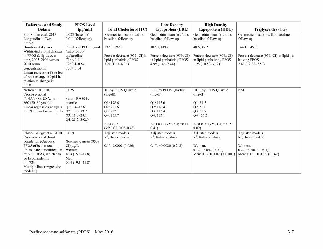
Reference and Study
Details PFOS Level
(µg/mL) Total Cholesterol (TC) Low Density
Lipoprotein (LDL) High Density
Lipoprotein (HDL) Triglycerides (TG) Fitz-Simon et al. 2013 Longitudinal (C8); n = 521 Duration: 4.4 years Within-individual changes in PFOS & lipids over time, 2005–2006 versus 2010 serum concentrations. Linear regression fit to log of ratio change in lipid in relation to change in PFOS
0.023 (baseline) 0.011 (follow-up) Tertiles of PFOS ng/ml (ratio follow up/baseline) T1: < 0.4 T2: 0.4–0.54 T3: > 0.54
Geometric mean (mg/dL): baseline, follow-up 192.5, 192.8 Percent decrease (95% CI) in lipid per halving PFOS 3.20 (1.63–4.76)
Geometric mean (mg/dL): baseline, follow-up 107.8, 109.2 Percent decrease (95% CI) in lipid per halving PFOS 4.99 (2.46–7.44)
Geometric mean (mg/dL): baseline, follow-up 48.6, 47.2 Percent decrease (95% CI) in lipid per halving PFOS 1.28 (−0.59–3.12)
Geometric mean (mg/dL): baseline, follow-up 144.1, 146.9 Percent decrease (95% CI) in lipid per halving PFOS 2.49 (−2.88–7.57)
Nelson et al. 2010 Cross-sectional (NHANES), USA. n = 860 (20–80 yrs old) Linear regression analysis for PFOS and serum lipids
0.025 Serum PFOS by quartile Q1: 1.4–13.6 Q2: 13.8–19.7 Q3: 19.8–28.1 Q4: 28.2–392.0
TC by PFOS Quartile (mg/dl): Q1: 198.6 Q2: 201.6 Q3: 202 Q4: 205.7 Beta 0.27 (95% CI; 0.05–0.48)
LDL by PFOS Quartile (mg/dl): Q1: 113.6 Q2: 116.4 Q3: 113.4 Q4: 123.1 Beta 0.12 (95% CI; −0.17– 0.41)
HDL by PFOS Quartile (mg/dl): Q1: 54.3 Q2: 56.0 Q3: 52.7 Q4 : 55.2 Beta 0.02 (95% CI; −0.05–0.09)
NM
Château-Degat et al. 2010 Cross-sectional, Inuit population (Quebec). PFOS effect on total lipids. Effect modification of n-3 PUFAs, which can be hypolipidemic n = 723 Multiple linear regression modeling
0.019 Geometric mean (95% CI) µg/L Women: 16.8 (15.8–17.8) Men: 20.4 (19.1–21.8)
Adjusted models R2, Beta (p value) 0.17, 0.0009 (0.086)
Adjusted models R2, Beta (p value) 0.17, −0.0020 (0.242)
Adjusted models R2, Beta (p value) Women: 0.12, 0.0042 (0.001) Men: 0.12, 0.0016 (< 0.001)
Adjusted models R2, Beta (p value) Women: 0.20, −0.0014 (0.04) Men: 0.16, −0.0009 (0.162)
Perfluorooctane sulfonate (PFOS) – May 2016 3-7

Reference and Study
Details PFOS Level
(µg/mL) Total Cholesterol (TC) Low Density
Lipoprotein (LDL) High Density
Lipoprotein (HDL) Triglycerides (TG) Eriksen et al. 2013 Cross-sectional, Middle aged Danish population n = 753 (663 men and 90 women) Generalized linear models used for analysis
0.036 Differences in TC (mg/dl) per 1 IQR increase Beta (95% CI): Total population: 3.7 (0.1, 7.3) Women: 11.7 (−0.2, 23.6) Men: 2.9 (−0.9, 6.7)
NM NM NM
Fisher et al. 2013 Cross-sectional, 2007–2009, Canadian Health Measures Survey (CHMS) Cycle 1. n = 2700 (aged 18–74) Used multivariate linear and logistic regression models to assess associations between PFOS and serum lipids.
0.0084 Unadjusted OR for high cholesterol compared to Q1 of PFOS exposure: OR (95% CI) Q1: Referrent Q2: 1.12 (0.89, 1.41) Q3: 1.15(0.91, 1.45) Q4: 1.66 (1.32, 2.09) p trend = 0.03 Null effects in adjusted model
Null effects Null effects NM
Children and Adolescents Frisbee et al. 2010 Cross-sectional (C8, children) GLM Analysis, n = 12,476 Differences of Estimated Marginal Mean (EMM) between Q1 and Q5 and regression analysis for Q trend
0.023 Differences in Estimated Marginal Mean (EMM), Beta (SE), p for trend: Age 1 to < 12: 5.5, 1.3 (0.3), < 0.001 Age 12 to < 18: 9.5, 2.1 (0.4), < 0.001
Differences in EMM, B(SE) p for trend: Age 1 to < 12: 3.4, 0.9 (0.3), .002 Age 12 to < 18: 7.5, 1.7 (0.2), < 0.001
Differences in EMM, B(SE), p for trend: Age 1 to < 12: 1.6, 0.3 (0.1), 0.007 Age 12 to < 18: 1.5, 0.4 (0.1), 0.001
Differences in EMM, B(SE), p for trend: Age 1 to < 12: 2.8, 0.1 (1.4), 0.99 Age 12 to <1 8: 2.8, −0.1 (1.0), 0.90
Geiger et al. 2014a Cross-sectional, NHANES, 1999–2008, dyslipidemia (TC, LDL, HDL, TG). n = 815 (Age < 18) Multivariate regression analysis. n = 815
T1: < 12.1 T2: 12.1–21.8 T3: > 21.8 ppb
TC (mg/dL) association with PFOS by tertiles T1: 1 T2: 1.73 (−2.89, 6.36) T3: 3.91 (−1.32, 9.14) p trend: 0.15 log transformed PFOS Beta 0.04 (95% CI: 0.00–0.08)
Association between PFOS and LDL: T1: 1 (referent) T2: 0.49 (−3.41, 4.38) T3: 4.59 (−0.17, 9.35) P trend: 0.0632 log transformed PFOS Beta 2.83 (95% CI: 0.03–5.37)
Association between PFOS and HDL: T1: 1 T2: 2.86 (0.44, 5.28) T3: 1.11 (−0.93, 3.15) P trend: 0.2931
Association between PFOS and TG: T1: 1 T2:−8.13 (−15.50, −0.77) T3: −8.89 (−15.67, −2.11) P trend: 0.0126 log transformed PFOS Beta −3.90 (95% CI: −7.72 to −0.08)
Perfluorooctane sulfonate (PFOS) – May 2016 3-8

Reference and Study
Details PFOS Level
(µg/mL) Total Cholesterol (TC) Low Density
Lipoprotein (LDL) High Density
Lipoprotein (HDL) Triglycerides (TG) Lin et al. 2009 Cross-sectional, NHANES, 1999–2000, 2003–2004. Adolescents and adults aged > 12 yrs n = 3,685
Mean (SEM) Log PFOS 12 to < 20 yrs olds: 3.11 (0.05) ng/mL 20 yrs old and older: 3.19 (0.04) ng/mL
NM NM OR (95% CI), p 1.61 (1.15–2.26), p < 0.05 in those 20 yrs or older
Null findings
Maisonet et al. 2015 Avon Longitudinal Study of Parents and Children. Prenatal PFOS compared to serum lipids in female offspring. n = 111 (age 7), n = 88 (age 15)
Mean (SD) 22.2 (11.4) mg/dl
Non-linear associations of TC with PFOS.
Non-linear associations of LDL with PFOS.
Null findings Null findings
Timmermann et al. 2014 Danish children, aged 8–10 years old. Linear regression models. 1997. n = 499
Median 41.5 ng/mL Null findings in normal weight children.
Null findings in normal weight children.
Null findings in normal weight children.
Null findings in normal weight children. In overweight children, 10 ng increase PFOS/mL plasma associated with 8.6% (95% CI: 1.2%–16.5%) higher triglyceride concentrations
Pregnant Women Starling et al. 2014 Cross-sectional (maternal at 14–26 weeks gestation), Norwegian Mother and Child Cohort (MoBa) 2003–2004. n = 891
0.013 Quartiles (ng/mL): Q1: < 10.31 Q2: 10.31–13.03 Q3: 13.04–16.59 Q4: > 16.60
B (95% CI) PFOS (ng/ml) and TC (mg/dL). Q1: Referrent Q2: −3.35 (−10.34, 3.64) Q3: 3.06 (−4.93, 11.05) Q4: 7.59 (−0.42, 15.60) TC change per IQR change in PFOS: 4.25 (0.81, 7.69)
B (95% CI) PFOS (ng/ml) and LDL(mg/dL). Q1: referrent Q2: −3.23 (−9.28, 2.83) Q3: 2.60 (−4.49, 9.70) Q4: 5.51 (−1.62, 12.64) LDL change per IQR PFOS change: 3.07 (−0.03, 6.18)
B (95% CI) PFOS (ng/ml) and HDL (mg/dL). Q1: Referrent Q2: 1.96 (−0.39, 4.31) Q3: 2.49 (0.00, 4.97) Q4: 4.45 (2.04, 6.86) HDL change per IQR change in PFOS: 2.08 (1.12, 3.04)
B (95% CI) PFOS (ng/ml) and TG (mg/dL). Q1: Referrent Q2: 0.00 (−0.06, 0.07) Q3: −0.03 (−0.10, 0.05) Q4: 0.00 (−0.07, 0.07) TG change per IQR PFOS change: −0.01 (−0.04, 0.02)
NM = Not Measured
Perfluorooctane sulfonate (PFOS) – May 2016 3-9

Some of the studies that examined serum LDL and HDL cholesterol also found significant
increases these measures. Neither of these lipoprotein complexes is a stand-alone indicator for cardiovascular decrease risk. Rather, it is the relationship across the lipoprotein complexes within the same individuals that is important with HDLs considered as protective and LDLs a biomarker for potential atherosclerosis. Relatively few studies of triglycerides noted a significant increase with the serum PFOS levels.
3.1.1.2 Liver Enzymes and Liver Disease
Cross-sectional studies and longitudinal studies of PFOS and liver enzymes in various populations are described below and summarized in Table 3-2.
Table 3-2. Summary of Epidemiology Studies of PFOS and Liver Enzymes Reference and Study Details PFOS Level (µg/mL) Results
Lin et al. 2010 n = 2,216 adults (1,076 men and 1,140 women) Age: > 20 years old Data from 1999–2000 and 2003–2004 NHANES Regression models used to analyze data and adjust for confounders
Mean levels Women: 0.0222 Men: 0.0274
Linear regression coefficients (standard error), p-value (adjusted for age, gender, race, lifestyle, measurement data, etc.) ALT (U/L): 1.01 (0.53), 0.066 (slight pos. association) GGT (U/L): 0.01 (0.03), p = 0.81 Total bilirubin (µmol): 0.30 (0.24), p = 0.22
Gallo et al. 2012 n = 47,092 Data from those enrolled in C8 Health Project Linear and logistic regression models used.
Mean level: 0.0233 Linear regression coefficients, (partial R2) Ln-ALT: 0.020, 95% CI: 0.014–0.026 (< 0.001)
Raised ALT in logistic regression odds ratio, (p-value) OR: 1.13, 95% CI: 1.07–1.18 (p < 0.001)
GGT: no association Direct bilirubin: less consistent results
Lin et al. (2010) investigated the association between low-dose serum PFOS (along with three other individual PFAS) and liver enzymes in the adult population of the United States by analyzing data from the 1999–2000 and 2003–2004 NHANES. The study population included 2,216 adults (1,076 males, 1,140 females) older than 20 years who were not pregnant or nursing; had fasted > 6 hours at the time of examination; were negative for hepatitis B or C virus; had body weight, height, educational attainment, and smoking status data available; and had serum tests for PFAS, liver function, and metabolic syndrome. Regression models were used to analyze the data and adjust for confounding factors. Mean PFOS levels were 0.0274 and 0.0222 µg/mL for males and females, respectively.
Serum PFOS concentration was divided into quartiles. Unadjusted liver enzymes, serum ALT, and log-GGT increased across quartiles of PFOS (p ≤ 0.03), but total bilirubin showed no trend. The linear regression models were adjusted for:
• Age, gender, and race/ethnicity. • Lifestyle (smoking status, drinking status, education level). • Biomarker data (BMI, metabolic syndrome, iron saturation status, insulin resistance).
Perfluorooctane sulfonate (PFOS) – May 2016 3-10

In the fully adjusted model, a slight positive association was found between serum PFOS
concentration and serum ALT (p = 0.066). A positive association was also found between serum PFOA concentration and serum ALT and PFOA concentration and serum GGT. Data interpretation was limited by the cross-sectional study design, and the fact that other environmental chemicals (possible covariates or explanatory variables) and medication use were not included in the study.
Gallo et al. (2012) investigated the correlation between serum PFOS levels and liver enzymes in a total of 47,092 samples collected from members enrolled in the C8 Health Project. The association of ALT, GGT, and direct bilirubin with PFOS was assessed using linear regression models adjusted for age, physical activity, body mass index, average household income, education level, race, alcohol consumption, and cigarette smoking. Median PFOS level was 0.0233 µg/mL with an interquartile range of 0.0137–0.0294 µg/mL. The ln-transformed values of ALT were significantly associated with ln-transformed PFOS levels (and PFOA) and showed a steady increase in fitted levels of ALT per decile of PFOS, leveling off after approximately 0.030 µg PFOS/mL. Fitted values of GGT showed no overall association with ln-transformed PFOS levels. A positive association was seen with direct bilirubin and PFOS levels in linear regression models, but this was not evident with logistic regression models. Limitations of the study include the cross-sectional design and self-reported lifestyle characteristics. Only a small number of ALT values were outside the normal range, making the results difficult to interpret in terms of health.
The epidemiological data supporting liver damage based on serum ALT and GGT as reported by Gallo et al. (2012) are not strong enough to support an association of serum PFOS alone with liver damage in humans, because in most of the epidemiology studies the serum contains a mixture of PFASs and possibly other exogenous chemicals.
3.1.1.3 Biomarkers of Kidney Function and Kidney Disease
Epidemiology studies of PFOS and kidney function and biomarkers in various populations are described below and summarized in Table 3-3.
Shankar et al. (2011) used data from the NHANES to determine whether there was a relationship between serum PFOS levels and chronic kidney disease. A total of 4,587 adult participants (51.1% females) with PFOS measurements available from the 1999–2000 and 2003–2008 cycles of the survey were examined. Chronic kidney disease was defined as glomerular filtration rate (GFR) < 60 mL/minute (min)/1.73 m2. Serum PFOS levels were categorized into quartiles: quartile 1 = < 0.012 µg/mL; quartile 2 = 0.012–0.019 µg/mL; quartile 3 = 0.019–0.030 µg/mL; quartile 4 = > 0.030 µg/mL. The multivariable odds ratio for chronic kidney disease for individuals in quartile 4 was 1.82 (95% CI: 1.01–3.27; p for trend = 0.019) compared with individuals in quartile 1. This association was shown to be independent for confounders of age, sex, race/ethnicity, body mass index, diabetes, hypertension, and serum cholesterol level. However, the authors noted that because of the cross-sectional nature of the study, the possibility of reverse causality could not be excluded. A low GFR would diminish the removal of PFOS from serum for excretion by the kidney, thus increasing the serum PFOS levels.
Perfluorooctane sulfonate (PFOS) – May 2016 3-11

Table 3-3. Summary of Epidemiology Studies of PFOS and Measures of Kidney Function Reference and Study Details PFOS levels (µg/mL) Results
Shankar et al. 2011 USA, NHANES n = 4587 adults PFOS from 1999–2000 and 2003–2008
Quartiles, µg/mL, n
1: < 0.012 µg/mL, 1,152 2: 0.012–0.019 µg/mL, 1,151 3: 0.019–0.030 µg/mL, 1,137 4: > 0.030 µg/mL, 1,147
Estimated glomerular filtration rate (eGFR) Chronic kidney disease defined as eGFR < 60 mL/minute/1.73 m2
Quartile, OR (95% CI) 1: Referent 2: 1.12 (0.64, 1.99) 3: 1.53 (0.87, 2.67) 4: 1.82 (1.01, 3.27) p = 0.02
Logistic regression adjusting for age, gender, race/ethnicity, education, smoking, alcohol, BMI, systolic blood pressure, diastolic blood pressure, diabetes, serum total cholesterol and glycohemoglobin Adjustment for PFOS did not alter association with PFOA
Multivariate regression of association PFOS with eGFR among subjects with and without chronic kidney disease β (SE) with −1.8 (0.8) and without −3.2 (0.6) chronic kidney disease (p < 0.05)
Steenland et al. 2010 USA, C8 Health Project participants n = 54,591 (≥ 20 yrs old)
Mean: 0.0234 ± 0.0161 Increased predicted uric acid of 0.2 to 0.3 µg/dL with increasing deciles of PFOS.
Odds Ratio, p-value Hyperuricemia (> 6.0 mg/dL for women and > 6.8 mg/dL for men): 1.00 1.02 (95% CI: 0.95−1.10), p < 0.0001 1.11 (95% CI: 1.04−1.20), p < 0.0001 1.19 (95% CI: 1.11−1.27), p < 0.0001 1.26 (95% CI: 1.17−1.35), p < 0.0001
Trend for increase uric acid more prominent with PFOA
Children Watkins et al. 2013 USA, C8 Health Project participants n = 9,660 (1 to < 18 yrs old)
Median: 0.020 β (95% CI) change in unit eGFR (mL/min/1.73 m2 ) per ln serum PFOS, −1.10 (−1.66 to −0.53), p = 0.0001 Linear regression adjusting for age, gender, race, smoking, and household income.
Geiger et al. 2014b USA, NHANES n = 1644 (12–18 yrs old)
Mean: 0.018 ± 0.005 Multivariable-adjusted OR (95% CI) between PFOS and hypertension
Quartile 1: 1 (referent) Quartile 2: 0.99 (0.55, 1.78) Quartile 3: 0.73 (0.36, 1.61) Quartile 4: 0.77 (0.37, 1.61) p = 0.36 Log transformed PFOS = 0.83 (0.58, 1.19)
Perfluorooctane sulfonate (PFOS) – May 2016 3-12

Steenland et al. (2010) reported on another analysis of the C8 Health Project participants
≥ 20 years old (n = 54,591) for a possible association between PFOS (and PFOA) serum levels and uric acid. Elevated uric acid is a risk factor for hypertension and may be an independent risk factor for stroke. The mean PFOS level was 0.0234 ± 0.0161 µg/mL. A statistically-significant (p < 0.0001) trend was observed between increasing PFOS levels (untransformed) and uric acid levels. A 0.2–0.3 µg/dL increase in uric acid was associated with an increase from the lowest to highest PFOS decile (0.010–0.050 µg/mL). Hyperuricemia (> 6.0 mg/dL for females and > 6.8 mg/dL for males) risk by quintiles increased slightly with PFOS levels (OR 1.00, 1.02, 1.11, 1.19, and 1.26). The serum of C8 study participants included several PFASs; PFOA appeared to have a greater influence on uric acid trends than PFOS in the models employed by Steenland et al. (2010).
Children. Watkins et al. (2013) evaluated the cross-sectional association between PFOS exposure and kidney function among children aged 1 to <18 years (mean 12.4 ± 3.8 years) enrolled in the C8 Health Project. A total of 9,660 participants had data available on serum PFOS (median = 0.020 µg/mL), serum creatinine, and height, which were used to calculated an estimated glomerular filtration rate (eGFR). Linear regression was used to evaluate the association between quartiles of measured serum PFOS concentration and eGFR. A shift from the lowest to the highest quartile of measured, natural log–transformed concentrations of PFOS in serum [IQR ln-(PFOS) = 0.64] was associated with a decrease in eGFR of 1.10 mL/min/1.73 m2 (95% CI: −1.66 to −0.53; p = 0.0001) adjusting for age, sex, race, smoking status, and house-hold income. With increasing quartile of serum PFOS concentrations, eGFR decreased mono-tonically with a decrease of 2.3, 2.6, and 2.9 mL/min/1.73 m2 for the second, third, and fourth quartile of serum PFOS, respectively, compared with the lowest quartile (p for trend across quartiles = 0.0001).
Geiger et al. (2014b) used data from the NHANES to determine whether there was a relationship between serum PFOS levels and hypertension in children. A total of 1,655 participants (aged 12–18 years) with PFOS measurements available from the 1999–2000 and 2003–2008 cycles of the survey were examined. Blood pressure was measured to determine the presence of hypertension and linear regression modeling was used to study the association between increasing quartiles of serum PFOS and mean changes in systolic and diastolic blood pressures. Mean PFOS level was 0.018 ± 0.005 µg/mL. No association was found between serum PFOS levels and hypertension in either unadjusted or multivariable-adjusted analyses. Compared with the lowest quartile, the multivariable-adjusted odds ratio (95% confidence interval) of hypertension in the highest quartile of exposure was 0.77 (0.37–1.61) (p-trend > 0.30).
3.1.1.4 Reproductive Hormones and Reproductive/Developmental Studies
Many of the studies of PFOS focused on pregnancy-related outcomes, including measures of fetal growth retardation, puberty, and other developmental endpoints, as well as pregnancy-related hypertension, preeclampsia, and gestational diabetes. Reproductive outcomes such as measures affecting fertility were also evaluated. Within each section, the discussion is divided into occupational exposure studies (if applicable) and general population studies. Epidemiology studies of PFOS and pregnancy-related outcomes in various populations are described below and summarized in Table 3-4.
Perfluorooctane sulfonate (PFOS) – May 2016 3-13

Table 3-4. Summary of Epidemiology Studies of PFOS and Pregnancy Outcomes
Study PFOS level
(µg/mL) Results Stein et al. 2009 United States (C8 Health Project) n = 5,262 pregnancies Self-reported pregnancy outcomes in mid-Ohio Valley in 2000–2006.
Median: 0.014 OR (95% CI), preeclampsia per IQR(lnPFOS) increase in PFOS: 1.1 (0.9, 1.3)
< 50th percentile 1.0 (referent) ≥ 50th percentile 1.3 (1.1, 1.7) < 50th percentile 1.0 (referent) 50th–<75th percentile 1.3 (1.0, 1.7) 75th–90th percentile 1.1 (0.8, 1.6) ≥ 90th percentile 1.6 (1.2, 2.3)
Darrow et al. 2013 United States (C8 Health Project) n = 1,630 live births from 1,330 women after January 1, 2005
Geometric mean: 0.0132
Pregnancy induced hypertension OR (95% CI) per log unit increase in PFOS: 1.47 (1.06, 2.04) By quintile:
Q1 up to 0.0086 µg/mL 1.0 (referent) Q2 0.0086– < 0.0121 1.46 (0.69, 3.11) Q3 0.0121– < 0.0159 2.71 (1.33, 5.52) Q4 0.0159– < 0.0214 2.21 (1.07, 4.54) Q5 ≥ 0.0214 1.56 (0.72, 3.38) Q1 up to 0.0086 µg/mL 1.0 (referent)
First pregnancy after PFOS measure 2.02 (1.11, 3.66)
Zhang et al. 2015 n = 258 women as part of LIFE study. Blood samples taken during 2005–2009.
Mean: 0.0131 with gestational diabetes and 0.012 without
Gestational diabetes OR (95% CI) associated with SD increment of preconception PFOS log-transformed concentration OR 1.13 (0.75, 1.72) (fully adjusted for age, BMI, smoking, etc.)
Pregnancy-related Outcomes. Stein et al. (2009) examined serum levels of PFOS and self-reported pregnancy outcomes of a population of females (5,262 pregnancies; aged 15–55 years) in the mid-Ohio Valley in 2000–2006. These females were enrollees in the C8 Health Project, a community health study of residents near a chemical plant that used PFOA in the manufacture of fluoropolymers. Pregnancies within the 5 years preceding the exposure measurements were analyzed. The mean level of PFOS in the serum of these females was 0.014 µg/mL. There was no association between PFOS levels and miscarriages. PFOS was associated with preeclampsia (adjusted odds ratio = 1.3; 95% CI: 1.1–1.7). Similarly, PFOA was not associated with miscarriage and only weakly associated with preeclampsia. The self-reported nature of pregnancy outcomes is a recognized limitation with uncertain impact on study results.
Darrow et al. (2013; 2014) analyzed pregnancy outcomes for the five years following enrollment in the C8 Health Project. Among the 69,030 females who provided serum for PFOS measurement in 2005–2006, 32,354 provided follow-up interviews on reproductive histories. After exclusions, 1,630 singleton live births from 1,330 females after January 1, 2005 were linked to birth records to identify outcomes of preterm birth (i.e., < 37 gestational weeks), pregnancy-induced hypertension, low birth weight (LBW) (i.e., < 2500 grams), and birth weight among full-term infants (Darrow et al. 2013). Effects on fetal growth measures are described in that section below. Another subset of 1,129 females with a total of 1,438 pregnancies was evaluated for an association between PFOS levels and miscarriage (Darrow et al. 2014). The baseline mean PFOS level for these females was 0.016–0.017 µg/mL. Confounders that were adjusted in each model for every outcome in the 2013 Darrow et al. study included maternal age,
Perfluorooctane sulfonate (PFOS) – May 2016 3-14

educational level, smoking status, parity, BMI, self-reported diabetes, time between conception and serum measurement. Parity was excluded, and race was included in the miscarriage analysis (Darrow et al. 2014).
An increased risk of pregnancy-induced hypertension was detected per log unit increase in PFOS (OR = 1.47; 95% CI: 1.06−2.04) and PFOA (OR = 1.27; 95% CI: 1.05−1.55). Although monotonicity was not evident, consistently increased odds were found across all upper PFOS (OR range: 1.46–2.72) and PFOA (OR range: 2.39–3.43) quintiles.
The odds of miscarriage per each log unit increase in PFOS was 1.21 (95% CI: 0.94−1.55) for all reported prospective pregnancies and 1.34 (95% CI: 1.02−1.76) when restricted the analysis to each woman’s first pregnancy. Miscarriage results were comparable across all PFOS quintiles in the primary analysis (OR range: 1.34–1.59) and those restricted to first pregnancy (OR range: 1.68–1.94). PFOA was not associated with miscarriage and was not a confounder of the observed association with PFOS. To address the potential for reverse causality related to PFAS levels decreasing from prior pregnancies, analyses were restricted to nulliparous and nulligravid females. Adjusted odds ratios were higher across all four quintiles for nulliparous (OR range: 1.88–3.08) and nulligravid females (OR range: 2.04–3.73). These studies represent prospective assessment of PFASs in relation to adverse pregnancy outcomes, which address some of the limitations in the available cross-sectional studies. The impact of measurement error resulting from unknown critical exposure windows and the time lag (> 99% of births were within 3 years) between the estimated conception date and the serum collection is unclear in these studies.
Preconception serum levels of PFOS (and other PFASs) were evaluated in females attempting pregnancy in relation to risk of developing gestational diabetes (Zhang et al. 2015). The 258 participants were members of the Longitudinal Investigation of Fertility and the Environment (LIFE) study with blood samples taken during 2005–2009. The ORs and 95% CIs of gestational diabetes associated with each SD increment of preconception serum PFOS concentration (log-transformed) (and six other PFAS) were estimated with the use of logistic regression after adjusting for age, pre-pregnancy body mass index, smoking, and parity, each conditional on the number of times a woman had been pregnant. Preconception mean serum PFOS levels were 0.0131 µg/mL in females with gestational diabetes and 0.012 µg/mL in females without gestational diabetes (p-value for mean difference = 0.10). A positive association was found between PFOS and risk of gestational diabetes in the fully adjusted model (OR = 1.13; 95% CI: 0.75−1.72). PFOA was the only PFAS that was significantly associated with developing gestational diabetes in this analysis.
Fetal Growth. Many different measures of fetal growth can be used in epidemiology studies. Birth weight is widely available (as it is routinely collected in medical records and birth certificates). LBW (defined as < 2500 g) can be a proxy measure for preterm birth (particularly when accurate gestational age dating is not available). Other measures of fetal growth, such as small for gestation age, tend to more accurately reflect fetal growth retardation. Epidemiology studies of PFOS and fetal growth are described below and summarized in Table 3-5.
Perfluorooctane sulfonate (PFOS) – May 2016 3-15

Perfluorooctane sulfonate (PFOS) – May 2016 3-16
Table 3-5. Summary of Epidemiology Studies of PFOS and Fetal Growth Study PFOS level (µg/mL) Results
Grice et al. 2007 United States (C8 Health Project) n = 263 women reporting 429 births Self-reported pregnancy outcomes in workers associated with perfluorinated chemical production factory.
Exposure to PFOS was based on job assignment and varied
Never exposed: 0.11–0.29 ppm Low exposure: 0.39–0.89 ppm High exposure: 1.30–1.97 ppm
No association between PFOS exposure and mean birth weight Regression coefficients for birth weight compared to never-exposed pregnancies, 95% CI (adjusted for maternal age, smoking, gravidity)
Ever exposed, low exposure −0.08 (−0.25, 0.09) Ever exposed, high exposure 0.07 (−0.14, 0.28) High exposure, > 1 yr 0.11 (−0.11, 0.33) Low or high exposure, > 1 yr −0.03 (−0.19, 0.13) Ever exposed, low or high - 0.05 (−0.20, 0.11)
Apelberg et al. 2007 United States (Baltimore) n = 293 newborns born between November 2004 and March 2005 Cord blood samples
Geometric mean: 0.005 Change in birth weight (g) per log unit increase (95% CI) −69 (−149, 10)
Fei et al. 2007 n = 1,400 women and their infants randomly selected from the group enrolled in the DNBC
Mean: 0.035 LBW
OR (95% CI) for LBW by quartile Q1 0.0064 to 0.026 µg/dL 1.0 (referent) Q2 0.026 to 0.033 µg/dL 3.5 (0.37, 31.16) Q3 0.033 to 0.043 µg/dL 6.0 (0.73, 49.34) Q4 ≥ 0.043 µg/dL 4.8 (0.56, 41.16) Trend: p = 0.13
Andersen et al. 2010 n = 1,010 women and their infants randomly selected from the group enrolled in the DNBC
Median: 0.0334 (range: 0.0064–0.1067)
PFOS concentrations per each 0.001 µg/mL increase inversely associated with: birth weight in girls: Beta = −3.2; 95% CI: −6.0 to −0.3 weight at 12 months in boys: Beta = −9.0; 95% CI: −15.9 to −2.2
Monroy et al. 2008 n = 101 pregnant women as part of a larger cohort study conducted at McMaster University Medical Center
Mean: 0.0183 in maternal serum (24–28 wks) 0.0162 in maternal serum at delivery 0.0072 in umbilical cord blood
No association between PFOS levels and infant birth weight
Change in PFOS per g change in birth weight Beta = 0.000853 (p = 0.73)
Washino et al. 2009 Japan n = 428 women and their infants between July 2002 and October 2005
Mean: 0.006 Change in birth weight per log unit increase (95% CI) For all: Beta = −149 g (−297.0, −0.5) For female infants: Beta = −269.4 g (−465.7, −73.0)
Hamm et al. 2010 Canada n = 252 women with blood samples taken between December 2005 and June 2006
Mean: 0.009 Change in birth weight per Ln unit increase (95% CI) 31.3 g (−43.3, 105.9)
Stein et al. 2009 Mean: 0.014 OR (95% CI), birth weight < 5.5 lbs. per IQR(lnPFOS) increase in PFOS: 1.3 (1.1, 1.6)
< 50th percentile 1.0 (referent) ≥ 50th percentile 1.5 (1.1, 1.9) < 50th percentile 1.0 (referent) 50th–<75th percentile 1.3 (0.9, 1.8) 75th–90th percentile 1.6 (1.1, 2.3) ≥ 90th percentile 1.8 (1.2, 2.8)
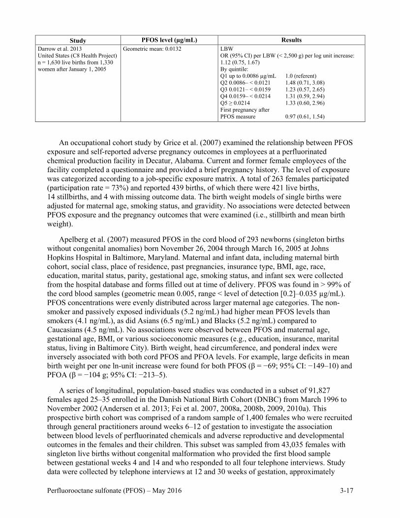
Study PFOS level (µg/mL) Results
Darrow et al. 2013 United States (C8 Health Project) n = 1,630 live births from 1,330 women after January 1, 2005
Geometric mean: 0.0132
LBW OR (95% CI) per LBW (< 2,500 g) per log unit increase: 1.12 (0.75, 1.67) By quintile: Q1 up to 0.0086 µg/mL 1.0 (referent) Q2 0.0086– < 0.0121 1.48 (0.71, 3.08) Q3 0.0121– < 0.0159 1.23 (0.57, 2.65) Q4 0.0159– < 0.0214 1.31 (0.59, 2.94) Q5 ≥ 0.0214 1.33 (0.60, 2.96) First pregnancy after PFOS measure 0.97 (0.61, 1.54)
An occupational cohort study by Grice et al. (2007) examined the relationship between PFOS exposure and self-reported adverse pregnancy outcomes in employees at a perfluorinated chemical production facility in Decatur, Alabama. Current and former female employees of the facility completed a questionnaire and provided a brief pregnancy history. The level of exposure was categorized according to a job-specific exposure matrix. A total of 263 females participated (participation rate = 73%) and reported 439 births, of which there were 421 live births, 14 stillbirths, and 4 with missing outcome data. The birth weight models of single births were adjusted for maternal age, smoking status, and gravidity. No associations were detected between PFOS exposure and the pregnancy outcomes that were examined (i.e., stillbirth and mean birth weight).
Apelberg et al. (2007) measured PFOS in the cord blood of 293 newborns (singleton births without congenital anomalies) born November 26, 2004 through March 16, 2005 at Johns Hopkins Hospital in Baltimore, Maryland. Maternal and infant data, including maternal birth cohort, social class, place of residence, past pregnancies, insurance type, BMI, age, race, education, marital status, parity, gestational age, smoking status, and infant sex were collected from the hospital database and forms filled out at time of delivery. PFOS was found in > 99% of the cord blood samples (geometric mean 0.005, range < level of detection [0.2]–0.035 µg/mL). PFOS concentrations were evenly distributed across larger maternal age categories. The non-smoker and passively exposed individuals (5.2 ng/mL) had higher mean PFOS levels than smokers (4.1 ng/mL), as did Asians (6.5 ng/mL) and Blacks (5.2 ng/mL) compared to Caucasians (4.5 ng/mL). No associations were observed between PFOS and maternal age, gestational age, BMI, or various socioeconomic measures (e.g., education, insurance, marital status, living in Baltimore City). Birth weight, head circumference, and ponderal index were inversely associated with both cord PFOS and PFOA levels. For example, large deficits in mean birth weight per one ln-unit increase were found for both PFOS (β = −69; 95% CI: −149–10) and PFOA (β = −104 g; 95% CI: −213–5).
A series of longitudinal, population-based studies was conducted in a subset of 91,827 females aged 25–35 enrolled in the Danish National Birth Cohort (DNBC) from March 1996 to November 2002 (Andersen et al. 2013; Fei et al. 2007, 2008a, 2008b, 2009, 2010a). This prospective birth cohort was comprised of a random sample of 1,400 females who were recruited through general practitioners around weeks 6–12 of gestation to investigate the association between blood levels of perfluorinated chemicals and adverse reproductive and developmental outcomes in the females and their children. This subset was sampled from 43,035 females with singleton live births without congenital malformation who provided the first blood sample between gestational weeks 4 and 14 and who responded to all four telephone interviews. Study data were collected by telephone interviews at 12 and 30 weeks of gestation, approximately
Perfluorooctane sulfonate (PFOS) – May 2016 3-17

6 and 18 months after birth, and when the children were 7 years of age. A food frequency questionnaire was filled out at home during approximately week 25 of pregnancy. Maternal blood samples were taken in the first and second trimester, and infant cord blood was sampled just after birth. Only blood results from the 1,400 females in the first trimester were reported. Mean plasma PFOS levels by age groups were: < 25 years: 0.039 µg/mL; 25–29 years: 0.037 µg/mL; 30–34 years: 0.034 µg/mL and ≥ 35 years: 0.033 µg/mL.
Potential confounders for which adjustments were made included: maternal age, maternal occupation and educational status, parity, pre-pregnancy BMI, smoking/alcohol consumption during pregnancy, gestational weeks at blood draw, child’s sex, child’s age at interview with mother, breast-feeding > 6 months (for 18-month interview), out-of-home child care, hours mother spent with child per day, and home density (the total number of rooms divided by the total number of people in the household). Although dietary data were available for at least 80% of the births, it is unclear why some of these studies did not examine these data as confounders (e.g., Fei et al. 2009). Although the DNBC had a low participation rate (31%), a previous study of various exposures in relation to three different outcomes (preterm birth, small-for-gestational-age, infancy and antepartum stillbirth) did not provide any evidence of non-participation bias (Nohr et al. 2006).
Using data from the DNBC, Fei et al. (2007) investigated the association between plasma levels of PFOS in pregnant females, length of gestation, preterm birth (i.e., < 37 gestational weeks), and infant birth weight. The average PFOS levels in maternal plasma were 0.035µg/mL (range: 0.0064–0.107 µg/mL). The data were adjusted for confounding factors that might also influence fetal growth or length of gestation and analyzed by analysis of variance and linear regression using both continuous PFOS concentrations and PFOS quartiles. No associations between PFOS and birth weight were found. PFOA concentrations based on the continuous exposure measures were inversely associated with birth weight (β= −10.6; 95% CI: −20.8 to −0.5) following adjustment for confounding (unadjusted β= −20.5; 95% CI: −31.5 to −9.6). Although most were not statistically-significant, ORs for preterm birth were consistent in magnitude (OR range: 1.43–2.94) across both the upper three PFOS and PFOA quartiles. Consistently elevated ORs were also detected (OR range: 3.39–6.00) for LBW across the upper three PFOS and PFOA quartiles, but all of these analyses were limited by very small cell sizes given low incidence of these outcomes. Although these ORs often lacked statistical significance due to low statistical power, the elevated odds detected between PFOS levels and various outcomes including preterm delivery and LBW warrant further research, especially given the potential generalizability limitations of this low-risk study population.
Fei et al. (2008a) also investigated the association between PFOS levels and placental weight, birth length, and head and abdominal circumference in the DNBC study population. Maternal PFOS levels were not associated with any of the fetal growth indicators when the lowest quartile was compared to the highest. In a stratified analysis of PFOS, inverse associations were found with birth length for post-term and pre-term infants and with ponderal index (relationship between mass and height) in multiparous females. In nulliparous females the association was positive. These associations were not statistically-significant.
Andersen et al. (2010) examined the association between maternal plasma PFOS concentration and offspring weight, length, and BMI at 5 and 12 months of age from participants in the DNBC. The mothers (n = 1,010) reported the information during an interview and weight and length measurements were used to calculate BMI. Median maternal plasma PFOS level was 0.0334 µg/mL with a range of 0.0064–0.1067 µg/mL. PFOS concentrations (per each 0.001
Perfluorooctane sulfonate (PFOS) – May 2016 3-18

µg/mL increase) were inversely associated with birth weight in girls (β= −3.2; 95% CI: −6.0 to −0.3), weight at 12 months in boys (β= −9; 95% CI: −15.9 to −2.2), and BMI at 12 months in boys (β= −0.017; 95% CI: −0.028 to −0.005) in models adjusted for maternal age, parity, prepregnancy BMI, smoking, gestational age at blood draw, socioeconomic status, and breastfeeding. Similar inverse associations were found with PFOA only in boys.
Monroy et al. (2008) examined the relationship between the maternal serum levels of PFOS and PFOA and infant birth weight from neonates born to 101 pregnant females enrolled in a large cohort study, the Family Study, conducted at McMaster University Medical Center in Ontario, Canada. Linear regression analyses were adjusted for parity, gestational length, BMI, gender, and smoking status as confounding factors. PFOS was measured in maternal serum from 24–28 weeks of gestation and at delivery and in umbilical cord blood (UCB) from 105 babies. PFOS was detected in all of the collected samples with mean levels of 0.0183, 0.0162, and 0.0072 µg/mL in maternal serum at 24–28 weeks, maternal serum at delivery, and in UCB, respectively. The concentration of PFOS in maternal serum was significantly higher than in UCB (mean ratio of UCB/maternal serum at delivery was 0.45). No statistically-significant associations were detected between levels of PFOS in the maternal serum or UCB and infant birth weight. Maternal PFOS levels were also not associated with maternal body mass index, gestational length, or gender. Results were similar for PFOA.
A prospective cohort study was conducted on birth weight between July 2002 and October 2005 at the Sapporo Toho Hospital in Hokkaido, Japan that included 428 native Japanese females and their infants (Washino et al. 2009). Females enrolled were at 23–35 weeks of gestation with a mean age of 30.5 years. Exclusion criteria included maternal pregnancy-induced hypertension, diabetes mellitus, fetal heart failure, and multiple births (i.e., restricted to singletons). A self-administered questionnaire survey after the second trimester of pregnancy was used by the subjects to report dietary habits, smoking status, alcohol consumption, caffeine intake, household income, and educational level. Other potential confounding factors collected from medical records included prepregnancy BMI, pregnancy complications, gestational age, infant sex, parity, infant disease, birth weight, and birth size. A blood sample was collected for measurement of PFOS and PFOA during the second trimester when the questionnaire was administered or after pregnancy for anemic mothers. The mean concentration of PFOS in the females was 0.006 µg/mL with detection in 100% of samples. The highest PFOS concentration identified was 0.016 µg/mL. The results indicated that large reductions in mean birth weight (β = −149 g; 95% CI: −297.0 to −0.5) were detected for each log-10 change in maternal serum PFOS exposure, especially among female infants (β = −269.4 g; 95% CI: −465.7 to −73.0). Large birth weight deficits were also detected per each unit increase in PFOA for both males (−68.1 g; 95% CI: −246.2–110.0) and females (−76.7 g; 95% CI: −234.7–81.3), with an overall change in mean birth weight of 75 grams (95% CI: −191.8–41.6).
A cohort study on pregnant females (≥ 18 years old) at 15–16 weeks gestation in the city of Edmonton, Alberta, Canada was undertaken to examine a possible association between perfluorinated chemicals, fetal growth, and gestational age (Hamm et al. 2010). The study population included 252 pregnant females who elected to undergo a second trimester prenatal triplescreen at 15–16 weeks of gestation for Down’s syndrome, trisomy 18, and open spina bifida. This population was restricted to mothers > 18 years of age who gave birth to live singletons without evidence of malformations, and who delivered at greater than or equal to 22 weeks of gestation. Serum samples collected from December 2005 to June 2006 during the second trimester had PFOS levels ranging from nondetectable to 0.035 µg/mL, with the mean
Perfluorooctane sulfonate (PFOS) – May 2016 3-19

and geometric mean being 0.009 µg/mL and 0.0074 µg/mL, respectively. Potential confounders included maternal age, maternal weight, maternal height, maternal smoking status, maternal race, gravida, gestational age at the time of serum collection, infant sex, infant birth weight, and infant gestational age at birth. Overall, there was no association with the level of PFOS and birth weight or length of gestation. Mean birth weight increased slightly by increasing PFOS tertiles (3,278 g for < 0.006 µg/mL; 3,380 g for 0.006–0.010 µg/mL; 3,387 g for > 0.010–0.035 µg/mL). The mean length of gestation for all groups was 38 weeks; the preterm delivery percentage was similar between groups. Similar associations were found for other PFASs, which were correlated with serum PFOS including PFOA (Spearman correlation coefficient = 0.52) and perfluorohexane sulfonate (Spearman correlation coefficient = 0.54).
In addition to the pregnancy-related outcomes discussed previously, Stein et al. (2009) examined fetal growth outcomes among females enrolled in the C8 Health Project. Pregnancies within the 5 years preceding the exposure measurements were analyzed. The mean level of PFOS in the serum of these females was 0.014 µg/mL at the time of measurement. There was no association between PFOS levels and preterm births. PFOS was, however, associated with an increased risk above the median (adjusted odds ratio = 1.5: 95% CI: 1.1–1.9) for LBW, and a dose-response relationship was reported for the 50th–75th, 75th–90th and > 90th percentile serum PFOS exposure concentrations (adjusted ORs = 1.3, 1.6, and 1.8, respectively). Similarly, PFOA was not associated with LBW and preterm birth. The self-reported nature of pregnancy outcomes is a recognized limitation with uncertain impact on study results. Although this 5-year window was intended to ensure that measured PFAS values at the time of study enrollment reflected exposure level at the time of pregnancy, this could have resulted in exposure misclassification given changes in maternal PFAS levels that could have occurred between the time of serum collection and pregnancy and lactation because measures had been implemented to decrease population exposures.
Darrow et al. (2013, 2014) analyzed pregnancy outcomes for the 5 years after enrollment in the C8 Health Project. Among the 69,030 females who provided serum for PFOS measurement in 2005–2006, 32,354 provided follow-up interviews on reproductive histories. After exclusions, 1,630 singleton live births from 1,330 females after January 1, 2005 were linked to birth records to identify outcomes of preterm birth (i.e., < 37 gestational weeks), LBW, and birth weight among full-term infants (Darrow et al. 2013). Another subset of 1,129 females with a total of 1,438 pregnancies was evaluated for an association between PFOS levels and miscarriage (Darrow et al. 2014). The baseline mean PFOS level for these females was 0.016–0.017 µg/mL. Confounders that were adjusted in each model for every outcome in the 2013 Darrow et al. study included maternal age, educational level, smoking status, parity, BMI, self-reported diabetes, and time between conception and serum measurement. Parity was excluded and race was included in the miscarriage analysis (Darrow et al. 2014). Maternal serum PFOS levels were not associated with preterm birth or LBW. An inverse association was found between PFOS and mean birth weight in full-term infants (−29 g per log unit increase; 95% CI: –66–7). PFOA was not associated with mean birth weight, and therefore was not a confounder of this association. These studies represent prospective assessments of PFASs in relation to adverse pregnancy outcomes thereby avoiding some of the limitations of the cross-sectional studies. The impact of measurement error resulting from unknown critical exposure windows and the time lag (> 99% of births were within 3 years) between the estimated conception date and the serum collection is unclear.
Perfluorooctane sulfonate (PFOS) – May 2016 3-20

Preeclampsia is a condition where the pregnant female is hypertensive because of reduced
renal excretion associated with a decrease in GFR. Preecampsia is often accompanied by LBW (Whitney et al. 1987). Morken et al. (2014) used a subset of the Norwegian Mother and Child Cohort to evaluate the relationship between GFR and fetal size. Participants included 470 preeclamptic patients and 483 non-preeclamptic females; plasma creatinine measured during the second trimester was used to estimate GFR. For the overall cohort, for each mL/min increase in GFR, infant weight at birth increased 0.73–0.83 g depending on the method used to calculate GFR. The increases in body weight with increased GFR were greater, and statistically-significant, in females with preeclampsia. Differences were not statistically-significant for the nonpreeclamptic group. Morken et al. (2014) was not a study of perfluorochemicals, and there were no serum measurements of any PFAS. However because PFOA/PFOS serum levels are expected to be higher with a lower GFR, the finding stimulated examination of the GFR as it relates to serum PFAS levels and the LBW identified in the epidemiology studies (Vesterinen et al. 2014; Verner et al. 2015).
Evidence for an inverse association between PFAS levels and birth weight raised the question of reverse causality linked to maternal GFR. PFOS excretion by the kidney is dependent, in part, by the GFR. Conditions that result in impairment of GFR (and, thus, increased serum PFOS) and are also related to fetal growth restriction could result in a confounded observation of an association between PFOS and decreased birth weight. Vesterinen et al. (2014), using the Navigation Guide systematic review methods, examined evidence pertaining to the relation between fetal growth and maternal GFR. They identified relevant studies that met the Navigation Guide criteria for inclusion in the analysis; none included consideration of PFOS or other PFASs. All studies were rated as low or very low quality leading to the conclusion that data were inadequate to determine an association between fetal growth and GFR.
Verner et al. (2015) modified the PK model of PFOS during pregnancy by Loccisano et al. (2013) described in section 2.5.1 to evaluate the association between GFR, serum PFOS levels and birth weight. When low GFR was accounted for in the model simulations, the reduction in birth weight associated with increasing serum PFOS was less than that found by the author’s meta-analysis of the same data. This finding suggests that a portion of the association between prenatal PFOS and birth weight could be confounded by maternal GFR differences within the populations studied. The true association for each 1 ng/mL increase in PFOS could be closer to a 2.72 g reduction (95% CI: −3.40 to −2.04) in body weight compared to the 5.00 g reduction (95% CI: −21.66 to −7.78) predicted by meta-analysis of the epidemiology data without a correction for low GFR.
Other Developmental Effects. Fei et al. (2010a) reported on the effects of PFOS and PFOA on the length of breastfeeding. Self-reported data on the duration of breastfeeding were collected during the telephone interviews at 6 and 18 months after birth of the child. Statistically-significant higher levels of PFOS were associated with a shorter duration of breastfeeding following adjustment for confounding. This is an expected consequence because PFOS is transferred from the mother during breast feeding; thus, the shorter the lactation period the greater the proportion of the serum PFOS at the time of birth remains with the mother. A 20% increase risk for the mother in weaning before 6 months was noted in both primiparous [OR = 1.20; 95% CI: 1.04–1.37] and multiparous females, [OR = 1.20; 95% CI: 1.06–1.37]) for each 0.010 µg/mL increase in PFOS concentration in the maternal blood.
Perfluorooctane sulfonate (PFOS) – May 2016 3-21

A dose-response relationship was noted only among multiparous females (OR range:
1.55–2.64) based on categorical PFOS exposures, as only the highest PFOS quartile showed an elevated effect estimate [OR = 1.52; 95% CI: 0.89–2.60]) among primiparous females. For analyses based on termination of exclusive breastfeeding before 4 months, associations were only seen among multiparous females for both PFOS and PFOA exposures. Given that the associations between length of breastfeeding and PFOA and PFOS exposures were largely only seen among multiparous females, reverse causality is a possible explanation since reductions of current PFOS and PFOA levels may have resulted from longer lactation periods for previous children.
Andersen et al. (2013) evaluated the association between maternal plasma PFOS levels and the children’s body mass index, waist circumference, and risk of being overweight at 7 years of age. From the subset of 1,400 randomly selected females from the DNBC who provided blood samples during their first trimester, only those children with weight and height information (n = 811) or waist measurements (n = 804) at age 7 years were included in the analysis. Maternal plasma PFOS levels were evaluated as both continuous and categorical exposures. Maternal PFOS concentrations were inversely associated with all of the children’s anthropometric endpoints, but statistical significance was not attained and a dose-response relationship was not observed. Neither maternal PFOS nor PFOA levels were associated with anthropometric measures in either boys or girls at age 7 in this prospective birth cohort.
A case-cohort study from the DNBC population was used to evaluate the relationship between prenatal PFAS exposure and the risk of congenital cerebral palsy (Liew et al. 2014). From a source population of 83,389 mother-child pairs, 156 cases of cerebral palsy were identified and matched to 550 randomly selected controls (including 440 boys). Stored maternal plasma samples collected in early or mid-pregnancy were analyzed for 16 PFAS; six compounds were quantifiable in > 90% of the samples. For the cerebral palsy cases and matched controls, median maternal PFOS levels were 0.0289 and 0.0276 µg/mL, respectively, for boys and 0.0275 and 0.0262 µg/mL, respectively, for girls. A statistically-significant increased risk of developing cerebral palsy in boys (rate ratio [RR] = 1.7; 95% CI: 1.0–2.8) was detected per each natural-log unit increase in maternal PFOS level. A dose-response relationship between cerebral palsy and categorical PFOS exposures was detected in boys. Positive associations were also found with PFOA and perfluoroheptanesulfonate (PFHpS), and the results for PFOS remained unchanged after adjusting for multiple PFAS in the regression models. No association was found between any PFAS level and risk of cerebral palsy in girls, although this analysis was much more limited by smaller numbers.
Fei and Olsen (2011) examined the association between prenatal PFOS (and PFOA) exposure and behavior or coordination problems in children aged 7 enrolled in the DNBC study. Behavioral problems were assessed using the Strengths and Difficulties Questionnaire (SDQ), and coordination problems were assessed using the Developmental Coordination Disorder Questionnaire (DCDQ) completed by the mothers. A total of 787 mothers completed the SDQ and 537 completed the DCDQ for children aged 7.01–8.47 years (mean age 7.15 years). The mean maternal PFOS concentration was 0.036 µg/mL, and PFOS levels were divided into quartiles: <LLOQ–0.00395, 0.00396–0.00532, 0.00535–0.00711, and 0.00714–0.02190 µg/mL. The primary analyses of dichotomized outcomes were examined using logistic regression. Linear regression and ordinal logistic regression were also used to examine the full scale of behavioral scores. There were no statistically-significant associations detected between 4th quartile PFAS exposures and various outcomes, including total difficulties, emotional symptoms, hyperactivity
Perfluorooctane sulfonate (PFOS) – May 2016 3-22
score, conduct problems, or peer problems. Odds ratios adjusted for different outcomes were adjusted for the following confounders: parity, maternal age, pre-pregnancy BMI, smoking and alcohol consumption during pregnancy, sex of the child, breastfeeding, birth year, housing density, gestational age at blood draw, and parental behavioral problem scores during their childhood. Overall, no associations between behavioral or coordination problems in children 7 years of age and prenatal PFOS (and PFOA) exposure were found.
A prospective birth cohort study called INUENDO3 was designed to examine biopersistent organochlorines in diet and human fertility (Høyer et al. 2015b). Pregnant females were enrolled between May 2002 and February 2004 with a total of 1,106 mother-child pairs at follow-up between January 2010 and May 2012 when the children were 7–9 years old. The study population consisted of 526 pairs from Greenland, 89 from Poland, and 491 from Ukraine. Since maternal blood samples for measurement of plasma PFOS levels were taken any time during pregnancy, median gestational age at time of collection varied by country (range: 23–33). Behavior of children was assessed with SDQ score, and logistic regression models were used in the analyses of PFOS tertiles and behavioral problems. Motor development was assessed with DCDQ score, and linear regression was used for analyses. All analyses were performed on the entire cohort, as well as by country although not all analyses could be performed on the Polish subset due to the small number of cases. Analyses were adjusted for the following potential confounders: maternal cotinine level during pregnancy, maternal alcohol consumption at conception, maternal age at pregnancy, gestational age at blood-sampling, and child gender.
The median maternal plasma PFOS level was 0.01 µg/mL for the combined population and 0.02, 0.005, and 0.008 µg/mL for the pregnant females from Greenland, Ukraine, and Poland, respectively. No associations were found between PFOS (and PFOA) levels and motor development score. Total SDQ score was not associated with PFOS levels; however, PFOS concentrations were associated with higher total SDQ score only in Greenland. The highest PFOS tertile was associated with a 0.5 point higher hyperactivity scores in the combined analysis in Greenland (0.3) and Poland (1.3), but no association was found in Ukraine. The adjusted OR for hyperactive behavior in the combined analysis was 1.4 (95% CI: 0.4–4.9) for the highest tertile compared to the lowest PFOS tertile, with comparable results found for Greenland and Ukraine. Although statistical adjustment in the regression models included country of participant, inter-country differences complicate interpretation of the study results especially given variability in exposure data collection periods and vastly different participation rates (e.g., 37% in Poland and 86% in Greenland). In addition to the potential for selection and information biases, the unknown critical exposure window(s), including the impact of unmeasured post-natal exposures, for these outcomes increases the uncertainty of these study results.
Fei et al. (2008b) examined the association between plasma levels of PFOS in pregnant females and the motor and mental development in their children. The developmental measures examined in the infants included Apgar score of child at birth and maternal reported questionnaire responses about child development milestones at 6 and 18 months. Using linear regression, no significant association between PFOS and Apgar score was observed after adjustment for potential confounders (OR = 1.20; 95% CI: 0.57–2.25). Although these data were limited by maternal reporting of the outcome data, there was no association between PFOS levels and motor or mental development as reported in the questionnaire at 6 months. In children at 18 months, mothers with higher PFOS levels were slightly more likely to report that their babies
3 Biopersistent Organochlorines in Diet and Human Fertility study.
Perfluorooctane sulfonate (PFOS) – May 2016 3-23
started sitting without support at a later age and “did not use word-like sounds to tell what he/she wants.” No statistically-significant associations were found with PFOA.
Hoffman et al. (2010) examined the associations between perfluorochemicals, including PFOS, and diagnosis of attention deficit hyperactivity disorder (ADHD) using the NHANES data from 1999–2000 and 2003–2004. The study population included 571 children aged 12–15 years including those who had been diagnosed as having ADHD (n = 48) and/or taking ADHD medications (n = 21). Various potential confounders were considered, including birth weight, admittance to a neonatal intensive-care unit, socioecomonic status, health insurance coverage, having a routine health care provider, preschool attendance, and lead exposure. NHANES sample cycle, age, sex, race/ethnicity, living with a smoker, and maternal smoking were adjusted for in the logistic regression models. The median serum PFOS levels were 0.023 µg/mL and ranged from 0.002 to 0.09 μg/mL. Serum PFOS was positively associated with parental report of ADHD (OR = 1.03, 95% CI: 1.01–1.05). The adjusted odds ratio per each 1 μg/L increase in serum PFOA for parental report of ADHD and ADHD medication use was 1.05 (95% CI: 1.02–1.08). Both PFOA and perfluorohexane sulfonate were also positively associated with parentally-reported ADHD. Data interpretation were limited by the cross-sectional study design, other potential confounders (e.g., alcohol consumption) that were not included in the available data, and measurement error resulting from using current PFOS levels as proxy measures of etiologically relevant exposures.
In a prospective study, Halldorsson et al. (2012) examined prenatal exposure to PFASs, including PFOS, and the risk of being overweight at 20 years of age. A birth cohort consisting of 965 singleton pregnancies were recruited from a midwife center in Aarhus, Denmark. Maternal PFOS levels were measured in serum samples collected during week 30 of gestation for assessment of in utero PFOS exposure and offspring anthropometry at 20 years of age. Among the 965 study subjects, 915 of their offspring were located and 665 agreed to participate. The median PFOS concentration was 0.0215 ± 0.0019 µg/mL with quartiles of 0.016 ± 0.0056, 0.0202 ± 0.0057, 0.0236 ± 0.0068, and 0.0285 ± 0.0021 µg/mL. Four PFASs, including PFOA, PFOS, PFOSA, and perfluorononanoic acid (PFNA) exhibited sufficient contrasts to examine quartiles of exposure; while eight of the other quantified PFASs did not. PFOS was positively associated with female offspring BMI at 20 years. Maternal PFOS concentrations were not associated with offspring anthropometry at 20 years. Associations of PFOS and other variables including smoking status; waist circumference; or insulin, leptin, or adiponectin concentrations at 20 years were not reported. Therefore, possible confounding cannot be assessed. Study strengths include a high rate of participation (69%) in the offspring analysis and for sample collection from the original cohort (72%).
The relationship between maternal PFOS (and PFOA) levels and prevalence of offspring overweight and waist-to-height ratio > 0.5 was investigated in a subset of the INUENDO (biopersistent organochlorines in diet and human fertility) prospective birth cohort (Høyer et al. 2015a). Pregnant females were enrolled between May 2002 and February 2004 with a total of 1,022 mother-child pairs at follow-up between January 2010 and May 2012 when the children were 7–9 years old. The study population consisted of 531 pairs from Greenland and 491 from Ukraine. The maternal blood samples for measurement of plasma PFOS levels were taken at a mean gestational age of 24 weeks, but there was a substantial range of collection windows in both Greenland (5–42 weeks) and Ukraine (9–40 weeks). The child’s weight and height were measured and used to calculate BMI. All analyses were performed on the entire cohort as well as by country.
Perfluorooctane sulfonate (PFOS) – May 2016 3-24

The median maternal plasma PFOS level was 0.0202 µg/mL in the pregnant females from
Greenland and 0.0050 µg/mL in the pregnant females from Ukraine. No associations were found between PFOS (and PFOA) levels and risk of being overweight in the combined analysis or in Ukraine. No associations were observed between PFOS and BMI score in either country. In the combined analysis, an association was detected for having waist-to-height ratio > 0.5 and the continuous (per each ln-unit increase) exposure (RR = 1.38, 95% CI: 1.05–1.82). Comparable results were noted for PFOA also and waist-to-height ratio > 0.5 in the combined analysis (Høyer et al. 2015a), although this was not statistically-significant (RR = 1.30, 95% CI: 0.97–1.74).
Reproductive Outcomes in Females. Using the C8 Health Project data, blood samples from a population of females aged 18–65 years (n = 25,957) were analyzed to determine whether the onset of menopause, levels of serum estradiol, and the amount of PFAS in the blood were inter-related (Knox et al. 2011). These data were cross-sectional, with a one-time serum measurement collected for participants. The mean PFOS level of all the females was 0.018 µg/mL. The analyses of menopause excluded participants who reported undergoing a hysterectomy. Logistic regression models were adjusted for age, smoking, alcohol consumption, BMI, and exercise. The analysis for menopause was determined upon three groups of females: childbearing (aged 30–42), perimenopausal (aged > 42–51) and menopausal (aged > 51– ≤ 65). These same groups were used for the estradiol concentrations except the childbearing group was extended to include those > 18 years; exclusions for this analyses included pregnant females, females with a full hysterectomy, or females taking hormones, fertility drugs, or selective estrogen receptor modulators.
Among females aged 51–65, statistically-significant ORs for menopause were detected across PFOS quintiles, including a monotonic dose-response relationship. Similar results were found with PFOA quintiles (OR range: 1.5–1.7). Although dose-response relationships were not evident, consistent ORs for menopause were detected among the perimenopausal age group, as well for both PFOS and PFOA exposures (OR range: 1.2–1.4). Inverse associations were detected between estradiol concentrations and PFOS in the perimenopausal group (β = −3.65; p < 0.0001) and menopausal group (β = −0.83; p < 0.007). Serum PFOA and estradiol concentrations were not associated. Despite the contaminated water supplies, the PFOS exposure levels were comparable to those from NHANES and likely represented general population levels. A study limitation was the one-time serum measurement and cross-sectional study design; thus, exposure misclassification is likely despite long half-lives reported for PFAS. The level of PFOS was significantly higher in the set of females that had undergone a hysterectomy. Menopause and having undergone a hysterectomy, therefore, may be associated with increased serum PFAS due to the loss of menstruation as a route for removing PFOS with the associated menstrual blood loss. Thus, reverse causation cannot be ruled out as an alternative explanation for the study findings.
Lopez-Espinosa et al. (2011) evaluated the relationship between pubertal timing and PFOS levels among 2,931 girls and 3,076 boys aged 8–18 years from the C8 study. A high proportion of available participants provided serum biomarkers among both boys (66%) and girls (67%). The median serum PFOS level was 0.018 µg/mL among these female participants, and exposures were examined continuous and categorical (quartiles) variables. Pubertal development was based on hormone levels (total > 50 ng/dL and free > 5 pg/mL testosterone in boys and estradiol > 20 pg/mL in girls) or onset of menarche. although participant age at survey and time of day of blood sampling were the only confounders that were identified and adjusted for, other covariates considered as potential confounders included BMI z-score, height annual household family
Perfluorooctane sulfonate (PFOS) – May 2016 3-25

income, ethnicity, ever smoking, and ever alcohol consumption. A reduced odds of having reached puberty was found with increasing PFOS levels, with girls having a difference of 138 days between the highest and lowest PFOS quartile. A reduced odds of postmenarche was found for both PFOS (138 days of delay) and PFOA (130 days of delay).
Christensen et al. (2011) used data from a prospective cohort study in the United Kingdom to conduct a nested case-control study examining the association between age at menarche and gestational exposure to perfluorinated chemicals including PFOS and PFOA. The study population from the Avon Longitudinal Study of Parents and Children included single-birth female subjects who had completed at least two puberty staging questionnaires between the ages of 8 and 13 years and whose mothers provided at least one prenatal serum sample. If more than one serum sample were available, the earliest sample provided was used for analysis. The study does not provide information as to when samples were collected. The females were divided into two groups, including those who experienced menarche prior to age 11.5 years (n = 218 cases), and a sample of those who experienced menarche after age 11.5 (n = 230 controls) from the 5,756 female offspring enrolled in the Avon study. Confounders including the mother’s pre-pregnancy BMI, age at delivery, age at menarche, educational level, and the child’s birth order and ethnic background were included in linear and logistic regression models used to analyze the data. The median maternal serum PFOS concentrations were 0.019 and 0.02 µg/mL for the early menarche and non-early menarche groups, respectively.
Although not statistically-significant, decreased adjusted odds ratios for earlier age at menarche were found for the prenatal PFOS examined as a continuous [OR = 0.68; 95% CI: 0.40–1.13] and the categorical [OR = 0.83; 95% CI: 0.56–1.23] exposure dichotomized as the median value (0.0198 µg/mL). Results were null for the continuous PFOA exposure measure and slightly elevated for the categorical exposure [OR = 1.29; 95% CI: 0.86–1.93] above the median value of 0.0037 µg/mL. The limitations of the study included having a small sample size, using a single maternal gestational serum sample for perfluorinated chemical measurement, and the self-reported nature of some covariates including menarche status and age at menarche.
The relationship between prenatal exposure to PFOS (and PFOA) and female and male reproductive function was evaluated in 343 females and 169 males whose mothers participated in an Aarhus, Denmark cohort in 1988–1989 (Kristensen et al. 2013; Vested et al. 2013). Maternal blood samples were collected during week 30 of gestation. Follow-up was initiated in 2008 when the offspring were approximately 20 years old. Median serum PFOS level was 0.0211 µg/mL for the mothers with daughters evaluated. Potential confounders adjusted for included maternal smoking during pregnancy, social class, and daughter’s BMI. No statistically-significant association was found between prenatal exposure to PFOS and age of menarche. In adjusted regression analysis, daughters from mothers in the highest PFOA tertile had a later age at menarche compared with those in the lowest tertile. No statistically-significant relationships were found between PFOS (or PFOA) exposure and cycle length, reproductive hormone levels, and number of follicles assessed by ultrasound (Kristensen et al. 2013). Study limitations included retrospective collection of some health outcome data, such as age of the menarche, which was queried 2–10 years afterward.
Fei et al. (2009) evaluated associations with PFOS levels and fecundity as indicated by the time to pregnancy (TTP) in the DNBC study population. In females who had a planned pregnancy (n = 1,240), there was a longer TTP with higher levels of PFOS (p < 0.001). PFOS was also associated with irregular menstrual periods (11.6% in the lowest quartile versus 14.2% in the upper three exposure quartiles). The proportion of females with infertility
Perfluorooctane sulfonate (PFOS) – May 2016 3-26

(TTP > 12 months) was higher in the upper three quartiles of PFOS versus the lowest quartile. These trends were statistically-significant. In females who had planned pregnancies (n = 1,240), there was a longer TTP with higher levels of PFOS (p < 0.001). Females with longer TTP were also older and had a history of spontaneous miscarriages or irregular menstrual cycles. The biological mechanism by which PFOS may reduce fecundity is unknown. Both TTP and infertility were also positively associated with serum PFOA levels. The selection of females who gave birth among only those with planned or partly planned pregnancies may limit study generalizability. Selection bias is also possible if excluded fertile females who did not plan their pregnancy had differentially higher or lower PFAS exposures. Additional analyses of unplanned pregnancies actually resulted in stronger association between PFAS levels and TTP.
Participants enrolled in the Maternal-Infant Research on Environmental Chemicals Study, a Canadian pregnancy and birth cohort, were evaluated for an association between serum PFOS levels (as well as PFOA and PFHxS) and TTP (Vélez et al. 2015). Females (n = 1,743) recruited from prenatal clinics across 10 Canadian cities between 2008 and 2011 (39% participation rate) were included in this analysis if they provided a first trimester blood sample collected between 6 and 14 gestational weeks. Infertility was defined as having a TTP of > 12 months or requiring infertility treatment for the current pregnancy. The geometric mean plasma PFOS level was 0.00459 µg/mL. No statistically-significant associations with fecundity were observed, although an increased risk was observed for infertility (OR = 1.14; 95% CI: 0.98–1.34) per one SD increased in PFOS. In contrast, statistically-significant associations were detected for infertility and reduced fecundity and both PFOA and PFHxS.
Reproductive Outcomes in Males. Lopez-Espinosa et al. (2011) also included 3,076 boys aged 8–18 years from the C8 database in their analysis, with a high proportion of available participants providing serum biomarkers (66%). The median serum PFOS level was 0.020 µg/mL among these male participants. Pubertal development was based on hormone levels (total > 50 ng/dL and free > 5 pg/mL testosterone). Reduced odds of reaching puberty in boys (i.e., raised testosterone) was detected with increasing PFOS (delay of 190 days between the highest and lowest quartile).
Reproductive function and other reproductive endpoints also were evaluated in the sons of the mothers who participated in the Aarhus, Denmark cohort (Kristensen et al. 2013). The median (25th–75th percentile) serum PFOA level was 0.0212 µg/mL (0.017.4–0.026.5 ng/mL) for the mothers with sons who were evaluated. PFOS was not associated with any outcome of reproductive function analyzed with multivariable regression models. No associations were found between PFOS (and PFOA) levels and percentage of progressive sperm, sperm morphology, semen volume, or testicular volume. Monotonic exposure-response relationships were detected for in utero PFOA exposure and sperm concentration, total sperm count, and percentage of progressive spermatozoa (based on the computer-assisted semen analysis), and positive associations for follicle-stimulating hormone (FSH) and luteinizing hormone (LH) levels were associated with PFOA (Vested et al. 2013).
Joensen et al. (2009) investigated the relationship between PFAS and semen quality in a cross-sectional study of 105 Danish males. The study participants were recruited in 2003 from a sample of 546 males from a compulsory medical examination for all young Danish males being considered for military service. They represented the individuals with the lowest and highest testosterone levels in that study population. Nine PFAS were measured from frozen, archived (5 years) samples, while the semen samples were collected during the 2003 examination. Confounders adjusted for in the various regression models included duration of abstinence and
Perfluorooctane sulfonate (PFOS) – May 2016 3-27

time between ejaculation and semen analysis. The median PFOS serum level in the 105 study participants was 0.025 µg/mL. Males with high combined levels of PFOA/PFOS had a median level of 6.2 million morphologically normal spermatozoa compared to 15.5 million in males with low PFOA/PFOS levels (p = 0.030).
There was no statistically significant association between testosterone levels and PFAS exposures and no difference in PFAS levels between high and low testosterone groups. To address previous study limitations and expand the generalizability of the findings, a later study by Joensen et al. (2013) was conducted to investigate the associations between serum PFOS concentration and reproductive hormones and semen quality. Study participants included a random sample of 247 healthy young Danish males (mean age 19.6 years) recruited in 2008–2009 from the same study population. Serum samples were analyzed for PFOS, as well as total testosterone (T), estradiol (E), sex hormone-binding globulin (SHBG), LH, FSH, and inhibin-B. Semen samples were collected the same morning as the blood samples, and self-administered questionnaires were also completed by the study participants. Confounders adjusted for in the various regression models included time to semen analysis, abstinence time, BMI, and smoking. The mean PFOS level was 0.0085 µg/mL. Inverse associations were detected for PFOS and various outcomes including T, calculated free T (FT), free androgen index (FAI), and ratios of T/LH, FT/LH, and FAI/LH (all p-values ≤ 0.05). PFOS was also inversely associated with estradiol, T/E ratio, and inhibin-B/FSH ratio, and positively associated with SHBG, LH, FSH, and inhibin-B, although statistical significance was not attained. No associations were detected between PFOS levels and any semen quality parameters. Study strengths included improved generalizability due to the random selection of subjects from the general population and a higher participation rate was (30%) compared to other population-based semen quality studies.
The relationship between serum PFOS exposures and 35 semen quality parameters was evaluated in 462 males enrolled in the LIFE Study cohort (Buck Louis et al. 2015). The males were recruited from 501 couples discontinuing contraception for the purposes of becoming pregnant and residing in 16 counties from Michigan and Texas. Forty-two percent of eligible couples enrolled in the study, and the 462 males provided at least one semen sample. Linear mixed models were adjusted for age, BMI, smoking, abstinence time, sample age, and study site. The study participants had a mean age of 31.8 years and mean PFOS levels were 0.017 µg/mL for Michigan residents and 0.021 µg/mL for Texas residents. Statistically-significant associations were detected between PFOS exposures and for a lower percentage of sperm with coiled tails; no associations were found for any other endpoint. In total, six PFAS (including PFOS) were associated with changes in 17 semen quality endpoints. Study strengths included improved generalizability, since participants were from the general population and had a higher participation rate (42%) compared to other population-based semen quality studies. A key study limitation of this and many of these types of epidemiology studies is the uncertainty related to the critical exposure window(s) relative to timing of the collected samples and the multiple comparisons (n = 245) that were examined.
Raymer et al. (2012) conducted a cross-sectional study of the relationships between PFAS and semen quality and reproductive hormones. The study population included 256 males recruited between 2002 and 2005 from Duke University Medical Center’s IVF Clinic. Reproductive health questionnaires were administered to participants. Blood and semen samples were used to detect PFAS and were both collected at the time of evaluation. Linear and logistic regression models were used to calculate effect estimates and were adjusted for age, period of abstinence, and tobacco use. The average PFOS levels in plasma were 0.0374 µg/mL and
Perfluorooctane sulfonate (PFOS) – May 2016 3-28

0.0008 µg/mL in semen. The strongest correlations detected between PFAS and hormones were between plasma PFOS and LH (r = 0.12), plasma PFOA and LH (r = 0.16), plasma PFOS and triiodothyronine (r = 0.14), as well as semen PFOS and FSH (r = 0.13). No statistically-significant associations were detected between PFOS and PFOA concentrations and reproductive hormones or different semen quality outcomes. The older population (mean age = 42 years) may limit comparability with previous studies and generalizability of study findings.
The INUENDO prospective birth cohort study of persistent organic pollutants and fertility was used to examine the relationship between PFAS and semen quality parameters (Toft et al. 2012). The study population included 588 males (97%) from Greenland, Poland and Ukraine who provided a semen sample among the underlying 607 male partners of 1710 pregnant females. PFOS levels were quantified from serum samples; these were categorized into tertiles and also examined as continuous exposures. Linear regression models and categorical analyses were adjusted for the following potential confounders: age, abstinence time, spillage, smoking, urogenital infections, BMI, and country of origin. For the categorical analysis combining the three cohorts, compared to the first tertile, the percent of normal sperm cells was decreased in the upper two serum PFOS tertiles with a decrease of 22% (95% CI: 1%–44%) and 35% (95% CI: 4%–66%) in the second and third PFOS tertiles, respectively. Exposure-response relationships were detected for the overall population based on the continuous PFOS exposure data, although this was only evident among the Polish and Ukrainian populations. No other associations between PFOS exposure and semen quality parameters were noted. The variable participation rates across study sites and potential for participation bias (i.e., if participation was related to fertility status and exposure levels) complicate interpretation of these results. The cross-sectional nature of this study also limits the ability to draw causal inference from these types of studies, especially since temporality could not be established some of the study population based on the timing of the blood and semen samples (e.g., nearly 60% of the Greenland samples were collected approximately a year before the semen samples).
Summary. Fetal growth retardation was examined through measures including mean birth weight, LBW, and small for gestational age. Mean birth weight examined as a continuous outcome was the most commonly examined endpoint for epidemiology studies of serum/cord PFOS exposures. Although three studies were null (Fei et al. 2008a; Hamm et al. 2010; Monroy et al. 2008), birth weight deficits ranging from 29 to 149 grams were detected in five studies (Apelberg et al. 2007; Chen et al. 2015; Darrow et al. 2013; Maisonet et al. 2012; Washino et al. 2009). Larger reductions (from 69 to 149 grams) were noted in three of these studies (Apelberg et al. 2007; Chen et al. 2015; Washino et al. 2009) on the basis of per unit increases in serum/cord PFOS exposures, while the lone categorical data showed an exposure-response deficit in mean birth weight up to 140 grams across the PFOS tertiles (Maisonet et al. 2012). Two (Chen et al. 2015; Whitworth et al. 2012) out of four (Fei et al. 2007; Hamm et al. 2009) studies of SGA and serum/cord PFOS exposures showed some suggestion of increased ORs (range: 1.3–2.3), while three (Chen et al. 2012; Fei et al. 2007; Stein et al. 2009) out of four (Darrow et al. 2014) studies of LBW showed increased risks (OR range: 1.5–4.8). Although a few of these studies showed some suggestion of dose-response relationships across different fetal growth measures (Fei et al. 2007; Maisonet et al. 2012; Stein et al. 2009), study limitations, including the potential for exposure misclassification, likely precluded the ability to adequately examine exposure-response patterns. While there is some uncertainty in the interpretation of the observed association between PFOS and birth weight given the potential impact of low GFR, the available information indicates that the association between PFOS exposure and birth weight for the general population cannot be ruled out. In humans with low GFR (which includes females
Perfluorooctane sulfonate (PFOS) – May 2016 3-29

with pregnancy induced hypertension or preeclampsia) the impact on body weight is likely due to a combination of the low GFR and the serum PFOS.
A small set of studies observed an association with gestational diabetes (preconception serum PFOS; Zhang et al. 2015), pre-eclampsia (Stein et al. 2009) and pregnancy-induced hypertension (Darrow et al. 2013) in populations with serum PFOS concentrations of 0.012 – 0.017 μg/mL. Zhang et al. (2015) and Darrow et al. (2013) used a prospective assessment of adverse pregnancy outcomes in relation to PFASs which addresses some of the limitations the available cross-sectional studies. Associations with these outcomes and serum PFOA also were observed.
Although there was some suggestion of an association between PFOS exposures and semen quality parameters in a few studies (Joensen et al. 2009; Toft et al. 2012), most studies were largely null (Buck Louis et al. 2015; Ding et al. 2013; Joensen et al. 2013; Raymer et al. 2012; Specht et al. 2012; Vested et al. 2013). For example, morphologically abnormal sperm associated with PFOS were detected in three (Buck Louis et al. 2015; Joensen et al. 2009; Toft et al. 2012) out of nine (Buck Louis et al. 2015; Ding et al. 2013; Joensen et al. 2013; Raymer et al. 2012; Specht et al. 2012; Vested et al. 2013) studies.
Small increased odds of infertility was found for PFOS exposures in studies by Jørgensen et al. (2014) [OR = 1.39; 95% CI: 0.93–2.07] and Vélez et al. (2015) [OR = 1.14; 95% CI: 0.98–1.34]. Although one study was null (Vestergaard et al. 2012), PFOS exposures were associated with decreased fecundability ratios (FRs), indicative of longer time to pregnancy, in studies by Fei et al. (2009) [FR = 0.74 (95% CI: 0.58–0.93) and in studies by Jørgensen et al. (2014) [FR = 0.90; 95% CI: 0.76–1.07]. Whitworth et al. (2012) data suggested that reverse causality may explain their observation of subfecundity odds of 2.1 (95% CI: 1.2–3.8) for the highest PFOS quartile among parous females, but a reduced odds among nulliparous females (OR = 0.7; 95% CI: 0.4–1.3). A recent analysis of the pooled DNBC study samples found limited evidence of reverse causality with an overall FR of 0.83 (95% CI: 0.72–0.97) for PFOS exposures, as well as comparable ratios for parous (0.86; 95% CI: 0.70–1.06) and nulliparous (0.78; 95% CI: 0.63–0.97) females (Bach et al. 2015). The same authors reported an increased infertility OR of 1.75 (95% CI: 1.21–2.53) and OR for parous (OR = 1.51; 95% CI: 0.86–2.65) and nulliparous (OR = 1.83; 95% CI: 1.10–3.04) females. Although there remains some concern over the possibility of reverse causation explaining some previous study results, these collective findings indicate a consistent association with fertility and fecundity measures and PFOS exposures.
3.1.1.5 Thyroid Effect Studies
Occupational Populations. In the cross-sectional study described above for production workers, thyroid hormone (TH) levels were also measured in male (n = 215) and female (n = 48) volunteers working at the Decatur, Alabama plant and male (n = 206) and female (n = 49) volunteers working at the Antwerp, Belgium plant (Olsen et al. 2001a). The mean PFOS level in all employees from the Decatur and Antwerp plants was 1.40 µg/mL (range: 0.11–10.06 µg/mL) and 0.96 µg/mL (range: 0.04–6.24 µg/mL), respectively. No significant associations were found for quartile of PFOS level and thyroid-stimulating hormone (TSH), serum thyroxine (T4), free thyroxine (FT4), triiodothyronine (T3), and thyroid hormone binding ratio.
General Population. The relationship between exposure to polyhalogenated compounds, including PFOS, and thyroid hormone homeostasis was examined in a cross-sectional study of the adult Inuit population of Nunavik, Quebec, Canada (Dallaire et al. 2009). Those using medication for thyroid disease and pregnant females were not included in the study.
Perfluorooctane sulfonate (PFOS) – May 2016 3-30

Concentrations of TSH, FT4, total triiodothyronine (TT3), and thyroxine-binding globulin (TBG) were measured in 623 individuals. Participants were given a survey to indicate smoking status, frequency of alcohol consumption, medications taken, and dietary fish consumption. The study detected PFOS in 100% of individuals, with a mean plasma PFOS concentration of 0.018 µg/mL (95% CI: 0.017–0.019 µg/mL). PFOS was negatively associated with circulating levels of TSH, TT3, and TBG and positively associated with FT4. The results suggest that human thyroid hormone levels could be affected by PFOS exposure. However, because the majority of individuals were reported by the authors to have normal thyroid gland function and the thyroid hormone levels were in the normal range, it is uncertain that these relationships are connected to thyroid disease or are a reflection of hormone variability in the human population.
NHANES data from three independent cross-sectional cycles (1999–2000; 2003–2004, and 2005–2006) were analyzed by Melzer et al. (2010) to estimate associations between serum PFOA and PFOS concentrations and thyroid disease in the general U.S. population. Overall, a total of 3,966 individuals ≥ 20 years of age (1,900 males and 2,066 females) were included. Of these, 292 females and 69 males reported thyroid disease. Overall mean PFOS levels were 0.025 µg/mL for males and 0.019 µg/mL for females. The data showed that males with PFOS levels in the highest quartile ≥ 0.037 µg/mL were more likely to report currently treated thyroid disease than males with PFOS levels in the lowest two quartiles combined, ≤ 0.026 µg/mL (OR = 2.68; 95% CI: 1.03–6.98; p = 0.043). Females had lower levels of PFOS than males and higher prevalence of thyroid disease, but serum PFOS concentration was not significantly associated with treated thyroid disease. With PFOA, the opposite was found, with females in the highest quartile, but not males, more likely to report thyroid disease. Further studies measuring thyroid hormone levels in a larger sample population could clarify whether pathology, changes in exposure, or altered pharmacokinetics can explain the association. Thyroid hormone levels were not reported by Melzer et al. (2010).
Another study of 1,181 members of NHANES for survey years 2007–2008 and 2009–2010 examined the association between serum PFOS levels (and 12 other PFASs) and thyroid hormone levels (Wen et al. 2013). Multivariable linear regression models were used with serum thyroid measures as the dependent variable and individual natural log-transformed PFAS concentration as a predictor along with confounders. The geometric mean serum PFOS level was 0.0142 µg/mL. No associations between PFOS level and thyroid hormones were found in males and females. However in 23 individuals defined as subclinical hypothyroid (TSH above normal range), a 1-unit increase in natural log-PFOS was positively associated with hypothyroidism (OR = 3.03; 95% CI: 1.14–8.07 in females; OR = 1.98; 95% CI: 1.19–3.28 for males; both p < 0.05).
Webster et al. (2015) also used NHANES 2007–2008 data from 1,525 adults to explore the contribution of PFOS exposure to those with risk factors for thyroid disease, low iodide status and/or high thyroid peroxidase antibody (TPOAb). Webster et al. 2015 saw that people with both elevated TPOAb and low iodide (those at risk for thyroid insufficiency) were more susceptible to PFOS associated disruption of thyroid hormone concentrations than were people without these two risk factors.
Bloom et al. (2010) examined the potential association between serum concentrations of eight polyhalogenated compounds, including PFOS, and human thyroid function. Levels of TSH and FT4 were measured in a subsample of participants in the cross-sectional New York State Angler Cohort Study (27 males and 4 females). A survey was conducted to determine smoking status, history of thyroid disease, medications used, and dietary fish consumption. None of the participants reported a thyroid condition or the use of thyroid medication. PFOS occurred at a
Perfluorooctane sulfonate (PFOS) – May 2016 3-31

high concentration compared to the other PFASs measured with a mean concentration of 0.0196 µg/mL (95% CI: 0.0163–0.0235). The results indicated no significant association between PFOS serum concentration (or PFOA) and thyroid hormone levels, potentially due to the study’s small sample size.
The relationship between thyroid biomarkers and serum levels of PFOS, PFOA, and other persistent organic pollutants was investigated in older adults (Shrestha et al. 2015). Levels of TSH, FT4, T4, and T3 were measured in 51 males and 36 females with a mean age of 63.6 years. None of the participants had thyroid disease or were taking thyroid medication. Covariates in the analysis included age, sex, education level, polychlorinated biphenyl (PCB) and PBDE exposure, smoking status, and alcohol consumption. The mean PFOS serum level was 0.0366 ± 0.023 µg/mL for all participants. In both unadjusted and adjusted models, PFOS was significantly (p < 0.05 or 0.01) and positively associated with FT4 and T4; a possible dose-response was not evaluated in this small sample.
The potential relationship between PFOS exposure and thyroid disease was investigated by Pirali et al. (2009) in a sample of 28 patients undergoing thyroid surgery (22 benign and 6 malignant) and a control group of 7 patients with no evidence of thyroid disease. PFOS was detected in thyroid tissue in 100% of the 8 males and 20 females with thyroid disease, with a median PFOS concentration of 5.3 ng/g, and no significant difference in levels between benign and malignant patients. The median PFOS concentration (4.4 ng/g) in the healthy glands of the control group was similar to that found in the diseased thyroid samples indicating that there was no association between PFOS concentration and thyroid disease.
A cross-sectional study of 903 pregnant females evaluated the association between plasma PFOS levels and plasma TSH (Wang et al. 2013). Twelve other perfluoroalkyl substances were also quantified and evaluated. The females were a cohort of the Norwegian Mother and Child Cohort Study, and the blood samples were drawn at approximately week 18 of gestation. The median PFOS concentration was 0.013 µg/mL with an interquartile range of 0.010–0.017 µg/mL. A trend was observed for increasing TSH across PFOS quartiles, with females in the third and fourth quartiles having significantly higher TSH levels compared with the first quartile. After adjustment, each 0.001 µg/mL increase in PFOS concentration was associated with a 0.8% (95% CI: 0.1%–1.6%) rise in TSH. The odds ratio of having an abnormally high TSH, however, was not increased. The plasma levels of other perfluoroalkyl substances were not related to TSH levels.
Expanding on the above study, Berg et al. (2015) investigated the association between a number of perfluoroalkyl substances, including PFOS, and TSH, T3, T4, free triiodothyronine (FT3), and FT4. A subset of 375 females on the Norwegian Mother and Child Cohort Study with blood samples at about gestational week 18 and at 3 days and 6 weeks after delivery were included. Seven compounds were detected in > 80% of the blood samples with PFOS present in the greatest concentration. The median PFOS level was 0.00803 µg/mL and the females were assigned to quartiles based on the first blood sample at week 18 of gestation. After adjustment for covariates (parity, age, thyroxin binding capacity, BMI), TSH was positively associated with PFOS. Females in the highest quartile had significantly higher mean TSH at all three time points compared to females in the first quartile. No associations were found between PFOS and the other thyroid hormone levels.
Maternal and umbilical cord blood concentrations of a number of fluorinated organic compounds, including PFOS, were determined in 15 females (17–37 years of age) and their
Perfluorooctane sulfonate (PFOS) – May 2016 3-32
newborns at Sapporo Toho Hospitals in Hokkaido, Japan from February 2003 to July 2003 (Inoue et al. 2004). PFOS was detected in 100% of the maternal and cord blood samples, with maternal blood PFOS ranging from 0.0049 to 0.0176 µg/mL, and cord blood PFOS ranging from 0.0016 to 0.0053 µg/mL. TSH and FT4 levels in the infants between days 4 and 7 of age were not related to cord blood PFOS concentration in this small study.
Chan et al. (2011) used blood from 974 serum samples collected in 2005–2006 from females in Canada (mean age 31.3 years) at 15–20 weeks gestation and measured thyroid hormones, FT4 and the level of PFAS to determine whether PFAS levels were associated with hypothyroxinemia. From the samples, there were 96 identified as cases of hypothyroxinemia and 175 identified as controls. The cases had normal TSH concentrations and free T4 concentrations in the lowest 10th percentile (≤ 8.8 pmol/L). The controls had normal TSH concentrations and free T4 concentrations between the 50th and 90th percentiles (12–14.1 pmol/L). The geometric mean for PFOS was 0.0074 µg/mL. The mean free T4 levels were 7.7 pmol/L in the cases and 12.9 in the controls. The mean TSH concentrations were 0.69 milli-Units/L in the cases and 1.13 in the controls. Analysis by conditional logistic regression indicated that the concentration of PFOS (or PFOA) was not significantly associated with hypothyroxinemia. For PFOS, the odds ratio for association of hypothyroxinemia with exposure to PFOS was 0.88 with a 95% CI of 0.63–1.24.
A similar study of 152 Canadian females evaluated maternal serum PFOS levels (and PFHxS, PFNA, PFOA) for associations with thyroid hormone levels during the early second trimester of pregnancy, weeks 15–18 (Webster et al. 2014). Mixed effects linear models were used to examine associations between PFOS levels and FT4, total T4, and TSH; associations were made for all females and separately for females with high levels of thyroid peroxidase antibody, a marker of autoimmune hypothyroidism. Median serum PFOS was 0.0048 µg/mL. No associations were found between levels of PFOS (or PFOA and PFHxS), and thyroid hormone levels in females with normal antibody levels. PFNA was positively associated with TSH. Clinically elevated thyroid peroxidase antibody levels were found in 14 (9%) of the study population. In the females with high antibody levels, PFOS, PFNA, and PFOA were strongly and positively associated with TSH. An IQR increase in maternal PFOS concentrations was associated with a 69% increase in maternal TSH compared to the median TSH level. PFNA and PFOA concentrations were associated with 46% and 54% increases, respectively, in maternal TSH.
Numerous epidemiologic studies have evaluated thyroid hormone levels, thyroid disease, or both in association with serum PFOS concentrations (Table 3-6). These epidemiologic studies provide limited support for an association between PFOS exposure and incidence or prevalence of thyroid disease, and they include large studies of representative samples of the general U.S. adult population (Melzer et al. 2010; Wen et al. 2013). These highly powered studies reported associations between PFOS exposure (serum PFOS concentrations) and thyroid disease but not thyroid hormone status. Melzer et al. (2010) studied thyroid disease with medication and Wen et al. (2013) studied subclinical thyroid disease. In studies of pregnant females, PFOS was associated with increased TSH levels (Berg et al. 2015; Wang et al. 2013; Webster et al. 2014). Thyroid function can be affected by iodide sufficiency and by autoimmune disease. Pregnant females testing positive for the anti-thyroid peroxidase (TPO) biomarker showed a positive association with PFOS and TSH (Webster et al. 2014). An association with PFOS and TSH and T3 was found in a subset of the NHANES population with both low iodide status and positive anti-TPO antibodies (Webster et al. 2015). These studies used anti-TPO antibody levels as an indication of stress to the thyroid system, not a disease state. Thus, the association between PFOS and altered thyroid hormone levels is stronger in people at risk for thyroid insufficiency.
Perfluorooctane sulfonate (PFOS) – May 2016 3-33

Table 3-6. Summary of Epidemiology Studies of PFOS and Thyroid Effects
Study PFOS level
(µg/mL) TSH T3 T4 Olsen et al. 2001a n = 263 Decatur, AL plant n = 255 Antwerp, Belgium plant
Decatur plant: 1.4 Antwerp plant: 0.96
No effect observed. No effect observed. No effects observed.
Dallaire et al. 2009 Canada n = 623 (adult Inuit population) Adjusted for sex, age, BMI, education, lipids and smoking
0.018 Adjusted Beta = −0.102 (p ≤ 0.05)
Adjusted Beta = −0.017 (p ≤ 0.05)
Adjusted Beta = 0.014 (p≤0.05)
Melzer et al. 2010 n = 3,966 adults, ≥ 20 yrs old NHANES (1999–2000; 2003–2004 and 2005–2006)
0.025 (men) 0.019 (women)
Men (µg/mL) Q1: 0.0003–0.018 Q2: 0.0182–0.0255 Q3: 0.0256–0.0367 Q4: 0.0368–0.435 Similar cut-points in women
Self-Reported on thyroid disease, with medication use (fully adjusted); OR (95% CI), p-value Men Women Q1: 1 (referent) Q1: 1 (referent) Q2: 0.43 (0.17, 1.08), p = 0.073 Q2: 1.05 (0.55, 2.00), p = 0.89 Q3: 0.95 (0.34, 2.70), p = 0.926 Q3: 0.81 (0.44, 1.51), p = 0.496 Q4: 1.89 (0.72, 4.93), p = 0.190 Q4: 1.31 (0.72, 2.36), p = 0.269 Q4 vs Q1&2: 2.68 (1.03, 6.98), p = 0.043 Q4 vs Q1&2: 1.27 (0.82, 1.97)
Wen et al. 2013 United States, NHANES 2007–2008, 2009–2010 n = 1,181 adults, aged ≥ 20 yrs Linear regression, adjusted, with sampling weights
0.0142 Subclinical hypothyroidism (fully adjusted); OR (95% CI), p-value Men Women 1.98 (1.19, 3.28), p < 0.05 3.03 (1.14, 8.07)
No associations between serum PFOS and thyroid hormones.
Webster et al. 2015 n = 1,525 adults NHANES (2007–2008)
Results are on those with high TPOAb and low iodine- n = 26
Geometric mean: 0.014
% difference in serum thyroid hormones for each IQ ratio increase in PFOS (95% CI), p-value (n = 26)
17.1 (6.6, 28.7), p < 0.05
% difference in serum thyroid hormones for each IQ ratio increase in PFOS (95% CI), p-value (n = 26)
4.7 (3.9,5.5), p < 0.05
% difference in serum thyroid hormones for each IQ ratio increase in PFOS (95% CI), p-value (n = 26)
−4.4 (−7.6,−1.1), p < 0.05
Bloom et al. 2010 n = 31 adults, subset of New York Angler Cohort study
0.0196 Log-PFOS and log-TSH, (95% CI), p-value
Beta = 0.04 (−0.52,0.59), p = 0.90
NM Log-PFOS and log-FT4, (95% CI), p-value
Beta = 0.03 (−0.17, 0.10), p = 0.62
Shrestha et al. 2015 n = 87 adults (mean age of 64) United States
Geometric mean: 0.036
Log-PFOS and log-TSH, (95% CI), p-value
Beta = 0.129 (−0.02, 0.28), p= 0.09
Log-PFOS and log-T3, (95% CI), p-value
Beta = 2.631 (−2.25, 7.51), p= 0.29
Log-PFOS and log-FT4, (95% CI), p-value Beta = 0.054 (0.002, 0.11), p = 0.04
Log-PFOS and log-T4, (95% CI), p-value Beta = 0.766 (0.33, 1.21), p = 0.001
Pirali et al. 2009 n = 28 patients undergoing thyroid surgery n = 7 control group
5.3 ng/g thyroid tissue
No association with PFOS concentration and thyroid disease
NM NM NM
Perfluorooctane sulfonate (PFOS) – May 2016 3-34

Study PFOS level
(µg/mL) TSH T3 T4 Wang et al. 2013 n = 903 women Norway (from case-control study of subfecundity in the Norwegian Mother and Child Cohort Study; cases and controls combined) Blood sample (mean 18 weeks pregnancy)
Median: 0.013 PFOS and ln-TSH (95% CI) Beta= 0.012 (0.005, 0.019)
NM NM
Berg et al. 2015 n = 375 women in the Norwegian Mother and Child Cohort Study Blood samples at week 18, and 3 days/6 weeks post-delivery
Median: 0.00803 (µg/mL) Q1: 0.0003–0.0057 Q2: 0.0058–0.008 Q3: 0.0081–0.011 Q4: 0.0111–0.0359
PFOS and ln-TSH mLU/L (95% CI), p-value Q1: 1 (referent) Q2: 0.18 (0.06, 0.31), p = 0.11 Q3: 0.26 (0.13, 0.40), p = 0.03 Q4: 0.35 (0.21, 0.50), p = 0.00
No association No association
Inoue et al. 2004 n = 15 women (17–37 yrs old) Japan
0.0016–0.0053 (cord blood) 0.0049–0.0176 (maternal blood)
No correlation between PFOS and TSH
NM No correlation between PFOS and free T4
Chan et al. 2011 n = 96 identified as cases of hypothyroxinemia n = 175 controls Canada (2005–2006)
Geometric mean: 0.0074
Association of hypothyroxinemia with PFOS exposure, OR (95% CI), adjusted OR = 0.88 (0.63, 1.24)
NM No association
Webster et al. 2014 n = 152 women Canada Blood samples taken during weeks 15–18 of pregnancy
Median: 0.0048 Beta per IQR PFOS and TSH, (95% CI, p-value) Normal TPOAb 0.07 (−0.06, 0.2), p = 0.28 High TPOAb 0.9 (0.2, 2), p = 0.02 [IQR PFOS = 0.0033 µg/mL]
NM Beta per IQR PFOS and free T4, (95% CI), p-value Normal TPOAb 0.05 (−0.1, 0.2), p = 0.58 High TPOAb −0.7 (−2, 0.3), p = 0.18 [IQR PFOS = 0.0033 µg/mL]
In people without diagnosed thyroid disease or without biomarkers of thyroid disease, thyroid hormones (TSH, T3, or T4) show mixed effects across cohorts. Studies of thyroid disease and thyroid hormone concentrations in children and pregnant females found mixed effects. TSH was the indicator most frequently associated with PFOS in studies of pregnant females. In cross-sectional studies where thyroid hormones were measured in association with serum PFOS, increased TSH was associated with PFOS exposure in the most cases (Berg et al. 2015; Wang et al. 2013; Webster et al. 2014), but this association was null in a smaller study with 15 participants (Inoue et al. 2004).
A case-control study of hypothyroxinemia (normal TSH and low free T4) in pregnant females (Chan et al. 2011), did not show associations of disease with PFOS exposure; in most other thyroid diseases, T4 and its compensatory TSH co-vary. In children from the C8 cohort, increasing PFOS was associated with increased T4 in children aged 1 to 17 years (Lopez-Espinosa et al. 2011); PFOS was not associated with hypothyroidism. A small South Korean study examined correlations between maternal PFASs during pregnancy and fetal thyroid
Perfluorooctane sulfonate (PFOS) – May 2016 3-35

hormones in cord blood (Kim et al. 2011). PFOS was associated with increased fetal TSH and with decreased fetal T3 (Kim et al. 2011). Studies of pregnant females show associations between TSH and PFOS, and studies in children show mixed results.
3.1.1.6 Immunotoxicity
Immune suppression
Immune function, and specifically immune system suppression, can affect numerous health outcomes, including risk of common infectious diseases (e.g., colds, flu, otitis media) and some types of cancer. The World Health Organization (WHO) guidelines for immunotoxicity risk assessment recommend measures of vaccine response as a measure of immune effects, with potentially important public health implications (WHO 2012).
Okada et al. (2012) investigated the relationship between maternal PFOS concentration (and PFOA) and otitis media (and allergic conditions), as well as cord blood Immunoglobulin E (IgE) levels during the first 18 months of life. The prospective birth cohort was based on infants delivered at the Sapporo Toho Hospital in Sapporo, Hokkaido, Japan between July 2002 and October 2005. PFOS levels were measured in maternal serum taken after the second trimester (n = 343) and total IgE concentration was measured in cord blood (n = 231) at the time of delivery. Infectious diseases and infant allergies were assessed through a self-administered questionnaire in mothers at 18 months post-delivery. Polynomial regression analyses, adjusted for potential confounders, were performed on log-transformed data. Mean maternal PFOS concentration was 0.0056 µg/mL and cord blood IgE level was 0.62 international units (IU)/mL. No significant associations were observed between maternal PFOS levels (or PFOA) and cord blood IgE levels or incidence of otitis media, wheeze, food allergy, or eczema in infants at 18 months of age.
The population from the DNBC studies evaluated by Fei et al. (2010b) was used to determine whether prenatal exposure to PFOS caused an increased risk of infectious diseases leading to hospitalization in early childhood. Information was collected by telephone interview. No clear pattern was identified when results were stratified by child’s age at the time of hospitalization for an infectious disease and the level of PFASs in the maternal blood, although effect modification by sex was indicated (i.e., associations were seen in girls but not in boys). Hospitalizations among girls increased with higher prenatal PFOS concentration (incidence rate ratio [IRR] for trend across PFOS quartiles = 1.18, 95% CI: 1.03–1.36). Mean maternal plasma levels were 0.0353 µg/mL, with a range of 0.0064–0.107 µg/mL.
Two other studies, described below, examined reported history of colds and gastroenteritis in children (up to age 3 years) (Granum et al. 2013) or colds and flu in adults (Looker et al. 2014). Neither study reported associations with PFOS concentration.
Three studies have examined response to one or more vaccine (e.g., measured by antibody titer) in relation to higher exposure to PFOS in children (Grandjean et al. 2012; Granum et al. 2013) or adults (Looker et al. 2014); the latter study was conducted in the high-exposure C8 community population (Table 3-7).
Perfluorooctane sulfonate (PFOS) – May 2016 3-36

Antibody responses to diphtheria and tetanus toxoids following childhood vaccinations were assessed in context of exposure to perfluorinated compounds (Grandjean et al. 2012). The prospective study included a birth cohort of 587 singleton births during 1999–2001 from the National Hospital in the Faroe Islands. Serum antibody concentrations were measured in children at age 5 years prebooster, approximately 4 weeks after the booster, and at age 7 years. Prenatal exposures to perfluorinated compounds were assessed by analysis of serum collected from the mother during week 32 of pregnancy (geometric mean 0.0273 µg/mL; IQR 0.0232–0.0331); postnatal exposure was assessed from serum collected from the child at 5 years of age (geometric mean 0.0167 µg/mL; IQR 0.0135–0.0211). Multiple regression analyses with covariate adjustments were used to estimate the percent difference in specific antibody concentrations per 2-fold increase in PFOS concentration in both maternal and 5-year serum.
Maternal PFOS serum concentration was inversely associated with antidiphtheria antibody concentration (−39%) at age 5 before booster. In addition, an association of antibody concentrations at age 7 was found with serum PFOS concentrations at age 5. A 2-fold increase in PFOS was associated with a difference in diphtheria antibody of −28% (95% CI: −46% to −3%). Additionally at ages 5 and 7, a small percentage of children had antibody concentrations below the clinically protective level of 0.1 IU/mL. At age 5, the odds ratios of antibody concentrations falling below this level for diphtheria were 2.48 (95% CI: 1.55–3.97) compared with maternal and 1.60 (95% CI: 1.10–2.34) compared with age 5 serum PFOS concentrations. For age 7 antibody levels associated with age 5 PFOS serum concentrations, odds ratios for inadequate antibody concentration were 2.38 (95% CI: 0.89–6.35) for diphtheria and 2.61 (95% CI: 0.77–8.92) for tetanus. Models were adjusted for maternal serum PCB concentration. Similar associations were also observed with PFOA concentrations.
The effects of prenatal exposure to perfluorinated compounds on vaccination responses and clinical health outcomes in early childhood were investigated in a subcohort of the Norwegian Mother and Child Cohort Study (Granum et al. 2013). A total of 56 mother-child pairs, for whom both maternal blood samples at delivery and blood samples from the children at 3 years of age, were evaluated. Antibody titers specific to measles, rubella, tetanus, and influenza were measured as these vaccines are part of the Norwegian Childhood Vaccination Program. Serum IgE levels were also measured. Mean maternal plasma PFOS concentration was 0.0056 µg/mL at delivery; the PFOA level was 0.0011 µg/mL and PFNA and PFHxS were below the limit of quantitation. PFOS levels in the children were not measured. A slight, but significant, inverse relationship between maternal PFOS level and anti-rubella antibodies in children at 3 years was found (β = −.08 [95% CI: −0.14 to −0.02]). No associations were found with PFOS or any perfluorinated compound and antibody levels to the other vaccines.
A cohort of 411 adult members of the C8 Health Project was evaluated in 2010 to determine whether there was an association between serum PFOS levels and antibody response following vaccination with an inactivated trivalent influenza vaccine (Looker et al. 2014). A prevaccination serum sample was collected at the time of vaccination and a post-vaccination serum sample was collected 21 ± 3 days later. The geometric mean serum PFOS level was 0.0083 µg/mL (95% CI: 0.0077–0.0091), and participants were divided into quartiles for analyses. Vaccine response, as measured by geometric mean antibody titer rise, was not affected by PFOS exposure.
Perfluorooctane sulfonate (PFOS) – May 2016 3-37

Table 3-7. Summary of Epidemiology Studies of PFOS and Immune Suppression
(Infectious Disease and Vaccine Response) Reference and Study Details PFOS level Results
General Population: Children Okada et al. 2012 Japan, birth cohort study, July 2002–October 2005 enrollment; follow-up to 18 months; n = 343 Log-transformed PFOS in blood after second trimester Logistic regression adjusting for maternal age, maternal educational level, parity, infant gender, breast-feeding period, environmental tobacco smoke at 18 months, day care attendance, period of blood sampling.
Mean 0.0056 µg/mL
Incidence otitis media 17.8% (n = 61) OR (95% CI) n Overall 1.40 (0.33, 6.00) n = 343 Males 1.38 (0.18, 10.60) n = 169 Females 1.43 (0.17, 12.30) n = 174
Fei et al. 2010b Denmark, birth cohort study, 1996–2002, follow-up through 2008; Number hospitalizations 219 girls, 358 boys Maternal blood sample median 8 weeks gestation Poisson regression adjusting for parity, maternal age, pre-pregnancy BMI, breastfeeding, smoking during pregnancy, socio-occupational status, home density, child’s age, gender of child, sibling age difference, gestational age at blood draw, birth year, and birth season.
Mean 0.0353 µg/mL Quartiles Q1 0.0064 – 0.026 Q2 0.0261 – 0.0333 Q3 0.0334 – 0.0432 Q4 ≥ 0.433
Adjusted IRR for hospitalization for infectious diseases by gender, IRR (95% CI) n
Overall Q1 1.0 n = 147 Q2 0.93 (0.71, 1.21) n = 142
Q3 0.90 (0.68, 1.18) n = 136 Q4 1.00 (0.76, 1.32) n = 152
Trend 1.00 (0.91, 1.09) Girls
Q1 1.0 n = 39 Q2 1.14 (0.73, 1.791) n = 48 Q3 1.61 (1.05, 2.47) n = 67 Q4 1.59 (1.02, 2.49) n = 65
Trend 1.18 (1.03, 1.36) Boys
Q1 1.0 n = 108 Q2 0.80 (0.57, 1.13) n = 94 Q3 0.61 (0.42, 0.89) n = 69 Q4 0.77 (0.54, 1.12) n = 87
Trend 0.90 (0.80, 1.02)
Grandjean et al. 2012 Faroe Islands Birth cohort, follow-up to age 7 yrs n = 587 Age 5 pre-booster (e.g., tetanus, diphtheria) and 4 weeks after booster and age 7 PFOS in 3rd trimester blood sample and in child (age 5) Linear regression, adjusted for sex, age, birth weight, maternal smoking, breastfeeding, and PCBs [and time since booster for post-booster analysis]
Geometric mean Maternal sample 0.027 µg/mL
Child’s sample 0.0167 µg/mL
Log PFOS and Log antibody Beta (95% CI) [% change in antibody titer per 2-fold increase in PFOS] Maternal PFOS Tetanus Diptheria Pre-booster −10.1 (−31.9, 18.7) −38.6 (−54.7, −16.9) Post-booster −2.3 (−28.6, 33.6) −20.6 (−37.5, 0.9) Year 7 35.3 (−3.9, 90.6) −19.7 (−41.8, 10.7) Year 7 (adjusted for age 5) 33.1 (1.5, 74.6) −10.0 (−32.6, 20.0) Child’s PFOS Tetanus Diptheria Pre-booster −11.9 (−30.0, 10.9) −16.0 (−34.9, 8.3) Post-booster −28.5 (−45.5, −6.1) −15.5 (−31.5, 4.3) Year 7 −23.8 (−44.3, 4.2) −27.6 (−45.8, −3.3) Year 7 (adjusted for age 5) −11.4 (−30.5, 12.8) −20.6 (−38.2, 2.1) Similar results seen with PFOA
Perfluorooctane sulfonate (PFOS) – May 2016 3-38

Reference and Study Details PFOS level Results
Granum et al. 2013 Norway Birth cohort, Norwegian Mother and Child Cohort Study n = 56 with maternal blood at delivery and child blood samples at 3 yrs Linear regression, considered potential confounders
Mean 0.0056 µg/mL
Beta (95% CI ) (p-value), PFOS and antibody titer Rubella −0.08 (−0.14, −0.02) (p = 0.007) Measles −0.05 (−0.10, 0.01) (p = 0.09) Tetanus −0.002 (−0.03, 0.02) (p = 0.87) Hib −0.16 (−1.02, 0.70) (p = 0.71) Similar results for other PFASs
General Population: Adults Looker et al. 2014 C8 Health Project, West Virginia 2005–2005 enrollment and baseline blood sample and questionnaires; 2010 follow-up n = 411 with pre-vaccination blood sample – flu vaccination – 21 day post vaccination blood sample Linear regression: antibody titer rise Logistic regression: seroconversion and seroprotection Considered possible confounders, retained in final model: age, gender, mobility (# addresses), and history of previous influenza vaccination
Geometric mean 0.0083 µg/mL Q1: 0.001–0.0058 Q2: 0.0059–0.0092 Q3: 0.0093–0.0145 Q4: 0.0147–0.0423
Percentage positive) OR (95% CI), by influenza strain: Seroconversion
(4-fold increase in antibody titer)
Seroproection (antibody titer 1:40 following vaccine)
Influenza B (62%) (66%) PFOS continuous 1.17 (0.63, 2.17) 0.85 (0.44, 1.64) Q1 1.0 (referent) 1.0 (referent) Q2 0.72 (0.39, 1.33) 0.67 (0.35, 1.25) Q3 0.81 (0.42, 1.53) 0.82 (0.42, 1.59) Q4 0.87 (0.43, 1.74) 0.73 (0.36, 1.47) A/H1Na (84%) (96%) PFOS continuous 1.10 (0.51, 2.37) 0.93 (0.23, 3.71) Q1 1.0 (referent) 1.0 (referent) Q2 0.97 (0.44, 2.14) 0.55 (0.13, 2.37) Q3 0.78 (0.35, 1.75) 1.81 (0.32, 10.22) Q4 0.94 (0.38, 2.31) 1.26 (0.24, 6.61) A/H3N2 (65%) (84%) PFOS continuous 1.17 (0.63, 2.15) 0.63 (0.26, 1.49) Q1 1.0 (referent) 1.0 (referent) Q2 1.08 (0.59, 1.97) 0.85 (0.38, 1.88) Q3 1.10 (0.59, 2.06) 1.09 (0.47, 2.56) Q4 1.41 (0.72, 2.78) 0.56 (0.24, 1.28)
Asthma
Humblet et al. (2014) evaluated a cohort from NHANES to investigate children’s PFAS serum levels, including PFOS, and their association with asthma-related outcomes. Sera were analyzed for 12 PFAS with focus on PFOA, PFOS, PFHxS, and PFNA. A total of 1,877 children 12–19 years old with at least one serum sample available were included. Asthma and related outcomes were self-reported. Median serum PFOS levels were 0.017 µg/mL for those ever having asthma and 0.0168 µg/mL for children without asthma. In the multivariable adjusted model, a doubling of PFOS level was inversely associated with the odds of ever having asthma (OR = 0.88, 95% CI: 0.74–1.04), but statistical significance was not attained. PFOA was significantly associated with asthma and no associations were found between the other PFASs and outcome.
The association between serum levels of perfluorinated compounds and childhood asthma was investigated by Dong et al. (2013). The cross-sectional study included a total of 231 children aged 10–15 years with physician-diagnosed asthma and 225 age-matched non-asthmatic controls. Between 2009 and 2010, asthmatic children were recruited from two hospitals in Northern Taiwan, while the controls were part of a cohort population in seven public schools in Northern Taiwan. Serum was collected for measurement of ten perfluorinated compounds,
Perfluorooctane sulfonate (PFOS) – May 2016 3-39

absolute eosinophil counts, total IgE, and eosinophilic cationic protein. A questionnaire was administered to asthmatic children to assess asthma control and to calculate an asthma severity score (including frequency of attacks, use of medicine, and hospitalization) during the previous 4 weeks. Associations of perfluorinated compound quartiles with concentrations of immunological markers and asthma outcomes were estimated using multivariable regression models.
Nine of ten perfluorinated compounds were detectable in ≥ 84.4% of all children with levels generally higher in asthmatic children compared with non-asthmatics. Serum concentrations of PFOS in asthmatic and non-asthmatic children were 0.0455 ± 0.0373 and 0.0334 ± 0.0264 µg/mL, respectively; similar levels were measured for perfluorotetradecanoic acid with much lower concentrations of the remaining six perfluorinated carboxylated and two sulfonates sulfonates. The adjusted odds ratios for asthma association with the highest versus lowest quartile levels were significantly elevated for seven of the PFAS compounds. For PFOS, the odds ratio was 2.63 (95% CI: 1.48–4.69). In asthmatic children, absolute eosinophil counts, total IgE, and eosinophilic cationic protein concentration were positively associated with PFOS levels with a significant monotonic trend with increasing serum concentration. None of these biomarkers was significantly associated with PFOS levels in non-asthmatic children. Serum PFOS levels, as well as three other compounds, were significantly associated with higher asthma severity scores.
A summary of the studies that examined the relationship between PFOS serum levels and markers of immunotoxicity in humans is presented in Table 3-7. A few studies have evaluated associations with measures indicating immunosuppression. Two studies reported decreases in response to one or more vaccines in children aged 3, 5, and 7 years (e.g., measured by antibody titer) in relation to increasing maternal serum PFOS levels during pregnancy or at 5 years of age (Grandjean et al. 2012; Granum et al. 2013). Decreased rubella and mumps antibody concentrations in relation to serum PFOS concentration were found among 12–19 year old children in the NHANES, particularly among seropositive children (Stein et al. 2015). A third study of adults found no associations with antibody response to influenza vaccine (Looker et al. 2014). In the three studies examining exposures in the background range among children (i.e., general population exposures, geometric means < 0.02 µg/ml), the associations with PFOS were also seen with other correlated PFAS, complicating conclusions specifically for PFOS. No clear associations were reported between prenatal PFOS exposure and incidence of infectious disease among children (Fei et al. 2010b; Okada et al. 2012), although an elevation in risk of hospitalizations for an infectious disease was found among girls suggesting an effect at the higher maternal serum levels measured in the Danish population (mean maternal plasma levels were 0.0353 µg/mL).
With regard to other immune dysfunction, serum PFOS levels were not associated with risk of ever having had asthma among children in the NHANES with median levels of 0.017 µg/mL (Humblet et al. 2014). A study among children in Taiwan with higher serum PFOS concentrations (median with and without asthma 0.0339 and 0.0289 µg/mL, respectively) found higher odds ratios for physician-diagnosed asthma with increasing serum PFOS quartile (Dong et al. 2013). Associations also were found for other PFASs. Among asthmatics, serum PFOS was also associated with higher severity scores, serum total IgE, absolute eosinophil counts and eosinophilic cationic protein levels.
Perfluorooctane sulfonate (PFOS) – May 2016 3-40

3.1.1.7 Other Effects
Metabolic syndrome is a combination of medical disorders and risk factors that increase the risk of developing cardiovascular disease and diabetes. Lin et al. (2009) investigated the association between serum PFOS (plus three other PFASs) and glucose homeostasis and metabolic syndrome in adolescents (12–20 years) and adults (> 20 years) by analyzing the 1999–2000 and 2003–2004 NHANES data. The National Cholesterol Education Program Adult Treatment Panel III guidelines were used to define adult metabolic syndrome and the modified guidelines were used to define adolescent metabolic syndrome. The study population included 1,443 subjects (474 adolescents, 969 adults) at least 12 years of age who had a morning examination and triglyceride measurement. There were 266 male and 208 female adolescents and 475 male and 493 female adults. Multiple linear regression and logistic regression models were used to analyze the data. Covariates included age, sex, race, smoking status, alcohol consumption, and household income. Log-transformed PFOS concentration was 3.11 ng/mL and 3.19 ng/mL for adolescents and adults, respectively. In adults, serum PFOS concentration was associated with increased β-cell function (β coefficient 0.15, p < 0.01). Serum PFOS concentration was not associated with metabolic syndrome, glucose concentration, homeostasis model of insulin resistance, or insulin levels in adults or adolescents.
3.1.1.8 Summary and conclusions from the human epidemiology studies
Numerous epidemiology studies have been conducted evaluating occupational PFOS exposure and environmental PFOS exposure including a large community highly-exposed to PFOA (the C8 Health Project) and background exposures in the general population in several countries. Occupational and general populations have evaluated the association of PFOS exposure to a variety of health endpoints. Health outcomes assessed include blood lipid and clinical chemistry profiles, thyroid effects, immune function, reproductive effects, pregnancy-related outcomes, fetal growth and developmental outcomes, and cancer.
Serum Lipids. Multiple epidemiologic studies have evaluated serum lipid status in association with PFOS concentration (Table 3-1). These studies provide support for an association between PFOS and small increases in total cholesterol. Hypercholesterolemia, which is clinically defined as cholesterol > 240 mg/dL, was associated with PFOS exposure in a Canadian cohort (Fisher et al. 2013) and in the C8 cohort (Steenland et al. 2009). Cross-sectional occupational studies demonstrated an association between PFOS and total cholesterol (Olsen et al. 2001a, 2001b, 2003b). Evidence for associations between other serum lipids and PFOS is mixed, including HDL cholesterol, LDL, VLDL, and non-HDL cholesterol, as well as triglycerides. The studies on serum lipids in association with PFOS serum concentrations are largely cross-sectional in nature and were largely conducted in adults, but some studies exist on children and pregnant females. The location of these cohorts varied from the U.S. population including NHANES volunteers, to the Avon cohort in the UK, to Scandinavian countries. Limitations to these studies include the frequently high correlation between PFOA and PFOS exposure; not all studies control for PFOA in study design. Also studied were populations with known elevated exposure to other environmental chemicals including PFOA in the C8 population and PBDEs and other persistent organic chemicals in the Inuit population.
Liver. Cross-sectional and longitudinal studies evaluated PFOS and liver enzymes in adults. Lin et al. (2010) looked at data from the NHANES, which is representative of the U.S. national population, and Gallo et al. (2012) reported an analysis of data from the C8 Health Project, reflective of a highly-exposed community. Both studies saw a slight positive association between
Perfluorooctane sulfonate (PFOS) – May 2016 3-41

serum PFOS levels and increased serum ALT values. The association between PFOS levels and increased serum GGT levels was less defined and overall did not appear to be affected. Total or direct bilirubin showed no association with PFOS in either study. In the Gallo et al. (2012) study, the cross-sectional design and self-reported lifestyle characteristics are limitations to the study, and while both studies showed a trend, it was not large in magnitude.
Kidney. Shankar et al. (2011) and Watkins et al. (2013) analyzed sub-sets or the entire population for an association between PFOS serum levels and either kidney disease or biomarkers that may be associated with kidney function. Shankar et al. (2011) used NHANES data and showed a positive association between increasing levels of PFOS and chronic kidney disease, as defined as an eGFR of < 60 mL/min/1.73 m2. The odds ratio for chronic kidney disease at > 0.030 µg/mL of PFOS was 1.82 (95% CI: 1.01–3.27), and while the possibility of reverse causality could not be excluded, the association between PFOS and eGFR when examined in those with and without chronic kidney disease supports an effect. Watkins et al. (2013) evaluated C8 Health Project children to look at PFOS levels and kidney function in children, as defined as decreased eGFR, and found a dose-related trend: the decrease was 1.10 mL/min/1.73 m2 (95% CI: −1.66 to −0.53). Geiger et al. (2014b) found no association in children between serum PFOS levels and hypertension. Steenland et al. (2010) evaluated C8 Health Project adults and found a positive association between PFOS serum levels and an increase in uric acid with odds ratios increasing from 1.02 to 1.26 with each decile. Overall, studies do suggest an association between chronic kidney disease, as defined by estimated glomerular filtration rate; however, reverse causality cannot be excluded.
Fertility, Pregnancy, and Birth Outcomes. Fetal growth retardation was examined through measures including mean birth weight, LBW, and small for gestational age. Mean birth weight examined as a continuous outcome was the most commonly examined endpoint for epidemiology studies of serum/cord PFOS exposures. Although three studies were null (Fei et al. 2008b; Hamm et al. 2010; Monroy et al. 2008), birth weight deficits ranging 29–149 grams were detected in five studies (Apelberg et al. 2007; Chen et al. 2015; Darrow et al. 2013; Maisonet et al. 2012; Washino et al. 2009). Larger reductions (69–149 grams) were noted in three of these studies (Apelberg et al. 2007; Chen et al. 2015; Washino et al. 2009) based on per unit increases in serum/cord PFOS exposures, while the lone categorical data showed an exposure-response deficit in mean birth weight up to 140 grams across the PFOS tertiles (Maisonet et al. 2012). Two (Chen et al. 2015; Whitworth et al. 2012) out of four (Fei et al. 2007; Hamm et al. 2009) studies of SGA and serum/cord PFOS exposures showed some suggestion of increased ORs (range: 1.3–2.3), while three (Chen et al. 2012; Fei et al. 2007; Stein et al. 2009) out of four (Darrow et al. 2014) studies of LBW showed increased risks (OR range: 1.5–4.8). Although a few of these studies showed some suggestion of dose-response relationships across different fetal growth measures (Fei et al. 2007; Maisonet et al. 2012; Stein et al. 2009), study limitations, including the potential for exposure misclassification, likely precluded the ability to adequately examine the exposure-response pattern.
Recent data also indicate an association between low maternal GFR and infant birth weight, supporting GFR as a confounder in epidemiology studies (Morken et al. 2014; Verner et al. 2015). In such cases the increased serum PFOS could be the result of the developmental milestone rather than a cause. However, while a proportion of the association between prenatal PFOS and birth weight may be confounded by low maternal GFR, a direct effect of PFOS on neonatal weight cannot be entirely dismissed based on the available data.
Perfluorooctane sulfonate (PFOS) – May 2016 3-42

A small set of studies observed an association with gestational diabetes (Zhang et al. 2015,
preconception serum PFOS), pre-eclampsia (Stein et al. 2009) and pregnancy-induced hypertension (Darrow et al. 2013) in populations with serum PFOS concentrations of 0.012–0.017 μg/mL. Zhang et al. (2015) and Darrow et al. (2013) used a prospective assessment of adverse pregnancy outcomes in relation to PFASs which addresses some of the limitations in the available cross-sectional studies. Associations with these outcomes and serum PFOA also were observed.
Although there was some suggestion of an association between PFOS exposures and semen quality parameters in a few studies (Joensen et al. 2009; Toft et al. 2012), most studies were largely null (Buck Louis et al. 2015; Ding et al. 2013; Joensen et al. 2013; Vested et al. 2013; Raymer et al. 2012; Specht et al. 2012; Vested et al. 2013). For example, morphologically abnormal sperm associated with PFOS were detected in three (Buck Louis et al. 2015; Joensen et al. 2009; Toft et al. 2012) out of eight (Buck Louis et al. 2015; Ding et al. 2013; Joensen et al. 2013; Raymer et al. 2012; Specht et al. 2012; Vested et al. 2013) studies.
Small increased odds of infertility was found for PFOS exposures in studies by Jørgensen et al. (2014) [OR = 1.39; 95% CI: 0.93–2.07] and Vélez et al. (2015) [OR = 1.14; 95% CI: 0.98–1.34]. Although one study was null (Vestergaard et al. 2012), PFOS exposures associated with decreased FRs, indicative of longer time to pregnancy, were noted in studies by Fei et al. (2009) [FR = 0.74 (95% CI: 0.58–0.93) and in studies by Jørgensen et al. (2014) [FR = 0.90; 95% CI: 0.76–1.07]. Whitworth et al. (2012) data suggested that reverse causality may explain their observation of subfecundity odds of 2.1 (95% CI: 1.2–3.8) for the highest PFOS quartile among parous females, but a reduced odds among nulliparous females (OR = 0.7; 95% CI: 0.4–1.3). A recent analysis of the pooled DNBC study samples did not find strong evidence of differences by parity status with an overall fecundability ratio of 0.83 (95% CI: 0.72–0.97) for PFOS exposures, as well as comparable ratios for parous (0.86; 95% CI: 0.70–1.06) and nulliparous (0.78; 95% : 0.63–0.97) females (Bach et al. 2015). The same authors reported an increased infertility OR of 1.75 (95% CI: 1.21–2.53) and OR for parous (OR = 1.51; 95% CI: 0.86–2.65) and nulliparous (OR = 1.83; 95% CI: 1.10–3.04) females. Although there remains some concern over the possibility of reverse causation explaining some previous study results, these collective findings indicate a consistent association with fertility and fecundity measures and PFOS exposures.
Thyroid. Numerous epidemiologic studies have evaluated thyroid hormone levels and/or thyroid disease in association with serum PFOS concentrations. These epidemiologic studies provide limited support for an association between PFOS exposure and incidence or prevalence of thyroid disease, and include large studies of representative samples of the general U.S. adult population (Melzer et al. 2010; Wen et al. 2013). These highly powered studies reported associations between PFOS exposure (serum PFOS concentrations) and thyroid disease but not thyroid hormone status. Melzer et al. (2010) studied thyroid disease with medication and Wen et al. (2013) studied subclinical thyroid disease. Thyroid function can be affected by iodide sufficiency and by autoimmune disease. People testing positive for the anti-TPO biomarker showed associations with PFOS and TSH or T4 (Webster et al. 2014); this association was stronger in people with both low iodide status and positive anti-TPO antibodies (Webster et al. 2015). These studies used anti-TPO antibody levels as an indication of stress to the thyroid system, not a disease state. Thus, the association between PFOS and altered thyroid hormone levels is stronger in people at risk for thyroid insufficiency. In people without diagnosed thyroid disease or without biomarkers of thyroid disease, thyroid hormones (TSH, T3, or T4) show mixed effects across cohorts.
Perfluorooctane sulfonate (PFOS) – May 2016 3-43

Immune Function. A few studies have evaluated associations with measures indicating immunosuppression. Two studies reported decreases in response to one or more vaccines in children aged 3, 5, and 7 years (e.g., measured by antibody titer) in relation to increasing prenatal serum PFOS levels or at 5 years of age (Grandjean et al. 2012; Granum et al. 2013). Decreased rubella and mumps antibody concentrations in relation to serum PFOS concentration were found among 12–19 year old children in the NHANES, particularly among seropositive children (Stein et al. 2015). A third study of adults found no associations with antibody response to influenza vaccine (Looker et al. 2014). In the three studies examining exposures in the background range among children (i.e., general population exposures, geometric means < 0.02 µg/ml), the associations with PFOS were also seen with other correlated PFASs, complicating conclusions specifically for PFOS.
No clear associations were reported between prenatal PFOS exposure and incidence of infectious disease among children (Fei et al. 2010b; Okada et al. 2012), although an elevation in risk of hospitalizations for an infectious disease was found among girls suggesting an effect at the higher maternal serum levels measured in the Danish population (mean maternal plasma levels were 0.0353 µg/mL). With regard to other immune dysfunction, serum PFOS levels were not associated with risk of ever having had asthma among children in the NHANES with median levels of 0.017 µg/mL (Humblet et al. 2014). A study among children in Taiwan with higher serum PFOS concentrations (median with and without asthma 0.0339 µg/mL and 0.0289 µg/mL, respectively) found higher odds ratios for physician-diagnosed asthma with increasing serum PFOS quartile (Dong et al. 2013). Associations also were found for other PFASs. Among asthmatics, serum PFOS was also associated with higher severity scores, serum total IgE, absolute eosinophil counts and eosinophilic cationic protein levels.
3.1.2 Carcinogenicity Studies
Occupational Exposure. Several analyses of various health outcomes have occurred on cohorts of workers at the 3M Decatur, Alabama plant (Alexander et al. 2003; Alexander and Olsen 2007; Mandel and Johnson 1995). Cause-specific mortality was examined in a cohort of 2,083 workers employed for at least 1 year among workers grouped into three PFOS exposure categories: non-exposed, low exposed, and high exposed. Exposure classifications were determined using PFOS serum concentrations measured in a subset of workers linked to specific jobs and work histories. Cumulative exposures were also estimated by applying a weight to each of the exposure categories and multiplying by the number of years of employment for that job for each individual. The geometric mean serum PFOS levels were 0.941 µg/mL for chemical plant employees and 0.136 µg/mL for non-exposed workers. Results of these studies are summarized in Table 3-8.
A total of 145 deaths were identified with 65 of them in high-exposure jobs. Standardized mortality ratios (SMRs) were calculated using the state of Alabama reference data and when analyzing the entire cohort, SMRs were not elevated for most of the cancer types and for non-malignant causes. SMRs that were above 1 included cancer of the esophagus, liver, breast, urinary organs, bladder, and skin. However, the number of cases was very small (1–3), resulting in wide confidence intervals. The SMRs for these causes (except breast cancer) were also elevated when the cohort was limited to the 65 employees ever employed in a high exposure job. The SMR for bladder cancer was 4.81 (95% CI: 0.99–14.06). Three male employees in the cohort died of bladder cancer (0.62 expected). All were employed at the Decatur plant for > 20 years and had worked in high exposure jobs for at least 5 years. The SMR for bladder
Perfluorooctane sulfonate (PFOS) – May 2016 3-44

cancer for workers who were ever employed in a high exposure job was 12.77 (0.23 expected, CI: 2.63–37.35). When the data were analyzed for workers with > 5 years of employment in a high exposure job, the SMR was 24.49. This effect remained when the data were analyzed using county death rates.
While the three deaths from bladder cancer were greater than the expected number observed in the general population, the small number of deaths (especially for females in all categories) precludes a definitive conclusion regarding an association with PFOS exposure. In addition, six death certificates were not obtained, and smoking status was not known for the cohort increasing the uncertainty with regard to the estimated risk.
Based on these results, another study of this cohort was conducted to evaluate bladder cancer incidence (Alexander and Olsen 2007). Cancer deaths were ascertained from death certificates and via questionnaire for bladder cancer cases, year of diagnosis, and smoking history. Eleven bladder cancer cases were identified: five deaths and six incident cases. Only two of the six self-reported cases were confirmed with medical records. Five of the six incident cases had a history of cigarette smoking. Standardized incidence ratios (SIR) were estimated for the three exposure categories described for the mortality study and compared to U.S. cancer rates. SIRs were 0.61, 2.26, and 1.74 for the nonexposed, ever low, and ever high exposure categories, respectively. Rate ratios by cumulative exposure index were increased in the higher categories (5 to < 10 and ≥ 10) when using either the U.S. population rates or an internal referent population, however the number of cases were few and confidence intervals were wide including the null. These results, while suggestive of an elevated risk of bladder cancer, were not conclusive.
Grice et al. (2007) evaluated associations between PFOS exposure at the 3M Decatur, Alabama plant and various malignant or benign tumors reported by the same study group evaluated by Alexander and Olsen (2007). Current and past employees at the plant answered questionnaires (n = 1,400; 1,137 male and 263 female) about diagnosis of cancers or non-cancerous conditions. Data were analyzed by PFOS exposure category: unexposed (< 0.29 µg/mL), low (0.39–0.89 µg/mL), or high exposure (1.30–1.97 µg/mL) and by categories of estimated cumulative exposure using the same weighted approach described in the previous studies of this cohort. Prostate, melanoma, and colon cancer were the most frequently reported malignancies. When cumulative exposure measures were analyzed, elevated odds ratios were reported for both colon and prostate cancer, however, they did not reach statistical significance. Length of follow-up may not have been adequate to detect cancer incidence in this cohort as approximately one-third of the participants had worked < 5 years in their jobs, and only 41.7% were employed ≥ 20 years.
C8 Health Project Community. Members of the C8 Health Project, 47,151 cancer-free adults and 203 cases, were evaluated for an association between serum PFOS levels and incidence of colorectal cancers (Innes et al. 2014). This cross-sectional study compared serum PFOS (and PFOA) levels at enrollment with diagnosis of primary colorectal cancer. Serum PFOS levels ranged from < 0.0005 to 0.759 µg/mL, with an average of 0.0234 µg/mL. A concentration-related inverse relationship was found between PFOS level and diagnosis of colorectal cancer with OR = 0.24 (95% CI 0.16, 0.37; highest to lowest quartile, p for trend < 0.00001). An inverse association was also found between PFOA and colorectal cancer.
General Population. A subset of females enrolled in the DNBC was evaluated for an association between plasma PFOS levels (as well as 15 other perfluoroalkylated substances) measured during pregnancy and risk of breast cancer during a follow-up period of 10–15 years
Perfluorooctane sulfonate (PFOS) – May 2016 3-45

(Bonefeld-Jørgensen et al. 2014). A total of 250 females diagnosed with breast cancer were matched for age and parity with 233 controls. The mean PFOS level in the controls was 0.0306 µg/mL while levels in the cases were divided into quintiles ranging from < 0.0204 up to > 0.0391 µg/mL. No association was found between PFOS levels and breast cancer risk in logistic regression models adjusted for age at blood draw, BMI before pregnancy, gravidity, use of oral contraceptives, age at menarche, smoking, alcohol consumption, maternal education and physical activity. A weak positive Relative Risk (1.04; 95% CI: 0.99–1.08) was found only with perfluorooctane-sulfonamide.
These same researchers had previously observed a borderline significant positive association with PFOS levels and breast cancer (adjusted OR = 1.03, 95% CI: 1.001–1.07) in a small cohort from Greenland (Bonefeld-Jørgensen et al. 2011). Logistic regression models were adjusted for age, BMI, total number of full-term pregnancies, breastfeeding, menopausal status, and serum cotinine, but the unadjusted results that included the entire study group were not different. Median serum PFOS levels were 0.0456 µg/mL (range: 0.0116–0.124 µg/mL) among 31 breast cancer patients and 0.0219 µg/mL (range: 0.0015–0.172 µg/mL) among 98 controls. A weak positive odds ratio of 1.03 (95% CI: 1.00–1.05) was also found for the sum of perfluorosulfonated compounds which included PFOS along with perfluorohexane sulfonate and perfluorooctane sulfonamide.
Eriksen et al. (2009) examined the association between plasma PFOS concentration and the risk of cancer in the general Danish population. The study population was chosen from individuals (50–65 years of age) who had enrolled in the prospective Danish cohort Diet, Cancer, and Health study between December 1, 1993 and May 31, 1997. The Danish Cancer Registry and Danish Pathology Data Bank were used to identify cancer patients diagnosed between December 1, 1993, and July 1, 2006. The cancer patients (n = 1,240) consisted of 1,111 males and 129 females whose median age was 59 years having prostate cancer (n = 713), bladder cancer (n = 332), pancreatic cancer (n = 128), and liver cancer (n = 67). The individuals (n = 772) in the subcohort comparison group were randomly chosen from the cohort study and consisted of 680 males and 92 females whose median age was 56 years. The participants answered a questionnaire upon enrollment in the cohort study, and data on known confounders were obtained from the questionnaires. The plasma PFOS concentrations, based on blood samples provided at enrollment (1993–1997) for cancer patients were as follows: males 0.0351 µg/mL, females 0.0321 µg/mL, prostate cancer 0.0368 µg/mL, bladder cancer 0.0323 µg/mL, pancreatic cancer 0.0327 µg/mL, and liver cancer 0.0310 µg/mL. The plasma PFOS concentrations for the subcohort comparison group were 0.0350, 0.0293, and 0.0343 µg/mL for the males, females, and combined, respectively. Incidence rate ratios, crude and adjusted for confounders, did not indicate an association between plasma PFOS concentration and bladder, pancreatic, or liver cancer in models adjusting for potential confounders. For prostate cancer, increased odds ratios 30% above the comparison group for quartiles 2 through 4 were observed, but there was no increasing trend in the analysis using PFOS concentration as a continuous variable. The plasma PFOS levels in the population were lower than those observed in occupational cohorts.
Hardell et al. (2014) investigated an association between prostate cancer and levels of PFAS in whole blood. Patients with newly diagnosed prostate cancer (n = 201) had median PFOS levels of 0.009 µg/mL, while the control group (n = 186) had a median level of 0.0083 µg/mL. PFOS levels, which were measured 1–3 years after cancer diagnosis, were not associated with higher risks of prostate cancer in logistic regression models adjusted for age, BMI, and year of
Perfluorooctane sulfonate (PFOS) – May 2016 3-46

blood sampling, or when analyzed according to Gleason score (pathology grade) and prostate-specific antigen. A significantly higher risk for prostate cancer was found for a group with PFOS levels above the median and a first-degree relative with prostate cancer indicating a potential genetic risk factor.
A small study found no differences in blood PFOS levels between cancer and non-cancer patients; the types of cancer in the patients were not defined. Vassiliadou et al. (2010) found median serum PFOS concentrations among 40 cancer patients (0.0113 µg/mL, males; 0.008 µg/mL, females) were similar to two control groups (0.0105 and 0.0137 µg/mL, males; 0.007 and 0.0085 µg/mL, females).
Results of the cancer epidemiology studies in the highly exposed and general populations are summarized in Table 3-8.
Table 3-8. Summary of PFOS Epidemiology Studies of Cancer Reference and Study Details Analysis Group Relative Risk Estimates
Occupational Exposure Studies Alexander et al. 2003 Fluorochemical production, Decatur, Alabama Film plant and chemical plant employees (current, retired and former), n = 2,083, follow-up through 1998 83% male, median age 50.9 yrs at follow-up, median 13.2 yrs of employment Mortality Comparisons by exposure group classified using matrix of work history (1961–1997) and job-specific serum PFOS concentration: No exposure, low and high potential workplace exposure; Cumulative exposure level based on exposure category weight (1,3, or 10) and years spent in specific jobs
Mortality through 1998 All (Alabama referent) Non-exposed jobs (0.11–0.29 μg/ml) Low exposure jobs (0.39–0.89 μg/ml) High exposure jobs (1.30–1.97 μg/ml) All (Alabama referent) Non-exposed jobs (0.11–0.29 μg/ml) Low exposure jobs (0.39–0.89 μg/ml) High exposure jobs (1.30–1.97 μg/ml)
Liver Cancer SMR (95% CI) 1.61 (0.20, 5.82) (n = 2) No cases 3.94 (0.10, 21.88) (n = 1) 2.00 (0.05, 11.01) (n = 1) Bladder Cancer SMR (95% CI) 4.81 (0.99, 14.06)(n = 3) No cases No cases 12.77 (2.63, 37.35) (n = 3)
Alexander and Olsen 2007; Grice et al. 2007) Fluorochemical production, Decatur, Alabama Film plant and chemical plant employees, n = 1,400 of 2,083 who completed questionnaire in 2002 and 188 decedents since mortality analysis. 495 declined; participation 73.9% of eligible, 43,739 person-years of follow-up. 81.2% male, Incidence (via questionnaire) with confirmation by physician for some
Incidence through 2002 All (U.S. population referent) Non-exposed jobs (0.11–0.29 μg/ml) Low exposure jobs (0.39–0.89 μg/ml) High exposure jobs (1.30–1.97 μg/ml) Ever low or high Low or high (≥ 1 yr)
Bladder cancer (2 of 6 reported confirmed; 5 deaths) SIR (95% CI) (n cases) 1.28 (0.64, 2.29) (n = 11) 0.61 (0.07, 2.19) (n = 2) 2.26 (0.91, 4.67) (n = 7) 1.74 (0.64, 3.79) (n = 6) 1.7 (0.77, 3.22) (n = 9) 1.31 (0.48, 2.85) (n = 6)
Non-exposed jobs Ever low or high Low or high (≥ 1 yr) High (> 1 yr)
Colon cancer (12 of 22 reported confirmed) OR, 95% CI, (n cases) 1.0 (n = 8) 1.21 (0.51, 2.87) (n = 15) 1.37 (0.57, 3.30) (n = 14) 1.69 (0.68, 4.17) (n = 7)
Perfluorooctane sulfonate (PFOS) – May 2016 3-47

Reference and Study Details Analysis Group Relative Risk Estimates
Non-exposed jobs Ever low or high Low or high (≥ 1 yr) High (> 1 yr)
Prostate cancer (22 of 29 reported confirmed) OR (95% CI) (n cases) 1.0 (n = 10) 1.34 (0.62, 2.91) (n = 19) 1.36 (0.61, 3.02) (n = 16) 1.08 (0.44, 2.69) (n = 9)
General Population Studies Bonefeld-Jørgensen et al. 2014 Denmark; case-control study nested in prospective cohort; DNBC, 1996–2002, follow-up to 2010. 250 women with breast cancer identified using cancer registry (mean age at blood draw 30.4 yr) and 233 controls (mean age at blood draw 29.6 yr), frequency matched on age and parity, selected at random from cohort at baseline. PFOS (and other perfluorochemicals) in blood drawn between gestation weeks 6 and 14.
Mean serum PFOS in controls 0.031 μg/mL; correlation PFOS and PFOA 0.69 Continuous PFOS Quintiles < 0.02 0.02–0.025 0.025–0.030 0.030–0.039 > 0.039
Breast Cancer Adjusted RR (95% CI) (n cases) 0.99 (0.98, 1.01) (n = 221) 1.0 (n = 42) 1.51 (0.081, 2.71) (n = 52) 1.51 (0.82, 2.84) (n = 49) 1.13 (0.59, 2.04) (n = 43) 0.90 (0.47, 1.7) (n = 35)
Hardell et al. 2014 Denmark; case-control study Prostate cancer cases from hospital admissions, 2007–2011 (n = 201, participation 79%, median age 67 yr); population-based controls matched on age geographical location (n = 186, participation 54%); Blood sampling for perfluorinated alkyl acids 2007–2011
Median blood PFOS in cases 0.009 μg/mL, controls 0.0083 μg/mL
Prostate Cancer Adjusted RR (95% CI) (n cases) 1.0 (0.60, 1.5) (n = 109)
Bonefeld-Jørgensen et al. 2011 Greenland, case-control study Inuit women with breast cancer registered at hospital (n = 31, 80% of all cases) in 2000–2003 (median age 50 yr). Age and district-matched (frequency) controls selected from cross-sectional biomonitoring study (n = 115, median age 54 yr)
Median serum PFOS (range) Cases: 0.0456 μg/mL (0.0116−0.124) Controls: 0.0219 μg/mL (0.0015–0.172)
Breast Cancer OR (95% CI), p-value, (n cases/n controls) Unadjusted 1.01 (1.003, 1.02), p = 0.02, (98 cases/31 controls) Adjusted 1.03 (1.001, 1.07), p = 0.05, (69 cases/9 controls)
Eriksen et al. 2009 Denmark Diet, Cancer and Health Study; enrolled December 1, 1993–May 31, 1997; cancer diagnoses between December 1, 1993–July 1, 2006. 1,240 cancer cases (1,111 male, 129 female), median age 59 years compared to 772 participants selected at random from cohort, median age 56 years. Analysis using Cox proportional hazards model stratified by sex (IRR)
Plasma PFOS concentrations at enrollment; range: 0.001–0.131 μg/mL.
Quartiles PFOS IRR (95% CI) Q1 Q2 Q3 Q4 Trend per 10 ng/mL increase
Bladder Cancer (n = 332) 1.0 0.76 (0.50, 1.16) 0.93 (0.61, 1.41) 0.70 (0.446, 1.07) 0.93 (0.83, 1.03)
Q1 Q2 Q3 Q4 Trend per 10 ng/mL increase
Liver Cancer (67) 1.0 0.62 (0.29, 1.33) 0.72 (0.33, 1.56) 0.59 (0.27, 1.27) 0.97 (0.79, 1.19)
Q1 Q2 Q3 Q4 Trend per 10 ng/mL increase
Prostate Cancer (n = 713) 1.0 1.35 (0.97, 1.87) 1.31 (0.94, 1.82) 1.38 (0.99, 1.93) 1.05 (0.97, 1.14)
Perfluorooctane sulfonate (PFOS) – May 2016 3-48
3.1.2.1 Summary and Conclusions from the Human Cancer Epidemiology Studies
A small number of epidemiology studies of PFOS exposure and cancer risk are available. While these studies do report elevated risk of bladder and prostate cancers, limitations in design and analysis preclude the ability to make definitive conclusions. While an elevated risk of bladder cancer mortality was associated with PFOS exposure in an occupational study (Alexander et al. 2003), a subsequent study to ascertain cancer incidence in the cohort observed elevated but statistically insignificant incidence ratios that were 1.7- to 2-fold higher among workers with higher cumulative exposure (Alexander and Olsen 2007). The risk estimates lacked precision because the number of cases was small. Smoking prevalence was higher in the bladder cancer cases, but the analysis did not control for smoking because data were missing for deceased workers, and therefore positive confounding by smoking is a possibility. Mean PFOS serum levels were 0.941 µg/mL. No elevated bladder cancer risk was observed in a nested case-control study in a Danish cohort with plasma PFOS concentrations at enrollment of 0.001–0.1305 µg/mL (Eriksen et al. 2009).
Elevated odds ratios for prostate cancer were reported for the occupational cohort examined by Alexander and Olsen (2007) and the Danish population-based cohort examined by Eriksen et al. (2009). However, the confidence intervals included the null, and no association was reported by another case-control study in Denmark (Hardell et al. 2014). A case-control study of breast cancer among Inuit females in Greenland with similar serum PFOS levels to those of the Danish population (0.0015–0.172 µg/mL) reported an association of low magnitude that could not be separated from other perfluorsulfonated acids, and the association was not confirmed in a Danish population (Bonefeld-Jørgensen et al. 2011, 2014). Some studies evaluated associations with serum PFOS concentration at the time of cancer diagnosis, and the impact of this potential exposure misclassification on the estimated risks is unknown (Bonefeld-Jørgensen et al. 2011; Hardell et al. 2014). No associations were adjusted for other perfluorinated chemicals in serum in any of the occupational and population-based studies.
3.2 Animal Studies
Acute and short-term studies in rats and mice provide data on lethality, systemic toxicity, neurotoxicity, and mode of action. Subchronic studies in monkeys and rats found decreased body weight, increased liver weight accompanied by microscopic lesions, and decreased serum cholesterol. The most prominent microscopic lesion of the liver in both monkeys and rats was centrilobular hepatocellular hypertrophy. In a chronic bioassay, rats had decreased body weight, increased liver weight with microscopic lesions, and an increased incidence of hepatocellular adenomas. Effects on development and reproduction were found in both rats and mice, including increased neonatal mortality. Other developmental and reproductive toxicity effects included decreased gestation length, lower birth weight, and developmental delays. Postnatal effects of gestational and lactational exposure included evidence of developmental neurotoxicity, changes in thyroid and reproductive hormones, altered lipid and glucose metabolism, and decreased immune function. Each of these studies is described in detail below, and a tabular summary of the animal studies is provided in Appendix C, Table C-2.
Perfluorooctane sulfonate (PFOS) – May 2016 3-49

3.2.1 Acute Toxicity
The few available acute toxicity studies of PFOS indicate a lethal dose for 50% (statistical median) of animals (LD50) of 251 mg/kg and an LC50 of 5.2 ppm in rats (Dean et al. 1978; Rusch et al. 1979). PFOS caused no irritation in a dermal irritation study although limited study details were available (OECD 2002). An eye irritation study was also conducted but few details were provided on effects observed (OECD 2002).
Oral Exposure
Dean et al. (1978) exposed 5 CD rats/sex/dose by gavage to a single dose of 0, 100, 215, 464, or 1,000 mg/kg of PFOS suspended in a 20% acetone/80% corn oil mixture. Rats were observed for abnormal signs for 4 hours after exposure and then daily for up to 14 days. All rats died in the 464 and 1,000 mg/kg group, and 3 of 10 rats died in the 215 mg/kg group. Clinical signs observed included hypoactivity, decreased limb tone, and ataxia. Necropsy results indicated stomach distension, lung congestion, and irritation of the glandular mucosa. Based on the findings, the acute oral LD50 was 233 mg/kg in males, 271 mg/kg in females, and 251 mg/kg combined.
Male Wistar rats and male ICR mice (n = 2–3 per group) were administered a single oral dose of PFOS at 0, 125, 250, or 500 mg/kg and monitored for any neurological signs (Sato et al. 2009). Animals of both species treated with ≥ 250 mg/kg had decreased body weight or delay of body weight gain during the 14 days post-exposure. One of three rats in the 250 mg/kg group and both rats in the 500 mg/kg group died. One mouse in each dose group died. No neurological signs were observed. No histopathological changes were observed in the neuronal or glial cells of the cerebrum and cerebellum in rats killed 24 hours after exposure. In these same rats, the highest concentration of PFOS was in the liver and the lowest was in the brain. Rats administered 250 mg/kg bw did not show any differences in the levels of catecholamines (norepinephrine, dopamine, and serotonin) or amino acids (glutamic acid, glycine, and gamma-aminobutyric acid [GABA]) when compared to the controls at 24 and 48 hours post-exposure.
Inhalation Exposure
Rusch et al. (1979) exposed Sprague-Dawley rats (5/sex/dose) to PFOS dust (in air) at concentrations of 0, 1.89, 2.86, 4.88, 6.49, 7.05, 13.9, 24.09, or 45.97 mg/L for 1 hour. Rats were observed for abnormal signs prior to exposure, every 15-min during exposure, at removal from the chamber, hourly for 4 hours after exposure, and then daily for up to 14 days. The 45.97 mg/L group was not used in determining the LC50 as this portion of the study was terminated on day 2 due to high mortality; the 13.9 mg/L group was also not part of the calculation as this group was terminated early due to mechanical problems. All rats in the 24.09 mg/L group died by day 6. Mortality for the other groups was 0%, 10%, 20%, 80%, and 80% in the 1.89, 2.86, 4.88, 6.49, and 7.05 mg/L groups, respectively. Clinical signs observed included emaciation, red material around the nose or other nasal discharges, dry rales, breathing disturbances, and general poor condition. Necropsy results indicated discoloration of the liver and lung. Based on the findings, the acute inhalation LC50 was 5.2 mg/L (ppm).
Dermal/Ocular Exposure
The only dermal and ocular irritation PFOS studies were performed by Biesemeier and Harris (1974) and were summarized in OECD (2002) with few details. In the dermal study, six albino rabbits were treated by placing 0.5 grams of the test material on their intact or abraded backs and covered. Erythema and edema were scored after 24 and 72 hours. The primary
Perfluorooctane sulfonate (PFOS) – May 2016 3-50

irritation score was zero indicating no irritation or edema. No information was provided on the guidelines followed, sex of the animals, and the vehicle used.
In the ocular study, six albino New Zealand White rabbits, fitted with Elizabethan collars, were treated with one tenth of a gram of the test substance instilled in one eye; the other eye was used as the untreated control. Reaction to the test material was recorded at 1, 24, 48, and 72 hours after treatment; however, the scale criteria were not presented or referenced. Scores were maximal at 1 hour and 24 hours after treatment, then decreased over the rest of the study. The raw data were not provided in the OECD (2002) report.
3.2.2 Short-Term Studies
Short-term oral toxicity studies in rats and mice included data on lethality, body weight, liver weight, and histopathology, as well as serum lipids. Body weight was decreased and liver weight increased at > 2 mg/kg/day in rats. Higher doses resulted in hepatocyte hypertrophy and decreased cholesterol in rats and mice. Mechanistic studies in mice indicate changes suggestive of hepatic hyperlipidemia or fatty liver disease.
Oral Exposure
Rat. Forty to seventy Sprague-Dawley Crl:CD (SD) IGS BR rats/sex/dose were administered PFOS in the diet at concentrations of 0, 0.5, 2.0, 5.0, or 20 ppm as part of a long term chronic cancer bioassay (Seacat et al. 2003). Five animals per dose group were sacrificed for interim necropsies at 4 weeks. Doses were equivalent to 0, 0.05, 0.18, 0.37, and 1.51 mg/kg in males and 0, 0.05, 0.22, 0.47, and 1.77 mg/kg in females. Animals were observed twice daily for mortality and moribundity, with a clinical exam performed weekly. Body weight and food consumption data were recorded weekly. Food efficiency was determined, and mean daily intake of PFOS, cumulative dose, and percentage of dose were identified in the liver and sera. Blood and urine were obtained from 10 animals/sex/dose during week 4 for clinical chemistry, hematology, and urinalysis evaluation. A thorough necropsy was performed on five animals/ sex/dose at the end of 4 weeks of treatment and liver samples were collected for palmitoyl CoA oxidase (PCoAO) activity, liver weight, cell proliferation index (PI), and PFOS concentration analysis. Microscopic analysis of tissues was performed on the control and high-dose animals. Analysis of PFOS in the liver and sera were determined by HPLC/MS/MS and results were considered quantitative to ± 30%.
A summary of findings in the study is provided in Table 3-9. For the animals treated for 4 weeks, terminal body weight in the 20 ppm animals was decreased, although not statistically-significant. Absolute liver weight was not affected, but relative liver weight was increased in the high dose males and females; the increase was significant only for males. Food consumption and food efficiency were decreased only in the 20 ppm females. No treatment-related effects were observed on hematology or urinalysis; male rats treated with 20 ppm had significant decreases in serum glucose. Analysis of PCoAO activity was weakly increased (< 2-fold) when compared to controls in the 20 ppm dose group males in one laboratory and similar to controls in another laboratory analysis. The 20 ppm (1.5 mg/kg/day) dose group was a LOAEL for males following a 4 week exposure.
Perfluorooctane sulfonate (PFOS) – May 2016 3-51

Table 3-9. Mean (± SD) Values for Select Parameters in Rats Treated for 4 Weeks Parameter PFOS (mg/kg/day)
Males 0 0.05 0.18 0.37 1.51
Body wt (g) 323 ± 34 315 ± 16 303 ± 25 309 ± 19 296 ± 21 Liver/body wt (%) 3.6 ± 0.2 4.1 ± 0.4 3.9 ± 0.2 3.5 ± 0.3 4.4* ± 0.3 PCNA LI (%) 0.042 ± 0.024 0.038 ± 0.014 0.069 ± 0.028 0.043 ± 0.025 0.065 ± 0.029 Glucose (mg/dL) 97 ± 11 97 ± 5 91 ± 11 94 ± 9 84* ± 5 AST (IU/L) 122 ± 26 146 ± 29 104 ± 23 114 ± 17 131 ± 20 PCoAO (IU/g) 9.0 ± 2.2 9.0 ± 2.3 7.0 ± 4.0 8.0 ± 0.8 6.0 ± 1.4
Females 0 0.05 0.22 0.47 1.77 Body wt (g) 213 ± 21 192 ± 11 202 ±15 206 ± 29 193 ± 17 Liver/body wt (%) 3.8 ± 0.2 3.7 ± 0.2 3.8 ± 0.2 3.7 ± 0.4 4.1 ± 0.3 PCNA LI (%) 0.53 ± 0.032 0.055 ± 0.015 0.059 ± 0.013 0.097 ± 0.036 0.183 ± 0.085 Glucose (mg/dL) 114 ± 7 11 ± 7a 113 ± 18 109 ± 11 107 ± 8 AST (IU/L) 123 ± 28 120 ± 37 101 ± 12 112 ± 24 92 ± 16 PCoAO (IU/g) 5.0 ± 1.5 6.0 ± 1.1 3.0 ± 1.7 2.0** ± 1.1 4.0 ± 1.1
Source: Data from Seacat et al. 2003 Notes: a Reviewer suspects this is a typo and should be 111 mg/dL as it was not marked significant and is not in the text. *Statistically-significant from controls, p < 0.05 PCNA LI = proliferating cell nuclear antigen labeling index IU = international unit
Curran et al. (2008) conducted two 28-day studies in groups of 15 Sprague-Dawley rats/sex/dose. In both studies, the animals were administered 0, 2, 20, 50, or 100 mg PFOS/kg diet which was equivalent to 0, 0.14, 1.33, 3.21, or 6.34 mg PFOS/kg body weight/day, respectively, in males and 0, 0.15, 1.43, 3.73, or 7.58 mg/kg body weight/day, respectively, in females. In the first study (Study 1), rats were assessed for changes in clinical chemistry, hematology, histopathology, and gene expression. In Study 2, blood pressure, erythrocyte deformability and liver fatty acid composition were assessed. Tissues were also analyzed for PFOS residues by LC/MS/MS. Tissue residue results showed a dose-dependent increase with most of the PFOS identified in the liver; values for the PFOS residue levels are reported in section 2.2, Distribution.
There were no treatment-related differences observed in hematology and urinalysis parameters. Statistically-significant (p ≤ 0.05) decreases in body weight and food consumption were observed in the males and females administered ≥ 50 mg PFOS/kg diet. Food consumption was also statistically decreased in males during week 3 of treatment in the 20 mg PFOS/kg diet group. No differences in blood pressure measurements were observed across the groups. Deformability index values in red blood cells over a range of shear stress levels were significantly lower in both males and females exposed to 100 mg PFOS/kg diet, relative to controls.
Absolute and relative liver weights were statistically-significantly increased in the male and female rats at ≥ 20 mg PFOS/kg diet. Relative liver weight was also statistically increased in the 2 mg PFOS/kg diet females. Histopathological changes were observed in the liver of the males treated with ≥ 50 mg PFOS/kg diet and included hepatocyte hypertrophy and an apparent increase in cytoplasmic homogeneity. Increased hepatocyte hypertrophy and cytoplasmic homogeneity in the females was seen at ≥ 50 mg PFOS/kg diet.
Perfluorooctane sulfonate (PFOS) – May 2016 3-52

Both males and females showed a significant increase in expression of the gene for peroxisomal
acyl-coenzyme A oxidase at concentrations ≥ 50 mg PFOS/kg diet. Cytochrome P-450 4A22 (CYP4A22) expression was increased 4%–15% greater than controls in the males in the ≥ 20 mg/kg diet groups and 3%–7% greater in the females administered ≥ 50 mg PFOS/kg diet. Liver fatty acid profiles showed increased total monounsaturated fatty acid levels and decreased total polyunsaturated fatty acids. A total of 67 fatty acid profiles were examined. The authors stated that the profile changes were similar to those induced by weak peroxisome proliferators.
At the high doses, the serum levels of conjugated bilirubin and total bilirubin were increased significantly. Serum cholesterol was significantly decreased for males and females at ≥ 50 mg PFOS/kg diet. Serum T4 and T3 levels were also decreased in males and females, with T4 levels being statistically-significantly decreased at ≥ 20 mg PFOS/kg diet, when compared to the control levels. Significant differences as observed in this study are provided in Table 3-10.
Table 3-10. Mean (± SD) Values for Select Parameters in Rats Treated for 28 Days
Parameter PFOS (mg/kg diet)
0 2 20 50 100 Males
Final body wt (g) 415.1 ± 40.1 412.3 ± 32.0 386.2 ± 25.9 363.7* ± 25.7 327.0* ± 21.6 Liver wt (g) 17.7 ± 2.7 17.1 ± 2.8 18.4 ± 3.2 20.8* ± 1.5 21.7* ± 2.3 Liver/body wt (%) 4.24 ± 0.41 4.13 ± 0.48 4.75* ± 0.67 5.73* ± 0.21 6.64* ± 0.41 Thyroid wt (g) 0.021± 0.004 0.022 ± 0.005 0.020 ± 0.004 0.020 ± 0.003 0.021 ± 0.055 Conjugated bilirubin (µmol/L)
0.57 ± 0.18 0.65 ± 0.22 0.62 ± 0.19 0.75 ± 0.27 2.13* ± 0.44
Total bilirubin (µmol/L)
2.75 ± 0.63 2.75 ± 0.89 2.47 ± 0.82 2.55 ± 0.91 4.01* ± 0.87
Cholesterol (mmol/L) 2.54 ± 0.63 2.46 ± 0.55 2.06 ± 0.43 1.63* ± 0.31 0.31* ± 0.18 Triglycerides (mmol/L)
1.74 ± 0.93 1.92 ± 0.78 1.77 ± 0.57 1.00* ± 0.42 0.20* ± 0.08
T4 (nmol/L) 80.94 ± 11.83 66.97 ± 14.75 14.36* ± 4.18 12.88* ± 2.67 13.29* ± 2.59 T3 (nmol/L) 1.60 ± 0.33 1.81 ± 0.19 1.36 ± 0.26 1.29 ± 0.26 1.21* ± 0.23
Females Final body wt (g) 247.2 ± 27.5 251.2 ± 13.1 245.9 ± 10.5 217.6* ± 15.1 197.6* ± 10.4 Liver wt (g) 9.1 ± 1.5 10.2 ± 1.2 11.0* ± 1.2 11.2* ± 1.2 12.2* ± 1.4 Liver/body wt (%) 3.64 ± 0.38 4.06* ± 0.39 4.45* ± 0.40 5.12* ± 0.38 6.24* ± 0.67 Thyroid wt (g) 0.016 ± 0.003 0.017 ± 0.004 0.018 ± 0.003 0.017 ± 0.003 0.018 ± 0.005 Conjugated bilirubin (µmol/L)
0.52 ± 0.14 0.47 ± 0.14 0.49 ± 0.17 0.85* ± 0.18 2.60* ± 0.73
Total bilirubin (µmol/L)
2.00 ± 0.75 1.67 ± 0.43 1.51 ± 0.54 2.20 ± 0.43 4.69* ± 1.04
Cholesterol (mmol/L) 2.06 ± 0.36 2.02 ± 0.51 1.66 ± 0.28 1.37* ± 0.24 0.52* ± 0.16 Triglycerides (mmol/L)
0.99 ± 0.46 1.68 ± 0.99 1.11 ± 0.70 0.65 ± 0.30 0.37* ± 0.30
T4 (nmol/L) 37.71 ± 15.41 32.39 ± 10.40 19.62* ± 2.49 15.05* ± 1.99 16.40* ± 4.61 T3 (nmol/L) 1.83 ± 0.17 1.72 ± 0.14 1.75 ± 0.27 1.41* ± 0.22 1.27* ± 0.20
Source: Data from Tables 2-3 and 6-7 in Curran et al. 2008 Note: *Statistically-significant from controls, p < 0.05 or p ≤ 0.05
The LOAEL was the 20 mg/kg dietary level (males: 1.33 mg PFOS/kg/day; females: 1.43 mg PFOS/kg/day) for a significant increase in absolute (females) and relative (males and females) liver weights and significant decrease in serum T4 (males and females). The NOAEL was the 2 mg/kg diet level (0.14–0.15 mg PFOS/kg/day).
Perfluorooctane sulfonate (PFOS) – May 2016 3-53

Ten three-month old male Sprague-Dawley rats/group were administered 0 (Milli-Q water
only), 5, or 20 mg/kg/day PFOS by oral gavage for 28 days (Cui et al. 2009). Rats were sacrificed after exposure, and blood and tissue samples were obtained. All rats (10/10) administered 20 mg/kg/day of PFOS died by study day 26. At necropsy, rats had bleeding around the eye socket and nose and yellow staining in the urogenital region. Prior to death, rats displayed significant weight loss and a decrease in food consumption when compared to controls. Rats administered 5 mg/kg/day also had a significant decrease in body weight when compared to controls at the study termination. Viscera indices were calculated including the hepatosomatic index (HSI), renal-somatic index (RSI), and gonad-somatic index to evaluate the hyperplasia, swelling and/or atrophy of the organs, and all three indices were statistically-significantly increased in all of the treated groups. The increases in the HSI and RSI showed a dose dependency. Rats administered 20 mg/kg/day had swelling and discoloration of the liver, with hepatocyte hypertrophy and cytoplasmic vacuolation observed on histopathological exam. Rats administered 20 mg/kg/day had congestion and thickened walls in the lungs with the pulmonary congestion also observed in the 5 mg/kg rats. Based on the results, a LOAEL of 5 mg/kg/day in rats was identified based on a significant decrease in body weight, dose-related effects in the liver and pulmonary congestion. A NOAEL could not be identified.
Mouse. The variability in the serum lipid profiles in humans suggests that response to PFOS exposure could be impacted by individual physiological differences and that environmental factors such as diet might contribute to intraspecies variability in response. L. Wang et al. (2014) reported on the differences in response of male BALB/c mice (4–5 weeks old) administered PFOS (0, 5, or 20 mg/kg) for 14 days while concurrently given diets that varied in fat [regular fat (RF) versus high fat (HF) content]. The high fat diet contained 10% more lard and 3% more cholesterol than the regular fat diet. Liver and serum responses were evaluated after a 14 day exposure period. The data were for the endpoints monitored were presented graphically.
Following PFOS exposure, there was an increase in liver fat content in both groups and a decrease in liver glycogen in rats on both diets. For the mice on the regular fat diet, the addition of PFOS led to a significant increase in liver fat content (an approximately two-fold increase). For the mice on the high fat diet, the addition of PFOS caused a slight a slight and nonsignificant increase in the liver fat content.
The fat content of the diet alone was associated with significantly higher serum levels of glucose, HDL cholesterol, LDL cholesterol, total cholesterol, and triglycerides. The differences were significant for glucose, albumin, and total cholesterol (p < 0.01). For glucose, cholesterol, HDL, and LDL, the serum levels declined as the dose of PFOS increased; for triglycerides the levels increased at a dose of 5 mg PFOS/kg/day and decreased at 20 mg PFOS/kg/day. PPARα expression at the end of 14 day PFOS treatment increased for the RF group, but it decreased for the HF groups (significant for the high dose).
The authors examined the expression of several genes involved with lipid metabolism (CPT1A and CYP7A1). CPT1A plays a role in transport of fatty acid into the mitochondria for beta oxidation, and CYP7A1 is involved with the transformation of cholesterol into bile acids. The high fat diet alone increased the expression of both genes. On the RF diet, the exposure to PFOS was associated with a significant dose-related increase in CPT1A expression, whereas for the high fat diet plus PFOS there was a significant decrease in expression. For CYP7A1 expression there was no significant impact of PFOS with the RF diet, whereas with the high fat diet there was a highly significant decrease in expression with PFOS. The study demonstrates a clear influence of diet alone on the liver and lipid profile of the treated mice, combined with
Perfluorooctane sulfonate (PFOS) – May 2016 3-54

some dose-related differences in the responses to PFOS exposure. The data support a possible role for PFOS in inhibiting pathways for cholesterol metabolism and export of liver lipids and identify some PFOS associated liver responses that are independent of PPARα activation.
A 21-day study by Wan et al. (2012) examined mechanistic aspects related to the role of PFOS in leading to hepatic steatosis in male CD-1 mice (4/dose). Animals were given PFOS in corn oil by gavage at doses of 0, 1, 5, or 10 mg/kg/day with sacrifice after 3, 7, 14, or 21 days. Liver weights were significantly (p < 0.05) increased for the highest two dose groups across the duration of the study and only at day 7 for the 1 mg/kg/day dose. The size of the liver was significantly increased (p < 0.0003) at 5 and 10 mg/kg/day and a yellowish coloration of the tissues was visually apparent. Histologically there was microvesicular steatosis on day 14 and macrovesicular steatosis on day 21 at 10 mg/kg/day. The level of liver triglycerides was significantly (p < 0.001) increased compared to control for the 5 and 10 mg/kg/day dose groups.
The Wan et al. (2012) study included a series of mechanistic components to investigate the mode of action for the effects observed. Both mRNA and protein expression for fatty acid translocase and lipoprotein lipase were significantly increased for the 10 mg/kg/day dose. Levels of mRNA in adipose tissue from the fat pad were not increased for either enzyme. Export of liver lipids appeared to decrease, leading to lower serum LDL/VLDL levels on days 14 and 21. The change correlated with increased liver weight and decreased expression of liver apolipoprotein B-100 (apob). By day 21, apob expression was significantly decreased (p < 0.001) even in the low dose group. Formation of hepatic VLDLs requires apob; the VLDLs are carriers of liver triglycerides and other lipids from liver to serum.
The authors also examined total hepatic β oxidation, peroxisomal β oxidation, and mitochondrial β oxidation using d31 palmitic acid. The results of this assay indicated that the PFOS was primarily responsible for a decrease in mitochondrial β oxidation as monitored on day 14. While total and peroxisomal β oxidation were slightly, but significantly, increased (p < 0.01) at 10 mg/kg/day, mitochondrial β oxidation was markedly decreased (p < 0.05 or 0.01) in all dose groups. Transcripts for mRNA for peroxisomal acyl-CoA oxidase, Cyp 4a14, and acyl-Co A dehydrogenase were significantly increased in the 5 and 10 mg/kg/day dose groups, suggesting breakdown of long chain fatty acids by peroxisomes. Increases in peroxisomal oxidation in the absence of increased mitochondrial beta oxidation can lead to accumulation of fatty acids in the liver (steatosis). The LOAEL identified for this study is 5 mg/kg/day. At 1 mg/kg/day there was increased liver weight in the absence of histopathological correlates. The 1 mg/kg/day dose is accordingly a NOAEL. The authors concluded that the hepatic changes observed in mice were similar to those associated with nonalcoholic fatty liver disease in humans and were not totally a reflection of PPARα activation.
Bijland et al. (2011) examined the molecular biology for the hepatic hyperlipidemia in APOE*3-Leiden.CETP mice, a strain that exhibits human-like lipoprotein metabolism. The experimental animals were fed a western-type diet containing 0.25% cholesterol, 1% corn oil, and 14% bovine fat for 4 weeks with or without 3 mg PFOS/kg/day. The diet contained 0.25% cholesterol, 1% corn oil, and 14% bovine fat. Plasma samples were collected via tail vein bleeding and analyzed for a variety of lipid related endpoints including TC, triglycerides, VLDL, and HDL. Following terminal sacrifice, the liver, heart, perigonadal fat, spleen, and skeletal femoralis muscle were collected for analysis. Fecal samples were collected for measurement of bile acids and neutral sterols.
Perfluorooctane sulfonate (PFOS) – May 2016 3-55
Significant decreases in triglycerides (−50%), total cholesterol (−60%), HDL (−74%), and
non-HDL (−60%) were found in mice given PFOS compared with controls. VLDL was also significantly less than that of controls, but the level was only presented graphically. Radiolabeled VLDL-like emulsion particles showed the plasma half-life of VLDL was reduced by 52% in PFOS treated mice compared with controls accompanied by significantly increased uptake by liver, heart, and muscle. VLDL production by the liver was markedly decreased (−87%) in treated animals. Liver weight and hepatic triglyceride content were significantly greater (p < 0.0001) and perigonadal fat pad weight was significantly less (p < 0.05) in PFOS treated mice compared to those of controls. Thus, PFOS was found to decrease hepatic VLDL production leading to increased retention of triglycerides (steatosis) and hepatomegaly. As a consequence, there was a decrease in plasma-free fatty acids and glycerol and the mass of perigonadal fat pad. Neutral sterols in the feces were not altered, but the presence of bile acids was decreased by 50%. Hepatic clearance of VLDL and HDL cholesterol were decreased primarily because of impaired hepatic production and clearance of these lipoprotein complexes.
Compared with the controls, PFOS treated animals had 3,986 differentially expressed genes. Impacted hepatic genes involved with lipid metabolism included those involved with VLDL metabolism, fatty acid uptake and transport, fatty acid oxidation, and triglyceride synthesis. Overall, the genes upregulated (1- to 2-fold) were those involved with fatty acid uptake and transport and catabolism; triglyceride synthesis; cholesterol storage; and VLDL synthesis. Genes involved with HDL synthesis, maturation, clearance, and bile acid formation and secretion were downregulated (1-fold for most genes to almost 4-fold for genes involved in secretion). These changes are consistent with increased hepatic hyperlipidemia, decrease in bile acid secretion, and serum hypolipidemia. Many of the genes activated are associated with the nuclear pregnane X receptor (PXR) to a greater extent than PPARα. Lipoprotein lipase activity and mRNA expressions were increased in the liver. This enzyme facilitates removal of TGs from serum LDLs, as well as uptake into the liver and other organs as free fatty acids and glycerol. Lipoprotein lipase activity in the liver is relatively low compared to that of peripheral tissues.
3.2.3 Subchronic Studies
Three monkey studies of oral PFOS exposure (two with rhesus- and one with cynomolgus-strains) and two rat subchronic studies are available. The study with cynomolgus monkeys was a GLP study. There are no subchronic studies by dermal or inhalation routes of exposure with PFOS. In monkeys, clinical signs of toxicity were observed at 0.5 mg/kg/day, while lower body weight, increased liver weight with hepatocellular hypertrophy, and decreased serum cholesterol occurred at 0.75 mg/kg/day. Rats given 1.3–1.6 mg/kg/day had increased liver weight with hepatocyte hypertrophy and decreased cholesterol.
Oral Exposure
Monkey. Two monkey studies were performed with rhesus monkeys (Goldenthal et al. 1978a and 1979). In the first study, 2 monkeys/sex/dose were administered 0, 10, 30, 100, or 300 mg/kg/day of PFOS in distilled water by gavage. The study was terminated on day 20 as all of the 300 mg/kg treated monkeys died beginning on day 4; deaths were also observed at all lower doses, but whether it was one or both of the animals was not stated. Clinical signs of toxicity were observed in all groups and included decreased activity, emesis, body stiffening, general body trembling, twitching, weakness, and convulsions. At necropsy, several of the
Perfluorooctane sulfonate (PFOS) – May 2016 3-56

100 and 300 mg/kg/day monkeys had a yellowish-brown discoloration of the liver although there were no microscopic lesions. A NOAEL or LOAEL was not determined for this study.
In the second study, 2 rhesus monkeys/sex/dose were administered 0, 0.5, 1.5, or 4.5 mg/kg/day of PFOS in distilled water by gavage for 90 days. All monkeys in the 4.5 mg/kg/day group died or were euthanized in extremis by week 7 and exhibited decreased body weight, signs of gastrointestinal tract toxicity (anorexia, emesis, black stool), decreased activity, and marked to severe rigidity and had a significant decrease in serum cholesterol. Histopathology of the 4.5 mg/kg/day monkeys showed diffuse lipid depletion in the adrenals (4/4), diffuse atrophy of the pancreatic exocrine cells (3/4) and moderate diffuse atrophy of the serous alveolar cells (3/4). All monkeys in the 0.5 and 1.5 mg/kg/day treated groups survived, but they exhibited occasional diarrhea, soft stools, and anorexia. These clinical signs showed a dose-related increase, and 1/4 of the 1.5 mg/kg/day monkeys had low serum cholesterol. Body weight was decreased in males and females at 1.5 mg/kg/day. There were no treatment-related effects observed in any of the 0.5 or 1.5 mg/kg/day monkeys at necropsy. Based on the findings, the LOAEL was 0.5 mg/kg/day, and the NOAEL could not be determined.
Seacat et al. (2002) administered 0, 0.03, 0.15, or 0.75 mg/kg/day of potassium PFOS orally in a capsule by intragastric intubation to 6 young-adult to adult cynomolgus monkeys/sex/dose, except for the 0.03 mg/kg/day group (4 monkeys/sex), daily for 26 weeks (182 days) in a GLP study. Two monkeys per sex in the control, 0.15, and 0.75 mg/kg/day groups were monitored for 1 year post-exposure for reversible or delayed toxic effects. Monkeys were observed twice daily for mortality, morbidity, clinical signs, and qualitative food consumption. Body weights were recorded pre-dosing and weekly thereafter, and ophthalmic examinations were performed pre- and post-treatment. PFOS levels were determined in serum and liver tissue and hematology and clinical chemistry were performed. Urine and fecal analyses were done and full histopathology performed at the scheduled sacrifice. Liver samples were also obtained for hepatic peroxisome proliferation determination and immunohistochemistry was performed by PCNA to look for cell proliferation. Selected results are shown in Table 3-11.
Two of the 0.75 mg/kg/day males died; one died on day 155 and one was found moribund and was sacrificed on day 179. The monkey that died had pulmonary necrosis and severe acute recurrences of pulmonary inflammation as its cause of death. The specific cause of the moribund condition was not established, however, the clinical chemistry results were suggestive of hyperkalemia. Overall mean body weight gain was significantly (p ≤ 0.05) less in the 0.75 mg/kg/day males and females (lost 8 ± 8% and 4 ± 5%, respectively) after the treatment when compared to controls (gained 14 ± 11% and 5 ± 5%, respectively). Mean absolute and relative (to body weight) liver weight was increased significantly in the 0.75 mg/kg/day males and females.
Males and females at 0.75 mg/kg/day had lower total serum cholesterol beginning on day 91 (27%–68% [males] and 33%–49% [females] lower than controls) and lower high density lipoprotein cholesterol beginning on day 153 (72%–79% and 61%–68% lower than controls) when compared to the control values. This effect was reversible, however, as the total cholesterol levels were similar to controls by week 5 during recovery and the total high density lipoprotein cholesterol was similar to controls by week 9. Estradiol values were lower at 0.75 mg/kg in males and females on day 182; however, the data were highly variable and the study authors stated that the change was not well understood. Total triiodothyronine (T3) values were significantly decreased and TSH was increased on day 182 in the high-dose monkeys, but a true dose-response was not observed and the monkeys had no indication of clinical hypothyroidism (TSH values within reference range, no hyperlipidemia, and no thyroid gland histopathological lesions).
Perfluorooctane sulfonate (PFOS) – May 2016 3-57

Table 3-11. Mean (± SD) Values for Select Parameters in Monkeys Treated for 182 Days
Parameter PFOS (mg/kg/day) Males
0 0.03 0.15 0.75 Body wt (g) 3.7 ± 0.7 3.9 ± 0.6 3.3 ± 0.3 3.2 ± 0.8 Body wt change (%) 14 ± 11 16 ± 8 8 ± 7 −8 ± 8* Liver wt (g) 54.9 ± 8.1 62.1 ± 5.3 57.3 ± 5.5 85.3 ± 38.4 Liver/body wt (%) 1.6 ± 0.2 1.7 ± 0.3 1.8 ± 0.1 2.7 ± 0.3* Cholesterol (mg/dL) 152 ± 28 110 ±17** 147 ± 24 48 ± 19** HDL (mg/dL) 63 ± 11 42 ± 4** 48 ± 14 13 ± 5** Total T3 (ng/dL) 146 ± 19.8 145 ± 18.0 129 ± 4.8 76 ± 22** TSH (µU/mL) 0.55 ± 0.44 0.56 ± 0.10 1.38 ± 0.78 1.43 ± 0.25* Estradiol (pg/mL) 23.0 ± 11.5 24.1 ± 14.2 23.2 ± 7.4 0.8 ± 1.0**
Females 0 0.03 0.15 0.75 Body wt (g) 3.0 ± 0.4 3.2 ± 0.7 3.1 ± 0.5 2.8 ± 0.4 Body wt change (%) 5 ± 5 6 ± 7 4 ± 5 −4 ± 5 Liver wt (g) 51.1 ± 9.4 56.8 ± 12.6 57.0 ± 3.1 75.3 ± 13.3* Liver/body wt (%) 1.8 ± 0.2 1.9 ± 0.0 2.1 ± 0.2 2.9 ± 0.3* Cholesterol (mg/dL) 160 ± 47 122 ± 22 129 ± 22 82 ± 15** HDL (mg/dL) 56 ± 16 42 ± 9 36 ± 12** 21 ± 7** Total T3 (ng/dL) 148 ± 21.6 139 ± 11.5 116 ± 16.8 99 ± 16.8* TSH (µU/mL) 1.02 ± 0.69 2.01 ± 2.09 1.33 ± 1.13 1.86 ± 1.29 Estradiol (pg/mL) 148.5 ± 110.1 125.2 ± 101.2 70.6 ± 62.7 39.9 ± 33.6
Source: Data from Seacat et al. 2002 Notes: *Statistically-significant from controls: *p < 0.05 ** Statistically-significant from controls: p < 0.01.
Hepatic peroxisome proliferation was measured by PCoAO activity and was increased significantly in the 0.75 mg/kg/day females; however, the increase was not dose-related and it was < two-fold. There were no treatment-related effects on cell proliferation in the liver, pancreas, or testes when analyzed by proliferating cell nuclear antigen immunohistochemistry cell labeling index. Two high dose males and one high-dose female had mottled livers on gross examination at sacrifice; this was also observed in the high-dose male that died during the study. All females and 3/4 males at the high-dose had centrilobular or diffuse hepatocellular hypertrophy.
Serum and liver samples collected during the study were analyzed for PFOS and animals showed a dose-dependent increase in concentrations. Values decreased with recovery but never returned to control levels. There was not any gender difference in the amount of PFOS identified in the sera or liver. Based on the decreased body weight gain, decreased serum cholesterol, increased absolute and relative liver weight and histopathological lesions in the liver, the LOAEL in male and female monkeys treated with potassium PFOS was 0.75 mg/kg/day and the NOAEL was 0.15 mg/kg/day. Serum concentrations associated with no adverse effect (0.15 mg/kg/day) were 82.6 µg/mL in males and 66.8 µg/mL in females. Serum concentrations associated with adverse effects (0.75 mg/kg/day) were 173 µg/mL in males and 171 µg/mL in females.
Perfluorooctane sulfonate (PFOS) – May 2016 3-58

Rat. Goldenthal et al. (1978b) administered 0, 30, 100, 300, 1,000, or 3,000 ppm of PFOS in the diet to five CD rats/sex/group for 90 days. Dietary levels were equivalent to 0, 2, 6, 18, 60, and 200 mg/kg/day, respectively. All rats at ≥ 300 ppm died starting on day 7 after exhibiting emaciation, convulsions, hunched back, increased sensitivity to stimuli, reduced activity, and red material around the nose/mouth At 100 ppm body weights were decreased (~ 16.5%), as was food consumption, when compared to controls. Relative liver weight and relative/absolute liver weight was significantly increased in the 100 ppm males and females, respectively. Both sexes had significant increases in relative kidney weight at 100 ppm. Three males and 2 females from the 100 ppm group died. All rats survived at 30 ppm, but there was a significant decrease in food consumption (males) and significant increase in absolute and relative liver weight (females). All treated animals had very slight to slight cytoplasmic hypertrophy of hepatocytes in the liver. Based on the significant decrease in food consumption and increase in absolute and relative liver weight, the LOAEL was 30 ppm (2 mg/kg/day) and the NOAEL could not be determined.
Seacat et al. (2003) also performed an interim sacrifice for five Sprague-Dawley Crl:CD (SD) IGS BR rats/sex/dose at the end of 14 weeks as part of the long-term cancer bioassay. The animals were administered PFOS in the diet at concentrations of 0, 0.5, 2.0, 5.0, or 20 ppm. Doses were equivalent to 0, 0.03, 0.13, 0.34, and 1.33 mg/kg in males and 0, 0.04, 0.15, 0.40, and 1.56 mg/kg in females, respectively for those sacrificed at 14 weeks. Animals were observed twice daily for mortality and moribundity with a clinical exam performed weekly. Body weight and food consumption data were recorded weekly. Other parameters recorded were food efficiency, mean daily intake of PFOS, and cumulative/percentage of dose in the liver and sera. Blood and urine were obtained from 10 animals/sex/dose during week 14 for clinical chemistry, hematology, and urinalysis evaluation. A thorough necropsy was performed at the end of 14 weeks of treatment for 5 animals/sex/dose, and liver samples were collected for PCoAO activity, cell PI, and PFOS concentration analysis. Microscopic analysis of tissues was performed on the control and high-dose animals. Analysis of PFOS in the liver and sera were determined by HPLC/MS/MS, and results were considered quantitative to ± 30%.
No effects were observed on body weight, food efficiency, urinalysis evaluation, or peroxisome proliferation (hepatic PCoAO was unchanged) at 14 weeks. All significant changes, when compared to controls, were observed in the highest dose group. Food consumption was decreased. Absolute and relative (to body weight) liver weights were increased significantly in the males and males/females, respectively. All hematology parameters were similar to controls. Clinical chemistry parameters that were significantly affected, compared to controls, included decreased serum cholesterol (males), increased alanine aminotransferase [ALT] (males), and increased urea nitrogen (males/females). Select data are provided in Table 3-12.
Histopathological changes were not observed in the kidney; however, centrilobular hepatocyte hypertrophy and mid-zonal to centrilobular vacuolization were observed in the livers of the males and females. Based on the findings, the LOAEL for male and female rats administered PFOS in the diet for up to 14 weeks was 20 ppm (1.33 mg/kg in males and 1.56 mg/kg in females), and the NOAEL was 5 ppm (0.34 mg/kg in males and 0.40 mg/kg in females).
Perfluorooctane sulfonate (PFOS) – May 2016 3-59

Table 3-12. Mean (± SD) Values for Select Parameters in Rats Treated for 14 Weeks
Parameter PFOS (mg/kg/day) Males
0 0.03 0.13 0.34 1.33 Body wt (g) 496 ± 56 481 ± 51 434 ± 31 424 ± 44 470 ± 40 Liver wt (g) 15.5 ± 1.1 15.5 ± 2.7 14.0 ± 1.4 18.8 ± 3.0 20.3*± 2.2 Liver/body wt (%) 3.2 ± 0.3 3.2 ± 0.2 3.2 ± 0.2 3.6 ± 0.3 4.3* ± 0.4 Seg. neutrophils (103/µL) 1.1 ± 0.4 1.3 ± 0.3 1.2 ± 0.3 1.2 ± 0.4 1.6* ± 0.4 Glucose (mg/dL) 102 ± 6.2 106 ± 11 91 ± 14 99 ± 9 95 ± 10 Cholesterol (mg/dL) 63 ± 13 53 ± 17 51 ± 15 57 ± 7 37* ± 13 ALT (IU/L) 36 ± 7 41 ± 6 41 ± 5 44 ± 14 65* ± 53 Urea nitrogen (mg/dL) 13 ± 2 14 ± 2 13 ± 2 14 ± 1 16* ± 2 PCoAO (IU/g) 4.6 ± 1.3 4.8 ± 3.3 5.4 ± 3.0 1.8 ± 1.8 5.4 ± 1.9
Females 0 0.04 0.15 0.40 1.56
Body wt (g) 284 ± 39 298 ± 41 266 ± 16 247 ± 18 249 ± 26 Liver wt (g) 9.3 ± 1.6 9.2 ± 1.3 8.4 ± 0.7 8.7 ± 1.0 10.6 ± 0.7 Liver/body wt (%) 3.3 ± 0.2 3.1 ± 0.1 3.2 ± 0.3 3.5 ± 0.3 4.3* ± 0.4 Seg. neutrophils (103/µL) 1.0 ± 0.5 1.0 ± 0.5 0.7 ± 0.2 0.9 ± 0.6 1.0 ± 0.6 Glucose (mg/dL) 106 ± 12 106 ± 9 108 ± 6 95* ± 8 99 ± 7 Cholesterol (mg/dL) 75 ± 15 88 ± 27 87 ± 24 70 ± 13 66 ± 14 ALT (IU/L) 34 ± 2.4 36 ± 9 37 ± 18 34 ± 5 39 ± 18 Urea nitrogen (mg/dL) 12 ± 2 13 ± 2 13 ± 2 14 ± 3 17* ± 2 PCoAO (IU/g) 1.8 ± 1.6 3.0 ± 2.6 1.0 ± 0.8 1.6 ± 2.6 5.0 ± 2.9
Source: Data from Table 1 in Seacat et al. 2003 Note: *Statistically-significant from controls, p < 0.05
3.2.4 Neurotoxicity
Available in vivo and in vitro studies focused on mechanistic endpoints to a greater extent neurobehavioral indications of neurotoxicity. Effects observed included altered levels of excitatory amino acids in the brain, changes in neurotransmitter levels and increases in miniature post-synaptic currents along with inward calcium currents. One study found effects on learning and memory in mice at approximately 2 mg/kg/day.
In vivo
Rat. Yang et al. (2009) determined the effect of PFOS on excitatory amino acids (EAAs) and glutamine synthetase (GS) in the rat central nervous system. Adult male Wistar rats (5/group) were administered a single dose of 0, 12.5, 25, or 50 mg/kg bw PFOS by oral gavage. The animals were sacrificed 5 days after administration. The EAAs analyzed in brain tissue were glutamate (Glu), aspartate, glycine, and GABA.
Rats in the 12.5, 25, and 50 mg/kg groups had significantly (p < 0.05) decreased body weights, by 15%, 22%, and 27%, respectively, compared to controls. Among the EAAs, the Glu content was significantly decreased in the hippocampus at the high dose (decrease of 77% compared to controls; p <0.05); no other significant differences were recorded. In the cortex, Glu was the only excitatory amino acid (EAA) affected with significant decreases at 25 (decrease of 33% compared to controls) and 50 (decrease of 47 compared to controls) mg/kg. GS activity was
Perfluorooctane sulfonate (PFOS) – May 2016 3-60

significantly increased in the hippocampus at 25 and 50 mg/kg bw. The study had a LOAEL of 12.5 mg/kg/day in rats based on the decreased body weight.
Mouse. Groups of 15 adult C57BL6 mice (8 weeks old; number of each sex not specified) were administered PFOS at doses of 0, 0.43, 2.15, or 10.75 mg/kg/day by gavage for three months (Long et al. 2013). Learning and memory were assessed in the Morris water maze. The apoptosis profile of hippocampal cells, as well as the levels of glutamate, GABA, dopamine, 3,4-dihydrophenylacetic acid (DOPAC), and homovanillic acid (HVA) were evaluated. In the water maze trial, animals in the mid- and high-dose groups exhibited a significantly longer latency to escape and spent significantly less time in the target quadrant. A significant increase in the percentage of apoptotic cells was observed in the hippocampus of the mid- and high-dose animals. Neurotransmitter levels were affected only in the high-dose group as based on decreased dopamine and DOPAC levels plus increased glutamate levels. HVA and GABA levels were unchanged by PFOS treatment.
Differential protein expression at the high dose included down-regulation of Mib1 protein (an E3 ubiquitin-protein ligase), Herc5 (hect domain and RLD 5 isoform 2), and Tyro3 (TYRO3 protein tyrosine kinase 3). Succinate dehydrogenase flavoprotein subunit (SDHA), Gzma (Isoform HF1 of Granzyme A precursor), Plau (Urokinase-type plasminogen activator precursor), and Lig4 (DNA ligase 4) were upregulated. The 0.43 mg/kg/day dose group was the NOAEL, and the 2.15 mg/kg/day dose group the LOAEL based on water maze performance.
In vitro. Slotkin et al. (2008) evaluated 10–250 µmol PFOS, PFOA, perfluorooctane sulfamide (PFOSA), and perfluorobutane sulfonate (PFBS) in vitro in differentiated and undifferentiated PC12 cells, a neurotypic cell line. The study evaluated the following endpoints as indications of effects:
• Inhibition of DNA synthesis. • Deficits in cell numbers and growth. • Oxidative stress. • Cell viability. • Shifts in differentiation toward or away from the dopamine and acetylcholine (ACh)
neurotransmitter phenotypes.
No effects on cell size, cell number, or neurocyte outgrowth were observed. PFOS decreased cell viability at 250 µmol and promoted differentiation into the ACh phenotype at the expense of the DA phenotype. The study suggests that the mechanisms for the observed effects in the neurotypic cell lines are not the same for the individual perfluoroalkyl acids tested. The rank order for the adverse effects measured in vitro was as follows: PFOSA > PFOS > PFBS = PFOA.
Liao et al. (2009) assessed the effect of varying chain lengths of the perfluorinated compounds on cultured Sprague-Dawley rat hippocampal neurons. Spontaneous miniature post-synaptic currents (mPSCs) were recorded in gap-free mode from hippocampal neurons at 8–15 days in vitro. The compounds were tested at 100 µmol and included a variety of perfluorinated compounds including PFOS. Testing showed the frequency of mPSCs increased in proportion to the increase in carbon chain length. PFOS had a statistically-significant (p < 0.001) increase in the mPSCs when compared to the four carbon PFBS. Inward calcium currents (ICa) were recorded in the presence or absence of the individual compounds with a ramp depolarization pulse. Voltage values were recorded and plotted versus the corresponding ICa every 5 mV and the resulting current-voltage relationship curve established. All three sulfonic compounds increased
Perfluorooctane sulfonate (PFOS) – May 2016 3-61

the ICa. The longer the chain length the greater was the effect. PFOS caused the greatest increase in ICa (% increase not provided).
In the same study, the chronic effects of perfluorinated compounds (50 µmol) on neuronal development were evaluated by measuring neurite outgrowth and branching. Among the sulfonic compounds, only PFOS statistically suppressed the length of neurites (p < 0.001; 25% below that of controls) and sum length of neurites per neuron (p < 0.001; 31% below that of controls). The study suggested that the effects of perfluorinated sulfonates on neurons were greater than the perfluorinated carboxylates. The study authors hypothesized that this reflects the fact that PFOS was more likely to be incorporated into the lipid bilayer of cell membranes. This is consistent with the results from a study by Matyszewska et al. (2008) who found that PFOS incorporation into a model biological membrane was superior to PFOA and that it caused a change in membrane fluidity and thickness depending on the amount incorporated.
3.2.5 Developmental/Reproductive Toxicity
Rats and mice were found to be affected in developmental/reproductive studies with orally-administered PFOS. Prenatal exposure of rats to PFOS caused an increase in neonatal mortality when dams were given doses ≥ 1 mg/kg/day and lowered pup body weight occurred at maternal doses of 0.4 mg/kg/day. Neonatal death was shown to be a direct effect of PFOS on the lung surfactant. Other developmental and reproductive toxicity effects included decreased gestation length and developmental delays. Higher doses resulted in fetal sternal defects and cleft palate in both rats and mice.
Many specialized developmental studies have also been conducted with PFOS to assess long-term effects in offspring (see section 3.2.6). Postnatal effects of gestational and lactational exposure included evidence of developmental neurotoxicity, changes in thyroid and reproductive hormones, altered lipid and glucose metabolism, and decreased immune function.
Reproductive Effects
Rat. A two-generation reproductive study was conducted in Crl:CD(SD)IGS VAF rats with five groups of 35 rats/sex/group administered 0, 0.1, 0.4, 1.6, or 3.2 mg/kg/day of PFOS by gavage for 6 weeks prior to and during mating (Luebker et al. 2005b). Treatment in males continued through the cohabitation interval, and females were treated throughout gestation, parturition, and lactation.
F0 Generation: Parental animals (F0) were observed twice daily for clinical signs, and body weight and food consumption monitored. Two sets of females in each dose group were treated and had Caesarean-sections (C-sections) performed on GD 10; others delivered naturally and were killed on LD 21. Typical reproductive parameters were monitored in the females. The F0 male rats were sacrificed and necropsied after the cohabitation interval, with the testes, epididymides, prostate, and seminal vesicles weighed. All livers from adults were removed, weighed, and examined. Blood samples were collected from five male rats at sacrifice and five female rats on LD 21 for pharmacokinetic analysis; livers of pups from the litters of these five dams were also collected for analysis.
In the F0 generation male rats, mortality, clinical signs, and mating/fertility parameters were unaffected. During pre-mating, decreases in terminal body weight, body weight gain, and food consumption occurred at 1.6 and 3.2 mg/kg/day in males. The only effect on weight of the
Perfluorooctane sulfonate (PFOS) – May 2016 3-62

organs evaluated was a significant reduction in the absolute weight of the seminal vesicles (with fluid) and prostate in males administered 3.2 mg/kg/day. In the F0 generation female rats, there were no deaths and no effects on the reproductive parameters measured in both dams sacrificed on GD 10 and those allowed to deliver naturally. The F0 dams administered ≥ 0.4 mg/kg/day had localized alopecia during pre-mating, gestation, and lactation, and a decrease in body weight and food consumption.
F1 Generation: The F1 generation pup viability was significantly reduced at 1.6 and 3.2 mg/kg/day, therefore only the 0.1 and 0.4 mg/kg/day dose groups were carried into the second generation. Twenty-five F1 rats/sex/dose were administered 0, 0.1, or 0.4 mg/kg/day of PFOS by oral gavage beginning at weaning on post-natal day (PND) 22 and continuing until sacrifice. One rat/sex/litter was tested in a passive avoidance paradigm at 24 days of age and one rat/sex/litter was evaluated in a water-filled M-maze on PND 70. On PND 28, females were evaluated for vaginal patency and on PND 34 males were examined for preputial separation. On PND 90, rats were assigned within each dose group to cohabitation, and once confirmed pregnant, the females were housed individually. The F1 generation male rats were sacrificed after mating, necropsied, and evaluated as described in the F0 generation. All F1 generation females were allowed to deliver and were sacrificed and necropsied on LD 21.
Mortality occurred in the F1 offspring of dams administered 1.6 or 3.2 mg/kg/day. At 1.6 mg/kg/day, over 26% of the pups were found dead between LDs 2 and 4. At 3.2 mg/kg/day, 45% of the pups were found dead on LD 1, with 100% dead by LD 2. The dams dosed with 3.2 mg/kg/day also had a significant increase in stillborn pups and the viability index was 0% at 3.2 mg/kg/day and 66% at 1.6 mg/kg/day. The lactation index was 94.6% at 1.6 mg/kg/day. At 3.2 mg/kg/day, there were significant decreases in gestation length and number of implantation sites, and reductions in litter size. Statistically-significant decreases in pup body weight were also observed at the two highest doses. Additional adverse effects in pups at 3.2 mg/kg/day included impacts on lactation (i.e., high number [~ 75%] of pups not nursing and not having milk present in the stomach), an increased incidence of stillborn pups, and a high incidence of maternal cannibalization of the pups.
In the F1 generation offspring, pups administered 3.2 mg/kg/day could only be evaluated on LD 1 due to the high mortality. All viable pups from the 1.6 mg/kg/day group had significantly (p < 0.05 or 0.01) delayed eye opening, pinna unfolding, surface righting, and air righting during lactation. No delays were observed in rats administered doses ≤ 0.4 mg/kg/day. Sexual maturation was not affected in the 0.1 and 0.4 mg/kg/day groups after weaning. The results from the passive avoidance (beginning at 24 days of age) and water maze tests (beginning at 70 days of age) for neurobehavioral effects showed no dose-related effects on learning and memory.
F2 Generation: F1 parental animals displayed no clinical signs or mortality. Food consumption was transiently decreased in F1 males, but it was not affected in F1 females. Reproductive performance was unaffected in the F1 dams.
All F2 generation pups were sacrificed, necropsied, and examined on LD 21 as previously described for the F1 generation pups. In the F2 generation pups, decreases in mean pup body weights were observed at 0.1 mg/kg/day on LDs 4 and 7, but mean pup body weights were similar to controls by LD 14. The pups in the 0.4 mg/kg/day group displayed significant decreases in body weight on LDs 7–14; after LD 21, body weights remained lower than controls, but were not statistically-significant. No other treatment-related effects were observed.
Perfluorooctane sulfonate (PFOS) – May 2016 3-63

Based on the decreases in body weight gain and food consumption, the LOAEL for both the
F0 male and female rats was 0.4 mg/kg/day and the NOAEL was 0.1 mg/kg/day. For the F1 rats, the NOAEL was 0.4 mg/kg/day and the LOAEL was not identified. For the F1 offspring, the LOAEL was 1.6 mg/kg/day based on the significant decrease in the pup viability, pup weight, and survival; the NOAEL was 0.4 mg/kg/day. In the F2 generation offspring, the LOAEL was 0.4 mg/kg/day, based on the significant decreases in mean pup body weight; the NOAEL was 0.1 mg/kg/day.
Because of the significant reductions in pup viability observed at 1.6 and 3.2 mg/kg/day, a cross-fostering study was conducted as a means of determining whether the effects observed in pups were a result of in utero exposure to PFOS or as a result of exposure during lactation (Luebker et al. 2005b). Twenty five female Sprague-Dawley rats/group were administered 0 or 1.6 mg/kg/day PFOS in 0.5% Tween-80 by gavage, beginning 42 days prior to mating with untreated males, and continuing throughout gestation until LD 21. Parental females were observed twice daily for viability and clinical observations were recorded. Maternal body weight and food consumption were recorded. All maternal rats were sacrificed on LD 22 and gross necropsy was performed; the number and distribution of implantation sites were recorded. After parturition, litters were immediately removed from their respective dams and placed with either a control- or PFOS-treated dam for rearing. This cross-fostering procedure resulted in four groups as follows:
• Control dams with litters from control dams (negative control). • Control dams with litters from PFOS-treated dams (in utero exposure only). • PFOS-treated dams with litters from control dams (post-natal exposure only). • PFOS-treated dams with litters from PFOS-treated dams (both in utero and post-natal
exposure).
There were no mortality or clinical signs associated with treatment in the dams. Mean maternal body weight gain and food consumption at 1.6 mg/kg/day was reduced compared to controls during premating and continuing throughout gestation, but not lactation. Significant reductions in gestation length, the average number of implantation sites, total litter size (live and dead), and live litter size were observed for treated dams.
Live litter sizes were comparable between treated and control groups following cross-fostering. However, on LDs 2–4, approximately 19% of the pups in the group exposed gestationally and lactationally were either found dead or presumed cannibalized compared to 1.6% for the negative control. For pups only exposed prenatally, mortality was 9% compared to 1.1% for those exposed during lactation only. Reductions in pup body weights on LD 1 were observed in groups exposed both gestationally and lactationally and in those with gestational exposure only. On LDs 4–21, pup body weights were reduced in all exposed groups when compared to the negative control (p < 0.05 or 0.01). The greatest deficit in body weight compared to controls was the group exposed during both gestation and lactation.
Sex ratios and the lactation index were comparable among all groups. Electron microscopic examination of the livers revealed an increase in the number of peroxisomes in pups from treated dams. No significant differences in pup lung histopathology were observed between the negative control group and the treated animals.
Serum PFOS concentrations in untreated dams ranged from below the limit of detection (0.05 µg/mL) to 5.34 µg/mL. Serum PFOS concentrations in the pups from the negative controls
Perfluorooctane sulfonate (PFOS) – May 2016 3-64

were below the limit of detection. Serum PFOS concentrations in the pups from treated dams, fostered with untreated dams (in utero exposures) ranged 47.6–59.2 µg/mL. Serum PFOS concentrations of treated dams ranged 59.2–157 µg/mL. Serum PFOS concentrations in the pups from untreated dams, fostered with treated dams (lactational exposure), ranged from below the limit of detection to 35.7 µg/mL. Serum PFOS concentrations in the pups from treated dams, fostered with treated dams (in utero plus lactational exposures), ranged 79.5–96.9 µg/mL. These data indicate that exposure to PFOS can occur both in utero and via milk from treated dams (3M Environmental Laboratory 1999). The accuracy of quantitation for the analyses was ± 30%.
In conclusion, pups from control dams that were cross-fostered with PFOS-treated dams (lactational exposure only) had the same low mortality rate (1.1%) as pups from control dams cross-fostered with control dams (1.6%; negative control). Mortality rates in the remaining two groups (gestational exposures and gestational plus lactational exposures) were much higher at 9% and 19%, respectively. Although the study is limited, the data to indicate that reduced pup survival is mainly a result of in utero exposure to PFOS and that post-natal exposure via milk in conjunction with in utero exposure increases the risk of mortality. In contrast, when the pups were nursed by dams that had been exposed there was no significant effect on pup viability even though the dams continued to receive PFOS during the period of lactation.
The dose-response curve for neonatal mortality in rat pups born to PFOS exposed dams and the associated biochemical and pharmacokinetic parameters were investigated in a companion study (Luebker et al. 2005a). At 6 weeks prior to mating, female Crl:CD(SD)IGS VAF/Plus rats were administered 0, 0.4, 0.8, 1.0, 1.2, 1.6, or 2.0 mg PFOS/kg bw/day by oral gavage. Dosing continued during the mating interval and through GD 20 for dams assigned to C-section which included eight dams in the control, 1.6, and 2.0 mg/kg/day groups, but none from the other dose groups. Another group (~ 20 dams per dose group) was allowed to deliver and nurse their pups through LD 4. These dams and their pups were sacrificed on LD 5.
The dams in the C-section group were examined for the number of corpora lutea, number of implantation sites, live/dead fetuses, and early/late resorptions. Maternal liver weights were determined and the maternal organs examined by gross necropsy. Fetuses were pooled by litter and mean weight recorded. For the dams that were allowed to deliver, reproductive and fetal parameters (Table 3-13) were measured and recorded. Biochemical parameters investigated in the dams and litters included: serum lipids, glucose, mevalonic acid, thyroid hormones (TT4 and FT4, TT3, and FT3, and TSH), milk cholesterol, and liver lipids. Mevalonic acid was included as it is a biomarker of 3-hydroxy-3-methylglutaryl coenzyme A (HMG-CoA) reductase activity. Some chemicals that are inhibitors of this enzyme are known to cause developmental effects in rats.
No mortality occurred and no effects were observed in reproductive parameters (corpora lutea, implantations, fetuses/litter) in those dams receiving C-sections. Overall absolute body weights of the dams were reduced slightly (5%–7% of that for the controls) in the 1.6 and 2.0 mg/kg/day group dams during gestation; the changes, although slight, were statistically-significant. Body weight change was significantly reduced (p < 0.05 or 0.01) during premating at 2 mg/kg/day and during lactation at ≥ 0.8 mg/kg/day. Food consumption showed a decreasing trend with increasing dose during pre-mating, gestation and lactation. For dams allowed to deliver, the fertility index, implantations per delivered litter, gestation index, live births, and delivered pups/litter were similar between treated and control dams. Based on the decreased body weight gain, the LOAEL for the F0 dams was 0.8 mg/kg/day and the NOAEL was 0.4 mg/kg/day.
Perfluorooctane sulfonate (PFOS) – May 2016 3-65

Table 3-13. Fertility and Litter Observations in Dams Administered 0 to 2.0 mg
PFOS/kg/day 0.0 0.4 0.8 1.0 1.2 1.6 2.0
Fertility indexa (%) 96.4 100.0 89.5 95.0 94.7 92.6 96.4 Implantations per delivered litter
14.7 ± 2.3 16.2 ± 1.8 15.1 ± 2.2 15.9 ± 2.0 15.3 ± 2.5 14.3 ± 2.1 14.4 ± 1.9
Gestation length (days)
22.9 ± 0.3 22.6 ± 0.5 22.5 ± 0.5* 22.4 ± 0.6** 22.3 ± 0.5** 22.0 ± 0.0** 22.2 ± 0.4**
Gestation indexb (%) 100 100 100 100 100 100 100 Delivered pups/litter 13.9 ± 2.6 15.0 ± 2.3 14.5 ± 2.3 15.1 ± 2.3 14.0 ± 2.9 13.6 ± 2.8 13.3 ± 2.5 Live births (%) 98.1 97.0 99.2 99.3 99.6 98.3 99.6 Dams with all pups dying on LDs 1–5
0 0 0 1 0 4 14**
Viability indexc (%) 97.3 97.6 93.1 88.8 81.7 49.3** 17.1** Source: Data from Luebker et al. 2005a Notes: a Number of dams pregnant/number of dams mated x 100 b Number of dams with live offspring/number of pregnant dams x 100 c Number of live pups on day 5 postpartum/number of live births x 100 *Statistically-significant at p ≤ 0.05 ** Statistically-significant at p ≤ 0.01
In the group sacrificed on LD 5, a significant decrease in gestation length was observed at doses ≥ 0.8 mg/kg. Offspring viability was decreased starting at 0.8 mg/kg and was statistically-significant at 1.6 and 2.0 mg/kg. The viability indices were 97.3%, 97.6%, 93.1%, 88.8%, 81.7%, 49.3%, and 17.1% at 0, 0.4, 0.8, 1.0, 1.2, 1.6, and 2.0 mg/kg, respectively (Table 3-13). Lipids, glucose utilization, and thyroid hormones were similar or slightly different for treated animals compared to controls. In all treated groups, pup body weight at birth on PND 5 was significantly less than that of controls. In one male and one female pup at 2.0 mg/kg/day, the heart and thyroid were collected and examined microscopically. No lesions were found when compared to the controls. The LOAEL for the F1 generation was 0.4 mg/kg/day based on decreased body weight and a NOAEL was not identified.
Several benchmark dose (BMD) estimates (BMD5 and benchmark dose for the lower 95th percentile confidence bound [BMDL5]) were presented in the study. They were as follows:
• Effect on gestation length: BMD5 = 0.45 mg/kg/day, BMDL5 = 0.31 mg/kg/day. • Birth weight effect: BMD5 = 0.63 mg/kg/day, BMDL5 = 0.39 mg/kg/day. • Decreased pup weight (day 5): BMD5 = 0.39 mg/kg/day, BMDL5 = 0.27 mg/kg/day. • Pup weight gain (day 5): BMD5 = 0.41 mg/kg/day, BMDL5 = 0.28 mg/kg/day. • Decreased survival of pups to day 6: BMD5 = 1.06 mg/kg/day, BMDL5 = 0.89
mg/kg/day.
The impact of PFOS exposure on the hypothalamic-pituitary-testicular axis in groups of 19 adult male rats was studied by López-Doval et al. (2014) following dosing at levels of 0, 0.5, 1, 3, or 6 mg/kg/day by gavage for 28 days. Serum LH, FSH, and testosterone were measured in all animals. The histology of the hypothalamus, pituitary gland, and testes were examined by light microscopy and by electron microscopy (two animals/dose group using each method). Noradrenaline concentration in the anterior and medial hypothalamus and median eminence and GnRH in the whole hypothalamus were also determined in five animals/dose group each. For the
Perfluorooctane sulfonate (PFOS) – May 2016 3-66

remaining five animals/dose group, GnRH gene expression in the hypothalamus and LH and FSH gene expression in the pituitary gland were assayed.
The pituitary gonadotrophic cells examined using an electron microscope showed structural abnormalities in all exposed animals, although under light microscopy, the cells at the lowest exposure levels appeared normal. At doses ≥ 3 mg/kg/day the most active gonadotrophic cells were classified as inactive based on the lack of homogeneous endoplasmic reticulum and a well-developed Golgi complex. Many cells in the process of degeneration were observed. The hypothalamus appeared to be normal at the two lowest doses, but not for doses ≥ 3 mg/kg/day at which basophilia, vacuolation, and irregular nuclear borders were seen. Histological abnormalities (edema around seminiferous tubules and malformed spermatids) in the testes were seen at doses ≥ 1 mg/kg/day. Gene expression for LH and FSH were increased compared to controls at the two lowest doses, with subsequent decreases at the higher doses. Serum LH and testosterone were significantly decreased and FSH was significantly increased at all doses. Gene expression for GnRH was significantly decreased compared to controls at all doses, while GnRH levels in the hypothalamus were increased at the high dose. The results are consistent with inhibition of the reproductive hypothalamus-pituitary-testicular axis at doses of 0.5 mg/kg/day and above. The 0.5 mg/kg/day was the LOAEL based on significantly decreased LH and testosterone concentration and increased FSH concentration. The authors stated that the various biochemical changes observed are linked and could be due to PFOS antiandrogenic and/or estrogenic properties as has been proposed by other researchers.
Developmental Studies
Rat. Thibodeaux et al. (2003) administered 0, 1, 2, 3, 5, or 10 mg/kg PFOS in 0.5% Tween-20 daily by gavage during gestational days (GDs) 2–20 to groups of 9–16 pregnant Sprague-Dawley rats. Maternal weight gain, food and water consumption, and serum clinical chemistries were monitored and recorded. Rats were euthanized on GD 21 and uterine contents examined. At sacrifice, PFOS levels were measured in the serum and maternal and fetal livers.
Maternal body weight, food consumption and water consumption were significantly decreased (p < 0.0001) in a dose-dependent manner at ≥ 2 mg/kg; these data were presented graphically. A dose-dependent increase in the serum PFOS concentration was observed with liver concentrations approximately four times higher than serum at each dose. Liver weight was not affected in the treated rats. Serum chemistry showed significant decreases in cholesterol (decrease of 14% compared to controls) and triglycerides (decrease of 34% compared to controls) at 10 mg/kg. Serum thyroxine (T4) and T3 were significantly decreased in all treated rats when compared to controls, however, a feedback response on TSH was not observed. The number of implantations or live fetuses at term was not affected by treatment. There was a decrease in fetal weight, and birth defects such as cleft palate, ventricular septal defect, and enlargement of the right atrium were observed at 10 mg/kg, but the litter incidence rates were not given. Benchmark dose estimates provided for different parameters were as follows:
• Maternal weight reduction BMD5 = 0.22 mg/kg and BMDL5 = 0.15 mg/kg (polynomial model).
• T4 effects on GD 7 BMD5 = 0.23 mg/kg and BMDL5 = 0.05 mg/kg (Hill model). • Fetal sternal defects BMD5 = 0.31 mg/kg and BMDL5 = 0.12 mg/kg (logistic model). • Fetal cleft palate BMD5 = 8.85 mg/kg and BMDL5 = 3.33 mg/kg (logistic model).
Perfluorooctane sulfonate (PFOS) – May 2016 3-67
Lau et al. (2003) conducted a companion study to the one by Thibodeaux et al. (2003) in
order to examine the post-natal impact of in utero exposure to PFOS. Sprague-Dawley rats were administered 0, 1, 2, 3, 5, or 10 mg/kg/day PFOS in 0.5% Tween-20 by gavage on GDs 2–21. On GD 22, dams were monitored for signs of parturition. The day after parturition was designated PND 1. The number of pups per litter, number of live pups in the litter and body weight were monitored. All pups were weaned on PND 21 and separated by gender. Additional pregnant rats were dosed in the same manner to 0, 1, 2, 3, or 5 mg/kg/day of PFOS, and four pups from each litter were sacrificed within 2–4 hours after birth and used to determine blood and liver PFOS concentrations and thyroid hormone analysis. The other pups were maintained in the study and used for serum collection and thyroid hormone analysis and as the subjects for the neurobehavioral tests.
In dams administered 10 mg/kg/day, the neonates became pale, inactive, and moribund within 30–60 minutes of birth and all died. In 5 mg/kg/day dams, the neonates became moribund after 8–12 hours, with 95% dying within the first 24 hours. A 50% fetal mortality was observed in dams administered 3 mg/kg/day. Pups from dams treated with 2 mg/kg/day still had significant increases in mortality, but those from dams administered 1 mg/kg/day were similar to controls (these data were presented graphically). No differences were observed in liver weight in the neonates. Pup body weight was significantly decreased in dams administered ≥ 2 mg/kg/day. A significant (p < 0.05) delay in eye opening was observed at the same dose in the pups, but no differences in onset of puberty were observed at that dose. On PND 2, serum levels of both total T4 and free T4 were decreased significantly in all the treated groups, but total T4 recovered to levels similar to those of controls by weaning. No changes were observed in serum T3 or TSH. The thyroid hormone data were presented graphically. Choline acetyltransferase activity in the prefrontal lobe, which is sensitive to thyroid status, was slightly reduced in rat pups, but activity in the hippocampus was not. T-maze testing did not demonstrate any learning deficiencies. Based on the findings, the developmental LOAEL is 2 mg/kg/day PFOS for mortality, decreased body weight, and a significant 1-day delay in eye opening; the NOAEL is 1 mg/kg/day. The authors calculated a BMDL5 for a 6 day survival of 7.02 mg/kg/day.
Because of the high number of fetal deaths, a sub-study was performed with newborns from the 5 mg/kg/day PFOS group wherein they were cross-fostered with control dams immediately after parturition. Survival was monitored for 3 days. Cross-fostering the pups from PFOS-treated rats (5 mg/kg/day) with control dams did not increase their survival. Conversely, all control pups fostered by PFOS treated dams survived, supporting the Luebker et al. (2005a) observations.
Grasty et al. (2003) exposed pregnant rats to 25 mg/kg/day by gavage for four consecutive days during critical windows of development (GDs 2–5, 6–9, 10–13, 14–17, or 17–20) or at 25 or 50 mg/kg/day on GDs 19–20. Litter size at birth was unaffected, but pup weight was decreased in dams exposed for each of the 4 day intervals. Neonates died after dosing in all the gestation time periods tested and the number of deaths increased as the time of dosing moved closer to the end of gestation period. Mortality was 100% when administered on GD 17–20. Most deaths occurred within 24 hours; all pups had died by PND 4.
In the dams treated only on GDs 19–20, survival of the pups was 98%, 66%, and 3% in the control, 25, and 50 mg/kg/day groups on PND 5, respectively. Histological examination of the lungs showed differences in the level of maturation between the control and treated pups.
Grasty et al. (2005) performed a study with a comparable design to their 2003 study in order to determine whether delayed lung surfactant maturation was responsible for neonatal deaths.
Perfluorooctane sulfonate (PFOS) – May 2016 3-68

Dams were given 25 or 50 mg/kg/day on GDs 19–20 and offspring evaluated on GD 21 or PND 0 immediately after birth. The newborns had normal pulmonary surfactant profiles. Morphometric measurements of the histological lung sections of newborns showed significantly (p < 0.05) increased proportion of solid tissue and decreased proportion of small airway space at both doses. Co-treatment of dams with dexamethasone or trans-retinol palmitate as rescue agents did not improve survival of newborns. These agents are used therapeutically to promote lung maturation and surfactant production.
While lung surfactant maturation did not appear to be the cause of death in the Grasty et al. (2003) study, some data support effects of PFOS on lung surfactants. Xie et al. (2007, 2010a, 2010b) found that PFOS interacts with dipalmitoylphosphatidylcholine, a major lung surfactant. As discussed in the distribution section, Borg et al. (2010) found that radiolabeled PFOS was localized in the perinatal lung on GD 18 after it was administered to the dams on GD 16. In these same pups, the PFOS levels in the lungs were three-fold higher than what was in the maternal blood on PND 1.
Chen et al. (2012) administered 0, 0.1, or 2.0 mg/kg/day PFOS in 0.05% Tween 80 in deionized water by gavage to 10 pregnant Sprague-Dawley rats/group on GDs 1–21. After parturition (PND 0), pups were counted and weighed, and 2 male and 2 female pups/litter were randomly selected for sacrifice and serum and lung collection. Six offspring/litter were kept until PND 21 when they were sacrificed for serum and lung collection. Lung tissue was assessed for markers of oxidative stress and cytoplasmic protein and examined histologically. The serum and lungs were also analyzed for PFOS concentration. Three additional groups of 10 rats/dose were treated as described above and the number of deaths/litter recorded until PND 4.
Body weight of the pups was decreased and postnatal pup mortality (by PND 3) was increased significantly (p < 0.05 and 0.01, respectively) at 2.0 mg/kg/day, when compared to the control litters. No treatment-related findings were observed at 0.1 mg/kg/day. Postnatal pup mortality in the control, 0.1, and 2.0 mg/kg/day groups on PND 3 was approximately 4%, 3%, and 23%, respectively. On PND 0, PFOS concentrations in the pup serum (µg/mL) were approximately 2 times greater than that found in the pup lung (µg/g) at both 0.1 and 0.2 mg/kg/day. PFOS concentrations decreased in both the serum and lungs on PND 21, but they were still greater compared to serum. PFOS was not detected in control pups at either timepoint.
Histopathological changes observed in pup lungs at 2.0 mg/kg/day on PND 0 included marked alveolar hemorrhage, thickened interalveolar septum, and focal lung consolidation. On PND 21, the lungs also had alveolar hemorrhage, thickened septum, and inflammatory cell infiltration. Numerous apoptotic cells were observed. No abnormalities were observed on examination of the control rats or the pups from dams receiving 0.1 mg/kg/day.
An increase in biomarkers associated with oxidative stress was found in pups from the 2.0mg/kg/day dams. The levels of malondialdehyde (MDA) were 473% and 305% of controls on PND 0 and 21, respectively, and glutathione levels and superoxide dismutase (SOD) activity decreased at both time-points compared to controls. Cytochrome c release from the inner mitochondrial membrane and increased caspase −3, −8, and −9 are biomarkers for apoptotic cell death. Each of these factors was significantly increased above that for controls at 2.0 mg/kg/day on both PNDs 0 and 21. No changes were observed in the pups from dams receiving 0.1 mg/kg/day. The NOAEL for histopathological lesions in the lung, oxidative stress, and apoptosis was 0.1 mg/kg/day with a LOAEL of 2 mg/kg/day.
Perfluorooctane sulfonate (PFOS) – May 2016 3-69

Ye et al. (2012) administered 0, 5, or 20 mg PFOS/kg/day by gavage in 0.5% Tween-20 to
Sprague-Dawley rats on GDs 12–18. Animals were sacrificed on GD 18.5 and the lungs analyzed for histological lesions and gene expression profiles. Maternal treatment with PFOS did not result in any apparent microscopic changes in the fetal lung. However, gene expression profiling showed a dose-dependent upregulated expression of 21 genes at 5 mg/kg/day and of 43 genes at 20 mg/kg/day. The genes included five PPARα target genes, four of which are involved in lipid metabolism; the remaining upregulated genes were involved in significant cytoskeletal, extracellular matrix remodeling, and transport and secretion of proteins.
Lv et al. (2013) investigated the impact of gestational and lactational exposure to PFOS on glucose and lipid homeostasis in offspring. Groups of 6 pregnant SPF Wistar rats were given doses of 0, 0.5, or 1.5 mg/kg/day dissolved in 0.5% Tween 20 from GD 0 to PND 20. After birth, pups were sexed, randomly selected and cross-fostered to insure there were equal pups per litter (5 male and 5 female). Pup weights were determined on PNDs 0, 5, 10, 15, and 21. Serum and liver samples were also collected at PND 0 and 21 from an unspecified number of pups. The remaining pups were maintained for 19 weeks after weaning before final sacrifice. Blood samples were collected at 10 and 15 weeks after weaning and examined for fasting serum triglycerides, total cholesterol, and fasting blood glucose. A glucose tolerance test was administered after a 16-hour overnight fast. The adult pups were sacrificed at 22 weeks of age for collection of total liver RNA with analysis for hepatic transcription factor SREBP-1c (sterol regulatory element binding protein 1c) as a reflection of lipogenesis linked to glucose. Other parameters evaluated included serum insulin, leptin, and adiponectin, and gonadal fat weight, pancreatic beta cell area, fat accumulation in the liver as monitored through oil red and hematoxylin and eosin staining.
Body weight of pups from treated dams was significantly reduced (p < 0.05) at birth, throughout lactation, and persisted until week 8 post-weaning. A dose-related increase in glucose intolerance was observed at 10 weeks post-weaning in pups from treated dams with statistical significance attained at 1.5 mg/kg/day. At 15 weeks, pups from the 0.5 mg/kg/day dams had significantly increased glucose intolerance, while that for high-dose pups was increased but did not attain statistical significance. Fasting glucose levels and serum glycosylated serum protein concentrations were similar between pups from treated and control dams at 10 and 15 weeks post-weaning. At 18 weeks after weaning, pups from dams given 1.5 mg/kg/day had significant increases in serum insulin, insulin resistance index, and serum leptin. Serum adiponectin was significantly decreased in pups from both treated groups compared with that of controls. At sacrifice, pups from both treated groups had a significant increase in epigonadal fat pad weight, and fat accumulation was observed in the liver of high-dose animals. The lowest dose tested (0.5 mg/kg/day) was a LOAEL for a significant decrease in birth weight that persisted until week 8 of the post-lactation period, a significant increase of the epigonadal fat pad weight at 19 weeks after weaning, impaired glucose tolerance at 15 weeks after weaning, and decreased serum adiponectin.
Mouse. As described for rats, a two-part developmental study with PFOS was performed in mice by Thibodeaux et al. (2003) and Lau et al. (2003). In the first study, groups of 20–29 CD-1 mice were administered 0, 1, 5, 10, 15, or 20 mg/kg/day PFOS during GDs 1–17 (Thibodeaux et al. 2003). Maternal weight gain, food and water consumption, and serum clinical chemistries were monitored and recorded. Mice were euthanized on GD 18. Parameters as described for the rat were also measured in the mice.
Perfluorooctane sulfonate (PFOS) – May 2016 3-70

Maternal body weight gain was significantly decreased at 20 mg/kg/day. Food and water
consumption were not affected by treatment. Increases in serum PFOS were comparable to the rat. PFOS treatment increased (p < 0.05) the liver weight in a dose-dependent manner in the mice. T4 was decreased in a dose-dependent manner on GD 6 with statistical significance (p < 0.05) attained for the 20 mg/kg/day group; levels of T3 and TSH were not affected by treatment. A significant increase in post-implantation loss was observed in animals administered 20 mg/kg/day, and reduced fetal weight (p < 0.05) was observed from dams in the 10 and 15 mg/kg/day groups. Birth defects such as cleft palate, ventricular septal defect, and enlargement of the right atrium were observed at doses ≥ 10 mg/kg.
In the second part of the developmental study, the post-natal effects of in utero exposure to PFOS were evaluated in the mouse (Lau et al. 2003). CD-1 mice were administered 0, 1, 5, 10, 15 or 20 mg/kg/day of PFOS in 0.5% Tween-20 by gavage on GDs 1–17.
Most mouse pups from dams administered 15 or 20 mg/kg/day did not survive for 24 hours after birth. Fifty percent mortality was observed at 10 mg/kg/day. Survival of pups in the 1 and 5 mg/kg/day treated dams was similar to controls. A significant (p < 0.0001) increase in absolute liver weight was observed at ≥ 5 mg/kg/day. A significant delay in eye opening was observed at ≥ 5 mg/kg/day. No dose- or treatment-related effects were observed on T4, T3, and TSH levels in the pups. The LOAEL for this study in mice was 5 mg/kg/day and the NOAEL was 1 mg/kg/day. The authors calculated a BLDL5 for survival at 6 days of 3.88 mg/kg/day.
Ten pregnant ICR mice/group were administered 0, 1, 10, or 20 mg/kg of PFOS daily by gavage from GD 1 to GD 17 or 18 (Yahia et al. 2008). Five dams/group were sacrificed on GD 18 for fetal external and skeletal effects and histological examination of the maternal liver, kidneys, lungs and brain; the other five were left to give birth. Body weight, food consumption, and water consumption were monitored in the dams. In the dams sacrificed on GD 18, the gravid uterus was removed and the number of live/dead fetuses, fetal body weight, and number of resorptions were recorded. Four pups/litter were sacrificed immediately after birth for examination of their lungs.
All dams survived and exhibited no clinical signs. A statistically-significant (p < 0.05 or p < 0.01) decrease in body weight was observed in the dams administered 20 mg/kg/day beginning on GD 10. Water consumption was increased. Maternal absolute liver weight increased in a dose-dependent manner, significantly in the 10 (59%) and 20 (60%) mg/kg/day groups.
All neonates in the 20 mg/kg/day dose group were born pale, weak, and inactive, and all died within a few hours of birth. At 10 mg/kg/day, 45% of those born died within 24 hours. Survival of the 1 mg/kg/day group was similar to that of controls. Neonatal weight was significantly decreased at 10 and 20 mg/kg/day. In the fetuses from dams treated with 20 mg/kg/day, there were large numbers of cleft palates (98.56%), sternal defects (100%), delayed ossification of phalanges (57.23%), wavy ribs (84.09%), spina bifida occulta (100%), and curved fetus (68.47%). Similar defects were observed in the fetuses from dams treated with 10 mg/kg/day except at a lower incidence. Results from this study are summarized in Table 3-14.
Histopathological exam showed that all fetuses examined on GD 18 from dams treated with 20 mg/kg were alive and had normal lung structures but mild to severe intracranial dilatation of the blood vessels. Neonates from the 20 mg/kg treated dams had fetal lung atelectasis (partial or complete collapse of the lung or a lobe of the lung) with reduction of alveolar space and
Perfluorooctane sulfonate (PFOS) – May 2016 3-71
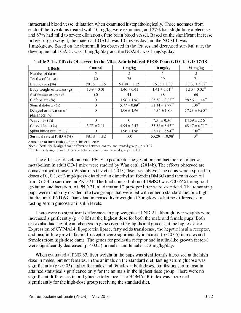
intracranial blood vessel dilatation when examined histopathologically. Three neonates from each of the five dams treated with 10 mg/kg were examined, and 27% had slight lung atelectasis and 87% had mild to severe dilatation of the brain blood vessel. Based on the significant increase in liver organ weight, the maternal LOAEL was 10 mg/kg/day and the NOAEL was 1 mg/kg/day. Based on the abnormalities observed in the fetuses and decreased survival rate, the developmental LOAEL was 10 mg/kg/day and the NOAEL was 1 mg/kg/day.
Table 3-14. Effects Observed in the Mice Administered PFOS from GD 0 to GD 17/18 Effects Control 1 mg/kg 10 mg/kg 20 mg/kg
Number of dams 5 5 5 5 Total # of fetuses 80 76 79 71 Live fetuses (%) 98.75 ± 1.25 98.88 ± 1.12 96.85 ± 1.97 90.06 ± 3.02* Body weight of fetuses (g) 1.49 ± 0.01 1.46 ± 0.01 1.41 ± 0.01** 1.10 ± 0.02** # of fetuses examined 60 44 68 60 Cleft palate (%) 0 1.96 ± 1.96 23.36 ± 8.27** 98.56 ± 1.44** Sternal defects (%) 0 15.77 ± 0.99** 52.44 ± 2.79** 100** Delayed ossification of phalanges (%)
0 1.96 ± 1.96 4.34 ± 1.80 57.23 ± 9.60**
Wavy ribs (%) 0 0 7.31 ± 0.34* 84.09 ± 2.56** Curved fetus (%) 3.55 ± 2.11 4.94 ± 2.47 33.38 ± 8.47** 68.47 ± 6.71** Spina bifida occulta (%) 0 1.96 ± 1.96 23.13 ± 3.94** 100** Survival rate at PND 4 (%) 98.18 ± 1.82 100 55.20 ± 18.98* 0**
Source: Data from Tables 2-3 in Yahia et al. 2008 Notes: *Statistically-significant difference between control and treated groups, p < 0.05 ** Statistically-significant difference between control and treated groups, p < 0.01
The effects of developmental PFOS exposure during gestation and lactation on glucose metabolism in adult CD-1 mice were studied by Wan et al. (2014b). The effects observed are consistent with those in Wistar rats (Lv et al. 2013) discussed above. The dams were exposed to doses of 0, 0.3, or 3 mg/kg/day dissolved in dimethyl sulfoxide (DMSO) and then in corn oil from GD 3 to sacrifice on PND 21. The final concentration of DMSO was < 0.05% throughout gestation and lactation. At PND 21, all dams and 2 pups per litter were sacrificed. The remaining pups were randomly divided into two groups that were fed with either a standard diet or a high fat diet until PND 63. Dams had increased liver weight at 3 mg/kg/day but no differences in fasting serum glucose or insulin levels.
There were no significant differences in pup weights at PND 21 although liver weights were increased significantly (p < 0.05) at the highest dose for both the male and female pups. Both sexes also had significant changes in genes regulating lipids and glucose at the highest dose. Expression of CYP4A14, lipoprotein lipase, fatty acids translocase, the hepatic insulin receptor, and insulin-like growth factor-1 receptor were significantly increased (p < 0.05) in males and females from high-dose dams. The genes for prolactin receptor and insulin-like growth factor-1 were significantly decreased (p < 0.05) in males and females at 3 mg/kg/day.
When evaluated at PND 63, liver weight in the pups was significantly increased at the high dose in males, but not females. In the animals on the standard diet, fasting serum glucose was significantly (p < 0.05) higher for males and females at both doses, but fasting serum insulin attained statistical significance only for the animals in the highest dose group. There were no significant differences in oral glucose tolerance. The HOMA-IR index was increased significantly for the high-dose group receiving the standard diet.
Perfluorooctane sulfonate (PFOS) – May 2016 3-72
The results from the glucose tolerance test (fasting blood glucose levels and blood glucose
levels over 2 hours following oral glucose challenge) became statistically-significant (p < 0.05) at the high dose in both sexes fed high fat diets on PND 63. Fasting serum insulin was significantly increased (p < 0.05) at 3 mg/kg/day in males and females on both diets, with the effects more pronounced in mice on the high fat diet than in mice on the standard diet. The HOMA-IR index was significantly increased (p < 0.01) at both doses for males and females on the high fat diet.
3.2.6 Specialized Developmental/Reproductive Studies
Hormonal Disruption
Rat. Yu et al. (2009a) fed pregnant adult Wistar rats (n = 20/group) a control diet or a diet containing 3.2 mg PFOS/kg feed. Doses to the dams were not calculated, and body weight and feed consumption data were not presented. Treatment continued for both groups throughout gestation and lactation. Dams were allowed to deliver, and on the day of delivery (PND 0) samples were collected from two control litters and two PFOS treated litters. The remaining litters were cross-fostered within 12 hours of birth to make the following groups:
• Litters from control dams fostered by control dams (CC, unexposed control; n = 8). • Litters from treated dams fostered by control dams (TC, prenatal exposure; n = 8). • Litters from control dams fostered by treated dams (CT, post-natal exposure; n = 8). • Litters from treated dams fostered by treated dams (TT, prenatal + postnatal exposure;
n = 10).
The pups were weaned on PND 21 and then fed the same diet as the foster dam. Pups were weighed and sacrificed on PNDs 0, 7, 14, 21, or 35. Serum thyroid hormone analysis was performed and included total thyroxine (T4), total triiodothyronine (T3), reverse T3 (rT3), and hepatic expression of genes involved in thyroid hormone (TH) transport, metabolism, and receptors. The genes associated with thyroid metabolism included type 1 deiodinase (DI01) and uridine diphosphoglucuronosyl transferase 1A1 and 1A6 (UGT1A1 and UGT1A6). Those associated with thyroid hormone transport included transthyretin (TTR). The genes for the thyroid hormone receptors α and β (TRα and TRβ) were also studied.
No mortality or clinical signs were observed in the dams. Body weight in offspring from PFOS treated groups did not differ significantly from controls. Liver weights in pups from the pre- and postnatal exposure (group TT) were significantly increased on PNDs 21 and 35. As observed in other studies, levels of PFOS in the dams and offspring were higher in the liver when compared to the serum. The levels of PFOS in both the serum and liver increased with time in the pups exposed postnatally (group CT) but decreased with time in those exposed only prenatally (group TC). The levels increased in those in the TT group. These results indicate that PFOS can be transferred by the placenta and through lactation.
The total T3 and rT3 were not affected by PFOS treatment of the pups. Compared to controls, pups in all treated groups had significant (p < 0.05 or 0.01) decreases in total T4 on PNDs 21 and 35, with the response in the CT and TT groups larger than that of the TC group. On PNDs 21 and 35, T4 levels were 71%–75% and 63%–64% of controls for the CT and TT groups, respectively, compared with 80%–81% of control for the TC group on both days. Pups in the TT group (exposed pre- and postnatally) had T4 levels that were significantly lower than the controls at PND 14. For gene expression, no statistically-significant differences were observed between
Perfluorooctane sulfonate (PFOS) – May 2016 3-73
litters born to control dams or litters born to treated dams on PND 0. The only significant finding in gene expression at the other sacrifice time-points was a significant (p < 0.01) increase (1.5 times greater than the controls) in TTR on PND 21 in the pups that had been treated both in the prenatal and postnatal interval. Lactational exposure appears to be an important contributor to the observed thyroid effects given that the serum PFOS levels were higher, and T4 levels lower, in the CT group than in the TC group.
The effects of PFOS on testosterone production by fetal Leydig cells were investigated following prenatal exposures (Zhao et al. 2014). Pregnant Sprague-Dawley rats (n = 4) were administered PFOS by gavage at doses of 0, 5, or 20 mg/kg/day on GDs 11–19; controls received the 0.05% Tween 20 vehicle. Dams were killed on GD 20 and the male pups removed, weighed, and measured for length and anogenital distance. The fetal testes were removed for analysis of testosterone production, fetal Leydig cell numbers, ultrastructure, and gene and protein expression levels. Dams given 20 mg/kg/day had significantly lower body weight and serum cholesterol levels on GD 20. Male fetuses had significantly lower body weight at 5 and 20 mg/kg/day. At 20 mg/kg/day there were significant differences in body length, anogenital distance, and testes weight; all measures were lower than those for controls.
Testicular mRNA levels of growth factors (Kitl), cholesterol transporters (Scarb1 and Star), steroidogenic enzymes (Cyp11a1, Cyp17a, and Hsd3b1), junction protein (Trmp2), and LH receptor (Lhcgr) were significantly reduced in fetuses from dams given 20 mg/kg/day. Fetuses from high-dose dams also had significantly lower testicular testosterone levels, enzyme activity, and protein levels for 3β-hydrosteroid dehydrogenase and 17α-hydroxylase/20-lyase. Liver cholesterol and testes HDL-cholesterol levels were reduced in fetuses from high dose dams. Histologically, the number of fetal Leydig cells was reduced and showed a decreased number of lipid droplets and features of apoptosis at 20 mg/kg/day. The 5 mg/kg/day dose was a LOAEL for effects on male fetal body weight.
Developmental Neurotoxicity
Rat. Twenty five female Sprague-Dawley rats/group were administered 0, 0.1, 0.3, or 1.0 mg/kg/day of potassium PFOS by gavage from GD 0 through PND 20 (Butenhoff et al. 2009). An additional 10 mated females/group were used to collect additional blood and tissue samples. Offspring were monitored through PND 72 for growth, maturation, motor activity, learning and memory, acoustic startle reflex, and brain weight.
There were no treatment-related effects on the pregnancy rates, gestation length, number of implantation sites, number of pups born, sex ratio, birth to PND 4 survival, PND 4–21 survival, pup body weights through PND 72, and gross internal findings. Maternal body weight and body weight gain during gestation were comparable between the treated and control groups. On LDs 1–4, dams in the 1.0 mg/kg/day group had slightly, but not significantly, lower weight gain and food consumption than those of controls resulting in significantly lower (p < 0.05 or 0.01) absolute body weight throughout lactation. Food consumption was transiently decreased (p < 0.05 or 0.01) on GDs 6–9 for the 0.3 mg/kg/day group and on GDs 6–12 for the 1.0 mg/kg/day group. These findings in the treated dams are not considered to be treatment-related or adverse. Based on results, the maternal toxicity NOAEL was 1.0 mg/kg/day and the LOAEL could not be determined.
No treatment related effects were observed on functional observational battery assessments performed on PNDs 4, 11, 21, 35, 45, and 60. Male offspring from dams administered 0.3 and
Perfluorooctane sulfonate (PFOS) – May 2016 3-74

1.0 mg/kg/day had statistically-significant (p < 0.05) increases in motor activity on PND 17, but this was not observed on PND 13, 21 or 61. No effect on habituation was observed in the 0.1 and 0.3 mg/kg/day males or in the 1.0 mg/kg/day females. On PND 17, males at 1.0 mg/kg/day showed a lack of habituation as evidenced by significantly (p < 0.05) increased activity counts for the sequential time intervals of 16–30, 31–45, and 46–60 minutes. The normal habituation response is for motor activity to be highest when the animals are first exposed to a new environment and to decline during later exposures to the same environment as they have learned what to expect. There were no effects in males or females on acoustic startle reflexes or in the Biel swimming maze trials. Mean absolute and relative (to body weight) brain weight and brain measurements (length, width) were similar between the control and treated animals. Based on the increased motor activity observed reflecting decreased habituation, the LOAEL for developmental neurotoxicity in male rats was 1.0 mg/kg/day and the NOAEL was 0.3 mg/kg/day.
Y. Wang et al. (2015) examined the effects of PFOS on spatial learning and memory following pre- and post-natal exposure. Pregnant Wistar rats were administered PFOS in the drinking water at 0, 5, or 15 mg/L beginning on GD 1 and continuing through lactation. Doses to the animals were not calculated, and body weight and water consumption data were not presented. Doses were estimated as 0, 0.8, or 2.4 mg/kg/day using subchronic values for female Wistar rats from USEPA (1988). Maternal serum levels in the treated groups were 25.7 and 99.3 µg/mL, respectively, on PND 7 and 64.3 and 207.7 µg/mL, respectively, on PND 35. On PND 1 pups were cross-fostered to establish groups for unexposed controls, only prenatal exposure, only post-natal exposure, and continuous exposure. After weaning, pups were given the same treated or control water as their foster dam. Three pups per group were sacrificed on PNDs 7 and 35 for measurement of protein and RNA levels in the hippocampus. On PND 35, 8–10 pups per group were tested in the Morris Water Maze which consisted of one day of visible platform tests, seven days of hidden platform tests, and a probe trial 24 hours after the last hidden platform test.
Offspring survival on PND 1 was significantly reduced from high-dose dams before cross-fostering; survival on PND 5 was not given. On water maze testing day 1, swimming speed and the time to reach the visible platform were similar between all treated and control groups. Thereafter, escape latency was significantly increased for all treated groups on one or more testing days. The most pronounced and significant effect was in pups exposed prenatally from dams given 15 mg/L and cross-fostered to control dams. Similar trends were observed for escape distance. During the probe trial for memory testing, pups continuously exposed pre- and post-natally to 15 mg/L spent less time in the target quadrant than the unexposed controls but statistical significance was not achieved as consistently as that for the group exposed only during gestation. Protein levels of growth-associated protein-43, neural cell adhesion molecule 1, nerve growth factor, and brain-derived neurotrophic factor were significantly decreased in the hippocampus on PND 35, especially in pups exposed prenatally to 15 mg/L and cross-fostered to control dams.
Ten pregnant Sprague-Dawley rats/group were administered 0, 0.1, 0.6, or 2.0 mg/kg/day of PFOS in 0.5% Tween 80 by oral gavage from GD 2 to GD 21 (Zeng et al. 2011). On GD 21, dams were monitored for parturition and the day of delivery was designated PND 0. On PND 0, five pups/litter were sacrificed and the trunk blood, cortex, and hippocampus were collected for examination. Astrocyte activation markers, glial fibrillary acidic protein (GFAP) and S100 calcium binding protein B, which are associated with morphological changes inside the cell,
Perfluorooctane sulfonate (PFOS) – May 2016 3-75

were evaluated with immunohistochemistry. The other pups were randomly redistributed to dams within the dosage groups and allowed to nurse until PND 21, when they were sacrificed with the same tissues collected as described for PND 0. PFOS concentration in the hippocampus, cortex, and serum increased in a dose-dependent manner, but overall was lower in all tissues on PND 21 than on PND 0.
The number of GFAP positive cells was significantly increased in the hippocampus and cortex of offspring from treated dams on PND 21. The protein levels of GFAP in PND 21 offspring were also increased in the hippocampus and cortex on Western Blot tests. The S100 calcium binding protein B was increased in the offspring’s hippocampus and cortex on PND 21 in those from dams treated with 0.6 and 2.0 mg/kg/day.
In other tests, PFOS increased the mRNA expression of two inflammatory cytokines, interleukin 1 beta (IL-1β) and tumor necrosis factor-α(TNF) The expression of IL-1β and TNF-α was significantly increased compared to controls in all treated offspring in the hippocampus on PND 0 and in those from dams administered ≥ 0.6 mg/kg on PND 21. In the cortex, IL-1β and TNF-α were only significantly increased in the 0.6 mg/kg group and 2.0 mg/kg group, respectively, on PND 0. On PND 21 in the cortex, IL-1β was increased at ≥ 0.6 mg/kg and TNF-α was increased in the high dose group.
To determine the mechanisms leading to the inflammatory effect after PFOS exposure, mRNA levels of three pro-inflammatory transcription factors in both brain tissues were examined. The greatest increase was observed in the hippocampus on with a significant increase in activation protein-1 (AP-1) in all dose groups and an increase in nuclear factor-ĸB (NF-ĸB) and cAMP response element-binding protein at ≥ 0.6 mg/kg groups at PND 0. Two synaptic proteins, synapsin 1 (Syn 1) and synaptophysin (Syp) were also affected; Syn 1 was decreased with PFOS exposure primarily in the hippocampus. Syp was decreased in the hippocampus, but increased in the cortex.
Mouse. Fuentes et al. (2007) treated 8–10 pregnant Charles River CD-1 mice/group to 0 or 6 mg/kg/day of PFOS dissolved in 0.5% Tween-20 daily by gavage on gestation days (GDs) 12–18. After treatment, mice were either left alone or restrained (immobilized) three times per day for 30 minutes to induce maternal stress. Maternal body weight and food and water consumption were monitored. At birth, the length of gestation, number of live/dead pups, and sex/weight of pups were recorded.
During the post-natal period, the body weight of the pups was recorded, landmarks for development were monitored, and neuromotor maturation tests (i.e., surface righting reflex, forelimb grip strength) were conducted. At 3 months of age, the pups were tested in open-field and rotarod tests to further assess development. The PFOS treatment had no effect on maternal body weight or food/water consumption. On PNDs 4 and 8, pups from dams treated with 6 mg/kg of PFOS had reduced body weight, as well as delayed (p < 0.05) eye opening, pinna detachment, and surface righting reflex. Female pups from dams exposed to 6 mg/kg of PFOS and stressed by immobilization exhibited reduced open-field activity. No differences in activity were observed for male pups and rotarod performance was not affected in any group by PFOS alone or combined with maternal stress.
Ten-day old male neonatal Naval Medical Research Institute (NMRI) mice (4–7/group) were exposed once to 0, 0.75, or 11.3 mg/kg bw of PFOS by oral gavage (Johansson et al. 2008). Spontaneous behavior (locomotion, rearing, and total activity) and habituation were examined in
Perfluorooctane sulfonate (PFOS) – May 2016 3-76

the mice at 2 and 4 months old. Behavior was tested in an automated device equipped with horizontal infrared beams. Motor activity was measured during a 60-minute period divided into three 20-minute sessions. Locomotion, rearing, and total activity were recorded.
No effects were observed on body weight. At 2 months old, mice exposed to 0.75 and 11.3 mg/kg bw of PFOS exhibited significant (p ≤ 0.01) decreases in locomotion, rearing, and total activity during the first 20 minutes compared to controls. After 60 minutes, activity was significantly increased in the 11.3 mg/kg bw dose group when compared to controls. The expected habituation response is for the highest activity pattern to occur in the first 20-minute period not the last period. The same trend was observed at 4 months in the mice exposed to 11.3 mg/kg bw. At 4 months the responses in the 0.75 mg/kg bw dose group were similar to the controls. Overall, a single PFOS treatment on PND 10 affected habituation even up to 4 months of age for mice in the high dose group (11.3 mg/kg/day). The LOAEL was 0.75 mg/kg based on decreased locomotion, rearing, and total activity in 2 month old mice.
Johansson et al. (2009) administered a single oral dose of 0 (3 litters) or 11.3 mg/kg (four litters) to NMRI male mice (10 days old). The exact number of male mice in each litter was not provided. Sacrifice occurred 24 hours after treatment and the brain was dissected. The cerebral cortex and hippocampus were homogenized to determine if PFOS affected the protein levels of calcium/calmodulin-dependent protein kinase II (CaMKII), growth-associated protein-43 (GAP-43), synaptophysin, and tau, which are all proteins involved in neuronal survival, growth, and synaptogenesis change during the brain growth spurt.
There were no clinical signs of acute toxicity, and no treatment-related body weight differences. The CaMKII and GAP-43 protein levels in the hippocampus were both increased in the PFOS treated males; levels were increased 57% (p < 0.001) and 22% (p < 0.01), respectively, when compared to controls. Protein values in the cerebral cortex were similar between the control and treated mice. Synaptophysin protein levels were increased significantly (p < 0.001; 48%) in the hippocampus and (p < 0.01; 59%) in the cerebral cortex of the treated mice. The tau protein levels in the cerebral cortex were increased significantly (p < 0.05; 80%) in treated animals compared to controls. Overall, the study indicates that a one-time treatment with 11.3 mg//kg PFOS had a significant effect on the neuronal proteins evaluated.
Tissue and Metabolic effects
Zeng et al. (2014) examined cardiac mitochondria mediated apoptosis in weaned rats exposed by way of their dams (10 per dose group) to 0, 0.1, 0.6, or 2 mg/kg/day in 0.05% Tween 80 by gavage on GDs 2–21. The pups were sacrificed at the end of the lactation period. Trunk blood and the heart were recovered. Apoptotic cells in the heart tissue from six animals per dose group were measured using a Terminal deoxynucleotidyl transferase dUTP nick end labeling (TUNEL) staining assay by an individual pathologist blinded to the exposure group. The apoptosis index was recorded as percent apoptotic cells per 1,000 cells in the same section. PFOS exposure was associated with a dose dependent increase in the percentage of TUNEL positive nuclei (p < 0.05). The 0.6 mg/kg/day dose was the LOAEL and the 0.1 mg/kg/day dose the NOAEL. The researchers found that biomarkers for apoptosis were supportive of the TUNEL results. The expression of BCL2-associated X protein and cytochrome c were upregulated and bcl-2 downregulated. The concentration of caspase 9 was significantly increased above the control levels at all doses and caspase 3 levels were significantly increased for all but the lowest dose level.
Perfluorooctane sulfonate (PFOS) – May 2016 3-77

3.2.7 Chronic Toxicity
Only a single chronic exposure study in animals is available (Thomford 2002/Butenhoff et al. 2012). It is the long term component of the Seacat et al. (2002) subchronic study reported in section 3.2.3. Sprague-Dawley Crl:CD (SD)IGS BR, rats (n = 40–70) were dosed using a PFOS containing diet for up to 105 weeks. Five per sex per dose group were sacrificed at 4 and 14 weeks as described earlier. Treatment resulted in decreased body weight, with increased liver weight with hepatocellular hypertrophy. A satellite group of animals received 20 ppm of the PFOS containing diet for 52 weeks, followed by the control diet until sacrifice at week 106.
The animals received dietary levels of 0, 0.5, 2, 5, or 20 ppm PFOS as the potassium salt. Corresponding PFOS doses were 0, 0.024, 0.098, 0.24, and 0.984 mg/kg/day, respectively, for males and 0, 0.029, 0.120, 0.299, 1.251 mg/kg/day, respectively, for females. Five animals/sex in the treated groups were sacrificed during week 53 and liver samples were obtained for mitochondrial activity, hepatocellular proliferation rate, and determination of palmitoyl-CoA oxidase activity; liver weight was recorded. The results from the 4-week and 14-week sacrifices (Seacat et al. 2002) from this study are provided in sections 3.2.2 and 3.2.3, respectively. Serum samples were collected at weeks 27 and 53 from 10 rats/sex/dose group and were examined for clinical effects associated with systemic toxicity; liver samples were obtained during and at the end of the study for determination of PFOS concentration. Data on chronic effects were not reported for the recovery group. The concentration of PFOS in serum was measured at weeks 4, 14, and 105. In males the serum levels decreased between week 14 and 105 by 50% for all but the 0.5 ppm group where the decrease in serum concentration was larger. A serum measurement was available at 53 weeks for the high dose males and was comparable to the value at 14 weeks. In females serum levels serum levels remained relatively constant at 14 and 105 weeks. In both males and females the concentrations in the liver were lower at 105 weeks than they were at 14 weeks.
The clinical serum observations for ALT at 53 weeks were consistent with those at 14 weeks in demonstrating significant (p < 0.05) increases for the high dose males but not females. At week 27, ALT was increased for high-dose males, but did not attain statistical significance. For males at 53 weeks in the 0, 0.5, 2, 5, and 20 ppm groups, ALT values were 54 ± 66, 62 ± 52, 40 ± 7.5, 44 ± 8.3, and 83 ± 84 IU/L, respectively. The large SDs were the result of high values in one animal in each of the control and 0.5 ppm groups and two animals in the 20 ppm group. Thus, some animals may be more sensitive to liver damage as a result of exposure than others. AST levels were not increased for either sex. Serum blood urea nitrogen (BUN) was significantly (p ≤ 0.05) increased at 20 ppm for males and females at weeks 14, 27, and 53 and in 5 ppm males and females at 27 and 53 weeks. The males in the 2 ppm group also had a significant (p ≤ 0.05) increase in BUN at 53 weeks. These data were presented graphically in Butenhoff et al. (2012).
Nonneoplastic lesions in the liver are shown in Table 3-15. At sacrifice, males at 2 ppm had a significant (p < 0.05) increase in hepatocellular centrilobular hypertrophy. In the males and females at 5 and 20 ppm, there were significant (p < 0.05) increases in centrilobular hypertrophy, centrilobular eosinophilic hepatocytic granules (females only), and centrilobular hepatocytic vacuolation (males only). At the high dose, there was a significant increase in the number of animals with single cell hepatic necrosis in both males and females at 53 weeks. Necrosis in the recovery animals was comparable to the controls.
Perfluorooctane sulfonate (PFOS) – May 2016 3-78

Table 3-15. Incidence of Nonneoplastic Liver Lesions in Rats
(Number Affected/Total Number)
Lesion 0 ppm
0 mg/kg/day (d) 0.5 ppm
0.024 mg/kg/d 2.0 ppm
0.098 mg/kg/d 5.0 ppm
0.242 mg/kg/d 20 ppm
0.984 mg/kg/d Males
Centrilobular hypertrophy 0/65 2/55 4/55* 22/55** 42/65** Eosinophilic granules 0/65 0/55 0/55 0/55 14/65* Vacuolation 3/65 3/55 6/55 13/55** 19/65** Single cell necrosis 5/65 4/55 6/55 5/55 14/65*
Females 0 mg/kg/d 0.029 mg/kg/d 0.120 mg/kg/d 0.299 mg/kg/d 1.251 mg/kg/d
Centrilobular hypertrophy 2/65 1/55 4/55 16/55** 52/65** Eosinophilic granules 0/65 0/55 0/55 7/55** 36/65** Single cell necrosis 7/65 6/55 6/55 6/55 15/65* Source: Data from Thomford 2002/Butenhoff et al. 2012 Notes: *Significantly increased over control: p < 0.05 ** Significantly increased over control: p < 0.01.
No effects were observed on hepatic palmitoyl-CoA oxidase activity or increases in proliferative cell nuclear antigen (PCNA) at weeks 4 and 14 or bromodeoxyuridine at week 53. PFOS was identified in the liver and serum samples of the treated animals and trace amounts were identified in the control animals. The LOAEL at termination for male rats was 2 ppm (0.098 mg/kg/day) and for female rats was 5 ppm (0.299 mg/kg/day) based on the liver histopathology. The NOAEL for the males was 0.5 ppm (0.024 mg/kg/day) and 2 ppm (0.120 mg/kg/day) for females. Additional details from the study in regard to carcinogenicity are provided in section 3.2.8.
Survival was not affected by PFOS administration. Males and females administered 20 ppm had statistically-significantly decreased mean body weight compared to controls during weeks 9–37 and 3–101, respectively, but was similar to controls by week 105. The females at 20 ppm had decreased food consumption during weeks 2–44. At the week 14 and 53 sacrifices, absolute and relative liver weights were significantly increased at 20 ppm in males and relative liver weight was increased at 20 ppm in females. At week 53, liver weight data were given only for the control and 20 ppm groups such that a dose-response could not be evaluated.
3.2.8 Carcinogenicity
Rat. Tumor data were collected as part of the chronic study (Thomford 2002/Butenhoff et al. 2012) described above. The tumor results are provided in Table 3-16. A significant positive trend (p = 0.0276) was noted in the incidence of hepatocellular adenoma in male rats. This was associated with a significant increase (p < 0.0456) in the high-dose group (7/60, 11.7%) over the control (0/60, 0%). No hepatocellular tumors were observed in the recovery group exposed for 52 weeks and sacrificed at 106 weeks. Liver tumors were observed in males at all doses (0%, 6%, 6%, 2%, and 11.7%). In females, significant positive trends were observed in the incidences of hepatocellular adenoma (p = 0.0153) and combined hepatocellular adenoma and carcinoma (p = 0.0057) at sacrifice. Here too, the response was not linear to dose with sequential values of 0%, 2%, 2%, 2%, and 8.3%. These cases were associated with significant increases in the high-dose group 5/60 (p = 0.0386; 8.3%) for adenomas and 6/60 (p = 0.0204; 10%) for combined adenomas and carcinomas compared to the control. The female recovery group had 2/20 liver adenomas (5%) and no carcinomas. The presence of increased levels of ALT in the males of the
Perfluorooctane sulfonate (PFOS) – May 2016 3-79

high dose group at 14, 27, and 53 weeks supports hepatic tissue damage with compensatory repair as a probable a possible mode of action (MOA) for the liver tumors. In all cases the SDs about the means are broad suggesting that some animals could be less resilient than others to the liver effects.
Table 3-16. Tumor Incidence (%)
Tumors 0 ppm
0 mg/kg/d 0.5 ppm
0.024 mg/kg/d 2.0 ppm
0.098 mg/kg/d 5.0 ppm
0.242 mg/kg/d 20 ppm
0.984 mg/kg/d
20 ppm recovery
1.144 mg/kg/day Males
Liver hepatocellular adenoma+ 0 (0/60) 6.0 (3/50) 6.0 (3/50) 2.0 (1/50) 11.7* (7/60) 0 (0/40) Thyroid follicular cell adenoma 5.0 (3/60) 10.2 (5/49) 8.0 (4/50) 8.2 (4/49) 6.8 (4/59) 23.1* (9/39) follicular cell carcinoma 5.0 (3/60) 2.0 (1/49) 2.0 (1/50) 4.1 (2/49) 1.7 (1/59) 2.6 (1/39) combined 10.0 (6/60) 12.2 (6/49) 10.0 (5/50) 10.2 (5/49) 8.5 (5/59) 25.6 (10/39)
Females 0 mg/kg/d 0.029 mg/kg/d 0.120 mg/kg/d 0.299 mg/kg/d 1.251 mg/kg/d 1.385 mg/kg/d Liver hepatocellular adenoma+ 0 (0/60) 2.0 (1/50) 2.0 (1/49) 2.0 (1/50) 8.3* (5/60) 5.0 (2/40) hepatocellular carcinoma 0 (0/60) 0 (0/50) 0 (0/49) 0 (0/50) 1.7 (1/60) 0 (0/40) combined+ 0 (0/60) 2.0 (1/50) 2.0 (1/49) 2.0 (1/50) 10.0* (6/60) 5.0 (2/40) Thyroid follicular cell adenoma 0 (0/60) 0 (0/50) 0 (0/49) 4.0 (2/50) 1.7 (1/60) 2.5 (1/40) follicular cell carcinoma 0 (0/60) 0 (0/50) 0 (0/49) 2.0 (1/50) 0 (0/60) 0 (0/40) follicular cell combined 0 (0/60) 0 (0/50) 0 (0/49) 6.0* (3/50) 1.7 (1/60) 2.5 (1/40) C-cell adenomas 20.0 (12/60) 12.0 (6/50) 12.2 (6/49) 16.0 (8/50) 8.3* (5/60) 15.0 (6/40) C-cell Carcinomas 0 (0/60) 2.0 (1/50) 0 (0/49) 0 (0/50) 0 (0/60) 2.5 (1/40) C-cell combined 20.0 (12/60) 14.0 (7/50) 12.2 (6/49) 16.0 (8/50) 8.3* (5/60) 17.5 (7/40) Mammary Fibroma/Adenoma 33.3 (20/60) 54.0* (27/50) 39.6 (19/48) 48.0 (24/50) 18* (11/60) 37.5 (15/40) Adenoma 11.7 (7/60) 12.0 (6/50) 10.4 (5/48) 14.0 (7/50) 6.7 (4/60) 10.0 (4/40) Combined adenomas 38.3 (23/60) 60.0* (30/50) 45.8 (22/48) 52.0 (26/50) 25.0* (15/60) 40.0 (16/40) carcinoma 18.3 (11/60) 24.0 (12/50) 31.3 (15/48) 22.0 (11/50) 23.3 (14/60) 10.0 (4/40)
Source: Data from Thomford 2002/Butenhoff et al. 2012. Notes: +Significant positive trend. * Significantly increased over the control: p < 0.05 ** Significantly increased over the control: p < 0.01.
There were cases of thyroid follicular cell adenomas and carcinomas in both the male and female rats but no pattern of dose-response or significant increases compared to controls. The incidence of thyroid follicular cell adenomas in the male recovery group was increased significantly (p = 0.028) over controls (23.1% vs 5%). The incidence of combined thyroid follicular cell adenoma and carcinoma in the recovery group males (10/39, 25.6%) did not attain statistical significance compared to that of the control group (6/60, 10%). The males that were continually dosed for 105 weeks had a much lower adenoma incidence than the recovery group (6.8% versus 23.1%). In no case were thyroid tumors determined to be a cause of death.
In females, there was a significant increase (p = 0.0471) for combined thyroid follicular cell adenoma and carcinoma in the mid-high (5.0 ppm) group (3/50, 6%) compared to the control group (0/60, 0%). The incidence data for thyroid follicular tumors lacked dose-response. C-cell thyroid adenomas had a higher incidence than the follicular cell tumors in female rats. The
Perfluorooctane sulfonate (PFOS) – May 2016 3-80
highest incidence was in the control group (20%); there was a lack of dose-response across groups (8%–18%). As was the case with the combined adenomas and carcinomas, the C-cell tumors were not identified as a cause of death.
There was a high background incidence in mammary gland tumors in the female rats, primarily combined fibroma adenoma and adenoma (25%–60%), but the incidence lacked dose-response for all tumor classifications. Significant (p = 0.0318) increases combined mammary fibroadenoma/adenoma (30/50, 60%; p = 0.0318) were observed in the low–dose (0.5 ppm) group compared to the respective controls but there was a lack of dose response with the high dose group having a lower incidence (25%) than the controls (38%). Mammary gland carcinomas also lacked dose-response and had a relatively comparable incidence across dose groups including the controls.
Mouse. The mouse model C57BL/6J-Min/+ for intestinal neoplasia was used to study the obesogenic and tumorigenesis effects of PFOS following in utero exposure (Ngo et al. 2014). The C57BL/6J-ApcMin/+ mouse has a heterozygote mutation in the tumor suppressor gene adenomatous polyposis coli (Apc), and is therefore a sensitive model in which to test whether chemicals can affect intestinal tumorigenesis. Wild-type females (Apc+/+), mated to heterozygous males (ApcMin/+), were given 0, 0.01, 0.1, or 3 mg/kg/day by gavage on GDs 1–17 and allowed to litter naturally. Offspring with ApcMin/+ genotype were terminated at 11 weeks of age for study of intestinal tumorigenesis and obesogenic effect while wild-type (Apc+/+) offspring were sacrificed at 20 weeks to assess any obesogenic effect at an older age. In the treated groups, whole litter loss occurred in 6/16, 10/28, and 7/14 dams, respectively, compared with 2/22 controls; the timing of loss, late, or early gestation, was not stated. No clinical signs of toxicity were observed during dosing and maternal body weight was similar between treated and control groups. For offspring of either genotype, terminal body weight, liver and spleen weights, and plasma glucose were not affected by in utero exposure. PFOS did not increase intestinal tumorigenesis in susceptible, ApcMin/+, offspring.
3.3 Other Key Data
3.3.1 Mutagenicity and Genotoxicity
Results of genotoxicity testing with PFOS are summarized in Tables 3-17 and 3-18. PFOS was tested for mutation in the Ames Salmonella/Microsome plate test and in the D4 strain of Saccharomyces cerevisiae (Litton Bionetics, Inc. 1979). It was also tested in a Salmonella-Escherichia coli/Mammalian-microsome reverse mutation assay with and without metabolic activation (Mecchi 1999), in an in vitro assay for chromosomal aberrations in human whole blood lymphocytes with and without metabolic activation (Murli 1999), and in an unscheduled DNA synthesis assay in rat liver primary cell cultures (Cifone 1999). In all these assays, PFOS was negative. In an in vivo mouse micronucleus assay, PFOS did not induce any micronuclei in the bone marrow of Crl:CD-1 BR mice (Murli 1996). A 50% w/w solution of the diethanolammonium salt of PFOS in water (T-2247 CoC) was also tested to determine whether induction of gene mutation in five strains of S. typhimurium and in S. cerevisiae strain D3 would take place with and without metabolic activation (Simmon 1978). The results were negative.
Governini et al. (2015) collected semen samples from 59 healthy-nonsmoking patients attending a Center for Couple Sterility at the University in Siena, Italy. The subjects were divided into those that were normozoospermic (13) and those that were oligoasthenoterato-
Perfluorooctane sulfonate (PFOS) – May 2016 3-81

zoospermic (46). PFOS was present in 25% of the seminal plasma samples and 84% of the serum samples. Conversely PFOA was present in 75% of the seminal plasma samples and only 16% of the blood samples. Sperm were evaluated for the presence of aneuploidy and diploidy, and sperm DNA was evaluated for fragmentation using the TUNEL assay. The frequencies of aneuploidy and diploidy were significantly greater in the PFAS positive samples than in the PFC negative samples (p < 0.001 and p < 0.05, respectively) suggesting the possibility for errors in cell division. The levels of fragmented chromatin were significantly increased (p < 0.001) for the PFC positive group compared with the PFAS negative group.
Table 3-17. Genotoxicity of PFOS in vitro Species
(test system) End-point With activation Without activation Reference Salmonella strains and D4 strain of Saccharomyces cerevisiae
Gene mutation negative negative Litton Bionetics, Inc. 1979
Salmonella strains and Escherichia coli WP2uvr
Gene mutation negative negative Mecchi 1999
5 strains of S. typhimurium and S. cerevisiae strain D3
Gene mutation negative negative Simmon 1978
Human lymphocytes Chromosome aberrations
negative negative Murli 1999
Hepatocytes from Fisher 344 male rats
DNA synthesis negative Cifone 1999
Table 3-18. Genotoxicity of PFOS in vivo Species
(test system) End-point Results Reference Crl:CD-1 BR mice Presence of micronuclei in bone marrow negative Murli 1996
3.3.2 Protein binding
The ability of PFOS to bind to serum proteins for distribution is discussed in section 2.2. PFC protein binding can also impact cellular function in cases where the proteins in question are transporters (serum albumin and fatty acid binding protein), enzymes (lysine decarboxylase), or membrane receptors such as members of the PPAR family and thyroid hormone receptors. The mechanistic studies of the membrane receptors are described in section 3.3.4.
Ren et al. (2015) examined the relative binding affinities of 16 perfluoroalkyl compounds for the human thyroid hormone receptor’s α ligand binding domain (TRα-LBD) using a fluorescence competitive binding assay. Solutions of 1µmol TRα-LBD were prepared in DMSO. Changes in TRα-LBD tryptophan fluorescence after binding to 10 µmol T3 in the absence or presence of the PFAS was used to determine the binding properties of the PFAS. IC50 values were calculated by linear extrapolation between two responses located in the vicinity of a 50% inhibition level. All the PFAS had a lower affinity for the receptor than T3, but the binding affinity of PFOS was greater than that for PFOA and the other sulfonates tested. The IC50 value for PFOS was 16 µmol, compared with 0.3 µmol for T3.
Perfluorooctane sulfonate (PFOS) – May 2016 3-82

Lysine decarboxylase is a key enzyme involved in the production of cadaverine from the
amino acid lysine. S. Wang et al. (2014) studied the impact of a series of 16 PFAS on the activity and conformation of this enzyme because of its involvement in growth and development. The interaction assays were carried out in vitro using a fluorescent probe to measure enzyme activity. The impact of a PFAS on enzyme activity caused a decrease in fluorescence that represented enzyme inhibition. Varying the PFAS concentrations provided the data for determining inhibition constants for each compound tested. Members of the sulfonate family were stronger inhibitors than the carboxylic acids, and enzyme inhibition increased as did the length of the carbon chain. Only the 4, 6, and 8 carbon members of the sulfonate family were tested.
Circular dichroism was used as a tool for determining changes in enzyme conformation in the presence of the tested PFAS (S. Wang et al. 2014). PFOS caused a greater change in enzyme conformation than PFOA. Cellular cadaverine production was decreased indicating the potential for PFOS to alter metabolism by way of enzyme inhibition as a consequence of its protein binding properties. To date there has been scant investigation of PFOS or other PFASs as enzyme inhibitors.
An in vitro study of the impact of PFOS (and other PFASs) on the conformation of several proteins (BSA, ovalbumin, and β-galactosidase) in solution found that the denaturing effect of the PFAS depended on the amino acid composition and conformation of the protein as well as the individual PFAS (Ospinal-Jiménez and Pozzo 2012). The PFOS concentration (1 millimole [mmol]) was higher than one would expect in vivo because the study was designed to examine denaturing potential.
Enzymes targeted by PFOS can vary. Molecular docking analysis of PFOS’s potential to bind with and change the activity of enzymes along metabolic pathways associated with its critical effects could provide important insights related to toxicity. The importance of the S. Wang et al. (2014) and Ospinal-Jiménez and Pozzo (2012) studies are the evidence they produced showing that the protein binding properties of a PFAS can impact the conformation, thereby possibly changing activity.
3.3.3 Immunotoxicity
Human–in vitro. In a pilot study, Brieger et al. (2011) examined the effects of PFOS on human leukocytes. Peripheral blood mononuclear cells (PBMC) were obtained from 11 voluntary donors (n = 6 females, 5 males). The mean plasma concentrations of PFOS were 0.004, 0.0028, and 0.0055 µg/mL for all, female, and male volunteers, respectively. PBMCs were incubated with varying concentrations of PFOS followed by assays for cell viability, proliferation, and natural killer (NK) cell activity. The human promyelocytic leukemia cell line, HL-60, was also used in cell viability and monocyte differentiation assays. The various components of the assays employed and the results are identified as follows:
1. In the cell viability assay, the PBMCs and HL-60 cells were incubated with 0–125 μg/mL of PFOS for 24 hours. Viability was determined after incubation by measuring neutral red uptake. No significant reduction of viability was observed up to 125 µg/mL; however, the highest concentration for PFOS could not be evaluated due to limited solubility. Therefore, 100 µg/mL was the highest concentration used thereafter.
2. In the proliferation assay, the PBMCs were incubated with 0, 1, 10, or 100 μg/mL of PFOS for 24 hours; labeled with 6-carboxyfluorescein succinimidyl ester (CFSE); stimulated with concanavalinA, a T-cell mitogen (ConA, 5 μg/mL to half of all samples);
Perfluorooctane sulfonate (PFOS) – May 2016 3-83

and incubated for an additional 72 hours. Proliferation was slightly increased at 100 µg/mL and slightly reduced with the presence of ConA, but neither effect was statistically-significant.
3. For the NK cell assays, PBMCs were incubated with 0, 1, 10, or 100 μg/mL of PFOS for 24 hours followed by incubation for 3 hours with K562 target cells (12.5:1 ratio) labeled with CFSE. K562 cells are a chronic myelogenous leukemia cell line known to be susceptible to NK cell induced cytotoxicity. PFOS significantly (p < 0.001) reduced NK cell cytotoxicity to K562 cells by 32% at 100 µg/mL.
4. In the monocyte differentiation assay, HL-60 cells were incubated with 0, 1, 10, or 100 μg/mL of PFOS for 72 hours. Half of each sample was stimulated with 25 nmol calcitrol, 1α,25-dehydroxyvitamin D3 (1,25D3) 24 hours into the incubation period. Expression of CD11b and CD14 were measured as markers of differentiation. In the presence of 1,25D3, PFOS had no significant effect on the percentage of HL-60 cells expressing CD11b and CD14. No differences in monocyte differentiation were observed in the absence of 1,25D3.
5. Whole blood was incubated with 0–100 μg/mL of PFOS in the presence or absence of 25 μg/mL phytohemagglutinin (PHA), a T-cell cytokine secretion stimulator, for 48 hours. Lipopolysaccharide (LPS, 0 or 250 ng/mL), a monocyte stimulator, was added to whole blood incubated with 0.1–100 μg/mL of PFOS either 4 or 24 hours prior to the end of the 48 hour incubation period. Release of the cytokines TNF-α and IL-6 from T-cells or monocytes was quantified. Cytokine release from T-cells was not affected by PFOS. PFOS significantly (p < 0.001) reduced the release of the pro-inflammatory cytokine TNF-α after monocyte LPS stimulation. The authors also looked at the correlation between basal PFOS concentration of the blood donor and cytokine release. A significant association was observed between PFOS concentration and the release of LPS-induced IL-6 by peripheral monocytes.
This study suggests some effects on immunity in humans; however the sample size used is small and the concentrations at which effects were observed are much higher than the levels of PFOS in human blood samples.
Midgett et al. (2014) examined the effects on IL-2 production using stimulated cultured human Jurkat cells and CD4+ T cells recovered from 11 healthy volunteers. Both cell types were stimulated with PHA/phorbal myristate acetate (PMA) or anti-CD3 to produce IL-2 and incubated with 0–100 µg PFOS/mL; separate experiments were conducted with human Jurkat cells in the presence or absence of a PPAR antagonist. Cell viability was not affected in either cell type up to and including the highest concentration of PFOS. In the human Jurkat cells stimulated with PHA/PMA a concentration of 10 µg/mL was a NOEL and 50 µg/mL a LOEL for inhibition of IL-2 production in the absence and presence of a PPARα inhibitor. In the presence of anti-CD3, the NOEL was 1 µg/mL and the LOEL 5 µg/mL. In primary human CD4+ T cells stimulated with PHA/PMA, the NOEL was the 10 µg/mL concentration and the LOEL 100 µg/mL for inhibition of IL-2 production. A decrease in T cell IL-2 production is a characteristic associated with autoimmune disorders, suggesting that this population could be sensitive to PFOS exposures. However, the authors caution that the results from the in vitro studies do not reflect any potential decrease in circulating PFOS as the result of protein binding to albumin or other serum proteins. In this study the observed IL-2 effects in the Jurkat cells were demonstrated to be independent of PPARα activation as the inhibition was similar with and without the PPAR antagonist.
Perfluorooctane sulfonate (PFOS) – May 2016 3-84

Mouse. Qazi et al. (2009a) administered diets containing 0, 0.001%, 0.005% , 0.02% (40 mg/kg bw/day), 0.05% (100 mg/kg bw/day), 0.1%, 0.25%, 0.5%, or 1% PFOS and 0.02% PFOA for 10 days to 4–6 six male (6–8 weeks old) C57Bl/6 mice/group. Doses for all dietary levels were not presented by the study authors. PFOS and PFOA were dissolved in 20 mL of acetone prior to being mixed with the chow and then dried to allow the odor of the acetone to dissipate prior to administration. At the end of 10 days, mice were bled for analysis of PFOA and PFOS, and then killed. Weights were obtained for the thymus, spleen, liver, and epididymal fat. The number of thymocytes and splenocytes were measured and checked for viability. Histology was also performed on the thymus and spleen.
The mice treated with dietary concentrations of > 0.02% (~ 40 mg PFOS/kg bw/day) PFOS exhibited pronounced weight loss (> 20%), a decrease in food consumption (> 40%), and lethargy and were withdrawn from the experiment after 5 days of exposure. The author stated that this was not due to taste aversion since it is also observed when PFOS is administered intraperitoneally or subcutaneously. The background levels of PFOS and PFOA were both similar in the control mice; however, after administration of 0.02% in the diet, the serum level of PFOS was approximately twice that of PFOA. Only the animals treated with 0.02% PFOS had a significant decrease in total body weight and in the wet weights of the thymus, spleen, and epididymal fat pads compared to the controls. However, all three doses resulted in a significant increase (p < 0.05 or 0.01) in liver weight, compared to controls. Similar findings slightly more pronounced were observed in mice administered PFOA. The mice administered 0.02% of PFOS demonstrated a marked decrease in the total number of thymocytes (84% of controls) and splenocytes (43% of controls), and they had thymocytes and splenocytes that were reduced in size. Finally, in the mice administered 0.02% PFOS or PFOA, the thymic cortex was small and devoid of cells and the cortical/medullary junction was not distinguishable. No obvious histological differences in the spleen of the mice administered any dose of PFOA or PFOS were observed.
Qazi et al. (2009b) also performed a study to see if exposure to PFOS influenced the cells of the innate immune system. Four male C57Bl/6 mice per dose were exposed to rat chow supplemented with 0%, 0.001%, or 0.02% PFOS for 10 consecutive days. A second, similar study was performed to determine if the PFAS exposure influenced innate immune response to bacterial LPS. Mice were exposed to PFOS as described above. On day 10, some mice were injected intravenously with 0.1 mL sterile saline containing 300 µg LPS (E. coli), while others received vehicle only. In the first study, mice were bled directly after the 10 day exposure and in the second study mice were bled 2 hours after administration of LPS. The spleen, thymus, epididymal fat, liver, and peritoneal and bone marrow cells were collected.
No effects were observed in any of the mice exposed to 0.001% PFOS. Exposure to 0.02% PFOS caused an increase in liver weight and a decrease in the weight of other organs and overall body weight. Food consumption in these mice was also decreased 25% when compared to control mice. The total intake of PFOS over the 10 days was approximately 6 mg (0.6 mg/kg/day), and the total concentration of PFOS in the serum was 340 ± 16 µg/mL (ppm). The overall total number of white blood cells and lymphocytes were decreased while the neutrophil counts were similar to controls. The number of macrophages in the bone marrow was increased but not those of the peritoneum and spleen. Cells isolated from the peritoneal cavity and bone marrow, but not spleen, of mice exposed to the high level of PFOS had enhanced levels of the pro-inflammatory cytokines, TNF-α, and IL-6 in response to stimulation by LPS. The levels of these cytokines in the serum were not elevated. This study indicates that PFOS can have
Perfluorooctane sulfonate (PFOS) – May 2016 3-85
an effect on the innate immune responses in mice following a 10-day exposure to about 0.6 mg/kg/day.
In Qazi et al. (2010), male C57BL/6 (H-2b) mice (n = 7) were administered PFOS in the diet at 0.005% (w/w) for 10 days to determine the effect on the histology and immune status of the liver. There was no effect on body weight, food intake, thymus, spleen or fat pad mass, serum levels of ALT or AST, hematocrit, hemoglobin, or the numbers of thymocytes and splenocytes. However, the liver mass was increased 1.6-fold when compared to untreated controls, and hypertrophic hepatocytes surrounded the central vein. No necrosis was noted. Total serum cholesterol was decreased and there was a moderate increase in serum ALP. At the end of the study, the total mean serum PFOS concentration for four mice was 125.8 µg/mL. PFOS increased only one type of intrahepatic immune cells (TER119+). The treated mice also had lower levels of the hepatic cytokines, TNF-α, IFN-γ, and IL-4, when compared to the control mice and an increase in hepatic erythropoietin. The IgM response of the intrahepatic B and T cells was normal.
Peden-Adams et al. (2008) gave PFOS in Milli-Q water containing 0.5% Tween 20, daily by gavage for 28 days to five adult male and female B6C3F1 mice/group. Equivalent daily PFOS doses to the seven dose groups were 0, 0.00017, 0.0017, 0.0033, 0.017, 0.033, and 0.166 mg/kg/day, respectively. Animals were euthanized at the end of treatment. Various immune parameters, including lymphocytic proliferation, NK cell activity, lysozyme activity, antigen specific IgM production, lymphocyte immunophenotypes, and serum PFOS concentrations were determined after exposure.
Survival, behavior, body weight, spleen, thymus, kidney, gonad and liver weights, and lymphocytic proliferation were not affected by treatment. Lysozyme activity increased significantly in females, but not males, at 0.0033 and 0.166 mg/kg/day, respectively compared to the control group; however, the response as not dose-related. NK cell activity was increased significantly (p ≤ 0.05) 2- to 2.5-fold in males at 0.017, 0.033, and 0.166 mg/kg/day, but was not affected in any of the females. Splenic T-cell immunophenotypes were slightly affected in females, but they were significantly altered in males treated with ≥ 0.0033 mg/kg/day. In both genders, thymus cell populations were less sensitive to PFOS. Male thymic T-cell subpopulations were not affected with PFOS treatment and in females were increased only at 0.033 and 0.166 mg/kg/day.
Because IgM suppression can result from effects on both T- and B-cells, antibody production was measured in response to sheep red blood cells (SRBC) (T-dependent) and a trinitrophenyl (TNP) LPS conjugate (T-independent). The SRBC plaque-forming response was suppressed and demonstrated a dose-response in males beginning at 0.0017 mg/kg/day and in females at 0.017 mg/kg/day. In males it was suppressed by 52%–78% and females by 50%–74%. For evaluation of T-independent (TI) responses, an additional group of female mice was treated with 0 or 0.334 mg/kg/day of PFOS orally for 21 days and challenged with a TI antigen TNP-LPS conjugate. Serum TNP-specific IgM titers were decreased after the TNP-LPS challenge with serum levels of TNP-specific IgM significantly suppressed by 62% compared with controls. Based on the IgM suppression observed in both the T-cell independent and dependent tests, humoral immune effects can be attributed to B-cells, rather than T-cells. Serum levels of PFOS were similar between males and females. Based on the results the LOAEL in mice is 0.0017 mg/kg/day in males and 0.017 mg/kg/day in females. The NOAELs are 0.00017 mg/kg/day in males and 0.0033 mg/kg/day in females.
Perfluorooctane sulfonate (PFOS) – May 2016 3-86

Potassium PFOS suspensions were made with deionized water with 2% Tween 80 and
administered orally by gavage at doses of 0, 5, 20, or 40 mg/kg bw to twelve male (8–10 weeks old) C57BL/6 mice/group daily for 7 days (Zheng et al. 2009). Food consumption and body weight were measured daily for 7 days. Mice were bled on the eighth day (24 hours after the last treatment) and subsequently sacrificed. The blood was analyzed for corticosterone and PFOS concentration. Spleen, thymus, liver, and kidneys were collected and weighed, and the spleen and thymus were processed into suspensions to look at functional immune endpoints and T-cell immunophenotype determinations.
Starting on about day 3, mean body weights were significantly decreased compared to the controls for the 20 and 40 mg/kg bw/day doses. However, food consumption decreased with treatment. At the end of treatment, the body weight, splenic, and thymic weights were all decreased at 20 and 40 mg/kg bw/day, compared to the controls. Liver weight was increased by 34%, 79%, and 117% over controls at 5, 20, and 40 mg/kg bw/day, respectively. A dose-dependent increase in PFOS was observed in the serum samples; levels in the controls were below the limit of detection. Serum corticosterone levels increased significantly in mice treated with doses ≥ 20 mg/kg/day. Splenic and thymic cellularity were significantly decreased (p ≤ 0.05) at 20 and 40 mg/kg bw/day; cellularity in the spleen and thymus in the mice administered 40 mg/kg/day was decreased by 51% and 61%, respectively, compared to the control mice. To determine population changes in functional cell types of spleen and thymic lymphocytes, CD4/CD8 marker analysis was performed. Significant decreases in CD4+ and CD8+ cells were observed in both the spleen and thymus in the mice administered ≥ 20 mg/kg/day PFOS.
A lactate-dehydrogenase release assay was performed to determine NK cell activity. The average NK-cell activity was decreased at 20 and 40 mg/kg/day compared to control mice, 18.04 ± 1.42 and 13.08 ± 1.11, respectively compared to 50.33 ± 4.08 in controls. No numeric data were provided for the 5 mg/kg/day group. Treatment in all groups of mice resulted in a significant suppression of the plaque-forming cell response after 7 days of treatment; results were 63%, 77%, and 86% that of controls at 5, 20, and 40 mg/kg bw/day, respectively. Based on the increase in liver weight and the suppression of the plaque-forming cell response, the LOAEL was 5 mg/kg/day in mice and the NOAEL could not be determined.
In order to observe chronic effects of immunotoxicity, adult male C57BL/6 mice (10/group) were administered 0, 0.008, 0.083, 0.417, 0.833, and 2.083 mg/kg/day PFOS with 2% Tween 80 in de-ionized water daily by gavage for 60 days (Dong et al. 2009). Parameters similar to those described above for Zheng et al. (2009) were measured.
At sacrifice, mice treated with ≥ 0.417 mg/kg/day had significantly lower body weight compared to the control mice, as well as significant decreases in spleen, thymus and kidney weight. Food consumption in the study was decreased in mice at 0.833 and 2.083 mg/kg/day. Liver weight was increased significantly in all dose groups compared to controls, 5.17 ± 0.12 g (control), 5.21 ± 0.17 g, 5.78 ± 0.13 g, 6.67 ± 0.11 g, 8.17 ± 0.21 g, and 11.47 ± 0.12 g, respectively. Serum corticosterone was decreased in mice at the two higher doses. As in the shorter-term study, thymic and splenic cellularity was decreased in a dose-dependent trend, with significant decreases observed in mice receiving ≥ 0.417 mg/kg/day. The CD4/CD8 marker analysis performed on splenic and thymic lymphocytes demonstrated that the numbers of T cell and B cell CD4/CD8 subpopulations were decreased starting at 0.417 mg PFOS/kg/day. Splenic NK cell activity was increased significantly compared to controls (31.14 ± 1.93%) in the mice at 0.083 mg/kg/day (45.43 ± 4.74%) with significant marked decreases at 0.833 mg/kg/day
Perfluorooctane sulfonate (PFOS) – May 2016 3-87

(20.28 ± 2.51%) and 2.083 mg/kg/day (15.67 ± 1.52%). The SRBC-specific IgM plaque forming cell response showed a dose-related decrease with statistical significance at 0.083 mg/kg/day and higher. Based on the findings in the 60 day study, the NOAEL was 0.008 mg/kg/day and the LOAEL was 0.083 mg/kg/day. The serum concentration at the LOAEL was 7.132 mg/L.
Keil et al. (2008) treated pregnant C57BL/6N females (bred with male C3H/HeJ mice) with PFOS to evaluate developmental immunity in their inbred B6C3F1 offspring. The females (10–12/group) were administered 0, 0.1, 1, or 5 mg/kg of PFOS in 0.5% Tween-20 by gavage daily on gestation days (GDs) 1–17. Pups remained with the dam for approximately 3 weeks with immunotoxicity evaluations performed at 4 and 8 weeks. Body weight was recorded for dams during the study and pups after delivery. Organ weights (spleen, liver, thymus and uterus) from the pups were recorded at sacrifice. Only litters with 6 to 9 pups were retained for the immunotoxicity studies. One male and one female were selected from the retained litters (total of 6 male and 6 female pups) for testing of the immunotoxicity parameters; positive controls were included for each assay.
NK cell activity was not altered in any pups at 4 weeks old. At 8 weeks, however, NK cell activity was suppressed in males treated with 1 and 5 mg/kg/day (42.5% and 32.1% decreases compared to controls, respectively) and in females at 5 mg/kg/day (35.1%, compared to controls). The positive control for NK cell activity produced the appropriate response. The plaque-forming cell response for SRBC IgM production by B cells was only assessed at 8 weeks and was significantly suppressed in the 5 mg/kg/day males (53%); no effect was observed in the females. The only significant differences in lymphocyte immunophenotypes was a 21% decrease in absolute numbers of B220+ cells in 4-week-old females in the 5 mg/kg/day group compared to controls; this effect was not observed at 8 weeks. The other significant change was a 25% decrease in CD3+ and 28% decrease in CD4+ thymocytes at 5 mg/kg/day in males at the 8-week evaluation. Functional responses (nitrite production) to LPS and interferon-gamma by peritoneal macrophages were not affected with treatment in the 8-week-old mice (not evaluated at 4 weeks). Based on the changes in the immunotoxicity parameters evaluated, the LOAEL in mice is 1 mg/kg/day in males and 5 mg/kg/day in females. The NOAEL is 0.1 mg/kg/day in males and 1 mg/kg/day in females.
Guruge et al. (2009) administered 0, 5, or 25 µg/kg PFOS (0, 0.005, or 0.025 mg/kg, respectively) in 30 female B6C3F1 mice/group for 21 days and then exposed them intranasally to 100 plaque forming units (pfu; in 30 µL of phosphate buffered saline) influenza A virus suspension. Mice were observed for 20 days past inoculation. Concentrations of PFOS in the plasma, spleen, thymus, and lung all showed a dose-dependent increase; however, there was not a significant change in body or organ weights (spleen, thymus, liver, kidney, and lung) of the treated mice compared to the controls. Survival rate was significantly decreased in the mice at 25 µg/kg PFOS after viral exposure. Survival rate in the mice on day 20 was 46% in the controls and 17% in the high-dose group.
The four studies in mice discussed above examined NK cell activity and SRBC response. The results from those studies are summarized in Table 3-19. Three of the studies showed effects on SRBC response, NK cell activity, or both at the same dose that caused increased liver weight. Based on the limited evidence, neither response appeared more sensitive than the other. The NK cell activity was enhanced at very low PFOS doses, while it was depressed at higher doses. The animal studies indicate that females are less susceptible to impacts on NK cell activity and the SRBC response than males.
Perfluorooctane sulfonate (PFOS) – May 2016 3-88

Table 3-19. Summary of SRBC and NK Cell Findings in Mice after PFOS Exposure
Study Strain Duration
Days
SRBC NK Cell activity Increased Liver wt.
NOAEL mg/kg/day
LOAEL mg/kg/day
NOAEL mg/kg/day
LOAEL mg/kg/day
LOAEL mg/kg/day
Dong et al. (2009)
C57BL/6 (M)
60 0.008 0.083 (↓) 0.008 0.083 (↑) 0.833 (↓)
0.083
Keil et al. (2008)
B6C3F1 (M, F pups)
GDs 1–17 Dams only*
1 (M) 5 (F)
5 (↓M) -
- 1 (F)
1 (↓M) 5 (↓F)
5 (M at 4 wks only)
Peden-Adams et al. (2008)
B6C3F1 (M, F)
28 0.00017 (M) 0.0033 (F)
0.0017(↓M) 0.017 (↓M)
0.0033 (M) 0.166 (F)
0.017 (↑M) None
Zheng et al. (2009)
C56BL/6 (M)
7 5 (↓) 5 20 (↓) 5
Notes: Direct dosing of the dams did not continue during the lactation period. The immune system response was evaluated in pups at 4 and 8 weeks. Effects were seen at 8 weeks but not at 4 weeks. The direct ion of the arrow indicates if the change from control was an increase or a decrease. M = male; F = female
3.3.4 Physiological or Mechanistic Studies of Noncancer Effects
Hormone Disruption
Martin et al. (2007) administered 10 mg PFOS/kg to adult male Sprague-Dawley rats (n = 5) for 1, 3, or 5 days by oral gavage and determined the impact of PFOS on hormone levels. Blood was collected via cardiac puncture, and the serum was analyzed for cholesterol, testosterone, free and total T4, and total T3. RNA extracted from the livers was used for gene expression profiling, genomic signatures, and pathway analyses to determine a mechanism of toxicity.
Following a 1-day, 3-day, and 5-day dose, a significant decrease (p < 0.05) was observed in total T4 (~ decrease of 47–80%) and free T4 (~ decrease of 60–82%). The total T3 was only significantly deceased after day 5 (decrease of ~ 23%). Serum cholesterol was significantly decreased (p < 0.05) after dosing for 3 and 5 days. Serum testosterone was similar to controls at all timepoints. PFOS treatment caused hepatomegaly, hepatocellular hypertrophy, and macrovesicular steatosis. Genes associated with the thyroid hormone release and synthesis pathway included type 3 deiodinase DIO3, which catalyzes the inactivation of T3 and type 1 deiodinase DIO1, which deiodinates prohormone T4 to bioactivate T3. Treatment with PFOS caused significant (p < 0.05) DIO1 repression and Dio3 induction only on day 5.
Chang et al. (2007) investigated whether the decrease of FT4 often observed in animals upon PFOS exposure was due to competition for carrier protein binding interference. The study used equilibrium dialysis radioimmunoassay (ED-RIA) for FT4 measurements in in vitro and in vivo protocols. PFOS did not decrease serum total thyroxine (TT4) or FT4 at concentrations up to 200 µmol in vitro. Female rats administered three daily 5 mg/kg oral doses of PFOS also had no changes to serum TSH and FT4 when checked by ED-RIA. However, FT4 was significantly decreased in the animals when measured with two analog methods, chemiluminescence immunoassay and simple RIA. The authors suggested that further testing for thyroid hormone parameters should use a reference method such as ED-RIA for determining serum FT4 as analog methods may falsely appear to decrease free thyroid hormones.
Perfluorooctane sulfonate (PFOS) – May 2016 3-89

Chang et al. (2008) investigated whether PFOS competed with thyroxine for serum binding
proteins in rats. Three different experimental designs were employed. In the first part, five to fifteen female Sprague-Dawley rats/group were given either a single oral dose of vehicle (0.5% Tween 20 in distilled water; three groups) or 15 mg potassium PFOS/kg bw (three groups) suspended in vehicle. Rats were killed at 2, 6, and 24 hours post-dosing, and blood samples were obtained. Serum FT4, total thyroxine (TT4), triiodothyronine (TT3), reverse triiodothyronine (rT3), and thyrotropin were measured at each timepoint. TSH was measured only at the 6 and 24 hour timepoints. PFOS concentrations in the blood and liver were also measured along with hepatic transcripts for UDP-glucuronosyltransferase 1A (UGT1A) (involved in glucuronidation and T4 turnover) and malic enzyme (ME). ME activity is an indicator for tissue response to thyroid hormone.
Serum TT4 decreased significantly (p < 0.05) compared to controls after 2 hours (decrease of 24%), 6 hours (decrease of 38%), and 24 hours (decrease of 53%). The TT3 and rT3 only decreased significantly at the 24-hour time-point, while FT4 was increased significantly at 2 and 6 hours (68% and 90% over control, respectively) before becoming similar to that of controls at the 24-hour time-point. Serum levels of PFOS were significantly (p < 0.05) higher than controls at all time-points (control: < LOQ; treated: 37.28, 66.90, and 61.58 µg/mL at 2, 6, and 24 hours, respectively). A similar trend was observed with the concentration of PFOS in the liver (control: < LOQ; treated: 30.60, 44.84, 45.00 µg/g at 2, 6, and 24 hours, respectively). The ME and UGT1A mRNA transcripts were significantly increased (p < 0.05) only at the 2 hour time-point, compared to controls, and the ME activity was increased significantly only at the 24-hour sampling.
In the second part of the study, Sprague-Dawley rats were injected intravenously with either 9.3 µCi (females; n = 5/group) or 11 µCi (males; n = 4/group) of 125I-T4 followed by a single oral dose of either vehicle or 15 mg potassium PFOS/kg bw. Urine and feces were collected for 24 hours after administration to determine the 125I elimination. At the end of the 24 hours, the animals were killed and liver and serum samples collected. Serum TT4 concentration was decreased by 55% in the PFOS treated males and females compared to controls. There was also a decrease in serum 125I in the treated males. Liver 125I radioactivity decreased by 40% and 30% in males and females, respectively, but the urine and feces 125I radioactivity increased, with the males exhibiting the most activity. This indicates a loss of thyroid hormones and increased turnover.
In the last part of the study, adult male Sprague-Dawley rats (4–6/group) were administered either vehicle only by gavage, 3 mg/kg bw of potassium PFOS suspended in vehicle by gavage, 10 µg/mL (10 ppm) propylthiouracil (PTU) in drinking water, or 10 ppm PTU in drinking water + 3 mg PFOS/kg bw for 7 consecutive days. PTU is an inhibitor of thyroid hormone synthesis. On days 1, 3, 7, and 8, TT4, TT3, and TSH were monitored and on day 8, the pituitaries were removed and placed in static culture to assess thyrotropin releasing hormone- (TRH)-mediated release of TSH. During the days of dosing with PFOS, TSH levels did not increase, but TT4 and TT3 were decreased. Pituitary response to TRH-mediated TSH release was not affected or lessened after the PFOS-only administration.
Results suggest that oral PFOS administration results in a transiently increased tissue availability of thyroid hormones, increased turnover of T4, and a reduction in TT4, but PFOS administration does not induce a typical hypothyroid state or alter the hypothalamic-pituitary-thyroid axis.
Perfluorooctane sulfonate (PFOS) – May 2016 3-90

In the study by Curran et al. (2008) (see section 3.2.2 of this document) where Sprague-
Dawley rats (15/sex/group) were administered 0, 2, 20, 50, or 100 mg PFOS/kg diet for 28 days, T4 and T3 levels were decreased. T4 levels were statistically-significantly decreased at ≥ 20 mg PFOS/kg diet, when compared to the control levels, in both males and females. T3 levels were decreased significantly at ≥ 50 mg/kg diet in the females and 100 mg/kg diet in the males. There were no treatment-related changes observed with absolute thyroid weight.
Yu et al. (2009a) fed adult pregnant Wistar rats (n = 20/group) a control diet or a diet containing 3.2 mg PFOS/kg feed. Treatment continued for both groups throughout gestation and lactation. Dams were allowed to deliver naturally and on the day of delivery (PND 0), samples were collected from two control litters and two PFOS treated litters. Litters were cross-fostered to help determine whether PFOS had more effect when administered prenatally, postnatally, or both. The total T3 and rT3 were not affected with PFOS treatment in the pups. Pups in all groups, except the controls, had significant (p < 0.05 or 0.01) decreases in total T4 on PNDs 21 and 35. Pups exposed pre- and postnatally were also significantly T4-deficient at PND 14.
Male Sprague-Dawley rats (8–10/group) were administered 0, 1.7, 5.0, or 15.0 mg/L PFOS in drinking water for 91 days (Yu et al. 2009b). At the end of exposure, serum was collected and analyzed for total thyroxine (T4), FT4, total triiodothyronine (T3), and TSH. Liver and thyroid organ weights were obtained as well. Also measured were messenger RNA (mRNA) levels for two isoforms of uridine diphosphoglucuronosyl transferase (UGT1A6 and UGT1A1) and DIO1 in liver; sodium iodide symporter (NIS), TSH receptor (TSHR), and DIO1 in thyroid; and activity of thyroid peroxidase (TPO).
No treatment-related effects were observed on body weight or thyroid absolute and relative weight. Absolute and relative (to body weight) liver weights were increased significantly (p < 0.05 or 0.01) in the rats administered 5 and 15 mg/L. Levels of the thyroid hormone activity measured are in Table 3-20 and show that total T4 decreased in a significant dose-dependent manner in the treated rats. Serum FT4 was only decreased at 5 mg/L, total T3 was only increased at 1.7 mg/L, and there was no effect on TSH.
Table 3-20. Thyroid Hormone Levels in PFOS Treated Rats Dose administered
mg/L Total T3
(µg/L) Total T4
(µg/L) Free T4 (pmol/L)
TSH (IU/L)
PFOS (mg/L)
0 0.29 ± 0.04 40.9 ± 1.8 19.0 ± 1.3 0.72 ± 0.30 < LOQ 1.7 0.48* ± 0.08 23.9** ± 1.3 16.7 ± 1.4 0.67 ± 0.27 5.0 ± 0.3 5.0 0.23 ± 0.05 16.4** ± 5.4 12.6* ± 1.5 1.12 ± 0.34 33.6 ± 2.1
15.0 0.23 ± 0.03 8.5**± 1.6 17.3 ± 1.1 1.62 ± 0.67 88.2 ± 4.2 Source: Data from Table 3 in Yu et al. 2009b Notes: *statistically-significant at p < 0.05 ** statistically-significant at p < 0.01 LOQ = limit of quantification
Hepatic UGT1A6 was not affected with treatment, but hepatic UGT1A1 mRNA expression was upregulated in the rats treated with 5 and 15 mg/L. Exposure to PFOS at ≥ 5 mg/L also lowered DIO1 mRNA in the liver when compared to controls. The DIO1 levels in the thyroid increased in these same treatment groups by 1.8- and 2.9-fold, respectively, compared to controls. PFOS treatment had no effect on NIS, TSHR, or TPO activity.
Perfluorooctane sulfonate (PFOS) – May 2016 3-91
Six female Wistar rats/dose were administered 0, 0.2, 1.0, or 3.0 mg/kg of PFOS by oral
gavage daily for 5 consecutive days (Yu et al. 2011). Groups of six were also administered propylthiouracil at 10 mg/kg or PTU (10 mg/kg) + PFOS (3.0 mg/kg) in the same manner. Serum and bile were evaluated for total T4 (TT4), TT3, transthyretin, and thyroglobulin. Serum TT4 and TT3 both decreased significantly at 1.0 and 3.0 mg/kg for the TT4 (~ 63.7% and 58.9% of controls) and 3.0 mg/kg for the TT3 (~ 62.9% of the control value). The values in bile were not affected and were similar to controls. Serum transthyretin and thyroglobulin were also similar to controls. As stated earlier (section 2.2.1), Yu et al. (2011) found that liver OATp2 was increased significantly (143% compared to controls) in rats at 3.0 mg/kg, indicating that this transporter may be involved in hepatic T4 uptake and could potentially lead to the decrease observed in serum TT3 and TT4. Relative liver weight and absolute and relative thyroid weight were all increased significantly with treatment of PFOS, PTU, and PFOS + PTU. In the thyroid, PTU had the most effect followed by the PFOS/PTU mixture and then the PFOS alone. In the liver, PFOS alone had the most effect.
Ren et al. (2015) examined the comparative agonist and antagonist properties of the PFCs as revealed using a T3 cell proliferation assay in GH2 cancer cells. Antagonist activity was measured using cell proliferation response in the presence of 0.2 nmol T3 and the PFAS. PFOS had the strongest potency as an agonist among the PFAS compounds tested but was still less potent than T3. PFOS also upregulated three thyroid hormone response genes and downregulated another three, one of those being the fatty acid binding protein gene in tadpoles. Molecular docking analysis was used to examine the mode of interaction between the PFOS and the TRα-ligand binding domain protein. PFOS and T3 both hydrogen bonded with Arg-228, with the PFOS sulfonate functional group facing into the pocket and the perfluorinated carbon chain oriented towards the exterior of the pocket.
Kjeldsen and Bonefeld-Jørgensen (2013) conducted an in vitro study in an attempt to elucidate the mechanisms by which PFAS, including PFOS, affect the estrogen receptor (ER) and androgen receptor (AR) transactivity, as well as aromatase activity. Estrogenic and antiestrogenic activities were assessed using the stably transfected MVLN cell line carrying an estrogen response element luciferase reporter vector. Androgenic and antiandrogenic activities were assessed using the Chinese hamster ovary cell line CHO-K1 transiently co-transfected with an MMTV-LUC reporter vector and an AR expression plasmid pSVAR0. Effects on aromatase activity were assessed using the human choriocarcinoma JEG-3 cell line. PFOS had no effect on aromatase activity, but it was cytotoxic at ≥ 1 × 10⁻⁴ M.
In the ER transactivation assay, PFOS was cytotoxic to MVLN cells at concentrations ≥ 6 × 10⁻⁵ M. The half maximal effective concentration (EC50) for PFOS was 2.9 × 10⁻⁵ M compared with 4.8 × 10⁻¹¹ M for 17β-estradiol (E2). Co-exposure of cells with E2 and PFOS enhanced the E2-induced ER response at the highest non-cytotoxic PFOS concentration. No evidence of antagonism was observed.
In the AR transactivation assay, PFOS was cytotoxic to CHO-K1 cells at concentrations ≥ 1 × 10⁻⁴ M. PFOS did not act as an agonist, however, it elicited a significant (p < 0.05) inhibiting effect (76%) on AR function at a relative high test concentration of 5 × 10⁻⁵ M. Co-exposure of cells with dihydrotestosterone and PFOS elicited a significant (p < 0.05) concentration-dependent antagonistic effect on DHT-induced AR transactivity; the IC50 was 4.7 × 10⁻⁶ M.
Perfluorooctane sulfonate (PFOS) – May 2016 3-92

PPAR activity
Studies have been conducted in order to determine if PFOS activates PPARs. The PPARs are members of the nuclear hormone receptor superfamily of ligand-activated transcription factors. These factors can alter gene expression in response to endogenous and exogenous ligands and are associated with lipid metabolism, energy homeostasis, and cell differentiation. The three types, PPARα, β/δ, or γ, are encoded by different genes, expressed in many tissues, and have specific roles during development as well as in the adult (Takacs and Abbott 2007).
In vitro. Shipley et al. (2004) tested PFOS to determine whether it activated human or mouse PPARα in a COS-1 cell-based luciferase reporter trans-activation assay. The COS-1 is a fibroblast-like cell line derived from monkey kidney. Concentrations at 8, 16, 32, 64, 125, 250, 500, and 1000 µmol were tested. The COS-1 cells were transfected with either a mouse or human PPARα expression plasmid along with the reporter plasmid, pHD(x3)luc, which has three PPAR binding sites that are linked to a minimal promoter controlling the gene for Firefly luciferase. Cells were also cotransfected with a plasmid encoding Renilla luciferase to serve as a control. A positive control, WY-14,643, was also used. In the experiments, PFOS activated both human and mouse PPARα. The highest PFOS-activation was 4- to 6-fold and was similar to that obtained with the positive control. The average EC50 was 13 µmol in the mouse and 15 µmol in the human PPARα.
Both PFOS and PFOA were tested to determine whether they could activate PPARs in a transient transfection cell assay (Takacs and Abbott 2007). The Cos-1 cells were cultured in Dulbecco’s Minimal Essential Medium (DMEM) with fetal bovine serum in 96-well plates and transfected with mouse or human PPARα, β/δ, or γ reporter plasmids. Transfected cells were then exposed to PFOS (1–250 µmol), positive controls (known agonists and antagonists), or negative controls (DMEM, 0.1% water and 0.1% dimethyl sulfoxide). The positive control agonists and antagonists were WY-14,643 and MK-886, respectively, for PPARα, and troglitazone and GW9662, respectively, for PPARγ. Only the agonist L165,041 was used for PPARβ/δ. After treatment for 24 hours, activity was measured using the Luciferase reporter assay. WY-14,643 was used for mouse and human PPARα, and it exhibited 15- and 1-fold increase, respectively over the luciferase response of the negative controls. L165,041 was the agonist for mouse and human PPARβ/δ. It exhibited 28- and 13-fold increases in the luciferase response, respectively, compared to the negative controls. Finally, troglitazone, the agonist for mouse and human PPARγ, increased the luciferase response 3- and 2-fold over the negative controls, respectively. The antagonists showed appropriate inhibitory responses with maximum inhibition of agonist activity of 90% and 60% for mouse and human PPAR α, respectively, and 47% and 45% for mouse and human PPARγ.
In this study, PFOS activated the mouse PPARα with a significant (p < 0.01) 1.5-fold increase in activity at 120 µmol PFOS, compared to the negative control. PFOS did not significantly increase activity in the human PPARα construct. PFOS activated the mouse PPARβ/δ but not the human PPARβ/δ construct. It did not activate the mouse or human PPARγ construct. Table 3-21 shows summary data. The authors concluded that PFOA activated PPARα more than PFOS and that the mouse was more responsive than the human. PFOA and PFOS both activated mouse but not human PPARβ/δ, and neither chemical activated human or mouse PPARγ.
Perfluorooctane sulfonate (PFOS) – May 2016 3-93

Table 3-21. Summary of PFAS Transactivation of Mouse and Human PPARα, β/δ, and γ
PPAR isoform PFAS Mouse LOECa Human LOECa
α PFOA PFOS
10 µmol 120 µmol
30 µmol NAb
β/δ PFOA PFOS
40 µmol 20 µmol
NA NA
γ PFOA PFOS
NA NA
NA NA
Source: Data from Table 1 in Takacs and Abbott 2007 Notes: a LOEC = lowest observed effect concentration; lowest concentration (µmol) at which there was a significant difference compared to the negative control (p < 0.05) c NA = not activated
Wolf et al. (2008) tested PFAS, including PFOS, to determine whether mouse and human PPARα activity could be induced in transiently transfected COS-1 cell assays. COS-1 cells were transfected with either a mouse or human PPAR-α receptor-luciferase reporter plasmid and after 24-hours were exposed to either negative controls (water or 0.1% DMSO), a positive control (WY-14,643), or PFOS at 1–250 µmol. At the end of 24-hours of exposure, the luciferase activity was measured. The no observed effect concentration (NOEC) for PFOS was 60 µmol in the mouse; the LOEC was 90 µmol (48.4 µg/mL), and the C20max was 94 µmol. The corresponding values for humans were: NOEC = 20 µmol, LOEC = 30 µmol (16.2 µg/mL), and C20max = 262 µmol.
Wolf et al. (2012) incubated transfected cells with PFAS at concentrations of 0.5 to 100 µmol, vehicle (water or 0.1% DMSO as negative control), or with 10 µmol WY-14,643 (positive control). Assays were performed with three identical plates per compound per species, with nine concentrations per plate and eight wells per concentration. Cell viability was assessed using the Cell Titer Blue cell viability kit and read in a fluorometer. The positive and negative controls had the expected results. All PFAS significantly induced human and mouse PPARα. The study also provided the C20max, which was the concentration at which a PFAS produced 20% of the maximal response elicited by the most active PFAS. For PFOS, this was 94 µmol in mouse PPARα and 262 µmol in human PPARα. For comparison, PFOA was 6 µmol and 7 µmol, respectively.
Several studies have suggested that PFOS may target PPARγ and influence metabolism via pathways under its control. L. Zhang et al. (2014) examined the direct binding properties of PFOS and other PFASs using the ligand binding domain of human PPARγ. Interactions between transfected B1.21(DE3) E. coli supported derivation of IC50 values for the different PFAS examined. The IC50 values were derived using a fluorescence displacement method and comparing the results from the tested chemicals with those of decanoic and octanoic acid. The PFAS binding increased with carbon chain length (C4 to C11). The authors also examined the PFAS binding to the PPARγ ligand binding domain. For compounds with fewer than 11 carbons there was a correlation between binding and chain length. The authors interpreted this as an indication that hydrophobic interactions between the amino acids of the binding domain and the PFAS are responsible for the stability of the complex. PFOA and PFOS induced receptor activation to a similar extent, 2.8 and 2.5 times greater than the control, respectively. The authors concluded that PFASs induce disruption of lipid homeostasis and inflammation by the PPARγ pathway as well as the PPARα pathway. Among the three members of the sulfonate family tested (4, 6, and 8 carbons), PFOS displayed the strongest activation potency.
Perfluorooctane sulfonate (PFOS) – May 2016 3-94

In vivo
Rats. Martin et al. (2007) administered PFOS to male Sprague-Dawley rats by oral gavage at doses of 0 or 10 mg/kg/day for 1, 3, or 5 consecutive days. Clinical chemistry, hematology, histopathology, and gene expression profiling of the livers from three rats/group were performed. Body weight was not affected with treatment, but relative liver weight increased after 5 days of treatment. PFOS exhibited peroxisome proliferator-activated receptor alpha agonist-like effects on genes associated with fatty acid homeostasis. Exposure also caused down-regulation of cholesterol biosynthesis genes. PFOS caused significant DIO1 repression and Dio3 induction on day 5 of exposure, which corresponded to decreases of T3 only on day 5 and total and free T4 decreases. DIO1 deiodinates thyroxine (T4) to bioactivate T3 and Dio3 catalyzes the inactivation of T3. PFOS was poorly correlated with peroxisome proliferators in the global gene expression patterns and indicated weak matches with hepatotoxicity related signatures and weak correlation to PPARα agonist treatment. Expression of HMG-CoA reductase was significantly upregulated, and cholesterol biosynthesis was downregulated in a manner suggesting a mechanism distinct from the statins. The authors suggested a link between PFOS, PPAR, and thyroid hormone homeostasis based on work by Miller et al. (2001) who observed decreased serum T4 and T3 levels and increased hepatic proliferation following exposure to peroxisome proliferators. They also noted that PFOS exhibited similarities to compounds that induce xenobiotic metabolizing enzymes through PPARγ and constitutive androstane receptor (CAR).
Wang et al. (2010) dosed albino Wistar female rats with 3.2 mg PFOS/kg diet from GD 1 to weaning (PND 21). Pups were allowed access to the treated feed until PND 35. To determine if prenatal or lactational exposure had more effect on altering gene expression, pups were divided into cross fostering groups on PND 2. These groups are listed below:
• Pups born to treated dams fostered by control dams. • Pups born to control dams fostered by treated dams. • Pups born to control dams fostered by other control dams. • Pups born to treated dams fostered by other treated dams.
Gene expression changes were examined on PNDs 1, 7, and 35. Significant effects were observed on genes involved in neuroactive ligand-receptor interaction, calcium signaling pathways, cell communication, the cell cycle, and peroxisome proliferator-activated receptor (PPAR) signaling. Transthyretin (TTR) which is a serum and cerebrospinal fluid carrier of thyroxine (T4) was decreased after PND 1. Based on analysis of PFOS in the serum, the half-life of PFOS in the neonates was approximately 14 days, and overall, prenatal exposure altered gene expression more than lactational exposure.
In a 4-week study in rats, the hepatic effects of PFOS, WY-14,648 and phenobarbital (PB) were compared (Elcombe et al. 2012). Groups of 30 male Sprague-Dawley rats were administered either 20 ppm PFOS, 100 ppm PFOS, 50 ppm WY-14,648, or 500 ppm PB in the diet ad libitum for either 1, 7, or 28 days. Control animals received only diet ad libitum for the duration of the study. Ten animals from each group were sacrificed on days 2, 8, and 29 for evaluation of liver weights, peroxisome proliferation, enzyme induction, cell proliferation, apoptosis, and other clinical and pathological parameters. The study showed that PFOS exhibits the combined effects of WY-14,643 and PB, behaving as a combined peroxisome proliferator and phenobarbital-like enzyme inducer. The data suggested that PFOS may activate not only PPARα, but also CAR and PXR.
Perfluorooctane sulfonate (PFOS) – May 2016 3-95

Mice. To assess PPAR involvement in developmental effects of PFOS, male and female 129S1/Svlm wild-type and PPARα knockout (KO) mice were bred and pregnancy confirmed (Abbott et al. 2009). The females (n = 8–20 dams/group) were administered either vehicle (0.5% Tween-20) or PFOS by gavage on GDs 15–18; the wild-type mice were administered 4.5, 6.5, 8.5, or 10.5 mg/kg/day PFOS and the KO mice, 8.5 or 10.5 mg/kg/day. Dams and pups were observed daily, and pups were weighed on PNDs 1 and 15. Eye opening was recorded on PNDs 12–15. Dams and pups were killed on PND 15, and body and liver weights were recorded and serum collected.
Reproductive parameters measured included maternal body weight, maternal body weight gain, implantation sites, total number of pups at birth, and the percent litter loss from implantation to birth. Pup body weight and pup body weight gain were not affected with treatment in either the KO or wild-type mice. PFOS exposure had no effect on absolute or relative liver weight in any of the dams. In both strains of pups, PFOS exposure at 10.5 mg/kg/day caused a significant increase in relative liver weight (sexes were combined). Survival of the pups was affected with treatment. Most post-natal deaths occurred between PNDs 1 and 2. Survival of the wild-type pups was significantly (p < 0.001) decreased and was 65% ± 10 (n = 16), 45% ± 14 (n = 8), 55% ± 6 (n = 7), 43% ± 9 (n = 20), and 26% ± 9 (n = 17) in the control, 4.5, 6.5, 8.5, and 10.5 mg/kg/day groups, respectively. Survival of the KO pups was significantly (p < 0.001) decreased and was 84% ± 9 (n = 12), 56% ± 12 (n = 13), and 62% ± 8 (n = 14) in the control, 8.5, and 10.5 mg/kg/day groups, respectively.
Post-natal development was also affected in the wild-type and KO pups. On PND 13, 44% of the control pups and none in the 8.5 mg/kg/day wild-type group had experienced their eye opening. In the KO mice, open eyes were reported in 23% of the 10.5 mg/kg pups and 59% of the controls on PND 14. All serum samples (pups and adults) showed a linear relationship between the amount of PFOS administered and the amount found in the serum, with levels in treated groups being significantly increased compared to the controls. As the results from the wild-type and KO pups were similar, the author concluded that PFOS-induced neonatal lethality and delayed eye opening were not dependent on the PPARα activation.
In another mechanistic developmental study, a PFOS solution with 0.5% Tween-20 was administered to timed-pregnant CD-1 mice by oral gavage at 0, 5, or 10 mg/kg/day for GD 1–17 (Rosen et al. 2009). Five dams per group were euthanized at term, and three fetuses per litter were collected for preparation of total RNA from liver and lung. Additional liver and lungs were collected from two more fetuses/litter for histological examination.
Treatment with PFOS had no effect on body weight, general appearance, or litter size. Hematoxylin and eosin stained sections from treated and control fetal livers showed eosinophilic granules characteristic of peroxisome proliferation in the PFOS treated dose groups. At 5 mg/kg/day, 753 fully annotated genes were altered in the fetal liver. In the fetal liver, PFOS upregulated a number of markers for fatty acid metabolism, xenobiotic metabolism, peroxisome biogenesis, cholesterol biosynthesis, bile acid biosynthesis, and glucose and glycogen metabolism. In the fetal lungs, up-regulation only occurred in a limited group of genes including: Cyp4a14, enoyl-Coenzyme A hydratase (Ehhadh), and fatty acid binding protein 1 (FABP1).
Qazi et al. (2009a) tested the effects of 0, 0.005%, or 0.02% PFOS on wild-type and PPARα-null 129/Sv mice. Dietary administration of 0.02% PFOS for 10 days resulted in a significant increase in liver weight and a reduction in the weight of the spleen in both the wild-type and null mice; the thymus and epididymal fat pad weights were both decreased in the wild-type mice
Perfluorooctane sulfonate (PFOS) – May 2016 3-96

only. The wild-type mice administered 0.02% PFOS in the diet had a pronounced decrease in the total number of thymocytes (by 84%) and splenocytes (by 43%), as well as a decrease in the size of all subpopulations of thymocytes and splenocytes. In the knock-out mice, the reduction in the total number of thymocytes and subpopulations was partially or almost totally attenuated; effects on splenocytes were mostly eliminated. There were no effects in the wild-type or knock-out mice administered 0.005%. The study indicated that the immunomodulation was partially dependent on PPARα activation.
Changes in gene expression were examined in wild-type and PPARα-null mice administered PFOS by gavage at 0, 3, or 10 mg/kg/day for 7 days (Rosen et al. 2010). At sacrifice, liver tissues were processed for histopathology and total RNA; microarray analysis was conducted using Affymetrix GeneChip 430_2 mouse genome arrays. Liver weight was increased at 10 mg/kg/day in both wild-type and null mice. Overall gene expression showed dose-related changes in wild-type mice, while the number of transcripts influenced by PFOS in null mice was similar across the dose groups. This finding suggests that there are PPARα-independent effects in null mice that also occur in wild-type mice. Thus, some of the liver effects in the wild-type animals are not necessarily a reflection of PPARα activation.
In wild-type mice, PFOS altered the expression of PPARα-regulated genes including those involved in lipid metabolism, peroxisome biogenesis, proteasome activation, and inflammatory response. Altered PPARα-independent genes included those associated with xenobiotic metabolism in both wild-type and null mice. PFOS caused induction of a constitutive androstane receptor (CAR) inducible gene, Cyp2b10, indicating the likelihood that PFOS also activates CAR. In null mice, changes induced by PFOS included up-regulation of genes in the cholesterol biosynthesis pathway and modest down-regulation of genes associated with oxidative phosphorylation and ribosome biogenesis (Figure 3-1). Unique in null mice, PFOS upregulated Cyp7a1, an important gene related to bile acid/cholesterol homeostasis. The results support those from other studies that indicate PFOS exposure results in metabolic changes both linked to, and independent of, PPAR-α.
The variability in the serum lipid profiles in humans suggests that response to PFOS exposure could be impacted by individual physiological differences and that environmental factors such as diet could contribute to intraspecies differences in response. L. Wang et al. (2014) reported on the differences in response of male BALB/c mice (4–5 weeks old) administered PFOS (0, 5, or 20 mg/kg) for 14 days while concurrently given diets that varied in dietary fat [regular fat (RF) versus high fat (HF)] content. Following PFOS exposure, there was an increase in liver fat content in both groups and a decrease in liver glycogen. However, the increase in fat content was more pronounced with the RF mice than in the mice on the HF diet. This study is described in more detail in section 3.2.2.
The fat content of the diet alone was associated with significantly higher serum levels of glucose, HDL cholesterol, LDL cholesterol, and total cholesterol. For glucose, cholesterol, HDL, and LDL the levels declined as the dose of PFOS increased. In the case of triglycerides, the levels increased with 5 mg/kg/day PFOS and decreased at 20 mg/kg/day. PPARα expression at the end of the 14-day PFOS treatment increased for the RF group but decreased for the HF groups (significant for the high dose).
Perfluorooctane sulfonate (PFOS) – May 2016 3-97

Figure 3-1. Functional Categories of Genes Modified by PFOS in Wild-Type and Null Mice
The high fat diet alone increased the expression of CPT1A and CYP7A1 genes involved with lipid metabolism. On the RF diet, the exposure to PFOS was associated with a significant dose-related increase in CTP1A expression, whereas for the high fat diet plus PFOS there was a significant decrease in expression. For CYP7A1 expression there was no significant impact of PFOS with the RF diet, whereas with the high fat diet there was a highly significant decrease in expression with PFOS.
The L. Wang et al. (2014) study demonstrates a clear influence of diet alone on the liver and lipid profile that was combined with some dose-related differences in the responses to PFOS exposure. The data support a possible role for PFOS in inhibiting pathways for metabolism and export of liver lipids and identify some PFOS associated liver responses that are independent of PPARα activation.
Tan et al. (2012) conducted a dose-response study of hepatic proteonomic responses following exposure of male Kunming mice (5/dose group) to PFOS at levels of 0.1, 1.5, or 5 mg/kg/day by interperitoneal (ip) injection for 7 days. Twenty-four hours after the last dose,
Perfluorooctane sulfonate (PFOS) – May 2016 3-98
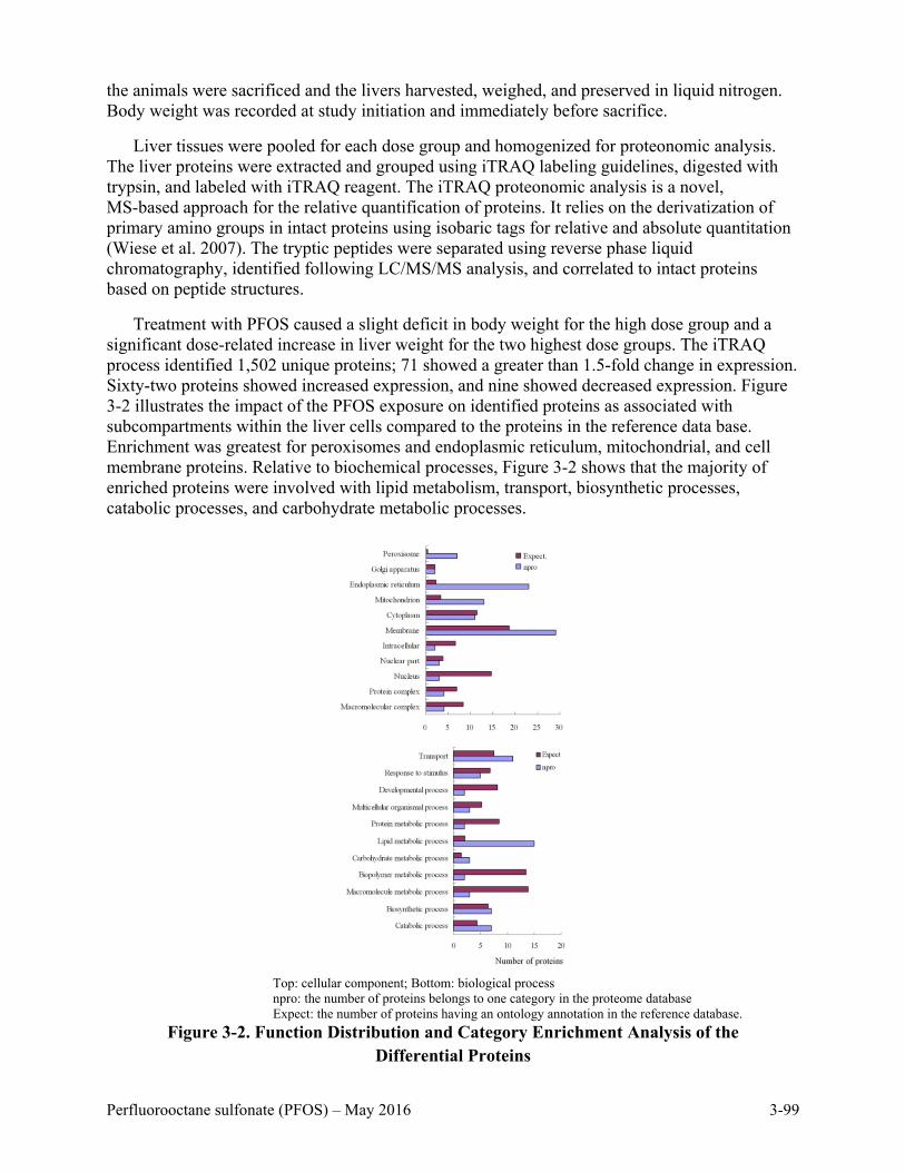
the animals were sacrificed and the livers harvested, weighed, and preserved in liquid nitrogen. Body weight was recorded at study initiation and immediately before sacrifice.
Liver tissues were pooled for each dose group and homogenized for proteonomic analysis. The liver proteins were extracted and grouped using iTRAQ labeling guidelines, digested with trypsin, and labeled with iTRAQ reagent. The iTRAQ proteonomic analysis is a novel, MS-based approach for the relative quantification of proteins. It relies on the derivatization of primary amino groups in intact proteins using isobaric tags for relative and absolute quantitation (Wiese et al. 2007). The tryptic peptides were separated using reverse phase liquid chromatography, identified following LC/MS/MS analysis, and correlated to intact proteins based on peptide structures.
Treatment with PFOS caused a slight deficit in body weight for the high dose group and a significant dose-related increase in liver weight for the two highest dose groups. The iTRAQ process identified 1,502 unique proteins; 71 showed a greater than 1.5-fold change in expression. Sixty-two proteins showed increased expression, and nine showed decreased expression. Figure 3-2 illustrates the impact of the PFOS exposure on identified proteins as associated with subcompartments within the liver cells compared to the proteins in the reference data base. Enrichment was greatest for peroxisomes and endoplasmic reticulum, mitochondrial, and cell membrane proteins. Relative to biochemical processes, Figure 3-2 shows that the majority of enriched proteins were involved with lipid metabolism, transport, biosynthetic processes, catabolic processes, and carbohydrate metabolic processes.
Top: cellular component; Bottom: biological process npro: the number of proteins belongs to one category in the proteome database Expect: the number of proteins having an ontology annotation in the reference database.
Figure 3-2. Function Distribution and Category Enrichment Analysis of the Differential Proteins
Perfluorooctane sulfonate (PFOS) – May 2016 3-99

Sixteen proteins were identified that showed dose-response for the increase in expression.
Four of these were related to peroxisomal beta-oxidation, four were related to CYP-450 aromatase activity, and three had transferase activity including GSTmu3 and GSTmu6. A GTP binding protein (GTP sar-1b) also displayed a dose-related response. One of the remaining four proteins exhibiting dose-response, cysteine sulfinic acid decarboxylase, is the rate limiting enzyme in taurine production and has been proposed as a biomarker for hepatocarcinogenesis.
In the developmental study by Butenhoff et al. (2009), mRNA transcript data for the control and 1.0 mg/kg/day dose group (GD 20 dams and fetuses and PND 21 male pups) was obtained by quantitative reverse transcription polymerase chain reaction. Results for this part of the study were reported by Chang et al. (2009). Statistically-significant changes included:
• Increased Cyp2b2 levels in dams and male pups (2.8-fold and 1.8-fold, respectively) than in controls on GD 20 and PND 21.
• Higher mean acyl CoA (ACoA) and Cyp4a1 levels in male pups (1.5-fold and 2.1-fold, respectively) than those of controls.
• Lower mean Cyp7a1 (3.5-fold) than that for controls.
These results suggest induction of PPARα as well as hepatic CAR. Transcripts possibly related to thyroid status were all similar between the treated dams and pups and the controls.
Oxidative Damage
Liu et al. (2009) conducted a study of KM mice in which 3–6 pups/sex/group were administered one subcutaneous injection of 0 or 50 mg PFOS/kg bw on PNDs 7, 14, 21, 28, or 35. The study was done in an attempt to determine the effects of treatment on the oxidation-antioxidation system by measuring MDA content, SOD activity, and total antioxidation capability (T-AOC). Animals were sacrificed 24 hours post-treatment, blood was collected, and liver and brain were removed and weighed.
No treatment-related effects were observed on body weight. Relative liver weight was significantly increased (p < 0.01 or p <0.05) when compared to controls in both males and females at most time-points. The levels of MDA in the brain and liver and SOD activity were similar between treated mice and the controls at most time-points. On PNDs 7 and 21 in the treated males, brain SOD activity was significantly lower when compared to controls by 19% and 13%, respectively. Liver SOD activity was lower (decrease of 19%) in the treated females on PND 14 when compared to controls. Male brain T-AOC was decreased at all stages of post-natal development but only significantly at PND 21 (decrease of 15%). Male liver T-AOC was decreased significantly at PND 7 (decrease of 25%) and 14 (decrease of 27%). Female brain T-AOC had no significant differences from controls and the liver T-AOC was decreased only at PND 21 (decrease of 15%). The study also demonstrated that distribution increased in the liver and lessened in the blood and brain with postnatal development in both the males and females. On PND 7, PFOS concentrations were 11.78%, 5.04%, and 14.84% in the male mouse blood, brain, and liver, respectively. On PND 28, the PFOS concentrations were 9.89%, 0.85%, and 63.39% in the male mouse blood, brain, and liver, respectively. PFOS brain levels were about 5-fold higher on PND 7 than they were on PND 28. A similar trend was observed in the females. The study suggested that oxidative damage from PFOS can occur, is more prominent in the younger neonates, and is slightly more pronounced in the males.
Perfluorooctane sulfonate (PFOS) – May 2016 3-100

Gap Junctional Intercellular Communication (GJIC)
Gap junctions are found in the cell’s plasma membrane and formed by proteins that connect to form an intercellular connection that allows a direct exchange of chemicals from the interior of one cell to that of adjacent cells without passage into the extracellular space. The GJIC is considered to be essential in maintaining healthy cells and thus disruptions are thought to cause abnormal cell growth, including tumor formation. They also appear to be linked to some neurological, reproductive or endocrine abnormalities.
Hu et al. (2002) tested PFOS exposure in vitro and in vivo to determine whether disruption to the GJIC could possibly be a mechanism for the effects observed with PFOS exposure. The study exposed a rat epithelial cell line (WB-F344) and a dolphin kidney epithelial cell line (CDK) to PFOS at concentrations of 0, 3.1, 6.25, 12.5, 50, 100, or 160 µmol for 30 minutes. GJIC effects were measured using the scrape loading dye technique. PFOS inhibited GJIC rapidly in a dose-dependent method starting at 12.5 µmol, but it was reversible once exposure ended. Additionally, 4 to 6 Sprague-Dawley rats/sex/group were exposed to 0 or 5 mg/kg PFOS by gavage for either 3 or 21 days. GJIC was significantly reduced in the liver tissue after 3 days of exposure. Inhibition also occurred in rats exposed for 21 days, but it was comparable to that seen after 3 days. No differences were observed between the male and female rats.
Wan et al. (2014a) isolated and cultured Sertoli cells from testes of 20-day old rats to examine PFOS’s effects on blood testes barrier function. By day 3 the cultures had established a functional tight junction barrier. Gap junction communication was assayed by means of fluorescence recovery using a photo bleaching assay that measured the ability of a fluorescent dye to move from one cell to another in the presence or absence of PFOS (20 µmol; a 3-hour exposure) in a 120 second period. Cells treated with PFOS displayed significantly lower fluorescence recovery than the control cells in the absence of cytotoxicity. The assays were performed in triplicate. The authors identified this as a matter of concern because it represents diminished function of the blood testes barrier in coordinating an intercellular junction necessary for intercellular communication during spermatogenesis. The authors also examined other characteristics of the blood testes barrier finding effects of PFOS on the cytoskeleton manifest in the form of shortened F-actin filaments.
Mitochondrial Function
Starkov and Wallace (2002) isolated mitochondria from the livers of adult male Sprague-Dawley rats and used them to measure mitochondrial membrane potential and oxygen consumption when exposed to PFOS. PFOS appeared to be a weak mitochondrial toxicant. At higher concentrations, PFOS caused a small increase in resting respiration rate and slightly decreased the membrane potential. The observed effects were attributed to a slight increase in nonselective permeability of the mitochondria membranes caused by the surface-active property of the compound.
Wallace et al. (2013) also examined the impact of 16 different PFASs on mitochondrial respiration rate and oxidative phosphorylation as measured in vitro using isolated rat liver mitochondria. Inhibition was determined through the reduction in oxygen consumption in response to the addition of ADP to isolated mitochondria. PFOS displayed a 20–30 times more potent inhibitory effect among the other sulfonates evaluated (PFBS and PFHxS). PFOS was three times more potent than PFOA. The inhibition mode of action seemed to vary across different PFAS families. In the case of PFOS, its impact on membrane fluidity appeared to be
Perfluorooctane sulfonate (PFOS) – May 2016 3-101

responsible for the observed respiratory inhibition. The authors’ proposed mode of action for this effect from PFOS is consistent with the findings of Matyszewska et al. (2008) that PFOS increased the membrane fluidity and thickness of a model biological membrane in a manner similar to that resulting from cholesterol insertion into a lipid by-layer.
3.3.5 Structure-Activity Relationship
In vitro. Bjork and Wallace (2009) performed a study to see whether the PPARα agonism was relevant in human cell lines and whether effects differed with various chain lengths. Primary rat and human hepatocytes and HepG2/C3A hepatoma cells were exposed to 25 µmol PFAS for 24 hours to determine the structure-activity relationships across various chain lengths. The concentration used was the maximum concentration that did not lead to cell injury in any of the cell lines. The PFAS tested included perfluorinated carboxylic acids with carbon chain lengths of 2 to 8 and perfluorinated sulfonic acids with chain lengths of 4 to 8.
The PFAS stimulated mRNA expression of either acyl CoA oxidase (Acox) or acyl CoA thioesterase (Cte-rats or Acot 1-humans) only in rat hepatocytes and within both series and transcripts; the degree of stimulation of gene expression increased with increasing carbon number. Maximum stimulation of Acox gene expression was 3-fold over control for PFOS; maximum stimulation for Cte/Acot 1 gene expression was 4-fold for PFOS. PFOS did not cause any significant stimulation of Acox or Cte/Acot 1 gene expression in human hepatocytes. The Cyp4a11 gene was not expressed or stimulated by any of the PFASs in HepG2/C3A cells. However, this gene expression was stimulated by PFAS exposure in both rat and human hepatocytes with the perfluorocarboxylates indicating a chain-length dependent structure activity relationship. Maximum gene expression stimulation was in the longer carbon PFAS, but the variability was large with little statistical difference between the 6 and 8 carbon molecules. Study results suggested that the PPARα related gene expression by PFAS was induced in primary rat hepatocytes, increased with carbon chain length, and appeared to be greater in the carboxylic acids (such as PFOA) when compared to the sulfonates (such as PFOS). There was no induction of peroxisome-related fatty acid oxidation gene expression (Acox and Cte/Acot 1) in either primary or transformed human liver cells in culture. This suggests that the PPARα mediated peroxisome proliferation observed in rodent liver may not be relevant as an indicator of human risk.
3.3.6 ToxCast Assays
The Toxicity Forecaster (ToxCast) database is a large high throughput screening compilation of public in vitro and in vivo assays on over 9,000 chemicals. PFOS was tested in 1,087 assays and was active in 175. Assay activation defines the occurrence of the molecular event within the assay (cytotoxicity, induction, binding, and so forth.) with the concentration resulting in 50% activity, AC50, used for comparison to other assays. Assays with < 50% reported efficacy or over-fitting issues are not included in the results discussed. Some of the data from the ToxCast assays such as the interactions with PPAR and CAR support the experimental data for PFOS and PFOA. In cases where effects were only observed at concentrations greater than those causing cytotoxicity, attributing the outcome to PFOS rather than the cytotoxicity is less certain.
Cytotoxicity. Of the active assays, 20 were examining endpoints based on cytotoxicity. Most cell types offered at least one cytotoxicity AC50 for comparison to other in vitro assays. If no cytotoxicity AC50 was reported for a specific cell type, the minimum in vitro cytotoxicity
Perfluorooctane sulfonate (PFOS) – May 2016 3-102

endpoint for PFOS was used for comparison. The lowest PFOS induced AC50 recorded in the ToxCast database is 5.34 μM in the assay for induction of tumor protein 53 using liver cells and the highest AC50 recorded is 172.02 μM for measuring tumor protein in intestinal cells (SD = 45.15; standard error = 9.9).
Endocrine Disruption. Four different estrogen receptor (ESR) assays reported activation following PFOS treatment, all of which were Estrogen Receptor 1- (ESR1-) related. Estrogen and its receptors are essential for sexual development and reproductive function, but they also play a role in other tissues, such as bone. Estrogen receptors are also involved in pathological processes including breast cancer, endometrial cancer, and osteoporosis (NCBI 2016). Two assays of the same cell type were related to ESR1 induction with the lowest AC50, 18.06 μM. This is lower than the cytotoxicity AC50 reported for the cell type used, 23.76 μM, and is indicative of ESR1 induction. In a different ESR1 assay, antagonism was recorded at an AC50 of 83.57 μM, a value higher than the cytotoxicity AC50 for that cell type, 66.31 μM. PFOS induced the estrogen DNA binding site with an AC50 of 87.42 μM. There was no cell-specific reference cytotoxicity value for comparison. The ToxCast assays suggest that PFOS has the ability to induce ESR1.
PFOS antagonized the androgen receptor (AR) at 12.57 μM in human cells and 4.27 μM in rats. Although there is no direct cellular cytotoxicity value to compare, PFOS rat AR antagonism occurred at lower concentrations than the minimum cytotoxicity value (5.34 μM). This implies that PFOS reacts with the AR receptor in the rat and perhaps the human. The progesterone receptor (PR) was also antagonized within the same human cell type, and had a higher AC50, 22.2 μM, than the minimum cytotoxicity AC50. Thyroid receptor (TR) antagonism AC50, 91.24 μM, was also higher than its respective cell specific cytotoxicity AC50 (33.323 μM). This signifies that assay activation (i.e., positive results) might have occurred due to cytotoxicity rather than PR, TR, or human AR antagonism. However, PFOS-induced ESR1 and antagonized rat AR was observed.
Immunotoxicity. PFOS activated a variety of genes related to immunotoxicity in the ToxCast database. These genes include: chemokine ligand (CXCL) 10, CXCL8, collagen type II alpha (COL3A), interleukin-1 alpha (IL-1α), plasminogen activator (PLA), plasminogen activator urokinase (PLAUR), vascular cell adhesion molecule (VCAM1), and the TNF receptor subfamily gene CD40 (CD40). All of the immunological assays were performed by the vendor BioSeek. The vendor did not have a cytotoxicity AC50 for every cell type utilized and used only two cytotoxicity AC50 values for comparison. Genes that had lower AC50 values than cell or BioSeek specific cytotoxicity AC50 were: CD40, PLAUR, PLA, VCAM1, and CXCL8. Given the limited cytotoxicity reference values it is difficult to determine if all gene activity can be attributed to PFOS. For PLAUR and VCAM1, AC50 results were lower than their cell specific cytotoxicity AC50 and have stronger translational potential. VCAM1 and PLAUR play a role in inducing chronic inflammation and vascularization in vivo (Kleinstreuer et al. 2014). This implies PFOS may play a role in inducing chronic inflammation and/or vascularization, both of which are important for the development of rheumatoid arthritis (Khansari et al. 2009).
Neurotoxicity (in vitro). PFOS activated five different neurological receptor families with seven different receptor types in cell-based assays. The receptors activated were: 5-hydroxytryptamine receptor (5HT) 5a, 6, and 7, the adenosine A2a receptor (ADORA2), the adrenoceptor alpha 2C (ADRA2C), and beta 1 (ADRB1), as well as the dopamine receptor D4 (DRD4). Cell-specific cytotoxicity AC50 values for reference were lacking for all of the in vitro assays; only ADORA2 had an activation AC50 lower than the lowest PFOS cytotoxicity AC50 of 5.34 μM. Therefore, it
Perfluorooctane sulfonate (PFOS) – May 2016 3-103

is difficult to draw any conclusions on the potential neurotoxicity of PFOS using the ToxCast data.
Fish Toxicity (in vivo). Oregon State University conducted a large number of toxicity studies using a zebrafish model. PFOS gave positive results in nine assays. Positive effects were recorded for limb malformations 5 days after a 1-time exposure during embryonic development. Other assays with positive results were those for Axis Malformation, Jaw Malformation, Pericaradiac Edema, Snout Malformation, Touch Response, Trunk Malformation, Yolk Sac Edema, and Mortality. Mortality had the lowest reported AC50 at 0.54 µmol. The results provide strong evidence for PFOS developmental toxicity in fish and suggest a potential for human developmental human toxicity.
PPAR/PXR/RAR Receptors. PFOS activated PPARs, PXR, constitutive adrostane receptor (CAR), and retinoic acid receptor (RAR) in assays conducted under the ToxCast program. PFOS induced the DNA sequences for PPAR alpha (PPARα), peroxisome proliferator hormone response elements (PPRE), and PPAR gamma (PPARγ) and antagonized the PPARγ receptor. The only PPAR assay AC50 that was above the cell-specific cytotoxicity AC50 was PPARγ antagonism at 5.91 μM. However, it is possible that cytotoxicity occurs due to PPAR induction or that the PPAR antagonism contributes to cytotoxicity. PFOS induced DNA sequences for PXR, AC50 9.42 μM, at concentrations lower than the cell-specific cytotoxicity AC50. CAR and RAR alpha antagonism were also observed but not at levels below the cell specific cytotoxicity values of 17.57 μM and 28.45 μM respectively. PPAR, PXR, CAR, and RAR pathways are all nuclear receptors that can form heterodimers with one another to induce translation of linked genes. Some of these genes are important for development, reproduction, waste degradation, and even induction of cytotoxicity. Therefore, PFOS induction of these assays are consistent with the experimental data on PPAR and CAR receptor activation.
Cytochrome P450s Activation. Cytochrome P450 (CYP) enzyme bindings were also examined within the ToxCast database in order to understand any metabolic potential for a chemical. Though PFOS is not known to be metabolically active, it showed activation in four acceptable CYP assays: CYP2C18, CYP2C19, and CYP2C9 in human cells, and CYP2C11 in rat. All of the CYP assays had activation at concentrations lower than the lowest cytotoxic AC50. The CYP2C class is known to be involved in the metabolism of xenobiotics including drugs, such as the anti-seizure medication diazepam, the beta blocker propranolol, and the selective serotonergic reuptake inhibitor citalopram. Though there is no evidence of metabolism of PFOS by these CYPs, it is possibly acting as a competitive or allosteric inhibitor for other substrates. This, coupled with PFOS’s high affinity for albumin, could significantly alter the pharmacokinetics of various necessary and habitual pharmaceuticals.
3.4 Hazard Characterization
3.4.1 Synthesis and Evaluation of Major Noncancer Effects
3.4.1.1 Liver Effects, Cholesterol, and Uric Acid
Good correlation between serum and hepatic levels of PFOS has been shown for human samples (Kärrman et al. 2010; Olsen et al. 2003a). However, no consistent adverse effects on the liver were found in epidemiology studies. Biomonitoring studies performed at the 3M Decatur, Alabama plant (Olsen et al. 2001a, 2001b, 2003b) identified occasional differences in hepatic
Perfluorooctane sulfonate (PFOS) – May 2016 3-104

clinical chemistry values but no associated hepatic disease and or hepatic carcinogenicity was reported.
Multiple epidemiologic studies have evaluated serum lipid status in association with PFOS concentration. These studies provide support for an association between PFOS and small increases in total cholesterol. Hypercholesterolemia, which is clinically defined as cholesterol greater than 240 mg/dL, was associated with PFOS exposure in a Canadian cohort (Fisher et al. 2013) and in the C8 cohort (Steenland et al. 2009). PFOS levels in these studies were 0.0084 µg/mL and 0.022 µg/mL, respectively. Cross-sectional occupational studies demonstrated an association between PFOS and total cholesterol (Olsen et al. 2001a, 2001b, 2003b), with much higher PFOS serum levels of up to 1.40 µg/mL. Evidence for associations between other serum lipids and PFOS is mixed, with some studies showing an association with measurements of concurrent HDL and/or LDL and others failing to measure the serum lipoprotien complexes. The studies on serum lipids in association with PFOS serum concentrations are largely cross-sectional in nature and were largely conducted in adults, but some studies exist on children and pregnant females. The location of these cohorts varied from the U.S. population including NHANES volunteers, to the Avon cohort in the UK, to Scandanivian countries. Limitations to these studies include the frequently high correlation between PFOA and PFOS exposure; not all studies control for PFOA in study design.
There are several characteristics of HDLs that explain the association of increased serum concentration of HDL with PFOS or PFOA levels. HDLs accept cholesterol from other serum lipoprotein complexes and bring it to the liver for degradation and conversion to bile salts (Montgomery et al. 1990). Competition between PFOS and bile salts for biliary transport could result in impeded removal of HDL lipids from serum and increase both HDL cholesterol and total cholesterol. The liver is the only tissue that can rid the body of excess cholesterol by secreting it in bile for removal with the feces (Montgomery et al. 1990). In addition, HDLs have the highest ratio of protein to lipid (50:50) among the serum lipoprotein complexes (Montgomery et al. 1990). Binding of PFOS to HDL protein could impede the HDL interaction with liver tissue receptors resulting in increased serum levels of HDL. LDLs contain 21% protein. LDL uptake by tissues is mediated by binding of the LDL apo-B-100 protein and by a receptor independent route (Montgomery et al. 1990). Thus, conformational changes in the lipoprotein proteins as a result of PFOS binding can also impact serum LDL levels.
PFOS, when absorbed, is primarily found in the liver tissue. In monkeys, rats, and mice, PFOS levels in the liver showed a dose-dependent increase that was consistently greater than serum levels (Curran et al. 2008; Goldenthal et al. 1978a; Liu et al. 2009; Seacat et al. 2002; Thomford 2002/Butenhoff et al. 2012). Chang et al. (2009) identified PFOS levels in the liver of rat offspring as early as GD 20, and Stein et al. (2012) measured PFOS in human amniotic fluid supporting placental transfer.
In experimental studies, increased absolute liver weight was observed in monkeys exposed to 0.75 mg/kg/day for 182 days (Seacat et al. 2002), in rats at ≥ 1.33 mg/kg/day for 14 weeks (Curran et al. 2008; Seacat et al. 2003), and in rats at ≥ 0.77 mg/kg/day for 53 weeks (Thomford 2002/Butenhoff et al. 2012). Microscopic lesions of the liver were observed in rats and monkeys. Lesions were found in rats at 1.33–1.56 mg/kg/day after 14 weeks (Seacat et al. 2003), in rats at 0.098–0.299 mg/kg/day after 104 weeks (Thomford 2002/Butenhoff et al. 2012), and in monkeys at 0.75 mg/kg/day after 53 weeks (Seacat et al. 2002). Liver lesions were similar in both species and included centrilobular hypertrophy and vacuolation after the subchronic and chronic exposures with eosinophilic granules also observed after chronic duration. Single cell necrosis
Perfluorooctane sulfonate (PFOS) – May 2016 3-105
was also found in rats at 0.984–1.251 mg/kg/day after 104 weeks (Thomford 2002/Butenhoff et al. 2012; Table 3-15). In these studies, no evidence of peroxisome proliferation was found in either species.
Hepatomegaly and increased liver weight alone are not considered adverse in cases where a chemical such as PFOA causes stimulation of PPAR-α, CAR, and/or PXR cellular receptors (Hall et al. 2012). However, when accompanied by necrosis (Thomford 2002/Butenhoff et al. 2012) and/or fatty acid steatosis (Bijland et al. 2011; Wan et al. 2012), liver weight increases are considered adverse.
In contrast with humans, rats, mice, and monkeys displayed a decrease in cholesterol levels and high density lipoprotein cholesterol following PFOS administration in short and long term studies (Curran et al. 2008; Seacat et al. 2003; L. Wang et al. 2014) when compared to the controls. Male rats had decreased serum cholesterol after 14 weeks at a dose of about 1.4 mg/kg/day (Curran et al. 2008). Wan et al. (2012) found evidence for hepatic macrovesicular steatosis in mice given ≥ 5 mg/kg/day that was not entirely due to PPARα activation. Steatosis was exacerbated when PFOS exposure was combined with a high fat diet.
As discussed above in section 3.3.4, mice administered PFOS showed differential expression of genes or proteins mainly involved in lipid metabolism, transport, biosynthetic processes, and response to stimulus (Tan et al. 2012; L. Wang et al. 2014) and in genes involved in cholesterol biosynthesis and xenobiotic metabolism (Rosen et al. 2010). More specific investigations into the genes involved in lipoprotein metabolism were conducted by Bijland et al. (2011) as described below. In addition, the nuclear hormone receptors CAR and PXR have been shown to be activated in mice (Bijland et al. 2011; Rosen et al. 2010) and rats (Elcombe et al. 2012). Taken together, these studies consistently show an effect on expression of genes involved in lipid metabolism and cholesterol transport and biosynthesis following in vivo PFOS exposure.
To further examine PFOS-specific effects on lipid metabolism, Bijland et al. (2011) examined the molecular biology of hepatic hyperlipidemia in APOE*3-Leiden.CETP mice, a strain that exhibits human-like lipoprotein metabolism. Details of the experimental procedure were given in section 3.2.2. Animals fed 3 mg/kg/day for 4 weeks had decreased hepatic VLDL production leading to increased retention of triglycerides and hepatomegaly, with concomitant decreased hepatic clearance of VLDL and HDL cholesterol. Fecal bile acid content was decreased by 50%.
Overall the genes upregulated were those involved with fatty acid uptake, transport, and catabolism; triglyceride synthesis; cholesterol ester synthesis; and VLDL synthesis and secretion. Genes involved with HDL synthesis, maturation, clearance, and bile acid formation were downregulated. Lipoprotein lipase activity and mRNA expression, both normally low in the liver, were increased.
Many of the genes activated are associated with the nuclear PXR receptor to a greater extent than PPARα. Lipoprotein lipase activity facilitates removal of TGs from serum LDLs, and uptake into the liver and other organs as free fatty acids and glycerol.
No animal studies were identified that examined serum uric acid levels following PFOS exposures.
Perfluorooctane sulfonate (PFOS) – May 2016 3-106

3.4.1.2 Developmental/Reproductive Toxicity
PFOS has been detected in amniotic fluid samples indicating that the chemical crosses the placenta. The median ratio of maternal serum:amniotic fluid concentration was 25.5:1, and PFOS was rarely detected in amniotic fluid until the serum concentration reached at least 0.0055 µg/mL (Stein et al. 2012). Studies evaluating the reproductive and developmental health in humans exposed to PFOS have been performed in both occupational settings and in the general population.
Although not entirely consistent, the set of studies evaluating fetal growth retardation suggest an association of prenatal serum PFOS with deficits in mean birth weight and with LBW. Although three studies were null (Fei et al. 2008a; Hamm et al. 2010; Monroy et al. 2008), birth weight deficits ranging from 29 to 149 grams were detected in 5 studies (Apelberg et al. 2007; Chen et al. 2015; Darrow et al. 2013; Maisonet et al. 2012; Washino et al. 2009). In these studies, PFOS serum levels ranged from 0.005 to 0.0132 µg/mL. Three (Chen et al. 2012; Fei et al. 2007; Stein et al. 2009) out of four (Darrow et al. 2014) studies of LBW showed increased risks (OR range: 1.5–4.8). Studies have questioned whether low maternal GFR is a positive confounder in epidemiology studies of birth weight and PFAS (Morken et al. 2014; Verner et al. 2015). The Verner et al. (2015) comparison between a meta-analysis and PBPK simulations suggests that the some but not all of the association reported between PFOS and birth weight could be attributable to low GFR.
A small set of studies observed an association with gestational diabetes (Zhang et al. 2015, preconception serum PFOS), pre-eclampsia (Stein et al. 2009), and pregnancy-induced hypertension (Darrow et al. 2013) in populations with serum PFOS concentrations of 0.012–0.017 μg/mL. Zhang et al. (2015), and Darrow et al. (2013) used a prospective assessment of adverse pregnancy outcomes in relation to PFAS which addresses some of the limitations the available cross-sectional studies. Associations with these outcomes and serum PFOA also were observed.
There also is generally consistent evidence of associations of serum PFOS with decreased fertility and fecundity (Bach et al. 2015; Fei et al. 2009; Jørgensen et al. 2014; Vélez et al. 2015); there was one null study (Vestergaard et al. 2012). While a concern over the possibility of reverse causation explaining observed associations has been raised (Whitworth et al. 2012), the collective findings, particularly from a more recent study (Bach et al. 2015), support a consistent association with fertility and fecundity measures and PFOS exposures. Although there was some suggestion of an association between PFOS exposures and semen quality parameters in a few studies (Joensen et al. 2009; Toft et al. 2012), most studies were largely null (Buck Louis et al. 2015; Ding et al. 2013; Joensen et al. 2013; Raymer et al. 2012; Specht et al. 2012; Vested et al. 2013).
No animal studies were identified that suggested effects on fertility in males or females. However, López-Doval et al. (2014) found structural effects on the hypothalamic-pituitary axis in adult male rats after exposure to PFOS. There were histopathological lesions of the testes at doses ≥ 1 mg/kg/day and only a few active gonadotrophic cells at doses ≥ 3 mg/kg/day. The lowest dose tested, 0.5 mg/kg/day, was accompanied by decreased LH and testosterone levels and increased FSH levels.
Perfluorooctane sulfonate (PFOS) – May 2016 3-107

Increased pup mortality was observed when rat dams were treated only during gestation as
part of developmental toxicity studies (Chen et al. 2012; Lau et al. 2003; Thibodeaux et al. 2003). Chen et al. (2012) found increased mortality, decreased body weight, and histopathological changes in the lungs (alveolar hemorrhage, thickened interalveolar septum) in rat offspring from dams treated with 2.0 mg/kg/day from GD 1 to 21. No effects were observed in those administered 0.1 mg/kg/day. Developmental delays were found in rat offspring at a lower dose than that affecting survival (1 mg/kg/day; Butenhoff et al. 2009) and in mice at a slightly higher dose (5 mg/kg/day; Lau et al. 2003; Thibodeaux et al. 2003).
Rat dams were treated with PFOS for 63 or 84 days in a one- or two-generation reproductive study, respectively (Luebker et al. 2005a, 2005b). No changes in maternal liver weight were observed on either protocol. The most sensitive endpoint was decreased pup body weight, with reduced survival also observed at higher maternal doses. A NOAEL for pup body weight effects is 0.1 mg/kg/day in the two-generation study (Luebker et al. 2005b); this dose was not tested in the one-generation study (Luebker et al. 2005a) where the LOAEL was 0.4 mg/kg/day for decreased pup body weight, decreased maternal body weight, and decreased gestation length. A 0.4 mg/kg/day dose was a LOAEL in the both the one and two generation studies. The dose associated with a significant decrease in pup survival for the two generation study was 1.6 mg/kg/day and the dose for a decreased viability index was 0.8 mg/kg/day (BMDL5 = 0.89 mg/kg/day) in the one-generation study.
To help characterize the mechanism of PFOS induced neonatal mortality, Grasty et al. (2003) examined critical windows of exposure by treating rats with a high dose of PFOS (25 mg/kg/day) for a 4-day period during various stages of pregnancy. Mortality was highest when treatment occurred on GDs 17–20, identifying late gestation as the sensitive window for neonatal death. In a subsequent experiment, exposure to 50 mg/kg/day of PFOS on GDs 19 and 20 alone was sufficient to produce almost 100% mortality to pups at birth.
Studies by Grasty et al. (2003, 2005) and Chen et al. (2012) describe significant histological and morphometric differences in the lungs between control and PFOS-exposed newborn pups, suggesting that lung maturation and pulmonary surfactant interactions are potential MOAs. Changes in lung morphology were noted in rat pups, but prenatal exposure to PFOS did not affect lung phospholipids or alter the expression of marker genes for alveolar differentiation associated with lung maturation (Grasty et al. 2005). Chen et al. (2012) found that PFOS caused oxidative stress and cell apoptosis in the lungs of offspring from mothers treated with 2.0 mg/kg/day during GDs 1–21.
Currently, the leading hypothesis for the MOA of PFOS-induced neonatal mortality is that PFOS interacts directly with components of natural lung surfactants (Grasty et al. 2005; Xie et al. 2010a, 2010b). PFOS interacts with the major phosphatidylcholine components of pulmonary surfactants and cell membranes and, therefore, has the potential to alter the dynamic properties of lung surfactant (Xie et al. 2010a). PFOS partitions into phospholipid membranes to increase membrane fluidity in several cell types (Xie et al. 2010b). This high tendency of PFOS to partition into phosphatidylcholine lipid bilayers is consistent with its resemblance to medium chain fatty acids and may be responsible for interfering with the normal physiological function of pulmonary surfactant.
Prenatal PFOS exposures appear to influence hormones during gestation, as well as in neonate and adult animals. Zhao et al. (2014) examined the testes from male Sprague-Dawley rat fetuses on GD 20 following maternal exposure to 0, 5, or 20 mg/kg/day on GDs 11–19. Fetal
Perfluorooctane sulfonate (PFOS) – May 2016 3-108

Leydig cells were found to be reduced in number with evidence of apoptosis. Levels of testosterone were reduced along with the levels of key enzymes or mRNA for proteins involved with testosterone production.
Studies have examined the impact of gestational and lactational exposures on the pups as adults (Lv et al. 2013 rats; Wan et al. 2014b, mice). In both cases early life exposure through maternal treatment with PFOS was associated with abnormal glucose control in the mature offspring. In both cases, serum glucose was significantly higher in the adult animals exposed during gestation and lactation than in controls and there was evidence of insulin resistance. The LOAEL was 0.5 mg/kg/day in the Lv et al. (2013) study and 3 mg/kg/day for pups fed a diet with normal fat levels (Wan et al. 2014b). In the Wan et al. (2014b) study, the NOAEL was 0.3 mg/kg/day. When accompanied by a high fat diet, 0.3 mg/kg/day was a LOAEL for increased insulin resistance.
3.4.1.3 Immunotoxicity
A few studies have evaluated associations with measures indicating immunosuppression. Two studies reported decreases in response to one or more vaccines (diphtheria, rubella) in children aged 3, 5, and 7 years (e.g., measured by antibody titer) in relation to increasing maternal serum PFOS levels (maternal levels ranging from 0.0056 to 0.027 µg/mL) during pregnancy or in the children at 5 years of age (mean child 0.0167 µg/mL) (Grandjean et al. 2012; Granum et al. 2013). Decreased rubella and mumps antibody concentrations in relation to serum PFOS concentration were found among 12–19 year old children in the NHANES, particularly among seropositive children (Stein et al. 2015). A third study of adults found no associations with antibody response to influenza vaccine (Looker et al. 2014). In the three studies examining exposures in the background range among children (i.e., general population exposures, geometric means < 0.02 µg/mL), the associations with PFOS were also seen with other correlated PFAS, complicating conclusions specifically for PFOS.
No clear associations were reported between prenatal PFOS exposure and incidence of infectious disease among children (Fei et al. 2010b; Okada et al. 2012), although there might be effect modification by sex. With regard to other immune dysfunction, serum PFOS levels were not associated with risk of ever having had asthma among children in the NHANES with median levels of 0.017 µg/mL (Humblet et al. 2014). A study among children in Taiwan with higher serum PFOS concentrations (median with and without asthma 0.0339 and 0.0289 µg/mL, respectively) found higher odds ratios for physician-diagnosed asthma with increasing serum PFOS quartile (Dong et al. 2013). Associations also were found for other PFASs. Among asthmatics, serum PFOS was also associated with higher severity scores, serum total IgE, absolute eosinophil counts, and eosinophilic cationic protein levels.
Other data on the immunotoxicity of PFOS in humans are limited to in vitro studies using cells recovered from human blood (PBMCs; Brieger et al. 2011 or CD4+ T cells; Midgett et al. 2014). In both cases the concentration of PFOS with a demonstrated significant effect was 100 µg/mL, and the concentration that lacked any effects was 10 µg/mL. A significant (p < 0.001) decrease in TNFα and a nonsignificant trend towards increasing IL-6 release from stimulated monocytes were seen, but no effects were measured on stimulated T cells (Brieger et al. 2011). T cell IL-2 production was decreased in the Midgett et al. (2014) study.
Perfluorooctane sulfonate (PFOS) – May 2016 3-109

Studies in mice examined NK cell activity and SRBC response following oral dosing with
PFOS. Three of four studies showed effects on SRBC response and/or NK cell activity at the same dose that caused increased liver weight (Dong et al. 2009; Keil et al. 2008; Zheng et al. 2009). Based on the limited evidence, neither response appeared more sensitive than the other. The animal studies indicate that females are less susceptible to impacts on NK cell activity and the SRBC response than males.
The NK cell activity was enhanced at very low PFOS doses, while it was depressed at higher doses. Peden-Adams et al. (2008) and Dong et al. (2009) showed increased NK cell activity in male mice following exposure to 0.0017 mg/kg/day and 0.083 mg/kg/day, respectively. The increased activity in Dong et al. (2009) correlated with a PFOS serum level of approximately 7.1 µg/mL. In the Dong et al. (2009) study, the NK cell activity was significantly decreased at a higher dose of 0.833 mg/kg/day, demonstrating a U-shaped response to dose. Doses ≥ 1 mg/kg/day resulted in decreased NK cell activity in offspring of dams treated during gestation (Keil et al. 2008) and in adult male mice (Zheng et al. 2009).
In the Peden-Adams et al. (2008) study, IgM suppression occurred after 28 days of treatment with 0.0017 mg/kg/day although there were not any overt signs of toxicity. Further investigation found that the IgM suppression was observed in both the T-cell independent and dependent tests making the humoral immune effects caused by B-cells. Other studies also showed a suppression of the SRBC response at higher doses of PFOS (Dong et al. 2009; Keil et al. 2008; Zheng et al. 2009). Guruge et al. (2009) found a decrease in survival in mice exposed to 0.025 mg/kg of PFOS after exposure to influenza A virus.
Qazi et al. (2009a) reported that approximately 40 mg/kg/day in the diet for 10 days in wild-type and PPARα-null 129/Sv knock-out mice caused a pronounced decrease in the total number of thymocytes and splenocytes, as well as a decrease in size of the those present in wild-type mice. Knock-out mice had a reduction in the total number of thymocytes that was less than that seen in the wild-type mice. Effects on splenocytes were mostly eliminated in knock-out mice. The study, thus, indicated that the immunomodulation was partially dependent on PPARα. Mechanisms that could cause these effects other than PPAR activation are not known. At the same dose, Qazi et al. (2009b) did not find elevated levels in serum or spleen of TNF-α and IL-6 in response to stimulation by LPS in C57Bl/6 mice, but levels were increased in the cells from the peritoneal cavity and bone marrow
3.4.1.4 Neurotoxicity
Developmental neurotoxicity and adult neurotoxicity studies have been conducted in rats and mice. Mechanistic studies have examined effects on excitatory amino acids and gene profiles following PFOS exposures.
Butenhoff et al. (2009) found significantly increased motor activity and decreased habituation of male offspring at one time point (PND 17) following gestational and lactational dosing of dams with 1.0 mg/kg/day. No effects were found on learning and memory with the Biel swimming maze. Luebker et al. (2005b) found no effects on passive avoidance behavior or water maze learning and memory in F1 offspring at a daily dose of 0.4 mg/kg/day. Y. Wang et al. (2015) used water maze testing on offspring from treated dams who were cross-fostered with either control or treated dams, and continued on the treatment of their lactational dam. Escape latency was significantly increased for all treated groups on one or more testing days with the most pronounced effect in pups exposed prenatally from dams given 15 mg/L drinking water and
Perfluorooctane sulfonate (PFOS) – May 2016 3-110

cross-fostered to control dams. Y. Wang et al. (2015) did not provide data on water intake or body weight data. A drinking water concentration of 5 mg/L was a NOAEL, and a concentration of 15 mg/L was a LOAEL for offspring. Estimated adult doses are 0.8 and 2.4 mg/kg/day, respectively, using the subchronic USEPA (1988) conventions for water intake and body weight. Long et al. (2013) found a significantly longer latency to escape, with significantly less time spent in the target quadrant in the Morris water maze test for learning and memory at a dose of 2.5 mg/kg/day in 8-week-old C57BL6 mice. The NOAEL for these effects was 0.43 mg/kg/day.
Effects were observed on excitatory amino acids in the central nervous systems of rats when administered 25 mg/kg/day of PFOS one time (Yang et al. 2009). Wang et al. (2010) found that pre-natal exposure to 3.2 mg/kg/day of PFOS in the feed had some effect on gene expression involved in neuroactive ligand-receptor interaction, calcium signaling pathways and PPAR signaling. Zeng et al. (2011) also found PFOS administered to pregnant rats as low as 0.1 mg/kg from GD 2 to 21 caused significant increases of PFOS in the brain (hippocampus and cortex) of the offspring, with effects on inflammatory markers and transcription factors. Two-month-old mice exposed to 0.75 mg/kg of PFOS when they were 10 days old (Johansson et al. 2008) displayed abnormal habituation responses in motor activity testing. Cultured hippocampal neurite growth and branching were suppressed by exposure to 50 µmol PFOS. The authors hypothesized that this was a consequence of PFOS incorporation into the neuronal lipid bilayer membrane (Liao et al. 2009). The effect of PFOS was greater than that of PFOA. PFOS was the only member of the sulfonate family to exhibit this effect.
3.4.1.5 Thyroid Effects
Numerous epidemiologic studies have evaluated thyroid hormone levels and/or thyroid disease in association with serum PFOS concentrations. These epidemiologic studies provide limited support for an association between PFOS exposure and incidence or prevalence of thyroid disease, and include large studies of representative samples of the general U.S. adult population (Melzer et al. 2010; Wen et al. 2013). These highly powered studies reported associations between PFOS exposure (serum PFOS concentrations) and thyroid disease but not thyroid hormone status. Melzer et al. (2010) studied thyroid disease with medication (PFOS level of 0.025 µg/mL in males and 0.019 µg/mL in females) and Wen et al. (2013) studied subclinical thyroid disease (mean serum 0.0142 µg/mL). Thyroid function can be affected by iodide sufficiency and by autoimmune disease. People testing positive for the anti-TPO biomarker for autoimmune thyroid disease showed associations with PFOS (0.0048 µg/mL) and TSH or T4 (Webster et al. 2014); this association was stronger in people with both low iodide status and positive anti-TPO antibodies, with a PFOS level of 0.014 µg/mL (Webster et al. 2015). These studies used anti-TPO antibody levels as an indication of stress to the thyroid system, not a disease state. Thus, the association between PFOS and altered thyroid hormone levels is stronger in people at risk for iodine deficiency than those receiving adequate dietary iodine. In people without diagnosed thyroid disease or without biomarkers of thyroid disease, thyroid hormones (TSH, T3, or T4) show mixed effects across cohorts.
Several animal models have described changes in thyroid hormone levels after administration of PFOS. In contrast to the human epidemiology studies, the most consistent finding in animals treated with PFOS was a decrease in T4 with slight, or no, changes in T3. Any changes found in T3 and T4 levels failed to activate the hypothalamic-pituitary-thyroid (HPT) feedback mechanism to produce significant elevations of serum TSH.
Perfluorooctane sulfonate (PFOS) – May 2016 3-111
Rats treated orally with PFOS for 1–5 days had significant decreases in total T4 at doses of
10 and 15 mg/kg, but not at 5 mg/kg (Chang et al. 2007, 2008; Martin et al. 2007). With treatment for 7 days, total T4 was decreased at 1 and 3 mg/kg (Yu et al. 2011).
In Cynomolgus monkeys treated with 0.03, 0.15, or 0.75 mg/kg/day of PFOS for 26 weeks, Seacat et al. (2002) saw significant reductions of total triiodothyronine (T3) (~ 50%), and a less consistent effect in total thyroxine (TT4, females only). This was more pronounced at the end of exposure period in the high-dose group but neither a dose-response nor evidence of hypothyroidism was observed. TSH levels were variable during the study, but increased 2-fold in the high-dose males at the end of exposure.
Exposure of pregnant rats to PFOS at 1 mg/kg/day, which corresponded to maternal serum concentrations of 14–26 μg/mL, resulted in decreases in T4 and T3 in dams (Thibodeaux et al. 2003) and decreased T4 in pups (Lau et al. 2003). No effect was observed on serum TSH. In contrast, no effects were found on thyroid hormones in either dams or pups when females were treated prior to mating and through LD 4 (Luebker et al. 2005a). Histological and morphometric evaluations of the fetal and neonatal thyroid glands indicated normal number and size distribution of follicles, and normal follicular epithelial cell heights and colloid areas (Chang et al. 2009).
In addition to the evaluation of PFOS’s effects on serum TT4, several studies have examined the levels of circulating FT4 (Lau et al. 2003; Luebker et al. 2005a; Thibodeaux et al. 2003; Yu et al. 2011). In these studies, FT4 was reduced after PFOS administration when measured by analog radioimmunoassays (RIA). However, when the FT4 was measured by an equilibrium dialysis step prior to the standard RIA (ED-RIA), FT4 levels in the PFOS-treated rats were comparable to those of controls (Luebker et al. 2005a).
Mechanisms underlying the PFOS-induced alterations in thyroid hormones are still under active investigation, but do not likely involve altered de novo biosynthesis of the hormones or compromised integrity of the HPT axis. Yu et al. (2009b) reported no significant effects of PFOS on the sodium iodide symporter gene expression (for iodide uptake) or thyroid peroxidase activity in the thyroid gland. Chang et al. (2008) showed that release of TSH from the pituitary in response to ex vivo TRH stimulation was not altered by PFOS exposure.
Weiss et al. (2009) demonstrated that perfluorinated chemicals (including PFOS) are capable of competing with T4 and displacing the hormone from binding to the human thyroid hormone transport protein transthyretin (TTR). In fact, PFOS ranks the second highest in binding potency among all perfluorinated compounds examined, although its TTR binding potency is only one-fifteenth of that for T4. Similarly, Ren et al. (2015) demonstrated that PFOS bound to the ligand binding domain of the human thyroid hormone receptor, although with a much lower affinity than T3.
Several possibilities might account for the differential findings of thyroid hormone disruption between animal models and human biomonitoring data. First, decreased T3 or T4 was observed in adult monkeys and rodents only when serum PFOS reached the 70–90 μg/mL range. Pregnant rats and neonatal rats appeared to be more sensitive, exhibiting TT4 depression when serum PFOS reached about 20 and 40 μg/mL, respectively. However, serum PFOS in general populations of humans is estimated to be 0.018–0.037 µg/mL, about three orders of magnitude lower than the effective body burden for thyroid hormone disruption in animal models. Secondly, TBG (rather than TTR as in rodents) is the major thyroid hormone transporter in
Perfluorooctane sulfonate (PFOS) – May 2016 3-112
humans. Although PFOS can bind to human TTR and effectively displaces T4 as illustrated in the rat model, its binding affinity to TBG is unknown. PFOS has been shown to have much a lower binding affinity for both TTR and the thyroid hormone receptor than do T4 and T3, respectively (Ren et al. 2015; Weiss et al. 2009).
3.4.2 Synthesis and Evaluation of Carcinogenic Effects
The small set of epidemiology studies of PFOS exposure do not suggest that there is an association with cancer, but the breadth and scope of the studies are not adequate to make definitive conclusions. While an elevated risk of bladder cancer mortality was associated with PFOS exposure in an occupational study (Alexander et al. 2003), a subsequent study to ascertain cancer incidence in the cohort observed elevated but statistically insignificant incidence ratios that were 1.7- to 2-fold higher among exposed workers (Alexander and Olsen 2007). Mean PFOS serum levels were 0.941 µg/mL. No elevated bladder cancer risk was observed in a nested case-control study in a Danish cohort with plasma PFOS concentrations at enrollment ranging 0.001–0.1305 µg/mL (Eriksen et al. 2009).
Elevated odds ratios for prostate cancer were reported for the occupational cohort examined by Alexander and Olsen (2007) and the Danish population-based cohort examined by Eriksen et al. (2009), however the confidence intervals included the null, and no association was reported by another case-control study in Denmark (Hardell et al. 2014). A case-control study of breast cancer among Inuit females in Greenland with similar serum PFOS levels to those of the Danish population (0.0015–0.172 µg/mL) reported an association of low magnitude that could not be separated from other perfluorsulfonated acids, and the association was not confirmed in a Danish population (Bonefeld-Jørgensen et al. 2011, 2014). Some studies evaluated associations with serum PFOS concentration at the time of cancer diagnosis and the impact of this potential exposure misclassification on the estimated risks is unknown (Bonefeld-Jørgensen et al. 2011; Hardell et al. 2014). No associations were adjusted for other perfluorinated chemicals in serum in any of the occupational and population-based studies.
The only chronic toxicity/carcinogenicity study in animals was a rat study (Thomford 2002/Butenhoff et al. 2012). Increased incidence of hepatocellular adenomas in the male (12% at the high dose) and female rats (8% at the high dose) and combined adenomas/carcinomas in the females (10% at the high dose) were observed, but they did not display a clear dose-related response. In males but not females the serum ALT levels were increased at 14, 27, and 53 weeks. At 105 weeks there was an increase in eosinophilic clear cell foci, and cystic hepatocellular degeneration in males given 2, 5, and 20 ppm PFOS. Low levels of single cell necrosis in all dose groups (males and females) were identified; the increase compared to controls was significant at the high dose in males and females (Table 3-15).
Thyroid tumors (adenomas and carcinomas) were seen in males receiving 0, 0.5, 2, 5, or 20 ppm and in females receiving 5 or 20 ppm in their diet. The tumor (adenomas + carcinomas) prevalence for males was consistent across dose groups. In males the incidence of thyroid tumors was significantly elevated only in the high-dose, recovery group males exposed for 52 weeks (10/39) but not in the animals receiving the same dose at 105 weeks. There were very few follicular cell acenomas/carcinomas in the females (5 total) with no dose-response. The most frequent thyroid tumor type in the females was C-cell adenomas, but the highest incidence was that for the controls and there was a lack of dose response among the exposed groups. C-cell adenomas were not observed in males (Thomford 2002/Butenhoff et al. 2012).
Perfluorooctane sulfonate (PFOS) – May 2016 3-113

There was a high background incidence in mammary gland tumors in the female rats,
primarily combined fibroma adenoma and adenoma, but the incidence lacked dose-response for all tumor classifications. Mammary gland carcinomas also lacked dose-response and had a relatively comparable incidence across dose groups including the controls (Thomford 2002/Butenhoff et al. 2012).
All genotoxicity studies including an Ames test, mammalian-microsome reverse mutation assay, an in vitro assay for chromosomal aberrations, an unscheduled DNA synthesis assay, and mouse micronucleus assay were negative.
3.4.3 Mode of Action and Implications in Cancer Assessment
Short-term genotoxicity assays suggested that PFOS is not a DNA-reactive compound. The results from five in vitro studies (Cifone 1999; Litton Bionetics, Inc. 1979; Mecchi 1999; Murli 1999; Simmon 1978) were negative, as was the result from an in vivo bone marrow micronucleus assay (Murli 1996).
Induction of peroxisome proliferation has been suggested as the mode of action for an increasing number of non-genotoxic carcinogens that induce liver tumors upon chronic administration to rats, mice, or both (Ashby et al. 1994; Rao and Reddy 1996). The liver-expressed peroxisome PPARα regulates the transcription of genes involved in peroxisome proliferation, cell cycle control, apoptosis, and lipid metabolism. The data for PFOS illustrate the ability of PFOS to activate PPARα (Martin et al. 2007; Shipley et al. 2004; Wolf et al. 2008, 2012). However, data are generally lacking for increased cell proliferation. No increase in hepatic cell proliferation was detected in the subchronic study (Seacat et al. 2003) or the cancer bioassay (Thomford 2002/Butenhoff et al. 2012); limited necrosis was observed across all doses and significantly (p < 0.05) increased for the 20 ppm males and females. In addition, no subchronic or longer term studies revealed evidence of preneoplastic foci in the liver. Liu et al. (2009) studied biomarkers for oxidative stress in the liver and brain in KD mice. Levels of MDA did not differ between controls and exposed animals; SOD activity was lower than that observed in the controls.
Other possible MOAs for carcinogenicity have been explored, including mitochondrial biogenetics and GJIC. While PFOS was shown to be a weak toxicant to isolated mitochondria (Starkov and Wallace 2002), it inhibited GJIC in a dose-dependent manner in two cell lines and in liver tissue from rats exposed orally (Hu et al. 2002). These are not clearly defined MOAs, and their importance relative to PFOS exposure is not certain. Ngo et al. (2014) used the mouse model C57BL/6J –Min/+ for intestinal neoplasia to determine effects following in utero exposure. Maternal treatment with PFOS at doses up to 0.3 mg/kg/day during gestation did not result in an increase of intestinal tumors in either wildtype or susceptible offspring up to 20 weeks of age.
3.4.4 Weight of Evidence Evaluation for Carcinogenicity
Under the EPA Guidelines for Carcinogen Risk Assessment (USEPA 2005a) there is suggestive evidence of carcinogenic potential of PFOS in humans. A single chronic cancer bioassay in animals is available for PFOS. Although liver adenomas were significantly increased in males and females at the highest dose and a positive trend was observed (p = 0.03), a dose-response pattern was not observed. In males the incidence of thyroid follicular tumors was
Perfluorooctane sulfonate (PFOS) – May 2016 3-114

elevated only in the high-dose, recovery group exposed for 52 weeks, where it was about 3 times greater than the incidence in rats given the same dose for 104 weeks. As was the case for the liver tumors, the thyroid adenoma data did not show a direct response to dose. Based on the available evidence, the data are inadequate to support a PPARα-linked MOA for the liver and thyroid adenomas observed by Thomford (2002)/Butenhoff et al. (2012) in the chronic 2 year bioassay in Sprague-Dawley Crl:CD(SD)IGS BR rats.
3.4.5 Potentially Sensitive Populations
In humans, single blood samplings of different populations within the United States do not support major gender differences in half-life or sensitivity to PFOS. Gender differences could not be determined by those exposed by occupational exposure, as the majority of those tested were males. Serum monitoring among the NHANES populations (2004–2008) found significantly (p < 0.05) higher PFOS levels in males (0.020 μg/L) than females (0.014 μg/L). However, this difference is more likely to be related to exposures than to sensitivity.
Evidence from animal studies does not suggest major differences between genders in the amount of PFOS identified in the serum and liver tissue of animals or in the toxicity. In the monkey studies and most developmental rat studies, there do not appear to be any differences between the males and females after administration of PFOS. However, in the chronic/carcinogenicity study in rats, the male rats do appear to be slightly more sensitive to liver toxicity. In animal studies of immunological effects, the response to NK cell suppression occurred at a lower dose in males than in females (Peden-Adams et al. 2008).
Animal studies clearly show that developmental exposure of rats or mice to PFOS administered during gestation results in rapid, dose-dependent effects on neonatal survival (Lau et al. 2003; Luebker et al. 2005a, 2005b). Additional long term effects on postnatal growth and delays in developmental landmarks (eye opening, pinna unfolding, surface righting, air righting) occur in surviving rat pups. The mechanistic cause of this developmental toxicity is unknown, but investigations of several potential modes of action are summarized here. Generally, there is a lack of consistency among the epidemiology studies regarding potential associations between PFOS levels during pregnancy and developmental birth outcomes. Some studies indicate a potential impact on birth weight, but this finding is not consistent across studies.
The animal data on LBW receive support from the epidemiolgy (Apelberg et al. 2007; Chen et al. 2015; Darrow et al. 2013; Maisonet et al. 2012; Washino et al. 2009). For humans with low GFR (females with pregnancy-induced hypertension or preeclampsia in late pregnancy), the impact on body weight is likely due to a combination of the low GFR and the serum PFOS (Verner et al. 2015). Low GFR in pregnant females will tend to cause an increase in serum PFOS compared to individuals with a normal GFR. Females with hypertension during pregnancy could have an increased risk for having a LBW baby.
The fat content of the diet appears to be an important variable that influences the effects from PFOS exposures. Elevated total cholesterol, HDL, and sometimes triglycerides are effects seen in a number of the human epidemiology studies. However, none of the studies evaluated appeared to control for fat content in the typical diet of the subjects. Martin et al. (2007), Bijland et al. (2011), and Wan et al. (2012) found hepatic steatosis in PFOS-treated animals. Liver fat increased with both a high fat diet alone and with a high fat diet plus PFOS (Wan et al. 2012). In the same study, significant increases in the expression of fatty acid translocase and lipoprotein lipase was observed at the 10 mg/kg/day PFOS dose. Mobilization of liver lipids appeared to
Perfluorooctane sulfonate (PFOS) – May 2016 3-115

decrease following the PFOS exposure leading to lower serum LDL/VLDL levels; VLDLs are carriers of liver triglycerides and other lipids from liver to serum.
To help characterize the mechanism of PFOS induced neonatal mortality, Grasty et al. (2003) examined critical windows of exposure by treating rats with a high dose of PFOS (25 mg/kg/day) for a 4-day period during various stages of pregnancy. Neonatal mortality occurred after all treatment periods, but the incidence of neonatal death increased when exposure occurred later in gestation. Mortality was highest when treatment occurred on gestation days (GDs) 17–20, identifying late gestation as a critical exposure window for increasing the risk of neonatal survival. The effects of PFOS at this stage of development could be related to an impact of PFOS on lung surfactants leading to respiratory distress syndrome. Both Luebker et al. (2005a) and Lau et al. (2003) identified pup mortality as adverse effects of gestational PFOS exposures.
Perfluorooctane sulfonate (PFOS) – May 2016 3-116

4. DOSE-RESPONSE ASSESSMENT
A Reference Dose (RfD) or Reference Concentration (RfC) is used as a benchmark for the prevention of long-term toxic effects other than carcinogenicity. RfD/RfC determination assumes that thresholds exist for toxic effects, such as cellular necrosis, significant body or organ weight changes, blood disorders, and so on. The RfD is expressed in terms of milligrams per kilogram per day (mg/kg/day), and the RfC is expressed in milligrams per cubic meter (mg/m3). The RfD and RfC are estimates (with uncertainties spanning perhaps an order of magnitude) of the daily exposure to the human population (including sensitive subgroups) that is likely to be without an appreciable risk of deleterious effects during a lifetime.
4.1 Dose-Response for Noncancer Effects
4.1.1 RfD Determination
Human Data. In humans, data have been obtained from studies evaluating both occupational and general population exposure scenarios. Some studies monitored similar populations over time to determine whether or not a trend was present. Pathways of exposure in the general population appear to be from drinking water, food (especially fish/seafood), and some environmental exposures (e.g., carpets, house dust). In general, PFOS levels in the serum of the general population have decreased since production was stopped in the United States.
Multiple epidemiology studies evaluated serum lipid status in association with PFOS concentration. These studies provide support for an association between PFOS and small increases in total cholesterol. Hypercholesterolemia, (clinically defined as cholesterol > 240 mg/dL) was associated with PFOS exposure in a Canadian cohort (Fisher et al. 2013) and in the C8 cohort (Steenland et al. 2009); PFOS levels in these studies were 0.0084 µg/mL and 0.022 µg/mL, respectively. Cross-sectional occupational studies demonstrated an association between PFOS and total cholesterol (Olsen et al. 2001a, 2001b, 2003b), with much higher PFOS serum levels of up to 1.40 µg/mL. Evidence for associations between PFOS and other serum lipids including HDL cholesterol, LDL, VLDL, non-HDL cholesterol, and triglycerides is mixed.
The studies on serum lipids in association with PFOS serum concentrations are largely cross-sectional in nature and were largely conducted in adults. Some studies exist on children and pregnant females. The location of these cohorts varied from the U.S. population including NHANES volunteers, to the Avon cohort in the UK, to and Scandanivian countries. Limitations to these studies include the frequently high correlation between PFOA and PFOS exposure; not all studies control for PFOA in study design.
Studies that evaluated thyroid hormone levels and/or thyroid disease in association with serum PFOS concentrations include large representative samples of the general U.S. adult population and provide limited support for an association between PFOS exposure and the incidence or prevalence of thyroid disease. PFOS levels in Melzer et al. (2010) were 0.025 µg/mL in males and 0.019 µg/mL in females, and in Wen et al. (2013) they were 0.0142 µg/mL. Pregnant females testing positive for the anti-TPO biomarker for autoimmune thyroid disease showed a positive association with PFOS (0.0048 µg/mL) and TSH (Webster et al. 2014). In a second study, Webster et al. (2015) found an association with PFOS (0.014 µg/mL) and TSH and T3 in a subset of the NHANES population with both low iodide status and positive anti-TPO antibodies. Anti-TPO antibody levels are an indication of stress to
Perfluorooctane sulfonate (PFOS) – May 2016 4-1

the thyroid system, not a disease state. Thus, the association between PFOS and altered thyroid hormone levels is stronger in people at risk for thyroid insufficiency or disease. In people without diagnosed thyroid disease or without biomarkers of thyroid disease, thyroid hormones (TSH, T3, or T4) show mixed effects across cohorts.
A few studies evaluated associations with measures of immunosuppression. Two studies reported decreases in response to one or more vaccines (diphtheria, rubella) in children aged 3, 5, and 7 years (e.g., measured by antibody titer) in relation to increasing maternal serum PFOS levels (ranging 0.0056–0.027 µg/mL) during pregnancy or at 5 years of age (Grandjean et al. 2012; Granum et al. 2013). Decreased rubella and mumps antibody concentrations in relation to serum PFOS concentration were found among 12–19 year old children in the NHANES, particularly among seropositive children (Stein et al. 2015). A study of adults found no associations with antibody response to influenza vaccine (Looker et al. 2014). In the three studies examining exposures in the background range among children (i.e., general population exposures, geometric means < 0.02 µg/ml), the associations with PFOS were also correlated with other PFASs, complicating conclusions as they applied to PFOS.
No clear associations were reported between prenatal PFOS exposure and incidence of infectious disease among children (Fei et al. 2010b; Okada et al. 2012), although an elevation in risk of hospitalizations for infectious disease was found among girls, suggesting effect modification by sex. PFOS levels were not associated with risk of ever having had asthma among children in the NHANES with median levels of 0.017 µg/mL (Humblet et al. 2014). A study among children in Taiwan with higher serum PFOS concentrations (median with and without asthma 0.0339 and 0.0289 µg/mL, respectively) found higher odds ratios for physician-diagnosed asthma with increasing serum PFOS quartile (Dong et al. 2013). Associations with other PFASs were also positive. Among asthmatics, serum PFOS was associated with higher severity scores, serum total IgE, absolute eosinophil counts, and eosinophilic cationic protein levels.
The set of studies evaluating fetal growth retardation suggest an association of prenatal serum PFOS with deficits in mean birth weight and with LBW, however it is not entirely consistent. Birth weight deficits ranging from 29 to 149 grams were detected in five studies (Apelberg et al. 2007; Chen et al. 2015; Darrow et al. 2013; Maisonet et al. 2012; Washino et al. 2009). In these studies, PFOS serum levels ranged from 0.005 to 0.0132 µg/mL. Three (Chen et al. 2012; Fei et al. 2007; Stein et al. 2009) out of four (Darrow et al. 2014) studies of LBW showed increased risks (OR range: 1.5–4.8). Studies have questioned whether low maternal GFR is a confounder in epidemiology studies of birth weight and PFOS (Morken et al. 2014; Verner et al. 2015). The Verner et al. (2015) study compared the results from a meta-analysis of the epidemiology data with PBPK simulations and concluded that the some, but not all, of the association reported between PFOS and birth weight is attributable to low GFR. Thus, the interpretation of the observed associations is unclear.
A small set of studies observed an association with gestational diabetes (Zhang et al. 2015, preconception serum PFOS), pre-eclampsia (Stein et al. 2009), and pregnancy-induced hypertension (Darrow et al. 2013) in populations with serum PFOS concentrations of 0.012 – 0.017 μg/mL. Zhang et al. (2015) and Darrow et al. (2013) used a prospective assessment of adverse pregnancy outcomes in relation to serum PFAS thereby avoiding some of the limitations of the available cross-sectional studies. Associations with serum PFOA and adverse pregnancy outcome were identified.
Perfluorooctane sulfonate (PFOS) – May 2016 4-2

There is consistent evidence of associations of serum PFOS with decreased fertility and
fecundity (Bach et al. 2015; Fei et al. 2009; Jørgensen et al. 2014; Vélez et al. 2015). While a concern over the possibility of reverse causation explaining observed associations has been raised (Whitworth et al. 2012), the collective findings, particularly from a more recent study (Bach et al. 2015), support a consistent association with fertility and fecundity measures and PFOS exposures. Although there was some suggestion of an association between PFOS exposures and semen quality parameters in a few studies (Joensen et al. 2009; Toft et al. 2012), most studies were largely null (Buck Louis et al. 2015; Ding et al. 2013; Joensen et al. 2013; Raymer et al. 2012; Specht et al. 2012; Vested et al. 2013).
Animal Data. Adequate studies were available for short-term, subchronic, chronic, developmental, and reproductive parameters in rats, mice, and primates. Subchronic, chronic, and reproductive toxicity animal studies, all with exposure duration greater than 60 days, have been summarized in Table 4-1. Shorter duration studies that focused on immunotoxicity endpoints and developmental toxicity studies are summarized in Table 4-2. Although the exposure durations are shorter in developmental studies, they are important in quantification of dose-response because the exposures occur during critical windows of development and are often symptomatic of effects that can occur later in life. It is noted, however, that in some of these studies, steady states of PFOS might not have been achieved due to the long half-life of PFOS in animal models (see discussion of steady state in section 4.1.1.1)
Seacat et al. (2002) treated monkeys with PFOS for up to 6 months and found increased liver weight and centrilobular or diffuse hepatocellular hypertrophy at 0.75 mg/kg/day, but no clear evidence of peroxisomal or cell proliferation. Hepatic peroxisome proliferation, measured by PCoAO activity, was increased significantly in the females at 0.75 mg/kg/day; however, the magnitude was less than the 2-fold increase typically indicating biological significance and PPARα activation. There were no treatment-related effects on cell proliferation in the liver, pancreas, or testes; survival was decreased among the males. At the dose with no effects observed (0.15 mg/kg/day), the serum concentration was 83 µg/mL in males and 67 µg/mL in females. At the effect level (0.75 mg/kg/day), the serum concentrations were 173 µg/mL in males and 171 µg/mL in females, about twice those for the no-effect serum level despite a 5-fold increase in dose.
Microscopic lesions of the liver were observed at doses of 1.33 mg/kg/day in males and 1.56 mg/kg/day in females after 14 weeks (Seacat et al. 2003) and at 0.098 mg/kg/day in males and 0.299 mg/kg/day in females after 105 weeks (Thomford 2002/Butenhoff et al. 2012). Liver lesions included centrilobular hypertrophy and vacuolation after the subchronic and chronic exposures with eosinophilic granules observed after 104 weeks. No evidence of peroxisome proliferation was found during either phase of the study. Mean no effect levels in males and females were 0.34 mg/kg/day and 0.40 mg/kg/day, respectively, after 14 weeks and 0.024 mg/ kg/day and 0.120 mg/kg/day, respectively, after 104 weeks.
Rat dams were treated with PFOS for 63 or 84 days in one- and two-generation reproductive studies, respectively (Luebker et al. 2005a, 2005b). No changes in maternal liver weight were observed with either protocol. The most sensitive endpoint was decreased pup body weight at 0.4 mg/kg/day in both the one- and two-generation study. A NOAEL for pup body weight effects was 0.1 mg/kg/day in the two-generation study; the one-generation study (Luebker et al. 2005a) lacked a NOAEL, as pup body weight was impacted at the lowest dose tested (0.4 mg/kg/day).
Perfluorooctane sulfonate (PFOS) – May 2016 4-3

Table 4-1. NOAEL/LOAEL and Effects for Longer-Term Duration Studies of PFOS
Species Study Duration NOAEL
(mg/kg/day) LOAEL
(mg/kg/day) Critical Effect(s) Reference Monkey 90 days ND 0.5 diarrhea, anorexia Goldenthal et al.
1979 Monkey 182 days (6
months) 0.15 0.75 ↓ survival, body wt gain
↑ liver wt; hepatocyte hypertrophy, ↓T3 and ↑TSH
Seacat et al. 2002
Rat 90 days ND 2.0 ↑ liver wt hepatocyte hypertrophy
Goldenthal et al. 1978b
Rat 98 days (14 weeks)
0.40 (F) 0.34 (M)
1.56 (F) 1.33 (M)
↑ liver wt ↓ cholesterol (M) ↑ ALT (M), ↑BUN (M/F) ↑ liver hypertrophy hepatic centrilobular vacuolization
Seacat et al. 2003
Rat 2 generation (84 days; 12 weeks)
0.1 0.4 ↓ adult body wt gain ↓ pup body wt
Luebker et al. 2005b
Rat 1 generation (females only) (63 days)
0.4 0.8 ↓ maternal wt gain ↓ gestation length ↓ pup survival
Luebker et al. 2005a
Rat 1 generation (females only) (63 days)
ND 0.4 ↓ pup body weight Luebker et al. 2005a
Rat 728 days (104 weeks; 2 yrs)
0.120 (F) 0.024 (M)
0.299 (F) 0.098 (M)
Cystic degeneration, centrilobular vacuolation (M) and centrilobular eosinophilic granules (F); ↑hepatic necrosis centrilobular vacuolation at higher doses
Thomford 2002/Butenhoff et al. 2012
Mouse 60 days 0.008 0.083 ↑ liver wt ↑ splenic NK cell activity; ↓ SRBC response
Dong et al. 2009
Mouse 90 days 0.43 2.15 Impaired spatial learning and memory
Long et al. 2013
Notes: ND = not determined BUN = blood urea nitrogen M = male; F = female
Offspring survival was affected in a dose-related manner in the one-generation study, with a biologically important decrease in viability index attained at 0.8 mg/kg/day and statistical significance reached at 1.6 mg/kg/day (Luebker et al. 2005a). In the two generation study (Luebker et al. 2005b), F1 offspring viability was markedly impacted at a dose of 1.6 mg/kg/day, resulting in discontinuation of that dose for production of the F2 generation
Some effects on thyroid-related parameters were noted in animals, but there did not appear to be any increase in hypothyroid or hyperthyroid disorders. In the Seacat et al. monkey study (2002), trends for reduced total triiodothyronine (T3) and increased TSH (males only) were observed and reached statistical significance for T3 in males and females. In the case of TSH, the decrease was significant only for males. The trend in females lacked clear dose-response. There was no evidence of hypothyroidism. PFOS-induced alterations of thyroid hormones were also seen studies on adult rats (Martin et al. 2007; Thibodeaux et al. 2003; Yu et al. 2009b, 2011);
Perfluorooctane sulfonate (PFOS) – May 2016 4-4
however, most reductions involved circulating TT4, instead of T3. In most animal studies, however, the changes in T3 and TT4 failed to activate the HPT feedback mechanism to produce significant elevations of serum TSH.
Across the range of longer-term studies, the lowest LOAEL is 0.098 mg/kg/day for histopathological changes in the liver of male Sprague-Dawley rats following a 104-week (2-year) exposure (Thomford 2002/Butenhoff et al. 2012). Histological changes observed included centrilobular eosinophilic granules, centrilobular vacuolation, and centrilobular hypertrophy with single cell necrosis at a higher dose. Significant increases in absolute and relative liver weights were not observed. The LOAEL for comparable effects in females was about 3 times higher. After 14 weeks, Seacat et al. (2003) reported increased absolute and relative liver weights in male and absolute liver weight in female Sprague-Dawley rats, accompanied by centrilobular hypertrophy and decreased cholesterol levels at a dose of 1.33 mg/kg/day for the males and 1.56 mg/kg/day for the females. An increase in serum ALT at the same dose is suggestive of liver damage, but these data were highly variable and did not notably progress in the Thomford 2002/Butenhoff et al. 2012 study at 27 and 53 weeks. In monkeys, decreased survival, increased relative liver weight, and decreased cholesterol were seen at a LOAEL of 0.75 mg/kg/day administered for 6 months (Seacat et al. 2002).
In the Dong et al. (2009) study, an increase in splenic NK cell activity, a decrease in the SRBC response, and increased liver weight were seen in male mice after 60 days of treatment with 0.083 mg/kg/day; resulting PFOS serum concentrations were approximately 7.1 mg/L. At a 10-fold higher dose, NK response was decreased and indicative of a U-shaped response to dose. No other studies of an immunological endpoint with a comparable exposure duration were identified.
The most severe of the effects observed in the longer-term studies was the decrease pup survival in the one-generation study by Luebker et al. (2005a) in rats at a LOAEL of 0.8 mg/kg/day, a dose not evaluated in the two-generation study. The LOAEL for the less serious effect of decreased pup body weight was 0.4 mg/kg/day in the one- and two-generation studies.
The short-term and developmental exposure studies compiled in Table 4-2 below support the concern for low dose-effects on pup body weight and survival. The majority of the short-term, dose-response studies of PFOS were designed to examine developmental end-points.
Similar to the decreased offspring survival described in the one-generation reproductive toxicity study (Luebker et al. 2005a), increased pup mortality was observed when rat dams were treated only during gestation as part of developmental toxicity studies (Chen et al. 2012; Lau et al. 2003; Thibodeaux et al. 2003). Chen et al. (2012) found increased mortality, decreased body weight, and histopathological changes in the lungs (alveolar hemorrhage, thickened interalveolar septum) in rat offspring from dams treated with 2.0 mg/kg/day from GD 1 to 21. No effects were observed in those administered 0.1 mg/kg/day. Data from Borg et al. (2010) demonstrated significantly increased levels of fetal and neonatal PFOS concentrations in the lung between GD 18 and PND 1 compared with their dams, providing a possible link to the changes observed by Chen et al. (2012). Thibodeaux et al. (2003) and Lau et al. (2003) both found decreased maternal and pup weight, but no effects on maternal liver weight, when dams were dosed at 2 mg/kg/day from GD 2 to 20.
Perfluorooctane sulfonate (PFOS) – May 2016 4-5

Table 4-2. NOAEL/LOAEL Data for Short-Term Oral Studies of PFOS
Species Study Duration NOAEL
(mg/kg/day) LOAEL
(mg/kg/day) Critical Effect(s) Reference Rat 28 days ND (F)
0.14 (M) 0.15 (F) 1.33 (M)
↑ relative liver wt (M/F), ↓T4 (M/F)
Curran et al. 2008
Rat GDs 1–21 0.1 2.0 ↓ pup survival histopathological changes to lungs (pups)
Chen et al. 2012
Rat GD 0 to PND 20 - 0.5 ↓ body weight impaired glucose tolerance
Lv et al. 2013
Rat GDs 11–19 - 5 ↓body weight, ↓ fetal Leydig cells, and ↓testosterone
Zhao et al. 2014
Rat GDs 2–20 1.0 2.0 ↓ dam and pup body weight ↓ pup survival
Thibodeaux et al. 2003; Lau et al. 2003
Rat GD 0–PND 20 0.3 1.0 ↑ motor activity and decreased habituation in male pups
Butenhoff et al. 2009
Rat GDs 0–20
0.8 2.5 ↑water maze escape distance and escapre latency
Y. Wang et al. 2015
Rat GD 0–LD 21 0.8 2.5 ↑water maze escape distance and escapre latency
Y. Wang et al. 2015
Mouse GDs 1–17 1.0 5.0 ↑ liver wt, dams and pups; delayed eye opening
Thibodeaux et al. 2003; Lau et al. 2003
Mouse GD 3–PND 21 (dams) (offspring evaluated on PND 63)
0.3 3.0 ↑ liver weight, increased insulin resistance
Wan et al. 2014a
Mouse 21 days 1 5 ↑liver weight hepatic steatosis
Wan et al. 2012
Mouse 28 days 0.00017 (M) 0.0033 (F)
0.0017 (M) 0.017 (F)
↓ SRBC plaque-forming cell response
Peden-Adams et al. 2008
Mouse GDs 1–17 (M) 1 (F)
1 (M) 5 (F)
↓ NK cell activity at postnatal week 8
Keil et al. 2008
Note: M = male; F = female
In the standard developmental neurotoxicity study by Butenhoff et al. (2009), male offspring showed increased motor activity and decreased habituation on PND 17 following a maternal dose of 1 mg/kg/day; no effects on body weight were reported. In Y. Wang et al. (2015), the NOAEL for learning and memory as reflected in Morris water maze results for rats exposed during gestation and gestation/lactation was 0.8 mg/kg/day and the LOAEL was 2.4 mg/kg/day. In the longer-term 90-day study by Long et al. (2013), the NOAEL for effects on learning and memory was 0.43 mg/kg/day with a LOAEL of 2.12 in mice first exposed at 8 weeks. Evaluating postnatal effects of in utero exposure in the mouse, Lau et al. (2003) reported increased liver weight and delayed eye opening in offspring from dams treated with 5 mg/kg/day.
The studies by Lv et al. (2013) in rats and Wan et al. (2014b) in mice provide evidence for long lasting impacts on blood glucose control in adult animals exposed to PFOS gestationally and lactationally. In both studies, dams were exposed throughout gestation and lactation, but the offspring were not directly treated. In the Lv et al. (2013) study, the animals were evaluated at 22 weeks of age and in the Wan et al. (2014b) study animals were evaluated at 63 days of age
Perfluorooctane sulfonate (PFOS) – May 2016 4-6

(9 weeks). In both cases, the rats exposed during gestation had signs of insulin resistance, resulting in elevated serum glucose levels.
Peden-Adams et al. (2008) identified immunotoxicity in male mice exposed to 0.0017 mg/kg/day. IgM production was suppressed after 28 days of treatment although no overt signs of toxicity were observed at any dose. In the Keil et al. (2008) study, crossbred mice exposed during gestation had decreased NK cell activity in males and females at postnatal week 8. The SRBC IgM response was suppressed in males at a higher dose (5 mg/kg/day), but not in females. The 52%–78% decrease in the SRBC plaque-forming cell response in male mice in the study by Peden-Adams et al. (2008) with a LOAEL of 0.0017 mg/kg/day and an NOAEL of 0.00017 mg/kg/day is the only effect at a LOAEL less than that in male rats (0.072 mg/kg/day) from the Thomford (2002)/Butenhoff et al. (2012) chronic study. The number of animals per dose group utilized by Peden-Adams et al. (2008) was small (n = 5). The SRBC response suppression in male pups (n = 6) from the Keil et al. (2008) developmental exposure was higher at 5 mg/kg/d; females showed no response. The longer duration study by Dong et al. (2009) also had a higher LOAEL at 0.083 mg/kg/day for SRBC suppression and increased liver weight.
Decreased NK cell activity occurred at a lower dose than the SRBC response in the Keil et al. (2008) study, at a higher dose in the Peden-Adams et al. (2008) study, and at the same dose in the Dong et al. (2009) study. The NK cell activity was enhanced at very low PFOS doses, while it was depressed at higher doses. These differences highlight the need for additional research to confirm the NOAEL and LOAEL for the immunological endpoints. In all three studies with the low dose responses, males responded at lower doses than females.
Studies in mice examined NK cell activity and SRBC response. Three of four studies showed effects on SRBC response, NK cell activity, or both at the same dose that caused increased liver weight (0.083 mg/kg/day, Dong et al. 2009; 5 mg/kg/day, Keil et al. 2008; Zheng et al. 2009). The extremely low-dose effects found in Peden-Adams et al. (2008) with a LOAEL for SRBC response of 0.0017 mg/kg/day after 28 days are not supported by the LOAEL of 0.083 mg/kg/day for a dosing duration of 60 days from Dong et al. (2009).
Taken together, the lower antibody titers associated with PFOS levels in humans and the consistent suppression of SRBC response in animals indicates a concern for adverse effects on the immune system. However, lack of human dosing information and lack of low-dose confirmation of effects in animals for the short-duration study precludes the use of these immunotoxicity data in setting the RfD.
4.1.1.1 Pharmacokinetic Model
Among the studies summarized in Tables 4-1 and 4-2, a number reported low-dose adverse effects and had data on measured serum concentrations that made them suitable for pharmacokinetic modeling in order predict a time-integrated average serum concentration for the exposure duration and experimental doses. Because of the complexities of the pharmacokinetic differences between animals and humans and across animal species, the average serum values are a superior point of departure (POD) for RfD derivation, rather than the external doses in the studies. Generally, it was assumed that animals were observed at the end of dosing. The published Wambaugh et al. (2013) model described in section 2.5.1 was applied to the selected studies. The use of the animal data and the available PK model allows for the incorporation of species differences in saturable renal resorption, dosing duration, and serum measurements for
Perfluorooctane sulfonate (PFOS) – May 2016 4-7

doses administered to determine human equivalent doses based on average serum concentration and clearance.
The results for studies in the rats are summarized in Table 4-3. For the Butenhoff et al. (2009) study two different AUCs were calculated—gestational only (for the male offspring endpoint) and gestational plus 20 days postnatal (for the maternal endpoint). This separation of the two exposures neglects lactational transfer of compound, which was not modeled.
The predicted results from studies in mice and the monkey are provided in Tables 4-4 and 4-5, respectively. The Lau et al. (2003) data on mice are representative of the impact of PFOS on developmental endpoints. Although the duration of this study is relatively short at 19 days, the average serum levels associated with the observed effects on pup body weight and developmental milestones merit consideration. The Seacat et al. (2002) study on monkeys is a long term (6 month), multiple dose study of systemic toxicity in which the LOAEL for effects on liver weight, liver histopathology, cholesterol, body weight gain, T3, and TSH was accompanied by early death in two of six monkeys.
The AUC for the LOAEL or NOAEL of each data set can be used to determine an average serum concentration by dividing it by the duration of the study in days with adjustment for the number of hours in a day. The average serum concentration given in Table 4-6 for the LOAEL or NOAEL was determined through numeric simulation. Averaging the serum concentrations for the duration of exposure is important because of the variability in the times of exposure across the studies (17–182 days).
Average serum concentration has the advantage of normalizing across the exposure durations to generate a uniform metric for internal dose in situations where the dosing durations varied and serum measurements were taken immediately prior to sacrifice. The averaged serum concentration is a hybrid of the AUC and the maximum serum concentration. As applied to the database for PFOS, average serum concentration appears to be a stable reflection of internal dosimetry.
Table 4-6 provides dosing duration and the predicted average serum concentration from each of the modeled studies. Internal doses associated with developmental toxicity were 19.9–25 µg/mL for reduced pup body weight (Luebker et al. 2005a, 2005b), 34.6 µg/mL for changes in motor activity (Butenhoff et al. 2009), and 35.1–39.7 µg/mL for pup survival (Lau et al. 2003; Luebker et al. 2005a). In comparison, internal doses associated with increased liver weight were 64.6–157 µg/mL (Seacat et al. 2002, 2003). Thus, the internal doses associated with the developmental and liver effect levels (LOAELs) differ by less than an order of magnitude (19.9–157 µg/mL), while the corresponding AUC values (Tables 4-3 through 4-5) differ by more than an order of magnitude (30,100 µg/mL*h–684,000 µg/mL*h).
Perfluorooctane sulfonate (PFOS) – May 2016 4-8

Table 4-3. Predicted Final Serum Concentration and Time Integrated Serum
Concentration (AUC) for Different Treatments of Rat
Study Species / Strain
Study Duration
Oral Doses mg/kg/day
Measured Serum
Concentration µg/mL
Species / Strain Used
for Prediction
Predicted Final Serum
Concentration µg/mL
Predicted AUC µg/mL*h
Seacat et al. 2003
Male Rat/ Crl:CD(SD) IGS BR
98 Days
0.03 4.04 (0.80) Male Rat/Sprague-Dawley
2.29 (0.0888) 3,430 (108) 0.13 17.1 (1.22) 9.94 (0.386) 14,900 (480) 0.34 43.9 (4.9) 25.9 (0.976) 38,900 (1,230) 1.33 148 (14) 101 (3.94) 152,000 (4,860)
Seacat et al. 2003
Female Rat/ Crl:CD(SD) IGS BR
98 Days
0.04 6.96 (0.99) Female Rat/Sprague-Dawley
4.86 (0.0978) 6,620 (143) 0.15 27.3 (2.3) 18.2 (0.364) 24,800 (561) 0.40 64.4 (5.5) 48.3 (1.07) 65,800 (1,500) 1.56 223 (22) 187 (7.98) 256,000 (7,500)
Butenhoff et al. 2009 and Chang et al. 2009
Rat/Sprague-Dawley
Gestation (22 Days)
0.1 1.722 (0.068) Female Rat/Sprague-Dawley
3.7 (0.121) 1,060 (37.7) 0.3 6.245 (0.096) 11.1 (0.367) 3,180 (114) 1 26.630 (3.943) 37.1 (1.2) 10,600 (376)
Butenhoff et al. 2009 and Chang et al. 2009
Rat/Sprague-Dawley
Gestation (21 Days) + Postnatal (20 Days)
0.1 3.159 (0.081) Female Rat/Sprague-Dawley
6.36 (0.167) 3,410 (105) 0.3 8.981 (0.275) 19.1 (0.512) 10,300 (323) 1 30.480 (1.294) 63.5 (1.67) 34,100 (1,040)
Thibodeaux et al. 2003 and Lau et al. 2003
Rat/Sprague-Dawley
GDs 2–20 (19 days)
1 19.69a Female Rat/Sprague-Dawley
32.4 (1.05) 8,020 (279) 2 44.33a 64.8 (2.23) 16,000 (594) 3 70.62a 97 (3.26) 24,000 (866) 5 79.39a 162 (5.61) 40,100 (1,430)
10 189.4a 321 (15) 79,800 (3,070) Luebker et al. 2005b
Rat/Crl:CD (SD)IGS VAF/Plus
6 wks prior to mating through gestation and lactation (84 Days)
0.1 4.52 (1.15) Female Rat/Sprague-Dawley
11 (0.226) 12,600 (312) 0.4 26.2 (16.1) 43.8 (0.882) 50,400 (1,180) 1.6 136 (86.5) 174 (5.73) 201,000 (5,250) 3.2 155 (39.3) 342 (24.5) 398,000 (17,700)
Luebker et al. 2005a
Rat/Crl:CD (SD)IGS VAF/Plus
6 wks prior to mating through gestation (63 Days)
0.4 NT Female Rat/Sprague-Dawley
35.7 (0.765) 30,100 (794) 0.8 NT 71.3 (1.65) 60,100 (1,640) 1.0 NT 88.9 (2.25) 75,000 (2,060) 1.2 NT 107 (2.91) 90,000 (2,600) 1.6 NT 142 (4.13) 120,000 (3,400) 2.0 NT 177 (6.38) 150,000 (4,530)
Notes: Numbers in parentheses indicate SD GD = gestation day; NT = not tested a Thibodeaux et al. (2003) data available only in a graph in the published paper; the values for the model obtained from author.
Table 4-4. Predicted Final Serum Concentration and Time Integrated Serum Concentration (AUC) for the Mouse
Study Species / Strain
Study Duration And Type
Administered Doses
mg/kg/day
Measured Final Serum
Concentration µg/mL
Species / Strain Used
for Prediction
Predicted Final Serum
Concentration µg/mL
Predicted AUC µg/mL*h
Lau et al. 2003
Female Mouse/CD-1
GDs 1–17 (17 days)
1 NT Female Mouse / CD1
54.8 (1.78) 13,500 (460) 5 NT 195 (38.4) 57,700 (5,220)
10 NT 259 (103) 88,900 (19,700) 15 NT 289 (158) 106,000 (35,000) 20 NT 312 (217) 118,000 (50,300)
Notes: Numbers in parentheses indicate SD GD = gestation day; NT = not tested
Perfluorooctane sulfonate (PFOS) – May 2016 4-9

Table 4-5. Predicted Final Serum Concentration and Time Integrated Serum
Concentration (AUC) for the Monkey
Study Species / Strain
Study Duration And Type
Administered Doses
mg/kg/day
Measured Final Serum
Concentration µg/mL
Species / Strain Used
for Prediction
Predicted Final Serum
Concentration µg/mL
Predicted AUC µg/mL*h
Seacat et al. 2002
Monkey / Cynomol-gus
182 days 0.03 F: 13.2 (1.4) M: 15.8 (1.4)
Monkey / Cynomol-gus
14.3 (0.228) 33,800 (547)
0.15 F: 66.8 (10.8) M: 82.6 (25.2)
68.8 (0.978) 166,000 (2460)
0.75 F: 171 (22) M: 173 (37)
225 (6.28) 684,000 (10,700)
Notes: Numbers in parentheses indicate SD M = male; F = female
Table 4-6. Average Serum Concentrations for the Duration of Dosing
Study
Dosing duration
days NOAEL
mg/kg/day
NOAEL (Av serum µg/mL)a
LOAEL mg/kg/day
LOAEL (Av serum µg/mL)a
Seacat et al. 2002 monkey: ↑liver weight + histopathology; ↓body weight; ↓T3; ↑TSH
182 0.15 38 (0.564) 0.75 157
(2.45)
Seacat et al. 2003 male rat: ↑liver weight, centrilobular vacuolization,↑ ALT, ↑BUN
98 0.34 16.5 (0.522) 1.33 64.6
(2.06)
Luebker et al. 2005b: ↓ rat pup body weightb 84 0.1 6.26
(0.155) 0.4 25 (0.583)
Luebker et al. 2005a: ↓ rat pup body weightb 63 None None 0.4 19.9
(0.525) Luebker et al. 2005a rat: ↓ maternal body weight, gestation length and pup survivalb
63 0.4 19.9 (0.525) 0.8 39.7
(1.09)
Butenhoff et al. 2009 rat developmental neurotoxictiy: ↑increased motor activity↓ habituation
41 0.3 10.4 (0.328) 1.0 34.6
(1.05)
Lau et al. 2003: ↓rat pup survival; ↓maternal and pup body weight
19 1.0 17.5 (0.609) 2.0 35.1
(1.3)
Notes: a Average serum concentrations predicted from PK simulations of dose regimens were performed using species-specific parameter distributions. The number in parentheses is the SD. b Multiple effects are included for the Luebker et al. (2005a, 2005b) studies to distinguish between the effects quantified for dose-response.
Perfluorooctane sulfonate (PFOS) – May 2016 4-10

The internal doses associated with no adverse effects on developmental and liver endpoints (NOAELs) were very similar with overlapping ranges; the average serum concentrations ranged 6.26–19.9 µg/mL for developmental/neurodevelopmental endpoints (Butenhoff et al. 2009; Lau et al. 2003; Luebker et al. 2005a, 2005b) and 16.5–38 µg/mL for liver weight changes and accompanying liver pathology and changes in serum biochemistry (Seacat et al. 2002, 2003). Despite the similarity in average serum concentrations, the AUC values differ by an order of magnitude (12,600 µg/mL*h–166,000 µg/mL*h). Given the differences in external doses, the projected serum levels are proportionally quite similar. Table 4-6 identifies 6.26 and 10.4 µg/mL as the lowest average serum concentrations associated with a NOAEL for offspring effects; the associated LOAELs were based on decreased pup body weight (Luebker et al. 2005b) and increased motor activity in male pups (Butenhoff et al. 2009). Average serum values for no increases in liver weight, liver histopathology, changes in body weight, and serum biochemistry in monkeys (38 µg/mL; Seacat et al. 2002) and male rats (16.5 µg/mL; Seacat et al. 2003) are very similar to the average no effect serum value in Lau et al. (2003) for decreased pup survival with a shorter averaging time (17.5 µg/mL). Thus, it appears that the NOAELs are consistent across gender, species, and treatment with respect to average serum concentration. Assuming that mode of action and susceptibility to toxicity do not vary and that pharmacokinetics alone explain variation, it is reasonable to expect similar concentrations to cause similar effects in humans.
The Wambaugh et al. (2013) model employed here to generate the average serum concentrations shown in Table 4-6 does not include a gestational or lactational component. However the results are in good agreement with those of Loccisano et al. (2012b) from their gestational and lactational model. Comparison of the average maternal serum concentrations calculated for developmental endpoints (Butenhoff et al. 2009; Lau et al. 2003; Luebker et al. 2005a) with those depicted graphically in Figure 3-7 (from Loccisano et al. 2012b), demonstrates good agreement between the two models. For example the LOAEL of 1 mg/kg/day for developmental neurotoxicity (Butenhoff et al. 2009) yields a calculated average maternal serum of 34.6 µg/mL as seen in Table 4-6, which is very similar to the approximately 25 µg/mL for the dams that can be estimated from the graph (Loccisano et al. 2012b). The slightly higher value calculated from the Wambaugh et al. (2013) model might be due to the longer dosing interval, 41 days, used by Butenhoff et al. (2009), versus GD 20 levels presented graphically by Loccisano et al. (2012b). Fetal PFOS serum concentration on GD 20 was published by Chang et al. (2009), but because the Wambaugh et al. (2013) model predicts maternal values, a direct comparison to the fetal plasma predicted by Loccisano et al. (2012b; Figure 3-7) cannot be made. However, despite the limitations in the fetal data, values generated by the Wambaugh et al. (2013) model can be accepted with reasonable confidence that the predicted AUC values accurately represent maternal levels during gestational and lactational exposures.
The Andersen et al. (2006) model, used to make the predictions in Tables 4-3 through 4-6, calls for numerical simulation in order to make predictions for serum concentrations resulting from a regimen of discrete doses. However, one can predict the steady-state concentration (Css) resulting from a fixed infusion dose rate (DR, in units of µmol/h):
Css =DR
free ∗ Qfil�1 +
TmaxQfil ∗ kT + DR�
The Css depends non-linearly on DR. The PFOS studies in Tables 4-1 and 4-2, used discrete, daily doses that can be converted to DR by dividing the daily dose (mg/kg/day) by 24 hours to
Perfluorooctane sulfonate (PFOS) – May 2016 4-11
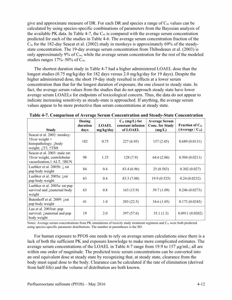
give and approximate measure of DR. For each DR and species a range of Css values can be calculated by using species-specific combinations of parameters from the Bayesian analysis of the available PK data. In Table 4-7, the Css is compared with the average serum concentration predicted for each of the studies in Table 4-6. The average serum concentration fraction of the Css for the 182-day Seacat et al. (2002) study in monkeys is approximately 69% of the steady-state concentration. The 19-day average serum concentration from Thibodeaux et al. (2003) is only approximately 9% of Css, while the average serum concentration for the rest of the modeled studies ranges 17%–50% of Css.
The shortest duration study in Table 4-7 had a higher administered LOAEL dose than the longest studies (0.75 mg/kg/day for 182 days versus 2.0 mg/kg/day for 19 days). Despite the higher administered dose, the short 19-day study resulted in effects at a lower serum concentration than that for the longest duration of exposure, the one closest to steady state. In fact, the average serum values from the studies that do not approach steady state have lower average serum LOAELs for endpoints of toxicological concern. Thus, the data do not appear to indicate increasing sensitivity as steady-state is approached. If anything, the average serum values appear to be more protective than serum concentrations at steady state.
Table 4-7. Comparison of Average Serum Concentration and Steady-State Concentration
Study
Dosing duration
days LOAEL
mg/kg/day
Css (mg/L) for constant infusion
of LOAEL
Average Serum Conc. for Study
(mg/L) Fraction of Css (Average / Css)
Seacat et al. 2002: monkey: ↑liver weight + histopathology; ↓body weight; ↓T3; ↑TSH
182 0.75 227 (6.95) 157 (2.45) 0.689 (0.0131)
Seacat et al. 2003: male rat: ↑liver weight, centrilobular vacuolization,↑ ALT, ↑BUN
98 1.33 128 (7.9) 64.6 (2.06) 0.504 (0.0211)
Luebker et al. 2005b: ↓ rat pup body weight 84 0.4 83.4 (6.96) 25 (0.583) 0.302 (0.027)
Luebker et al. 2005a: ↓rat pup body weight 63 0.4 83.3 (7.08) 19.9 (0.525) 0.24 (0.0232)
Luebker et al. 2005a: rat pup survival and ↓maternal body weight
63 0.8 163 (15.9) 39.7 (1.09) 0.246 (0.0273)
Butenhoff et al. 2009: ↓rat pup body weight 41 1.0 203 (22.5) 34.6 (1.05) 0.173 (0.0245)
Lau et al. 2003rat: pup survival; ↓maternal and pup body weight
19 2.0 397 (57.6) 35.1 (1.3) 0.0911 (0.0202)
Notes: Average serum concentrations from PK simulations of toxicity study treatment regimens and Css were both predicted using species-specific parameter distributions. The number in parentheses is the SD.
For human exposure to PFOS one needs to rely on average serum calculations since there is a lack of both the sufficient PK and exposure knowledge to make more complicated estimates. The average serum concentrations of the LOAEL in Table 4-7 range from 19.9 to 157 µg/mL; all are within one order of magnitude. The predicted toxic serum concentrations can be converted into an oral equivalent dose at steady state by recognizing that, at steady state, clearance from the body must equal dose to the body. Clearance can be calculated if the rate of elimination (derived from half-life) and the volume of distribution are both known.
Perfluorooctane sulfonate (PFOS) – May 2016 4-12
A reliable measure of half-life in humans is available from a retired worker population
followed for 5 years. Olsen et al. (2007) calculated the PFOS half-life in this former worker population as 5.4 years (see section 2.5.2). Thompson et al. (2010) give a volume of distribution of 0.23 L/kg bw (see section 2.5.3). These values combined give a clearance of 8.1×10⁻⁵ L/kg bw/day as determined by the following equation:
CL = Vd x (ln 2 ÷ t1/2) = 0.23 L/kg bw x (0.693 ÷ 1,971 days) = 0.000081 L/kg bw/day
Where:
Vd = 0.23 L/kg ln 2 = 0.693 t1/2 = 1,971 days (5.4 years x 365 days/year = 1,971 days)
These values combined give a clearance of 8.1 x 10⁻⁵ L/kg bw/day.
Scaling the derived average concentrations (in µg/mL) for the NOAELs and LOAELs in Table 4-6 gives predicted oral HEDs in mg/kg bw/day for each corresponding serum measurement. The HED values are the predicted human oral exposures necessary to achieve serum concentrations equivalent to the NOAEL or LOAEL in the animal toxicity studies. Note that this scaling uses linear human kinetics in contrast to the non-linear phenomena observed at high doses in animals.
Thus, HED = average serum concentration (in µg/mL) x CL
Where:
Average serum is from model output in Table 4-6 CL = 0.000081 L/kg bw/day
The resulting HED values are shown in Table 4-8. Endpoints considered as critical effects in multiple studies include offspring growth and survival, liver weight changes, liver histopathology, and changes in serum biochemistry indicative of systemic effects. Each study selected for modeling was of high quality and show effects at low doses. In all cases but one (Luebker et al. 2005a) the POD for the analysis was a NOAEL rather than a LOAEL. The developmental effects of reduced pup body weight and survival occurred in the absence of changes in maternal liver weight, indicating that maternal toxicity and PPARα were not confounding variables.
The external dose NOAELs and LOAELs from other studies summarized in Tables 4-1 and 4-2 that lacked serum information are comparable to those in the modeled studies. For example, the NOAEL in the Long et al. (2013) 90-day mouse study for effects on learning and memory is 0.43 mg/kg/day (Table 4-1) compared to the 0.3 mg/kg/day for Butenhoff et al. (2009) in rats and the LOAEL for mice is 2.15 mg/kg/day compared to the value of 1 mg/kg/day for rats. The LOAEL from Luebker et al. (2005a) of 0.4 mg/kg/day for decreased pup body weight is not unlike the 0.5 mg/kg/day observed by Lv et al. (2013) for decreased pup body weight and increased insulin resistance (Table 4-2). The 1.0 mg/kg/day NOAEL and 2.0 mg/kg/day LOAEL for decreased body weight in rat dams and pups combined with decreased pup survival (Lau et al. 2003; Thibodeaux et al. 2003) are quite similar to the corresponding values of 1 and 5 mg/kg/day, respectively, in the study of mice conducted by the same authors (increased maternal liver weight and delayed pup eye opening).
Perfluorooctane sulfonate (PFOS) – May 2016 4-13

Table 4-8. Human Equivalent Doses Derived from the Modeled Animal Average
Serum Values
Study
Dosing duration
days NOAEL mg/kg/d
NOAEL Av serum
µg/mL HED
mg/kg/d LOAEL mg/kg/d
LOAEL Av serum
µg/mL HED
mg/kg/d Seacat et al. 2002 monkey: ↑liver weight + histopathology; ↓body weight; ↓T3; ↑TSH
182 0.15 38 0.0031 0.75 157 0.013
Seacat et al. 2003 male rat: ↑liver weight, centrilobular vacuolization,↑ ALT, ↑BUN
98 0.34 16.5 0.0013 1.33 64.6 0.0052
Luebker et al. 2005b rat: ↓pup body weight 84 0.1 6.26 0.00051 0.4 25 0.002
Luebker et al. 2005a rat: ↓ pup body weight 63 None None None 0.4 19.9 0.0016
Luebker et al. 2005a rat: ↓maternal body weight, gestation length and pup survival
63 0.4 19.9 0.0016 0.8 39.7 0.0032
Butenhoff et al. 2009 rat developmental neurotoxictiy: ↑motor activity, ↓habituation
41 0.3 10.4 0.00084 1.0 34.6 0.0028
Lau et al. 2003 rat: ↓ pup survival; maternal and pup body weight
19 1.0 17.5 0.0014 2.0 35.1 0.0028
4.1.1.2 RfD Quantification
Several acceptable PODs can be used in the process of RfD development based on the modeled human equivalent doses (Table 4-9).
All modeled studies identified a NOAEL for PFOS except for the endpoint of offspring growth as measured by body weight in the one-generation study by Luebker et al. (2005a) with a LOAEL of 0.4 mg/kg/day. The same external dose was also a LOAEL for the same effect in the two-generation study by Luebker et al. (2005b), with a NOAEL of 0.1 mg/kg/day, a dose not tested in the one-generation study. The calculated HED values associated with no adverse effects on developmental and liver endpoints (NOAELs) were very similar with a range of 0.00051–0.0031 mg/kg/day.
Two effect-level doses were modeled from the Luebker et al. (2005a) one-generation rat study: (1) the NOAEL for the effects on pup survival (0.4 mg/kg/day), which was the LOAEL for the body weight effect, and (2) the LOAEL (0.8 mg/kg/day) for the pup survival effect to illustrate the importance of the body weight LOAEL in both the one- and two-generation Luebker et al. (2005a, 2005b) studies. In the two-generation study, 1.6 mg/kg/day resulted in the death of > 26% of the pups between LD 2 and 4. Support for the pup survival serum level LOAEL is provided by the Lau et al. (2003) rat study, with a HED for the same end point that is comparable to that in the Luebker et al. (2005b) study (0.0028 mg/L and 0.0032 mg/L, respectively).
Perfluorooctane sulfonate (PFOS) – May 2016 4-14

Table 4-9. POD Outcomes for the HEDs from the Pharmacokinetic Model
Average Serum Values
POD POD Value mg/kg/day UFH UFA UFL UFS UFD UFtotal
Candidate RfD
mg/kg/day PK-HED (Seacat et al. 2003; rat, NOAEL,↑ ALT, ↑BUN
0.0013 10 3 1 1 1 30 0.00004
PK-HED (Lau et al. 2003; rat, NOAEL ↓pup survival)
0.0014 10 3 1 1 1 30 0.00005
PK-HED (Butenhoff et al. 2009; rat, NOAEL ↑motor activity ↓habituation)
0.00084 10 3 1 1 1 30 0.00003
PK-HED (Luebker et al. 2005b; rat, NOAEL↓pup body wt)
0.00051 10 3 1 1 1 30 0.00002
PK-HED LOAEL (Luebker et al. 2005a; rat, LOAEL↓pup body wt)
0.0016 10 3 3 1 1 100 0.00002
PK-HED (Luebker et al. 2005a; rat, NOAEL ↓pup survival)
0.0016 10 3 1 1 1 30 0.00005
Notes: UFH: Intra-individual uncertainty factor, UFA: Interspecies uncertainty factor, UFS: Subchronic to chronic uncertainty factor, UFL: LOAEL to NOAEL uncertainty factor, UFD: incomplete database uncertainty factor, UFtotal: Total (multiplied) uncertainty factor
The pharmacokinetically-modeled average serum values from the animal studies are restricted to the animal species selected for their low dose response to oral PFOS intakes. However, the modeled average serum values from animals are several orders of magnitude greater than measured values in humans. Thus, extrapolation to humans adds a layer of uncertainty that needs to be accommodated in deriving the RfD.
HED PODs. The PK HEDs derived from Seacat et al. (2003), Lau et al. (2003), Butenhoff et al. (2009), and Luebker et al. (2005a, 2005b) were each examined as the potential basis for the RfD (ph). The Seacat et al. (2002) results for male monkeys were not utilized in the derivation of the RfD because of the premature deaths in two of the six males at the LOAEL. Each of these studies, except one, contained a NOAEL from which the HED could be derived. The outcomes for potential RfD values are similar demonstrating the ability of the model to normalize the animal data across species, gender, and exposure duration.
Uncertainty Factors
An uncertainty factor for intraspecies variability (UFH) of 10 is assigned to account for variability in the responses within the human populations because of both intrinsic (genetic, life stage, health status) and extrinsic (life style) factors that can influence the response to exposure. No information was available relative to variability in the human population that supports a factor other than 10.
An uncertainty factor for interspecies variability (UFA) of three was applied to account for uncertainty in extrapolating from laboratory animals to humans (i.e., interspecies variability). The 3-fold factor is applied to account for toxicodynamic differences between the animals and
Perfluorooctane sulfonate (PFOS) – May 2016 4-15

humans. The HEDs were derived using average serum values from a model to account for pharmacokinetic differences between animals and humans.
An uncertainty factor for LOAEL to NOAEL extrapolation (UFL) of one was applied to all PODs, except the LOAEL of 0.4 mg/kg/day for effects on pup body weight in the one-generation Luebker et al. (2005a) study. A value of three is assigned for this study based on the fact that the NOAEL for this effect was 0.1 mg/kg/day in the two-generation (Luebker et al. 2005b) study, a dose that was not used in the one-generation study. The LOAEL in the two-generation study was 0.4 mg/kg/day, demonstrating that the difference between a NOAEL and LOAEL for the body weight is not a factor of 10, the default value for NOAEL/LOAEL extrapolation.
An uncertainty factor for extrapolation from a subchronic to a chronic exposure duration (UFS) of one was applied because the PODs are based on average serum concentrations for all studies except Seacat et al. (2013). The studies for developmental endpoints are not adjusted for lifetime exposures because they cover a critical window of exposure with lifetime consequences. The average serum value associated with the developmental (Luebker et al. 2005b) POD is lower than that for any of the other modeled studies including those with systemic effects after longer exposures. It is accordingly more protective of adverse effects than the POD for any of the longer-term studies despite the limited exposure duration. The serum from the Seacat et al. (2013) study was collected at 14 weeks. Some of the animals in the study continued to be dosed for a total of 105 weeks, but the effects observed at the LOAEL did not increase in magnitude. Serum measurements taken before sacrifice were 2-fold higher at 14 weeks in males than they were at 105 weeks. Concentrations of PFOS in the liver were lower at 105 weeks than they were at 14 weeks. The PFOS concentrations in the diet were constant. SDs about the monitored ALT and BUN were broad indicating higher sensitivity is some animals than others. The serum and effects data for the male rats justify the subchronic to chronic adjustment to the study NOAEL for this study.
A database uncertainty factor (UFD) of one was applied to account for deficiencies in the database for PFOS. The epidemiology data provide strong support for the identification of hazards observed following exposure to PFOS in the laboratory animal studies and human relevance. However, uncertainties in the use of the available epidemiology data precluded their use at this time in the quantification of the effect level for derivation of the drinking water health advisory. In animals, comprehensive oral short term, subchronic, and chronic studies in three species and several strains of laboratory animals have been conducted and published in the peer reviewed literature. Additionally, there are several neurotoxicity studies (including developmental neurotoxicity) and several reproductive (including one- and two-generation reproductive toxicity studies) and developmental toxicity studies including assessment of immune effects following developmental exposure.
RfD Selection
Based on the consistency of the response and of the use of the most sensitive endpoint, developmental toxicity, as the critical effect, the RfD of 0.00002 mg/kg/day from Luebker et al. (2005a) is selected as the RfD for PFOS. This RfD is derived from reduced pup body weight in the two-generation study in rats. The POD for the derivation of the RfD for PFOS is the HED of 0.00051 mg/kg/day that corresponds to a NOAEL that represents approximately 30% of steady-state concentration. An UF of 30 (10 UFH and 3 UFA) was applied to the HED NOAEL to derive an RfD of 0.00002 mg/kg/day. This is supported by the 0.00002 mg/kg/day value derived from the LOAEL for the same effect in the one-generation Luebker et al. (2005a) study and the
Perfluorooctane sulfonate (PFOS) – May 2016 4-16

0.00003 mg/kg/day value for neonatal neurodevelopmental effects in the Butenhoff et al. (2009) study.
Low body weights in neonates are a biomarker for developmental deficits and linked to problems often manifest later in life. A study by Lv et al. (2013) that lacked serum data for pharmacokinetic modeling identified 0.5 mg/kg/day as a LOAEL for effects on body weight in Wistar rat pups exposed during gestation, an observation that was accompanied by increased insulin resistance, problems with glucose homeostasis, and hepatic fat accumulation in the pups as adults. A similar effect on glucose homeostasis was observed in CD-1 mice at PND 63 in a study by Wan et al. (2014b), with a dose of 3 mg/kg/day for animals receiving a diet with regular fat content. For animals receiving a high fat diet, the LOAEL was 0.3 mg/kg/day. Support for the neurodevelopmental effects in Butenhoff et al. (2009) at a dose 1 mg/kg/day kg/day is provided by the NOAEL (0.43 mg/kg/day) in the Long et al. (2013) 90-day mouse study for effects on learning and memory.
Use of the developmental toxicity endpoint is directly relevant to human health because in utero and lactational exposures have been demonstrated. PFOS has been measured in the blood of newborns (Spliethoff et al. 2008), in human breast milk (Kärrman et al. 2010), and in serum samples from children aged 5–15 years (Dong et al. 2013; Grandjean et al. 2012). A human epidemiology study found no association with maternal PFOS levels and motor or mental development of their children; the mean maternal serum concentration was approximately 0.035 µg/mL (Fei et al. 2008b).
4.1.2 RfC Determination
The only inhalation study available is an acute lethality inhalation study in rats (Rusch et al. 1979); no inhalation data are available in humans. Thus, data are insufficient for the development of an RfC for PFOS.
4.2 Dose-Response for Cancer Effects
Under the EPA (2005a) Guidelines for Carcinogen Risk Assessment, when the evidence from the epidemiology studies and the cancer bioassays is suggestive for carcinogenicity, a quantitative estimate of risk is generally not performed unless there is a well-conducted study that could serve a useful purpose by providing a sense of the magnitude and uncertainty of potential risks, ranking potential hazards, or setting research priorities. In the case of PFOS, the existing evidence does not support a strong correlation between the tumor incidence and dose to justify a quantitative assessment.
Perfluorooctane sulfonate (PFOS) – May 2016 4-17

5. REFERENCES
3M Company. 2000. Determination of Serum Half-Lives of Several Fluorochemicals. Interim Report #1, June 8, 2000. 3M Company, St. Paul, MN.
3M Environmental Laboratory. 1999. Analytical Laboratory Report on the Determination of the Presence and Concentration of Potassium Perfluorooctanesulfonate (CAS No. 2795-39-3) in the Serum of Sprague-Dawley Rats Exposed to Potassium Perfluorooctanesulfonate via Gavage. Laboratory Report No. U2779. 3M Company, St. Paul, MN.
Abbott, B.D., C.J. Wolf, K.P. Das, R.D. Zehr, J.E. Schmid, A.B. Lindstrom, M.J. Strynar, and C. Lau. 2009. Developmental toxicity of perfluorooctane sulfonate (PFOS) is not dependent on expression of peroxisome proliferator activated receptor-alpha (PPARα) in the mouse. Reproductive Toxicology 27:258–265.
Alexander, B.H., G.W. Olsen, J.M. Burris, J.H. Mandel, and J.S. Mandel. 2003. Mortality of employees of a perfluorooctanesulfonyl fluoride manufacturing facility. Occupational and Environmental Medicine 60:722–729.
Alexander, B.H., and G.W. Olsen. 2007. Bladder cancer in perfluorooctanesulfonyl fluoride manufacturing workers. Annals of Epidemiology 17:471–478.
Andersen, M.E., H.J. Clewell, Y.-M. Tan, J.L. Butenhoff, and G.W. Olsen. 2006. Pharmacokinetic modeling of saturable, renal absorption of perfluoroalkylacids in monkeys- probing the determinants of long plasma half-lives. Toxicology 227:156–164.
Andersen, M.E., J.L. Butenhoff, S.-C. Chang, D.G. Farrar, G.L. Kennedy, C. Lau, G.W. Olsen, and K.B. Wallace. 2008. Review: Perfluoroalkyl acids and related chemistries- toxicokinetics and modes of action. Toxicological Sciences 102:3–14.
Andersen, C.S., C. Fei, M. Gamborg, E.A. Nohr, T.I.A. Sørensen, and J. Olsen. 2010. Prenatal exposures to perfluorinated chemicals and anthropometric measures in infancy. American Journal of Epidemiology 172:1230–1237.
Andersen, C.S., C. Fei, M. Gamborg, E.A. Nohr, T.I.A. Sørensen, and J. Olsen. 2013. Prenatal exposures to perfluorinated chemicals and anthropometry at 7 years of age. American Journal of Epidemiology 178:921–927.
Apelberg, B.J., L.R. Goldman, A.M. Calafat, J.B. Herbstman, Z. Kuklenyik, J. Heidler, L.L. Needham, R.U. Halden, and F.R. Witter. 2007. Determinants of fetal exposure to polyfluoroalkyl compounds in Baltimore, Maryland. Environmental Science & Technology 41:3891–3897.
Ashby, J., A. Brady, C.R. Elcombe, B.M. Elliot, J. Ishmael, J. Odum, J.D. Tugwood, S. Kettle, and I.F.H. Purchase. 1994. Mechanistically-based human hazard assessment of peroxisome proliferator-induced hepatocarcinogenesis. Human & Experimental Toxicology 13(Suppl. 2):S1–S117.
Ashford, R.D. 1994. Ashford's Dictionary of Industrial Chemicals: Properties, Production, Uses. Wavelength Publications, Ltd.
Perfluorooctane sulfonate (PFOS) – May 2016 5-1

ATSDR (Agency for Toxic Substances and Disease Registry). 2015. Draft Toxicological
Profile for Perfluoroalkyls. United States Department of Health and Human Services, Agency for Toxic Substances and Disease Registry, Public Health Service, Atlanta, GA. Accessed May 2016. http://www.atsdr.cdc.gov/ToxProfiles/tp200.pdf.
Augustine, L.M., R.J. Markelewicz, K. Boekelheide, and N.J. Cherrington. 2005. Xenobiotic and endobiotic transporter mRNA expression in the blood-testis barrier. Drug Metabolism and Disposition 33(1):182–189.
Bach, C.C., Z. Liew, B.H. Bech, E.A. Nohr, C. Fei, E.C. Bonefeld-Jørgensen, T.B. Henriksen, and J. Olsen. 2015. Perfluoroalkyl acids and time to pregnancy revisited: An update from the Danish National Birth Cohort. Environmental Health 14:59.
Barrett, E.S., C. Chen, S.W. Thurston, L.S. Haug, A. Sabaredzovic, F.N. Fjeldheim, H. Frydenberg, S.F. Lipson, P.T. Ellison, and I. Thune. 2015. Perfluoroalkyl substances and ovarian hormone concentrations in naturally cycling women. Fertility and Sterility 103(5):1261–1270.
Beesoon, S., G.M. Webster, M. Shoeib, T. Harner, J.P. Benskin, and J.W. Martin. 2011. Isomer profiles of perfluorochemicals in matched maternal, cord, and house dust samples: Manufacturing sources and transplacental transfer. Environmental Health Perspectives 119:1659–1664.
Beesoon, S., and J.W. Martin. 2015. Isomer-specific binding affinity of perfluorooctanesulfonate (PFOS) and perfluorooctanoate (PFOA) to serum proteins. Environmental Science & Technology 49:5722–5731.
Berg, V., T.H. Nøst, S. Hansen, A. Elverland, A.-S. Veyhe, R. Jorde, J.Ø. Odland, and T.M. Sandanger. 2015. Assessing the relationship between perfluoroalkyl substances, thyroid hormones and binding proteins in pregnant women; a longitudinal mixed effects approach. Environment International 77:63–69.
Biesemeier, J.A., and D.L. Harris. 1974. Eye and Skin Irritation Report on Sample T-1117. Project No. 4102871. WARF Institute, Inc.
Bijland, S., P.C.N. Rensen, E.J. Pieterman, A.C.E. Maas, J.W. van der Hoorn, M.J. van Erk, L.M. Havekes, K.W. van Dijk, S.-C. Chang, E.J. Ehresman, J.L. Butenhoff, and H.M.G. Princen. 2011. Perfluoroalkyl sulfonates cause alkyl chain length-dependent hepatic steatosis and hypolipidemia mainly by impairing lipoprotein production in APOE*3-Leiden CETP mice. Toxicological Sciences 123:290–303.
Bjork, J.A., and K.B. Wallace. 2009. Structure-activity relationships and human relevance for perfluoroalkyl acid-induced transcriptional activation of peroxisome proliferation in liver cell cultures. Toxicological Sciences 111:89–99.
Bjork, J.A., J.L. Butenhoff, and K.B. Wallace. 2011. Multiplicity of nuclear receptor activation by PFOA and PFOS in primary human and rat hepatocytes. Toxicology 228:8–17.
Bloom, M.S., K. Kannan, H.M. Spliethoff, L. Tao, K.M. Aldous, and J.E. Vena. 2010. Exploratory assessment of perfluorinated compounds and human thyroid function. Physiology & Behavior 99:240–245.
Perfluorooctane sulfonate (PFOS) – May 2016 5-2

Bogdanska, J., D. Borg, M. Sunström, U. Bergström, K. Halldin, M. Abedi-Valugerdi, Å.
Bergman, B. Nelson, J. DePierre, and S. Nobel. 2011. Tissue distribution of 35S-labelled perfluorooctane sulfonate in adult mice after oral exposure to a low environmentally relevant dose or a high experimental dose. Toxicology 284:54–62.
Bonefeld-Jørgensen, E.C., M. Long, R. Bossi, P. Ayotte, G. Asmund, T. Krüger, M. Ghisari, G. Mulvad, P. Kern, P. Nzulumiki, and E. Dewaily. 2011. Perfluorinated compounds are related to breast cancer risk in Greenlandic Inuit: A case control study. Environmental Health 10:88.
Bonefeld-Jørgensen, E.C., M. Long, S.O. Fredslund, R. Bossi, and J. Olsen. 2014. Breast cancer risk after exposure to perfluorinated compounds in Danish women: A case-control study nested in the Danish National Birth Cohort. Cancer Causes Control 25(11):1439–1448.
Borg, D., J. Bogdanska, M. Sundström, S. Nobel, H. Håkansson, Å. Bergman, J.W. DePierre, K. Halldin, and U. Bergström. 2010. Tissue distribution of 35S-labelled perfluorooctane sulfonate (PFOS) in C57Bl/6 mice following late gestational exposure. Reproductive Toxicology 30:550–557.
Brieger, A., N. Bienefeld, R. Hasan, R. Goerlich, and H. Haase. 2011. Impact of perfluorooctane sulfonate and perfluorooctanoic acid on human peripheral leukocytes. Toxicology in Vitro 25(4):960–968.
Buck Louis, G.M., Z. Chen, E.F. Schisterman, S. Kim, A.M. Sweeney, R. Sundaram, C.D. Lynch, R.E. Gore-Langton, and D.B. Barr. 2015. Perfluorochemicals and human semen quality: The LIFE study. Environmental Health Perspectives 123(1):57–63.
Burris, J.M., J.K. Lundberg, G.W. Olsen, C. Simpson, and J. Mandel. 2002. Determination of Serum Half-Lives of Several Fluorochemicals. Interim Report #2. 3M Company, St. Paul, MN.
Butenhoff, J., G. Costa, C. Elcombe, D. Farrar, K. Hansen, H. Iwai, R. Jung, G. Kennedy, Jr., P. Lieder, G. Olsen, and P. Thomford. 2002. Toxicity of ammonium perfluorooctanoate in male cynomolgus monkeys after oral dosing for 6 months. Toxicological Sciences 69:244–257.
Butenhoff, J., G.L. Kennedy, Jr., P.M. Hindliter, P.H. Lieder, R. Jung, K.J. Hansen, G.S. Gorman, P.E. Noker, and P.J. Thomford. 2004. Pharmacokinetics of perfluorooctanoate in cynomolgus monkeys. Toxicological Sciences 82:394–406.
Butenhoff, J., and S. Chang. 2007. ADME Study of Perfluorooctane Sulfonate in Rats. ST-179 Final Report. 3M Strategic Toxicology Laboratory, 3M Center, St. Paul, MN.
Butenhoff, J.L., D.J. Ehresman, S.-C. Chang, G.A. Parker, and D.G. Stump. 2009. Gestational and lactational exposure to potassium perfluorooctanesulfonate (K+PFOS) in rats: Developmental neurotoxicity. Reproductive Toxicology 27:319–330.
Butenhoff, J. L., S.C. Chang, G.W. Olsen, and P.J. Thomford. 2012. Chronic dietary toxicity and carcinogenicity study with potassium perfluorooctane sulfonate in Sprague Dawley rats. Toxicology 293:1–15.
Perfluorooctane sulfonate (PFOS) – May 2016 5-3

Chan, E., I. Burstyn, N. Cherry, F. Bamforth, and J.W. Martin. 2011. Perfluorinated acids and
hypothyroxinemia in pregnant women. Environmental Research 111:559–564.
Chang, S.-C., J.R. Thibodeaux, M.L. Eastvold, D.J. Ehresman, J.A. Bjork, J.W. Froehlich, C. Lau, R.J. Singh, K.B. Wallace, and J.L. Butenhoff. 2007. Negative bias from analog methods used in the analysis of free thyroxine in rat serum containing perfluorooctanesulfonate (PFOS). Toxicology 234:21–33.
Chang, S.-C., J.R. Thibodeaux, M.L. Eastvold, D.J. Ehresman, J.A. Bjork, J.W. Froehlich, C. Lau, R.J. Singh, K.B. Wallace, and J.L. Butenhoff. 2008. Thyroid hormone status and pituitary function in adult rats given oral doses of perfluorooctanesulfonate (PFOS). Toxicology 243:330–339.
Chang, S.-C., D.J. Ehresman, J.A. Bjork, K.B. Wallace, G.A. Parker, D.G. Stump, and J. Butenhoff. 2009. Gestational and lactational exposure to potassium perfluorooctanesulfonate (K+PFOS) in rats: Toxicokinetics, thyroid hormone status and related gene expression. Reproductive Toxicology 27:387–399.
Chang, S.-C., P.E. Noker, G.S. Gorman, S.J. Gibson, J.A. Hart, D.J. Ehresman, and J.L. Butenhoff. 2012. Comparative pharmacokinetics of perfluorooctanesulfonate (PFOS) in rats, mice and monkeys. Reproductive Toxicology 33:428–440.
Chang, E.T., H. Adami, P. Boffetta, C. Cole, T.B. Starr, and J.S. Mandel. 2014. A critical review of perfluorooctanoate and prefluorooctanesulfonate exposure and cancer risk in humans. Critical Reviews in Toxicology 44(51):1–81.
Château-Degat, M.-L., D. Pereg, R. Dallaire, P. Ayotte, S. Dery, and É. Dewailly. 2010. Effects of perfluorooctanesulfonate exposure on plasma lipid levels in the Inuit population of Nunavik (Northern Quebec). Environmental Research 110:710–717.
Chen, Y.-M., and L.-H. Guo. 2009. Fluorescence study on site-specific binding of perfluoroalkyl acids to human serum albumin. Archives of Toxicology 83:255–261.
Chen, T., L. Zhang, J.-Q. Yue, Z.-Q. Lv, W. Xia, Y.-J. Wan, Y.-Y. Li, and S.-Q. Xu. 2012. Prenatal PFOS exposure induces oxidative stress and apoptosis in the lung of rat off-spring. Reproductive Toxicology 33:538–545.
Chen, H., P. He, H. Rao, F. Wang, H. Liu, and J. Yao. 2015. Systematic investigation of the toxic mechanism of PFOA and PFOS on bovine serum albumin by spectroscopic and molecular modeling. Chemosphere 129:217–224.
Christensen, K.Y., M. Maisonet, C. Rubin, A. Holmes, A.M. Calafat, K. Kato, W.D. Flanders, J. Heron, M.A. McGeehin, and M. Marcus. 2011. Exposure to polyfluoroalkyl chemicals during pregnancy is not associated with offspring age at menarche in a contemporary British cohort. Environment International 37:129–135.
Cifone, M.A. 1999. Unscheduled DNA Synthesis in Rat Liver Primary Cell Cultures with PFOS. Final Report, Covance Study No. 207840447. Covance Laboratories Inc., Vienna, VA.
Perfluorooctane sulfonate (PFOS) – May 2016 5-4

Corley, R.A., A.L. Mendrala, F.A. Smith, D.A. Staats, M.L. Gargas, R.B. Conolly, M.E.
Andersen, and R.H. Reitz. 1990. Development of a physiologically based pharmacokinetic model for chloroform. Toxicology and Applied Pharmacology 103:512–527.
Corsini, E., A. Avogadro, V. Galbiati, M. Dell'Agli, M. Marinovich, C.L. Galli, and D.R. Germolec. 2011. In vitro evaluation of the immunotoxic potential of perfluorinated compounds (PFCs). Toxicology and Applied Pharmacology 250(2):108–116.
Corsini, E., R.W. Luebke, D.R. Germolec, and J.C. DeWitt. 2014. Perfluorinated compounds: Emerging POPs with potential immunotoxicity. Toxicology Letters 230(2):263–270.
Corsini, E., E. Sangiovanni, A. Avogadro, V. Galbiati, B. Viviani, M. Marinovich, C.L. Galli, M. Dell'Agli, and D.R. Germolec. 2012. In vitro characterization of the immunotoxic potential of several perfluorinated compounds (PFCs). Toxicology and Applied Pharmacology 258(2):248–255.
Cui, L., Q.-F. Zhou, C.-Y. Liao, J.-J. Fu, and G.-B. Jiang. 2009. Studies on the toxicological effects of PFOA and PFOS on rats using histological observation and chemical analysis. Archives of Environmental Contamination and Toxicology 56:338–349.
Cui, L., C.-Y. Liao, Q.-F. Zhou, T.-M. Xia, Z.-J. Yun, and G.-b. Jiang. 2010. Excretion of PFOA and PFOS in male rats during a subchronic exposure. Archives of Environmental Contamination and Toxicology 58:205–213.
Curran, I., S.L. Hierlihy, V. Liston, P. Pantazopoulos, A. Nunnikhoven, S. Tittlemier, M. Barker, K. Trick, and G. Bondy. 2008. Altered fatty acid homeostasis and related toxicologic sequelae in rats exposed to dietary potassium perfluorooctanesulfonate (PFOS). Journal of Toxicology and Environmental Health, Part A 71:1526–1541.
D’Alessandro, M.L., D.A. Ellis, J.A. Carter, N.L. Stock, and R.E. March. 2013. Competitive binding of aqueous perfluoroctanesulfonic acid and ibuprofen with bovine serum albumin studied by electrospray ionization mass spectrometry. International Journal of Mass Spectrometry 345–347:28–36.
Dallaire, R., É. Dewailly, D. Pereg, S. Déry, and P. Ayotte. 2009. Thyroid function and plasma concentrations of polyhalogenated compounds in Inuit adults. Environmental Health Perspectives 117:1380–1386.
Dankers, A.C., M.J.E. Roelofs, A.H. Piersma, F.C.G.J. Sweep, F.G.M. Russel, M. van den Berg, M.B.M. van Duursen, and R. Masereeuw. 2013. Endocrine disruptors differentially target ATP-binding cassette transporters in the blood-testis barrier and affect Leydig cell testosterone secretion in vitro. Toxicological Sciences 136(2):382–391.
Darrow, L.A., C.R. Stein, and K. Steenland. 2013. Serum perfluorooctanoic acid and perfluorooctane sulfonate concentrations in relation to birth outcomes in the Mid-Ohio Valley, 2005–2010. Environmental Health Perspectives 121:1207–1213.
Darrow, L.A., P.P. Howards, A. Winquist, and K. Steenland. 2014. PFOA and PFOS serum levels and miscarriage risk. Epidemiology 25:e1–8.
Perfluorooctane sulfonate (PFOS) – May 2016 5-5

Davies, B., and T. Morriss. 1993. Physiology parameters in laboratory animals and human.
Pharmaceutical Research 10:1093–1095.
Dean, W.P., D.C. Jessup, G. Thompson, G. Romig, and D. Powell. 1978. Fluorad Fluorochemical Surfactant FC-95 Acute Oral Toxicity (LD50) Study in Rats. Study No. 137-083. International Research and Development Corporation.
DeWitt. J.C., W.C. Williams, J. Creech, and R.W. Luebke. 2015. Supression of antigen-specific antibody responses in mice exposed to perfluorooctanoic acid: Role of PPARα and T- and B-cell targeting. Journal of Immunotoxicology 13(1):38-45.
Ding, G., J. Zhang, Y. Chen, L. Wang, M. Wang, D. Xiong, and Y. Sun. 2013. Combined effects of PFOS and PFOA on zebrafish (Danio rerio) embryos. Archives of Environmental Contamination and Toxicology 64(4):668–675.
Donauer, S., A. Chen, Y. Xu, A.M. Calafat, A. Sjodin, and K. Yolton. 2015. Prenatal exposure to polybrominated diphenyl ethers and polyfluoroalkyl chemicals and infant neurobehavior. The Journal of Pediatrics 166(3):736–742.
Dong, G.-H., Y.-H. Zhang, L. Zheng, W. Liu, Y.-H. Jin, and Q.-C. He. 2009. Chronic effects of perfluorooctane sulfonate exposure on immunotoxicity in adult male C57BL/6 mice. Archives of Toxicology 83:805–815.
Dong, G.-H., K.-Y. Tung, C.-H. Tsai, M.-M. Liu, D. Wang, W. Liu, Y.-H. Jin, W.S. Hsieh, Y.L. Lee, and P.-C. Chen. 2013. Serum polyfluoroalkyl concentrations, asthma outcomes, and immunological markers in a case-control study of Taiwanese children. Environmental Health Perspectives 121:507–513.
EFSA (European Food Safety Authority). 2008. Opinion of the scientific panel on contaminants in the food chain on perfluorooctane sulfonate (PFOS), perfluorooctanoic acid (PFOA) and their salts. The EFSA Journal 653:1–131.
Egeghy, P., and M. Lorber. 2011. An assessment of the exposure of Americans to perfluorooctane sulfonate: A comparison of estimated intakes with values inferred from NHANES data. Journal of Exposure Science and Environmental Epidemiology 21:150–168.
Elcombe, C.R., B.M. Elcombe, J.R. Foster, S.-C. Chang, D.J. Ehresman, and J.L. Butenhoff. 2012. Hepatocellular hypertrophy and cell proliferation in Sprague-Dawley rats from dietary exposure to potassium perfluorooctanesulfonate results from increased expression of xenosensor nuclear receptors PPARα and CAR/PXR. Toxicology 293:16–29.
Environment Canada. 2006. Ecological Screening Assessment Report on Perfluorooctane Sulfonate, Its Salts and Its Precursors that Contain the C8F17SO2 or C8F17SO3, or C8F17SO2N Moiety. Accessed May 2016. http://www.ec.gc.ca/lcpe-cepa/documents/substances/spfo-pfos/ecological_sar_pfos_eng.pdf.
Eriksen, K., M. Sørensen, J.K. McLaughlin, L. Lipworth, A. Tjønneland, K. Overvad, and O. Raaschou-Nielsen. 2009. Perfluorooctanoate and perfluorooctanesulfonate plasma levels and risk of cancer in the general Danish population. Journal of the National Cancer Institute 101:605–609.
Perfluorooctane sulfonate (PFOS) – May 2016 5-6

Eriksen, K.T., O. Raaschou-Nielsen, J.K. McLaughlin, L. Lipworth, A. Tjønneland, K.
Overvad, and M. Sørensen. 2013. Association between plasma PFOA and PFOS levels and total cholesterol in a middle-aged Danish population. PLOS ONE 8:e56969.
Fàbrega, F., V. Kumar, M. Schuhmacher, J.L. Domingo, and M. Nadal. 2014. PBPK modeling for PFOS and PFOA: Validation with human experimental data. Toxicology Letters 230:244–251.
Fei, C., J.K. McLaughlin, R.E. Tarone, and J. Olsen. 2007. Perfluorinated chemicals and fetal growth: A study within the Danish National Birth Cohort. Environmental Health Perspectives 115:1677–1682.
Fei, C., J.K. McLaughlin, R.E. Tarone, and J. Olsen. 2008a. Fetal growth indicators and perfluorinated chemicals: A study in the Danish National Birth Cohort. American Journal of Epidemiology 168:66–72.
Fei, C., J.K. McLaughlin, L. Lipworth, and J. Olsen. 2008b. Prenatal exposure to perfluorooctanoate (PFOA) and perfluorooctane sulfonate (PFOS) and maternally reported developmental milestones in infancy. Environmental Health Perspectives 116:1391–1395.
Fei, C., J.K. McLaughlin, L. Lipworth, and J. Olsen. 2009. Maternal levels of perfluorinated chemicals and subfecundity. Human Reproduction 1:1–6.
Fei, C., J.K. McLaughlin, L. Lipworth, and J. Olsen. 2010a. Maternal concentrations of perfluorooctanesulfonate (PFOS) and perfluorooctanoate (PFOA) and duration of breastfeeding. Scandinavian Journal of Work, Environment & Health 36:413–421.
Fei, C., J.K. McLaughlin, L. Lipworth, and J. Olsen. 2010b. Prenatal exposure to PFOA and PFOS and risk of hospitalization for infectious diseases in early childhood. Environmental Research 110:773–777.
Fei, C., and J. Olsen. 2011. Prenatal exposure to perfluorinated chemicals and behavioral or coordination problems at age 7. Environmental Health Perspectives 119:573–578.
Fenton, S.E. 2015. A special issue dedicated to a complex tissue. Reproductive Toxicology 54:1–5.
Filgo, A.J., E.M. Quist, M.J. Hoenerhoff, A.E. Brix, G.E. Kissling, and S.E. Fenton. 2015. Perfluorooctanoic acid (PFOA)-induced liver lesions in two strains of mice following developmental exposures: PPARα is not required. Toxicologic Pathology 45:558-568.
Fisher, M., T.E. Arbuckle, M. Wade, and D.A. Haines. 2013. Do perfluoroalkyl substances affect metabolic function and plasma lipids? – Analysis of the 2007–2009, Canadian Health Measures Survey (CHMS) Cycle 1. Environmental Research 121:95–103.
Fitz-Simon, N., T. Fletcher, M.I. Luster, K. Steenland, A.M. Calafat, K. Kato, and B. Armstrong. 2013. Reductions in serum lipids with a 4-year decline in serum perfluorooctanoic acid and perfluorooctanesulfonic acid. Epidemiology 24:569–576.
Perfluorooctane sulfonate (PFOS) – May 2016 5-7

Fletcher, T., T.S. Galloway, D. Melzer, P. Holcroft, R. Cipelli, L.C. Pilling, D. Mondal, M.
Luster, and L.W. Harries. 2013. Associations between PFOA, PFOS and changes in the expression of genes involved in cholesterol metabolism in humans. Environment International 57–58:2–10.
Frisbee, S.J., A. Shankar, S.S. Knox, K. Steenland, D.A. Savitz, T. Fletcher, and A. Ducatman. 2010. Perfluorooctanoic acid, perfluorooctanesulfonate, and serum lipids in children and adolescents: Results from the C8 health project. Archives of Pediatrics & Adolescent Medicine 164:860–869.
Fu, Y., T. Wang, Q. Fu, P. Wang, and Y. Lu. 2014. Associations between serum concentrations of perfluoroalkyl acids and serum lipid levels in a Chinese population. Ecotoxicololgy and Environmental Safety 106:246–52.
Fuentes, S., M.T. Colomina, P. Vicens, N. Franco-Pons, and J. Domingo. 2007. Concurrent exposure to perfluorooctane sulfonate and restraint stress during pregnancy in mice: Effects on post-natal development and behavior of the offspring. Toxicological Sciences 98:589–598.
Gallo, V., G. Leonardi, B. Genser, M.-J. Lopez-Espinosa, S.J. Frisbee, L. Karlsson, A.M. Ducatman, and T. Fletcher. 2012. Serum perfluorooctanoate (PFOA) and perfluorooctane sulfonate (PFOS) concentrations and liver function biomarkers in a population with elevated PFOA exposure. Environmental Health Perspectives 120:655–660.
Geiger, S.D., J. Xiao, A. Ducatman, S. Frisbee, K. Innes, and A. Shankar. 2014a. The association between PFOA, PFOS and serum lipid levels in adolescents. Chemosphere 98:78–83.
Geiger, S.D., J. Xiao, and A. Shankar. 2014b. No association between perfluoroalkyl chemicals and hypertension in children. Integrated Blood Pressure Control 7:1–7.
Genuis, S.J., D. Birkholz, M. Ralitsch, and N. Thibault. 2010. Review paper: Human detoxification of perfluorinated compounds. Public Health 124:367–375.
Ghisari, M., H. Eiberg, M. Long, and E.C. Bonefeld-Jørgensen. 2014. Polymorphisms in phase I and phase II genes and breast cancer risk and relations to persistent organic pollutant exposure: A case-control study in Inuit women. Environmental Health 13(1):19.
Goldenthal, E.I., D.C. Jessup, R.G. Geil, and J.S. Mehring. 1978a. Ninety-Day Subacute Rhesus Monkey Toxicity Study. Study No. 137-092. International Research and Development Corporation, Mattawan, MI.
Goldenthal, E.I., D.C. Jessup, R.G. Geil, N.D. Jefferson, and R.J. Arceo. 1978b. Ninety-Day Subacute Rat Study. Study No. 137-085. International Research and Development Corporation, Mattawan, MI.
Goldenthal, E.I., D.C. Jessup, R.G. Geil, and J.S. Mehring. 1979. Ninety-Day Subacute Rhesus Monkey Toxicity Study. Study No. 137-087. International Research and Development Corporation, Mattawan, MI.
Perfluorooctane sulfonate (PFOS) – May 2016 5-8

Governini, L., C. Guerranti, V. De Leo, L. Boschi, A. Luddi, M. Gori, R. Orvieto, and P.
Piomboni. 2014. Chromosomal aneuploides and DNA fragmentation of human spermatozoa from patients exposed to perfluorinated compounds. Andrologia 47:1012–1019.
Governini, L., C. Guerranti, V. De Leo, L. Boschi, A. Luddi, M. Gori, R. Orvieto, and P. Piomboni. 2015. Chromosomal aneuploides and DNA fragmentation of human spermatozoa from patients exposed to perfluorinated compounds. Andrologia 47:1012–1019.
Grandjean, P., E.W. Andersen, E. Budtz-Jørgensen, F. Nielsen, K. Mølbak, P. Weihe, and C. Heilmann. 2012. Serum vaccine antibody concentrations in children exposed to perfluorinated compounds. Journal of the American Medical Association 307:391–397.
Grandjean, P., and R. Clapp. 2015. Perfluorinated alkyl substances emerging insights into health risks. New Solutions: A Journal of Environmental and Occupational Health Policy 15(2):147–163
Granum, B., L.S. Haug, E. Namork, S.B. Stølevik, C. Thomsen, I.S. Aaberge, H. van Loveren, M. Løvik, and U.C. Nygaard. 2013. Pre-natal exposure to perfluoroalkyl substances may be associated with altered vaccine antibody levels and immune-related health outcomes in early childhood. Journal of Immunotoxicology 10:373–379.
Grasty, R.C., B.E. Grey, C.S. Lau, and J.M. Rogers. 2003. Prenatal window of susceptibility to perfluorooctane sulfonate-induced neonatal mortality in the Sprague-Dawley rat. Birth Defects Research Part B: Developmental and Reproductive Toxicology 68:465–471.
Grasty, R.C., J.A. Bjork, K.B. Wallace, D.C. Wolf, C. Lau, and J.M. Rogers. 2005. Effects of prenatal perfluorooctane sulfonate exposure on lung maturation in the perinatal rat. Birth Defects Research Part B: Developmental and Reproductive Toxicology 74:405–416.
Grice, M., B. Alexander, R. Hoffbeck, and D. Kampa. 2007. Self-reported medical conditions in perfluorooctanesulfonyl fluoride manufacturing workers. Journal of Occupational and Environmental Medicine 49:722–729.
Guruge, K.S., H. Hikono, N. Shimada, K. Murakami, J. Hasegawa, L.W.Y. Yeung, N. Yamanaka, and N. Yamashita. 2009. Effect of perfluorooctane sulfonate (PFOS) on influenza A virus-induced mortality in female B6C3F1 mice. The Journal of Toxicological Sciences 34:687–691.
Hall, A.P., C.R. Elcombe, J.R. Foster, T. Harada, W. Kaufmann, A. Knippel, K. Küttler, D.E. Malarkey, R.R. Maronpot, A. Nishikawa, T. Nolte, A. Schulte, V. Strauss, and M.J. York. 2012. Liver hypertrophy: A review of adaptive (adverse and non-adverse) changes – conclusions from the 3rd International ESTP Expert Workshop. Toxicologic Pathology 40:971–994.
Halldorsson, T.I., D. Rytter, L.S. Haug, B.H. Bech, I. Danielsen, G. Becher, T.B. Henriksen, and S.F. Olsen. 2012. Prenatal exposure to perfluorooctanoate and risk of overweight at 20 years of age: A prospective cohort study. Environmental Health Perspectives 120:668–673.
Perfluorooctane sulfonate (PFOS) – May 2016 5-9

Hamm, M., N.M. Cherry, E. Chan, J. Martin, and I. Burstyn. 2010. Maternal exposure to
perfluorinated acids and fetal growth. Journal of Exposure Science and Environmental Epidemiology 20(7):589–97.
Harada, K.H., S. Hashida, T. Kaneko, K. Takenaka, M. Minata, K. Inoue, N. Saito, and A. Koizumi. 2007. Biliary excretion and cerebrospinal fluid partition of perfluorooctanoate and perfluorooctane sulfonate in humans. Environmental Toxicology and Pharmacology 24:134–139.
Hardell, E., A. Kärrman, B. van Bavel, J. Boa, M. Carlberg, and L. Hardell. 2014. Case-control study on perfluorinated alkyl acids (PFAAs) and the risk of prostate cancer. Environment International 63:35–39.
Higgins, C.P. and R.G. Luthy. 2006. Sorption of perfluorinated surfactants on sediments. Environmental Science & Technology 40:7251–7256.
Hoffman, K., T.F. Webster, M.G. Weisskopf, J. Weinberg, and V.M. Vieira. 2010. Exposure to polyfluoroakyl chemicals and attention deficit hyperactivity disorder in U.S. children ages 12–15 years. Environmental Health Perspectives 118:1732–1767.
Høyer, B.B., C.H. Ramlau-Hansen, M. Vrijheid, D. Valvi, H.S. Pedersen, V. Zviezdai, B.A.G. Jönsson, C.H. Lindh, J.P. Bonde, and G. Toft. 2015a. Anthropometry in 5- to 9-year old Greenlandic and Ukrainian children in relation to prenatal exposure to perfluorinated alkyl substances. Environmental Health Perspectives 123:841–846.
Høyer, B.B., C.H. Ramlau-Hansen, C. Obel, H.S. Pedersen, A. Hernik, V. Ogniev, B.A.G. Jönsson, C.H. Lindh, L. Rylander, A. Rignell-Hydbom, J.P. Bonde, and G. Toft. 2015b. Pregnancy serum concentrations of perfluorinated alkyl substances and offspring behavior and motor development at age 5–9 years – a prospective study. Environmental Health 14:2.
HSDB (Hazardous Substances Data Bank). 2012. Hazardous Substances Data Bank. National Institutes of Health, Health & Human Services, U.S. National Library of Medicine, Bethesda, MD. Accessed May 2016. http://toxnet.nlm.nih.gov/cgi-bin/sis/htmlgen?HSDB.
Hu, W., P.D. Jones, B.L. Upham, J.E. Trosko, C. Lau, and J.P. Giesy. 2002. Inhibition of gap junctional intercellular communication by perfluorinated compounds in rat liver and dolphin kidney epithelial cell lines in vitro and Sprague-Dawley rats in vivo. Toxicological Sciences 68:429–436.
Humblet, O., L.G. Diaz-Ramirez, J.R. Balmes, S.M. Pinney, and R.A. Hiatt. 2014. Perfluoroalkyl chemicals and asthma among children 12–19 years of age: NHANES (1999–2008). Environmental Health Perspectives 122:1129–1133.
Innes, K.E., J.H. Wimsatt, S. Frisbee, and A.M. Ducatman. 2014. Inverse association of colorectal cancer prevalence to serum levels of perfluorooctane sulfonate (PFOS) and perfluorooctanoate (PFOA) in a large Appalachian population. BMC Cancer 14:45.
Perfluorooctane sulfonate (PFOS) – May 2016 5-10

Inoue, K., F. Okada, R. Ito, S. Kato, S. Sasaki, S. Nakajima, A. Uno, Y. Saijo, F. Sata, Y.
Yoshimura, R. Kishi, and H. Nakazawa. 2004. Perfluorooctane sulfonate (PFOS) and related perfluorinated compounds in human maternal and cord blood samples: Assessment of PFOS exposure in a susceptible population during pregnancy. Environmental Health Perspectives 112:1204–1207.
Jain, R.B. 2014. Contribution of diet and other factors to the levels of selected perfluorinated compounds: Data from NHANES 2003–2008. International Journal of Hygiene and Environmental Health 217:52–61.
Jain, R.B., 2015. Estimation of the total concentration of perfluoroalkyl acids (PFAA) in human serum: Data from NHANES 2005–2012. Chemosphere, 134: 387-394.
Joensen, U.N., R. Bossi, H. Leffers, A.A. Jensen, N. Skakkebæk, and N. Jørgensen. 2009. Do perfluoroalkyl compounds impair human semen quality? Environmental Health Perspectives 117:923–927.
Joensen, U.N., B. Veyrand, J.-P. Antignac, M.B. Jensen, J.H. Petersen, P. Marchand, N.E. Skakkebæk, A.-M. Andersson, B. Le Bizec, and N. Jørgensen. 2013. PFOS (perfluorooctanesulfonate) in serum is negatively associated with testosterone levels, but not with semen quality, in healthy men. Human Reproduction 28:599–608.
Johansson, N., A. Fredriksson, and P. Eriksson. 2008. Neonatal exposure to perfluorooctane sulfonate (PFOS) and perfluorooctanoic acid (PFOA) causes neurobehavioural defects in adult mice. NeuroToxicology 29:160–169.
Johansson, N., P. Eriksson, and H. Viberg. 2009. Neonatal exposure to PFOS and PFOA in mice results in changes in proteins which are important for neuronal growth and synaptogenesis in the developing brain. Toxicological Sciences 108:412–418.
Jørgensen, K.T., I.O. Specht, V. Lenters, C.C. Bach, L. Rylander, B.A. Jönsson, C.H. Lindh, A. Giwercman, D. Heederik, G. Toft, and J.P. Bonde. 2014. Perfluoroalkyl substances and time to pregnancy in couples from Greenland, Poland and Ukraine. Environmental Health 13(1):116.
Kärrman, A., J.L. Domingo, X. Llebaria, M. Nadal, E. Bigas, B. van Bavel, and G. Lindström. 2010. Biomonitoring perfluorinated compounds in Catalonia, Spain: Concentrations and trends for human liver and milk samples. Environmental Science and Pollution Research 17:750–758.
Keil, D., T. Mehlmann, L. Butterworth, and M. Peden-Adams. 2008. Gestational exposure to perfluorooctane sulfonate suppresses immune function in B6C3F1 mice. Toxicological Sciences 103:77–85.
Kerger, B.D., T.L. Copeland, and A.P. DeCaprio. 2011. Tenuous dose-response correlations for common disease states: Case study of cholesterol and perfluorooctanoate/sulfonate (PFOA/PFOS) in the C8 Health Project. Drug and Chemical Toxicology 34:396–404.
Kerstner-Wood, C., L. Coward, and G. Gorman. 2003. Protein Binding of Perfluorohexane Sulfonate, Perfluorooctane Sulfonate and Perfluorooctanoate to Plasma (Human, Rat and Monkey) and Various Human-Derived Plasma Protein Fractions. Study ID 9921.7. Southern Research Institute.
Perfluorooctane sulfonate (PFOS) – May 2016 5-11

Khansari, N., Y. Shakiba, and M. Mahmoudi. 2009. Chronic inflammation and oxidative stress
as a major cause of age-related diseases and cancer. Recent Patents on Inflammation & Allergy Drug Discovery 3(1):73–80.
Kim, S.K., K.T. Lee, C.S. Kang, L. Tao, K. Kannan, K.R. Kim, C.K. Kim, J.S. Lee, P.S. Park, Y.W. Yoo, and J.Y. Ha. 2011. Distribution of perfluorochemicals between sera and milk from the same mothers and implications for prenatal and postnatal exposures. Environmental Pollution 159(1):169–174.
Kjeldsen, L.S., and E.C. Bonefeld-Jørgensen. 2013. Perfluorinated compounds affect the function of sex hormone receptors. Environmental Science and Pollution Research 20(11):8031–8044.
Klaassen, C.D., and L.M. Aleksunes. 2010. Xenobiotic, bile acid, and cholesterol transporters: Function and regulation. Pharmacological Reviews 62:1-96.
Kleinstreuer, N.C., J. Yang, E.L. Berg, T.B. Knudsen, A.M. Richard, M.T. Martin, D.M. Reif, R.S. Judson, M. Polokoff, D.J. Dix, and R.J. Kavlock. 2014. Phenotypic screening of the ToxCast chemical library to classify toxic and therapeutic mechanisms. Nature Biotechnology 32(6):583–591.
Knox, S.S., T. Jackson, B. Javins, S.J. Frisbee, A. Shankar, and A.M. Ducatman. 2011. Implications of early menopause in women exposed to perfluorocarbons. The Journal of Clinical Endocrinology & Metabolism 96:1747–1753.
Koustas, E., J. Lam, P. Sutton, P.I. Johnson, D.S. Atchley, S. Sen, K.A. Robinson, D.A. Axelrad, and T.J. Woodruff. 2014. The Navigation Guide – evidence-based medicine meets environmental health: systematic review of nonhuman evidence for PFOA effects on fetal growth. Environmental Health Perspectives 122:1015-1027.
Kristensen, S.L., C.H. Ramlau-Hansen, E. Ernst, S.F. Olsen, J.P. Bonde, A. Vested, T.I. Halldorsson, G. Becher, L.S. Haug, and G. Toft. 2013. Long-term effects of prenatal exposure to perfluoroalkyl substances on female reproduction. Human Reproduction 28(12):3337–48.
Kuklenyik, Z., J.A. Reich, J.S. Tully, L.L. Needham, and A.M. Calafat. 2004. Automated solid-phase extraction and measurement of perfluorinated organic acids and amides in human serum and milk. Environmental Science & Technology 38:3698–3704.
Lau, C., J.R. Thibodeaux, R.G. Hanson, J.M. Rogers, B.E. Grey, M.E. Stanton, J.L. Butenhoff, and L.A. Stevenson. 2003. Exposure to perfluorooctane sulfonate during pregnancy in rat and mouse. II: Postnatal evaluation. Toxicological Sciences 74:382–392.
Lau, C., K. Anitole, C. Hodes, D. Lai, A. Pfahles-Hutchens, and J. Seed. 2007. Perfluoroalkyl acids: A review of monitoring and toxicological findings. Toxicological Sciences 99:366–394.
Lewis, R.J., Sr., ed. 2004. Sax's Dangerous Properties of Industrial Materials. 11th ed. Wiley-Interscience, Wiley & Sons, Inc., Hoboken, NJ.
Perfluorooctane sulfonate (PFOS) – May 2016 5-12

Liao, C., T. Wang, L. Cui, Q. Zhou, S. Duan, and G. Jiang. 2009. Changes in synaptic
transmission, calcium current, and neurite growth by perfluorinated compounds are dependent on the chain length and functional group. Environmental Science & Technology 43:2099–2104.
Liew, Z., B. Ritz, E.C. Bonefeld-Jørgensen, T.B. Henriksen, E.A. Nohr, B.H. Bech, C. Fei, R. Bossi, O.S. von Ehrenstein, E. Streja, P. Uldall, and J. Olsen. 2014. Prenatal exposure to perfluoroalkyl substances and the risk of congenital cerebral palsy in children. American Journal of Epidemiology 180:574–581.
Lin, C.-Y., Y.-C. Lin, P.-C. Chen, and L.-Y. Lin. 2009. Association among serum perfluoroalkyl chemicals, glucose homeostasis, and metabolic syndrome in adolescents and adults. Diabetes Care 32:702–707.
Lin, C.-Y., L.-Y. Lin, C.-K. Chiang, W.-J. Wang, Y.-N. Su, K.-Y. Hung, and P.-C. Chen. 2010. Investigation of the associations between low-dose serum perfluorinated chemicals and liver enzymes in US adults. The American Journal of Gastroenterology 105:1354–1363.
Litton Bionetics, Inc. 1979. Mutagenicity Evaluation of T-2014 CoC in the Ames Salmonella/Microsome Plate Test. Final Report. LBI Project No. 20838. Litton Bionetics, Inc.
Liu, L., W. Liu, J. Song, H. Yu, Y. Jin, K. Oami, I. Sato, N. Saito, and S. Tsuda. 2009. A comparative study on oxidative damage and distributions of perfluorooctane sulfonate (PFOS) in mice at different postnatal developmental stages. The Journal of Toxicological Sciences 34:245–254.
Liu, W., B. Yang, L. Wu, W. Zou, X. Pan, T. Zou, F. Liu, L. Xia, X. Wan, and D. Zhang. 2015. Involvement of NRF2 in perfluorooctanoic acid-induced testicular damage in male mice. Biology of Reproduction 93(2):1-7.
Loccisano, A.E., J.L. Campbell, M.E. Andersen, and H.J. Clewell. 2011. Evaluation and prediction of pharmacokinetics of PFOA and PFOS in the monkey and human using a PBPK model. Regulatory Toxicology and Pharmacology 59:157–175.
Loccisano, A.E., J.L. Campbell, J.L. Butenhoff, M.E. Andersen, and H.J. Clewell. 2012a. Comparison and evaluation of pharmacokinetics of PFOA and PFOS in the adult rat using a physiologically based pharmacokinetic model. Reproductive Toxicology 33:452–467.
Loccisano, A.E., J.L. Campbell, J.L. Butenhoff, M.E. Andersen, and H.J. Clewell. 2012b. Evaluation of placental and lactational pharmacokinetics of PFOA and PFOS in the pregnant, lactating, fetal and neonatal rat using a physiologically based pharmacokinetic model. Reproductive Toxicology 33:468–490.
Loccisano, A.E., M.P. Longnecker, J.L. Campbell, Jr., M.E. Andersen, and H.J. Clewell. 2013. Development of PBPK models for PFOA and PFOS for human pregnancy and lactation life stages. Journal of Toxicology and Environmental Health, Part A 76:25–57.
Long, Y., Y. Wang, G. Ji, L. Yan, F. Hu, and A. Gu. 2013. Neurotoxicity of perfluorooctane sulfonate to hippocampal cells in adult mice. PLOS ONE 8(1):e54176.
Perfluorooctane sulfonate (PFOS) – May 2016 5-13

Looker, C., M.I. Luster, A.M. Calafat, V.J. Johnson, G.R. Burleson, F.G. Burleson, and T.
Fletcher. 2014. Influenza vaccine response in adults exposed to perfluorooctanoate and perfluorooctanesulfonate. Toxicological Sciences 138:76–88.
López-Doval, S., R. Salgado, N. Pereiro, R. Moyano, and A. Lafuente. 2014. Perfluorooctane sulfonate effects on the reproductive axis in adult male rats. Environmental Research 134:158–168.
Lopez-Espinosa, M.-J., T. Fletcher, B. Armstrong, B. Genser, K. Dhatariya, D. Mondal, A. Ducatman, and G. Leonard. 2011. Association of perfluorooctanoic acid (PFOA) and perfluorooctane sulfonate (PFOS) with age of puberty among children living near a chemical plant. Environmental Science & Technology 45:8160–8166.
Lu, Y., B. Luo, J. Li, and J. Dai. 2015. Perfluorooctanoic acid disrupts the blood-testes barrier and activates TNFα/p38 MAPK signaling pathway in vivo and in vitro. Archives of Toxicology 90(4):971-983.
Luebker, D.J., K.J. Hansen, N.M. Bass, J.L. Butenhoff, and A.M. Seacat. 2002. Interactions of fluorochemicals with rat liver fatty acid-binding protein. Toxicology 176:175–185.
Luebker, D.J., R.G. York, K.J. Hansen, J.A. Moore, and J.L. Butenhoff. 2005a. Neonatal mortality from in utero exposure to perfluorooctanesulfonate (PFOS) in Sprague-Dawley rats: Dose-response and biochemical and pharmacokinetic parameters. Toxicology 215:149–169.
Luebker, D.J., M.T. Case, R.G. York, J.A. Moore, K.J. Hansen, and J.L. Butenhoff. 2005b. Two-generation reproduction and cross-foster studies of perfluorooctanesulfonate (PFOS) in rats. Toxicology 215:126–148.
Lv, Z., G. Li, Y. Li, C. Ying, J. Chen, T. Chen, J. Wei, Y. Lin, Y. Jiang, Y. Wang, B. Shu, B. Xu, and S. Xu. 2013. Glucose and lipid homeostasis in adult rat is impaired by early-life exposure to perfluorooctane sulfonate. Environmental Toxicology 28:532–542.
Maisonet, M., M.L. Terrell, M.A. McGeehin, K.Y. Christensen, A. Holmes, A.M. Calafat, and M. Marcus. 2012. Maternal concentrations of polyfluoroalkyl compounds during pregnancy and fetal and postnatal growth in British girls. Environmental Health Perspectives 120(10):1432.
Maisonet, M., S. Näyhä, D.A. Lawlor, and M. Marcus. 2015. Prenatal exposures to perfluoroalkyl acids and serum lipids at ages 7 and 15 in females. Environment International 82:49–60.
Maloney, E.K., and D.J. Waxman. 1999. trans-Activation of PPARα and PPARγ by structurally diverse environmental chemicals. Toxicology and Applied Pharmacology 161:209–218.
Mandel, J., and R. Johnson. 1995. Mortality Study of Employees at 3M Plant in Decatur, Alabama. University of Minnesota, School of Public Health, Division of Environmental and Occupational Health, Minneapolis, MN.
Perfluorooctane sulfonate (PFOS) – May 2016 5-14

Martin, M.T., R.J. Brennan, W. Hu, E. Ayanoglu, C. Lau, H. Ren, C.R. Wood, J.C. Corton,
R.J. Kavlock, and D.J. Dix. 2007. Toxicogenomic study of triazole fungicides and perfluoroalkyl acids in rat livers predict toxicity and categorizes chemicals based on mechanisms of toxicity. Toxicological Sciences 97:595–613.
Matyszewska, D., J. Leitch, R. Bilewic, and J. Lipkowski. 2008. Polarization modulation infrared reflection – absorption spectroscopy studies of the influence of perfluorinated compounds on the properties of a model biological membrane. Langmuir 24:7408–7412.
Mecchi, M.S. 1999. Salmonella-Escherichia coli-Mammalian-Microsome Reverse Mutation Assay with PFOS. Final Report. Covance Study No. 20784-0-409. Covance Laboratories, Vienna, VA.
Melzer, D., N. Rice, M. Depledge, W. Henley, and T. Galloway. 2010. Association between serum perfluorooctanoic acid (PFOA) and thyroid disease in the NHANES study. Environmental Health Perspectives 118:686–692.
Midgett, K., M.M. Peden-Adams, G.S. Gilkeson, and D.L. Kamen. 2014. In vitro evaluation of the effects of perfluorooctanesulfonic acid (PFOS) and perfluorooctanoic acid (PFOA) on OL-2 production in human T-cells. Journal of Applied Toxicology 35(5):459–465.
Miller, R.T., L.A. Scappino, S.M. Long, and J.C. Corton. 2001. Role of thyroid hormones in hepatic effects of peroxisome proliferators. Toxicologic Pathology 29:149–155.
Mondal, D., M.J. Lopez-Espinosa, B. Armstrong, C.R. Stein, and T. Fletcher. 2012. Relationships of perfluorooctanoate and perfluorooctane sulfonate serum concentrations between mother-child pairs in a population with perfluorooctanoate exposure from drinking water. Environmental Health Perspectives 120(5):752.
Mondal, D., R.H. Weldon, B.G. Armstrong, L.J. Gibson, M.J. Lopez-Espinosa, H.M. Shin, and T. Fletcher. 2014. Breastfeeding: A potential excretion route for mothers and implications for infant exposure to perfluoroalkyl acids. Environmental Health Perspectives 122(2):187–192.
Monroy, R., K. Morrison, K. Teo, S. Atkinson, C. Kubwabo, B. Stewart, and W. Foster. 2008. Serum levels of perfluoroalkyl compounds in human maternal and umbilical cord blood samples. Environmental Research 108:56–62.
Montgomery, R., T.W. Conway, and A.A. Spector. 1990. Biochemistry: A Case-Oriented Approach. 5th ed. The C.V. Mosby Company, St. Louis, MO.
Mørck, T.A., F. Nielsen, J.K. Nielsen, V.D. Siersma, P. Grandjean, and L.E. Knudsen. 2015. PFAS concentrations in plasma samples from Danish school children and their mothers. Chemosphere 129:203–209.
Morken, N.-H., G.S. Travlos, R.E. Wilson, M. Eggesbø, and M.P. Longnecker. 2014. Maternal glomerular filtration rate in pregnancy and fetal size. PLOS ONE 9:e101897.
Murli, H. 1996. Mutagenicity Test on T-6295 in an In-vivo Mouse Micronucleus Assay. Final Report. CHV Study No. 17403-0-455. Corning Hazelton Inc., Vienna, VA.
Perfluorooctane sulfonate (PFOS) – May 2016 5-15

Murli, H. 1999. Chromosomal Aberrations in Human Whole Blood Lymphocytes with PFOS.
Final Report. Covance Study No. 2784-0-499. Covance Laboratories Inc., Vienna, VA.
NCBI (National Center for Biotechnology Information). 2016. ESR1 estrogen receptor 1 [Homo sapiens (human)]. National Institutes of Health, National Center for Biotechnology Information, Washington, DC. Accessed May 2016. http://www.ncbi.nlm.nih.gov/gene/2099.
Nelson, J.W., E.E. Hatch, and T.F. Webster. 2010. Exposure to polyfluoroalkyl chemicals and cholesterol, body weight, and insulin resistance in the general US population. Environmental Health Perspectives 118:197–202.
Ngo, H.T., R.B. Hetland, A. Sabaredzovic, L.S. Haug, and I.-L. Steffensen. 2014. In utero exposure to perfluorooctanoate (PFOA) or perfluorooctane sulfonate (PFOS) did not increase body weight or intestinal tumorigenesis in multiple intestinal neoplasia (Min/+) mice. Environmental Research 132:251–263.
Nohr, E.A., M. Frydenberg, T.B. Henriksen, and J. Olsen. 2006. Does low participation in cohort studies induce bias? Epidemiology 17:413–418.
Noker, P., and G. Gorman. 2003. A Pharmacokinetic Study of Potassium Perfluorooctanesulfonate in the Cynomolgus Monkey. U.S. Environmental Protection Agency, Washington, DC. U.S. EPA docket AR-226-1356.
NRC (National Research Council). 1983. Risk Assessment in the Federal Government: Managing the Process. National Research Council, Committee on the Institutional Means for Assessment of Risks to Public Health, Commission on Life Sciences. National Academy Press, Washington, DC.
OECD (Organisation for Economic Co-operation and Development). 2002. Hazard Assessment of Perfluorooctane Sulfonate (PFOS) and its Salts. ENV/JM/RD(2002)17/FINAL. Report of the Environment Directorate, Joint Meeting of the Chemicals Committee and the Working Party on Chemicals, Pesticides and Biotechnology, Co-operation on Existing Chemicals, Paris, November 21, 2002.
Okada, E., S. Sasaki, Y. Saijo, N. Washino, C. Miyashita, S. Kobayashi, K. Konishi, Y.M. Ito, R. Ito, A. Nakata, Y. Iwasaki, K. Saito, H. Nakazawa, and R. Kishi. 2012. Prenatal exposure to perfluorinated chemicals and relationship with allergies and infectious diseases in infants. Environmental Research 112:118–125.
Okada, E., S. Sasaki, I. Kashino, H. Matsuura, C. Miyashita, S. Kobayashi, K. Itoh, T. Ikeno, A. Tamakoshi, and R. Kishi. 2014. Prenatal exposure to perfluoroalkyl acids and allergic diseases in early childhood. Environment International 65:127–134.
Olsen, G.W., M.M. Burlew, J.M. Burris, and J.H. Mandel. 2001a. A Cross-Sectional Analysis of Serum Perfluorooctane Sulfonate (PFOS) and Perfluorooctanoate (PFOA) in Relation to Clinical Chemistry, Thyroid Hormone, Hematology and Urinalysis Results from Male and Female Employee Participants of the 2000 Antwerp and Decatur Fluorochemical Medical Surveillance Program. Final Report. 3M Company, St. Paul, MN.
Perfluorooctane sulfonate (PFOS) – May 2016 5-16

Olsen, G.W., M.M. Burlew, J.M. Burris, and J.H. Mandel. 2001b. A Longitudinal Analysis of
Serum Perfluorooctane Sulfonate (PFOS) and Perfluorooctanoate (PFOA) Levels in Relation to Lipid and Hepatic Clinical Chemistry Test Results from Male Employee Participants of the 1994/95, 1997 and 2000 Fluorochemical Medical Surveillance Program. Final Report. 3M Company, St. Paul, MN.
Olsen, G., K. Hansen, L. Stevenson, J. Burris, and J. Mandel. 2003a. Human donor liver and serum concentrations of perfluorooctanesulfonate and other perfluorochemicals. Environmental Science & Technology 37:888–891.
Olsen, G., J.M. Burris, M.M. Burlew, and J.H. Mandel. 2003b. Epidemiologic assessment of worker serum perfluorooctanesulfonate (PFOS) and perfluorooctanoate (PFOA) concentrations and medical surveillance examinations. Journal of Occupational and Environmental Medicine 45:260–270.
Olsen, G.W., J.M. Burris, D.J. Ehresman, J.W. Froehlich, A.M. Seacat, J.L. Butenhoff, and L.R. Zobel. 2007. Half-life of serum elimination of perfluorooctanesulfonate, perfluorohexanesulfonate and perfluorooctanoate in retired fluorochemical production workers. Environmental Health Perspectives 115:1298–1305.
Olsen, G.W., J.L. Butenhoff, and L.R. Zobel. 2009. Perfluoroalkyl chemicals and human fetal development: An epidemiologic review with clinical and toxicological perspectives. Reproductive Toxicology 27:212–230.
Ospinal-Jiménez, M., and D.C. Pozzo. 2012. Structural analysis of protein denaturation with alkyl perfluorinated sulfonates. Langmuir 28:17749–17760.
Osuna, C.E., P. Grandjean, P. Weihe, and H.A. El-Fawal. 2014. Autoantibodies associated with prenatal and childhood exposure to environmental chemicals in Faroese children. Toxicological Sciences 142(1):158-66.
Peden-Adams, M.M., J.M. Keller, J.G. EuDaly, J. Berger, G.S. Gilkeson, and D.E. Keil. 2008. Suppression of humoral immunity in mice following exposure to perfluorooctane sulfonate. Toxicological Sciences 104:144–154.
Pérez, F., M. Nadal, A. Navarro, F. Fabrega, J. Domingo, J.L. Barceló, D. Barceló, and M. Farré. 2013. Accumulation of perfluoroalkyl substances in human tissues. Environment International 59:354–362.
Pirali, B., S. Negri, S. Chytiris, A. Perissi, L. Villani, L. La Manna, D. Cottica, M. Ferrari, M. Imbriani, M. Rotondi, and L. Chiovato. 2009. Perfluorooctane sulfonate and perfluorooctanoic acid in surgical thyroid specimens of patients with thyroid disease. Thyroid 19:1407–1412.
Post, G.B., P.D. Cohn, and K.R. Cooper. 2012. Perfluorooctanoic acid (PFOA), an emerging drinking water contaminant: a critical review of recent literature. Environmental research 116:93–117.
Perfluorooctane sulfonate (PFOS) – May 2016 5-17

Qazi, M.R., Z. Xia, J. Bogdanska, S.-C. Chang, D.J. Ehresman, J.L. Butenhoff, B.D. Nelson,
J.W. DePierre, and M. Abedi-Valugerdi. 2009a. The atrophy and changes in the cellular compositions of the thymus and spleen observed in mice subjected to short-term exposure to perfluorooctane sulfonate are high-dose phenomena mediated in part by peroxisome proliferator-activated receptor-alpha (PPARα). Toxicology 260:68–76.
Qazi, M.R., J. Bogdanska, J.L. Butenhoff, B.D. Nelson, J.W. DePierre, and M. Abedi-Valugerdi. 2009b. High-dose, short-term exposure of mice to perfluorooctanesulfonate (PFOS) or perfluorooctanoate (PFOA) affects the number of circulating neutrophils differently, but enhances the inflammatory responses of macrophages to lipopolysaccharide (LPS) in a similar fashion. Toxicology 262:207–214.
Qazi, M.R., M.R. Abedi, B.D. Nelson, J.W. DePierre, and M. Abedi-Valugerdi. 2010. Dietary exposure to perfluorooctanate or perfluorooctane sulfonate induces hypertrophy in centrilobular hepatocytes and alters the hepatic immune status in mice. International Immunopharmacology 10:1420–1427.
Quist, E.M., A.J. Filgo, C.A. Cummings, G.E. Kissling, and M.J. Hoenerhoff. 2015. Hepatic mitochondrial alteration in CD-1 mice associated with prenatal exposures to low doses of perfluorooctanoic acid (PFOA). Toxicologic Pathology 41:546-557.
Rao, M.S., and J.K. Reddy. 1996. Hepatocarcinogenesis of the peroxisome proliferators. Annals of the New York Academy of Sciences 804:573.
Raymer, J.H., L.C. Michael, W.B. Studabaker, G.W. Olsen, C.S. Sloan, T. Wilcosky, and D.K. Walmer. 2012. Concentrations of perfluorooctane sulfonate (PFOS) and perfluorooctanoate (PFOA) and their associations with human semen quality measurements. Reproductive Toxicology 33(4):419–427.
Ren, X.-M., Y.-F. Zhang, L.-H. Guo, Z.-F. Qin, Q.-Y. Lv, and L.-Y. Zhang. 2015. Structure-activity relations in binding of perfluoroalkyl compounds to human thyroid hormone T3 receptor. Archives of Toxicology 89:233–242.
Rigden, M., G. Pelletier, R. Poon, J. Zhu, C. Auray-Blais, R. Gagnon, C. Kubwabo, I. Kosarac, K. Lalonde, S. Cakmak, and B. Xiao. 2015. Assessment of urinary metabolite excretion after rat acute exposure to perfluorooctanoic acid and other peroxisomal proliferators. Archives of Environmental Contamination and Toxicology 68(1):148–158.
Rosen, M.B., J.E. Schmid, K.P. Das, C.R. Wood, R.D. Zehr, and C. Lau. 2009. Gene expression profiling in the liver and lung of perfluorooctane sulfonate-exposed mouse fetuses: Comparison to changes induced by exposure to perfluorooctanoic acid. Reproductive Toxicology 27:278–288.
Rosen, M.B., J.R. Schmid, J.C. Corton, R.D. Zehr, K.P. Das, B.D. Abbott, and C. Lau. 2010. Gene expression profiling in wild-type and PPARα-null mice exposed to Perfluorooctane sulfonate reveals PPARα-independent effects. PPAR Research Volume 2010, Article ID 794739, 23 pp.
Perfluorooctane sulfonate (PFOS) – May 2016 5-18

Roth, N., and M.F. Wilks. 2014. Neurodevelopmental and neurobehavioural effects of
polybrominated and perfluorinated chemicals: A systematic review of the epidemiological literature using a quality assessment scheme. Toxicology Letters 230(2):271–281.
Rusch, G.M., W.E. Rinehart, and C.A. Bozak. 1979. An Acute Inhalation Toxicity Study of T-2306 CoC in the Rat. Project No. 78-7185. Bio/dynamics, Inc.
Salvalaglio, M., I. Muscionico, and C. Cavallotti. 2010. Determination of energies and sites of binding of PFOA and PFOS to human serum. The Journal of Physical Chemistry B 114:14860–14874.
Sato, I., K. Kawamoto, Y. Nishikawa, S. Tsuda, M. Yoshida, K. Yaegashi, N. Saito, W. Liu, and Y. Jin. 2009. Neurotoxicity of perfluorooctane sulfonate (PFOS) in rats and mice after single oral exposure. The Journal of Toxicological Sciences 34:569–574.
Seacat, A.M., P.J. Thomford, K.J. Hansen, G.W. Olsen, M.T. Case, and J.L. Butenhoff. 2002. Subchronic toxicity studies on perfluorooctanesulfonate potassium salt in cynomolgus monkeys. Toxicological Sciences 68:249–264.
Seacat, A.M., P.J. Thomford, K.J. Hansen, L.A. Clemen, S.R. Eldridge, C.R. Elcombe, and J.L. Butenhoff. 2003. Sub-chronic dietary toxicity of potassium perfluorooctanesulfonate in rats. Toxicology 183:117–131.
Shabalina, I.G., A.V. Kalinovich, B. Cannon, and J. Nedergaard. 2015 (epub). Metabolically inert perfluorinated fatty acids directly activate uncoupling protein 1 in brown-fat mitochondria. Archives of Toxicology 90(5):1117–28.
Shankar, A., J. Xiao, and A. Ducatman. 2011. Perfluoroalkyl chemicals and chronic kidney disease in US adults. American Journal of Epidemiology 174(8):893–900.
Sheng, N., J. Li, H. Liu, A. Zhang, and J. Dai. 2016. Interaction of perfluoroalkyl acids with human liver fatty acid-binding protein. Archives of Toxicology 90(1):217–227.
Shipley, J.M., C.H. Hurst, S.S. Tanaka, F.L. DeRoos, J.L. Butenhoff, A.M. Seacat, and D. Waxman. 2004. Trans-activation of PPARα and induction of PPARα target genes by perfluorooctane-based chemicals. Toxicological Sciences 80:151–160.
Shrestha, S., M.S. Bloom, R. Yucel, R.F. Seegal, Q. Wu, K. Kannan, R. Rej, and E.F. Fitzgerald. 2015. Perfluoroalkyl substances and thyroid function in older adults. Environment International 75:206–214.
Simmon, V.F. 1978. In-vitro Microbiological Mutagenicity Assays of 3M Company Compounds T-2247 CoC and T-2248 CoC. Final Report. SRI Project LSC-4442-016. SRI International, Menlo Park, CA.
Slitt, A.L., K. Allen, J. Morrone, L.M. Aleksunes, C. Chen, J.M. Maher, J.E. Manautou, N.J. Cherrington, and C.D. Klaassen. 2007. Regulation of transporter expression in mouse liver, kidney, and intestine during extrahepatic cholestasis. Biochimica et Biophysica Acta (BBA)-Biomembranes 1768(3):637–647.
Perfluorooctane sulfonate (PFOS) – May 2016 5-19

Slotkin, T., E. MacKillop, R. Melnick, K. Thayer, and F. Seidler. 2008. Developmental
neurotoxicity of perfluorinated chemicals modeled in vitro. Environmental Health Perspectives 116:716–722.
Specht, I.O., K.S. Hougaard, M. Spano, D. Bizzaro, G.C. Manicardi, C.H. Lindh, G. Toft, B.A. Jonsson, A. Giwercman, and J.P. Bonde. 2012. Sperm DNA integrity in relation to exposure to environmental perfluoroalkyl substances—a study of spouses of pregnant women in three geographical regions. Reproductive Toxicology 33:577–583.
Spliethoff, H.M., L. Tao, S.M. Shaver, K.M. Aldous, K.A. Pass, K. Kannan, and G.A. Eadon. 2008. Use of newborn screening program blood spots for exposure assessment: Declining levels of perfluorinated compounds in New York state infants. Environmental Science & Technology 42:5361–5367.
SRC (Syracuse Research Corporation). 2016. PHYSPROP Database. SRC, Inc., North Syracuse, NY. Accessed May 2016. http://www.srcinc.com/what-we-do/environmental/scientific-databases.html.
Starkov, A.A., and K.B. Wallace. 2002. Structural determinations of fluorochemical-induced mitochondrial dysfunction. Toxicological Sciences 66:244–252.
Starling, A.P., S.M. Engel, K.W. Whitworth, D.B. Richardson, A.M. Stuebe, J.L. Daniels, L.S. Haug, M. Eggesbø, G. Becher, A. Sabaredzovic, C. Thomsen, R.E. Wilson, G.S. Travlos, J.A. Hoppin, D.D. Baird, and M.P. Longnecker. 2014. Perfluoroalkyl substances and lipid concentrations in plasma during pregnancy among women in the Norwegian Mother and Child Cohort Study. Environment International 62:104–112.
Steenland, K., S. Tinker, S. Frisbee, A. Ducatman, and V. Vaccarino. 2009. Association of perfluorooctanoic acid and perfluorooctane sulfonate with serum lipids among adults living near a chemical plant. American Journal of Epidemiology 170:1268–1278.
Steenland, K., S. Tinker, A. Shankar, and A. Ducatman. 2010. Association of perfluorooctanoic acid (PFOA) and perfluorooctane sulfonate (PFOS) with uric acid among adults with elevated community exposure to PFOA. Environmental Health Perspectives 118:229–233.
Steenland, K., L. Zhao, and A. Winquist. 2015. A cohort incidence study of workers exposed to perfluoroctanoic acid (PFOA). Occupational & Environmental Medicine 0:1–8.
Stein, C.R., D.A. Savitz, and M. Dougan. 2009. Serum levels of perfluorooctanoic acid and perfluorooctane sulfonate and pregnancy outcome. American Journal of Epidemiology 170:837–846.
Stein, C.R., M.S. Wolff, A.M. Calafat, K. Kato, and S.M. Engel. 2012. Comparison of polyfluororalkyl compound concentration in maternal serum and amniotic fluid: a pilot study. Reproductive Toxicology 34:312–316.
Stein, C.R., K.J. McGovern, A.M. Pajak, P.J. Maglione, and M.S. Wolff. 2015 (epub). Perfluoroalkyl and polyfluoroalkyl substances and indicators of immune function in children aged 12–19 y: National Health and Nutrition Examination Survey. Pediatric Research 79:348–357.
Perfluorooctane sulfonate (PFOS) – May 2016 5-20

Takacs, M.L., and B.D. Abbott. 2007. Activation of mouse and human peroxisome
proliferator-activated receptors (α, β/δ, γ) by perfluorooctanoic acid and perfluorooctane sulfonate. Toxicological Sciences 95:108–117.
Tan, Y.-M., H.J. Clewell, III, and M.E. Andersen. 2008. Time dependencies in perfluorooctylacids disposition in rat and monkeys: A kinetic analysis. Toxicology Letters 177:38–47.
Tan, F., Y. Jin, W. Liu, X. Quan, J. Chen, and Z. Liang. 2012. Global liver proteome analysis using iTRAQ labeling quantitative proteomic technology to reveal biomarkers in mice exposed to perfluorooctane sulfonate (PFOS). Environmental Science & Technology 46:12170–12177.
Tan, X., G. Xie, X. Sun, Q. Li, W. Zhong, P. Oiao, X. Sun, W. Jai, and Z. Zhou. 2013. High fat diet feeding exaggerates perfluorooctanoic acid-induced liver injury in mice via modulating multiple metabolic pathways. PLOS One 8(4):e61409.
Taylor, K.W., K. Hoffman, K.A. Thayer, and J.L. Daniels. 2014. Polyfluoroalkyl chemicals and menopause among women 20–65 years of age (NHANES). Environmental Health Perspectives 122:145–150.
Thibodeaux, J.R., R.G. Hanson, J.M. Rogers, B.E. Grey, B.D. Barbee, J.H. Richards, J.L. Butenhoff, L.A. Stevenson, and C. Lau. 2003. Exposure to perfluorooctane sulfonate during pregnancy in rat and mouse. I: Maternal and prenatal evaluations. Toxicological Sciences 74:369–381.
Thomford, P.J. 2002. 104-Week Dietary Chronic Toxicity and Carcinogenicity Study with Perfluorooctane Sulfonic Acid Potassium Salt (PFOS; T-6295) in Rats. Final Report. Volumes I-IX. Covance Study No. 6329-183. 3M Company, St. Paul, MN.
Thompson, J., M. Lorber, L.-M.L. Toms, K. Kato, A.M. Calafat, and J.F. Mueller. 2010. Use of pharmacokinetic modeling to characterize exposure of Australians to perfluorooctanoic acid and perfluorooctane sulfonate. Environment International 36:392–397.
Timmermann, C.A., L.I. Rossing, A. Grøntved, M. Ried-Larsen, C. Dalgård, L.B. Andersen, P. Grandjean, F. Nielsen, K.D. Svendsen, T. Scheike, and T.K. Jensen. 2014. Adiposity and glycemic control in children exposed to perfluorinated compounds. The Journal of Clinical Endocrinology & Metabolism 99(4):E608–14.
Toft, G., B.A.G. Jönsson, C.H. Lindh, A. Giwercman, M. Spano, D. Heederik, V. Lenters, R. Vermeulen, L. Rylander, H.S. Pedersen, and J.K. Ludwicki. 2012. Exposure to perfluorinated compounds and human semen quality in Arctic and European populations. Human Reproduction 27(8):2532–2540.
Tucker, D.E., M.B. Macon, M.J. Strynar, S. Dragnino, E. Andersen, and S.E. Fenton. 2015. The mammary gland is a pensitive pubertal target in CD-1 and C57BL/6 mice following perinatal perfluorooctamoic acid (PFOA) exposure. Reproductive Toxicology 54:26-36.
Perfluorooctane sulfonate (PFOS) – May 2016 5-21
UNEP (United Nations Environment Programme). 2006. Report of the Persistent Organic Pollutants Review Committee on the Work of its Second Meeting. Addendum: Risk Profile on Perfluorooctane Sulfonate. UNEP/POPS/POPRC.2/17/Add.5. United Nations Environment Programme. Accessed May 2016. http://chm.pops.int/Default.aspx?tabid=2301.
USEPA (U.S. Environmental Protection Agency). 1986a. Guidelines for the Health Risk Assessment of Chemical Mixtures. U.S. Environmental Protection Agency. Federal Register 51(185):34014–34025.
USEPA (U.S. Environmental Protection Agency). 1986b. Guidelines for Mutagenicity Risk Assessment. U.S. Environmental Protection Agency. Federal Register 51(185):34006–34012.
USEPA (U.S. Environmental Protection Agency). 1988. Recommendations for and Documentation of Biological Values for Use in Risk Assessment. EPA 600/6-87/008. U.S. Environmental Protection Agency, Washington, DC.
USEPA (U.S. Environmental Protection Agency). 1991. Guidelines for Developmental Toxicity Risk Assessment. U.S. Environmental Protection Agency. Federal Register 56(234):63798–63826.
USEPA (U.S. Environmental Protection Agency). 1994a. Interim Policy for Particle Size and Limit Concentration Issues in Inhalation Toxicity Studies. U.S. Environmental Protection Agency. Federal Register 59(206):53799.
USEPA (U.S. Environmental Protection Agency). 1994b. Methods for Derivation of Inhalation Reference Concentrations and Application of Inhalation Dosimetry. EPA/600/8-90/066F. U.S. Environmental Protection Agency, Washington, DC. Accessed May 2016. https://cfpub.epa.gov/ncea/risk/recordisplay.cfm?deid=71993&CFID=57984601&CFTOKEN=76387071.
USEPA (U.S. Environmental Protection Agency). 1995. Use of the Benchmark Dose Approach in Health Risk Assessment. EPA/630/R-94/007. U.S. Environmental Protection Agency, Washington, DC. Accessed May 2016. http://hero.epa.gov/index.cfm/reference/download/reference_id/5992.
USEPA (U.S. Environmental Protection Agency). 1996. Guidelines for Reproductive Toxicity Risk Assessment. U.S. Environmental Protection Agency. Federal Register 61(212):56274-56322. Accessed May 2016. https://www.gpo.gov/fdsys/pkg/FR-1996-10-31/pdf/96-27473.pdf.
USEPA (U.S. Environmental Protection Agency). 1998. Guidelines for Neurotoxicity Risk Assessment. U.S. Environmental Protection Agency. Federal Register 63(93):26926–26954. Accessed May 2016. https://www.epa.gov/sites/production/files/2014-11/documents/neuro_tox.pdf.
Perfluorooctane sulfonate (PFOS) – May 2016 5-22

USEPA (U.S. Environmental Protection Agency). 2000a. Science Policy Council Handbook:
Peer Review. 2nd ed. EPA 100-B-00-001. U.S. Environmental Protection Agency, Office of Science Policy, Office of Research and Development, Washington, DC. Accessed May 2016. http://nepis.epa.gov/EPA/html/DLwait.htm?url=/Exe/ZyPDF.cgi/50000UAG.PDF?Dockey=50000UAG.PDF.
USEPA (U.S. Environmental Protection Agency). 2000b. Supplemental Guidance for Conducting for Health Risk Assessment of Chemical Mixtures. EPA/630/R-00/002. U.S. Environmental Protection Agency, Washington, DC. Accessed May 2016. https://cfpub.epa.gov/ncea/risk/recordisplay.cfm?deid=20533.
USEPA (U.S. Environmental Protection Agency). 2002. A Review of the Reference Dose and Reference Concentration Processes. EPA/630/P-02/0002F. U.S. Environmental Protection Agency, Washington, DC. Accessed May 2016. https://www.epa.gov/osa/review-reference-dose-and-reference-concentration-processes.
USEPA (U.S. Environmental Protection Agency). 2005a. Guidelines for Carcinogen Risk Assessment. EPA/630/P-03/001B. U.S. Environmental Protection Agency, Washington, DC. Accessed May 2016. https://www.epa.gov/risk/guidelines-carcinogen-risk-assessment.
USEPA (U.S. Environmental Protection Agency). 2005b. Supplemental Guidance for Assessing Susceptibility from Early-Life Exposure to Carcinogens. EPA/630/R-03/003F. U.S. Environmental Protection Agency, Washington, DC. Accessed May 2016. https://www.epa.gov/risk/supplemental-guidance-assessing-susceptibility-early-life-exposure-carcinogens.
USEPA (U.S. Environmental Protection Agency). 2006a. Peer Review Handbook: 3rd Edition. EPA/100/B-06/002. U.S. Environmental Protection Agency, Science Policy Council, Washington, DC. Accessed May 2016. https://www.epa.gov/sites/production/files/2015-09/documents/peer_review_handbook_2006_3rd_edition.pdf.
USEPA (U.S. Environmental Protection Agency). 2006b. A Framework for Assessing Health Risks of Environmental Exposures to Children. EPA/600/R-05/093F. U.S. Environmental Protection Agency, National Center for Environmental Assessment, Washington, DC. Accessed May 2016. https://cfpub.epa.gov/ncea/risk/recordisplay.cfm?deid=158363.
USEPA (U.S. Environmental Protection Agency). 2011. Exposure Factors Handbook: 2011 Edition. EPA/600/R-090/052F. U.S. Environmental Protection Agency, National Center for Environmental Assessment, Washington, DC. Accessed May 2016. https://cfpub.epa.gov/ncea/risk/recordisplay.cfm?deid=236252.
USEPA (U.S. Environmental Protection Agency). 2012. Benchmark Dose Technical Guidance. EPA/100/R-12/001. U.S. Environmental Protection Agency, Washington, DC.
USEPA (U.S. Environmental Protection Agency). 2014a. Child-Specific Exposure Scenarios Examples (Final Report). EPA/600/R-14-217F. U.S. Environmental Protection Agency, Washington, DC.
Perfluorooctane sulfonate (PFOS) – May 2016 5-23
USEPA (U.S. Environmental Protection Agency). 2014b. Emerging Contaminants Fact Sheet
– Perfluorooctane Sulfonate (PFOS) and Perfluorooctanoic Acid (PFOA). EPA 505-F-14-001. U.S. Environmental Protection Agency, Washington, DC. Accessed May 2016. http://nepis.epa.gov/Exe/ZyPDF.cgi/P100LTG6.PDF?Dockey=P100LTG6.PDF.
USEPA (U.S. Environmental Protection Agency). 2014c. Framework for Human Health Risk Assessment to Inform Decision Making. EPA/100/R-14/001. U.S. Environmental Protection Agency, Washington, DC. Accessed May 2016. https://www.epa.gov/risk/framework-human-health-risk-assessment-inform-decision-making.
USEPA (U.S. Environmental Protection Agency). 2015. ToxCast & Tox21 Summary Files from invitrodb_v2. U.S. Environmental Protection Agency, Washington, DC. Accessed May 2016. http://www2.epa.gov/chemical-research/toxicity-forecaster-toxcasttm-data.
Vanden Heuvel, J.P. 2013. Comment on “Associations between PFOA, PFOS and changes in the expression of genes involved in cholesterol metabolism in humans” by Fletcher et al., Environment International 57-58 (2013) 2-10. Environment International 61:150–153.
Vassiliadou, I., D. Costopoulou, A. Ferderigou, and L. Leondiadis. 2010. Levels of perfluorooctanesulfonate (PFOS) and perfluorooctanoate (PFOA) in blood samples from different groups of adults living in Greece. Chemosphere 80:1199–1206.
Vélez, M.P., T.E. Arbuckle, and W.D. Fraser. 2015. Maternal exposure to perfluorinated chemicals and reduced fecundity: The MIREC study. Human Reproduction 30:701–709.
Verner, M.A., and M.P. Longnecker. 2015. Comment on “Enhanced elimination of perfluorooctanesulfonic acid by menstruating women: Evidence from population-based pharmacokinetic modeling. Environmental Science & Technology 49(9):5836–5837.
Verner, M.-A., A.E. Loccisano, N.-H. Morken, M. Yoon, H. Wu, R. McDougall, M. Maisonet, M. Marcus, R. Kishi, C. Miyashita, M.-H. Chen, W.-S. Hsieh, M.E. Andersen, H.J. Clewell, and M.P. Longnecker. 2015. Associations of perfluoroalkyl substances (PFASs) with lower birth weight: An evaluation of potential confounding by glomerular filtration rate using a physiologically based pharmacokinetic model (PBPK). Environmental Health Perspectives 123:1317–1324.
Vested, A., C.H. Ramlau-Hansen, S.F. Olsen, J.P. Bonde, S.L. Kristensen, T.I. Halldorsson, G. Becher, L.S. Haug, E.H. Ernst, and G. Toft. 2013. Associations of in utero exposure to perfluorinated alkyl acids with human semen quality and reproductive hormones in adult men. Environmental Health Perspectives 121:453–458.
Vestergaard, S., F. Nielsen, A.M. Andersson, N.H. Hjøllund, P. Grandjean, H.R. Andersen, and T.K. Jensen. 2012. Association between perfluorinated compounds and time to pregnancy in a prospective cohort of Danish couples attempting to conceive. Human Reproduction 27(3):873–880.
Vesterinen, H.M., P.I. Johnson, D.S. Atchley, P. Sutton, J. Lam, M.G. Zlatnik, S. Sen, and T.J. Woodruff. 2014. Fetal growth and maternal glomerular filtration rate: A systematic review. Journal of Maternal-Fetal and Neonatal Medicine 28:2176–2181.
Perfluorooctane sulfonate (PFOS) – May 2016 5-24

Völkel, W., O. Genzel Boroviczény, H. Demmelmair, C. Gebauer, B. Koletzko, D. Twardella,
U. Raab, and H. Fromme. 2008. Perfluorooctane sulphonate (PFOS) and perfluorooctanoic acid (PFOA) in human breast milk: Results of a pilot study. International Journal of Hygiene and Environmental Health 211:440–446.
Von Ehrenstein, O., S. Fenton, K. Kato, Z. Kuklenyik, A. Calafat, and E. Hines. 2009. Polyfluoroalkyl chemicals in the serum and milk of breast-feeding women. Reproductive Toxicology 27:239–245.
Wallace, K.B., G.E. Kissling, R.L. Melnick, and C.R. Blystone. 2013. Structure-activity relationships for perfluoroalkane-induced in vitro interference with rat liver mitochondrial respiration. Toxicology Letters 222:257–264.
Wambaugh, J.F., R.W. Setzer, A.M. Pitruzzello, J. Liu, D.M. Reif, N.C. Kleinstreuer, N. Ching, Y. Wang, N. Sipes, M. Martin, K. Das, J.C. DeWitt, M. Strynar, R. Judson, K.A. Houck, and C. Lau. 2013. Dosimetric anchoring of in vivo and in vitro studies for perfluorooctanoate and perfluorooctanesulfonate. Toxicological Sciences 136:308–327.
Wan, H.T., Y.G. Zhao, X. Wei, K.Y. Hui, J.P. Giesy, and C.K.C. Wong. 2012. PFOS-induced hepatic steatosis, the mechanistic actions on β-oxidation and lipid transport. Biochimica et Biophysica Acta 1820:1092–1101.
Wan, H.T., D.D. Mruk, K.C. Wong, and C.Y. Cheng. 2014a. Perfluorooctanesulfomate (PFOS) perturbs male rat sertoli cell blood-testes barrier function affecting F-actin organization via p-FAK-Tyr407: An in vitro study. Endocrinology 155(1):249–262.
Wan, H.T., Y.G. Zhao, P.Y. Leung, and C.K.C. Wong. 2014b. Perinatal exposure to perfluorooctane sulfonate affects glucose metabolism in adult offspring. PLOS ONE 9:e87137.
Wang, F., W. Liu, Y. Jin, J. Dai, W. Yu, X. Liu, and L. Liu. 2010. Transcriptional effects of prenatal and neonatal exposure to PFOS in developing rat brain. Environmental Science & Technology 44:1847–1853.
Wang, Y., A.P. Starling, L.S. Haug, M. Eggesbo, G. Becher, C. Thomsen, G. Travlos, D. King, J.A. Hoppin, W.J. Rogan, and M.P. Longnecker. 2013. Association between perfluoroalkyl substances and thyroid stimulating hormone among pregnant women: A cross-sectional study. Environmental Health 12:76.
Wang, L., Y. Wang, Y. Liang, J. Li, Y. Liu, J. Zhang, A. Zhang, J. Fu, and G. Jiang. 2014. PFOS induced lipid metabolism disturbances in BALB/c mice through inhibition of low density lipoproteins excretion. Scientific Reports 4:4582.
Wang, S., Q. Lv, Y. Yang, L.-H. Guo, B. Wan, and L. Zhao. 2014. Cellular target recognition of perfluoroalkyl acids: In vitro evaluation of inhibitory effects on lysine decarboxylase. Science of the Total Environment 496:381–388.
Wang, F., W. Liu, Y. Jin, F. Wang, and J. Ma. 2015. Prenatal and neonatal exposure to perfluorooctane sulfonic acid results in aberrant changes in miRNA expression profile and levels in developing rat livers. Environmental Toxicology 30(6):712–723.
Perfluorooctane sulfonate (PFOS) – May 2016 5-25

Wang, Y., W. Liu, Q. Zhang, H. Zhao, and X. Quan. 2015. Effects of developmental
perfluorooctane sulfonate exposure on spatial learning and memory ability of rats and mechanism associated with synaptic plasticity. Food and Chemical Toxicology 76:70–76.
Washino, N., Y. Saijo, S. Sasaki, S. Kato, S. Ban, K. Koishi, R. Ito, A. Nakata, Y. Iwasaki, K. Saito, H. Nakazawa, and R. Kishi. 2009. Correlations between prenatal exposure to perfluorinated chemicals and reduced fetal growth. Environmental Health Perspectives 117:660–667.
Watkins, D.J., J. Josson, B. Elston, S.M. Bartell, H.-M. Shin, V.M. Vieira, D.A. Savitz, T. Fletcher, and G.A. Wellenius. 2013. Exposure to perfluoroalkyl acids and markers of kidney function among children and adolescents living near a chemical plant. Environmental Health Perspectives 121:625–630.
Webster, G.M., S.A. Venners, A. Mattman, and J.W. Martin. 2014. Associations between perfluoroalkyl acids (PFASs) and maternal thyroid hormones in early pregnancy: A population-based cohort study. Environmental Research 133:338–347.
Webster, G.M., S.A. Rauch, N. Ste Marie, A. Mattman, B.P. Lanphear, and S.A. Venners. 2015. Cross-sectional associations of serum perfluoroalkyl acids and thyroid hormones in U.S. adults: Variation according to TPOAb and Iodine status (NHANES 2007–2008). Environmental Health Perspectives.
Weiss, J.M., P.L. Andersson, M.H. Lamoree, P.E.G. Leonards, S.P.J. van Leeuwen, and T. Hamers. 2009. Competitive binding of poly-and perfluorinated compounds to the thyroid hormone transport protein transthyretin. Toxicological Sciences 109:206–216.
Wen, L.-L., L.-Y. Lin, T.-C. Su, P.-C. Chen, and C.-Y. Lin. 2013. Association between serum perfluorinated chemicals and thyroid function in U.S. adults: The National Health and Nutrition Examination survey 2007–2010. The Journal of Clinical Endocrinology & Metabolism 98(9):E1456–E1464.
Whitney, E.N., Cataldo, C.B. and S.R. Rolfes. 1987. Understanding Normal and Clinical Nutrition. West Publishing Company, St. Paul, MN.
Whitworth, K.W., L.S. Haug, D.D. Baird, G. Becher, J.A. Hoppin, R. Skjaerven, C. Thomsen, M. Eggesbo, G. Travlos, R. Wilson, and M.P. Longnecker. 2012. Perfluorinated compounds and subfecundity in pregnant women. Epidemiology 23(2):257.
WHO (World Health Organization). 2012. Guidance for Immunotoxicity Risk Assessment for Chemicals. Harmonization Project Document No. 10. WHO Document Production Services, Geneva, Switzerland.
Wiese, S., K.A. Reidegeld, H.E. Meyer, and B. Wardheild. 2007. Protein labeling by iTRAQ: A new tool for quantitative mass spectrometry in proteome research. Proteomics 7(3):340–350.
Wolf, C.J., M.L. Takacs, J.E. Schmid, C. Lau, and B.D. Abbott. 2008. Activation of mouse and human peroxisome proliferator- activated receptor alpha by perfluoroalkyl acids of different functional groups and chain lengths. Toxicological Sciences 106:162–171.
Perfluorooctane sulfonate (PFOS) – May 2016 5-26

Wolf, C.J., J.E. Schmid, C. Lau, and B.D Abbott. 2012. Activation of mouse and human
peroxisome proliferator- activated receptor-alpha (PPARα) by perfluoroalkyl acids (PFAAs); further investigation of C4-C12 compounds. Reproductive Toxicology 33:546–551.
Wong, F., M. MacLeod, J.F. Mueller, and I.T. Cousins. 2014. Enhanced elimination of perfluorooctane sulfonic acid by menstruating women: Evidence from population-based pharmacokinetic modeling. Environmental Science & Technology 48:8807–8814.
Xie, W., I. Kania-Korwel, P.M. Bummer, and H.-J. Lehmler. 2007. Effect of potassium perfluorooctanesulfonate, perfluorooctanoate and octanesulfonate on the phase transition of dipalmitoylphosphatidycholine (DPPC) bilayers. Biochimica et Biophysica Acta 1768:1299–1808.
Xie, W., G.D. Bothun, and H.-J. Lehmler. 2010a. Partitioning of perfluorooctanoate into phosphatidylcholine bilayers is chain length-independent. Chemistry and Physics of Lipids 163:300–308.
Xie, W., G. Ludewig, K. Wang, and H.-J. Lehmler. 2010b. Model and cell membrane partitioning of perfluorooctanesulfonate is independent of the lipid chain length. Colloids and Surfaces B: Biointerfaces 76:128–136.
Yahia, D., C. Tsukuba, M. Yoshida, I. Sato, and S. Tsuda. 2008. Neonatal death of mice treated with perfluorooctane sulfonate. The Journal of Toxicological Sciences 33:219–226.
Yan, S., H. Zhang, F. Zheng, N. Sheng, X. Guo, and J. Dai. 2015. Perfluorooctanoic acid exposure for 28 days affects glucose homeostasis and induces insulin hypersensitivity in mice. Scientific Reports 5:11029.
Yang, X., L. Wang, W. Sun, and Z. Xue. 2009. Effects of perfluorooctane sulfonate on amino acid neurotransmitters and glutamine synthetase in rats. Wei Sheng Yan Jiu 38:19–21.
Ye, L., B. Zhao, K. Yuan, Y. Chu, C. Li, C. Zhao, Q.-Q. Lian, and R.-S. Ge. 2012. Gene expression profiling in fetal rat lung during gestational perfluorooctane sulfonate exposure. Toxicology Letters 209:270–276.
Yeung, L.W.Y., K.S. Guruge, S. Taniyasu, N. Yamashita, P.W Angus, and C.B. Herath. 2013. Profiles of perfluoroalkyl substances in the liver and serum of patients with liver cancer and cirrhosis in Australia. Ecotoxicology and Environmental Safety 96:139–146.
Yu, W.-G., W. Liu, Y.-H. Jin, X.-H. Liu, F.-Q. Wang, L. Liu, and S.F. Nakayama. 2009a. Prenatal and postnatal impact of perfluorooctane sulfonate (PFOS) on rat development: A cross-foster study on chemical burden and thyroid hormone system. Environmental Science & Technology 43:8416–8422.
Yu, W.-G., W. Liu, and Y.-H. Jin. 2009b. Effects of perfluorooctane sulfonate on rat thyroid hormone biosynthesis and metabolism. Environmental Toxicology and Chemistry 28:990–996.
Yu, W.-G., W. Lu, L. Liu, and Y.-H. Jin. 2011. Perfluorooctane sulfonate increased hepatic expression of OAPT2 and MRP2 in rats. Archives of Toxicology 85:613–621.
Perfluorooctane sulfonate (PFOS) – May 2016 5-27

Yu, N., X. Wang, B. Zhang, J. Yang, J. Li, W. Shi, S. Wei, and Y. Yu. 2015. Deistribution of
perfluorooctane sulfonate isomers and predicted risk of thyroid hormonal perturbation in drinking water. Water Research 76:171–180.
Zeng, H.-C., L. Zhang, Y.-Y. Li, Y.-J. Wang, W. Xia, Y. Lin, J. Wei, and S.-Q. Xu. 2011. Inflammation-like glial response in rat brain induced by prenatal PFOS exposure. NeuroToxicology 32:130–139.
Zeng, H.-X., Q.-Z. He, Y.-Y. Li, C.-Q. Wu, Y.-M. Wu, and S.-q. Xu. 2014. Prenatal exposure to PFOS caused mitochondria-mediated apoptosis in heart of weaned rat. Environmental Toxicology 30:1082–1090.
Zhang, X., L. Chen, X.-C. Fei, Y.-S. Ma, and H.-W. Gao. 2009. Binding of PFOS to serum albumin and DNA: Insight into the molecular toxicity of perfluorochemicals. BMC Molecular Biology 10:16.
Zhang, T., H. Sun, Y. Lin, Y. Qin, X. Geng, and L. Kannan. 2013. Distribution of poly- and perfluoroalkyl substances in matched samples from pregnant women and carbon chain length related maternal transfer. Environmental Science & Technology 47:7974–7981.
Zhang, L., X.-M. Ren, and L.-H. Guo. 2013. Structure-based investigation on the interaction of perfluorinated compounds with human liver fatty acid binding protein. Environmental Science & Technology 47:11293–11301.
Zhang, Y., S. Beesoon, L. Zhu, and J.W. Martin. 2013. Biomonitoring of perfluoroalkyl acids in human urine and estimates of biological half-life. Environmental Science & Technology 47(18):10619–10627.
Zhang, T., H. Sun, X. Qin, Z. Gan, and K. Kannan. 2014. PFOS and PFOA in paired urine and blood from general adults and pregnant women: Assessment of urinary elimination. Environmental Science and Pollution Research 22(7):5572–5579.
Zhang, L., X.-M. Ren, B. Wan, and L.-H. Guo. 2014. Structure-dependent binding and activation of perfluorinated compounds on human peroxisome proliferator-actovated receptor γ. Toxicology and Applied Pharmacology 279:275–283.
Zhang, W., F. Wang, P. Xu, C. Miao, X. Zeng, X. Cui, C. Lu, H. Xie, H. Yin, F. Chen, and J. Ma. 2014. Perfluorooctanoic acid stimulates breast cancer cells invasion and up-regulates matrix metalloproteinase-2/-9 expression mediated by activating NF-κB. Toxicology Letters 229(1):118–125.
Zhang, C., R. Sundaram, J. Maisog, A.M. Calafat, D. Boyd Barr, and G.M. Buck Louis. 2015. A prospective study of prepregnancy serum concentrations of perfluorochemicals and the risk of gestational diabetes. Fertility and Sterility 103:184–189.
Zhao, B., L. Li, J. Liu, H. Li, C. Zhang, P. Han, Y. Zhang, X. Yuan, R.S. Ge, and Y. Chu. 2014. Exposure to perfluorooctane sulfonate in utero reduces testosterone production in rat fetal Leydig cells. PLOS ONE 9:e78888.
Zheng, L., G.-H. Dong, Y.-H. Jin, and Q.-C. He. 2009. Immunotoxic changes associated with a 7-day oral exposure to perfluorooctane sulfonate (PFOS) in adult male C57BL/6 mice. Archives of Toxicology 83:679–689.
Perfluorooctane sulfonate (PFOS) – May 2016 5-28

Appendix A: Literature Search Strategy Developing the Search
The literature search strategy was planned with input from EPA library services staff. CAS numbers served as the basis for identification of relevant search terms. Trial searches were conducted and results were evaluated to refine the search strategy (e.g., to prevent retrieval of citations unrelated to health and occurrence). The search string was refined to improve the relevancy of the results. All searches were conducted in the PubMed database, which contains peer-reviewed journal abstracts and articles in various biological, medical, public health, and chemical topics. The first search string (as well as future iterations) is presented below.
Every two weeks, a search was run in PubMed and a bibliography of the search results was compiled.
In 2012, the State of New Jersey Department of Environmental Protection (NJDEP) initiated a monthly search in PubMed for emerging literature on perfluorinated chemicals primarily from the carboxylic acid and sulfonate families. These searches were provided to the EPA on a monthly basis. There was a high degree of overlap with the results from the EPA search, thus increasing the confidence in the search strategy.
In 2013, the EPA search strategy was expanded to cover other members of the perfluorocarboxylic acids (C-4 to C-12) and sulfonate families (C-4, C-6, C-8). The search string was altered in June of 2013 to rely more on the search features offered by PubMed.
A change in the PubMed database structure in 2015 required some modification to the search strategy. A search in August 2015 returned more than 4,000 records, a number that was inconsistent with prior searches. The cause was PubMed’s lack of recognition of the search term, “Heptadecafluorooctane-1-sulphonic acid” and interpreting the term as “ACID.” The resolution is highlighted in the search strings below.
All search iterations are noted below.
Search Strategy Examples: (Arranged from most recent to oldest).
2015
Search: perfluorooctanoate OR “perfluorooctanoic acid” OR “perfluoroctanoic acid” OR pfoa OR “perfluorinated chemicals” OR “perfluorinated compounds” OR “perfluorinated homologue groups” OR “perfluorinated contaminants” OR “perfluorinated surfactants” OR perfluoroalkyl acids OR “perfluorinated alkylated substances” OR “perfluoroalkylated substances” OR pfba OR “perfluorobutanoic acid” OR perfluorochemicals OR “telomer alcohol” OR “telomer alcohols” OR “fluorotelomer alcohols” OR “polyfluoroalkyl compounds” OR “perfluorooctane sulfonate” OR pfos OR “perfluorooctanesulfonic acid” OR “perfluorooctane sulfonic acid” OR “perfluorooctane sulphonate” OR perfluorooctane sulfonate OR “perfluorooctanyl sulfonate” OR “Heptadecafluorooctane-1-sulphonic” OR “Heptadecafluoro-1-octanesulfonic acid” OR perfluorononanoate OR pfhxa OR “perfluorohexanoic acid” OR “fluorinated surfactants”
Filters: English. Frequency: Every 2 weeks
September 2013
Perfluorooctane sulfonate (PFOS) – May 2016 A-1

Search: perfluorooctanoate OR “perfluorooctanoic acid” OR “perfluoroctanoic acid” OR pfoa OR “perfluorinated chemicals” OR “perfluorinated compounds” OR “perfluorinated homologue groups” OR “perfluorinated contaminants” OR “perfluorinated surfactants” OR perfluoroalkyl acids OR “perfluorinated alkylated substances” OR “perfluoroalkylated substances” OR pfba OR “perfluorobutanoic acid” OR perfluorochemicals OR “telomer alcohol” OR “telomer alcohols” OR “fluorotelomer alcohols” OR “polyfluoroalkyl compounds” OR “perfluorooctane sulfonate” OR pfos OR “perfluorooctanesulfonic acid” OR “perfluorooctane sulfonic acid” OR “perfluorooctane sulphonate” OR perfluorooctane sulfonate OR “perfluorooctanyl sulfonate” OR “Heptadecafluorooctane-1-sulphonic acid” OR “Heptadecafluoro-1-octanesulfonic acid” OR perfluorononanoate OR pfhxa OR “perfluorohexanoic acid” OR “fluorinated surfactants”
Filters: English. Frequency: Every 2 weeks
June 2013
Search: (PFOA[tw] OR perfluorooctanoic acid[tw] OR 335-67-1[tw] OR PFBA[tw] OR perfluorobutanoate[tw] OR 3794-64-7[tw] OR PFDA[tw] OR perflurordecanoic acid[tw] OR 335-76-2[tw] OR PFHpA[tw] OR perfluoroheptanoic acid[tw] OR 375-85-9[tw] OR PFHxA[tw] OR perfluorohexanoic acid[tw] OR 307-24-4[tw] OR PFNA[tw] OR perfluorononanoic acid[tw] OR 375-95-1[tw] OR PFPtA[tw] OR perfluoropentanoic acid[tw] OR 2706-90-3[tw] OR PFPA[tw] OR pentafluoropropionic acid[tw] OR 422-64-0[tw]) AND (human* [tw] OR mammal*[tw]) NOT (environment* OR ecolog*)
Filters: English. Frequency: Every 2 weeks
February 2013
Search: perfluorooctanoate OR “perfluorooctanoic acid” OR “perfluoroctanoic acid” OR pfoa OR “perfluorinated chemicals” OR “perfluorinated compounds” OR “perfluorinated homologue groups” OR “perfluorinated contaminants” OR “perfluorinated surfactants” OR perfluoroalkyl acids OR “perfluorinated alkylated substances” OR “perfluoroalkylated substances” OR pfba OR “perfluorobutanoic acid” OR perfluorochemicals OR “telomer alcohol” OR “telomer alcohols” OR “fluorotelomer alcohols” OR “polyfluoroalkyl compounds” OR “perfluorooctane sulfonate” OR pfos OR “perfluorooctanesulfonic acid” OR “perfluorooctane sulfonic acid” OR “perfluorooctane sulphonate” OR perfluorooctane sulfonate OR “perfluorooctanyl sulfonate” OR “Heptadecafluorooctane-1-sulphonic acid” OR “Heptadecafluoro-1-octanesulfonic acid” OR perfluorononanoate OR pfhxa OR “perfluorohexanoic acid” OR “fluorinated surfactants”
Filters: English. Frequency: Every 2 weeks
June 2011
Search (perfluorooctanoate OR “perfluorooctanoic acid” OR “perfluoroctanoic acid” OR pfoa OR “perfluorinated chemicals” OR “perfluorinated compounds” OR “perfluorinated homologue groups” OR “perfluorinated contaminants” OR “perfluorinated surfactants” OR perfluoroalkylacids OR “perfluorinated alkylated substances” OR “perfluoroalkylated substances” OR pfba OR “perfluorobutanoic acid” OR perfluorochemicals OR “telomer alcohol” OR “telomer alcohols” OR “fluorotelomer alcohols” OR “polyfluoroalkyl compounds” OR
Perfluorooctane sulfonate (PFOS) – May 2016 A-2

“perfluorooctane sulfonate” OR pfos OR “perfluorooctanesulfonic acid” OR “perfluorooctane sulfonic acid” OR “perfluorooctane sulphonate” OR perfluorooctanesulfonate OR “perfluorooctanyl sulfonate” OR “Heptadecafluorooctane-1-sulphonic acid” OR “Heptadecafluoro-1-octanesulfonic acid” OR perfluorononanoate OR pfhxa OR “perfluorohexanoic acid” OR “fluorinated surfactants” OR 335-67-1 [rn])
Limits: Publication Date [Dates will change for each search], English Language only.
June 2009
Search (perfluorooctanoate OR “perfluorooctanoic acid” OR “perfluoroctanoic acid” OR pfoa OR “perfluorinated chemicals” OR “perfluorinated compounds” OR “perfluorinated homologue groups” OR “perfluorinated contaminants” OR “perfluorinated surfactants” OR perfluoroalkylacids OR “perfluorinated alkylated substances” OR “perfluoroalkylated substances” OR pfba OR “perfluorobutanoic acid” OR perfluorochemicals OR “telomer alcohol” OR “telomer alcohols” OR “fluorotelomer alcohols” OR “polyfluoroalkyl compounds” OR “perfluorooctane sulfonate” OR pfos OR “perfluorooctanesulfonic acid” OR “perfluorooctane sulfonic acid” OR “perfluorooctane sulphonate” OR perfluorooctanesulfonate OR perfluorononanoate OR pfhxa OR “perfluorohexanoic acid” OR “fluorinated surfactants” OR 335-67-1 [rn] OR 1763-23-1 [rn])
Limits: Entrez Date from 2009/04/07 to 2009/04/12.
New Jersey Search Terms
Search: perfluorinated OR perfluorooctanoate OR perfluorononanoate OR perfluorooctanesulfonate OR perfluorooctanesulphonate OR perfluoroalkylated OR perfluoroalkyl OR polyfluoroalkyl OR polyfluorinated OR PFBA OR PFBS OR PFDA OR PFHA OR PFHPA OR PFHXA OR PFHXS OR PFNA OR PFOA OR PFOAs OR PFOS OR PFUNDA OR “perfluorooctanoic acid” OR “perfluoro octanoic acid” OR “perfluorooctane sulfonate” OR “perfluorooctane sulfonic acid” OR “perfluorooctanesulfonic acid” OR “perfluorooctane sulphonate” OR “perfluorooctanyl sulfonate” OR “perfluorobutanoic acid” OR “perfluoroalkyl acids” OR “perfluorononanoic acid” OR “perfluorohexanoic acid” OR “perfluorohexane sulfonate” OR “perfluorohexane sulphonate” OR perfluorobutanoate OR “perfluoro butanoate” OR perfluorohexanoate OR “perfluoro hexanoate”
Filters: 1
Perfluorooctane sulfonate (PFOS) – May 2016 A-3

Appendix B: Studies Evaluated Since August 2014
The tables that follow identify the papers that were retrieved and reviewed for inclusion following the August 2014 peer review for the draft PFOS Health Effects Support Document. The papers listed include those recommended by the peer reviewers or public commenters, as well as those identified from the literature searches between the completion of the peer review draft and December 2015. Papers included in the final Health Effects Support Document (HESD) are noted and reasons provided for those that were not included in the final document.
The tables for document retrieval and review are followed by updated versions of the summaries of the epidemiology summary tables from the peer reviewed draft as recommended by the peer reviewers. They are a useful tool to facilitate a high level comparison of the study outcomes for each of the epidemiological study groupings.
The criteria utilized in determining the papers that were included in the HESD after the peer review and presented in the Background were the following:
1. The study examines a toxicity endpoint or population that had not been examined by studies already present in the draft assessment.
2. Aspects of the study design, such as the size of the population exposed or quantification approach, make it superior to key studies already included in the draft document.
3. The data contribute substantially to the weight of evidence for any of the toxicity endpoints covered by the draft document.
4. There are elements of the study design that merit its inclusion in the draft assessment based on its contribution to the mode of action or the quantification approach.
5. The study elucidates the mode of action for any toxicity endpoint or toxicokinetic property associated with PFOS exposure.
6. The effects observed differ from those in other studies with comparable protocols.
Table B-1. PFOS Epi Papers—Post Peer Review (Retrieved and Reviewed) Authors and year Topic—key words Status/Notes
Andersen et al. 2013 Postnatal growth Added PFOA/PFOS Back et al. 2015 Time to pregnancy Added PFOA Barrett et al. 2015 Ovarian hormone Not Added—No association observed for
PFOA; PFOS was not included in the assessment
Berg et al. 2015 Thyroid Added PFOA/PFOS Bonefeld-Jørgenson et al. 2014 Breast cancer Added PFOA/PFOS Bonefeld-Jørgenson et al. 2011 Breast cancer Added PFOA/PFOS Brieger et al. 2011 Immune effects Already presented in PFOS/PFOA Buck Louis et al. 2015 Semen quality Added PFOA/PFOS Chang et al. 2014 Analysis of human cancer
studies Added PFOA in the cancer weight of evidence section
Chen et al. 2015 Birth weight Added PFOS Dankers et al. 2013 Blood-testis barrier Reviewed,—not added; Study of an assay
that used PFOA as one chemical in the test battery
Darrow et al. 2013 Reproductive outcome Added PFOA/PFOS Darrow et al. 2014 Miscarriage Added PFOA/PFOS
Perfluorooctane sulfonate (PFOS) – May 2016 B-1

Authors and year Topic—key words Status/Notes Donauer et al. 2015 Infant Neurobehavior Not added—negative for PFOS; No
statistical differences in PFOA levels during pregnancy and any neuro endpoint. Better studies.
Eriksen et al. 2013 Total cholesterol—Danish Added PFOA/PFOS Fitz-Simon et al. 2013 Serum lipids Added PFOA/PFOS Fisher et al. 2013 Plasma lipids Added PFOA/PFOS Fletcher et al. 2013 Cholesterol–genes Added PFOA/PFOS Fu et al. 2014 Serum lipids in Chinese
subjects Not added: Chinese population, dataset available on U.S. population. More branched chain isomers found among the people in China.
Geiger et al. 2014a Lipids/children Added PFOA/PFOS Geiger et al. 2014b Hypertension/children Added PFOA/PFOS Ghisari et al. 2014 Breast cancer—Inuit Not added; same population as Bonefeld-
Jørgensen et al. 2014; this study focuses on gene polymorphisms
Governini et al. 2015 DNA effects in sperm Added PFOA/PFOS Grandjean and Clapp 2015 Health Risks Not added; the primary studies are already
included in the documents. Granum et al. 2013 Immune children Added PFOA/PFOS Hardell et al. 2014 Prostate cancer Added PFOA/PFOS Høyer et al. 2015a Human weight Added PFOA/PFOS Høyer et al. 2015b Behavior motor development Added PFOA/PFOS Humblet et al. 2014 Asthma Added PFOA/PFOS Jain 2014 NHANES Added PFOA/PFOS Innes et al. 2014 Colorectal cancer Added PFOA/PFOS Joensen et al. 2013 Sperm Added PFOA/PFOS Kerger et al. 2011 Cholesterol C8 Added; demographics for cholesterol and
PFOS in summary section of epi studies Kjeldsen and Bonefeld-Jørgensen 2013
Sex hormones Covered multiple PFAS in vitro no impact on weight of evidence
Kristensen et al. 2013 Prenatal female repro Added PFOA/PFOS Liew et al. 2014 Cerebral palsy children Added PFOA/PFOS Looker et al. 2014 Immune Added PFOA/PFOS López-Doval et al. 2014 Male repro Added PFOS Maisonet et al. 2015 Gestational diabetes Added PFOA/PFOS Maisonet et al. 2012 Birth weight Added PFOA/PFOS Mørck et al. 2015 PFAS levels in children Not added; No significant impact Okada et al. 2014 Allergy children Added PFOS Osuna et al. 2014 Antibodies PFOS PFOA Not added; focus more on methylHg and
PCB than PFAS; only n = 38 as preliminary study
Roth and Wilks 2014 Neurodevelopmental Not added; no significant impact Shrestha et al. 2015 Thyroid Added PFOA/PFOS Starling et al. 2014 Plasma lipids Added PFOA/PFOS Steenland et al. 2015 Workers Added PFOA
Perfluorooctane sulfonate (PFOS) – May 2016 B-2

Authors and year Topic—key words Status/Notes Stein et al. 2009 Pregnancy Added PFOA Taylor et al. 2014 Menopause Added PFOA/PFOS Vanden Heuvel 2013 Serum lipids Not added; is a rebuttal of Fletcher et al.
2013 conclusions. No significant impact Vassiliadou et al. 2010 PFOS in cancer vs non-cancer
patients Added PFOA/PFOS
Vélez et al. 2015 Fertility Added PFOA/PFOS Verner et al. 2015 Fetal growth GFR Added PFOA/PFOS Verner and Longnecker 2015 Menstruation/excretion Added PFOS Vested et al. 2013 Semen quality and hormones Added PFOS/PFOS Vesterinen et al. 2014 Fetal Growth GFR Added PFOA/PFOS Wang et al. 2013 Thyroid Added PFOA/PFOS Watkins et al. 2013 Kidney function Added PFOA/PFOS Webster et al. 2014 Maternal thyroid Added PFOA/PFOS Webster et al. 2015 Thyroid—iodine statue Added PFOS Wen et al. 2013 Thyroid Added PFOA/PFOS Yeung et al. 2013 Liver cancer Added PFOA/PFOS Zhang et al. 2015 Gestational diabetes Added PFOA/PFOS
Table B-2. PFOA Post Peer Review Animal Toxicity Studies Authors and year Topic Action notes
Bjork et al. 2011 Nuclear receptor activation In vitro, mechanistic findings comparable to studies already included
Corsini et al. 2014 Immune data review Not added; no significant impact Corsini et al. 2012 Immune in vitro data review Not added; no significant impact Dewitt et al. 2015 Immunotoxicity Added PFOA Fenton 2015 Repro editorial Not added Filgo et al. 2015 Liver tumors in females
developmentally exposed Added PFOA
Hall et al. 2012 PPARα and cancer Cited in synthesis. Paper on adversity of liver hypertrophy PFOA/ PFOS
Koustas et al. 2014 Fetal growth (animal studies) navigation guide
Added PFOA
Liu et al. 2015 Testes Added PFOA Long et al. 2013 Neurotoxicity adult PFOS Added PFOS Lu et al. 2015 Testes Added PFOA Ngo et al. 2014 Tumors mice Min/+ PFOS Added PFOS Post et al. 2012 Review paper Not added. Key studies included in the document;
no significant impact Quist et al. 2015 Liver histopathology/high fat
diet post weaning exposure Added PFOA
Rigden et al. 2015 Acute liver effects Added PFOA Shabalina et al. 2015 Brown fat uncoupling protein 1 Not added. Mechanistic; no significant impact Sheng et al. 2016 Binding to liver fatty acid
binding protein Not added; no significant impact, topic covered by other papers
Perfluorooctane sulfonate (PFOS) – May 2016 B-3

Authors and year Topic Action notes
Tan et al. 2012 Gene activation Added PFOA/PFOS Tan et al. 2013 Gene activation dietary fat Added PFOA Tucker et al. 2015 Mammary gland Added PFOA Wallace et al. 2013 Mitochodrial respiration Not added. No significant impact, topic covered
by other papers Wan et al. 2014b Glucose metabolism Added PFOS Wan et al. 2012 Hepatic steatosis Added PFOS Wan et al. 2014a Sertoli cells Added PFOS F. Wang et al. 2015 MiRNA liver PFOS early life Not added; no significant impact S. Wang et al. 2014 Lysine decarboxylase Added PFOA/PFOS L. Wang et al. 2014 Inhibition of LDL Added PFOS Y. Wang et al. 2015 Special learning and memory Added PFOS Yan et al. 2015 Glucose homeostasis Not added. Dose-response in Wan (2014b)
presented (more robust). Single dose for whole animal
Yu et al. 2015 Thyroid PFOS isomers Added PFOS Zeng et al. 2014 Mitochondrial mediated
apoptosis of the heart Added PFOS
L. Zhang et al. 2013 Fatty acid binding protein Added PFOA/PFOS Y. Zhang et al. 2013 Biological half-life Added PFOA/PFOS W. Zhang et al. 2014 Breast cancer cell invasion—
mechanistic Not added; in vitro, no significant impact
Zhao et al. 2014 Testosterone reduction in Leydig cells PFOS
Added PFOS
Perfluorooctane sulfonate (PFOS) – May 2016 B-4

Table B-3. Toxicokinetics: Post Peer Review
Authors and year Topic Action Notes D’Alessandro et al. 2013 Serum albumin Added PFOS Augustine et al. 2005 Transporter expression testes Not added background paper on testes transporters
–no relationship to PFOA PFOA or any PFAS Beesoon et al. 2011 Isomer profile Added PFOA Beesoon and Martin 2015 Albumin binding Added PFOA Cui et al. 2010 Excretion subchronic Added PFOA/PFOS Fàbrega et al. 2014 PK model Added PFOA/PFOS Kerstner-Wood et al. 2003 Plasma protein binding Added—PFOA/PFOS Klaassen and Aleksunes 2010
Transporter paper—Provided diagram of kidney transporters
Added PFOA
Loccisano et al. 2013 PK model—Human Added PFOA/PFOS Mondal et al. 2014 Breast milk Added PFOS/PFOA Ospinal-Jimenez and Pozzo 2012
Protein denaturation Added PFOS
Pérez et al. 2013 Human tissue levels New PFOA/PFOS Ren et al. 2015 Thyroid hormone receptor
binding (in vitro) Added PFOA/PFOS
Rigden et al. 2015 Liver and excretion Added PFOA Shabalina et al. 2015 Brown fat Not added; No information on MOA for body
weight effects in the animal or human studies Slitt et al. 2007 Transporter expression PFOA Not added. Reported on transporters during
extrahepatic cholestasis. No data on PFOA and PFOS. No significant impact.
Tucker et al. 2015 Menstruation-excretory route Added PFOA Verner and Longnecker 2015
Excretion PFOS Added PFOS
Wambaugh et al. 2013 PK model Added PFOA/PFOS Wong et al. 2014 Menstrual blood as excretory
route Added PFOA/PFOS
T. Zhang et al. 2014 Excretion general population and pregnancy
Added PFOA/PFOS
L. Zhang et al. 2014 PPAR gamma Added PFOS Y. Zhang et al. 2013 Excretion, half-life Added PFOA/PFOS T. Zhang et al. 2013 Maternal transfer Added PFOA/PFOS
Perfluorooctane sulfonate (PFOS) – May 2016 B-5

Table B-4. Association of Serum PFOS with Serum Lipids and Uric Acid
Reference Study type n Mean or median
serum PFOS (µg/mL) TC VLDL LDL HDL Non-HDL TG UA Occupational Populations Olsen et al. 2001a, 2003b
Cross-sectional 263 (Decatur) 255 (Antwerp)
1.40
0.96
↑ NM NM ↔ NM ↑ NM
Olsen et al. 2001b, 2003b
Longitudinal; ~5 years
175 (Decatur and Antwerp combined for analysis)
2.62 (baseline)
1.67 (follow-up) (Decatur)
1.87 (baseline) 1.16
(follow-up) (Antwerp)
↔ NM NM ↔ NM ↔ NM
General Populations Steenland et al. 2009
Cross-sectional (C8)
46,294 0.022 ↑ NM ↑ ↔ NM ↑ NM
Steenland et al. 2010
Cross-sectional (C8)
54,951 0.023 NM NM NM NM NM NM ↑
Frisbee et al. 2010
Cross-sectional (C8, children)
12,476 0.023 ↑ NM ↑ ↑ NM ↔ NM
Fitz-Simon et al. 2013
Longitudinal; 4.4 years (C8)
521 0.023 (baseline) 0.011 (follow-up)
↔ NM ↔ ↔ NM ↔ NM
Nelson et al. 2010
Cross-sectional (NHANES)
860 0.025 ↑ NM ↔ ↔ ↑ NM NM
Lin et al. 2009
Cross-sectional (NHANES)
3,685 0.0031 (12–< 20 yrs) 0.0032 (≥ 20 yrs)
NM NM NM ↑ NM ↔ NM
Maisonet et al. 2015
Longitudinal; prenatal and aged 7 and 15 years
111 (age 7 years) 88 (age 15 years)
0.022 ↔ NM ↔ ↔ NM ↔ NM
Timmermann et al. 2014
Cross-sectional (children 8–10 years)
499 0.0412 ↔ NM ↔ ↔ NM ↔ (normal wt) ↑ (overweight)
NM
Château-Degat et al. 2010
Cross-sectional 723 0.019 ↔ NM ↔ ↑ NM ↔ NM
Eriksen et al. 2013
Cross-sectional 663 0.036 ↑ NM NM NM NM NM NM
Perfluorooctane sulfonate (PFOS) – May 2016 B-6

Reference Study type n Mean or median
serum PFOS (µg/mL) TC VLDL LDL HDL Non-HDL TG UA Starling et al. 2014
Cross-sectional (maternal at 14–26 weeks gestation)
891 0.013 ↑ NM ↑ ↑ NM ↔ NM
Fisher et al. 2013
Cross-sectional 2,700 0.0084 ↔ NM ↔ ↔ NM NM NM
Notes: ↑ = positive association; ↓= negative association; ↔ = no association; TC = total cholesterol; VLDL= very low density lipoprotein; LDL= low density lipoprotein; non-HDL= TC(VLDL,IDL, LDL)-HDL; HDL= high density lipoprotein; TG = triglycerides; UA = uric acid; NM = not measured
Perfluorooctane sulfonate (PFOS) – May 2016 B-7

Table B-5. Association of Serum PFOS with Reproductive and Developmental Outcomes
Study Study type n
Mean or median serum PFOS
(µg/mL) Outcome Measures at
birth Growth/
Development Fecundity/
Fertility Occupational Populations
Grice et al. 2007 Survey 263 Not measured; exposure
categorized by job
↔ (any adverse) NM NM NM
General Populations—Measures at Birth Fei et al. 2007, 2008a, 2010a
Cross-sectional 1,400 0.033–0.039 (first trimester)
↔ (gestation length)
↓ (length of breastfeeding)
↔ (weight) ↔ (size)
↔ (Apgar score)
NM NM
Monroy et al. 2008 Cross-sectional 101 0.018 (maternal at 24–28 weeks)
0.016 (maternal at
delivery) 0.0072 (umbilical
cord blood)
↔ (gestation length)
↔ (weight) NM NM
Washino et al. 2009
Cross-sectional 428 0.0056 (maternal) NM ↑ (low weight females only)
NM NM
Hamm et al. 2009 Cross-sectional 252 0.009 (maternal) ↔ (gestation length)
↔ (weight) NM NM
Stein et al. 2009 Cross-sectional (C8)
5,262 0.014 ↔ (miscarriage) ↑ (low weight) NM NM
Darrow et al. 2013, 2014
Cross-sectional (C8)
1330 0.016–0.017 ↔ (preterm) ↑ (miscarriage)
↔ (low weight) ↑ (birth weight
decreased)
NM ↑ (hypertension)
Apelberg et al. 2007
Cross-sectional 293 0.005 (cord blood) ↔ (gestational age)
↓ (weight, head circumference, ponderal index)
NM NM
General Populations—Measures of Postnatal Growth Fei et al. 2008b Cross-sectional 1,400 0.033–0.039 (first
trimester) NM NM ↔ (at 6 months)
↑ (at 18 months; sitting up later)
MN
Liew et al. 2014 Cross-sectional 156 cases 550 controls
0.026–0.029 (first trimester)
NM NM ↑ (cerebral palsy in boys)
NM
Perfluorooctane sulfonate (PFOS) – May 2016 B-8

Study Study type n
Mean or median serum PFOS
(µg/mL) Outcome Measures at
birth Growth/
Development Fecundity/
Fertility Andersen et al. 2010
Cross-sectional 1,010 0.0334 (first trimester)
NM ↓ (birth weight in girls)
↓ (weight and BMI at 12
months in boys)
NM
Andersen et al. 2013
Cross-sectional 811 (children at age 7 years)
0.033–0.039 (first trimester)
NM NM ↔ (height, weight, waist measurement,
risk of overweight)
NM
Fei and Olsen 2011
Cross-sectional 787 (behavior) 537
(coordination)
0.036 (first trimester)
NM NM ↔ (behavior and coordination at 7
years)
NM
Høyer et al. 2015b Cross-sectional 1,106 0.01 (maternal) NM NM ↔ (motor skills, hyperactivity)
NM
Hoffman et al. 2010
Cross-sectional (NHANES)
571 (children) 0.023 NM NM ↑ (ADHD) NM
Høyer et al. 2015a Cross-sectional 1,022 (children) 0.005–0.0202 (maternal)
NM NM ↔ (overweight) ↑ (waist-to-height
ratio)
NM
Lopez-Espinosa et al. 2011
Cross-sectional (C8)
3,076 boys 2,931 girls
0.0098–0.036 NM NM ↑ (delayed puberty)
NM
Kristensen et al. 2013; Vested et al. 2013
Cross-sectional 343 women 169 men
(~ 20 years)
0.0211–0.0212 (maternal)
NM NM NM ↔ (measures of reproductive function)
Christensen et al. 2011
Cross-sectional 448 girls 0.019–0.02 (maternal)
NM NM ↔ (age at menarche)
NM
Halldorsson et al. 2012
Cross-sectional 665 0.0285 (maternal) NM NM ↑ (overweight in females at 20
years)
NM
General Populations—Male and Female Fertility Zhang et al. 2015 Cross-sectional 258 0.012–0.0131
(preconception) ↑ (gestational
diabetes) NM NM NM
Vélez et al. 2015 Cross-sectional 1,743 0.005 NM NM NM ↔ (time to pregnancy)
↔ (infertility) Fei et al. 2009 Cross-sectional 1,400 0.033–0.039 (first
trimester) NM NM NM ↑ (time to
pregnancy) ↑ (infertility)
Perfluorooctane sulfonate (PFOS) – May 2016 B-9

Study Study type n
Mean or median serum PFOS
(µg/mL) Outcome Measures at
birth Growth/
Development Fecundity/
Fertility Knox et al. 2011 Cross-sectional
(C8) 25,957 0.018 NM NM NM ↑ (early
menopause) Joensen et al. 2009 (PFOA/PFOS combined)
Cross-sectional 105 0.025 NM NM NM ↑ (lower number normal sperm)
↔ (testosterone) Joensen et al. 2013 Cross-sectional 247 0.0085 NM NM NM ↔ (semen
parameters) ↓ (testosterone)
Buck Louis et al. 2014
Cross-sectional 462 0.017–0.021 NM NM NM ↑ (lower % sperm with coiled tail)
(total of six PFAS associated with
changes in sperm quality)
Notes: ↑ = positive association; ↓= negative association; ↔ = no association; NM = Not Measured
Perfluorooctane sulfonate (PFOS) – May 2016 B-10
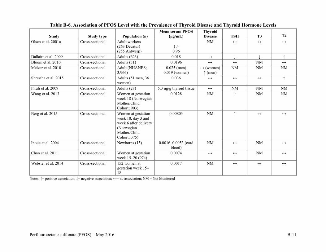
Table B-6. Association of PFOS Level with the Prevalence of Thyroid Disease and Thyroid Hormone Levels
Study Study type Population (n) Mean serum PFOS
(µg/mL) Thyroid Disease TSH T3 T4
Olsen et al. 2001a Cross-sectional Adult workers (263 Decatur) (255 Antwerp)
1.4
0.96
NM ↔ ↔ ↔
Dallaire et al. 2009 Cross-sectional Adults (623) 0.018 ↔ ↓ ↓ ↑ Bloom et al. 2010 Cross-sectional Adults (31) 0.0196 ↔ ↔ NM ↔ Melzer et al. 2010 Cross-sectional Adult (NHANES;
3,966) 0.025 (men)
0.019 (women) ↔ (women)
↑ (men) NM NM NM
Shrestha et al. 2015 Cross-sectional Adults (51 men, 36 women)
0.036 ↔ ↔ ↔ ↑
Pirali et al. 2009 Cross-sectional Adults (28) 5.3 ng/g thyroid tissue ↔ NM NM NM Wang et al. 2013 Cross-sectional Women at gestation
week 18 (Norwegian Mother/Child Cohort; 903)
0.0128 NM ↑ NM NM
Berg et al. 2015 Cross-sectional Women at gestation week 18, day 3 and week 6 after delivery (Norwegian Mother/Child Cohort; 375)
0.00803 NM ↑ ↔ ↔
Inoue et al. 2004 Cross-sectional Newborns (15) 0.0016–0.0053 (cord blood)
NM ↔ NM ↔
Chan et al. 2011 Cross-sectional Women at gestation week 15–20 (974)
0.0074 ↔ ↔ NM ↔
Webster et al. 2014 Cross-sectional 152 women at gestation week 15–18
0.0017 NM ↔ ↔ ↔
Notes: ↑= positive association; ↓= negative association; ↔= no association; NM = Not Monitored
Perfluorooctane sulfonate (PFOS) – May 2016 B-11

Table B-7. Association of Serum PFOS with Markers of Immunotoxicity
Study Study type Population (n)
Mean or median serum PFOS
(µg/mL)
Disease prevalence in
children Vaccine response
Okada et al. 2012 Prospective cohort
Maternal, third trimester (343)
0.0056 ↔ NM
Fei et al. 2010b Cross-sectional Maternal, first trimester (1,400)
0.0353 ↔ NM
Grandjean et al. 2012
Prospective cohort
Maternal at gestation week 32 (587)
0.0273 (maternal) NM ↓ (antibody titer in child at age 5
yrs) Grandjean et al. 2012
Prospective cohort
Children age 5 years (587)
0.0167 (child at age 5 years)
NM ↓ (antibody titer in child at age 7
yrs) Granum et al. 2013
Prospective cohort
Women at delivery (56)
0.0056 (maternal) ↔ ↓ (antibody titer in child at age 3
years) Humblet et al. 2014
Cross-sectional Children at 12–19 years (1,877)
0.017 (asthmatics) 0.0168 (non-asthmatics)
↔ for asthma NM
Dong et al. 2013 Cross-sectional Children age 10–15 years (231 asthmatics and 225 controls)
0.0455 (asthmatics) 0.0334 (non-asthmatics)
↑ for asthma NM
Looker et al. 2014 Cross-sectional Adults (411) 0.0083 NM ↔ Notes: ↑= positive association; ↓= negative association; ↔= no association; NM = Not Measured
Perfluorooctane sulfonate (PFOS) – May 2016 B-12

Appendix C: Summary of Data
Perfluorooctane sulfonate (PFOS) – May 2016 C-1

Table C-1. PFOS Toxicokinetic Information
Species Dose Route of exposure Effects observed
PFOS in liver (µg/g)
PFOS in blood (µg/mL)
Reference M F M F Human NA Unknown ↑ TC; ↑ TG NS 0.96–1.40 Olsen et al.
2001b, 2003b Human NA Unknown None observed
on cholesterol NS
1.16–2.62 Olsen et al. 2001b, 2003b
Human NA Drinking water ↑ TC; ↑ TG; ↑ LDL; ↑ UA
NS NS 0.022–0.023 Steenland et al. 2009, 2010
Human NA Drinking water ↑ TC; ↑ LDL; ↑ HDL
NS NS 0.023 Frisbee et al. 2010
Human NA Drinking water None observed on cholesterol
NS NS 0.011–0.023 Fitz-Simon et al. 2013
Human NA Unknown ↑ TC; ↑ non-HDL
NS NS 0.025 Nelson et al. 2010
Human NA Unknown ↑ HDL NS NS 0.019 Château-Degat et al. 2010
Human NA Unknown ↑ TC NS NS 0.036 Eriksen et al. 2013
Human NA Unknown ↑ TC; ↑ LDL; ↑ HDL
NS NS 0.013 Starling et al. 2014
Human NA Unknown None observed on cholesterol
NS NS 0.0084 Fisher et al. 2013
Human NA Unknown/ drinking water
Developmental delays
NS NS NS 0.0098–0.039 Fei et al. 2008b; Lopez-Espinosa et al. 2011
Human NA Unknown/ drinking water
LBW NS NS NS 0.0056–0.016 Washino et al. 2009; Stein et al. 2009; Darrow et al. 2013
Human NA Unknown None on birth outcome; birth weight and length; growth to 7 years
NS NS NS 0.009–0.039 Fei et al. 2007, 2008a; Monroy et al. 2008; Hamm et al. 2009; Andersen et al. 2013
Human NA Unknown ↑ time to pregnancy
NS NS NS 0.033–0.039 Fei et al. 2009
Perfluorooctane sulfonate (PFOS) – May 2016 C-2

Species Dose Route of exposure Effects observed
PFOS in liver (µg/g)
PFOS in blood (µg/mL)
Reference M F M F Human NA Unknown Effects on sperm
numbers and morphology
NS NS 0.017–0.025 NS Joensen et al. 2009; Buck Louis et al. 2014
Human NA Unknown None on semen parameters
NS NS 0.0085 NS Joensen et al. 2013
Human NA Unknown None on thyroid hormones
NS NS 0.96–1.4 Olsen et al. 2001b; 2003b
Human NA Unknown ↓TSH, T3; ↑T4 NS NS 0.018 Dallaire et al. 2009
Human NA Unknown None on thyroid hormones
NS NS 0.0196 Bloom et al. 2010
Human NA Unknown ↑ incidence of thyroid disease (men only)
NS NS 0.025 0.019 Melzer et al. 2010
Human NA Unknown ↑T4 NS NS 0.036 Shrestha et al. 2015
Human NA Unknown ↑TSH NS NS NS 0.008–0.0128 (gestation wk 18)
Wang et al. 2013; Berg et al. 2015
Human NA Unknown None on thyroid hormones
NS NS NS 0.0074 (gestation wk
15–20)
Chan et al. 2011
Human NA Unknown None on diseases in children
NS NS NS 0.0056 (maternal, third
trimester)
Okada et al. 2012
Human NA Unknown None on diseases in children
NS NS NS 0.0353 (maternal, first
trimester)
Fei et al. 2010b
Human NA Unknown ↓ antibody titer in children
NS NS NS 0.0273 (maternal,
gestation wk 32)
Grandjean et al. 2012
0.0167 (child age 5 years) Human NA Unknown ↓ antibody titer
in children NS NS NS 0.0056
(maternal at delivery)
Granum et al. 2013
Human NA Unknown ↑asthma NS NS 0.0455 (asthmatic children) Dong et al. 2013
Perfluorooctane sulfonate (PFOS) – May 2016 C-3

Species Dose Route of exposure Effects observed
PFOS in liver (µg/g)
PFOS in blood (µg/mL)
Reference M F M F Human NA Unknown None on vaccine
response NS NS 0.0083 Looker et al.
2014 Monkey 0.15 mg/kg/day
for 26 weeks with 52 week recovery
capsule None observed NS NS (serum) wk 1: 4.60
wk 26: 82.6 wk 35: 84.5 wk 79: 19.1
(serum) wk 1: 3.71
wk 26: 66.8 wk 35: 74.7 wk 79: 21.4
Seacat et al. 2002
Monkey 0.75 mg/kg/day for 26 weeks with 52 week recovery
capsule ↑ liver wt ↓ cholesterol and body wt
NS NS (serum) wk 1: 21.0 wk 26: 173 wk 35: 181 wk 79: 41.1
(serum) wk 1: 20.4 wk 26: 171 wk 35: 171 wk 79: 41.4
Seacat et al. 2002
Rat 0.018–0.023
mg/kg/day for 104 weeks
diet None observed wk 0: 11.0
wk 10: 23.8 wk 105: 7.83
wk 0: 8.71
wk 10: 19.2 wk 105: 12.9
(serum) wk 0: 0.91
wk 14: 4.04 wk 105: 1.31
(serum) wk 0: 1.61
wk 14: 6.96 wk 105: 4.35
Thomford 2002
Rat 0.184–0.247 mg/kg/day for 104 weeks
diet ↑ liver histopath. lesions
wk 0: 47.6 wk 10: 358
wk 105: 70.5
wk 0: 83.0 wk 10: 370
wk 105: 131
(serum) wk 0: 7.57
wk 14: 43.9 wk 105: 22.5
(serum) wk 0: 12.6
wk 14: 64.4 wk 105: 75.0
Thomford 2002
Rat 0.765–1.10 mg/kg/day for 104 weeks
diet ↑ body and liver wt ↑ hepatocellular adenoma
wk 0: 282 wk 10: 568
wk 105: 189
wk 0: 373 wk 10: 635
wk 105: 381
(serum) wk 0: 41.8 wk 14: 148
wk 105: 69.3
(serum) wk 0: 54.0 wk 14: 223
wk 105: 233
Thomford 2002
Rat (male only)
5 mg/kg for 28 days
oral gavage ↓ body wt 345 NS (whole blood) 72.0
NS Cui et al. 2009
Rat (male only)
20 mg/kg for 28 days
oral gavage 10/10 died (day 26) hepatic hypertrophy
648 NS (whole blood) NS
NS Cui et al. 2009
Perfluorooctane sulfonate (PFOS) – May 2016 C-4

Species Dose Route of exposure Effects observed
PFOS in liver (µg/g)
PFOS in blood (µg/mL)
Reference M F M F Rat 0.4 mg/kg
42 days prior to cohabitation through GD 21
oral gavage
↓ maternal and pup body wt
NS GD 21: dams = 107
fetuses = 30.6
NS (serum) GD 1: 40.7 GD 7: 40.9
GD 21: dams = 26.2
fetuses = 34.3
Luebker et al. 2005b
Rat 1.6 mg/kg 42 days prior to cohabitation through GD 21
oral gavage
↓ maternal and pup body wt ↓ pup survival
NS GD 21: dams = 388
fetuses = 86.5
NS (serum) GD 1: 160 GD 7: 154
GD 21: dams = 136
fetuses = 101
Luebker et al. 2005b
Rat 3.2 mg/kg 42 days prior to cohabitation through GD 21
oral gavage
100% pup mortality by PND 2
NS GD 21: dams = 610
fetuses = 230
NS (serum) GD 1: 318 GD 7: 306
GD 21: dams = 155
fetuses = 164
Luebker et al. 2005b
Rat 0.1 mg/kg GD 0 to PND 20
oral gavage None observed in dams or offspring
PND 21: Offspring =
5.98 PND 72:
Offspring = 0.98
GD 20: Dams = 8.35 Offspring =
3.21 PND 21:
Dams = NS Offspring =
5.28 PND 72:
Dams = NS Offspring =
0.80
(serum) PND 21:
Offspring = 1.73
PND 72: Offspring =
0.04
(serum) GD 20:
Dams = 1.72 Offspring = 3.91
PND 21: Dams = 3.16
Offspring = 1.77 PND 72:
Dams = NS Offspring = 0.21
Butenhoff et al. 2009; Chang et al. 2009
Perfluorooctane sulfonate (PFOS) – May 2016 C-5

Species Dose Route of exposure Effects observed
PFOS in liver (µg/g)
PFOS in blood (µg/mL)
Reference M F M F Rat 1.0 mg/kg GD 0
to PND 20 oral gavage ↑ motor activity
and ↓ habituation in male offspring
PND 21: Offspring =
44.89 PND 72:
Offspring = 7.17
GD 20: Dams = 48.88 Offspring =
20.03 PND 21:
Dams = NS Offspring =
41.23 PND 72:
Dams = NS Offspring = 7.2
(serum) PND 21:
Offspring = 18.61
PND 72: Offspring =
0.56
(serum) GD 20:
Dams = 26.63 Offspring =
31.46 PND 21:
Dams = 30.48 Offspring =
18.01 PND 72:
Dams = NS Offspring = 1.99
Butenhoff et al. 2009; Chang et al. 2009
Rat 1.0 mg/kg/day GDs 2–20
oral gavage none NS NS NS 19.69 Thibodeaux et al. 2003; Lau et al. 2003
Rat 2.0 mg/kg/day GDs 2–20
oral gavage ↓ dam and pup wt; ↓pup survival
NS NS NS 44.33 Thibodeaux et al. 2003; Lau et al. 2003
Note: NS = no sample obtained or recorded
Perfluorooctane sulfonate (PFOS) – May 2016 C-6

Table C-2. Summary of Animal Studies with Exposure to PFOS Method of exposure Length of study Species Concentration Results Reference
oral gavage 20 days monkey 0, 10, 30, 100, or 300 mg/kg/day 2 monkeys/sex/dose
NOAEL= NA LOAEL= 10 mg/kg/day from deaths at all doses
Goldenthal et al. 1978a
oral gavage 90 days monkey 0, 0.5, 1.5, or 4.5 mg/kg/day 2 monkeys/sex/dose
NOAEL= NA LOAEL= 0.5 mg/kg/day based on diarrhea and anorexia
Goldenthal et al. 1979
oral (capsule)
182 days monkey 0, 0.03, 0.15, or 0.75 mg/kg/day 4–6 monkeys/sex/dose
NOAEL= 0.15 mg/kg/day LOAEL= 0.75 mg/kg/day from ↓ body weight, ↑liver wt and ↓cholesterol
Seacat et al. 2002
oral gavage single dose rat 0, 100, 215, 464, or 1,000 mg/kg 5 rats/sex/dose
LD50 = 251 mg/kg (combined)
Dean et al. 1978
oral gavage single dose rat 0, 12.5, 25, or 50 mg/kg 5 male rats/dose
NOAEL= NA LOAEL= 12.5 mg/kg based on ↓ body weight
Yang et al. 2009
oral gavage single dose thyroid hormone activity
rat 0 or 15 mg/kg 5/15 female rats/group
Total thyroxine (TT4)- significant ↓ at 2, 6 and 24 hrs Triiodothyronine (TT3) and reverse triiodothyronine (rT3)- significant ↓ at 24 hrs Free thyroxine- significant ↑ at 2 and 6 hrs; normal at 24 hrs
Chang et al. 2008
inhalation 1 hour rat 0, 1.89, 2.86, 4.88, 6.49, 7.05, 13.9, 24.09, or 45.97 ppm 5 rats/sex/dose
LC50 = 5.2 ppm Rusch et al. 1979
oral (in diet) 28 days rat 0, 0.05, 0.18, 0.37, or 1.51 mg/kg/day—males 0, 0.05, 0.22, 0.47, or 1.77 mg/kg/day— females (0, 0.5, 2, 5, or 20 ppm) 5 rats/sex/dose
NOAEL = 0.37 mg/kg/day in males and 0.47 mg/kg/day in females LOAEL = 1.51 mg/kg/day in males and 1.77 mg/kg/day in females, based on ↓ body wt (M/F) and ↓ food consumption (F)
Seacat et al. 2003
oral (in diet) 28 days rat 0.14, 1.33, 3.21, 6.34 mg/kg/day—males 0.15, 1.43, 3.73, 7.58 mg/kg/day—females (0, 2, 20, 50, or 100 mg/kg diet) 15 rats/sex/dose
NOAEL = 0.14 mg /kg/day in males and NA in females LOAEL = 1.33 mg /kg/day in males and 0.15 mg/kg/day in females based on ↑ relative liver wt
Curran et al. 2008
Perfluorooctane sulfonate (PFOS) – May 2016 C-7

Method of exposure Length of study Species Concentration Results Reference
oral gavage 28 days rat 0, 5, or 20 mg/kg/day 10 males/dose
NOAEL= NA LOAEL= 5 mg/kg/day based on ↓ body wt and lung congestion
Cui et al. 2009
oral gavage 28 days rat 0, 0.5, 1, 3, or 6 mg/kg/day 19 males/dose
NOAEL = NA LOAEL = 0.5 mg/kg/day based on ↓ LH and testosterone and ↑ FSH
López-Doval et al. 2014
oral (in diet) 90 days rat 0, 2, 6, 18, 60, or 200 mg/kg/day 5 rats/sex/dose
NOAEL= NA LOAEL= 2 mg/kg/day, from ↑ liver wt, ↓ food consumption
Goldenthal et al. 1978b
oral (in diet) 98 days rat 0, 0.5, 2.0, 5.0, or 20 ppm 0, 0.03, 0.13, 0.34 or 1.33 mg/kg/day- males 0, 0.04, 0.15, 0.40 or 1.56 mg/kg/day- females 5 rats/sex/dose
NOAEL = 0.34 mg/kg/day in males and 0.40 mg/kg/day in females LOAEL = 1.33 mg/kg/day in males and 1.56 mg/kg/day in females, based on ↑ liver wt (M) and ↑ relative liver wt (M/F)
Seacat et al. 2003
oral gavage GD 0 to PND 20a
developmental neurotoxicity study
rat 0, 0.1, 0.3, or 1.0 mg/kg/day 25 females/dose
Maternal NOAEL= 1 mg/kg/day LOAEL= NA Developmental NOAEL= 0.3 mg/kg/day LOAEL= 1 mg/kg/day based on ↑ motor activity
Butenhoff et al. 2009
oral gavage GDs 2–20 rat 0, 1, 2, 3, 5, or 10 mg/kg
Maternal NOAEL= 1 mg/kg LOAEL= 2 mg/kg based on ↓ body wt Developmental NOAEL= 1 mg/kg LOAEL= 2 mg/kg based on ↓ survival BMDL5 corresponding to maternal dose for survival of rat pups at PND 8 was 0.58 mg/kg
Thibodeaux et al. 2003 and Lau et al. 2003
oral gavage GDs 2–21 rat 0, 0.1, 0.6, or 2.0 mg/kg
Offspring NOAEL = cannot be determined LOAEL= 0.1 mg/kg based on changes in the cortex and hippocampus (astrocyte activation markers, pro-inflammatory transcription factors)
Zeng et al. 2011
Perfluorooctane sulfonate (PFOS) – May 2016 C-8

Method of exposure Length of study Species Concentration Results Reference
oral gavage GDs 2–21 rat 0, 0.1, 0.6, or 2 mg/kg/day
Offspring on PND 21 NOAEL = 0.1 mg/kg/day LOAEL = 0.6 mg/kg/day based on increased apoptosis in heart cells
Zeng et al. 2014
oral gavage GDs 1–21 rat 0, 0.1, or 2.0 mg/kg/day
Offspring NOAEL= 0.1 mg/kg/day LOAEL = 2.0 mg/kg/day based on histopathological changes in lungs, ↓ body wt and ↑ mortality
Chen et al. 2012
oral gavage GD 0–PND 20 rat 0, 0.5, or 1.5 mg/kg/day 6 dams/dose
NOAEL = NA LOAEL = 0.5 mg/kg/day based on ↓ offspring body wt, impaired glucose tolerance
Lv et al. 2013
oral gavage GDs 11–19 rat 0, 5, or 20 mg/kg/day 4 dams/dose
NOAEL = NA LOAEL = 5 mg/kg/day based on ↓ offspring body wt
Zhao et al. 2014
oral gavage 6 wks prior to mating and Males—22 days Females— through gestation, parturition and lactation reproductive study
rat 0, 0.1, 0.4, 1.6, or 3.2 mg/kg/day 35 rats/sex/dose
F0 (M/F) parents NOAEL= 0.1 mg/kg/day LOAEL= 0.4 mg/kg/day based on ↓ bwt gain/food consumption F1 (M/F) parents NOAEL = 0.4 mg/kg LOAEL = NA, higher dose not tested F1 offspring NOAEL= 0.4 mg/kg/day LOAEL= 1.6 mg/kg/day based on ↓ viability, body wt F2 offspring NOAEL= 0.1 mg/kg/day LOAEL= 0.4 mg/kg/day based on ↓ body wt
Luebker et al. 2005b
Perfluorooctane sulfonate (PFOS) – May 2016 C-9

Method of exposure Length of study Species Concentration Results Reference
oral gavage 6 wks prior to mating and continued through mating, gestation and LD 4 reproductive study
rat 0, 0.4, 0.8. 1.0, 1.2, 1.6 and 2.0 mg/kg/day 20–28 dams/dose
F0 dams NOAEL= 0.4 mg/kg/day LOAEL= 0.8 mg/kg/day based on ↓ bwt gain F1 offspring NOAEL= not identified LOAEL= 0.4 mg/kg/day based on ↓ pup body weight BMDL5 estimates for decreased gestation length was 0.31 and viability was 0.89 mg/kg/day
Luebker et al. 2005a
oral (diet) 104 weeks rat 0, 0.024, 0.098, 0.242, or 0.984 mg/kg/day—males 0, 0.029, 0.120, 0.299, or 1.251 mg/kg/day—females 0, 0.5, 2, 5, or 20 ppm 40–70 males and females
Males NOAEL= 0.024 mg/kg/day Males LOAEL= 0.098 mg/kg/day based on liver histopathology Females NOAEL = 0.120 mg/kg/day Females LOAEL = 0.299 mg/kg/day based on liver histopathology Suggestive of carcinogenicity
Thomford 2002/ Butenhoff et al. 2012
oral gavage 1 time on PND 10 developmental neurotoxicity
mouse 0, 0.75, or 11.3 mg/kg 4–7 males/group
Mice at both concentrations showed ↓ activity and ↑ neuroprotein levels in the hippocampus
Johansson et al. 2008, 2009
oral gavage 7 days immunotoxicity study
mouse 0, 5, 20, or 40 mg/kg 12 male mice/dose
NOAEL= NA LOAEL= 5 mg/kg based on ↑ liver wt and suppression of the plaque forming cell response
Zheng et al. 2009
oral gavage GDs 1–17 developmental immunotoxicity
mouse 0, 0.1, 1, or 5 mg/kg 10–12 female mice/dose
Males NOAEL = 0.1 mg/kg Males LOAEL = 1 mg/kg based on ↓NK cell activity Females NOAEL = 1 mg/kg Females LOAEL = 5 mg/kg based on ↓NK cell activity
Keil et al. 2008
Perfluorooctane sulfonate (PFOS) – May 2016 C-10

Method of exposure Length of study Species Concentration Results Reference
oral gavage GDs 1–17 mouse 0, 1, 5, 10, 15, or 20 mg/kg
Maternal NOAEL= 1 mg/kg LOAEL= 5 mg/kg based on ↑ liver wt Developmental NOAEL= 1 mg/kg LOAEL= 5 mg/kg based on ↑ liver wt, delayed eye opening BMDL5 corresponding to maternal dose for survival of mouse pups at PND 6 was 3.88 mg/kg
Thibodeaux et al. 2003; Lau et al. 2003
oral gavage GDs 12–18 developmental
mouse 0 or 6 mg/kg/day 8–10 mice/dose
Maternal NOAEL= 6 mg/kg/day LOAEL= NA Developmental NOAEL= NA LOAEL= 6 mg/kg/day based on ↓ body wt
Fuentes et al. 2007
oral gavage GDs 1–17/18 developmental
mouse 0, 1, 10, or 20 mg/kg/day 10 mice/dose
Maternal NOAEL = 1 mg/kg/day LOAEL= 10 mg/kg/day, based on ↑ liver organ wt. Developmental NOAEL= 1 mg/kg/day LOAEL= 10 mg/kg/day, based on fetal abnormalities and ↓survival
Yahia et al. 2008
oral gavage 14 days With regular or high fat diet
mouse 0, 5, or 20 mg/kg/day 16 males/dose/diet
NOAEL = NA LOAEL = 5 mg/kg/day based on wt loss on high fat diet
L. Wang et al. 2014
oral gavage 3–21 days mouse 0, 1, 5, or 10 mg/kg/day 4 males/dose
NOAEL = 1 mg/kg/day LOAEL = 5 mg/kg/day based on ↑ liver wt, changes in oxidation biochemical parameters
Wan et al. 2012
oral gavage GD 0–PND 21 mouse 0, 0.3, 3 mg/kg/day 6 dams/dose
NOAEL = 0.3 mg/kg/day LOAEL = 3 mg/kg/day based on ↑ liver wt in dams and male offspring, ↑ fasting serum insulin in males
Wan et al. 2014b
Perfluorooctane sulfonate (PFOS) – May 2016 C-11

Method of exposure Length of study Species Concentration Results Reference
oral gavage 28 days immunotoxicity
mouse 0, 0.00017, 0.0017, 0.0033, 0.017, 0.033, or 0.166 mg/kg 5 mice/dose
Males NOAEL= 0.00017 mg/kg Males LOAEL= 0.0017 mg/kg based on ↓ plaque forming cell response Females NOAEL= 0.0033 mg/kg Females LOAEL= 0.017 mg/kg based on ↓ plaque forming cell response
Peden-Adams et al. 2008
oral gavage 60 days immunotoxicity
mouse 0, 0.008, 0.083, 0.417, 0.833, or 2.083 mg/kg 10 male mice/group
NOAEL = 0.008 mg/kg/day LOAEL = 0.083 mg/kg based on ↑ splenic NK cell activity and ↑ liver weight
Dong et al. 2009
oral gavage 90 days neurotoxicity
mouse 0, 0.43, 2.15, or 10.75 mg/kg/day 15/group, sex not specified
NOAEL = 0.43 mg/kg/day LOAEL = 2.15 mg/kg/day based on changes in water maze and histopath. in hippocampus
Long et al. 2013
dermal single dose rabbit 0.5 g* (no data on gender)
No irritation Biesemeier and Harris 1974
ocular single dose rabbit 0.5 g* (no data on gender)
Exact score not provided except maximal score at 1 and 24 hrs
Biesemeier and Harris 1974
Notes: *Exact dose not provided; NA= not applicable; could not be determined a GD = gestation day and PND = post natal day M = male; F = female
Perfluorooctane sulfonate (PFOS) – May 2016 C-12