energy metabolism of monocytic ehrlichia
TRANSCRIPT

Energy Metabolism of Monocytic EhrlichiaAuthor(s): Emilio Weiss, Jim C. Williams, Gregory A. Dasch and Yuan-Hsu KangSource: Proceedings of the National Academy of Sciences of the United States of America,Vol. 86, No. 5 (Mar. 1, 1989), pp. 1674-1678Published by: National Academy of SciencesStable URL: http://www.jstor.org/stable/33262 .
Accessed: 02/05/2014 09:56
Your use of the JSTOR archive indicates your acceptance of the Terms & Conditions of Use, available at .http://www.jstor.org/page/info/about/policies/terms.jsp
.JSTOR is a not-for-profit service that helps scholars, researchers, and students discover, use, and build upon a wide range ofcontent in a trusted digital archive. We use information technology and tools to increase productivity and facilitate new formsof scholarship. For more information about JSTOR, please contact [email protected].
.
National Academy of Sciences is collaborating with JSTOR to digitize, preserve and extend access toProceedings of the National Academy of Sciences of the United States of America.
http://www.jstor.org
This content downloaded from 130.132.123.28 on Fri, 2 May 2014 09:56:26 AMAll use subject to JSTOR Terms and Conditions

Proc. Natl. Acad. Sci. USA Vol. 86, pp. 1674-1678, March 1989 Microbiology
Energy metabolism of monocytic Ehrlichia (intracellular bacteria/animal pathogens/human pathogens)
EMILIO WEISS*t, JIM C. WILLIAMSt?, GREGORY A. DASCH*, AND YUAN-HSU KANG*
*Naval Medical Research Institute, Bethesda, MD 20814; tU.S. Army Medical Research Institute of Infectious Diseases, Fort Detrick, Frederick, MD 21201; and ?Office of the Director of Intramural Research Program, National Institute of Allergy and Infectious Diseases, Bethesda, MD 20892
Communicated by Carl R. Woese, December 1, 1988
ABSTRACT We investigated if the monocytic Ehrlichia are totally dependent on their host cells for energy, or, as Rickettsia, are capable of some ATP synthesis in vitro. The Miyayama strain of Ehrlichia sennetsu and the Maryland and Illinois strains of Ehrlichia risticii were cultivated in a mouse macrophage cell line, separated from host cell constituents by procedures that included Renografin or Percoll gradient cen- trifugation, and tested after cryopreservation. Cells incubated without a metabolizing substrate contained little, if any, ATP. When the Ehrlichia cells were incubated for 1 hr at 34?C with glutamine, significant amounts of ATP were detected. The amounts of ATP attained with glutamine were decreased in some instances by the addition of atractyloside, an inhibitor of adenine nucleotide translocase in mitochondria, and were decreased consistently and to a greater extent by 2,4-dinitro- phenol. When ATP, instead of glutamine, was added to the ehrlichiae, upon incubation the amount of ATP was markedly decreased. Comparable responses under all these conditions were obtained with Rickettsia typhi, although the final ATP levels were higher. Control preparations derived from unin- fected mouse macrophages or from the discards of the Ehrlichia purification procedures contained negligible amounts of ATP, which were not increased by incubation with glutamine. We conclude that with respect to ATP metabolism, the monocytic Ehrlichia resemble Rickettsia more closely than Chiamydia, even though Ehrlichia resemble Chlamydia in their intracellular location in the phagosomes and in possibly having a develop- mental cycle.
The three species of Ehrlichia that grow preferentially in the phagosomes of monocytic cells, E. canis, E. sennetsu, and E. risticii, the latter two species in particular, greatly resemble each other in morphological and developmental characteris- tics and antigenic composition (1). This similarity contrasts with their differences in host affinity and geographic distri- bution. E. sennetsu, isolated in 1953 in Japan (2), is a human pathogen believed to be confined to the Far East (3), while E. risticii, isolated in 1985 (4, 5), is associated with a disease of horses, observed in the eastern United States about a decade ago. This contrast renders the understanding of the natural history and phylogeny of these microorganisms quite diffi- cult. Several approaches are required to reach such an understanding, including an investigation of the biochemical characteristics of Ehrlichia.
In previous studies (6, 7) we have shown that E. sennetsu and E. risticii, separated from host cell constituents, utilize glutamine in preference to other substrates that have been tested, including glutamate. There is no indication that they have a glycolytic pathway. In this respect they resemble the obligate intracellular rickettsiae, although these microorga- nisms utilize glutamate somewhat better than glutamine (8). In the study here reported we show that Ehrlichia derive
The publication costs of this article were defrayed in part by page charge payment. This article must therefore be hereby marked "advertisement" in accordance with 18 U.S.C. ?1734 solely to indicate this fact.
some ATP from the metabolism of glutamine, as is the case in rickettsiae (8, 9).
MATERIALS AND METHODS Bacterial Strains. The isolation histories of the Miyayama
strain of E. sennetsu and the Maryland and Illinois strains of E. risticii and their cultivation in the P388D1 mouse mac- rophage cell line were described in a previous publication (7). The Wilmington strain of Rickettsia typhi was cultivated and harvested from the yolk sacs of chicken embryos, as de- scribed (9).
Separation of Ehrlichia and Rickettsia from Host Constitu- ents. For the early experiments, the separation of the ehrli- chiae from host constituents was done by the Renografin gradient centrifugation procedure without prior proteolytic digestion, as previously described (7). The diluent used in this procedure consisted of 0.2 M sucrose and 0.05 M potassium phosphate buffer, pH 7.4 (SPK). For the final step in the procedure, for cryopreservation, and for the metabolic tests, MgSO4 was added to a final concentration of 5 mM. For the more recent tests, the preparations of the heavily infected cells, after mechanical disruption, were subjected to three 20-min steps of enzymatic digestion at room temperature. First, trypsin was added to a final concentration of 500 ,g/ ml, then DNase in two steps to a final concentration of 250 ,ug/ml. The DNase contained sufficient MgSO4 to bring the final magnesium concentration to 5 mM. The DNase added in the final step also contained Bowman-Birk trypsin-chymo- trypsin inhibitor (Sigma) to a final concentration of 125 ,ug/ ml. After digestion, the preparations were further purified by filtration through type AP-20 glass microfiber depth filter (Millipore) and concentrated by centrifugation. The ehrli- chiae were separated from host constituents by isopycnic centrifugation in 25 ml of 32% Percoll gradients (Pharmacia) using a Ti 70 rotor (Beckman) for 30 min at 63,000 x gmax. To the SPK diluent were added glutamine, final concentration 1 mM, and for some of the steps in the procedure 1 mM citric acid. R. typhi was purified by the Renografin procedure as described (9).
Tests for ATP Formation. The tests were done in 1.8-ml microcentrifuge tubes, kept in ice water until ready for incubation. All reagents were prepared in SPK. To each tube were added 0.1 ml of one or two reagents, as indicated, or SPK in their place, and 0.2 ml of cell preparation, to a final volume of 0.4 ml. When incubated, the specimens were placed in a water bath for 1 hr at 34?C and oscillated at moderate speed. The reaction was stopped by placing the tubes in ice water, adding 40 ,l of 70% (wt/vol) perchloric acid and, after rapid mixing, adding 65 ,ul of 7.5 M KOH containing 0.05 M EDTA, a volume previously determined to neutralize the perchloric acid in the buffer used. After vigorous mixing followed by a 5-min wait for the precipitate
Abbreviation: DNP, 2,4-dinitrophenol. tTo whom reprint requests should be addressed.
1674
This content downloaded from 130.132.123.28 on Fri, 2 May 2014 09:56:26 AMAll use subject to JSTOR Terms and Conditions
Microbiology: Weiss et al. Proc. Natl. Acad. Sci. USA 86 (1989) 1675
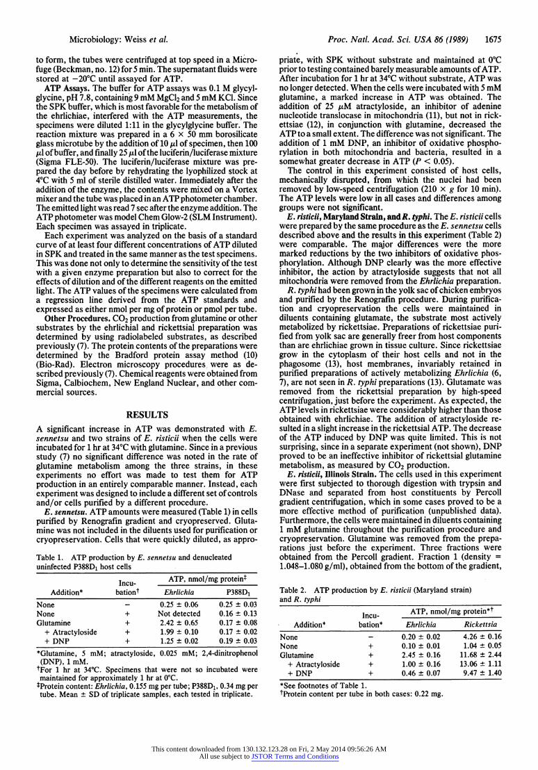
to form, the tubes were centrifuged at top speed in a Micro- fuge (Beckman, no. 12) for 5 min. The supernatant fluids were stored at -20?C until assayed for ATP.
ATP Assays. The buffer for ATP assays was 0.1 M glycyl- glycine, pH 7.8, containing 9 mM MgCl2 and 5 mM KCl. Since the SPK buffer, which is most favorable for the metabolism of the ehrlichiae, interfered with the ATP measurements, the specimens were diluted 1:11 in the glycylglycine buffer. The reaction mixture was prepared in a 6 x 50 mm borosilicate glass microtube by the addition of 10 ,ul of specimen, then 100 ,ul of buffer, and finally 25 ,ul of the luciferin/luciferase mixture (Sigma FLE-50). The luciferin/luciferase mixture was pre- pared the day before by rehydrating the lyophilized stock at 4?C with 5 ml of sterile distilled water. Immediately after the addition of the enzyme, the contents were mixed on a Vortex mixer and the tube was placed in an ATP photometer chamber. The emitted light was read 7 sec after the enzyme addition. The ATP photometer was model Chem Glow-2 (SLM Instrument). Each specimen was assayed in triplicate.
Each experiment was analyzed on the basis of a standard curve of at least four different concentrations of ATP diluted in SPK and treated in the same manner as the test specimens. This was done not only to determine the sensitivity of the test with a given enzyme preparation but also to correct for the effects of dilution and of the different reagents on the emitted light. The ATP values of the specimens were calculated from a regression line derived from the ATP standards and expressed as either nmol per mg of protein or pmol per tube.
Other Procedures. CO2 production from glutamine or other substrates by the ehrlichial and rickettsial preparation was determined by using radiolabeled substrates, as described previously (7). The protein contents of the preparations were determined by the Bradford protein assay method (10) (Bio-Rad). Electron microscopy procedures were as de- scribed previously (7). Chemical reagents were obtained from Sigma, Calbiochem, New England Nuclear, and other com- mercial sources.
RESULTS A significant increase in ATP was demonstrated with E. sennetsu and two strains of E. risticii when the cells were incubated for 1 hr at 34WC with glutamine. Since in a previous study (7) no significant difference was noted in the rate of glutamine metabolism among the three strains, in these experiments no effort was made to test them for ATP production in an entirely comparable manner. Instead, each experiment was designed to include a different set of controls and/or cells purified by a different procedure.
E. sennetsu. ATP amounts were measured (Table 1) in cells purified by Renografin gradient and cryopreserved. Gluta- mine was not included in the diluents used for purification or cryopreservation. Cells that were quickly diluted, as appro-
Table 1. ATP production by E. sennetsu and denucleated uninfected P388D1 host cells
Incu- ATP, nmol/mg proteint Addition* bationt Ehrlichia P388D1
None - 0.25 ? 0.06 0.25 ? 0.03 None + Not detected 0.16 ? 0.13 Glutamine + 2.42 ? 0.65 0.17 ? 0.08
+ Atractyloside + 1.99 ? 0.10 0.17 ? 0.02 + DNP + 1.25 ? 0.02 0.19 ? 0.03
*Glutamine, 5 mM; atractyloside, 0.025 mM; 2,4-dinitrophenol (DNP), 1 mM.
fFor 1 hr at 34?C. Specimens that were not so incubated were maintained for approximately 1 hr at 0?C. tProtein content: Ehrlichia, 0.155 mg per tube; P388D1, 0.34 mg per tube. Mean ? SD of triplicate samples, each tested in triplicate.
priate, with SPK without substrate and maintained at 0?C prior to testing contained barely measurable amounts of ATP. After incubation for 1 hr at 34?C without substrate, ATP was no longer detected. When the cells were incubated with 5 mM glutamine, a marked increase in ATP was obtained. The addition of 25 ytM atractyloside, an inhibitor of adenine nucleotide translocase in mitochondria (11), but not in rick- ettsiae (12), in conjunction with glutamine, decreased the ATP to a small extent. The difference was not significant. The addition of 1 mM DNP, an inhibitor of oxidative phospho- rylation in both mitochondria and bacteria, resulted in a somewhat greater decrease in ATP (P < 0.05).
The control in this experiment consisted of host cells, mechanically disrupted, from which the nuclei had been removed by low-speed centrifugation (210 x g for 10 min). The ATP levels were low in all cases and differences among groups were not significant.
E. risticii, Maryland Strain, andR. typhi. The E. risticii cells were prepared by the same procedure as the E. sennetsu cells described above and the results in this experiment (Table 2) were comparable. The major differences were the more marked reductions by the two inhibitors of oxidative phos- phorylation. Although DNP clearly was the more effective inhibitor, the action by atractyloside suggests that not all mitochondria were removed from the Ehrlichia preparation.
R. typhi had been grown in the yolk sac of chicken embryos and purified by the Renografin procedure. During purifica- tion and cryopreservation the cells were maintained in diluents containing glutamate, the substrate most actively metabolized by rickettsiae. Preparations of rickettsiae puri- fied from yolk sac are generally freer from host components than are ehrlichiae grown in tissue culture. Since rickettsiae grow in the cytoplasm of their host cells and not in the phagosome (13), host membranes, invariably retained in purified preparations of actively metabolizing Ehrlichia (6, 7), are not seen in R. typhi preparations (13). Glutamate was removed from the rickettsial preparation by high-speed centrifugation, just before the experiment. As expected, the ATP levels in rickettsiae were considerably higher than those obtained with ehrlichiae. The addition of atractyloside re- sulted in a slight increase in the rickettsial ATP. The decrease of the ATP induced by DNP was quite limited. This is not surprising, since in a separate experiment (not shown), DNP proved to be an ineffective inhibitor of rickettsial glutamine metabolism, as measured by CO2 production.
E. risticii, Illinois Strain. The cells used in this experiment were first subjected to thorough digestion with trypsin and DNase and separated from host constituents by Percoll gradient centrifugation, which in some cases proved to be a more effective method of purification (unpublished data). Furthermore, the cells were maintained in diluents containing 1 mM glutamine throughout the purification procedure and cryopreservation. Glutamine was removed from the prepa- rations just before the experiment. Three fractions were obtained from the Percoll gradient. Fraction 1 (density = 1.048-1.080 g/ml), obtained from the bottom of the gradient,
Table 2. ATP production by E. risticii (Maryland strain) and R. typhi
Incu- ATP, nmol/mg protein*t Addition* bation* Ehrlichia Rickettsia
None - 0.20 t 0.02 4.26 ? 0.16 None + 0.10 ? 0.01 1.04 ? 0.05 Glutamine + 2.45 ? 0.16 11.68 ? 2.44
+ Atractyloside + 1.00 ? 0.16 13.06 ? 1.11 + DNP + 0.46 ? 0.07 9.47 ? 1.40
*See footnotes of Table 1. tProtein content per tube in both cases: 0.22 mg.
This content downloaded from 130.132.123.28 on Fri, 2 May 2014 09:56:26 AMAll use subject to JSTOR Terms and Conditions

1676 Microbiology: Weiss et al. Proc. Natl. Acad. Sci. USA 86 (1989)
consisted of cells with greatest electron density and smallest contamination with host constituents (Fig. 1A), although some host membranes were retained (Fig. 1B). These cells also had the highest rate of glutamine metabolism (Table 3). Fraction 2 (1.044-1.048 g/ml), obtained from the intermedi- ate section of the gradient, contained some Ehrlichia particles that were less electron dense and numerous empty vesicles and other structures, presumably of host origin (Fig. IC). Glutamine specific activity was considerably lower. Fraction 3 (1.020-1.044 g/ml) is the top section of the gradient, which is generally discarded. Although some Ehrlichia particles are undoubtedly trapped, most of the other structures appear to be of host origin (Fig. 1D). Glutamine specific activity was quite low.
The ATP determinations of the three fractions (Table 3) reveal some similarities and some differences from those obtained in the previous two experiments (Tables 1 and 2). The amount of ATP in fraction 1 prior to incubation was considerably higher than the amounts obtained previously. There is a remarkable drop to a nonmeasurable level upon incubation without substrate. Upon incubation with gluta- mine there was an increase over the initial amount compa- rable to that obtained in the other experiments. Atractyloside had no apparent effect, while DNP was clearly inhibitory.
Table 3. ATP production by Percoll fractions separating E. risticii (Illinois strain) from host constituents
ATP, nmol/mg protein
Incu- Fraction Fraction Fraction Addition* bation* it 2t t
None - 1.57 + 0.22 0.37 ? 0.07 0.20 None + Not detected 0.02 0.20 Glutamine + 3.73 ? 0.11 0.76 ? 0.15 0.27
+ Atractyloside + 3.74 ? 0.25 0.77 ? 0.02 0.24 + DNP + 1.62 ? 0.08 0.37 ? 0.05 0.20
*See footnotes of Table 1. tFractions 1, 2, and 3 corresponded, respectively, to the bottom, intermediate, and top fractions of the Percoll gradient described in the text. Their appearance in the electron microscope is illustrated in Fig. 1. Protein contents were 0.11, 0.11, and 0.22 mg per tube, respectively. Glutamine metabolism (determined in a separate experiment), expressed as umol of CO2 per mg of protein produced from 5 mM glutamine, were 0.41, 0.12, and 0.02 per hr.
The results obtained with fraction 2 were qualitatively similar to those obtained with fraction 1, but much lower. They are compatible with the view that fraction 2 contained, propor- tionately, a much smaller number of Ehrlichia cells or cells of
AM
A.~ ~ ~ ~ ~ ~ ~ ~ ~ ~ ~ ~ ~ ~ ~ ~ ~ ~ ~~~~~~~~~~~I
~~~ - ~~~~~~~#M kXqR Pr . >4# -~~~~~~A
2. 't bt~A97
Z~ ~~4 1Q J~~~~~~~4 ST ~
4~~~~~~~~~~~~~~~~4
FIG. 1. Transmission electron micrographs of fractions 1, 2, and 3 of Percoll-purified preparation of E. risticii (Illinois strain) described in
the text, and tested as shown in Table 3. (A) Fraction 1. (X 6800.) (B) Fraction 1, field adjacent to the one shown in A. Large and small arrows
point, respectively, to what appear to be host and bacterial membranes. (x27,000.) (C and D) Fractions 2 and 3, respectively. (x6800.)
This content downloaded from 130.132.123.28 on Fri, 2 May 2014 09:56:26 AMAll use subject to JSTOR Terms and Conditions

Microbiology: Weiss et al. Proc. Natl. Acad. Sci. USA 86 (1989) 1677
Table 4. ATP recovery after addition to preparations of uninfected denucleated P388D1 cells, to purified R. typhi, and to Percoll fractions separating E. risticii (Illinois strain) from host constituents*
ATP recovered, prmol per tube E. risticii fractions (Table 3) ATP added, Incu- P388D1 R. typhi
pmnol per tube bation (Table 1) (Table 2) 1 2 3 0 - 84 ? 11 929 ? 35 177 ? 14 41 ? 8 44 ? 3
+ 52?42 226?11 0 2 43?15 400 - 82 ? 12 1187 ? 89 445 ? 25 86 ? 0 51 ? 5
+ 78 ? 35 280 ? 31 7? 7 16 ? 0 36 ? 7 1200 - 85 ? 3 Not done 1001 ? 110 278 ? 50 125 ? 26
+ 45 ? 20 Not done 24? 14 16 ? 4 43 ? 7 *See footnotes of Tables 1-3.
reduced metabolic activity. The results obtained with frac- tion 3 were virtually identical to those obtained with denu- cleated uninfected cells (Table 1).
ATP Catabolism. The results illustrated in Table 3 with fraction 1 suggest that ehrlichiae catabolize ATP, thereby decreasing the internal ATP concentrations when the cells are incubated without substrate. In the experiments de- scribed above, we also attempted to determine if ATP degradation could be shown when ATP itself was added to the cells. An unexpected difficulty encountered in such determinations was that, although ATP was stable in the diluent used, it rapidly disappeared when added to control preparations, even before incubation. Table 4 illustrates results obtained with several of the preparations used in the experiments depicted in Tables 1-3. The results, in contrast to those previously presented, are given as pmol of ATP per tube, rather than nmol per mg of protein, to better correlate the amounts of ATP added to those recovered. With unin- fected denucleated P388D1 cells, the amounts of ATP were uniformly moderately low, irrespective of amounts of ATP added (0, 400, or 1200 pmol per tube). These amounts were not substantially changed by incubation for 1 hr at 34?C. By contrast, when 400 pmol per tube was added to R. typhi, before incubation the amounts of ATP were higher than those in comparable tubes to which no ATP had been added. After incubation the amounts of ATP in the two groups were about the same. Somewhat similar results were obtained with fraction 1 of E. risticii. The additions of 400 or 1200 pmol per tube were reflected in higher ATP levels before incubation. Surprisingly, the ATP levels after incubation were lower than those obtained with the P388D1. With fraction 3 the results were similar to those obtained with P388D1. The results with fraction 2 appeared to be intermediate between those with fractions 1 and 3, although more closely resembling those of fraction 3. With the other two Ehrlichia preparations (Tables 1 and 2), possibly because of less complete separation from host cell debris, the results were intermediate between those for fractions 1 and 2 (not shown).
It is obvious that the kinetics of ATP destruction by eukaryotic debris (P388D1 and fraction 3) and those by ehrlichial or rickettsial preparations are quite different. Whether the destruction of ATP by ehrlichiae was due to an ATPase or some other enzyme, such as a phosphatase, kinase, or pyrophosphatase, has not yet been investigated. The results are consistent with the expectation that ehrlichiae can catabolize extracellular ATP, although a role by host enzymes in these reactions cannot be excluded. It remains to be shown whether ATP is also transported intact by Ehrli- chia, as found for typhus rickettsiae (12).
DISCUSSION Our results clearly demonstrate that two monocytic Ehrli- chia, E. sennetsu and E. risticii, are capable of limited ATP
synthesis in vitro when incubated with glutamine. It is tempting, therefore, to compare this and other biological properties of ehrlichiae with those of other obligate intracel- lular bacteria. Our understanding of the biological properties of ehrlichiae, however, has not kept pace with our expanding knowledge of ehrlichiae as agents of human and animal diseases (1, 7). For example, casual examination of electron micrographs of ehrlichiae (Fig. 1 A and B; refs. 4, 5, 7, 14, and 15) would tend to link them to chlamydiae (16, 17). Ehrli- chiae, as well as chlamydiae, multiply in the phagosomes of host cells and during growth are surrounded by a host cell membrane. There is good evidence that chlamydiae have surface components that inhibit fusion of the phagosome with lysosomes (16). Such a mechanism may exist in ehrlichiae, but it has not yet been demonstrated. The only biochemical evidence supporting the intraphagosomal location of ehrli- chiae is the pH optimum for metabolic activity, which is moderately alkaline (7). In addition, electron micrographs of ehrlichiae clearly display forms varying in size and electron density that suggest the existence of a developmental cycle. In chlamydiae a cycle was clearly demonstrated with differ- entiation into elementary and reticulate bodies. The elemen- tary bodies are infectious, inhibit phagosome-lysosome fu- sion, and are toxic for mice and macrophages. The reticulate bodies have none of these properties (16) but, in contrast to elementary bodies, are capable of metabolic activity and multiplication (17). There is no indication that such a clear differentiation exists in ehrlichiae, but the cycle of develop- ment of ehrlichiae has not been subjected to critical scrutiny. Another characteristic of chlamydiae, which is quite uncom- mon among bacteria (18), is the lack of a peptidoglycan layer (17). Although ehrlichiae may have low amounts of pepti- doglycan and lipopolysaccharide, as is the case in Rickettsia tsutsugamushi (19), the outer membranes of ehrlichiae have not been studied in sufficient detail to permit such a com- parison.
The main difference between ehrlichiae and rickettsiae is the location of their intracellular growth, since rickettsiae multiply in the cytoplasm of their host cells without being surrounded by a host membrane (13). This location is attributed to the stimulation of phospholipase A activity, which permits the rickettsia to escape from the phagosome as it induces its own phagocytosis (20). Biochemically, ehrli- chiae are otherwise more closely akin to rickettsiae than to chlamydiae. Although experiments identical to those re- ported here, to our knowledge, have not been performed with chlamydiae, there is good evidence that they are totally dependent on exogenously supplied ATP (17, 21). Ehrlichiae resemble rickettsiae in the type of substrate they utilize (7) and ATP formation in vitro.
In conclusion, the similarity between the monocytic ehr- lichiae and the rickettsiae in metabolic activity contrasts with the difference in the nature of their intracellular parasitism. The examination of the 16S rRNA sequences of Rickettsia
This content downloaded from 130.132.123.28 on Fri, 2 May 2014 09:56:26 AMAll use subject to JSTOR Terms and Conditions

1678 Microbiology: Weiss et al. Proc. Natl. Acad. Sci. USA 86 (1989)
prowazekii and E. risticii showed that E. risticii is specifically related to the rickettsia in the a subdivision of the purple bacteria (reviewed in ref. 22). Chlamydiae, on the other hand, are not closely related to any of the other eubacteria and are only distantly related to the planctomyces and related species (23). These results tend to indicate that the difference in intracellular location between the ehrlichiae and rickettsiae is not of paramount phylogenetic importance. Other factors, such as ecology in mammalian and, presumably, arthropod hosts (1), might be more significant.
Note Added in Proof. The inhibition of phagosome-lysosome fusion by ehrlichiae has recently been demonstrated (24).
We are greatly indebted to Nancy A. Robinson and Judith G. Pace of the Pathophysiology Division of the U.S. Army Medical Research Institute for Infectious Diseases for their instruction in the use of the equipment for ATP assay and to Robert Williams of the Naval Medical Research Institute for technical assistance in the electron microscopy preparations. This work was supported by the Naval Medical Research and Development Command, Department of the Navy, Research Project 3M161102.BS10.AK.111, and the U.S. Army Medical Research Institute for Infectious Diseases.
1. Ristic, M. (1986) in Microbiology-1986, ed. Leive, L. (Am. Soc. Microbiol., Washington, DC), pp. 182-187.
2. Misao, T. & Kobayashi, Y. (1955) Kyushu J. Med. Sci. 6, 145- 162.
3. Rapmund, G. (1984) J. Infect. Dis. 149, 330-338. 4. Holland, C. J., Ristic, M., Cole, A. I., Johnson, P., Baker, G.
& Goetz, T. (1985) Science 227, 522-524.
5. Holland, C. J., Weiss, E., Burgdorfer, W., Cole, A. I. & Kakoma, I. (1985) Int. J. Syst. Bacteriol. 35, 524-526.
6. Weiss, E., Dasch, G. A. & Kang, Y. (1985) in Rickettsiae and Rickettsial Diseases, ed. Kazar, J. (Slovak Academy of Sci- ence, Bratislava, Czechoslovakia), pp. 38-45.
7. Weiss, E., Dasch, G. A., Kang, Y. & Westfall, H. N. (1988) J. Bacteriol. 170, 5012-5017.
8. Weiss, E. (1973) Bacteriol. Rev. 37, 259-283. 9. Williams, J. C. & Weiss, E. (1978) J. Bacteriol. 134, 884-892. 10. -Bradford, M. M. (1976) Anal. Biochem. 72, 238-254. 11. Bruni, A., Contessa, A. R. & Luciani, S. (1962) Biochim.
Biophys. Acta 60, 301-311. 12. Winkler, H. H. (1976) J. Biol. Chem. 251, 389-396. 13. Weiss, E. (1982) Annu. Rev. Microbiol. 36, 345-370. 14. Ristic, M. & Huxsoll, D. L. (1984) in Bergey's Manual of
Systematic Bacteriology, eds. Krieg, N. R. & Holt, J. G. (Williams & Wilkins, Baltimore), pp. 704-709.
15. Rikihisa, Y. (1986) in Microbiology-1986, ed. Leive, L. (Am. Soc. Microbiol., Washington, DC), pp. 200-202.
16. Moulder, J. W. (1985) Microbiol. Rev. 49, 298-337. 17. Moulder, J. W. (1984) ASM News 50, 353-362. 18. Woese, C. R. (1987) Microbiol. Rev. 51, 221-271. 19. Amano, K., Tamura, A., Ohashi, N., Urakami, H., Kaya, S. &
Fukushi, K. (1987) Infect. Immun. 55, 2290-2292. 20. Winkler, H. H. (1986) Ann. Microbiol. (Paris) 137A, 333-336. 21. Weiss, E. & Wilson, N. N. (1969) J. Bacteriol. 97, 719-724. 22. Weisburg, W. G. (1989) in Intracellular Parasitism, ed. Moul-
der, J. W. (CRC, Boca Raton, FL), in press. 23. Weisburg, W. G., Hatch, T. P. & Woese, C. R. (1986) J.
Bacteriol. 167, 570-574. 24. Wells, M. Y. & Rikihisa, Y. (1988) Infect. Immun. 56, 3209-
3215.
This content downloaded from 130.132.123.28 on Fri, 2 May 2014 09:56:26 AMAll use subject to JSTOR Terms and Conditions