structural prediction analysis of ehrlichia …
TRANSCRIPT
STRUCTURAL PREDICTION ANALYSIS OF EHRLICHIA CHAFFEENSIS
OUTER MEMBRANE PROTEINS, P28 OMP-14 AND P28 OMP-19 ASSESSED BY
CIRCULAR DICHROSIM AND PORIN ASSAYS
by
GANGADAAR THOTAKURA
B.S., ANDHRA UNIVERSITY, VISAKHAPATNAM, A.P, INDIA, 2007
A THESIS
Submitted in partial fulfillment of the requirements for the degree
MASTER OF SCIENCE
Department of Diagnostic Medicine/ Pathobiology
College of Veterinary Medicine
KANSAS STATE UNIVERSITY
Manhattan, Kansas
2011
Approved by;
Major Professor
Roman Reddy Ganta
CORE Metadata, citation and similar papers at core.ac.uk
Provided by K-State Research Exchange
ABSTRACT
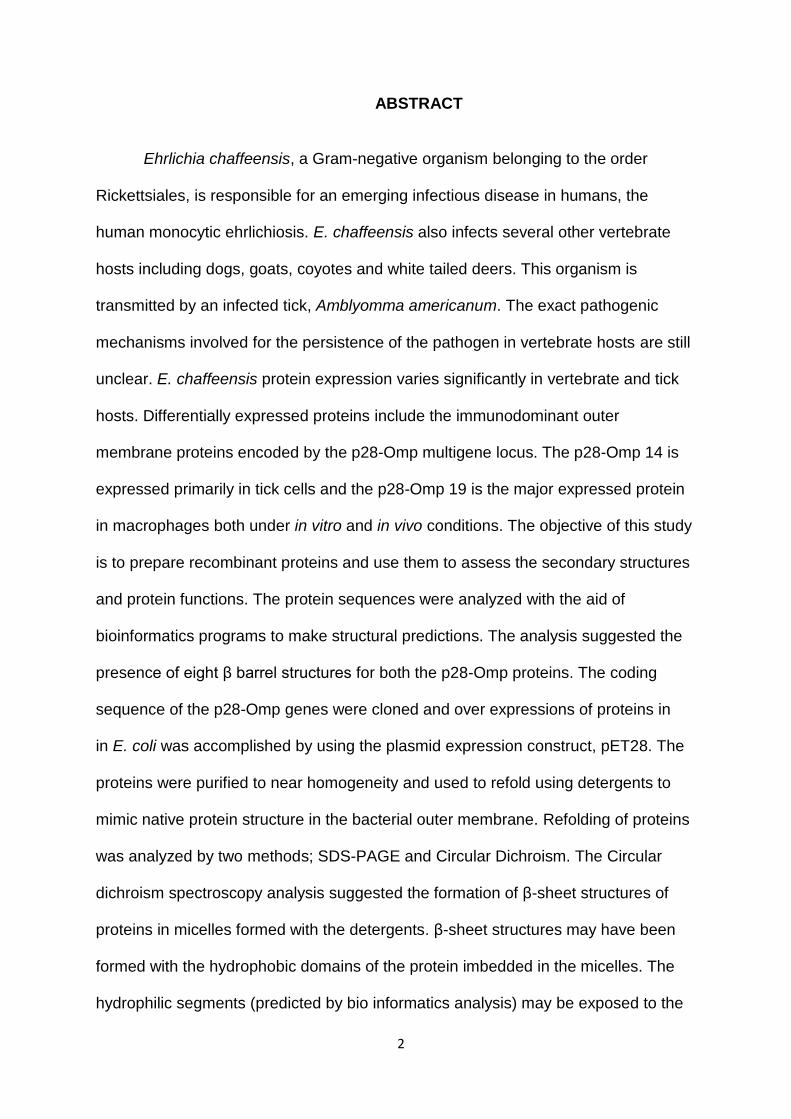
Ehrlichia chaffeensis, a Gram-negative organism belonging to the order
Rickettsiales, is responsible for an emerging infectious disease in humans, the
human monocytic ehrlichiosis. E. chaffeensis also infects several other vertebrate
hosts including dogs, goats, coyotes and white tailed deers. This organism is
transmitted by an infected tick, Amblyomma americanum. The exact pathogenic
mechanisms involved for the persistence of the pathogen in vertebrate hosts are still
unclear. E. chaffeensis protein expression varies significantly in vertebrate and tick
hosts. Differentially expressed proteins include the immunodominant outer
membrane proteins encoded by the p28-Omp multigene locus. The p28-Omp 14 is
expressed primarily in tick cells and the p28-Omp 19 is the major expressed protein
in macrophages both under in vitro and in vivo conditions. The objective of this study
is to prepare recombinant proteins and use them to assess the secondary structures
and protein functions. The protein sequences were analyzed with the aid of
bioinformatics programs to make structural predictions. The analysis suggested the
presence of eight β barrel structures for both the p28-Omp proteins. The coding
sequence of the p28-Omp genes were cloned and over expressions of proteins in
in E. coli was accomplished by using the plasmid expression construct, pET28. The
proteins were purified to near homogeneity and used to refold using detergents to
mimic native protein structure in the bacterial outer membrane. Refolding of proteins
was analyzed by two methods; SDS-PAGE and Circular Dichroism. The Circular
dichroism spectroscopy analysis suggested the formation of β-sheet structures of
proteins in micelles formed with the detergents. β-sheet structures may have been
iii
formed with the hydrophobic domains of the protein imbedded in the micelles. The
hydrophilic segments (predicted by bio informatics analysis) may be exposed to the
aqueous phase. The recombinant proteins were also used to prepare
proteoliposomes and tested for the porin activity. The analysis demonstrated the
porin activity for both p28-Omp 14 and 19 recombinant proteins by using mono-, di-
and tetra- saccharides as well as for amino acid L-glutamine. This study forms the
basis for initiating studies to compare the structural difference between the two
differentially expressed proteins of E. chaffeensis.

ACKNOWLEDGEMENTS
I would like to express my deepest appreciation to my major professor, Dr. Roman R.
Ganta, who supported me in my research with his patience and knowledge. His
assistance and guidance provides me with an excellent atmosphere for doing
research. I want to say special thanks to Dr. Roman R. Ganta for introducing MoU
between Andhra University and Kansas State University, which provided a great
platform for me to learn and explore the knowledge.
I would also like to show my sincere gratitude to all members of my advisory
committee: Dr. Om Prakash and Dr Dr. Kyeong-Ok Chang for their kind assistance
and extensive evaluation of my M.S program. They were very beneficial in providing
me with ideas to better my overall project. I truly appreciate the time and effort they
dedicated to helping me in the completion of my program.
I would like to thank Dr. Daisuke (NMR Lab) for his guidance and providing help for
completion of my research.
I would especially like to thank Chuanmin Cheng for her guidance as well as
friendship. A special thanks also goes to my lab members: Huitao Liu, Chakravarthy
Katragadda, Dr. Latitha Peddireddi and Bonto Faburay. To Dr. Bonto, I would like to
thank for your encouragement and support all the time.
I would also like to thank faculty members in Andhra University, Biochemistry
department for giving opportunity to join in the MoU program

v
Dedication
I dedicate this dissertation to my family members, teachers, and friends.
vi
TABLE OF CONTENTS
LIST OF FIGURES VII
LIST OF TABLES X
CHAPTER ONE LITERATURE REVIEW 1
CHAPTER TWO: STRUCTURAL ANALYSIS OF EHRLICHIA CHAFFEENSIS OUTER MEMBRANE PROTEINS, P28 OMP-14 AND P28 OMP-19 27
PROTOCOLS FOR MOLECULAR BIOLOGY TECHNIQUES 68
LIST OF REFERENCS 75
vii
LIST OF FIGURES
CHAPTER I Page Number: 21-24
Figure 1: Amblyomma americanum (Lone star tick) and its distribution
Figure 2: White-tailed deer.
Figure 3: Proposed life cycle of Ehrlichia chaffeensis.
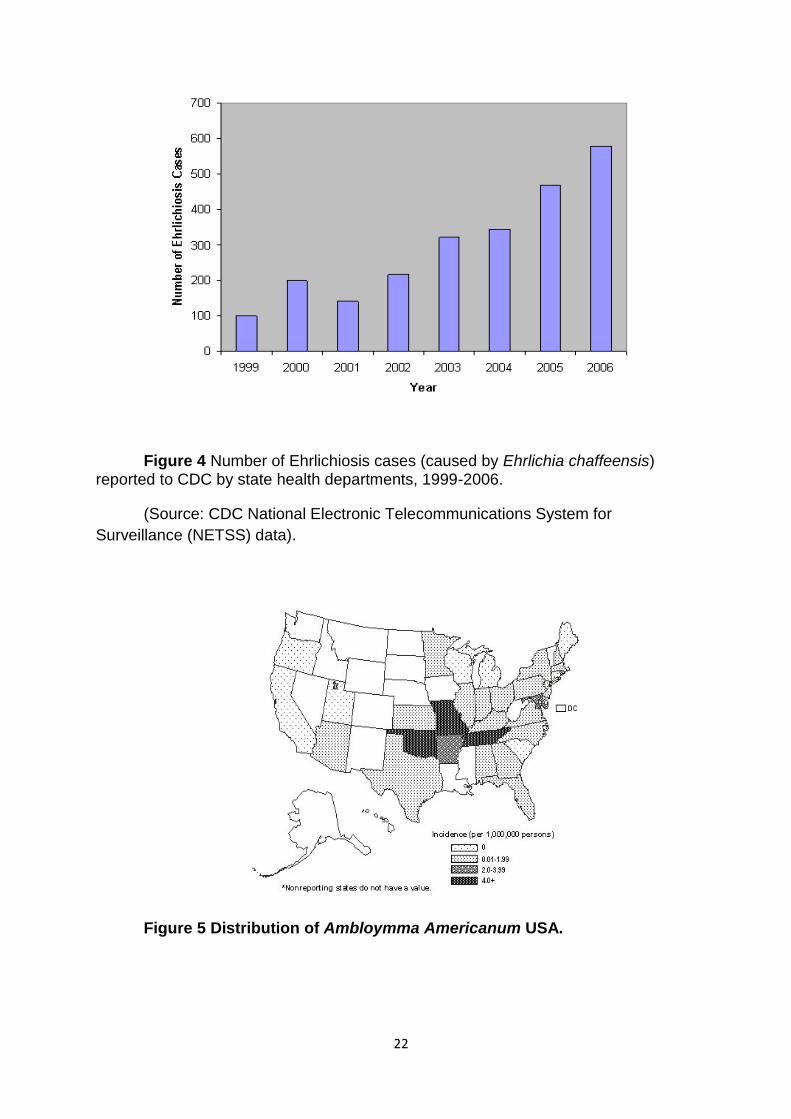
Figure 4: Number of Ehrlichiosis cases (caused by Ehrlichia chaffeensis) reported to
CDC by state health departments, 1999-2006.
Figure 5: Distribution of Amblyomma americanum USA.
Figure 6: Two morphological forms of E. chaffeensis.
Figure 7: A cartoon representing the p28-Omp loci.
Figure 8: Northern blot analysis of infected DH82 (lanes 1 and 2) and tick cell line.
CHAPTER II Page Number: 49-67
Figure 9 (A) (B): Calculating Hydrophobicity of p28-Omp14 and p28-Omp 19 by
PROTSCALE program (Kyte and Doolittle).
Figure 10 (A) (B): Predication of p28-Omp14 and p28-Omp 19in transmembrane
using PREDTMBB program
viii
Figure 11 (A) (B): Predicated 3D model of p28-Omp 14 and p28-Omp 19by using
CPH model 3.0
Figure 12: SDS PAGE analysis for recombinant expressed protein
Figure 13 (A) (B): Ion exchange Chromatography analysis by U.V.
spectrophotometer of recombinant gene 14 and 19protein expressed
Figure 14 (A) (B): Fractions from p28-Omp 14 and p28-Omp 19 ion exchange
chromatography anayalsed on 15% SDS-PAGE
Figure 15 (A) (B): Size exclusion chromatography analysis by U.V.
spectrophotometer of recombinant gene 14 and 19protein expressed
Figure 16: Protein fraction assessed from various purification steps
Figure 17: Various concentration of DHPC micelle was used p28-Omp19 refolding
Figure 18: Various concentration of DPC micelle treatment with p28-Omp19
Figure 19: p28- Omp 14 and 19 treated with DPC micelles and analyzed in 15%
SDS Page gel
Figure 20: p28- Omp 14 and 19 treated with DHPC micelles and analyzed in 15%
SDS Page gel
Figure 21 (A) (B): Circular dichroism work on p28 Omp14 with DPC and DHPC
Figure 22 (A) (B) (C): Proteoliposome assay p28-Omp 14
Figure 23(A) (B) (C): Proteoliposome assay p28-Omp 19

ix
Figure 24: Proteoliposome assay (p28-Omp 19) with four different concentration
of L-glutamine

x
LIST OF TABLES
Chapter I
Table 1: List of Anaplasmataceae diseases 25
Table 2: Rate of proteoliposome swelling assay 66

1
CHAPTER I: LITERATURE REVIEW
2
ABSTRACT
Ehrlichia chaffeensis, a Gram-negative organism belonging to the order
Rickettsiales, is responsible for an emerging infectious disease in humans, the
human monocytic ehrlichiosis. E. chaffeensis also infects several other vertebrate
hosts including dogs, goats, coyotes and white tailed deers. This organism is
transmitted by an infected tick, Amblyomma americanum. The exact pathogenic
mechanisms involved for the persistence of the pathogen in vertebrate hosts are still
unclear. E. chaffeensis protein expression varies significantly in vertebrate and tick
hosts. Differentially expressed proteins include the immunodominant outer
membrane proteins encoded by the p28-Omp multigene locus. The p28-Omp 14 is
expressed primarily in tick cells and the p28-Omp 19 is the major expressed protein
in macrophages both under in vitro and in vivo conditions. The objective of this study
is to prepare recombinant proteins and use them to assess the secondary structures
and protein functions. The protein sequences were analyzed with the aid of
bioinformatics programs to make structural predictions. The analysis suggested the
presence of eight β barrel structures for both the p28-Omp proteins. The coding
sequence of the p28-Omp genes were cloned and over expressions of proteins in
in E. coli was accomplished by using the plasmid expression construct, pET28. The
proteins were purified to near homogeneity and used to refold using detergents to
mimic native protein structure in the bacterial outer membrane. Refolding of proteins
was analyzed by two methods; SDS-PAGE and Circular Dichroism. The Circular
dichroism spectroscopy analysis suggested the formation of β-sheet structures of
proteins in micelles formed with the detergents. β-sheet structures may have been
formed with the hydrophobic domains of the protein imbedded in the micelles. The
hydrophilic segments (predicted by bio informatics analysis) may be exposed to the

3
aqueous phase. The recombinant proteins were also used to prepare
proteoliposomes and tested for the porin activity. The analysis demonstrated the
porin activity for both p28-Omp 14 and 19 recombinant proteins by using mono-, di-
and tetra- saccharides as well as for amino acid L-glutamine. This study forms the
basis for initiating studies to compare the structural difference between the two
differentially expressed proteins of E. chaffeensis.
4
Introduction
Vector borne diseases
Three major factors contribute to the spread of vector-borne diseases;
infectious microorganisms (eg: viruses, bacteria, parasites), vectors (eg:
mosquitoes, ticks, fleas) and reservoir hosts [1, 2]. Vectors-borne disease agents are
mostly transmitted by arthropod vectors. Vectors act as vehicles by which infectious
agents are transmitted from an infected host to another susceptible host [2]. Major
vector-borne infectious agents are maintained in nature by persisting in reservoir
hosts and arthropod vectors [3, 4]. Some of the important infectious diseases in
humans are the result of infections caused by vectors such as mosquitoes and ticks
[5, 6]. For example, malaria is caused by a mosquito-borne infection with
Plasmodium species. Human malaria is still considered the most important disease
because it is responsible for significant morbidity and mortality [7]. Nearly 350-400
million human cases of malaria are reported annually caused by Plasmodium
falciparum and Plasmodium vivax [8]. About half a million of these result in fatalities
[8-11]. Similarly, tick-borne diseases are a significant health concern to humans and
domestic animals [2, 3]. For example, the Lyme disease is a major problem for
humans and various vertebrate animals. It has a widespread distribution throughout
the world. It is discovered in 1975 as a human disease and is responsible for 1000s
of human cases in the USA each year [12, 13]. Louse-borne epidemic typhus,
caused by Rickettsia prowozakii, is another vector-borne disease responsible for
significant fatalities in people [14, 15]. Among populations concentrated at places
such as concentration camps during wars or civil disturbances, R. prowozakii

5
infections prevail and cause significant mortality (up to 30%). For example, deaths
resulting from this disease are often more in wars than war casualties. For example
in 1997 at refugee camps in Burundi, Africa, nearly 30,000 people died due to R.
prowozakii infections [6, 16].
Rocky Mountain spotted fever, African sleeping sickness, sandfly fever,
Chages disease, and louse borne typhus are among the vector borne disease
indentified in early 1900s [5]. From 1984 to 2004, nine new tick borne diseases
caused by rickettsial agents have been reported [17]. Human Ehrlichiosis and
Anaplasmosis are among the rickettsial diseases, discovered during the last three
decades [17-21]
Vector-borne diseases also have a high an impact on the economy of the
world due to the diseases caused to agriculture animals. Every year, millions of
dollars of economic losses occur in the world as a result of the vector-borne
diseases [22, 23]. For example, outbreak of babesiosis and anaplasmosis in the early
1990s in Latin America reported an estimated annual economic loss between 875 to
1,365 million dollars [24].
Tick borne diseases
Ticks are the second major vectors after mosquitoes for spreading infectious
diseases to animals and people [25-27]. Ticks are blood sucking parasitic arthropods
(obligate, hematophagous) and are found in every region of the world [5, 28]. Ancient
Greeks mentioned about ticks, but an actual demonstration of ticks as the pathogen
transmitting vectors is not reported until the 19th century where Smith and Kilbourne
6
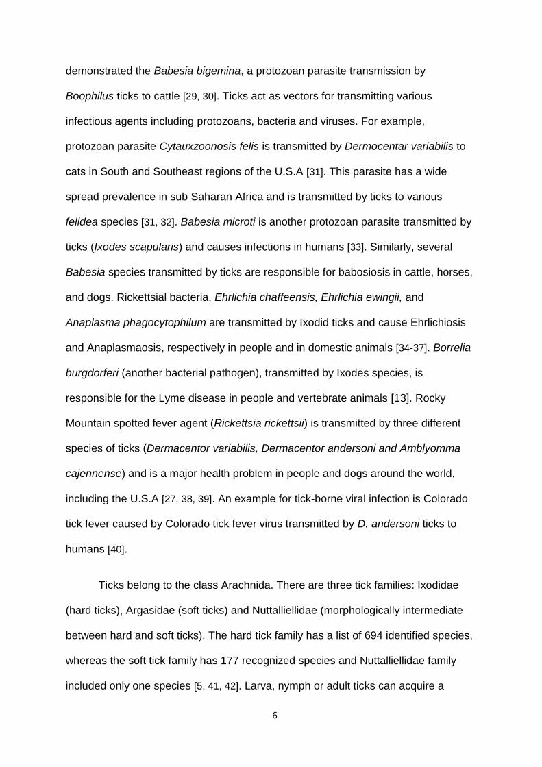
demonstrated the Babesia bigemina, a protozoan parasite transmission by
Boophilus ticks to cattle [29, 30]. Ticks act as vectors for transmitting various
infectious agents including protozoans, bacteria and viruses. For example,
protozoan parasite Cytauxzoonosis felis is transmitted by Dermocentar variabilis to
cats in South and Southeast regions of the U.S.A [31]. This parasite has a wide
spread prevalence in sub Saharan Africa and is transmitted by ticks to various
felidea species [31, 32]. Babesia microti is another protozoan parasite transmitted by
ticks (Ixodes scapularis) and causes infections in humans [33]. Similarly, several
Babesia species transmitted by ticks are responsible for babosiosis in cattle, horses,
and dogs. Rickettsial bacteria, Ehrlichia chaffeensis, Ehrlichia ewingii, and
Anaplasma phagocytophilum are transmitted by Ixodid ticks and cause Ehrlichiosis
and Anaplasmaosis, respectively in people and in domestic animals [34-37]. Borrelia
burgdorferi (another bacterial pathogen), transmitted by Ixodes species, is
responsible for the Lyme disease in people and vertebrate animals [13]. Rocky
Mountain spotted fever agent (Rickettsia rickettsii) is transmitted by three different
species of ticks (Dermacentor variabilis, Dermacentor andersoni and Amblyomma
cajennense) and is a major health problem in people and dogs around the world,
including the U.S.A [27, 38, 39]. An example for tick-borne viral infection is Colorado
tick fever caused by Colorado tick fever virus transmitted by D. andersoni ticks to
humans [40].
Ticks belong to the class Arachnida. There are three tick families: Ixodidae
(hard ticks), Argasidae (soft ticks) and Nuttalliellidae (morphologically intermediate
between hard and soft ticks). The hard tick family has a list of 694 identified species,
whereas the soft tick family has 177 recognized species and Nuttalliellidae family
included only one species [5, 41, 42]. Larva, nymph or adult ticks can acquire a
7
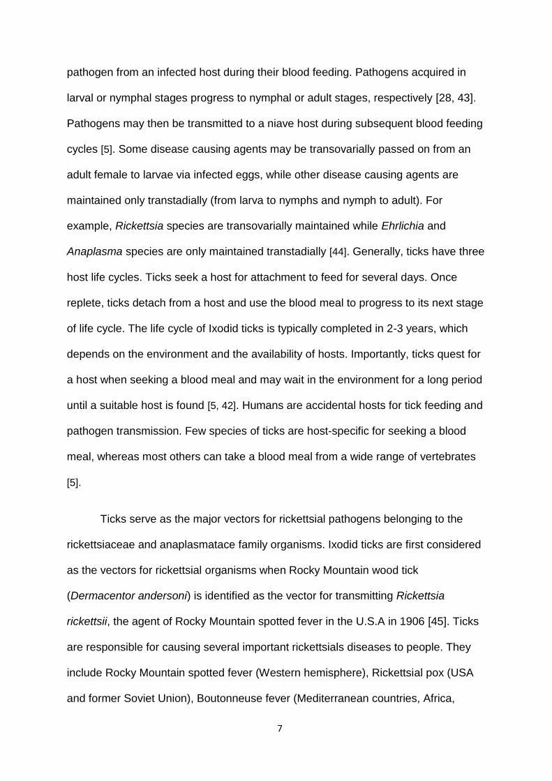
pathogen from an infected host during their blood feeding. Pathogens acquired in
larval or nymphal stages progress to nymphal or adult stages, respectively [28, 43].
Pathogens may then be transmitted to a niave host during subsequent blood feeding
cycles [5]. Some disease causing agents may be transovarially passed on from an
adult female to larvae via infected eggs, while other disease causing agents are
maintained only transtadially (from larva to nymphs and nymph to adult). For
example, Rickettsia species are transovarially maintained while Ehrlichia and
Anaplasma species are only maintained transtadially [44]. Generally, ticks have three
host life cycles. Ticks seek a host for attachment to feed for several days. Once
replete, ticks detach from a host and use the blood meal to progress to its next stage
of life cycle. The life cycle of Ixodid ticks is typically completed in 2-3 years, which
depends on the environment and the availability of hosts. Importantly, ticks quest for
a host when seeking a blood meal and may wait in the environment for a long period
until a suitable host is found [5, 42]. Humans are accidental hosts for tick feeding and
pathogen transmission. Few species of ticks are host-specific for seeking a blood
meal, whereas most others can take a blood meal from a wide range of vertebrates
[5].
Ticks serve as the major vectors for rickettsial pathogens belonging to the
rickettsiaceae and anaplasmatace family organisms. Ixodid ticks are first considered
as the vectors for rickettsial organisms when Rocky Mountain wood tick
(Dermacentor andersoni) is identified as the vector for transmitting Rickettsia
rickettsii, the agent of Rocky Mountain spotted fever in the U.S.A in 1906 [45]. Ticks
are responsible for causing several important rickettsials diseases to people. They
include Rocky Mountain spotted fever (Western hemisphere), Rickettsial pox (USA
and former Soviet Union), Boutonneuse fever (Mediterranean countries, Africa,
8
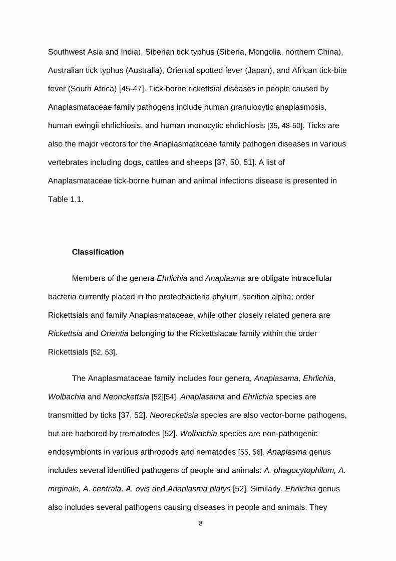
Southwest Asia and India), Siberian tick typhus (Siberia, Mongolia, northern China),
Australian tick typhus (Australia), Oriental spotted fever (Japan), and African tick-bite
fever (South Africa) [45-47]. Tick-borne rickettsial diseases in people caused by
Anaplasmataceae family pathogens include human granulocytic anaplasmosis,
human ewingii ehrlichiosis, and human monocytic ehrlichiosis [35, 48-50]. Ticks are
also the major vectors for the Anaplasmataceae family pathogen diseases in various
vertebrates including dogs, cattles and sheeps [37, 50, 51]. A list of
Anaplasmataceae tick-borne human and animal infections disease is presented in
Table 1.1.
Classification
Members of the genera Ehrlichia and Anaplasma are obligate intracellular
bacteria currently placed in the proteobacteria phylum, secition alpha; order
Rickettsials and family Anaplasmataceae, while other closely related genera are
Rickettsia and Orientia belonging to the Rickettsiacae family within the order
Rickettsials [52, 53].
The Anaplasmataceae family includes four genera, Anaplasama, Ehrlichia,
Wolbachia and Neorickettsia [52][54]. Anaplasama and Ehrlichia species are
transmitted by ticks [37, 52]. Neorecketisia species are also vector-borne pathogens,
but are harbored by trematodes [52]. Wolbachia species are non-pathogenic
endosymbionts in various arthropods and nematodes [55, 56]. Anaplasma genus
includes several identified pathogens of people and animals: A. phagocytophilum, A.
mrginale, A. centrala, A. ovis and Anaplasma platys [52]. Similarly, Ehrlichia genus
also includes several pathogens causing diseases in people and animals. They
9
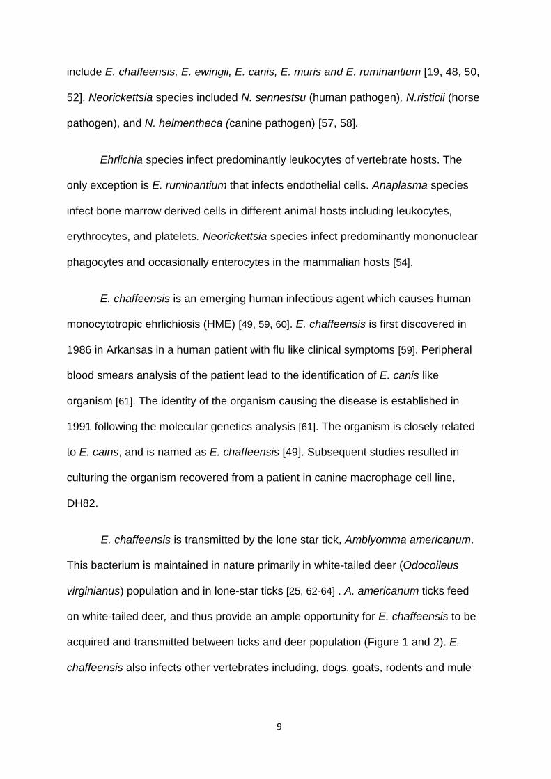
include E. chaffeensis, E. ewingii, E. canis, E. muris and E. ruminantium [19, 48, 50,
52]. Neorickettsia species included N. sennestsu (human pathogen), N.risticii (horse
pathogen), and N. helmentheca (canine pathogen) [57, 58].
Ehrlichia species infect predominantly leukocytes of vertebrate hosts. The
only exception is E. ruminantium that infects endothelial cells. Anaplasma species
infect bone marrow derived cells in different animal hosts including leukocytes,
erythrocytes, and platelets. Neorickettsia species infect predominantly mononuclear
phagocytes and occasionally enterocytes in the mammalian hosts [54].
E. chaffeensis is an emerging human infectious agent which causes human
monocytotropic ehrlichiosis (HME) [49, 59, 60]. E. chaffeensis is first discovered in
1986 in Arkansas in a human patient with flu like clinical symptoms [59]. Peripheral
blood smears analysis of the patient lead to the identification of E. canis like
organism [61]. The identity of the organism causing the disease is established in
1991 following the molecular genetics analysis [61]. The organism is closely related
to E. cains, and is named as E. chaffeensis [49]. Subsequent studies resulted in
culturing the organism recovered from a patient in canine macrophage cell line,
DH82.
E. chaffeensis is transmitted by the lone star tick, Amblyomma americanum.
This bacterium is maintained in nature primarily in white-tailed deer (Odocoileus
virginianus) population and in lone-star ticks [25, 62-64] . A. americanum ticks feed
on white-tailed deer, and thus provide an ample opportunity for E. chaffeensis to be
acquired and transmitted between ticks and deer population (Figure 1 and 2). E.
chaffeensis also infects other vertebrates including, dogs, goats, rodents and mule
10
deer [20, 65-68]. White tailed deer is considered the primary reservoir host for the
transmission cycle of this organism [64].
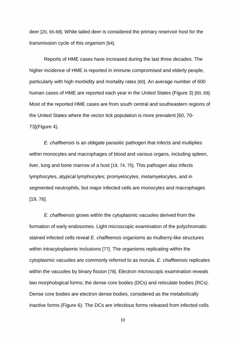
Reports of HME cases have increased during the last three decades. The
higher incidence of HME is reported in immune compromised and elderly people,
particularly with high morbidity and mortality rates [60]. An average number of 600
human cases of HME are reported each year in the United States (Figure 3) [60, 69].
Most of the reported HME cases are from south central and southeastern regions of
the United States where the vector tick population is more prevalent [60, 70-
73](Figure 4).
E. chaffeensis is an obligate parasitic pathogen that infects and multiplies
within monocytes and macrophages of blood and various organs, including spleen,
liver, lung and bone marrow of a host [19, 74, 75]. This pathogen also infects
lymphocytes, atypical lymphocytes; promyelocytes, metamyelocytes, and in
segmented neutrophils, but major infected cells are monocytes and macrophages
[19, 76].
E. chaffeensis grows within the cytoplasmic vacuoles derived from the
formation of early endosomes. Light microscopic examination of the polychromatic
stained infected cells reveal E. chaffeensis organisms as mulberry-like structures
within intracytoplasmic inclusions [77]. The organisms replicating within the
cytoplasmic vacuoles are commonly referred to as morula. E. chaffeensis replicates
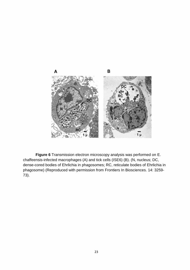
within the vacuoles by binary fission [78]. Electron microscopic examination reveals
two morphological forms; the dense core bodies (DCs) and reticulate bodies (RCs).
Dense core bodies are electron dense bodies, considered as the metabolically
inactive forms (Figure 6). The DCs are infectious forms released from infected cells
11
by lysis or by exocytosis. The RCs are metabolically active replicating forms mostly
seen within the phagosomes.
Clinical signs
The clinical signs of E. chaffeensis infection causing HME disease may
appear in patients within one to two weeks following blood feeding by an infected A.
americanum tick and the pathogen transmission [79]. The early clinical signs vary
from asymptomatic to mild flu-like symptoms [60, 79]. Most common symptoms
include malaise, headache, lower back pain, muscle aches, chills, nausea,
gastrointestinal symptoms, and development of sudden onset fever ~102˚F [80, 81].
Sometimes, the symptoms may include cough, pharyngitis, swollen lymph nodes,
and vomiting [73, 82]. Some patients also develop rashes (30-40%). The infection
may progress to severe illness with multiorgans failure. Greater than 50% patients
are reported to exhibit moderate leucopenia and a sharp decrease in the white blood
cell count [73, 82]. Nearly 90% of patients reported a decrease in the number of
platelets. Along with abnormalities in the blood, elevated levels of liver transaminase
are often noted because of infection [80, 81]. Approximately 20% of infected patients
develop signs and symptoms of central nervous system of meningitis syndrome [49].
Although people of all age groups are susceptible, but most severe cases are
observed in elderly age people with compromised immunity. The fatality of an
individual may occur due to hemorrhage, organ failure, or development of secondary
bacterial infections [49, 83].
Diagnosis for E. chaffeensis infection is more often based on patient’s history
(such as exposure to tick bites) [60], a blood smear analysis revealing the presence

12
of infected monocytes with other laboratory techniques, such as the indirect
immunofluroscence assay for E. chaffeensis antibodies and PCR amplification of a
specific genomic region of the pathogen. Studying blood smears is a common
diagnosing practice, but it is considered as insensitive in detecting the pathogen.
The pathogen is identified by staining blood smears by Romanovsky type
polychromatic stains [84]. Indirect immunoflorscence assay (IFA) is used for the
detection of antibodies against E. chaffeensis antigens, but due to cross reactivity
with other closely related organisms, the test can lead to false positives [85]. In
patients at early stages of infection, the low amount of antibodies may result in false
negatives. PCR assays are used for identifying DNA recovered from a whole blood
sample or a serum sample. The PCR assays are rapid, sensitive, and specific in
diagnosing a patient sample, but are not routinely used for human clinical diagnosis.
The targets used for PCR assays include GroESL gene, Variable Length PCR
Target gene (VLPT) or species-specific segment of a 16S rRNA gene [60, 61, 80, 86,
87].
Treatment
E. chaffeensis appears to be resistant to most of the antibiotics, including
ciprofloxacin and penicillin. Tetracycline and its derivatives, broad-spectrum
antimicrobials which inhibit protein synthesis in various bacterial species have
proven very effective in treating the infection. Doxycycline in particular is considered
as an effective drug of choice for the HME cases [19, 79].
13
Molecular biology:
The genome of E. chaffeensis is 1.18 mb and includes 1115 open reading
frames [88, 89]. The E. chaffeensis genome is considerably smaller than E. coli
genome. It is about one quarter in size when compared to E. coli genome. Many
genes in E. chaffeensis have been lost in the course of evolution, possibly for its
adaptation to obligate parasitic life [89]. They include genes required for the
biosynthesis of lipopolysaccharides and peptidoglycans [90, 91]. This organism is an
auxotroph and depends on the host for amino acids and other metabolites.
Several studies reported the molecular characterization of various genes in
this organism. They include various unnamed genes for p19, p22, p28, p32, p44,
p106, p120, p200 kDa protein coding genes and quinolate synthatase gene [91-94].
In addition, Type VI secretin system and two components regulator protein system
also have been partially characterized [95, 96].
Several immune reactive proteins of E. chaffeensis were identified from this
organism. They include, p28 kDa, p47 kDa, p120 kDa proteins are expressed on the
outer membrane of E. chaffeensis and appear to interact with the host cells [77, 92].
The p120 kDa and p47 kDa proteins are differentially expressed in dense core
bodies in infected monocytes, while the p28 proteins are shown to be expressed
differentially in the tick cells and macrophages [77, 85]. Several genes of E.
chaffeensis reported in literature contain tandem repeat sequence within the protein
coding sequences. They are p32 (previously known as variable length PCR tandem
repeat), p47, and p120 and p200. The tandem repeat sequences include amino
acids serine and threonine. These tandem repeat sequences of proteins are strongly
14
recognized by the immune sera of E. chaffeensis infected hosts, including humans
and dogs [85, 97, 98] .
The p47 is 285 amino acids longer, immunoreactive protein and includes 19
amino acids tandem repeats. Approximately half of the protein is represented by the
repeated sequences [98]. The p47 protein shows homology with renin
receptor/ATP6AP2/CAPER protein and with DNA III polymerase subunit gamma.
These proteins were expressed only on the surface of the dense core forms of E
chaffeensis [98, 99].
The p120 kDa protein is another immunoreactive differentially expressed
protein of this organism [97]. Its expression is higher in the dense core bodies. This
protein is also composed of multiple repeat sequences, which includes serine
residues. This protein may act as an adhesion and interacts with the host cell to
facilitate pathogen survival [87]. The immunogenic p140 kDa protein of E. canis is
homologues to p120 kDa protein of E chaffeensis [87, 97].
The p200-kDa protein, previously considered as glycoprotein, is also a
tandem repeat protein. The tandem repeats in this protein are 19 ankyrin sequences
[98]. This protein is similar to p200 and AnKA of E. canis and A. phagocytophilum,
respectively [100]. (Which also contain ankyrine repeats). These proteins are
translocated into the nuclei of infected host cells. They interact with the adenine –
rich motif of host gene promoters and intergenic Alu repeat sequence [98, 101]. The
p200-kDa protein may also play a role in inhibiting apoptosis of the infected cells.
The implications of these properties are that the bacterium may alter the host gene
expression in support of its survival [101].

15
The p28 proteins are the major expressed proteins on the outer membrane of
the organism [102-105]. The homologues of these proteins are identified in other
Ehrlichia species, Such as E. rumantium, E. muris, E. canis [103, 104]. These
proteins also share considerable homology to other surface proteins of A.
phagocytophilum and A. marginala. A. phagocytophilum and A. marginala proteins
have been shown to be involved in antigenic variation in support of the pathogen to
escape from host immunity [106-110]. The p28 proteins in E. chaffeensis are
differentially expressed in macrophages of vertebrate hosts and infected tick cells
under both in vitro and in vivo conditions [111-115]. The differentially expressed p28
proteins in macrophages and tick cells are p28-Omp14 and 19, respectively [112-
114].
Several other proteins are also expressed in the intercellular development of
the pathogen during its replication and maturation. They include type four secretin
system (T4SS) apparatus proteins and two-component regulator system proteins.
The T4SS is an ATP dependent bacterial transport system which transports
macromolecules (such as proteins and DNA) across the bacterial and host cell
membrane [95, 116]. The T4SS may be used by Gram negative bacteria to deliver
virulence factors to modulate the host genomes in support of their survival. The
T4SS proteins expression is observed in the organism present in infected host cells.
The T4SS proteins are constitute of a complex assembly of proteins. They include
proteins made from four virB2, one virB3, two virB4, four virB6, two virB8, two virB9,
one virB10, one virB11and one virD4 [95, 117].
Recent studies suggest that T4SS is used to deliver AnkA repeat proteins
from E. chaffeensis and A. phagocytophilum to infected host cells [95, 118, 119]. The

16
AnkA a protein of A. phagocytophilum has also been shown to be transported into
host cell nucleus and interact with promoters and various repeat sequences in the
genome. The significance of these interactions remains to be established [118-120].
However, like other Gram-negative bacteria, the proteins secreted by T4SS may be
important in altering the host gene expression in support of the pathogen for its
survival.
E. chaffeensis genome includes genes required for the expression of two
component regulatory system [121, 122]. Recent studies demonstrate that three
histidine kinase proteins and three response regulators are expressed by E.
chaffeesnsis when it replicates in human leucocytes [121]. Furthermore, inhibition of
the histidine kinase function by the drug closantel (a known inhibitor of histidine
kinase) resulted in the complete blocking of infection with this organism to host cells.
These results suggest that the two component regulatory system is needed for the
pathogen’s survival in vertebrate host cells [54, 116]. The two component regulatory
system is also functional in the A. pahagocytophilum [89].
E. chaffeensis obtains cholesterol or related sterols form the hosts or its
environment to stabilize the cytoplasm membrane as it lacks genes for the
synthesizing sterols. E. chaffeensis also lacks genes for synthesizing
lipopolysaccharides (LPS) [90, 123]. In the absence of LPS on the cell surface, the
organism is only protected with the membrane containing cholesterols [90, 124].
17
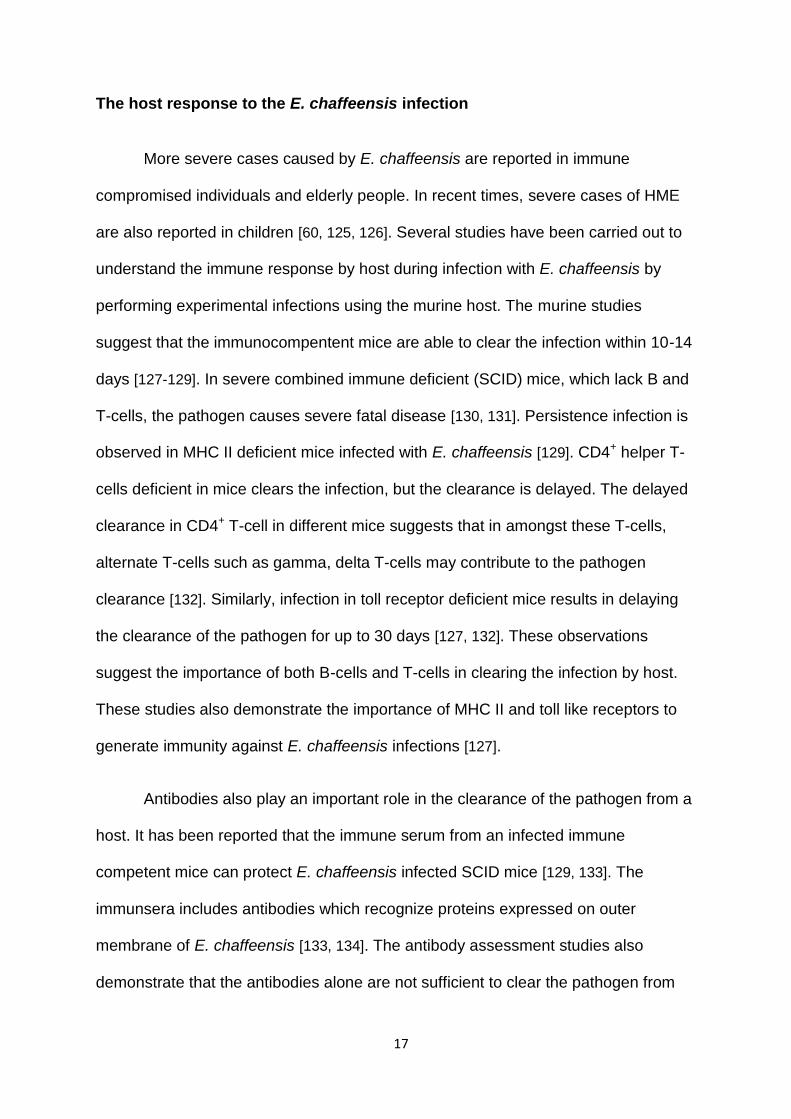
The host response to the E. chaffeensis infection
More severe cases caused by E. chaffeensis are reported in immune
compromised individuals and elderly people. In recent times, severe cases of HME
are also reported in children [60, 125, 126]. Several studies have been carried out to
understand the immune response by host during infection with E. chaffeensis by
performing experimental infections using the murine host. The murine studies
suggest that the immunocompentent mice are able to clear the infection within 10-14
days [127-129]. In severe combined immune deficient (SCID) mice, which lack B and
T-cells, the pathogen causes severe fatal disease [130, 131]. Persistence infection is
observed in MHC II deficient mice infected with E. chaffeensis [129]. CD4+ helper T-
cells deficient in mice clears the infection, but the clearance is delayed. The delayed
clearance in CD4+ T-cell in different mice suggests that in amongst these T-cells,
alternate T-cells such as gamma, delta T-cells may contribute to the pathogen
clearance [132]. Similarly, infection in toll receptor deficient mice results in delaying
the clearance of the pathogen for up to 30 days [127, 132]. These observations
suggest the importance of both B-cells and T-cells in clearing the infection by host.
These studies also demonstrate the importance of MHC II and toll like receptors to
generate immunity against E. chaffeensis infections [127].
Antibodies also play an important role in the clearance of the pathogen from a
host. It has been reported that the immune serum from an infected immune
competent mice can protect E. chaffeensis infected SCID mice [129, 133]. The
immunsera includes antibodies which recognize proteins expressed on outer
membrane of E. chaffeensis [133, 134]. The antibody assessment studies also
demonstrate that the antibodies alone are not sufficient to clear the pathogen from
18
the infected host. Cytokines are also involved in clearing of the pathogen [135].
Cytokines have an important role in mediating E. chaffeensis clearance from a
vertebrate host. Human monocytic cell line (THP I) infected with E. chaffeensis
suppresses the expression of several cytokines including IL-1α, IL-4, IL-6, IL-12, IL-
15, and IL-18 [135]. Cytokines IL 15 and IL18 usually serve as the activators of IFN
which activates the macrophage cells to kill a pathogen [54, 135, 136].
Recent proteomic studies from our laboratory identified 278 expressed
proteins of E. chaffeensis [137]. The expressed proteins included numerous
differentially expressed proteins of the organism grown in macrophages and tick
cells [138]. The differentially expressed proteins also included several outer
membrane proteins. Our laboratory also reported differences in the host response
against E. chaffeensis originating from tick cells and macrophages in the murine
host. These studies suggest that the pathogen clearance is delayed in vertebrate
host when E. chaffeensis originated from tick cells [132]. The antibody and cytokine
responses in mice infected with tick cell derived bacteria differed considerably
compared to mice infected with organisms originating from vertebrate macrophages.
The antibody response also steadily increased in mice infected with tick cell derived
bacteria [128, 139]. Together these data suggest that the host cell specific differential
protein expression by E. chaffeensis aids in the pathogens adaptation and
persistence in a vertebrate host. As differentially expressed proteins included p28-
Omp multigene locus proteins (described details below), the secondary structure
analysis (conducted in the current studies) will be important to assess the biological
significance of these proteins to the pathogen.
19
p28- Outer membrane proteins (p28-Omps)
p28-Omps of E. chaffeensis are encoded by a multigene locus (p28-Omp
locus). This multigene locus contains 22 tandemly arranged paralogos genes which
are separated by intergenic sequence varying from 9 to 600 bp [88, 111, 113, 115,
137, 139-141]. As the estimated molecular weight of the proteins encoded from p28
locus is between 28 to 32 kDa, the genes are referred to as p28-Omps. The p28-
Omp gene coding sequences share extensive homology, throughout the coding
sequences, except for three hypervariable regions [140]. The hypervariable regions
contain immunogenic B-cell epitopes and are recognized by sera from the E.
chaffeensis infected people and animals [134, 142]. The p28-Omp locus also differs
considerably in several E. chaffeensis isolates [115].
The homologs of p28-Omp locus are also found in other Ehrlichia species.
The p28-Omp locus (referred as p30-Omp locus) in E. canis also includes 22
tandemly arranged genes. In E. ruminantium, this locus is referred as MAP1 and it
includes 19 genes [143]. E. ewingii and E. muris has 16 and 21 paralogous genes,
respectively [104, 105]. (A cartoon representation of the p28-Omp multigene locus
including the host specifically expressed genes of E. chaffeensis, E. canis, E.
ruminantum is shown in Figure 7)
Several studies have been performed to map gene expression of the p28-
Omp loci in Ehrlichia species. Based on the transcriptional analysis, multiple genes
of the p28-Omp locus in E. chaffeensis are shown to be transcriptionally active in
macrophages with maximum expression observed for p28-Omp 19 [105, 141, 143,
144]. In tick cells, p28 Omp 14 gene product is the only one detected. The
differential expression is conformed in infected vertebrate hosts and ticks [104, 112,

20
145-147]. The differential expression is also reported for homologs of the p28-Omp
multigene loci E. canis and E. rumianatium. These differentially expressed proteins
of E. chaffeensis and E. canis are also post transnationally modified to include
glycosylation and phosphorylation moieties [138, 141]. The major surface protein
(msp2) and outer membrane protein (p44) of A. marginale and A. phagocytophilum,
respectively, are closely related to p28-Omp proteins [106-110]. The msp2 and p44
proteins are also made from multigene loci and include highly immunogenic variable
regions [52, 145]. The antigenic variants generated from msp 2 and p44 genes of A.
marginala and A. phagocytophilum are shown to play a role in immune evasion [51,
148-150].
Despite the detailed knowledge about the p28-Omp genes primary structure,
their expression patterns, their recognitions by B-cells, very little is known about their
secondary and tertiary structure in the outer membrane, similarly, their biological
function is not well described. In an effort to understand the structure and functional
relations, kumagai et al performed structure predication analysis and also examined
the porin activity for two p28-Omp 18 and 19 proteins. The studies also suggest that
two of the p28-Omp protein 18 and Omp 19 possess porin activity [151].
21
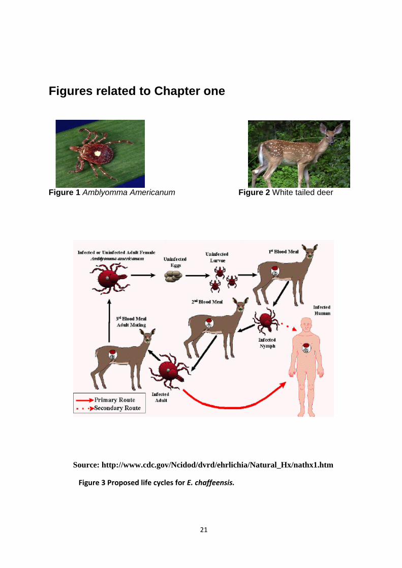
Figures related to Chapter one
Figure 1 Amblyomma Americanum Figure 2 White tailed deer
Source: http://www.cdc.gov/Ncidod/dvrd/ehrlichia/Natural_Hx/nathx1.htm
Figure 3 Proposed life cycles for E. chaffeensis.
22
Figure 4 Number of Ehrlichiosis cases (caused by Ehrlichia chaffeensis) reported to CDC by state health departments, 1999-2006.
(Source: CDC National Electronic Telecommunications System for
Surveillance (NETSS) data).
Figure 5 Distribution of Ambloymma Americanum USA.
23
Figure 6 Transmission electron microscopy analysis was performed on E.
chaffeensis-infected macrophages (A) and tick cells (ISE6) (B). (N, nucleus; DC,
dense-cored bodies of Ehrlichia in phagosomes; RC, reticulate bodies of Ehrlichia in
phagosome) (Reproduced with permission from Frontiers In Biosciences. 14: 3259-
73).

24
Figure 7 A cartoon representing the p28-Omp loci of E. chaffeensis, E. canis
and E. ruminantium with identified expressed proteins from the p28-Omp genes in
vertebrate macrophages (hatched boxes) and tick cells (checker board boxes) are
presented (Reproduced with permission from Frontiers In Biosciences. 14: 3259-73).
Figure 8 Northern blot analysis: DH82 (lanes 1 and 2) and tick cell line, ISE6
(lanes 3 and 4) assessed by Northern blot analysis using p28-Omp 14 or 19 gene-
specific 32P-labeled probes. Gene 19 transcript of the size 0.9 kb is detected only
in macrophage-derived RNA. Similarly, the tick cell-derived RNA contained 0.9 kb
transcript for gene

25

26
27
CHAPTER II:-
STRUCURAL PREDICTION ANALYSIS OF EHRLICHIA CHAFFEENSIS
OUTER MEMBRANE PROTEINS, P28 OMP-14 AND P28 OMP-19 ASSESSED BY
CIRCULAR DICHROSIM AND PORIM ASSAY
28
Introduction
Ehrlichia chaffeensis, obligate intercellular bacterium, causes human
monocytic ehrlichiosis (HME) [49, 61]. HME is considered as an emerging disease in
people. E. chaffeensis resides in phaogosomes of monocytes or macrophages of
vertebrate hosts [61]. This organism also infects several other vertebrates including
dog, goat, coyote, and white tailed deer [20, 65-68]. Amblyomma americanum tick
serves as the vector and white tailed deer serves as the reservoir host for this
pathogen [62-64, 152].
E. chaffeensis may have evolved unique protein expression strategies in
support of its growth in tick and vertebrate host environments. This organism
persists in both vertebrate and tick hosts. Recent studies demonstrated changes in
the protein expression of the organism replicating in macrophages and tick cells
[143]. Host cell-specific differential protein expression may be essential for E.
chaffeensis to adapt to vertebrate and tick hosts [112, 137, 138, 141]. Differentially
expressed proteins of E. chaffeensis include several outer membrane proteins. The
p28 Omp proteins are the most abundant outer membrane proteins expressed by E.
chaffeensis [112, 141, 143].
In tick cells and macrophages, the p28-Omp proteins are differentially
expressed. P28-Omp14 is the expressed protein in tick cells while p28-Omp19 is the
major expressed protein in macrophages [128, 138]. The differential expression of
these proteins is also confirmed for the pathogen in tick and vertebrate hosts [112,
137, 138, 141].

29
The p28 Omp proteins consist of three hyper variable regions are which
contain hydrophilic domains and are recognized by the immune sera against E.
chaffeensis [139, 140, 146]. Immunization with the p28-Omp recombinant proteins
protects SCID mice against of E. chaffeensis infections [134, 142].
Typically, the outer membrane proteins serve as the permeability barriers to
excrete noxious substances of bacteria and for the uptake of nutrients from the
environment [153]. The precise functions of the p28-Omps in E. chaffeensis remain
to be established.
In this study, we expressed the p28-Omp14 and p28-Omp 19 gene products
of E. chaffeensis using E. coli expression system. The recombinant proteins were
purified to near homogeneity and used to study the refolding in detergents to mimic
structures in the outer membrane of the organism. The protein function of p28-Omp
14 and 19 was assessed by evaluating the porin activity after reconstituting the
protein in a liposome.
Materials and method
In silco analysis:
We analyzed the protein sequences of p28-Omp 14 and 19 to predict the
hydrophobicity using the ProtoScale program available at
(http://expasy.org/tools/protscale.html). By calculating the average hydrophobicity
and average amphilicity index, can structural predications can be made for a protein.
The hydropathy plot is calculated with the help of index of Kyte and Doolittle and
amphiphilicity indexes [154, 155]. The amphilphilicity of polar side chains is the
30
second parameter with a calculated index of amino acid charges. The transfer
energy of the hydrocarbon part of a polar side chains are also used by the program
to prepare the hydrophobicity plot [154].
Analysis of the two dimensional structures of the p28-Omp 14 and 19 was done with
the help of the PRED-TMBB protein prediction program [155, 156]
(http://biophysics.biol.uoa.gr/PRED-TMBB/). CPHmodels-3.0 Program is helpful in the
predicting the 3D MODEL of the protein (http://www.cbs.dtu.dk/services/CPHmodels/)
[157]. This program is useful in predicting a 3D structure of a protein. The 3D model
predictions for p28 Omp-14 and 19 were performed by uploading the entire protein
coding sequence except the first 25 amino acids into the program. To generate a 3D
model, hydrophobic values and ampiphilic values obtained from ProtoScale program
analysis were used as per the instruction provided at the program website.
Cloning and expression p28 Omp14 and p28 Omp19 gene products:
The protein coding sequences of the p28 Omp14 and p28 Omp19 were
amplified from E. chaffeensis genomic DNA and cloned into pET 28 expression
plasmid vector (Novgen, Gibbstown,NJ). Nco I and Xho I sites of were engineered
into the PCR products to aid in the directional cloning into the pET 28 plasmid. The
coding sequences excluded the first 25 amino acids, as they were considered as the
signal sequences. Standard molecular cloning protocols were followed for preparing
the recombinant plasmid constructs. Briefly, the PCR products and pET 28 plasmid
were digested with Nco1 and Xho1 and the DNAs were purified by
phenol/chloroform and ethanol purification precipitation method. The PCR products
31
and the linerzed plasmids were ligated (5 to 1 ratio) using T4 DNA ligase. The
ligated products were then transformed into E. coli XL1blue and plated on agar plate
containing kanamycin. Colonies were randomly picked and grown in LB media
(Kanamycin resistance is conferred for the pET 28 plasmid). The presence of inserts
in a recombinant plasmid was verified after preparing plasmid DNA from 3 ml liquid
culture and the performing restriction enzyme digestion with Nco1 and Xho1.
Plasmids containing ~0.9 kb inserts were identified following resolving the digested
DNA on a 1% agarose gel. The integrity of sequence a recombinant plasmid was
confirmed by performing DNA sequence analysis of the plasmid DNAs. The
plasmids containing desired gene segments were retransformed into E. coli strain
BL21 (DE3). Transformed BL21 (DE3) strain cultures were assessed for the
presence of recombinant proteins. For this experiment, Culture was grown in 10 ml
LB medium with kanamycin (10µg/ml final concentration) and when the culture
reached to an optimum density of ~ 0.6 at 600, protein expression was induced with
1mM IPTG. The expression of the recombinant proteins was confirmed after
analyzing cell lysates prepared from the cultures The 10 ml liquid culture were
centrifuged for 5 min at 4000 rpm (Ependroff centrifugal 5810R). The supernatant
was discarded and the bacterial pellet, I ml of lysis buffer ( 50 mM Tris-HCl, 5 Mm
EDTA, 100 Mm NaCl, 0.5% Triton 100-X) containing lysozyme (2 µg/ml final
concentrations) was added. The culture was resuspended by vortexing several times
and subjected to sonication for 1 min using Sonic Dismembrator (Fisher scientific) at
setting of 9. The cell lysates were separated to soluble and insoluble fractions by
centrifugation at 4ºC for 5 min at 12,000 rpm (Ependroff centrifugal, 5810R). The
presence of expressed proteins was evaluated in both the fractions, equal volumes
of supernatant and 2x SDS gel loading buffer were mixed to prepare the protein
32
solution for analyzing on protein gel. To the insoluble fraction, 100µl of 1x SDS gel
loading buffer was added and vortexed several times to solubilize the proteins.
Twenty microliter each of the supernatant and insoluble fractions derived proteins
were used to resolve on a 15 % polyacrylamide gel containing 10% SDS. The
protocols to SDS for polyacrylamide gel electrophoresis were followed as described
in book of Sambrook et al. The electrophoresed gel was stained with coomasie blue
G250 staining solution and examined for the presence of proteins. The recombinant
expressed proteins were identified in the insoluble fractions of expression constructs
prepared for both p28-Omp14 and 19 genes. To verify the presence of specific
products, lysates prepared from a non-recombinant plasmid transformed into E. coli
BL21 (DE3) strain were used. Predicted 28 kDa proteins were observed in the
insoluble fraction desired proteins of the p28-Omp 14 and 19 gene.
Purification of recombinant proteins:
The E. coli strains containing pET 28 recombinant plasmids were grown at
37ºC in 10 ml of Laurie broth (LB) medium containing kanamycin (10µg/ml final
concentration) for about 14 hours. These cultures were transferred to 1 liter of LB
medium with kanamycin and incubated in a shaker incubator at 37ºC for expanding
the cultures. When the cultures grown to 0.5 optical density units (measured at 600
nm), protein expression was induced by adding 1 mM isopropyl beta D-
thiogalactopyranoside (IPTG). The cultures were continued to grow for six hours at
37ºC. The bacterial cultures were collected as fractions of 50 ml in Falcon tubes and
centrifuged at 4000 rpm for 10 min (Ependroff Centrifuge, 5810 R). The culture
pellets were pooled to four falcon tubes after resuspending in 25 ml of lysis buffer
[500 mM Tris HCl, 100 mM NaCl, 5 mM EDTA, 0.1 mM
33
phenylmethanesulfonylfluoride (PMSF) containing of 25 µl lysozyme (20 mg/ml)] and
then incubated at 37ºC for 30 min. The culture lysates were sonicated using Sonic
Dismembrator (Fisher scientific) for 5 min at setting 9. The lysate was centrifuged at
4000 rpm for 10 min to separate the insoluble and soluble fractions. To the pellet
containing the insoluble fraction were dissolved in 25 ml of lysis buffer and vortexed
to mix the insoluble fraction. The mixture was then centrifuged from 10 min at 4000
rpm as described above and the supernatant was discarded. This step was repeated
one more time to recover clean insoluble fractions that contained inclusion bodies
with recombinant proteins. The final recovered inclusion bodies were solubilized in
20 ml of denature buffer [20 mM Tris-HCl buffer (pH 8.5) containing 8 M urea] and
placed in a 65ºC water bath for 30 min to dissolve the pellet (votexing for every 10
min, If needed). After 30 min, the dissolved solution was centrifuged at 36,000 rpm
for 90 min (Beckman ultracentrifuge- optima max high capacity, SW 50.1) to remove
any non-soluble proteins or macromolecules. Clean supernatant containing
solubilized proteins was transformed to a sterile 50 ml Falcon tube and stored at -20
ºC for further use. The presence of recombinant protein in this fraction was
evaluated by subjecting 25 µl each of the fractions in a 15% SDS –polyacrylamide
gel and stained in coomassie blue staining. After verifying the presence of
recombinant proteins, the solutions were used to purify using ion exchange and size
exclusion chromatography methods to prepare purified recombinant proteins.
Ion exchange and size exclusion chromatography:
Anion exchange chromatography was used for the initial steps of purification
for p28-Omp14 and p28-Omp19 recombinant proteins. The size of the anion
exchange column used for purification is 2.5 cm x 10 cm (Bio-Rad, Hercules, CA).
34
The column was packed up to 8 cm height of the column with anion-exchange beads
(Q Sepharose Fast Flow, Amersham Pharmacia Biotech, Piscataway, NJ) and was
equilibrated with 100 ml of 20 mM Tris HCl (pH 8.5) containing 8 M urea (denature
buffer). Subsequently, the solution containing a recombinant protein was loaded on
to the column and washed with another 100 ml of denature buffer. Bound proteins
were eluted with a linear gradient of 0 to 1 M NaCl (200 ml volume) prepared in
denature buffer. Total 100 fractions (2 ml each) were collected. The presence of
protein in fractions was assessed at 280 nm by using U.V. spectrophotometer.
Based on the values obtained from the U.V spectrophotometer, graph was plotted
with fractions on X-axis and optical density values on Y-axis. Fractions containing
the major protein peak(s) were further analyzed to identify the presence of
recombinant proteins. Twenty-five microliter each of the fractions were resolved in a
15% SDS polyacrylamide gel and stained by following silver staining protocol. The
fractions having the desired protein (~28 kDa) at the highest concentration were
pooled (Figure: 2.10 for p28-Omp14 and Figure: 2.11 for p28-Omp19).
Size exclusion chromatography:
The ion exchange column purified pooled proteins representing ~ 28 kDa size
proteins were concentrated to 1 ml by using Amicon ultra centrifugal filters (10,000
Da capacity, Millipore). Proteins solutions were transferred to the filtration unit and
centrifuged at 4,500 rpm for 40 min in Ependroff Centrifuge, 5810 R. The
concentrated p28 protein solutions were subjected to size exclusion
chromatography. The size of the column used for this analysis is 1 cm x 170 cm
(Bio-rad). It was packed with Superdex beads (Superdex TM Prep grade, Amersham
Pharmacia Biotech, Piscataway, NJ) to fill the column bed up to 150 cm height
35
(approximately 200 ml of Superdex beads were used). The column was saturated
with 20 mM Tris HCl (pH 8.5) containing 8 M urea (denature buffer) by washing with
about 200 ml buffer. Subsequently, concentrated protein solution of p28-Omp 14 or
19 was loaded on to the column and eluted with 200 ml of denature buffer. Flow was
adjusted to 1 ml per 5 min. Total 200 fractions (1 ml per fractions) were collected
and analyzed using U.V spectrophotometer set at 280 nm to identify the presence of
a protein. Graph was plotted with optical density on X- axis and fractions collected
on Y-axis. Proteins fractions containing the highest absorption at 280 nm were
collected and the presence of p28 proteins was identified by performing PAGE in
presence of SDS (Outlined were mentioned above). The fractions containing 28 kDa
proteins were pooled and concentrated to 1 ml solution by using Amicon ultra
centrifugal filters as mentioned above. The protein concentration was estimated
using the kit for RD-DC method as per the manufacturing instructions (Bio-Rad,
Hercules, CA). The concentrated proteins were then stored at -20ºC until further use.
Refolding of recombinant protein by micelle formation:
The recombinant p28-Omp 14 or 19 proteins were concentrated further to 10
mg in 0.3 ml volume of Tris HCl buffer (pH8.5) containing 8 M urea. The proteins
were diluted into 20 ml of detergent solution. Typically, 20 µl each of the protein
solution was added to the detergent solution while vortexing for using a magnetic
stirrer at 4ºC. The slow addition of protein solution was completed in about 2.5
hours. Three different detergents were used for the refolding of proteins. They are
Dodecylphosphocholine (DPC), Dihexanoylphosphatidylcholine (DHPC) or β n-
Octyl-β-D-Glucopyranoside (β-OG). The detergent solutions were prepared in 20 ml
36
of 20 mM sodium borate buffer (pH 10) containg150 mM NaCl and 1 mM EDTA for
DPC (15 mM). The DHPC (24 mM) and β-OG (24 mM) detergent solutions were
prepared using 20 mM sodium phosphate buffer (pH 10) containing 150 mM NaCl
and 1 mM EDTA.
CD Spectrophotometer: The secondary structure of proteins was evaluated by
performing CD Spectroscopy scans at wavelengths from 190–250 nm as described
earlier [158]. CD spectroscopy analysis was performed for the detergent treated
proteins in which proteins were imbedded to form micelles in a buffer. CD
measurements were performed using a Jasco J-715 spectrometer (Jasco.co,
Oklahoma City, OK). The spectra were measured in a 200 µl capacity 1 mm
thickness cuvette with 1 cm path length using the protein solution imbedded in
micelles (approximately 0.4 mg of protein/ml of solution). The CD measurements
were expressed in terms of molar ellipticity. The CD data were assessed to identify
the presence of α helices and β sheet folding structures. The protein structure was
predicted, based on the negative minima observed in the wavelength scans. The
minima occur at 222 nm and 210 nm is related to the presence of α helices
structures. While bending is observed at the 218 nm is related to β sheet structure
[158-161].
Liposome assay (for determining the porin activity):
Porin activity of the recombinant proteins was determined after reconstituting
protein in detergents to form proteoliposomes as previously described with few
modification [162, 163]. Typically, mixture of 2.4 picomoles of acetone-washed egg
phosphatidylcholine (Sigma-Aldrich, St. Louis, MO) and 0.2 picomoles of
diacetylphosphate (Sigma-Aldrich, St. Louis, MO) were dried under a stream of
37
nitrogen gas at the bottom of a test tube. Then the lipid film was resuspended in 0.2
ml of 20 mM Tris-HCl buffer containing 2 µg of purified protein solubilized using β-
OG. The resuspension step was completed by vortexing for about a minute and then
by a brief sonication (1 to 2 min) in a Branson bath-type 1510 sonicator (Qsonica,
LLC, Newtown, CT) at setting of 2.5 for 5 min. The lipid-protein mixture was then
dried by using a vacuum desiccator ( Labconco, FreeZone Freeze Dry System,
Labconco Corporation Kansas City, Missouri ). To make proteoliposomes, the dried
protein-lipid film was resuspended in 0.3 ml of 10 mM Tris-HCl buffer (pH 7.5)
containing 15% dextran T-40 (Sigma- Aldrich, St Louis,MO). Resuspension of the
lipid film in the solution was carried out by occasional gentle rotation of the test tube,
followed by 30 min of occasional hand shaking of the tubes. Seventeen microliters of
proteoliposome suspensions mixed in 600 µl a solution containing solutes was used
for proteoliposome swelling assays. Proteoliposome swelling was measured by
recording the change in the optical density at 400 nm at various time intervals (0-500
sec). Proteoliposome swelling, an indication of solute uptake, was monitored by the
decrease in the optical density after proteoliposome and solutes were mixed. The
assays were performed for the following solutions at 33 mM; glucose (MW=180),
fructose (MW=180), sucrose (MW=342), stachyose (MW=666) and L-glutamine
(MW=146). Assays were also repeated four different L-glutamine concentrations (10,
20, 33, 40 mM).
38
Results
In silco analysis:
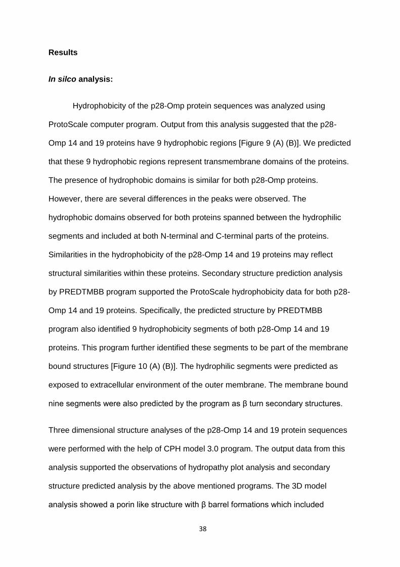
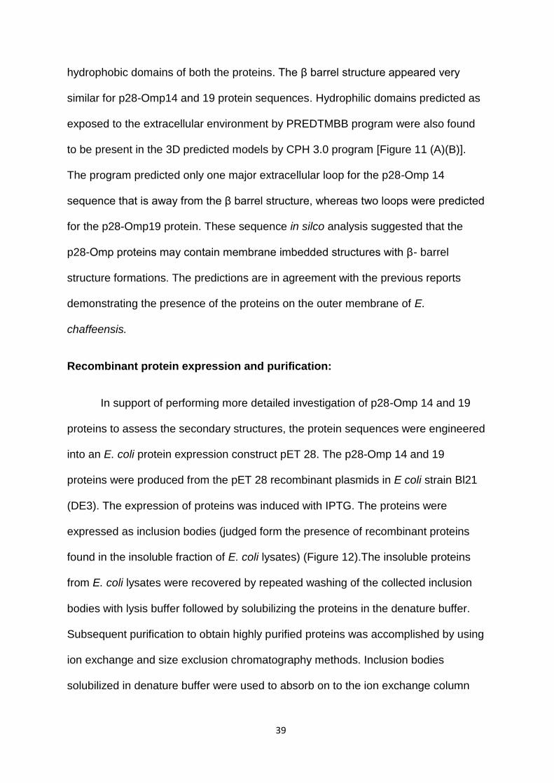
Hydrophobicity of the p28-Omp protein sequences was analyzed using
ProtoScale computer program. Output from this analysis suggested that the p28-
Omp 14 and 19 proteins have 9 hydrophobic regions [Figure 9 (A) (B)]. We predicted
that these 9 hydrophobic regions represent transmembrane domains of the proteins.
The presence of hydrophobic domains is similar for both p28-Omp proteins.
However, there are several differences in the peaks were observed. The
hydrophobic domains observed for both proteins spanned between the hydrophilic
segments and included at both N-terminal and C-terminal parts of the proteins.
Similarities in the hydrophobicity of the p28-Omp 14 and 19 proteins may reflect
structural similarities within these proteins. Secondary structure prediction analysis
by PREDTMBB program supported the ProtoScale hydrophobicity data for both p28-
Omp 14 and 19 proteins. Specifically, the predicted structure by PREDTMBB
program also identified 9 hydrophobicity segments of both p28-Omp 14 and 19
proteins. This program further identified these segments to be part of the membrane
bound structures [Figure 10 (A) (B)]. The hydrophilic segments were predicted as
exposed to extracellular environment of the outer membrane. The membrane bound
nine segments were also predicted by the program as β turn secondary structures.
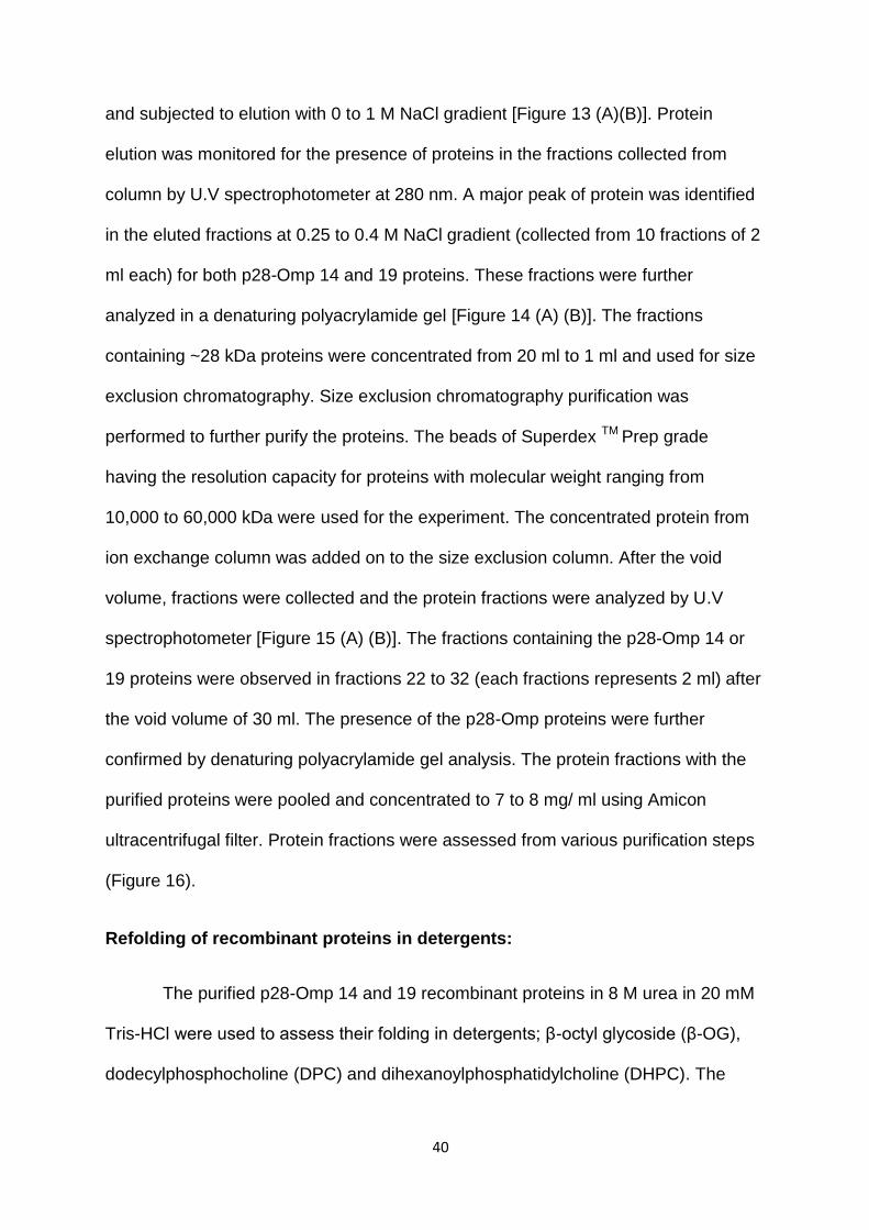
Three dimensional structure analyses of the p28-Omp 14 and 19 protein sequences
were performed with the help of CPH model 3.0 program. The output data from this
analysis supported the observations of hydropathy plot analysis and secondary
structure predicted analysis by the above mentioned programs. The 3D model
analysis showed a porin like structure with β barrel formations which included
39
hydrophobic domains of both the proteins. The β barrel structure appeared very
similar for p28-Omp14 and 19 protein sequences. Hydrophilic domains predicted as
exposed to the extracellular environment by PREDTMBB program were also found
to be present in the 3D predicted models by CPH 3.0 program [Figure 11 (A)(B)].
The program predicted only one major extracellular loop for the p28-Omp 14
sequence that is away from the β barrel structure, whereas two loops were predicted
for the p28-Omp19 protein. These sequence in silco analysis suggested that the
p28-Omp proteins may contain membrane imbedded structures with β- barrel
structure formations. The predictions are in agreement with the previous reports
demonstrating the presence of the proteins on the outer membrane of E.
chaffeensis.
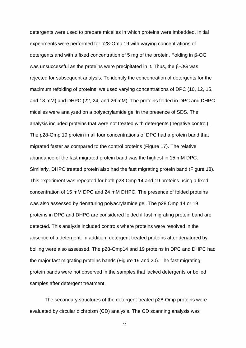
Recombinant protein expression and purification:
In support of performing more detailed investigation of p28-Omp 14 and 19
proteins to assess the secondary structures, the protein sequences were engineered
into an E. coli protein expression construct pET 28. The p28-Omp 14 and 19
proteins were produced from the pET 28 recombinant plasmids in E coli strain Bl21
(DE3). The expression of proteins was induced with IPTG. The proteins were
expressed as inclusion bodies (judged form the presence of recombinant proteins
found in the insoluble fraction of E. coli lysates) (Figure 12).The insoluble proteins
from E. coli lysates were recovered by repeated washing of the collected inclusion
bodies with lysis buffer followed by solubilizing the proteins in the denature buffer.
Subsequent purification to obtain highly purified proteins was accomplished by using
ion exchange and size exclusion chromatography methods. Inclusion bodies
solubilized in denature buffer were used to absorb on to the ion exchange column
40
and subjected to elution with 0 to 1 M NaCl gradient [Figure 13 (A)(B)]. Protein
elution was monitored for the presence of proteins in the fractions collected from
column by U.V spectrophotometer at 280 nm. A major peak of protein was identified
in the eluted fractions at 0.25 to 0.4 M NaCl gradient (collected from 10 fractions of 2
ml each) for both p28-Omp 14 and 19 proteins. These fractions were further
analyzed in a denaturing polyacrylamide gel [Figure 14 (A) (B)]. The fractions
containing ~28 kDa proteins were concentrated from 20 ml to 1 ml and used for size
exclusion chromatography. Size exclusion chromatography purification was
performed to further purify the proteins. The beads of Superdex TM Prep grade
having the resolution capacity for proteins with molecular weight ranging from
10,000 to 60,000 kDa were used for the experiment. The concentrated protein from
ion exchange column was added on to the size exclusion column. After the void
volume, fractions were collected and the protein fractions were analyzed by U.V
spectrophotometer [Figure 15 (A) (B)]. The fractions containing the p28-Omp 14 or
19 proteins were observed in fractions 22 to 32 (each fractions represents 2 ml) after
the void volume of 30 ml. The presence of the p28-Omp proteins were further
confirmed by denaturing polyacrylamide gel analysis. The protein fractions with the
purified proteins were pooled and concentrated to 7 to 8 mg/ ml using Amicon
ultracentrifugal filter. Protein fractions were assessed from various purification steps
(Figure 16).
Refolding of recombinant proteins in detergents:
The purified p28-Omp 14 and 19 recombinant proteins in 8 M urea in 20 mM
Tris-HCl were used to assess their folding in detergents; β-octyl glycoside (β-OG),
dodecylphosphocholine (DPC) and dihexanoylphosphatidylcholine (DHPC). The
41
detergents were used to prepare micelles in which proteins were imbedded. Initial
experiments were performed for p28-Omp 19 with varying concentrations of
detergents and with a fixed concentration of 5 mg of the protein. Folding in β-OG
was unsuccessful as the proteins were precipitated in it. Thus, the β-OG was
rejected for subsequent analysis. To identify the concentration of detergents for the
maximum refolding of proteins, we used varying concentrations of DPC (10, 12, 15,
and 18 mM) and DHPC (22, 24, and 26 mM). The proteins folded in DPC and DHPC
micelles were analyzed on a polyacrylamide gel in the presence of SDS. The
analysis included proteins that were not treated with detergents (negative control).
The p28-Omp 19 protein in all four concentrations of DPC had a protein band that
migrated faster as compared to the control proteins (Figure 17). The relative
abundance of the fast migrated protein band was the highest in 15 mM DPC.
Similarly, DHPC treated protein also had the fast migrating protein band (Figure 18).
This experiment was repeated for both p28-Omp 14 and 19 proteins using a fixed
concentration of 15 mM DPC and 24 mM DHPC. The presence of folded proteins
was also assessed by denaturing polyacrylamide gel. The p28 Omp 14 or 19
proteins in DPC and DHPC are considered folded if fast migrating protein band are
detected. This analysis included controls where proteins were resolved in the
absence of a detergent. In addition, detergent treated proteins after denatured by
boiling were also assessed. The p28-Omp14 and 19 proteins in DPC and DHPC had
the major fast migrating proteins bands (Figure 19 and 20). The fast migrating
protein bands were not observed in the samples that lacked detergents or boiled
samples after detergent treatment.
The secondary structures of the detergent treated p28-Omp proteins were
evaluated by circular dichroism (CD) analysis. The CD scanning analysis was
42
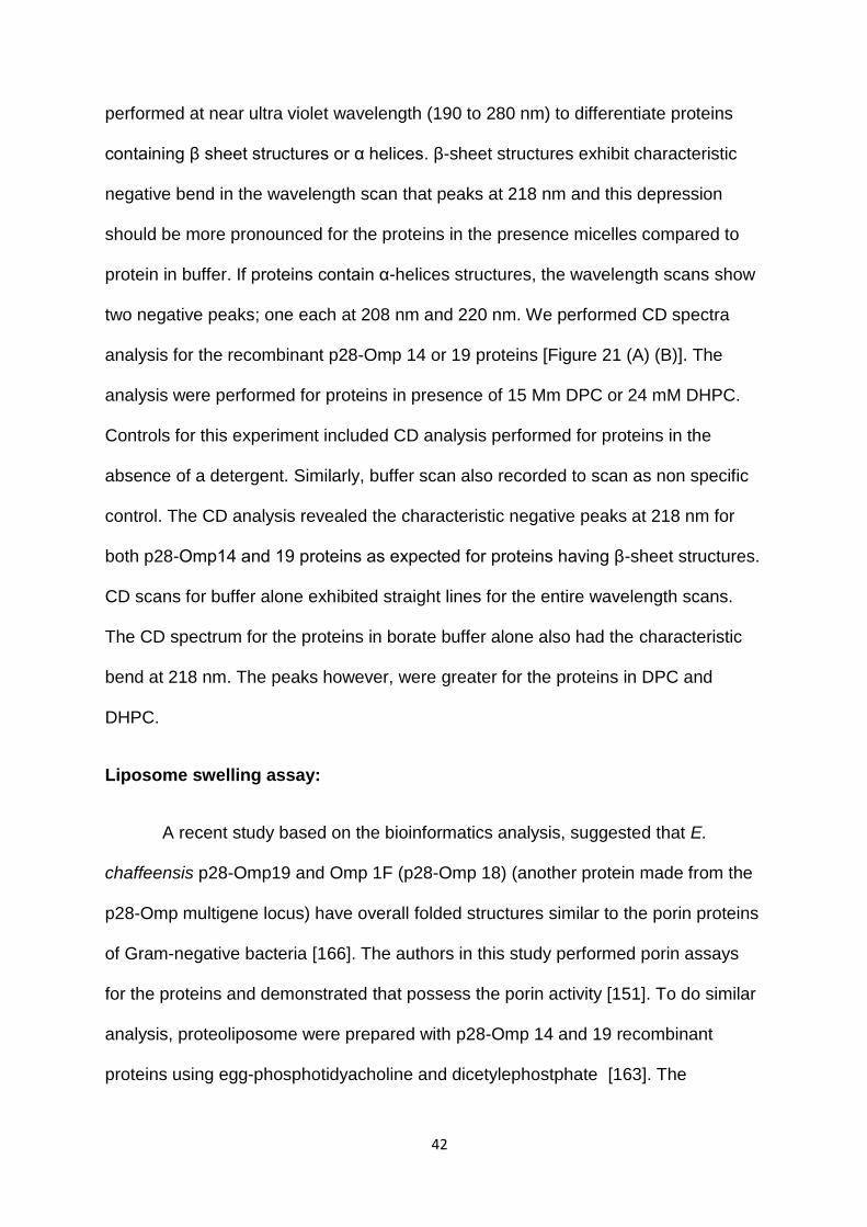
performed at near ultra violet wavelength (190 to 280 nm) to differentiate proteins
containing β sheet structures or α helices. β-sheet structures exhibit characteristic
negative bend in the wavelength scan that peaks at 218 nm and this depression
should be more pronounced for the proteins in the presence micelles compared to
protein in buffer. If proteins contain α-helices structures, the wavelength scans show
two negative peaks; one each at 208 nm and 220 nm. We performed CD spectra
analysis for the recombinant p28-Omp 14 or 19 proteins [Figure 21 (A) (B)]. The
analysis were performed for proteins in presence of 15 Mm DPC or 24 mM DHPC.
Controls for this experiment included CD analysis performed for proteins in the
absence of a detergent. Similarly, buffer scan also recorded to scan as non specific
control. The CD analysis revealed the characteristic negative peaks at 218 nm for
both p28-Omp14 and 19 proteins as expected for proteins having β-sheet structures.
CD scans for buffer alone exhibited straight lines for the entire wavelength scans.
The CD spectrum for the proteins in borate buffer alone also had the characteristic
bend at 218 nm. The peaks however, were greater for the proteins in DPC and
DHPC.
Liposome swelling assay:
A recent study based on the bioinformatics analysis, suggested that E.
chaffeensis p28-Omp19 and Omp 1F (p28-Omp 18) (another protein made from the
p28-Omp multigene locus) have overall folded structures similar to the porin proteins
of Gram-negative bacteria [166]. The authors in this study performed porin assays
for the proteins and demonstrated that possess the porin activity [151]. To do similar
analysis, proteoliposome were prepared with p28-Omp 14 and 19 recombinant
proteins using egg-phosphotidyacholine and dicetylephostphate [163]. The
43
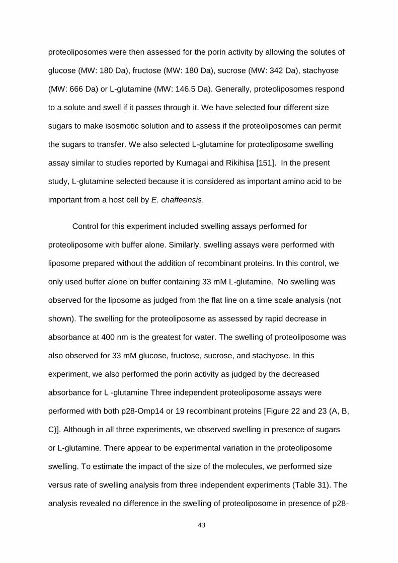
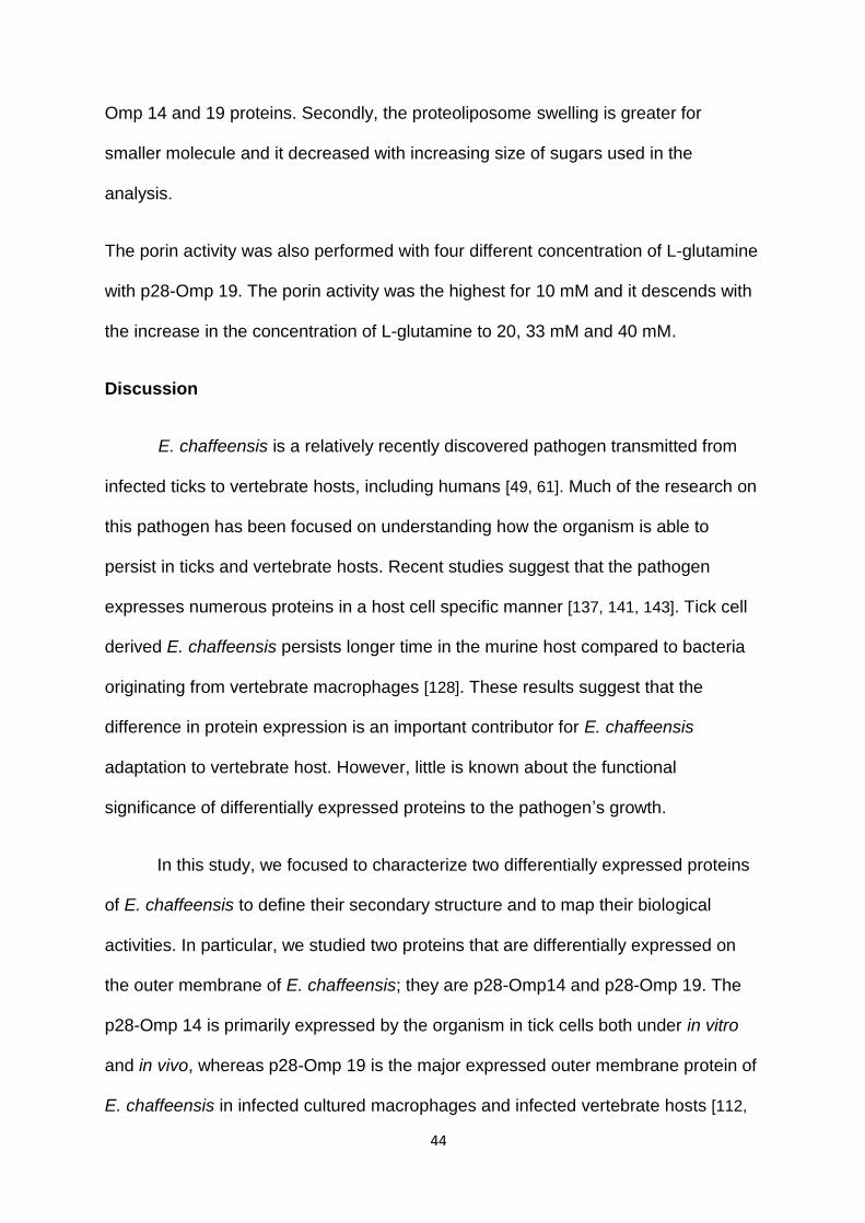
proteoliposomes were then assessed for the porin activity by allowing the solutes of
glucose (MW: 180 Da), fructose (MW: 180 Da), sucrose (MW: 342 Da), stachyose
(MW: 666 Da) or L-glutamine (MW: 146.5 Da). Generally, proteoliposomes respond
to a solute and swell if it passes through it. We have selected four different size
sugars to make isosmotic solution and to assess if the proteoliposomes can permit
the sugars to transfer. We also selected L-glutamine for proteoliposome swelling
assay similar to studies reported by Kumagai and Rikihisa [151]. In the present
study, L-glutamine selected because it is considered as important amino acid to be
important from a host cell by E. chaffeensis.
Control for this experiment included swelling assays performed for
proteoliposome with buffer alone. Similarly, swelling assays were performed with
liposome prepared without the addition of recombinant proteins. In this control, we
only used buffer alone on buffer containing 33 mM L-glutamine. No swelling was
observed for the liposome as judged from the flat line on a time scale analysis (not
shown). The swelling for the proteoliposome as assessed by rapid decrease in
absorbance at 400 nm is the greatest for water. The swelling of proteoliposome was
also observed for 33 mM glucose, fructose, sucrose, and stachyose. In this
experiment, we also performed the porin activity as judged by the decreased
absorbance for L -glutamine Three independent proteoliposome assays were
performed with both p28-Omp14 or 19 recombinant proteins [Figure 22 and 23 (A, B,
C)]. Although in all three experiments, we observed swelling in presence of sugars
or L-glutamine. There appear to be experimental variation in the proteoliposome
swelling. To estimate the impact of the size of the molecules, we performed size
versus rate of swelling analysis from three independent experiments (Table 31). The
analysis revealed no difference in the swelling of proteoliposome in presence of p28-
44
Omp 14 and 19 proteins. Secondly, the proteoliposome swelling is greater for
smaller molecule and it decreased with increasing size of sugars used in the
analysis.
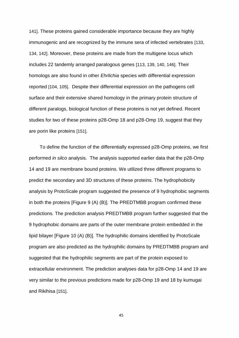
The porin activity was also performed with four different concentration of L-glutamine
with p28-Omp 19. The porin activity was the highest for 10 mM and it descends with
the increase in the concentration of L-glutamine to 20, 33 mM and 40 mM.
Discussion
E. chaffeensis is a relatively recently discovered pathogen transmitted from
infected ticks to vertebrate hosts, including humans [49, 61]. Much of the research on
this pathogen has been focused on understanding how the organism is able to
persist in ticks and vertebrate hosts. Recent studies suggest that the pathogen
expresses numerous proteins in a host cell specific manner [137, 141, 143]. Tick cell
derived E. chaffeensis persists longer time in the murine host compared to bacteria
originating from vertebrate macrophages [128]. These results suggest that the
difference in protein expression is an important contributor for E. chaffeensis
adaptation to vertebrate host. However, little is known about the functional
significance of differentially expressed proteins to the pathogen’s growth.
In this study, we focused to characterize two differentially expressed proteins
of E. chaffeensis to define their secondary structure and to map their biological
activities. In particular, we studied two proteins that are differentially expressed on
the outer membrane of E. chaffeensis; they are p28-Omp14 and p28-Omp 19. The
p28-Omp 14 is primarily expressed by the organism in tick cells both under in vitro
and in vivo, whereas p28-Omp 19 is the major expressed outer membrane protein of
E. chaffeensis in infected cultured macrophages and infected vertebrate hosts [112,
45
141]. These proteins gained considerable importance because they are highly
immunogenic and are recognized by the immune sera of infected vertebrates [133,
134, 142]. Moreover, these proteins are made from the multigene locus which
includes 22 tandemly arranged paralogous genes [113, 139, 140, 146]. Their
homologs are also found in other Ehrlichia species with differential expression
reported [104, 105]. Despite their differential expression on the pathogens cell
surface and their extensive shared homology in the primary protein structure of
different paralogs, biological function of these proteins is not yet defined. Recent
studies for two of these proteins p28-Omp 18 and p28-Omp 19, suggest that they
are porin like proteins [151].
To define the function of the differentially expressed p28-Omp proteins, we first
performed in silco analysis. The analysis supported earlier data that the p28-Omp
14 and 19 are membrane bound proteins. We utilized three different programs to
predict the secondary and 3D structures of these proteins. The hydrophobicity
analysis by ProtoScale program suggested the presence of 9 hydrophobic segments
in both the proteins [Figure 9 (A) (B)]. The PREDTMBB program confirmed these
predictions. The prediction analysis PREDTMBB program further suggested that the
9 hydrophobic domains are parts of the outer membrane protein embedded in the
lipid bilayer [Figure 10 (A) (B)]. The hydrophilic domains identified by ProtoScale
program are also predicted as the hydrophilic domains by PREDTMBB program and
suggested that the hydrophilic segments are part of the protein exposed to
extracellular environment. The prediction analyses data for p28-Omp 14 and 19 are
very similar to the previous predictions made for p28-Omp 19 and 18 by kumugai
and Rikihisa [151].
46
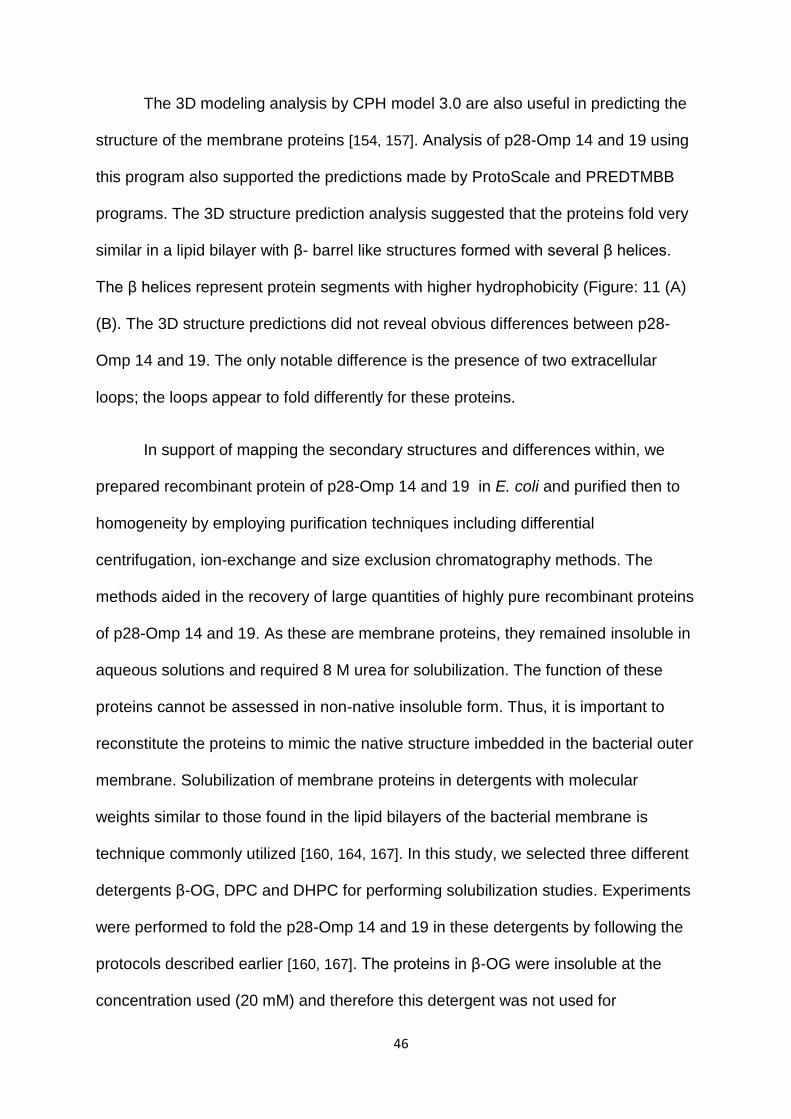
The 3D modeling analysis by CPH model 3.0 are also useful in predicting the
structure of the membrane proteins [154, 157]. Analysis of p28-Omp 14 and 19 using
this program also supported the predictions made by ProtoScale and PREDTMBB
programs. The 3D structure prediction analysis suggested that the proteins fold very
similar in a lipid bilayer with β- barrel like structures formed with several β helices.
The β helices represent protein segments with higher hydrophobicity (Figure: 11 (A)
(B). The 3D structure predictions did not reveal obvious differences between p28-
Omp 14 and 19. The only notable difference is the presence of two extracellular
loops; the loops appear to fold differently for these proteins.
In support of mapping the secondary structures and differences within, we
prepared recombinant protein of p28-Omp 14 and 19 in E. coli and purified then to
homogeneity by employing purification techniques including differential
centrifugation, ion-exchange and size exclusion chromatography methods. The
methods aided in the recovery of large quantities of highly pure recombinant proteins
of p28-Omp 14 and 19. As these are membrane proteins, they remained insoluble in
aqueous solutions and required 8 M urea for solubilization. The function of these
proteins cannot be assessed in non-native insoluble form. Thus, it is important to
reconstitute the proteins to mimic the native structure imbedded in the bacterial outer
membrane. Solubilization of membrane proteins in detergents with molecular
weights similar to those found in the lipid bilayers of the bacterial membrane is
technique commonly utilized [160, 164, 167]. In this study, we selected three different
detergents β-OG, DPC and DHPC for performing solubilization studies. Experiments
were performed to fold the p28-Omp 14 and 19 in these detergents by following the
protocols described earlier [160, 167]. The proteins in β-OG were insoluble at the
concentration used (20 mM) and therefore this detergent was not used for
47
subsequent experiments. Following the solubilization in DPC or DHPC, the presence
of folded proteins was assessed by SDS PAGE analysis. Folded proteins migrate
faster in the gel compared to non-folded proteins and therefore this method is useful
in identifying the presence and abundance of folded proteins. Folded proteins for
p28-Omp14 and 19 proteins are clearly visible in the polyacrylamide gel as judged
by rapid migration of protein bands compared to proteins in the absence of DPC and
DHPC. The folding is high at 15 mM DPC and 24 mM DHPC. These concentrations
of DPC and DHPC are considered critical concentration of detergents required for
forming micelles [168]. The micelles folded proteins were assessed further by
performing CD analysis to examine the nature of the secondary structures in the
folded proteins. The CD analysis suggested a characteristic bend at 218 nm, which
is an indication of the presence of rich β sheet structures that are typically present in
membrane bond proteins of Gram-negative bacteria [159, 169]. Both DPC and DHPC
treated proteins show a negative peak at 218 nm. These results indicating the
presence of large amounts of β sheet structures. Hydrophobic domains of a
membrane proteins form β sheet structures and get integrated into the lipid bilayers.
The hydrophobic domains of the proteins are imbedded within the membrane
surface of a lipid bilayer, thus, allowing membrane proteins to interact with their
hydrophobic environments. Previous studies on two p28-Omp proteins of E.
chaffeensis [p28-Omp 19 and p28-Omp 1F(Omp18)] suggest that the proteins form
porin like structure and possibly serve as porins to allow metabolite exchange
between the host and pathogen [151]. In this study, we assessed the proteoliposome
as a measure for porin activity swelling for both p28-Omp 14 and 19 recombinant
proteins after reconstituting the proteins to form in a proteoliposomes. The analysis
suggested that the p28-Omp 14 and 19 possess the porin activity. Proteoliposome
48
assays of p28-Omp 14 and 19 proteins had shown the permeability for L-glutamine
and for four sugars; glucose, fructose, sucrose, and stachyose. The swelling of
proteoliposome appeared to be dependent on the size of a molecule and the
concentration of solutes. Three independent proteoliposome assays suggested the
highest permeability for L- glutamine fallowed by fructose, glucose, sucrose, and
stachyose. The proteoliposome assay performed with p28-Omp 19 and L-glutamine
suggested that the porin activity is concentration dependent and increasing
concentration cause decrease in porin activity.
The availability of the recombinant proteins in pure form is valuable to further
the critical structural differences in these highly homogenous proteins and to define
the significance of differences in the primary structure to functional variations. The
functional differences may be understood by more structural analysis performed by
such as by performing X-ray crystallography and nuclear magnetic resonance
studies.
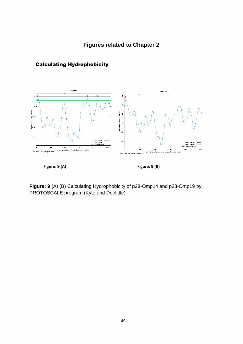
49
Figures related to Chapter 2
Figure: 9 (A) (B) Calculating Hydrophobicity of p28-Omp14 and p28-Omp19 by
PROTOSCALE program (Kyte and Doolittle)
Figure: 9 (A) Figure: 9 (B)
Calculating Hydrophobicity
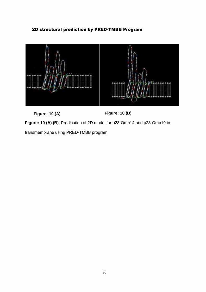
50
Figure: 10 (B)
Figure: 10 (A) (B): Predication of 2D model for p28-Omp14 and p28-Omp19 in
transmembrane using PRED-TMBB program
2D structural prediction by PRED-TMBB Program
Figure: 10 (A)
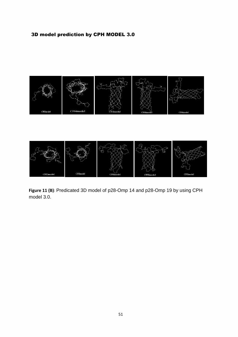
51
Figure 11 (B): Predicated 3D model of p28-Omp 14 and p28-Omp 19 by using CPH
model 3.0.
3D model prediction by CPH MODEL 3.0
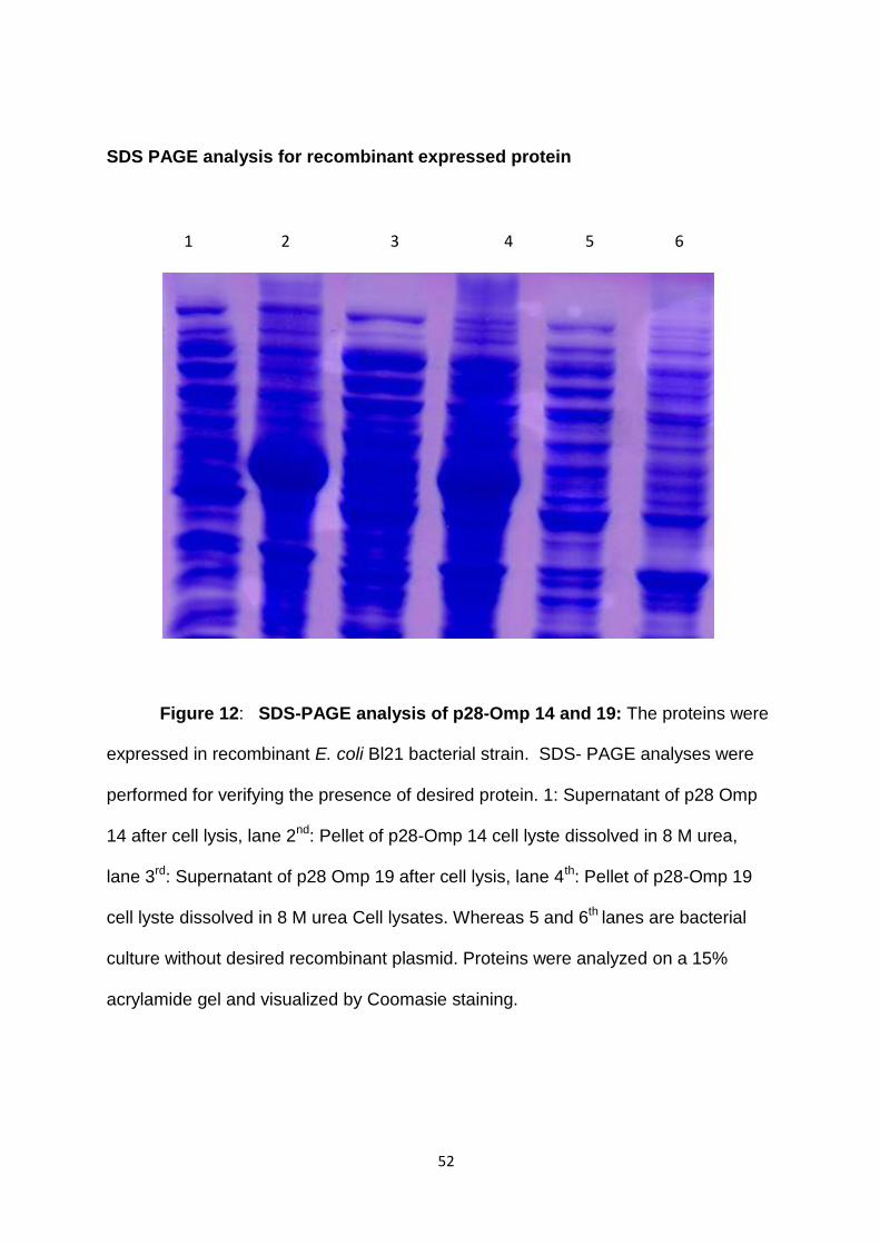
52
SDS PAGE analysis for recombinant expressed protein
1 2 3 4 5 6
Figure 12: SDS-PAGE analysis of p28-Omp 14 and 19: The proteins were
expressed in recombinant E. coli Bl21 bacterial strain. SDS- PAGE analyses were
performed for verifying the presence of desired protein. 1: Supernatant of p28 Omp
14 after cell lysis, lane 2nd: Pellet of p28-Omp 14 cell lyste dissolved in 8 M urea,
lane 3rd: Supernatant of p28 Omp 19 after cell lysis, lane 4th: Pellet of p28-Omp 19
cell lyste dissolved in 8 M urea Cell lysates. Whereas 5 and 6th lanes are bacterial
culture without desired recombinant plasmid. Proteins were analyzed on a 15%
acrylamide gel and visualized by Coomasie staining.
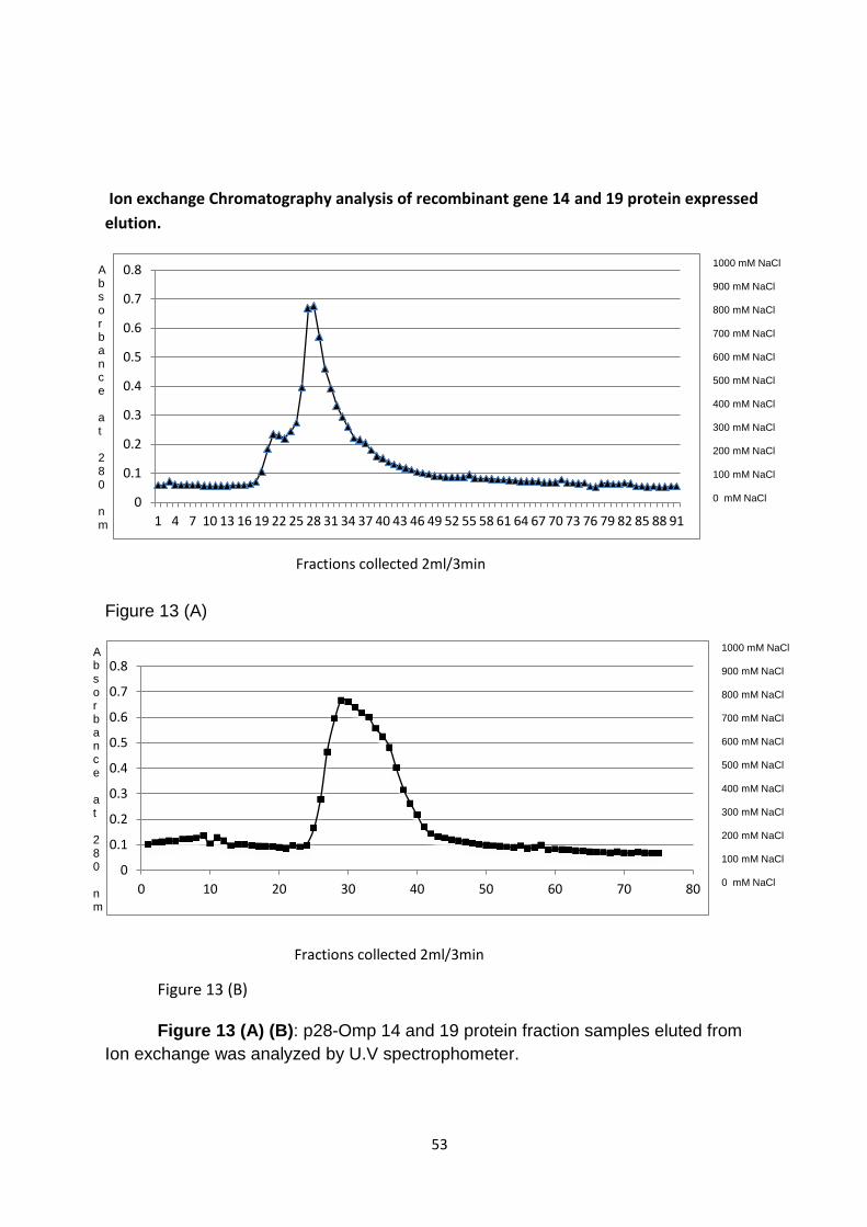
53
Ion exchange Chromatography analysis of recombinant gene 14 and 19 protein expressed
elution.
Figure 13 (A)
Figure 13 (B)
Figure 13 (A) (B): p28-Omp 14 and 19 protein fraction samples eluted from
Ion exchange was analyzed by U.V spectrophometer.
0
0.1
0.2
0.3
0.4
0.5
0.6
0.7
0.8
1 4 7 10 13 16 19 22 25 28 31 34 37 40 43 46 49 52 55 58 61 64 67 70 73 76 79 82 85 88 91
0
0.1
0.2
0.3
0.4
0.5
0.6
0.7
0.8
0 10 20 30 40 50 60 70 80
1000 mM NaCl 900 mM NaCl 800 mM NaCl 700 mM NaCl 600 mM NaCl 500 mM NaCl 400 mM NaCl 300 mM NaCl 200 mM NaCl 100 mM NaCl 0 mM NaCl
Fractions collected 2ml/3min
Absorbance at 280 nm
Fractions collected 2ml/3min
1000 mM NaCl 900 mM NaCl 800 mM NaCl 700 mM NaCl 600 mM NaCl 500 mM NaCl 400 mM NaCl 300 mM NaCl 200 mM NaCl 100 mM NaCl 0 mM NaCl
Absorbance at 280 nm
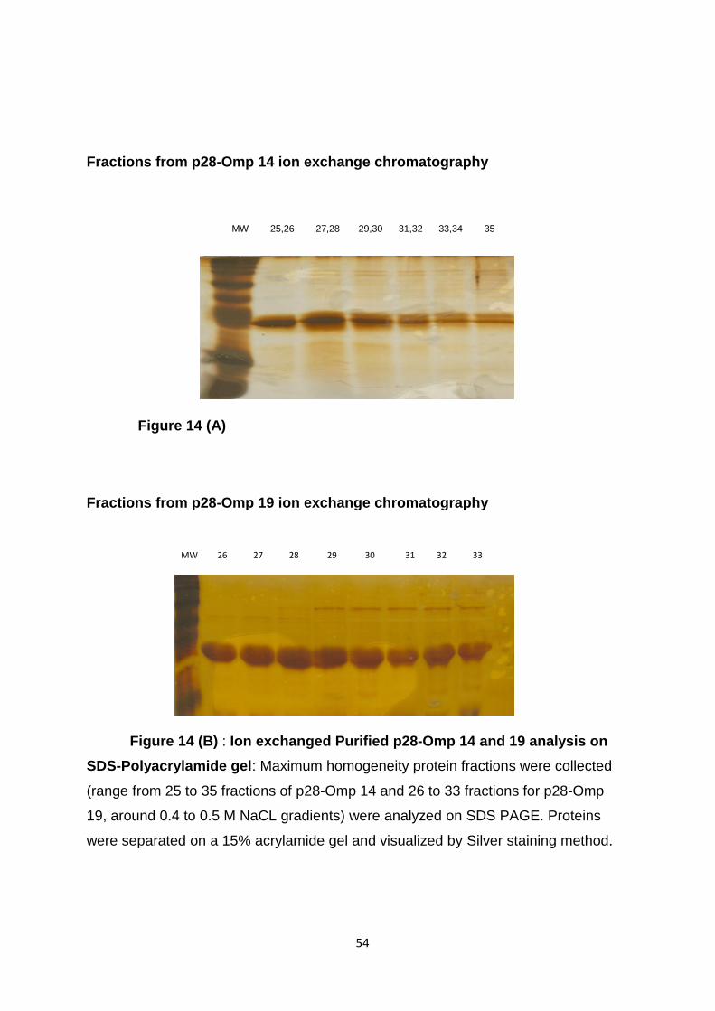
54
Fractions from p28-Omp 14 ion exchange chromatography
Figure 14 (A)
Fractions from p28-Omp 19 ion exchange chromatography
Figure 14 (B) : Ion exchanged Purified p28-Omp 14 and 19 analysis on
SDS-Polyacrylamide gel: Maximum homogeneity protein fractions were collected
(range from 25 to 35 fractions of p28-Omp 14 and 26 to 33 fractions for p28-Omp
19, around 0.4 to 0.5 M NaCL gradients) were analyzed on SDS PAGE. Proteins
were separated on a 15% acrylamide gel and visualized by Silver staining method.
MW 26 27 28 29 30 31 32 33
MW 25,26 27,28 29,30 31,32 33,34 35
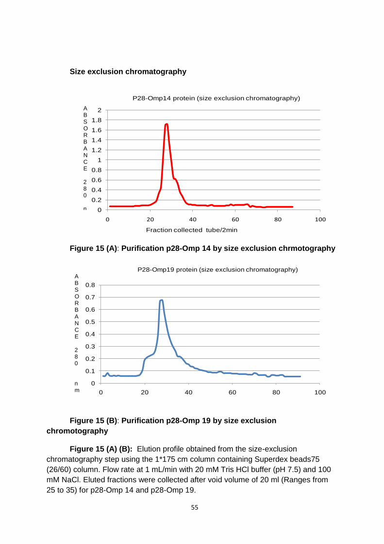
55
Size exclusion chromatography
Figure 15 (A): Purification p28-Omp 14 by size exclusion chrmotography
Figure 15 (B): Purification p28-Omp 19 by size exclusion
chromotography
Figure 15 (A) (B): Elution profile obtained from the size-exclusion
chromatography step using the 1*175 cm column containing Superdex beads75
(26/60) column. Flow rate at 1 mL/min with 20 mM Tris HCl buffer (pH 7.5) and 100
mM NaCl. Eluted fractions were collected after void volume of 20 ml (Ranges from
25 to 35) for p28-Omp 14 and p28-Omp 19.
0
0.1
0.2
0.3
0.4
0.5
0.6
0.7
0.8
0 20 40 60 80 100
P28-Omp19 protein (size exclusion chromatography)
Ab
sorb
ace
28
0 n
m
0
0.2
0.4
0.6
0.8
1
1.2
1.4
1.6
1.8
2
0 20 40 60 80 100
Fraction collected tube/2min
P28-Omp14 protein (size exclusion chromatography)
A B S O R B A N C E 2 8 0 nm
A B S O R B A N C E 2 8 0 nm
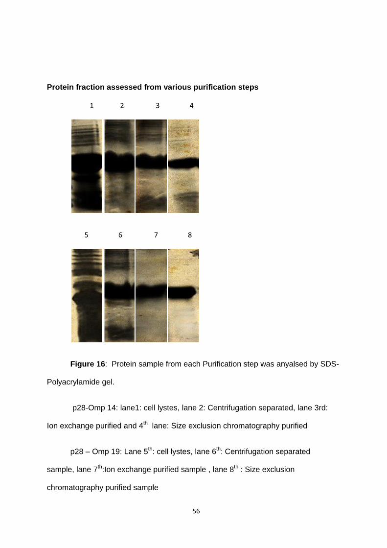
56
Protein fraction assessed from various purification steps
1 2 3 4
5 6 7 8
Figure 16: Protein sample from each Purification step was anyalsed by SDS-
Polyacrylamide gel.
p28-Omp 14: lane1: cell lystes, lane 2: Centrifugation separated, lane 3rd:
Ion exchange purified and 4th lane: Size exclusion chromatography purified
p28 – Omp 19: Lane 5th: cell lystes, lane 6th: Centrifugation separated
sample, lane 7th:Ion exchange purified sample , lane 8th : Size exclusion
chromatography purified sample
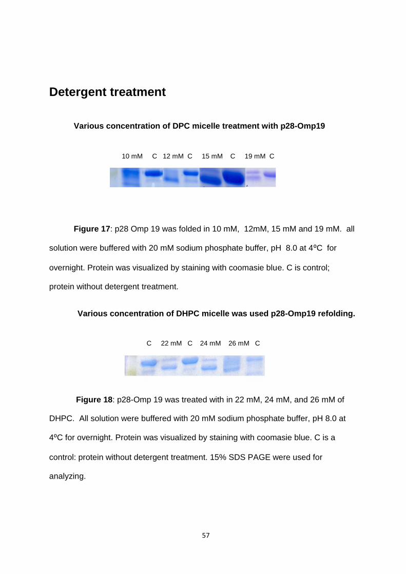
57
Detergent treatment
Various concentration of DPC micelle treatment with p28-Omp19
Figure 17: p28 Omp 19 was folded in 10 mM, 12mM, 15 mM and 19 mM. all
solution were buffered with 20 mM sodium phosphate buffer, pH 8.0 at 4⁰C for
overnight. Protein was visualized by staining with coomasie blue. C is control;
protein without detergent treatment.
Various concentration of DHPC micelle was used p28-Omp19 refolding.
Figure 18: p28-Omp 19 was treated with in 22 mM, 24 mM, and 26 mM of
DHPC. All solution were buffered with 20 mM sodium phosphate buffer, pH 8.0 at
4⁰C for overnight. Protein was visualized by staining with coomasie blue. C is a
control: protein without detergent treatment. 15% SDS PAGE were used for
analyzing.
C 22 mM C 24 mM 26 mM C
10 mM C 12 mM C 15 mM C 19 mM C
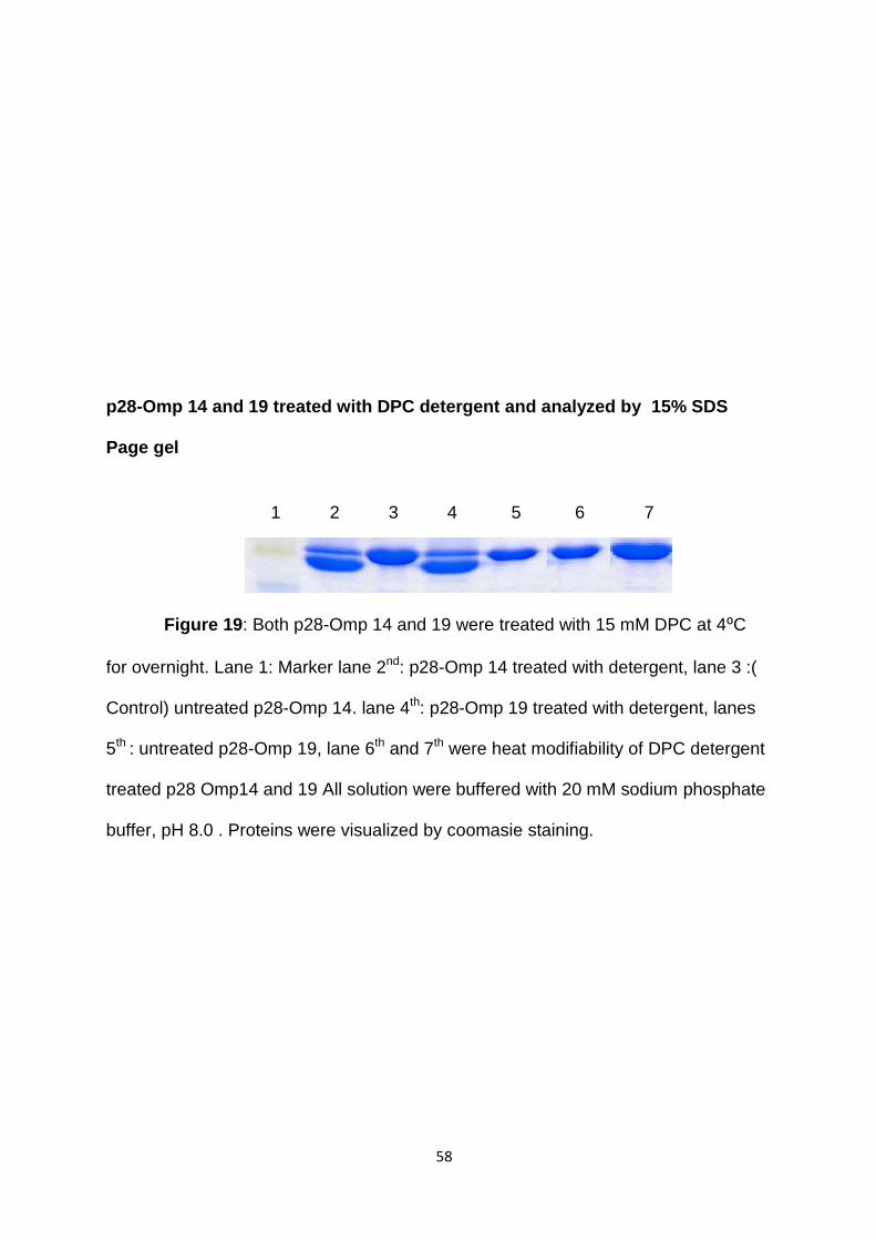
58
p28-Omp 14 and 19 treated with DPC detergent and analyzed by 15% SDS
Page gel
Figure 19: Both p28-Omp 14 and 19 were treated with 15 mM DPC at 4⁰C
for overnight. Lane 1: Marker lane 2nd: p28-Omp 14 treated with detergent, lane 3 :(
Control) untreated p28-Omp 14. lane 4th: p28-Omp 19 treated with detergent, lanes
5th : untreated p28-Omp 19, lane 6th and 7th were heat modifiability of DPC detergent
treated p28 Omp14 and 19 All solution were buffered with 20 mM sodium phosphate
buffer, pH 8.0 . Proteins were visualized by coomasie staining.
1 2 3 4 5 6 7

59
p28-Omp 14 and 19 treated with DHPC detergent and analyzed by 15% SDS
Page gel
Figure 20: Analysis of DHPC detergent folded protein: Both p28-Omp 14
and 19 were treated with 24 mM DHPC at 4⁰C for overnight. Lane 1: Marker lane
2nd: p28-Omp 14 treated with detergent, lane 3rd: Heat modifiability of DHPC
detergent treated p28-Omp 14, lane 4th: p28-Omp 19 treated with detergent, lanes
5th : Heat modifiability of DHPC detergent treated p28-Omp 19, lane 6th and 7th were
untreated p28 Omp14 and 19 proteins. All solution were buffered with 20 mM
sodium borate buffer, pH 8.0 . Proteins were visualized by coomasie staining.
1 2 3 4 5 6 7
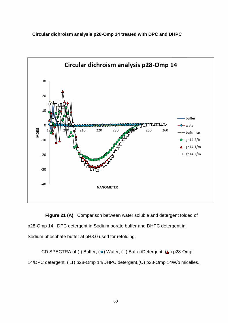
60
Figure 21 (A): Comparison between water soluble and detergent folded of
p28-Omp 14. DPC detergent in Sodium borate buffer and DHPC detergent in
Sodium phosphate buffer at pH8.0 used for refolding.
CD SPECTRA of (-) Buffer, ( ) Water, (--) Buffer/Detergent, ( ) p28-Omp
14/DPC detergent, ( ) p28-Omp 14/DHPC detergent,(O) p28-Omp 14W/o micelles.
-40
-30
-20
-10
0
10
20
30
190 200 210 220 230 240 250 260
MD
EG
NANOMETER
Circular dichroism analysis p28-Omp 14
buffer
water
buf/mice
gn14.2/b
gn14.1/m
gn14.2/m
Circular dichroism analysis p28-Omp 14 treated with DPC and DHPC
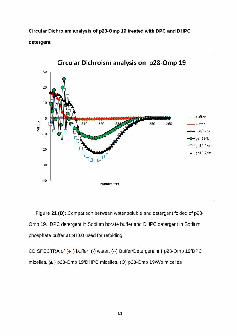
61
Circular Dichroism analysis of p28-Omp 19 treated with DPC and DHPC
detergent
Figure 21 (B): Comparison between water soluble and detergent folded of p28-
Omp 19. DPC detergent in Sodium borate buffer and DHPC detergent in Sodium
phosphate buffer at pH8.0 used for refolding.
CD SPECTRA of ( ) buffer, (-) water, (--) Buffer/Detergent, ( ) p28-Omp 19/DPC
micelles, ( ) p28-Omp 19/DHPC micelles, (O) p28-Omp 19W/o micelles
-40
-30
-20
-10
0
10
20
30
190 200 210 220 230 240 250 260
MD
EG
Nanometer
Circular Dichroism analysis on p28-Omp 19
buffer
water
buf/mice
gen19/b
gn19.1/m
gn19.2/m
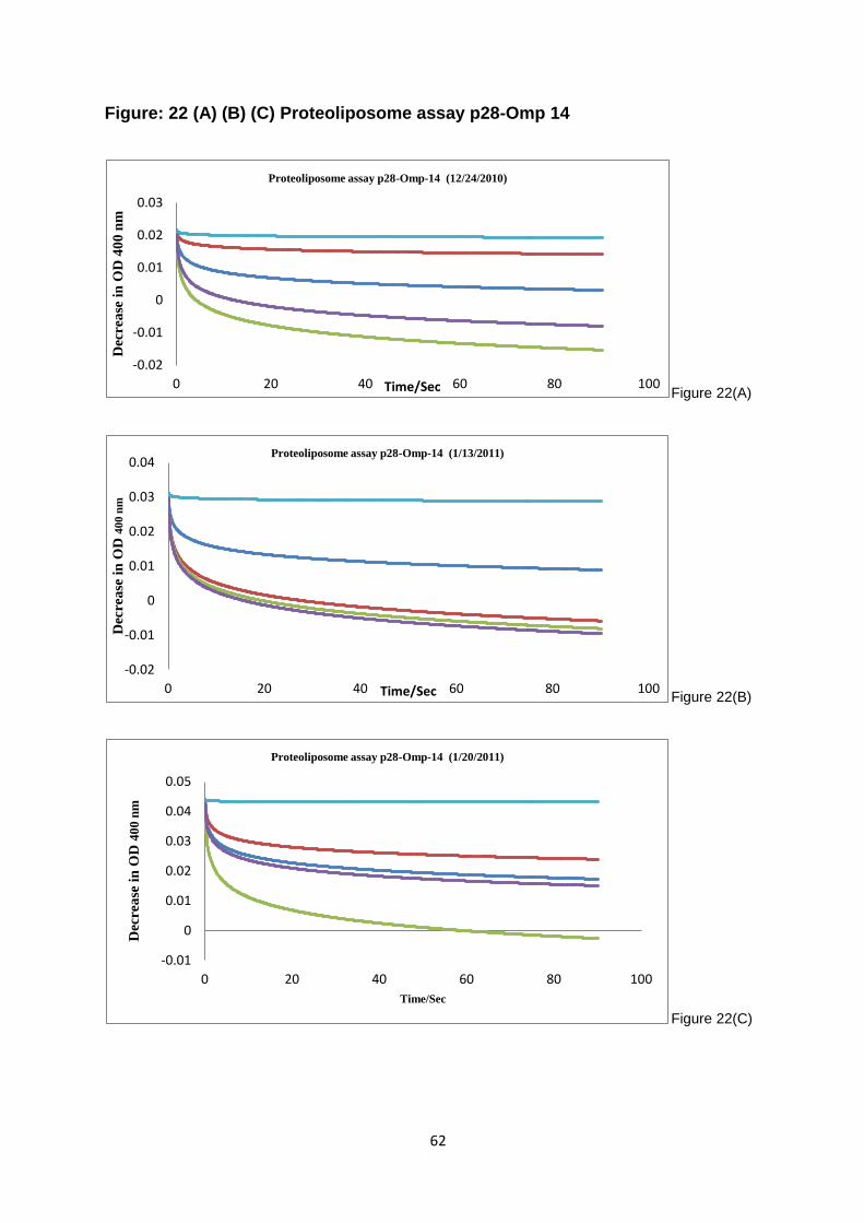
62
Figure: 22 (A) (B) (C) Proteoliposome assay p28-Omp 14
Figure 22(A)
Figure 22(B)
Figure 22(C)
-0.02
-0.01
0
0.01
0.02
0.03
0 20 40 60 80 100
Dec
rea
se i
n O
D 4
00
nm
Time/Sec
Proteoliposome assay p28-Omp-14 (12/24/2010)
-0.02
-0.01
0
0.01
0.02
0.03
0.04
0 20 40 60 80 100
Dec
rea
se i
n O
D 4
00
nm
Time/Sec
Proteoliposome assay p28-Omp-14 (1/13/2011)
-0.01
0
0.01
0.02
0.03
0.04
0.05
0 20 40 60 80 100
Dec
rea
se i
n O
D 4
00
nm
Time/Sec
Proteoliposome assay p28-Omp-14 (1/20/2011)
63
Figure 22 (A) (B) (C): Porin activity of the E. chaffeensis outer membrane
p28-Omp 14(recombinant). The diffusion rates of solutes into proteoliposome
reconstituted with E. chaffeensis outer membrane proteins were monitored as the
decrease in OD400. When proteoliposome were diluted in isosmotic solution of 33
mM Glucose (-) (MW=180), 33 mM sucrose (-) (MW=342), 33 mM fructose (-)
(MW=180), 33 mM, L- glutamine (-) (MW=156) and 33Mm stachyose (-) (MW=666).
(Three independent experiments are shown performed on (A) 12/24/2010, (B)
1/13/2011, (C) 1/20/2011).
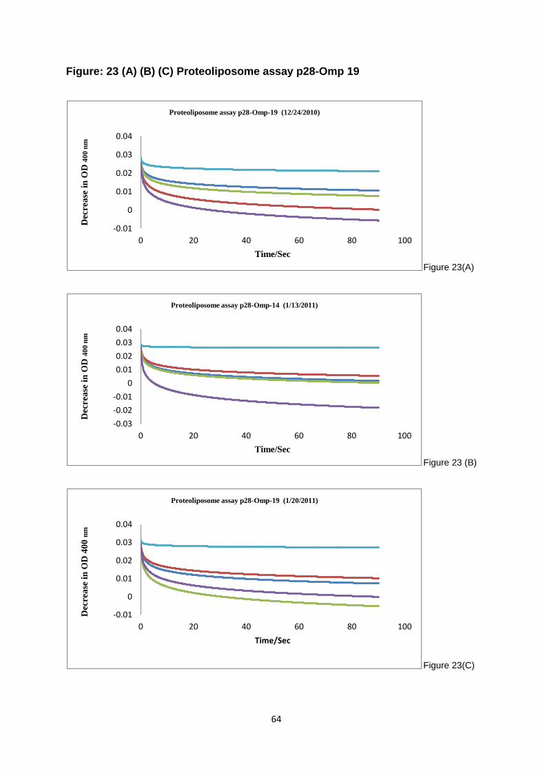
64
Figure: 23 (A) (B) (C) Proteoliposome assay p28-Omp 19
Figure 23(A)
Figure 23 (B)
Figure 23(C)
-0.01
0
0.01
0.02
0.03
0.04
0 20 40 60 80 100
Decrea
se i
n O
D 4
00
nm
Time/Sec
Proteoliposome assay p28-Omp-19 (12/24/2010)
-0.03
-0.02
-0.01
0
0.01
0.02
0.03
0.04
0 20 40 60 80 100
Dec
rea
se i
n O
D 4
00
nm
Time/Sec
Proteoliposome assay p28-Omp-14 (1/13/2011)
-0.01
0
0.01
0.02
0.03
0.04
0 20 40 60 80 100
Dec
rea
se i
n O
D 4
00
nm
Time/Sec
Proteoliposome assay p28-Omp-19 (1/20/2011)
65
Figure 23 (A) (B) (C): Porin activity of the E. chaffeensis outer membrane p28-Omp
19(recombinant). The diffusion rates of solutes into proteoliposome reconstituted
with E. chaffeensis outer membrane proteins were monitored as the decrease in OD
400. When proteoliposome were diluted in isosmotic solution of 33 mM Glucose (-)
(MW=180), 33 mM sucrose (-) (MW=342), 33 mM fructose (-) (MW=180), 33 mM, L-
glutamine (-) (MW=156) and 33Mm stachyose (-) (MW=666). (Three independent
experiments are shown performed on (A) 12/24/2010, (B) 1/13/2011, (C) 1/20/2011).
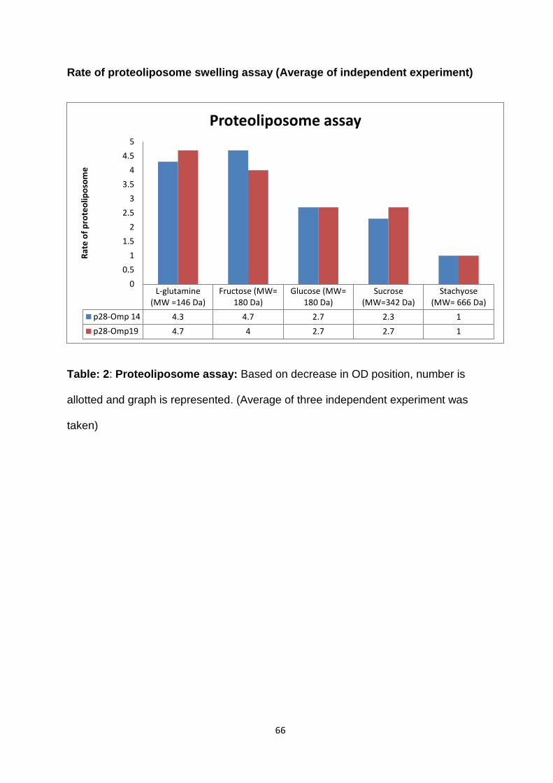
66
Rate of proteoliposome swelling assay (Average of independent experiment)
Table: 2: Proteoliposome assay: Based on decrease in OD position, number is
allotted and graph is represented. (Average of three independent experiment was
taken)
L-glutamine(MW =146 Da)
Fructose (MW=180 Da)
Glucose (MW=180 Da)
Sucrose(MW=342 Da)
Stachyose(MW= 666 Da)
p28-Omp 14 4.3 4.7 2.7 2.3 1
p28-Omp19 4.7 4 2.7 2.7 1
0
0.5
1
1.5
2
2.5
3
3.5
4
4.5
5
Rat
e o
f p
rote
olip
oso
me
Proteoliposome assay
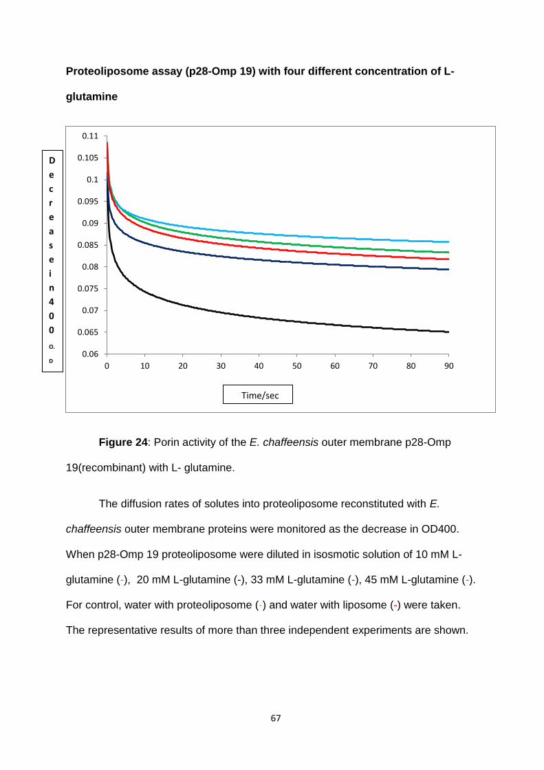
67
Proteoliposome assay (p28-Omp 19) with four different concentration of L-
glutamine
Figure 24: Porin activity of the E. chaffeensis outer membrane p28-Omp
19(recombinant) with L- glutamine.
The diffusion rates of solutes into proteoliposome reconstituted with E.
chaffeensis outer membrane proteins were monitored as the decrease in OD400.
When p28-Omp 19 proteoliposome were diluted in isosmotic solution of 10 mM L-
glutamine (-), 20 mM L-glutamine (-), 33 mM L-glutamine (-), 45 mM L-glutamine (-).
For control, water with proteoliposome (-) and water with liposome (-) were taken.
The representative results of more than three independent experiments are shown.
0.06
0.065
0.07
0.075
0.08
0.085
0.09
0.095
0.1
0.105
0.11
0 10 20 30 40 50 60 70 80 90
Time/sec
D
e
c
r
e
a
s
e
i
n
4
0
0
O.
D
68
Protocols for molecular biology techniques
69
Polymerase chain reaction (PCR): All PCR‘s were performed in final reaction
volume of 25 μl. Each reaction contains a final concentration of 1X PCR reaction
buffer, 50 nmoles of MgCl2, 10 nmoles of dNTP's, 10 pico moles each of forward
and reverse primers, about 1ng of template DNA, and 1 unit of taq DNA polymerase
(Invitrogen technologies, Carlsbad, CA). For PCR reactions that require proof
reading, the PCR conditions are; 1x Pfx amplification buffer, 1 U of platinum Pfx
DNA polymerase (Invitrogen Technologies, Carlsbad, CA), 5 nmoles of dNTP's, and
rest of the reagents concentrations are maintained same as in above described
reaction. The PCR temperature cycles include an initial heating to 95°C for 2 min,
followed by 40 cycles of 95°C denaturation for 30 sec, primer annealing for 30 sec
and carried out at appropriate temperatures calculated for each primer set, 72 °C
extension for 30 sec. The extension temperature for platinum Pfx DNA polymerase
is 68 °C. The extension temperatures were increased to 1min per each kb of
expected amplicons length. Each reaction set included a negative control, which
lacked a template but contained all the other reaction components. After reaction is
complete the products were resolved on agarose gels containing ethidium bromide
and visualized under UV light.
Restriction enzyme digestions: Typically restriction enzyme digestion reaction is
performed in a 20 μl volume. The reaction included 1x restriction enzyme reaction
buffer, 1 μg of DNA, 1-5 units of enzyme, 0.5 μl of BSA (10 μg/ μl), and water to a
final volume of 20 μl. Typically the restriction enzyme digestion is carried out at 37°C
for 2 h unless an enzyme requires a specific temperature. For all the reactions that
utilized two restriction enzymes, a buffer optimal for both the enzymes is utilized.
70
Phenol purification of DNA: DNA fragments from PCR, restriction enzyme
digestion, and filling-in reactions are purified phenol purification method. Typically, 3
M sodium acetate is added to final concentration of 0.3 M into a microcentrifuge tube
containing DNA and final volume was adjusted to 200 μl with TE buffer. Two
hundred micro liters of phenol (pH, 8.0) is added, vertexed to mix and centrifuged at
15,000 g for 15 min at 4°C. The top aqueous layer is transferred into a clean
microcentrifuge tube and added a 200 μl of phenol:chloroform:isoamylalcohol
(25:24:1) mixture. The contents are mixed by vortexing and centrifuged at 4°C at
15,000 g for 15 min. The top layer is transferred into another clean microcentrifuge
tube. Thease steps were repeated with pheol:chloroform:isoamylalacohol and then
with chloroform:isoamylalacohol. To the final removed aqueous layer, 0.5 ml
absolute cold ethanol is added, incubated at -20°C for 15 min followed by
centrifugation at 15,000g for 15 min. The DNA pellet is washed with 0.5 ml of 70%
ethanol. Final pellet was air dried, resuspended in 20 μl TE buffer and stored at -20
°C until use.
Ligation reactions: A typical ligation reaction included approximately 25 ng of
linearized purified plasmid vector DNA, 5 to10 molar excess of insert DNA, 1x
ligation buffer, 5 units of T4 DNA ligase (Promega Corporation, Madison, WI) in a 20
μl reaction volume. The ligation reaction is carried out by incubating the contents at
15°C for 16 h. Following the ligation, 1 μl of ligation mix is used for transformation by
chemical method. Alternatively, for use in transformation by electroporation method,
the DNA is purified by phenol:choloform:isoamyl alcohol method described above.
Preparation of E. coli cells for use in chemical transformation methods: The E.
coli strains utilized to prepare competent cells included Top 10 cells (Invitrogen
71
Technologies, Carlsbad, CA), DH5α (Stratagene, La Jolla, CA), and BL21 (DE3)
(Novagen Inc., Madison, WI). Top 10 stain of E. coli is always grown in the presence
of streptomycin (35μg/ml). DH5α and BL21 (DE3) stains are grown in a plain LB
medium. To prepare chemical competent cells, an E. coli colony of a desired strain
is cultured in 3 ml of LB medium overnight in a 37°C incubator, shaking at 250 rpm.
Subsequently, E. coli culture is re-inoculated into 100ml LB medium and grown in a
37°C incubator. After the cells are grown to 0.4 OD (measured at 600nm), the
cultures are harvested by centrifuging at 2,500 g for 5 min at 4°C. The cell pellet is
resuspended in 10 ml of freshly prepared 10 mM Tris-HCl (pH, 7.5) and 50 mM
CaCl2, and incubated on ice for 30 min. The cultures was centrifuged again at 2,500
g for 5 min at 4°C and the pellet is suspended in 2 ml of 10 mM Tris-HCl (pH, 7.5)
and 50 mM CaCl2 and stored at 4°C. The competent cells made by this procedure
are utilized within 24 h after their preparation.
Transformation: Transformation of ligated products into E. coli cells is achieved
either by a chemical method or through an electroporation procedure. To transform
by chemical method, 200 μl of chemical competent E. coli cells are mixed with 50 of
100mM CaCl2 and 49 μl of sterile water. One μl of ligation products are added to
this suspension and mixed by gentle tapping of the tube. The contents are then
incubated in ice for 15 min, followed by a heat shock at 42°C for 2 min. The cells are
incubated at room temperature for 10 min, added 1 ml of LB medium, and incubated
at 37 °C in a shaker incubator set at 200 rpm for 1h.
Preparation of Luria-Bertani (LB) media: The LB liquid medium and LB agar
plates were utilized to grow E. coli cultures. To prepare 1 lit of LB liquid medium, 15
72
g tryptone, 10 g of yeast extract and 10 g of sodium chloride were dissolved in 1 lit of
double distilled water and pH of the solution was adjusted to 7.0 with the help of 10N
NaOH. The LB medium was autoclaved at a liquid cycle. LB agar plates preparation
included similar preparation as described above but 15 g of cell culture grade agar
powder was added to the medium prior to autoclaving. After autoclaving, the LB agar
medium was allowed to cool to nearly 60°C and a desired concentration of
appropriate antibiotic was added to the medium. Approximately 15 ml of medium
was poured into sterile agar plates. After solidification of the agar medium, the plates
were wrapped and stored at 4°C until use.
Selection of recombinant clones: Two hundred micro liters of transformed
bacterial cultures are transferred onto LB plates containing appropriate antibiotic
specific to a recombinant plasmid. The culture is uniformly dispersed onto the agar
plate using a bacterial culture spreader. To grow the transformed E. coli the plates
were incubated overnight in a 37°C incubator. The presence of transformants is
assessed by comparing plates containing appropriate controls (ligation controls, no
transformation controls). Subsequently, several colonies are selected, inoculated in
a culture tube containing LB medium with appropriate antibiotic and are grown
overnight at 37 °C in a shaker incubator to isolate the plasmid DNA.
Isolation of Plasmid DNA: From overnight grown E. coli cultures plasmid DNA is
isolated by following boiling preparation method (336). To isolate plasmid DNA, 1.5
ml of overnight grown bacterial cultures are transferred into a micro centrifuge tube
and centrifuged 12,000 g for 5min. The supernatant was aspirated carefully with the
73
help of a vacuum device and cell pellet is resuspended in 0.4 ml plasmid lysis buffer
(10mM Tris-HCl pH 8.0, 0.1 M NaCl, 1mM EDTA, 5% v/v Triton X-100) with the help
of a tooth pick. Twenty five micro liters of freshly prepared lysozyme (10 mg/ml) is
added. Lysozyme was prepared by dissolving 10mg of lysozyme powder in 1 ml of
10 mM Tris-HCl buffer (pH, 8.0) to get a final concentration of 10 mg/ml. The
contents of the tube are vertexed to mix, placed in a boiling water bath exactly for 40
sec, and centrifuged at 12,000 g for 15 min at 4°C. The pellet containing cell debris
is removed with the help of a tooth pick. Four hundred and twenty μl of 100%, cold (-
20°C), isopropanol is added to the supernatant and mixed by vertexing, incubated at
room temperature for 5 min and centrifuged at 12,000 g for 15 min to recover
plasmid DNA. Supernatant is discarded and the DNA pellet is washed with 70%
ethanol and dried in a speed-vac system (Labconco Centrivap Concentrator, Kansas
City, MO) typically for about 5 min. Final pellet is resuspended in 100 μl of TE buffer
and contaminating bacterial RNA was removed by treating with 1 μl of RNase A (1
mg/ml) at 37°C for 5 min. The presence and quality of the plasmid DNA was
checked by agarose gel electrophoresis (described separately).
Isolation of genomic DNA: Genomic DNA of E. chaffeensis grown in tick or
macrophage cultures is isolated by sodium dodecyl sulfate-proteinase K-phenol,
chloroform-isoamyl-alcohol method (336). Briefly, 1.5 ml of E. chaffeensis cultures
are harvested by centrifugation at 12,000 g for 15 min and the cell pellet is
resuspended in 0.5 ml of DNA extraction buffer (10 Mm Tris-Cl pH 8.0, 0.1 M EDTA,
and 0.5% SDS) containing 0.5 mg/ml proteinase K (Sigma Chemical Co., St. Louis,
MO). The contents are mixed by vertexing and incubated for 2 h at 60°C.

74
Phenol:chloroform:isoamyl alcohol extraction method is used to remove
proteinecious material and ethanol is added to concentrate DNA as described
previously. DNA pellet is air dried and resuspended in 100 of TE buffer. To remove
the contaminating RNA, the DNA is treated with 1 μl RNase A (10 mg/ml) at room
temperature for 10 min. The DNA samples were stored at -20°C until use.
Agarose gel electrophoresis: Plasmid DNA, restriction digestion products or PCR
products are analyzed by resolving them on a 0.9% agarose gels by subjecting to
electrophoresis. The agarose gels are prepared after dissolving agarose powder in
1x TAE buffer (40 mM Tris-acetate, 1 mM EDTA; final pH 8.0) containing 0.1 μg/ml
of ethidium bromide. The contents are poured on a gel holding device and are
allowed to solidify at room temperature. The gel is placed in an electrophoresis
chamber containing 1X TAE buffer with 0.1 μg/ml of ethidium bromide dissolved in it.
About 5 μl of DNA is loaded into the wells. Molecular weights markers are also
loaded in a separate well and resolved to help in determining the approximate
molecular weight of the DNA. The DNA is subjected to electrophoresis in the
agarose gel at 70 V for 60-90 minutes and is visualized under UV illumination. The
images are captured using Kodak gel imaging system.
Automated sequencing: Recombinant DNA clones were sequenced to verify the
accuracy and orientation of the insert DNA using CEQ Genetic Analysis System and
by following the manufacturer’s recommendation (Beckman & Coulter, Fullerton,
CA). Prior to performing a sequencing reaction, the recombinant plasmid DNA is
purified by phenol:chloroform:isoamyl alcohol (25:24:1) method as described above.
Following the purification, the concentration of the DNA was estimated by Nanodrop
75
method. The purified plasmid DNA is sequenced using a forward or reverse primer
(plasmid derived sequence primers) specific to a sequence upstream or downstream
to insert DNA, respectively. Sequencing reaction is performed utilizing DTCS
sequencing kit by following the manufacturer’s recommendations (Beckman and
Coulter, Fullerton, CA). Sequence analysis is performed using Genetics Computer
Group (GCG) (87) or Vector.
Silver stain preparation: Dilute Silver Stain 10 folds (e.g. Dilute 5 ml of the stain in
45 ml de-ionized water), then add 65:l of Sensitizer-I per 50ml diluted Silver Stain.
Soak the gel in diluted and Sensitizer-I added Silver Stain for 20-30 min with gentle
rocking of the gel, depending upon the thickness of the gel.
Silver staining Developer preparation:
While the gel is staining, prepare the developer. Add one heaping spoonful (3-5gm)
of Developer to 100ml of de-ionized water. After the developer is dissolved, add 65 :l
of Sensitizer-I and 65:l of Sensitizer-II. Rinse the gel 10-20 seconds with de-ionized
water. Soak the gel in Developer-Sensitizer-I & II. Gently rock the gel until bands are
visible. Band intensity will develop quickly.As soon as band intensity reaches an
acceptable level, stop development with 2% acetic acid. Transfer the gel into
theacetic acid solution and incubate for 10 min. Gel may be stored in 2% acetic acid
or water.
76
List of References
References
1. Gratz NG: Emerging and resurging vector-borne diseases. Annu Rev Entomol 1999, 44(1):51.
2. Gubler DJ SG: Insects in Disease Transmission. 1991, 2nd edition(Hunter tropical medicine):981-1000.
3. Goddard J: Infectious Diseases and Arthropods: 2nd ed. A Humana Press product; 2000.
4. Kalluri S, Gilruth P, Rogers D, Szczur M: Surveillance of arthropod vector-borne infectious diseases using remote sensing techniques: a review. PLOS pathogens 2007, 3(10):1361.
5. Sonenshine DE: Biology of Ticks: Volume 1 ed. Oxford University Press; 1991.
6. Sattenspiel L: Tropical environments, human activities, and the transmission of infectious diseases. Am J Phys Anthropol 2000, Suppl 31:3.
7. Snow RW M, Deichmann U: Mortality, morbidity and disability due to malaria among Africa population. Infectious Diseases and Arthropods ,2000, 77(8):624.
8. Snow RW, Craig M, Deichmann U, Marsh K: Estimating mortality, morbidity and disability due to malaria among Africa's non-pregnant population. Bull World Health Organ 1999, 77(8):624-640.
9. Collins FH, Paskewitz SM: Malaria: current and future prospects for control. Annu Rev Entomol 1995, 40:195.
10. Casalino E, Le Bras J, Chaussin F, Fichelle A, Bouvet E: Predictive factors of malaria in travelers to areas where malaria is endemic. Arch Intern Med 2002, 162(14):1625-1630.
11. Kiszewski A, Teklehaimanot A: A review of the clinical and epidemiologic burdens of epidemic malaria. Am J Trop Med Hyg 2004, 71(2 Suppl):128-135.
12. Steere AC, Malawista SE, Snydman DR, Shope RE, Andiman WA, Ross MR, Steele FM: Lyme arthritis: an epidemic of oligoarticular arthritis in children and adults in three connecticut communities. Arthritis rheumatism 1977, 20(1):7.
13. Steere AC, Malawista SE: Cases of Lyme disease in the United States: locations correlated with distribution of Ixodes dammini. Ann Intern Med 1979, 91(5):730-733.
77
14. Fournier P, Ndihokubwayo J, Guidran J, Kelly P, Raoult D: Human pathogens in body and head lice. Emerging infectious diseases 2002, 8(12):1515.
15. Raoult D, Roux V: The body louse as a vector of reemerging human diseases. Clinical infectious diseases 1999, 29(4):888-911.
16. Raoult D, Roux V, Ndihokubwayo JB, Bise G, Baudon D, Marte G, Birtles R: Jail fever (epidemic typhus) outbreak in Burundi. Emerging infectious diseases 1997, 3(3):357-360.
17. McNabb SJN, Jajosky R, Hall-Baker P, Adams D, Sharp P, Worshams C, Anderson W, Javier A, Jones G, Nitschke D, Rey A, Wodajo M: Summary of notifiable diseases--United States, 2006. Morb Mortal Weekly Rep 2008, 55(53):1.
18. Louw M, Allsopp MTEP, Meyer EC: Ehrlichia ruminantium, an emerging human pathogen--a further report. SAMJ.South African medical journal 2005, 95(12):948.
19. Paddock CD, Sumner JW, Shore GM, Bartley DC, Elie RC, McQuade JG, Martin CR, Goldsmith CS, Childs JE: Isolation and characterization of Ehrlichia chaffeensis strains from patients with fatal ehrlichiosis. J Clin Microbiol 1997, 35(10):2496-2502.
20. Yabsley M: Natural history of Ehrlichia chaffeensis: vertebrate hosts and tick vectors from the United States and evidence for endemic transmission in other countries. Vet Parasitol 2010, 167(2-4):136-148.
21. Smallman-Raynor M, Cliff AD, Haggett P, Stroup DF, Williamson GD: Spatial and temporal patterns in final amendments to provisional disease counts. Journal of public health management and practice 1999, 5(3):68.
22. Gratz NG: Emerging and resurging vector-borne diseases. Annu Rev Entomol 1999, 44(1):51.
23. Gubler DJ: Resurgent vector-borne diseases as a global health problem. Emerging infectious diseases 1998, 4(3):442.
24. Fuente J: A ten-year review of commercial vaccine performance for control of tick infestations on cattle. Animal health research reviews 2007, 8(1):23.
25. Parola P, Raoult D: Ticks and tickborne bacterial diseases in humans: an emerging infectious threat. Clinical infectious diseases 2001, 32(6):897-928.
26. Taege AJ: Tick trouble: overview of tick-borne diseases. Cleve Clin J Med 2000, 67(4):241.
27. Walker DH: Tick-transmitted infectious diseases in the United States. Annu Rev Public Health 1998, 19:237.
78
28. Hocking B: Blood-sucking behavior of terrestrial arthropods. Annu Rev Entomol 1971, 16:1-26.
29. Riek RF: The life cycle of Babesia bigemina (Smith and Kilborne, 1893) in the tick vector Boophilus microplus (Canestrini). Aust J Agric Res 1964, 15(5):802.
30. Smith RD, James MA, Ristic M, Aikawa M, Vega y Murguia CA: Bovine babesiosis: protection of cattle with culture-derived soluble Babesia bovis antigen. Science 1981, 212(4492):335-338.
31. Blouin EF, Kocan AA, Glenn BL, Kocan KM, Hair JA: Transmission of Cytauxzoon felis Kier, 1979 from bobcats, Felis rufus (Schreber), to domestic cats by Dermacentor variabilis (Say). J Wildl Dis 1984, 20(3):241-242.
32. Hoover JP, Walker DB, Hedges JD: Cytauxzoonosis in cats: eight cases (1985-1992). J Am Vet Med Assoc 1994, 205(3):455-460.
33. Rodgers S, Mather T: Human Babesia microti incidence and Ixodes scapularis distribution, Rhode Island, 1998-2004. Emerging infectious diseases 2007, 13(4):633-635.
34. Burkot TR, Maupin GO, Schneider BS, Denatale C, Happ CM, Rutherford JS, Zeidner NS: Use of a sentinel host system to study the questing behavior of Ixodes spinipalpis and its role in the transmission of Borrelia bissettii, human granulocytic ehrlichiosis, and Babesia microti. Am J Trop Med Hyg 2001, 65(4):293.
35. Pancholi P, Kolbert CP, Mitchell PD, Reed KD, Dumler JS, Bakken JS, Telford SR, Persing DH: Ixodes dammini as a potential vector of human granulocytic ehrlichiosis. J Infect Dis 1995, 172(4):1007.
36. Parola P, Davoust B, Raoult D: Tick- and flea-borne rickettsial emerging zoonoses. Vet Res 2005, 36(3):469-492.
37. Parola P, Paddock C, Raoult D: Tick-borne rickettsioses around the world: emerging diseases challenging old concepts. Clin Microbiol Rev 2005, 18(4):719-756.
38. Walker DH: Rocky Mountain spotted fever: a disease in need of microbiological concern. Clin Microbiol Rev 1989, 2(3):227-240.
39. Burgdorfer W, Lane RS, Barbour AG, Gresbrink RA, Anderson JR: The western black-legged tick, Ixodes pacificus: a vector of Borrelia burgdorferi. Am J Trop Med Hyg 1985, 34(5):925-930.
40. Eklund CM: Distribution of Colorado tick fever and virus-carrying ticks. JAMA 1955, 157(4):335.
79
41. Keirans JE: Pictorial key to the adults of hard ticks, family Ixodidae (Ixodida: Ixodoidea), east of the Mississippi River. J Med Entomol 1989, 26(5):435.
42. Sonenshine, D. E. L. R. S. a. N. W. L.: Ticks (Ixodida). In Edited by G. a. L. D. E. Mullen. Academic Press.; 2002:518-556.
43. Cupp EW: Biology of ticks. The Veterinary clinics of North America.Small animal practice 1991, 21(1):1-26.
44. Cupp EW: Biology of ticks. The Veterinary clinics of North America.Small animal practice 1991, 21(1):1-26.
45. Azad AF, Beard CB: Rickettsial pathogens and their arthropod vectors. Emerging infectious diseases 1998, 4(2):179-186.
46. Uchida T, Tashiro F, Funato T, Kitamura Y: Isolation of a spotted fever group Rickettsia from a patient with febrile exanthematous illness in Shikoku, Japan. Microbiol Immunol 1986, 30(12):1323.
47. Kelly P, Matthewman L, Beati L, Raoult D, Mason P, Dreary M, Makombe R: African tick-bite fever: a new spotted fever group rickettsiosis under an old name. Lancet 1992, 340(8825):982.
48. Buller RS, Arens M, Hmiel SP, Paddock CD, Sumner JW, Rikhisa Y, Unver A, Gaudreault-Keener M, Manian FA, Liddell AM, Schmulewitz N, Storch GA: Ehrlichia ewingii, a newly recognized agent of human ehrlichiosis. N Engl J Med 1999, 341(3):148-155.
49. Dawson JE, Anderson BE, Fishbein DB, Sanchez JL, Goldsmith CS, Wilson KH, Duntley CW: Isolation and characterization of an Ehrlichia sp. from a patient diagnosed with human ehrlichiosis. J Clin Microbiol 1991, 29(12):2741-2745.
50. Anderson BE, Greene CE, Jones DC, Dawson JE: Ehrlichia ewingii sp. nov., the etiologic agent of canine granulocytic ehrlichiosis. Int J Syst Bacteriol 1992, 42(2):299-302.
51. Murphy GL, Ewing SA, Whitworth LC, Fox JC, Kocan AA: A molecular and serologic survey of Ehrlichia canis, E. chaffeensis, and E. ewingii in dogs and ticks from Oklahoma. Vet Parasitol 1998, 79(4):325-339.
52. Dumler JS, Barbet AF, Bekker CP, Dasch GA, Palmer GH, Ray SC, Rikihisa Y, Rurangirwa FR.: Reorganization of genera in the families Rickettsiaceae and Anaplasmataceae in the order Rickettsiales: unification of some species of Ehrlichia with Anaplasma, Cowdria with Ehrlichia and Ehrlichia with Neorickettsia, descriptions of six new species combinations and designation of Ehrlichia equi and 'HGE agent' as subjective synonyms of Ehrlichia phagocytophila.. 2001, 51:2145-2165.

80
53. Fournier P, Dumler JS, Greub G, Zhang J, Wu Y, Raoult D: Gene sequence-based criteria for identification of new rickettsia isolates and description of Rickettsia heilongjiangensis sp. nov. J Clin Microbiol 2003, 41(12):5456.
54. Rikihisa Y: Mechanisms to create a safe haven by members of the family Anaplasmataceae. Ann N Y Acad Sci 2003, 990:548-555.
55. Werren JH, Windsor DM: Wolbachia infection frequencies in insects: evidence of a global equilibrium? Proceedings - Royal Society.Biological sciences 2000, 267(1450):1277.
56. Bandi C, Anderson TJ, Genchi C, Blaxter ML: Phylogeny of Wolbachia in filarial nematodes. Proceedings - Royal Society.Biological sciences 1998, 265(1413):2407.
57. Pusterla N, Johnson EM, Chae JS, Madigan JE: Digenetic trematodes, Acanthatrium sp. and Lecithodendrium sp., as vectors of Neorickettsia risticii, the agent of Potomac horse fever. J Helminthol 2003, 77(4):335-339.
58. Brown JL, Huxsoll DL, Ristic M, Hildebrandt PK: In vitro cultivation of Neorickettsia helminthoeca, the causative agent of salmon poisoning disease. Am J Vet Res 1972, 33(8):1695-1700.
59. Maeda K, Markowitz N, Hawley RC, Ristic M, Cox D, McDade JE: Human infection with Ehrlichia canis, a leukocytic rickettsia. N Engl J Med 1987, 316(14):853.
60. Paddock C, Childs J: Ehrlichia chaffeensis: a prototypical emerging pathogen. Clin Microbiol Rev 2003, 16(1):37-64.
61. Anderson BE, Dawson JE, Jones DC, Wilson KH: Ehrlichia chaffeensis, a new species associated with human ehrlichiosis. J Clin Microbiol 1991, 29(12):2838-2842.
62. Anderson BE, Sims KG, Olson JG, Childs JE, Piesman JF, Happ CM, Maupin GO, Johnson BJ: Amblyomma americanum: a potential vector of human ehrlichiosis. Am J Trop Med Hyg 1993, 49(2):239-244.
63. Dawson JE, Childs JE, Biggie KL, Moore C, Stallknecht D, Shaddock J, Bouseman J, Hofmeister E, Olson JG: White-tailed deer as a potential reservoir of Ehrlichia spp. J Wildl Dis 1994, 30(2):162.
64. Lockhart JM, Davidson WR, Stallknecht DE, Dawson JE, Howerth EW: Isolation of Ehrlichia chaffeensis from wild white-tailed deer (Odocoileus virginianus) confirms their role as natural reservoir hosts. J Clin Microbiol 1997, 35(7):1681-1686.
65. Yu D, Li Y, Yoon J, Lee J, Lee M, Yu I, Chae J, Park J: Ehrlichia chaffeensis infection in dogs in South Korea. Vector borne and zoonotic diseases 2008, 8(3):355-358.

81
66. Yabsley M, Davidson W, Stallknecht D, Varela A, Swift P, Devos J, Dubay S: Evidence of tick-borne organisms in mule deer (Odocoileus hemionus) from the western United States. Vector borne and zoonotic diseases 2005, 5(4):351-362.
67. Dugan V, Gaydos J, Stallknecht D, Little S, Beall A, Mead D, Hurd C, Davidson W: Detection of Ehrlichia spp. in raccoons (Procyon lotor) from Georgia. Vector borne and zoonotic diseases 2005, 5(2):162-171.
68. Dugan VG, Little SE, Stallknecht DE, Beall AD: Natural infection of domestic goats with Ehrlichia chaffeensis. J Clin Microbiol 2000, 38(1):448-449.
69. Dumler JS, Bakken JS: Ehrlichial diseases of humans: emerging tick-borne infections. Clinical infectious diseases 1995, 20(5):1102-1110.
70. Dumler JS: Anaplasma and Ehrlichia infection. Ann N Y Acad Sci 2005, 1063:361-373.
71. Walker DH, Dumler JS: Emergence of the ehrlichioses as human health problems. Emerging infectious diseases 1996, 2(1):18-29.
72. Bakken JS, Goellner P, Van Etten M, Boyle DZ, Swonger OL, Mattson S, Krueth J, Tilden RL, Asanovich K, Walls J, Dumler JS: Seroprevalence of human granulocytic ehrlichiosis among permanent residents of northwestern Wisconsin. Clinical infectious diseases 1998, 27(6):1491.
73. Olano J, Hogrefe W, Seaton B, Walker D: Clinical manifestations, epidemiology, and laboratory diagnosis of human monocytotropic ehrlichiosis in a commercial laboratory setting. Clin Diagn Lab Immunol 2003, 10(5):891.
74. Barenfanger J: Identifying human ehrlichiosis. Lab Med 1996, 27:372.
75. Marty AM, Dumler JS, Imes G, Brusman HP, Smrkovski LL, Frisman DM: Ehrlichiosis mimicking thrombotic thrombocytopenic purpura. Case report and pathological correlation. Hum Pathol 1995, 26(8):920.
76. Dumler JS, Dawson JE, Walker DH: Human ehrlichiosis: hematopathology and immunohistologic detection of Ehrlichia chaffeensis. Hum Pathol 1993, 24(4):391.
77. Popov VL, Yu Xj, Walker DH: The 120 kDa outer membrane protein of Ehrlichia chaffeensis: preferential expression on dense-core cells and gene expression in Escherichia coli associated with attachment and entry. Microb Pathog 2000, 28(2):71.
78. Zhang J, Popov V, Gao S, Walker D, Yu X: The developmental cycle of Ehrlichia chaffeensis in vertebrate cells. Cell Microbiol 2007, 9(3):610-618.
82
79. Dumler JS, Madigan J, Pusterla N, Bakken J: Ehrlichioses in humans: epidemiology, clinical presentation, diagnosis, and treatment. Clinical infectious diseases 2007, 45 Suppl 1:S45-S51.
80. Everett ED, Evans KA, Henry RB, McDonald G: Human ehrlichiosis in adults after tick exposure. Diagnosis using polymerase chain reaction. Ann Intern Med 1994, 120(9):730.
81. Fishbein DB, Dawson JE, Robinson LE: Human ehrlichiosis in the United States, 1985 to 1990. Ann Intern Med 1994, 120(9):736.
82. Fordham LA, Chung CJ, Specter BB, Merten DF, Ingram DL: Ehrlichiosis: findings on chest radiographs in three pediatric patients. AJR, American journal of roentgenology 1998, 171(5):1421.
83. Dawson JE, Paddock CD, Warner CK, Greer PW, Bartlett JH, Ewing SA, Munderloh UG, Zaki SR: Tissue diagnosis of Ehrlichia chaffeensis in patients with fatal ehrlichiosis by use of immunohistochemistry, in situ hybridization, and polymerase chain reaction. Am J Trop Med Hyg 2001, 65(5):603.
84. Childs JE, Sumner JW, Nicholson WL, Massung RF, Standaert SM, Paddock CD: Outcome of diagnostic tests using samples from patients with culture-proven human monocytic ehrlichiosis: implications for surveillance. J Clin Microbiol 1999, 37(9):2997.
85. Doyle CK, Nethery K, Popov V, McBride J: Differentially expressed and secreted major immunoreactive protein orthologs of Ehrlichia canis and E. chaffeensis elicit early antibody responses to epitopes on glycosylated tandem repeats. Infect Immun 2006, 74(1):711-720.
86. Chapman A, Bakken J, Folk S, Paddock C, Bloch K, Krusell A, Sexton D, Buckingham S, Marshall G, Storch G, Dasch G, McQuiston J, Swerdlow D, Dumler S, Nicholson W, Walker D, Eremeeva M, Ohl C: Diagnosis and management of tickborne rickettsial diseases: Rocky Mountain spotted fever, ehrlichioses, and anaplasmosis--United States: a practical guide for physicians and other health-care and public health professionals. Morbidity and mortality weekly report.Recommendations and reports 2006, 55(RR-4):1-27.
87. Ge Y, Rikihisa Y: Surface-exposed proteins of Ehrlichia chaffeensis. Infect Immun 2007, 75(8):3833-3841.
88. Dunning Hotopp JC, Lin M, Madupu R, Crabtree J, Angiuoli S, Eisen J, Eisen J, Seshadri R, Ren Q, Wu M, Utterback T, Smith S, Lewis M, Khouri H, Zhang C, Niu H, Lin Q, Ohashi N, Zhi N, Nelson W, Brinkac L, Dodson R, Rosovitz MJ, Sundaram J, Daugherty S, Davidsen T, Durkin A, Gwinn M, Haft D, Selengut J, Sullivan S, Zafar N, Zhou L, Benahmed F, Forberger H, Halpin R, Mulligan S, Robinson J, White O, Rikihisa Y, Tettelin H: Comparative genomics of emerging human ehrlichiosis agents. PLoS Genetics 2006, 2(2):e21.

83
89. Rikihisa Y: Molecular events involved in cellular invasion by Ehrlichia chaffeensis and Anaplasma phagocytophilum. Vet Parasitol 2010, 167(2-4):155-166.
90. Lin M, Rikihisa Y: Ehrlichia chaffeensis and Anaplasma phagocytophilum lack genes for lipid A biosynthesis and incorporate cholesterol for their survival. Infect Immun 2003, 71(9):5324.
91. Dunning Hotopp JC, Lin M, Madupu R, Crabtree J, Angiuoli S, Eisen J, Eisen J, Seshadri R, Ren Q, Wu M, Utterback T, Smith S, Lewis M, Khouri H, Zhang C, Niu H, Lin Q, Ohashi N, Zhi N, Nelson W, Brinkac L, Dodson R, Rosovitz MJ, Sundaram J, Daugherty S, Davidsen T, Durkin A, Gwinn M, Haft D, Selengut J, Sullivan S, Zafar N, Zhou L, Benahmed F, Forberger H, Halpin R, Mulligan S, Robinson J, White O, Rikihisa Y, Tettelin H: Comparative genomics of emerging human ehrlichiosis agents. PLoS Genetics 2006, 2(2):e21-e21.
92. Yu XJ, Crocquet-Valdes P, Cullman LC, Walker DH: The recombinant 120-kilodalton protein of Ehrlichia chaffeensis, a potential diagnostic tool. J Clin Microbiol 1996, 34(11):2853-2855.
93. McBride J, Ndip L, Popov V, Walker D: Identification and functional analysis of an immunoreactive DsbA-like thio-disulfide oxidoreductase of Ehrlichia spp. Infect Immun 2002, 70(5):2700-2703.
94. McBride J, Comer J, Walker D: Novel Immunoreactive glycoprotein orthologs of Ehrlichia spp. Ann N Y Acad Sci 2003, 990:678.
95. Cheng Z: Regulation of type IV secretion apparatus genes during Ehrlichia chaffeensis intracellular development by a previously unidentified protein. J Bacteriol 2008, 190(6):2096.
96. Ohashi N, Zhi N, Lin Q, Rikihisa Y: Characterization and transcriptional analysis of gene clusters for a type IV secretion machinery in human granulocytic and monocytic ehrlichiosis agents. Infect Immun 2002, 70(4):2128.
97. Luo T, Zhang X, McBride J: Major species-specific antibody epitopes of the Ehrlichia chaffeensis p120 and E. canis p140 orthologs in surface-exposed tandem repeat regions. Clinical and vaccine immunology 2009, 16(7):982-990.
98. Luo T, Zhang X, Nicholson W, Zhu B, McBride J: Molecular characterization of antibody epitopes of Ehrlichia chaffeensis ankyrin protein 200 and tandem repeat protein 47 and evaluation of synthetic immunodeterminants for serodiagnosis of human monocytotropic ehrlichiosis. Clinical and vaccine immunology 2010, 17(1):87-97.
99. Wakeel A, Kuriakose J, McBride J: An Ehrlichia chaffeensis tandem repeat protein interacts with multiple host targets involved in cell signaling, transcriptional regulation, and vesicle trafficking. Infect Immun 2009, 77(5):1734-1745.
84
100. Caturegli P, Asanovich KM, Walls JJ, Bakken JS, Madigan JE, Popov VL, Dumler JS: ankA: an Ehrlichia phagocytophila group gene encoding a cytoplasmic protein antigen with ankyrin repeats. Infect Immun 2000, 68(9):5277-5283.
101. Zhu B, Nethery K, Kuriakose J, Wakeel A, Zhang X, McBride J: Nuclear translocated Ehrlichia chaffeensis ankyrin protein interacts with a specific adenine-rich motif of host promoter and intronic Alu elements. Infect Immun 2009, 77(10):4243-4255.
102. CROCQUETVALDES PA, CROCQUET-VALDES: Analysis of ehrlichial p28 gene expression in a murine model of persistent infection. Ann N Y Acad Sci 2005, 1063(1):420.
103. Gusa AA, Buller RS, Storch GA, Huycke MM, Machado LJ, Slater LN, Stockham SL, Massung RF: Identification of a p28 gene in Ehrlichia ewingii: evaluation of gene for use as a target for a species-specific PCR diagnostic assay. J Clin Microbiol 2001, 39(11):3871-3876.
104. van Heerden H, Collins N, Brayton K, Rademeyer C, Allsopp B: Characterization of a major outer membrane protein multigene family in Ehrlichia ruminantium. Gene 2004, 330:159-168.
105. Zhang C, Xiong Q, Kikuchi T, Rikihisa Y: Identification of 19 polymorphic major outer membrane protein genes and their immunogenic peptides in Ehrlichia ewingii for use in a serodiagnostic assay. Clinical and vaccine immunology 2008, 15(3):402.
106. Barbet A, Byrom B, Mahan S: Diversity of Ehrlichia ruminantium major antigenic protein 1-2 in field isolates and infected sheep. Infect Immun 2009, 77(6):2304-2310.
107. Barbet AF, Yi J, Lundgren A, McEwen BR, Blouin EF, Kocan KM: Antigenic variation of Anaplasma marginale: major surface protein 2 diversity during cyclic transmission between ticks and cattle. Infect Immun 2001, 69(5):3057.
108. Caspersen K, Park J, Patil S, Dumler JS: Genetic variability and stability of Anaplasma phagocytophila msp2 (p44). Infect Immun 2002, 70(3):1230-1234.
109. Dreher UM, de la Fuente J, Hofmann-Lehmann R, Meli ML, Pusterla N, Kocan KM, Woldehiwet Z, Braun U, Regula G, Staerk KDC, Lutz H: Serologic cross-reactivity between Anaplasma marginale and Anaplasma phagocytophilum. Clin Diagn Lab Immunol 2005, 12(10):1177.
110. IJdo J, Wu C, Telford S, Fikrig E: Differential expression of the p44 gene family in the agent of human granulocytic ehrlichiosis. Infect Immun 2002, 70(9):5295.
85
111. Yu X, McBride JW, Zhang X, Walker DH: Characterization of the complete transcriptionally active ehrlichia chaffeensis 28 kDa outer membrane protein multigene family. Gene 2000, 248(1-2):59-68.
112. Unver A, Rikihisa Y, Stich R, Ohashi N, Felek S: The omp-1 major outer membrane multigene family of Ehrlichia chaffeensis is differentially expressed in canine and tick hosts. Infect Immun 2002, 70(8):4701-4704.
113. Ohashi N, Zhi N, Zhang Y, Rikihisa Y: Immunodominant major outer membrane proteins of Ehrlichia chaffeensis are encoded by a polymorphic multigene family. Infect Immun 1998, 66(1):132-139.
114. Long SW, Zhang X, Qi H, Standaert S, Walker D, Yu X: Antigenic variation of Ehrlichia chaffeensis resulting from differential expression of the 28-kilodalton protein gene family. Infect Immun 2002, 70(4):1824-1831.
115. Cheng C, Paddock C, Ganta R: Molecular heterogeneity of Ehrlichia chaffeensis isolates determined by sequence analysis of the 28-kilodalton outer membrane protein genes and other regions of the genome. Infect Immun 2003, 71(1):187-195.
116. Cheng Z, Kumagai Y, Lin M, Zhang C, Rikihisa Y: Intra-leukocyte expression of two-component systems in Ehrlichia chaffeensis and Anaplasma phagocytophilum and effects of the histidine kinase inhibitor closantel. Cell Microbiol 2006, 8(8):1241-1252.
117. Bao W, Kumagai Y, Niu H, Yamaguchi M, Miura K, Rikihisa Y: Four VirB6 paralogs and VirB9 are expressed and interact in Ehrlichia chaffeensis-containing vacuoles. J Bacteriol 2009, 191(1):278-286.
118. Rikihisa Y, Lin M, Niu H, Cheng Z: Type IV secretion system of Anaplasma phagocytophilum and Ehrlichia chaffeensis. Ann N Y Acad Sci 2009, 1166:106-111.
119. Zhu B: Nuclear translocated Ehrlichia chaffeensis ankyrin protein interacts with a specific adenine-rich motif of host promoter and intronic Alu elements. Infect Immun 2009, 77(10):4243.
120. Zhang J, Sinha M, Luxon B, Yu X: Survival strategy of obligately intracellular Ehrlichia chaffeensis: novel modulation of immune response and host cell cycles. Infect Immun 2004, 72(1):498-507.
121. Rikihisa Y: Anaplasma phagocytophilum and Ehrlichia chaffeensis: subversive manipulators of host cells. Nature reviews.Microbiology 2010, 8(5):328-339.
122. Kumagai Y, Cheng Z, Lin M, Rikihisa Y: Biochemical activities of three pairs of Ehrlichia chaffeensis two-component regulatory system proteins involved in inhibition of lysosomal fusion. Infect Immun 2006, 74(9):5014-5022.
86
123. Lin M, Rikihisa Y: Ehrlichia chaffeensis downregulates surface Toll-like receptors 2/4, CD14 and transcription factors PU.1 and inhibits lipopolysaccharide activation of NF-kappa B, ERK 1/2 and p38 MAPK in host monocytes. Cell Microbiol 2004, 6(2):175-186.
124. Xiong Q, Lin M, Rikihisa Y: Cholesterol-dependent anaplasma phagocytophilum exploits the low-density lipoprotein uptake pathway. PLOS pathogens 2009, 5(3):e1000329-e1000329.
125. Safdar N, Love R, Maki D: Severe Ehrlichia chaffeensis infection in a lung transplant recipient: a review of ehrlichiosis in the immunocompromised patient. Emerging infectious diseases 2002, 8(3):320.
126. Gardner S, Holman R, Krebs J, Berkelman R, Childs J: National surveillance for the human ehrlichioses in the United States, 1997-2001, and proposed methods for evaluation of data quality. Ann N Y Acad Sci 2003, 990:80.
127. Ganta R, Wilkerson M, Cheng C, Rokey A, Chapes S: Persistent Ehrlichia chaffeensis infection occurs in the absence of functional major histocompatibility complex class II genes. Infect Immun 2002, 70(1):380.
128. Ganta R, Cheng C, Miller E, McGuire B, Peddireddi L, Sirigireddy K, Chapes S: Differential clearance and immune responses to tick cell-derived versus macrophage culture-derived Ehrlichia chaffeensis in mice. Infect Immun 2007, 75(1):135-145.
129. Winslow GM, Yager E, Shilo K, Collins DN, Chu FK: Infection of the laboratory mouse with the intracellular pathogen Ehrlichia chaffeensis. Infect Immun 1998, 66(8):3892.
130. Winslow GM, Yager E, Shilo K, Collins DN, Chu FK: Infection of the laboratory mouse with the intracellular pathogen Ehrlichia chaffeensis. Infect Immun 1998, 66(8):3892-3899.
131. Feng H, Walker D: Mechanisms of immunity to Ehrlichia muris: a model of monocytotropic ehrlichiosis. Infect Immun 2004, 72(2):966.
132. Ganta R, Cheng C, Wilkerson M, Chapes S: Delayed clearance of Ehrlichia chaffeensis infection in CD4 T-cell knockout mice. Infect Immun 2004, 72(1):159.
133. Winslow G, Yager E, Li J: Mechanisms of humoral immunity during Ehrlichia chaffeensis infection. Ann N Y Acad Sci 2003, 990:435.
134. Li JS, Yager E, Reilly M, Freeman C, Reddy GR, Reilly AA, Chu FK, Winslow GM: Outer membrane protein-specific monoclonal antibodies protect SCID mice from fatal infection by the obligate intracellular bacterial pathogen Ehrlichia chaffeensis. The journal of immunology 2001, 166(3):1855-1862.
87
135. Lee EH, Rikihisa Y: Anti-Ehrlichia chaffeensis antibody complexed with E. chaffeensis induces potent proinflammatory cytokine mRNA expression in human monocytes through sustained reduction of IkappaB-alpha and activation of NF-kappaB. Infect Immun 1997, 65(7):2890-2897.
136. Lee EH, Rikihisa Y: Absence of tumor necrosis factor alpha, interleukin-6 (IL-6), and granulocyte-macrophage colony-stimulating factor expression but presence of IL-1beta, IL-8, and IL-10 expression in human monocytes exposed to viable or killed Ehrlichia chaffeensis. Infect Immun 1996, 64(10):4211-4219.
137. Seo G, Cheng C, Tomich J, Ganta R: Total, membrane, and immunogenic proteomes of macrophage- and tick cell-derived Ehrlichia chaffeensis evaluated by liquid chromatography-tandem mass spectrometry and MALDI-TOF methods. Infect Immun 2008, 76(11):4823-4832.
138. Singu V, Peddireddi L, Sirigireddy K, Cheng C, Munderloh U, Ganta R: Unique macrophage and tick cell-specific protein expression from the p28/p30-outer membrane protein multigene locus in Ehrlichia chaffeensis and Ehrlichia canis. Cell Microbiol 2006, 8(9):1475-1487.
139. Reddy GR, Streck CP: Variability in the 28-kDa surface antigen protein multigene locus of isolates of the emerging disease agent Ehrlichia chaffeensis suggests that it plays a role in immune evasion. Molecular cell biology research communications 1999, 1(3):167-175.
140. Reddy GR, Sulsona CR, Barbet AF, Mahan SM, Burridge MJ, Alleman AR: Molecular characterization of a 28 kDa surface antigen gene family of the tribe Ehrlichiae. Biochem Biophys Res Commun 1998, 247(3):636-643.
141. Singu V, Liu H, Cheng C, Ganta R: Ehrlichia chaffeensis expresses macrophage- and tick cell-specific 28-kilodalton outer membrane proteins. Infect Immun 2005, 73(1):79-87.
142. Li J, Chu F, Reilly A, Winslow G: Antibodies highly effective in SCID mice during infection by the intracellular bacterium Ehrlichia chaffeensis are of picomolar affinity and exhibit preferential epitope and isotype utilization. The journal of immunology 2002, 169(3):1419-1425.
143. Ohashi N, Rikihisa Y, Unver A: Analysis of transcriptionally active gene clusters of major outer membrane protein multigene family in Ehrlichia canis and E. chaffeensis. Infect Immun 2001, 69(4):2083-2091.
144. Zhang J, Guo H, Winslow G, Yu X: Expression of members of the 28-kilodalton major outer membrane protein family of Ehrlichia chaffeensis during persistent infection. Infect Immun 2004, 72(8):4336-4343.
145. Unver A, Rikihisa Y, Ohashi N, Cullman LC, Buller R, Storch GA: Western and dot blotting analyses of Ehrlichia chaffeensis indirect fluorescent-antibody assay-positive and -negative human sera by using native and recombinant E. chaffeensis and E. canis antigens. J Clin Microbiol 1999, 37(12):3888-3895.
88
146. Ohashi N, Unver A, Zhi N, Rikihisa Y: Cloning and characterization of multigenes encoding the immunodominant 30-kilodalton major outer membrane proteins of Ehrlichia canis and application of the recombinant protein for serodiagnosis. J Clin Microbiol 1998, 36(9):2671-2680.
147. Bekker CPJ, Postigo M, Taoufik A, Bell-Sakyi L, Ferraz C, Martinez D, Jongejan F: Transcription analysis of the major antigenic protein 1 multigene family of three in vitro-cultured Ehrlichia ruminantium isolates. J Bacteriol 2005, 187(14):4782-4791.
148. Eid G, French DM, Lundgren AM, Barbet AF, McElwain TF, Palmer GH: Expression of major surface protein 2 antigenic variants during acute Anaplasma marginale rickettsemia. Infect Immun 1996, 64(3):836-841.
149. IJdo JW, Meek JI, Cartter ML, Magnarelli LA, Wu C, Tenuta SW, Fikrig E, Ryder RW: The emergence of another tickborne infection in the 12-town area around Lyme, Connecticut: human granulocytic ehrlichiosis. J Infect Dis 2000, 181(4):1388.
150. Barbet AF, Meeus PFM, Blanger M, Bowie MV, Yi J, Lundgren AM, Alleman AR, Wong SJ, Chu FK, Munderloh UG, Jauron SD: Expression of multiple outer membrane protein sequence variants from a single genomic locus of Anaplasma phagocytophilum. Infect Immun 2003, 71(4):1706.
151. Kumagai Y, Huang H, Rikihisa Y: Expression and porin activity of P28 and OMP-1F during intracellular Ehrlichia chaffeensis development. J Bacteriol 2008, 190(10):3597-3605.
152. Ewing SA, Dawson JE, Kocan AA, Barker RW, Warner CK, Panciera RJ, Fox JC, Kocan KM, Blouin EF: Experimental transmission of Ehrlichia chaffeensis (Rickettsiales: Ehrlichieae) among white-tailed deer by Amblyomma americanum (Acari: Ixodidae). J Med Entomol 1995, 32(3):368-374.
153. Voulhoux R, Bos M, Geurtsen J, Mols M, Tommassen J: Role of a highly conserved bacterial protein in outer membrane protein assembly. Science 2003, 299(5604):262-265.
154. Kyte J, Doolittle RF: A simple method for displaying the hydropathic character of a protein. J Mol Biol 1982, 157(1):105-132.
155. Esposti MD, Crimi M, Venturoli G: A critical evaluation of the hydropathy profile of membrane proteins. European journal of biochemistry 1990, 190(1):207-219.
156. Hirokawa T, Boon-Chieng S, Mitaku S: SOSUI: classification and secondary structure prediction system for membrane proteins. Bioinformatics 1998, 14(4):378-379.
89
157. Nielsen M, Lundegaard C, Lund O, Petersen T: CPHmodels-3.0--remote homology modeling using structure-guided sequence profiles. Nucleic Acids Res 2010, 38(Web Server issue):W576-W581.
158. van Stokkum IH, Spoelder HJ, Bloemendal M, van Grondelle R, Groen FC: Estimation of protein secondary structure and error analysis from circular dichroism spectra. Anal Biochem 1990, 191(1):110-118.
159. Sreerama N, Venyaminov SY, Woody RW: Estimation of protein secondary structure from circular dichroism spectra: inclusion of denatured proteins with native proteins in the analysis. Anal Biochem 2000, 287(2):243-251.
160. Tamm LK: Folding and assembly of [beta]-barrel membrane proteins. Biochimica et biophysica acta.Biomembranes 2004, 1666(1-2):250.
161. Compton LA: Analysis of protein circular dichroism spectra for secondary structure using a simple matrix multiplication. Anal Biochem 1986, 155(1):155.
162. Nikaido H, Rosenberg EY: Porin channels in Escherichia coli: studies with liposomes reconstituted from purified proteins. J Bacteriol 1983, 153(1):241.
163. Nikaido H, Nikaido K, Harayama S: Identification and characterization of porins in Pseudomonas aeruginosa. J Biol Chem 1991, 266(2):770.
164. White SH: Membrane protein folding and stability: physical principles. Annu Rev Biophys Biomol Struct 1999, 28(1):319.
165. Anderson BE: Ehrlichia chaffeensis, a new species associated with human ehrlichiosis. J Clin Microbiol 1991, 29(12):2838.
166. Benz R: Structure and function of porins from gram-negative bacteria. Annu Rev Microbiol 1988, 42:359.
167. Clark EDB: Refolding of recombinant proteins. Curr Opin Biotechnol 1998, 9(2):157-163.
168. E. Ruckenstein RN: Critical micelle concentration and the transition point for micellar size distribution . J Phys Chem 1981, 85(20):3010-3014.
169. Compton LA, Johnson WC: Analysis of protein circular dichroism spectra for secondary structure using a simple matrix multiplication. Anal Biochem 1986, 155(1):155-167.