anaplasma, coxiella and bartonella rickettsia, orientia, ehrlichia, · rickettsia, orientia,...
TRANSCRIPT

Rickettsia, Orientia, Ehrlichia, Anaplasma, Coxiella and Bartonella
History of Rickettsial Diseases
• Epidemic typhus - 16th century• Associated with wars and famine• WWI and WWII - 100,000 people affected• Ricketts identifies causative agent of Rocky
Mountain spotted fever - 20th century • Arthropod vectors identified• Arthropod control measures instituted

“Typhus is not dead. It will continue to break into the open whenever human stupidity and brutality give it a chance, as most likely they occasionally will. But its freedom of action is being restricted and more and more it will be confined, like other savage creatures, in the zoologic gardens of controlled diseases”
Hans Zinsser in Rats, Lice and History
Rickettsia, Orientia, Ehrlichia Anaplasma and Coxiella Biology
• Small obligate intracellular parasites • Once considered to be viruses • Separate unrelated genera• Gram-negative bacteria
– Stain poorly with Gram stain (Giemsa)• “Energy parasites”
– Transport system for ATP • Reservoirs - animals, insects and humans• Arthropod vectors (except Coxiella)
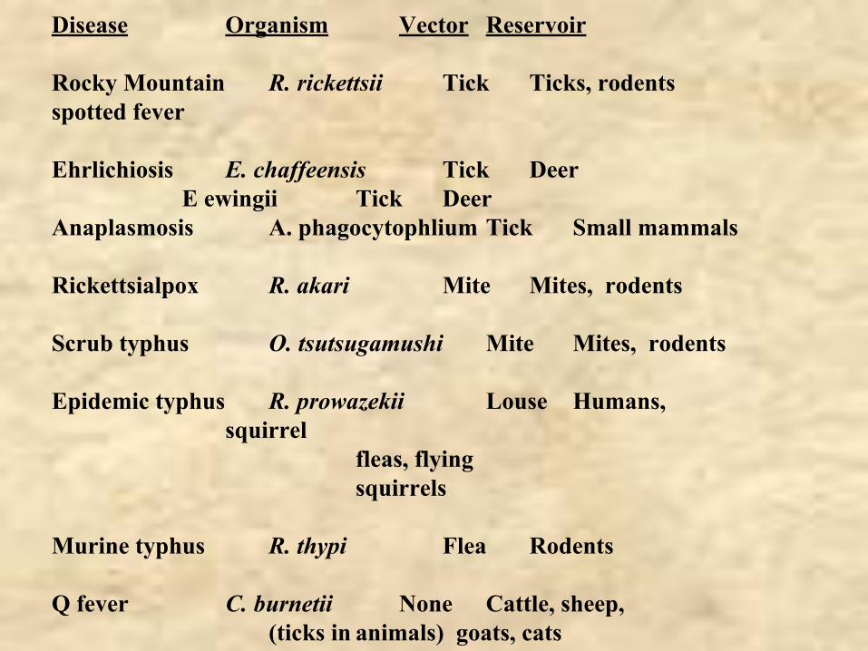
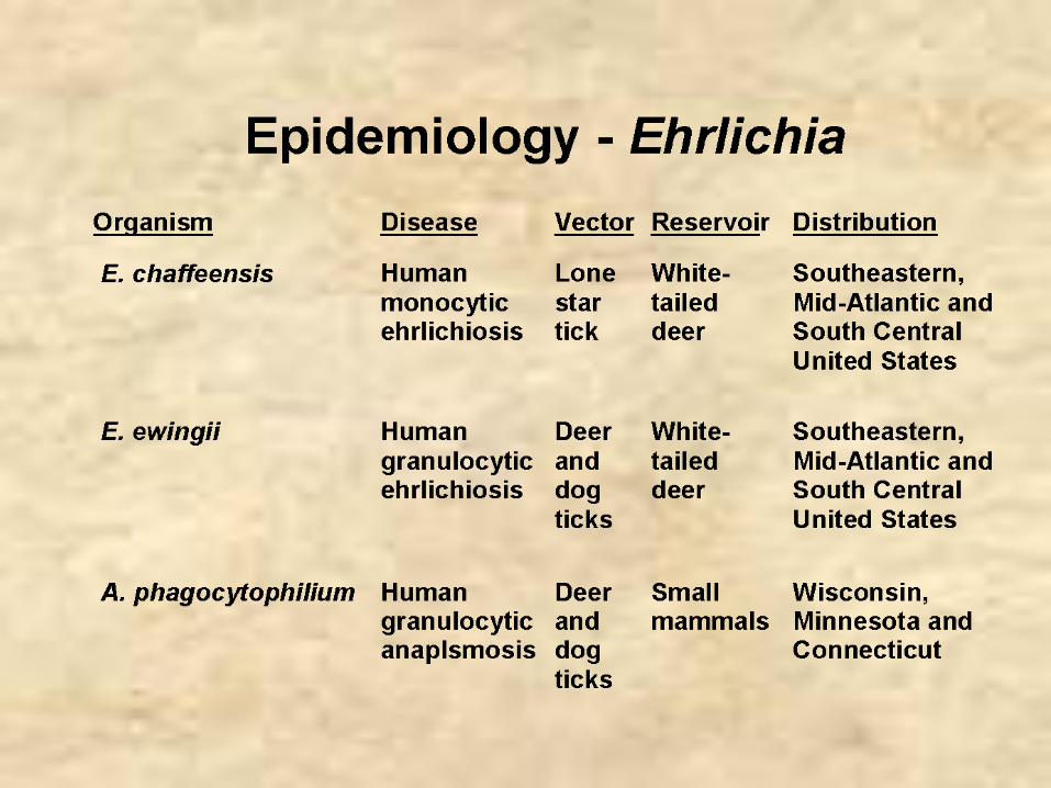
Disease Organism Vector Reservoir
Rocky Mountain R. rickettsii Tick Ticks, rodentsspotted fever
Ehrlichiosis E. chaffeensis Tick DeerE ewingii Tick Deer
Anaplasmosis A. phagocytophlium Tick Small mammals
Rickettsialpox R. akari Mite Mites, rodents
Scrub typhus O. tsutsugamushi Mite Mites, rodents
Epidemic typhus R. prowazekii Louse Humans, squirrel
fleas, flyingsquirrels
Murine typhus R. thypi Flea Rodents
Q fever C. burnetii None Cattle, sheep,(ticks in animals) goats, cats

Rickettsia and Orientia
N.B. Orientia was formerly Rickettsia
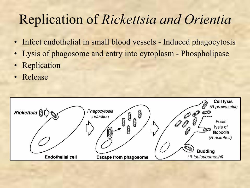
Replication of Rickettsia and Orientia• Infect endothelial in small blood vessels - Induced phagocytosis• Lysis of phagosome and entry into cytoplasm - Phospholipase• Replication• Release
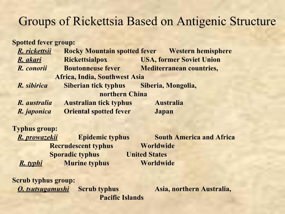
Groups of Rickettsia Based on Antigenic StructureSpotted fever group: R. rickettsii Rocky Mountain spotted fever Western hemisphere R. akari Rickettsialpox USA, former Soviet Union R. conorii Boutonneuse fever Mediterranean countries,
Africa, India, Southwest Asia R. sibirica Siberian tick typhus Siberia, Mongolia,
northern China R. australia Australian tick typhus Australia R. japonica Oriental spotted fever Japan
Typhus group: R. prowazekii Epidemic typhus South America and Africa
Recrudescent typhus Worldwide Sporadic typhus United States
R. typhi Murine typhus Worldwide
Scrub typhus group: O. tsutsugamushi Scrub typhus Asia, northern Australia,
Pacific Islands
Pathogenesis and Immunity
• No known toxins or immunopathology• Destruction of cells
– Leakage of blood into tissues (rash)– Organ and tissue damage
• Humoral and cell mediated immunity important for recovery– Antibody-opsonized bacteria are killed– CMI develops

Spotted Fever Group
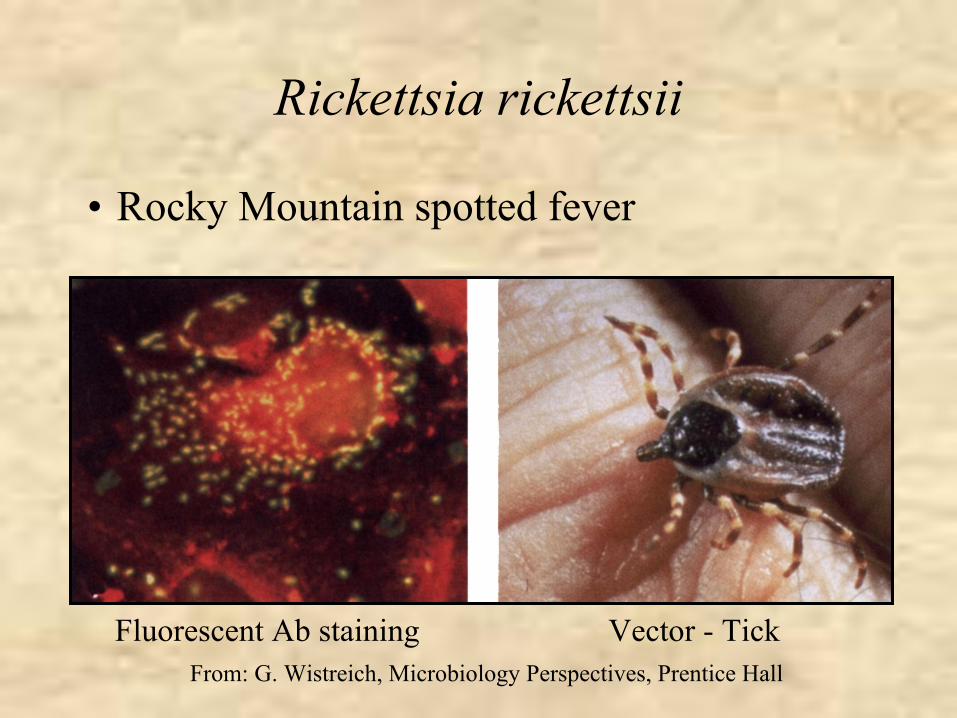
Rickettsia rickettsii
• Rocky Mountain spotted fever
Vector - TickFluorescent Ab stainingFrom: G. Wistreich, Microbiology Perspectives, Prentice Hall
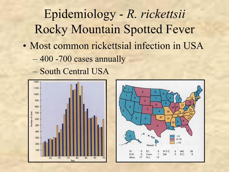
Epidemiology - R. rickettsiiRocky Mountain Spotted Fever
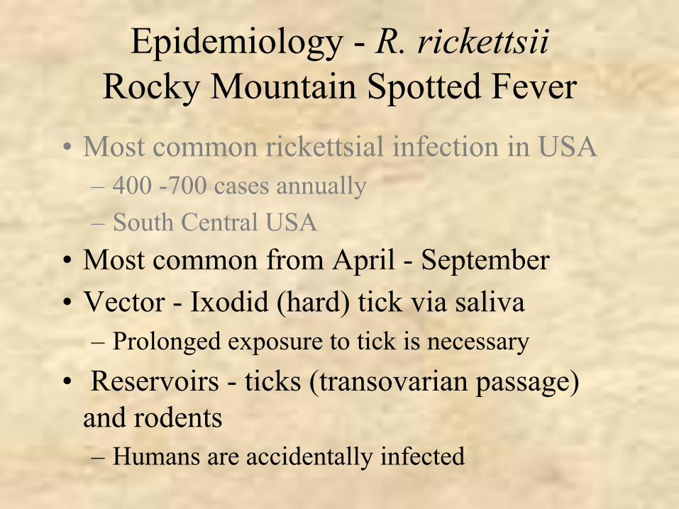
• Most common rickettsial infection in USA– 400 -700 cases annually– South Central USA
Epidemiology - R. rickettsiiRocky Mountain Spotted Fever
• Most common from April - September• Vector - Ixodid (hard) tick via saliva
– Prolonged exposure to tick is necessary• Reservoirs - ticks (transovarian passage)
and rodents– Humans are accidentally infected
• Most common rickettsial infection in USA– 400 -700 cases annually– South Central USA
Epidemiology - R. rickettsiiRocky Mountain Spotted Fever
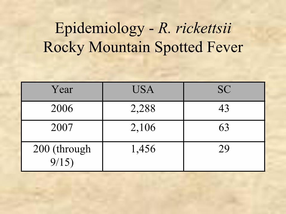
Year USA SC
2006 2,288 43
2007 2,106 63
200 (through 9/15)
1,456 29
Clinical Syndrome - Rocky Mountain Spotted Fever
• Incubation period - 2 to 12 days• Abrupt onset fever, chills, headache and myalgia• Rash appears 2 -3 days later in most (90%) patients
– Begins on hands and feet and spreads to trunk (centripetal spread)
– Palms and soles common – Maculopapular but can become petechial or hemorrhagic
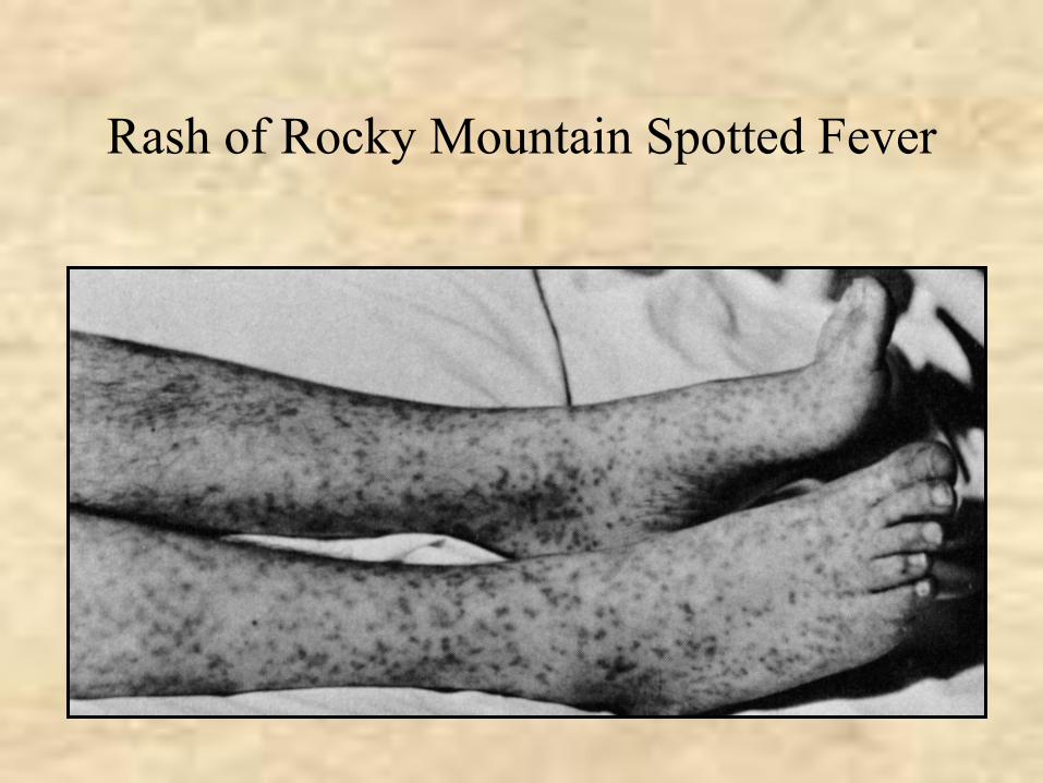
Rash of Rocky Mountain Spotted Fever
Clinical Syndrome - Rocky Mountain Spotted Fever
• Complications from widespread vasculitis– Gastrointestinal, respiratory, seizures, coma, renal failure– Most common when rash does not appear
• Mortality in untreated cases - 20%
• Incubation period - 2 to 12 days• Abrupt onset fever, chills headache and myalgia• Rash appears 2 -3 days later in most (90%) patients
– Begins on hands and feet and spreads to trunk (centripetal spread)
– Palms and soles common – Maculopapular but can become petechial or hemorrhagic
Laboratory Diagnosis - R. rickettsii
• Initial diagnosis - clinical grounds• Fluorescent Ab test for Ag in punch biopsy
- reference labs• PCR based tests - reference labs• Weil-Felix test - no longer recommended• Serology
– Indirect fluorescent Ab test for Ab– Latex agglutination test for Ab
Treatment, Prevention and ControlR. rickettsii
• Tetracycline and chloramphenicol– Prompt treatment reduces morbidity and
mortality• No vaccine• Prevention of tick bites (protective clothing,
insect repellents)• Prompt removal of ticks• Can’t control the reservoir
Consequences of Delayed Diagnosis of RMSF
• In Oklahoma on July 7 a 6 year-old presented with 1-day history of fever, headache, myalgia, and a macular rash on the arms, legs, palms, and soles
• On July 1 a tick had been removed from the patients neck• Diagnosis: Viral illness; patient given oral cephalosporin• On July 11 the patient was hospitalized with dehydration, irritability,
confusion and throbocytopenia• On July 12-13 patient developed disseminated intravascular
coagulation and iv doxycycline was administered.• The patient subsequently developed gangrene, requiring limb
amputation and removal of the upper stomach and distal esophagus• August 19 the patient died.• Serum samples from July 12 and August 3 tested positive for
antibodies to R. rickettsii

Rickettsia akari
• Rickettsialpox
Epidemiology - R. akariRickettsialpox
• Sporadic infection in USA• Vector - house mite• Reservoir - mites (transovarian
transmission) and mice• Humans accidentally infected

Clinical Syndrome -Rickettsialpox
• Phase I (1 week incubation period)– papule at bite site– Eschar formation
• Phase II (1 -3 week later)– Sudden onset of fever, chills headache and
myaglia– Generalized rash - papulovessicular, crusts
• Mild disease; fatalities are rare
Laboratory Diagnosis - R. akari
• Not available except in reference laboratories
Treatment, Prevention and ControlR. akari
• Tetracycline and chloramphenicol• Control of mouse population

Typhus Group
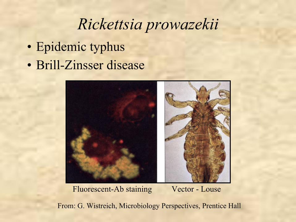
Rickettsia prowazekii• Epidemic typhus• Brill-Zinsser disease
Fluorescent-Ab staining Vector - Louse
From: G. Wistreich, Microbiology Perspectives, Prentice Hall
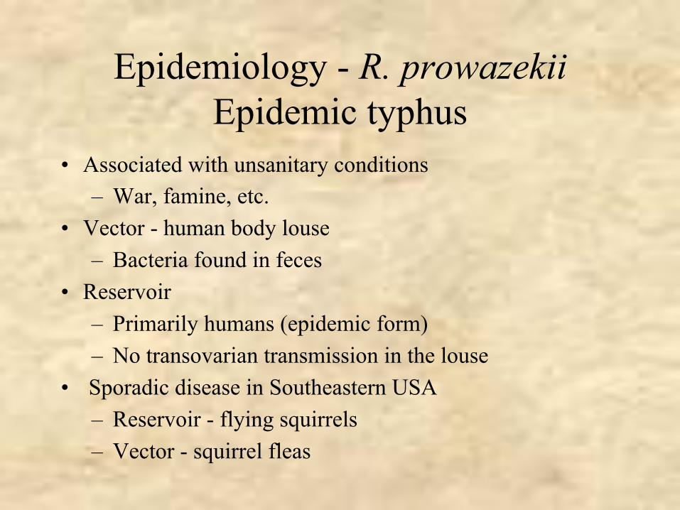
Epidemiology - R. prowazekiiEpidemic typhus
• Associated with unsanitary conditions– War, famine, etc.
• Vector - human body louse– Bacteria found in feces
• Reservoir– Primarily humans (epidemic form)– No transovarian transmission in the louse
• Sporadic disease in Southeastern USA– Reservoir - flying squirrels– Vector - squirrel fleas
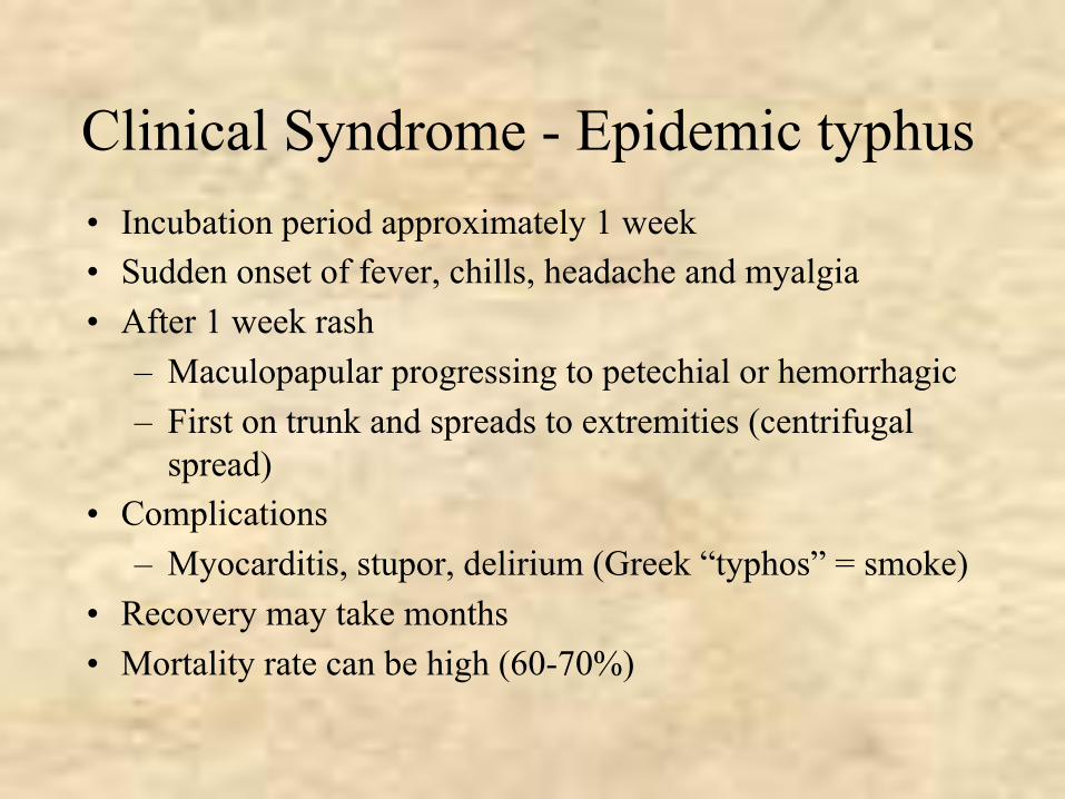
Clinical Syndrome - Epidemic typhus• Incubation period approximately 1 week• Sudden onset of fever, chills, headache and myalgia• After 1 week rash
– Maculopapular progressing to petechial or hemorrhagic– First on trunk and spreads to extremities (centrifugal
spread)• Complications
– Myocarditis, stupor, delirium (Greek “typhos” = smoke)• Recovery may take months• Mortality rate can be high (60-70%)
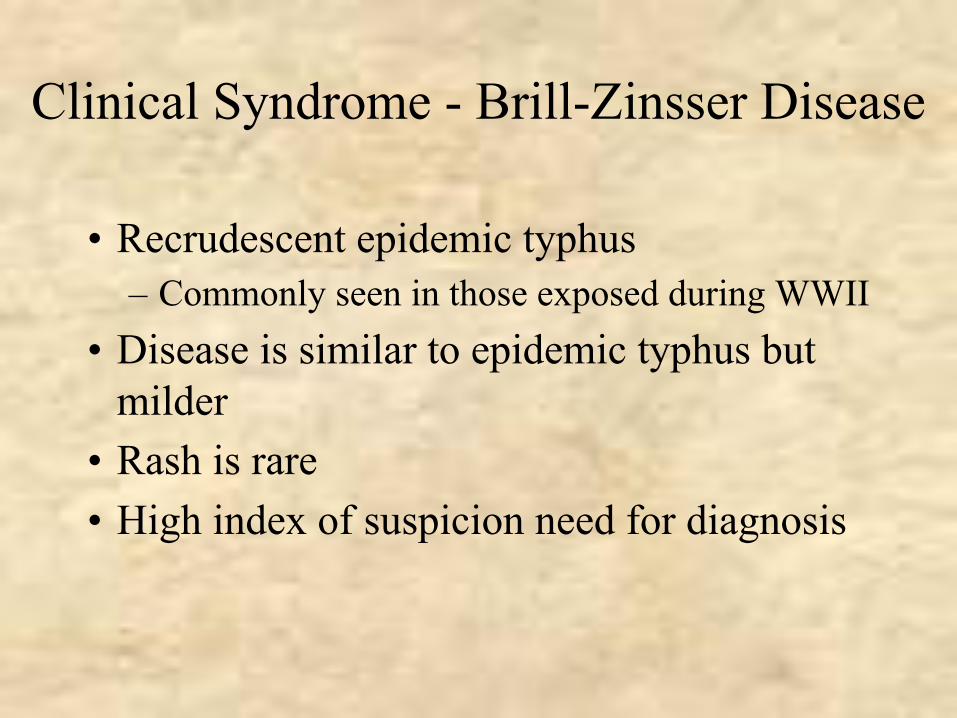
Clinical Syndrome - Brill-Zinsser Disease
• Recrudescent epidemic typhus– Commonly seen in those exposed during WWII
• Disease is similar to epidemic typhus but milder
• Rash is rare• High index of suspicion need for diagnosis
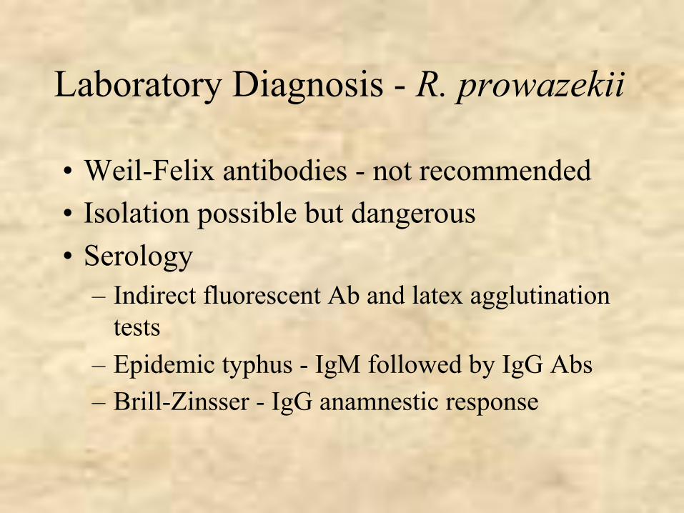
Laboratory Diagnosis - R. prowazekii
• Weil-Felix antibodies - not recommended• Isolation possible but dangerous• Serology
– Indirect fluorescent Ab and latex agglutination tests
– Epidemic typhus - IgM followed by IgG Abs– Brill-Zinsser - IgG anamnestic response
Treatment, prevention and ControlR. prowazekii
• Tetracycline and chloramphenicol• Louse control measures• Vaccine available for high risk populations
Rickettsia typhi
• Murine or endemic typhus
Epidemiology - R. typhiMurine or endemic typhus
• Occurs worldwide• Vector - rat flea
– Bacteria in feces• Reservoir - rats
– No transovarian transmission– Normal cycle - rat to flea to rat
• Humans accidentally infected
Clinical Syndrome- Murine Typhus
• Incubation period 1 - 2 weeks• Sudden onset of fever, chills, headache and
myalgia• Rash in most cases
– Begins on trunk and spreads to extremities (centrifugal spread)
• Mild disease - resolves even if untreated
Laboratory Diagnosis - R. typhi
• Serology– Indirect fluorescent antibody test
Treatment, Prevention and ControlR. typhi
• Tetracycline and chloramphenicol• Control rodent reservoir

Scrub Typhus Group
Orientsia (Rickettsia) tsutsugamushi
• Scrub typhus• Japanese “tsutsuga” = small and dangerous
and “mushi” = creature• “Scrub” - associated with terrain with scrub
vegetation
Epidemiology - O. tsutstugamushiScrub Typhus
• Vector - chiggers (mite larva)• Reservoir - chiggers and rats
– Transovarian transmission– Normal cycle - rat to mite to rat
• Humans are accidentally infected
Clinical Syndrome - Scrub Typhus
• Incubation period - 1 to 3 weeks• Sudden onset of fever, chills, headache and
myalgia• Maculopapular rash
– Begins on trunk and spreads to extremities (centrifugal spread)
• Mortality rates variable
Laboratory Diagnosis - O. tsutsugamushi
• Serology
Treatment, Prevention and ControlO. tsutsugamushi
• Tetracycline and chloramphenicol• Measures to avoid exposure to chiggers

Ehrlichia and Anaplasma
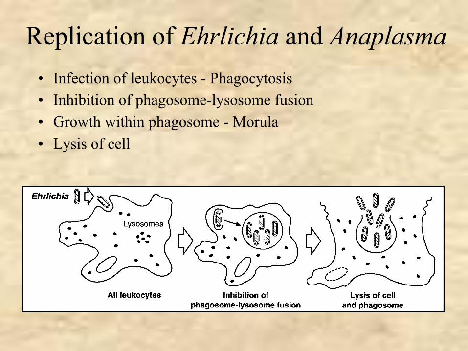
Replication of Ehrlichia and Anaplasma• Infection of leukocytes - Phagocytosis• Inhibition of phagosome-lysosome fusion• Growth within phagosome - Morula• Lysis of cell
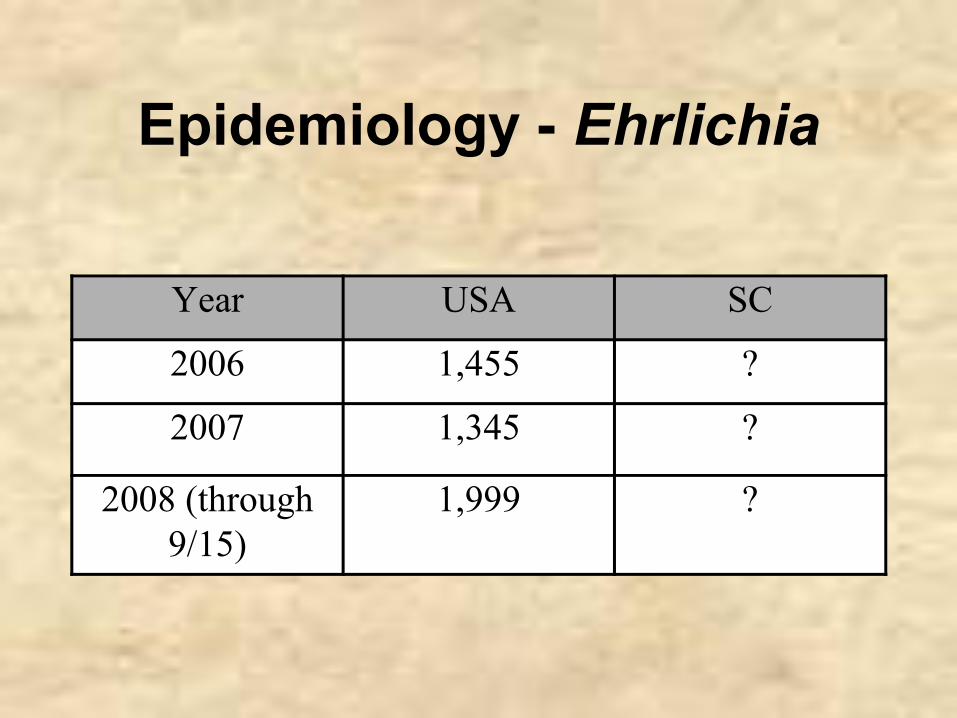
Epidemiology - Ehrlichia
Year USA SC
2006 1,455 ?
2007 1,345 ?
2008 (through 9/15)
1,999 ?
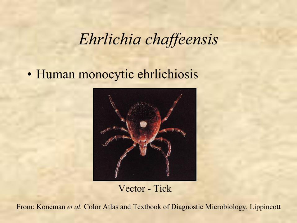
Ehrlichia chaffeensis
• Human monocytic ehrlichiosis
From: Koneman et al. Color Atlas and Textbook of Diagnostic Microbiology, Lippincott
Vector - Tick
Clinical Syndrome - Human Monocytic Ehrlichiosis
E. chaffeensis• Sudden onset of fever, chills, headache and
myalgia• No rash in most (80%) patients• Leukopenia, thrombocytopenia and elevated
serum transaminases • Mortality rates low (<5%)
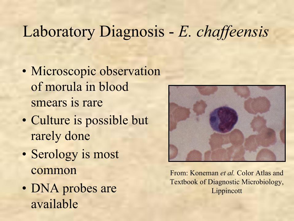
Laboratory Diagnosis - E. chaffeensis
• Microscopic observation of morula in blood smears is rare
From: Koneman et al. Color Atlas and Textbook of Diagnostic Microbiology,
Lippincott
• Culture is possible but rarely done
• Serology is most common
• DNA probes are available
Treatment, Prevention and ControlE. chaffeensis
• Doxycycline• Avoidance of ticks
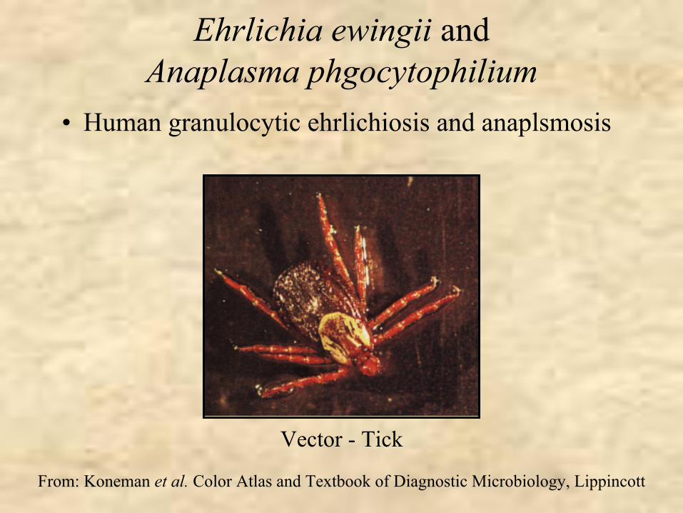
Ehrlichia ewingii and Anaplasma phgocytophilium
• Human granulocytic ehrlichiosis and anaplsmosis
From: Koneman et al. Color Atlas and Textbook of Diagnostic Microbiology, Lippincott
Vector - Tick
Clinical Syndrome - Human Granulocytic Ehrlichiosis or Anaplasmosis
E. ewingii or Anaplasma phgocytophilium
• Sudden onset of fever, chills, headache and myalgia
• No rash in most (80%) patients• Leukopenia , thrombocytopenia and
elevated serum transaminases • Mortality rates low (<5%)
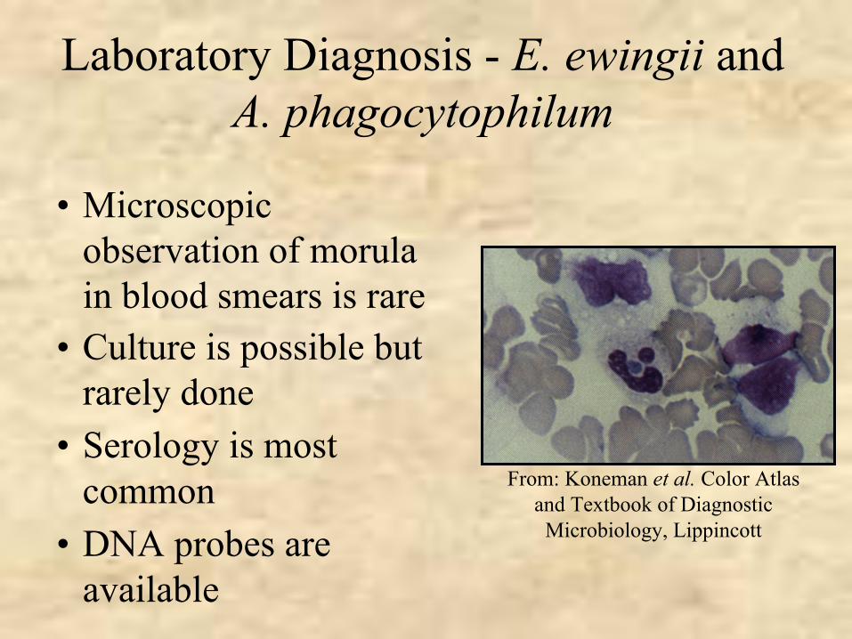
Laboratory Diagnosis - E. ewingii and A. phagocytophilum
• Microscopic observation of morula in blood smears is rare
From: Koneman et al. Color Atlas and Textbook of Diagnostic
Microbiology, Lippincott
• Culture is possible but rarely done
• Serology is most common
• DNA probes are available
Treatment, Prevention and ControlE. ewingii and A. phagocytophilum
• Doxycycline• Avoidance of ticks

Coxiella
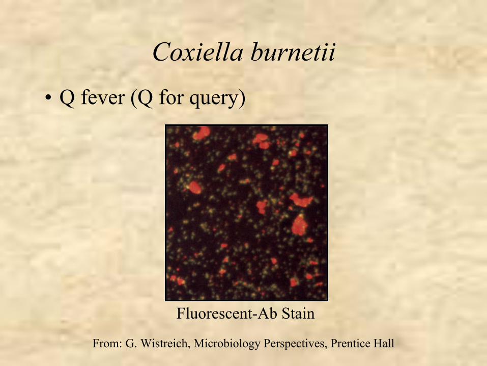
Coxiella burnetii• Q fever (Q for query)
Fluorescent-Ab Stain
From: G. Wistreich, Microbiology Perspectives, Prentice Hall
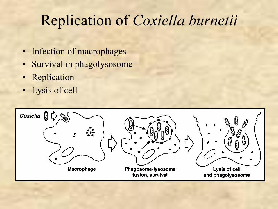
Replication of Coxiella burnetii
• Infection of macrophages• Survival in phagolysosome• Replication• Lysis of cell
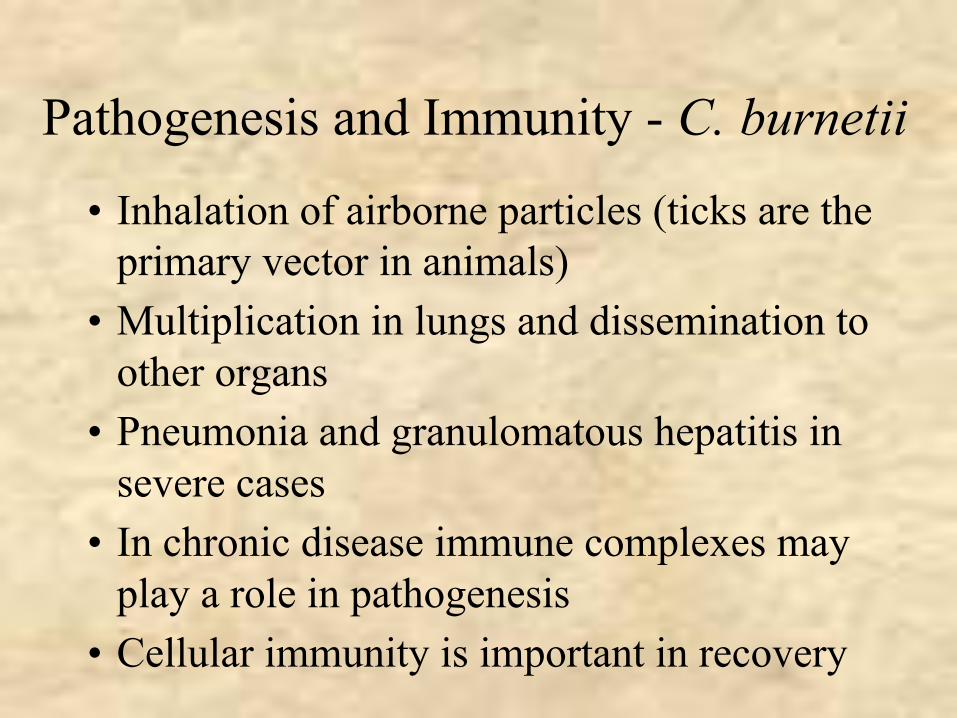
Pathogenesis and Immunity - C. burnetii
• Inhalation of airborne particles (ticks are the primary vector in animals)
• Multiplication in lungs and dissemination to other organs
• Pneumonia and granulomatous hepatitis in severe cases
• In chronic disease immune complexes may play a role in pathogenesis
• Cellular immunity is important in recovery
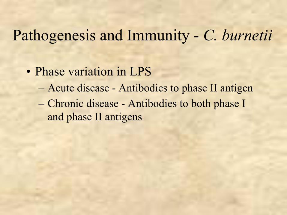
Pathogenesis and Immunity - C. burnetii
• Phase variation in LPS– Acute disease - Antibodies to phase II antigen – Chronic disease - Antibodies to both phase I
and phase II antigens
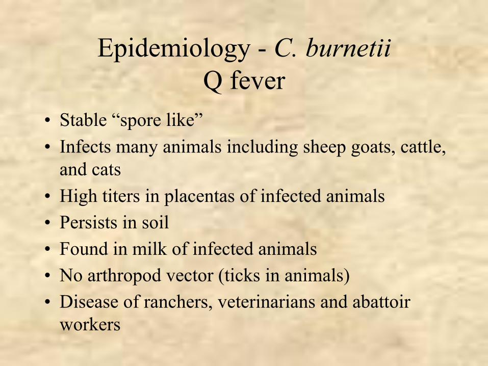
Epidemiology - C. burnetiiQ fever
• Stable “spore like”• Infects many animals including sheep goats, cattle,
and cats• High titers in placentas of infected animals• Persists in soil• Found in milk of infected animals• No arthropod vector (ticks in animals)• Disease of ranchers, veterinarians and abattoir
workers
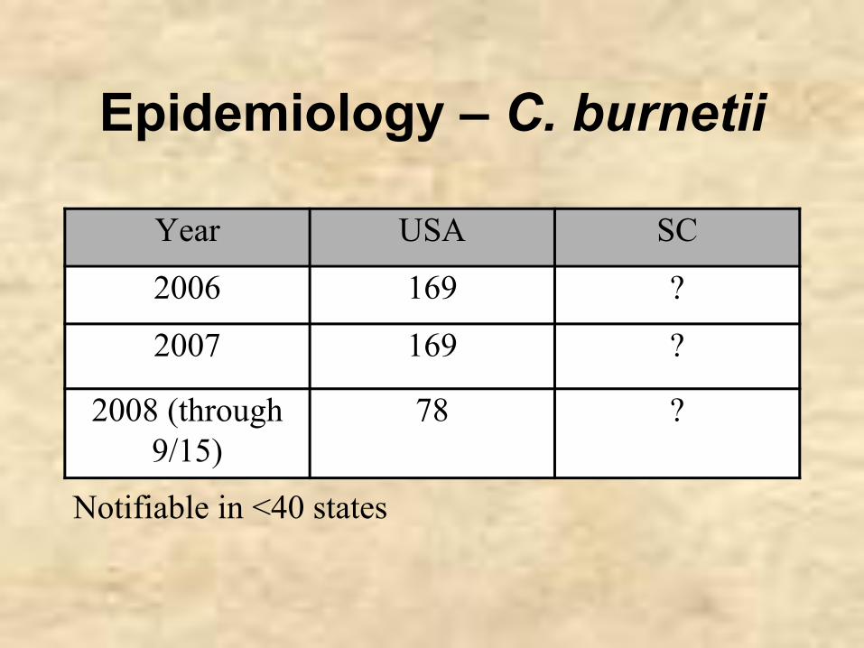
Epidemiology – C. burnetii
Year USA SC
2006 169 ?
2007 169 ?
2008 (through 9/15)
78 ?
Notifiable in <40 states
Clinical Syndrome - Q Fever• Acute Q fever
– Can be mild or asymptomatic – fever, chills, headache and myalgia– Respiratory symptoms usually mild (atypical
pneumonia)– Hepatomegaly and splenomegaly can be observed– Granulomas in the liver are observed histologically
• Chronic Q fever– Typically presents as endocarditis on a damaged heart
valve– Prognosis is poor
Laboratory Diagnosis - C. burnetii
• Serology– Acute disease - Ab to phase II antigen– Chronic disease - Ab to both phase I and phase
II antigens
Treatment, Prevention and ControlC. burnetii
• Acute Q fever - tetracycline• Chronic Q fever - combination of antibiotics• Vaccine is available but it is not approved
for use in the USA

Case Study – Coxiella burnetii• A 56 year-old woman presented with a high fever (104o), hepatomegaly and
elevated liver enzymes• Diagnosis: Acute cholecystitis; cholecystectomy performed• Patient’s symptoms persisted• Chest CT scan performed 4 weeks later revealed nonspecific interstitial lung
disease.• Serum samples obtained at the time of the CT scan and 6 weeks later revealed
antibodies to C. burnetii phase II antigens • Her husband also developed a febrile illness 3 days after her illness started and
his serum samples revealed the presence of antibodies to C. burnetii phase II antigens
• The patients were both treated with doxycycline and their symptoms resolved• They did not own livestock but drove on an unpaved road past a neighbor who
raised goats.
• The goats tested positive for antibodies to C. burnetii

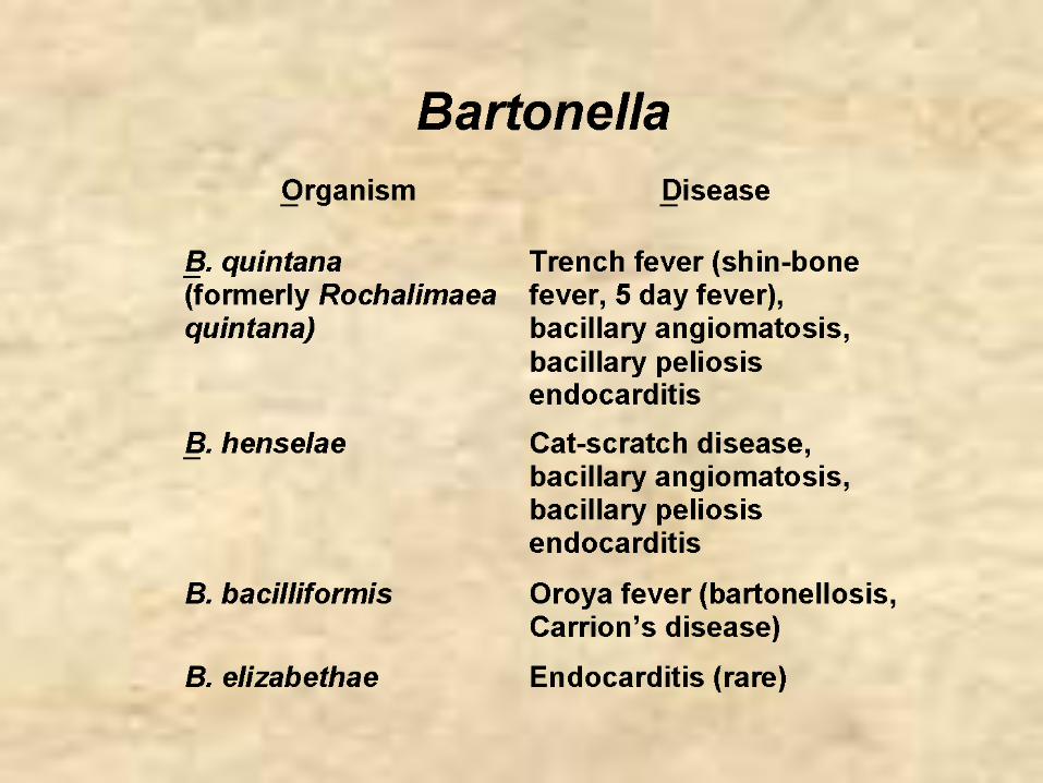
Bartonella
Microbiology - Bartonella
• Small Gram-negative aerobic bacilli• Difficult to culture• Infect animals but do not cause disease in
animals• Insects are thought to be the vectors in
human disease• Some species infect erythrocytes other
attach to cells
Bartonella quintana
• Trench fever– Shin-bone fever– 5 day fever
Epidemiology - B. quintanaTrench Fever
• Associated with war and famine• Vector - human body louse
– Organism found in feces• Reservoir - humans
– No transovarian transmission– Cycle - human to louse to human
Clinical Syndrome - Trench Fever
• Infection may be asymptomatic or severe• Sudden onset of fever, chills, headache and
myalgia• Severe pain in the tibia (shin-bone fever)• Symptoms may appear at 5 day intervals (5 day
fever)• Maculopapular rash may or may not develop on
the trunk• Mortality rates very low.
Laboratory Diagnosis - B. quintana
• Serology - reference laboratories• PCR - reference laboratories
Treatment, Prevention and ControlB. quintana
• Various antibiotics• Control of body louse
Bartonella henselae
• Cat-scratch disease
Epidemiology - B. henselaeCat-scratch Disease
• Acquired from cat bite or scratch and possibly from cat fleas
Clinical SyndromeCat-scratch Disease
• Benign disease• Chronic regional lymphadenopathy

Laboratory Diagnosis - B. henselae
• Serology

Treatment - B. henselae
• Does not respond to antimicrobial therapy