viral glycoproteins: biological role and application in diagnosis
TRANSCRIPT

REVIEW ARTICLE
Viral glycoproteins: biological role and application in diagnosis
Nilotpal Banerjee1 • Sumi Mukhopadhyay1
Received: 21 September 2015 / Accepted: 10 December 2015 / Published online: 18 January 2016
� Indian Virological Society 2016
Abstract The viruses that infect humans cause a huge
global disease burden and produce immense challenge
towards healthcare system. Glycoproteins are one of the
major components of human pathogenic viruses. They have
been demonstrated to have important role(s) in infection
and immunity. Concomitantly high titres of antibodies
against these antigenic viral glycoproteins have paved the
way for development of novel diagnostics. Availability of
appropriate biomarkers is necessary for advance diagnosis
of infectious diseases especially in case of outbreaks. As
human mobilization has increased manifold nowadays,
dissemination of infectious agents became quicker that
paves the need of rapid diagnostic system. In case of viral
infection it is an emergency as virus spreads and mutates
very fast. This review encircles the vast arena of viral
glycoproteins, their importance in health and disease and
their diagnostic applications.
Keywords Viral glycoprotein � Immunodiagnostics �Biomarker � Viral pathogenesis
Introduction
Being an obligate intracellular parasite [32], virus is the
most deadly microbe to be dealt with. Globally it accounts
for extremely high morbidity and mortality throughout the
age groups of people [3, 62]. Thousands of new viral
strains are discovered till date affecting people producing a
huge global burden of viral infections resulting immense
challenge towards healthcare system [9, 20, 48]. With the
capability of fast mutation, viruses affect the host cells with
new and newer mechanisms. So to detect them at the
earliest, there is an extreme need of dynamic diagnostic
system.
Glycans are major components of the outermost surface
of viruses. Thus, majority of the interactions of viral
pathogens with their hosts are influenced by the pattern of
glycans and glycan-binding receptors that each expresses.
[5, 95, 98] Glycans are most complex biomolecules due to
extensive branching of carbohydrates, and a variety of
glycoproteins have been identified in human viral patho-
gens. These pathogenic glycans either virus encoded or
host derived usually elicit high humoral responses in
human body [34]. These virus specific high levels of glycan
specific antibodies have been exploited to develop novel
diagnostic assays.
Viral diagnostic tests can be broadly classified into three
categories in general. Those are direct detection, indirect
examination (virus isolation), and by serology. In case of
direct detection, the clinical sample is examined directly to
identify any presence of virus particles, virus antigen or
viral nucleic acids. In case of indirect examination, the
sample has to be added into cell culture, eggs or animals to
grow the virus in vitro. This is known as virus isolation.
Serology always constitutes the bulk of the work of any
virology laboratory, especially in overpopulated third
world countries. Serological diagnosis is generally made by
detecting titres of antibody in infection [8]. Generally, the
majority of common viral infections are diagnosed by
serology [86]. Viruses can be directly detected through
electron microscopy. It can also be enumerated by
molecular biological techniques like PCR/RTPCR by
detecting viral genomes. These techniques are extremely
& Sumi Mukhopadhyay
1 Department of Laboratory Medicine, School of Tropical
Medicine, 108, C.R Avenue, Kolkata 700073, India
123
VirusDis. (January–March 2016) 27(1):1–11
DOI 10.1007/s13337-015-0293-5

useful but are technically demanding, costly and require
skilled personnel. On the other hand, indirect detection by
virus isolation is dependent on cell culture techniques. The
major problem of cell culture is it takes a long time (up to
4 weeks). Also, the sensitivity is poor and depends on
many factors, such as the specimen condition and the
condition of the cell line. Cell cultures are also very sus-
ceptible to microbial contamination and toxins present in
the specimen. Also, many viruses do not grow at all in cell
culture e.g. Hepatitis B and C, viruses causing diarrhea,
parvovirus etc. Serology is the mainstream of viral diag-
nosis [8, 44]. With increase in the growth of sophisticated
immunoassay techniques, effective viral immunodiagnostic
assays are now available in the market [13, 91, 96].
The detection of structural glycoproteins of viruses or
early glycoprotein antigen formation in the host due to
viral infection or the quantification of titres of antibodies
against viral antigenic glycoprotein is an emerging dis-
cipline in viral immunodiagnostics [47]. The detection of
these structural glycoproteins of viruses is done by lec-
tins or monoclonal antibodies acting as probe or by
measuring the titres of host antibodies against antigenic
glycoprotein.
There are several good review works on viral glyco-
proteins. Namely, the work of Kazuya I.P.J. Hidari and
Takashi Suzuki on Glycan receptor in influenza Virus [43].
Yuan et al. [116] worked on receptor glycoprotein inter-
action in Zaire Ebola Virus (ZEV). This review attempts to
conglomerate the importance of glycoprotein in widely
studied viral infection and their application in diagnosis.
Viral glycoproteins
A fully assembled infectious virus is known as virion. The
simplest virions consist of two basic components, namely
nucleic acid (single- or double-stranded RNA or DNA) and
a capsid, which is a protein coat, functions as a shell to
protect the viral genome from nucleases. This capsid comes
into play during infection to attach the virion to specific
receptors exposed on the prospective host cell. Capsid
proteins are coded by the viral genome. Due to its limited
size, the genome codes for only a few structural proteins
(besides non-structural regulatory proteins involved in
virus replication). Capsids are formed as single or double
protein shells and consist of only one or a few structural
protein species. Therefore, multiple protein copies must
self assemble to form the continuous three-dimensional
capsid structure [35]. The structural viral proteins are
extremely important to the virus, so as to facilitate the
transfer of the viral nucleic acid from one host cell to
another. The proteins determine the antigenicity of the
virus. Host’s primary immune response is directed against
the antigenic determinants of these proteins rather glyco-
protein in major cases.
There are enveloped Viruses and these envelopes are
made up of either lipid or glycoprotein. Viral envelopes
mainly consist of Envelope proteins (E), Membrane pro-
teins (M) and Spike proteins (S) [24]. Lipid envelopes are
derived from the host cell. Whereas the envelope glyco-
proteins are virus encoded. However, there are sugars
attached to the viral glycoproteins which often reflect the
host cell that harboured the virus. The surface glycopro-
teins of an enveloped virus attach the virion to a target host
cell by properly interacting with a cellular receptor [22].
Structural biological analysis of viral envelope glycopro-
teins reveals that viruses have wide range of folds to
facilitate their attachment with proper host receptors.
Bowden et al. [10] stated that Arenaviridae group of
viruses have a/b fold, whereas Filoviridae possess ‘Chal-
ice’ of GP1. Similarly, Paramyxoviridae shows six bladed
b propeller and large trimeric haemaglutinin is shown by
Orthomyxoviridae. Glycosylated GP120 trimer is observed
in the Lentiviruses of Retroviridae. Viruses exhibit
‘Semaphorins’ which are family of cell surface signalling
glycoproteins [19]. These semaphorins binds with cell
surface receptors to initiate important physiological pro-
cesses. These observations are made by recent study of
viral glycoproteins by employing Macromolecular crys-
tallography [10]. The M and S proteins of the virus are
usually rich in N glycosylated proteins, which have been
demonstrated as important virulent factor of viruses [98].
Thus, E, M and S viral glycoproteins are involved in viral
host binding and subsequent virus-host membrane fusion to
establish the pathogenesis of the virus. Two envelope
glycoproteins, namely E1 and E2 develop the viral spike of
the virions of Flaviviridae family [61] are involved in the
engagement with host receptor and conformational change
required for membrane fusion (Fig. 1c). Studies show that
E2 can express independently but E1 is dependent upon E2
in case of HCV. SARS Coronavirus possess a
spike(S) glycoprotein [70], which itself performs the
membrane fusion for the entry of the virion and its fusion
with host cell [115]. In case of Chikungunya virus,
attachment is facilitated by the E2 glycoprotein [89] and
fusion mainly by the E1 glycoprotein, thus both the pro-
cesses are mutually exclusive, whereas in Dengue virus, it
is carried by the same E protein (http://www.uniprot.org/
uniprot/Q8JUX5). Interestingly, the dengue virus apart
from synthesizing the basic capsid, membrane and envel-
ope proteins also produces seven non-structural secretory
glycoproteins NS1,2A, 2B, 3, 4A,4B,5 [46]. These proteins
are not integrated in the virus but secreted in the host.
Studies have found heterogeneity in the E glycoprotein of
Dengue virus [56]. Five different glycans are present in this
glycoconjugate including Mannose, GalNac and GlcNac,
2 N. Banerjee, S. Mukhopadhyay
123

Fucose and Sialic Acid. B cell and T-cell epitopes are
predicted in a study by analysing this E glycoprotein [46].
The Dengue Viral envelope is more ordered than the inner
viral core, as the envelope is composed of 90 glycoprotein
E dimer icosahedral scaffold [58]. Computational studies
are there to develop vaccines against Dengue virus [4, 97].
There are three glycoproteins present in HIV [1];
namely gp 120, gp 160 and gp 41 [38]. All these are
encoded by the ENV gene [76]. The HIV envelope gly-
coprotein gp120 contains nine disulphide bridges and is
highly glycosylated, carrying on average 24 N-linked
glycans (Fig. 1d) [73]. Experiments proved that the glycan
part of the gp 41 protein has important role in the efficient
intracellular transport of another glycoprotein gp 160.
Those gp 160 proteins lack gp41 are arrested in golgi
complex after their biosynthesis [27]. Zaire Ebola Virus is
the member of Filoviridae group, and the Glycoproteins
(GP) have found to be major pathogenic determinants [24–
26, 63, 75, 99]. In the Ebola virion GP gene is the 4th gene
among total seven genes in the linear gene order. This
synthesizes several proteins. Among them two are pre-
dominant. Those are sGP and D-peptide (delta peptide).
These two proteins are produced due to a furin cleavage of
a precursor pre-sGP protein. The GP is actually a Spike
protein which is composed of two subunits joined by
disulphide linkage Gp1-Gp2 [92].
The Chikungunya Virus on the other hand are known to
produce 4 Non structural glycoproteins (nsP1-4) [91] these
nsPs have been demonstrated to have important role in
keeping the replicase complex of the virus intact in the host
as well as to circumvent important host immune responses.
Chikungunya Virus has two envelope proteins, namely E1
and E2 [11]. Thus, viral glycoproteins have diverse struc-
ture and function. Taken together, glycoproteins are
important components of the virus structure and each have
unique role to establish pathogenesis. [17].
Viral glycoproteins have a definite role in their patho-
genesis. The primary goal of viral infection is to identify a
receptor on the host cell surface and binding with it.
Subsequently this will pave the way of viral entry into the
host cell. In most cases, the first attachment site of the virus
is a glycan, either a glycoprotein or a glycolipid. So, gly-
coproteins play a crucial role in viral pathogenesis. The
study of glycoproteins in viral infection is most important
to know the disease process as well as to develop antiviral
treatments. Glycoprotein–receptor interactions also play
Fig. 1 Distribution of Glycoproteins on the surfaces of different viruses a influenza virus, b SARS Coronavirus, c Hepatitis C virus, d human
immunodeficiency virus, e Ebola virus, f Dengue virus and g Chikungunya virus
Viral glycoproteins: biological role and application in diagnosis 3
123

important roles in pathogen pattern recognition and in the
regulatory signals that control the activities of cells of the
immune system. The most important cause behind viral
infection is that it has evolved to present its own sugars and
receptors in a manner that mimics or interferes with host
glycan-based immune functions. Glycomic studies are
ongoing in several viruses. Several advanced technologies
are there to decipher structural and functional aspects of
glycans like Glycan microarray [40], Mass Spectrometry
and Nano LC. Glycan array represents the actual in vivo
interaction in silico. The arrayed multivalent demonstration
of polysaccharides mimics the cell surface display. There
are two types of carbohydrate microarray. Those are
polysaccharide and oligosaccharide microarray [53]. Nat-
ural polysaccharides are randomly immobilized on solid
matrices exploiting hydrophobic physical absorption or
charge-based interaction. Polysaccharide microarrays are
useful for comparative antigenicity analyses. Being
hydrophilic in nature, oligosaccharides need chemical
derivatization before arraying. Through oligosaccharide
microarray we can study structure–activity relationships
[52]. Microarrays were developed on maleimide-function-
alized surfaces using seven thiol-containing synthetic high-
mannose oligosaccharides for the identification of human
immunodeficiency virus (HIV) vaccine candidate antigens
[2]. The binding profile reveals that several proteins which
interact with gp120 of HIV, like the receptor of the innate
immune system known as DC-SIGN (CD 209). In case of
Influenza glycans, there are protocols for fluorescent
labeling of virus, coupling of virus to a glycan microarray,
analysis of a glycan microarray slide experiment, and data
interpretation. Studies have shown that there are a2,3-linked sialic acid motif (SA2, 3Gal) in avian, equine, and
canine species. Whereas a2, 6-linked sialic acid motif
(SA2, 6Gal) is present in humans. SAa2, 3Gal and
SAa2,6Gal are present in swine, these are causing corre-
sponding host tropism. Zhao et al. [117] showed that,
association mining results of glycan microarray [2] data
with 211 influenza viruses from five host groups: humans,
swine, canine, migratory waterfowl, and terrestrial birds
[72]. The study suggest that besides Neu5Aca2-6Galb,human-origin viruses could bind glycans with Neu5Aca2-8Neu5Aca2-8Neu5Ac and Neu5Gca2-6Galb1-4GlcNAcsubstructures; Galb and GlcNAcb terminal substructures,
without sialic acid branches these were linked with the
binding of human, swine, and avian origin viruses. Sulfated
Neu5Aca2-3 substructures were associated with the bind-
ing of human- and swine-origin viruses. Finally, through
three-dimensional structure characterization, it has been
revealed that the role of glycan chain shapes is more
important than that of torsion angles [117].
Though characterization of Glycoproteins is tough but,
through Mass Spectrometry, it is now easier to identify
structural details of complex glycoproteins. Mass spec-
trometry derived glycoproteomics [118] helps us to pre-
cisely identify viral and cellular proteins that are
functionally, structurally, and dynamically altered during
virus infection, but enables us to identify important pro-
teins having active role in the infection pathway. Addi-
tionally, isolation and purification techniques along with
quantitative strategies in conjunction with MS signifi-
cantly improve its sensitivity to detect low-abundant
proteins. With time, more virus and host genomes are
being sequenced and MS-based glycoproteomics is
becoming a very important tool for virology. A work by
Barrientos et al. [7] revealed that post translational
modification of secretory glycoprotein of Zaire Ebola
Virus can be characterized by Mass spectrometry.
MALDI-TOF MS (Matrix-assisted laser desorption/ion-
ization–time-of-flight mass spectrometry) also enables to
identify regions susceptible to limited proteolysis in sGP
of ZEV.
Another work by Anastassia et al. [54] shows that 0.1
microgram of viral glycoprotein can be purified by Nano-
Liquid Chromatography. After Nano-LC the sample is
analysed through mass spectrometry. One more work
showed that they purified Heat shock protein 90 by
NanoLC-MS of Respiratory syncitial virus which have
important role in virus particle assembly [54, 80]. So it is
evident that using these modern techniques, the biological
roles of glycoproteins can be studied more conveniently.
Viral glycoproteins and their biological role
Virus is a nucleic acid surrounded by proteins. This
infective particle is called a virion. In most cases this virion
is covered with a fascinating coat composed of glycopro-
teins through which the virus communicates with its host.
The co-evolution of host and virus leads the way of making
the glycoprotein coat so fascinating [23]. It is evident that
the infectivity of a virus rather of its nucleic acid is fully
dependent on its glycoproteins. Enveloped viruses gener-
ally encode membrane proteins and these special proteins
are necessary to mediate the specific binding against host
cell ligands. This also directs initial events of membrane
fusion and viral internalization. These fascinating envelope
proteins are generally glycosylated [15]. The process of
glycosylation takes place in the endoplasmic reticulum
(ER)-Golgi complex secretory pathway. The host cell
encoded glycosyl-transferase enzyme catalyses the glyco-
sylation. This glycosylation is necessary to make the virion
host compatible, which is needed by the virus for its
pathogenesis. So glycans present at the envelop proteins
acts as immunological barriers to resist evasion by the host
immune system [22].
4 N. Banerjee, S. Mukhopadhyay
123
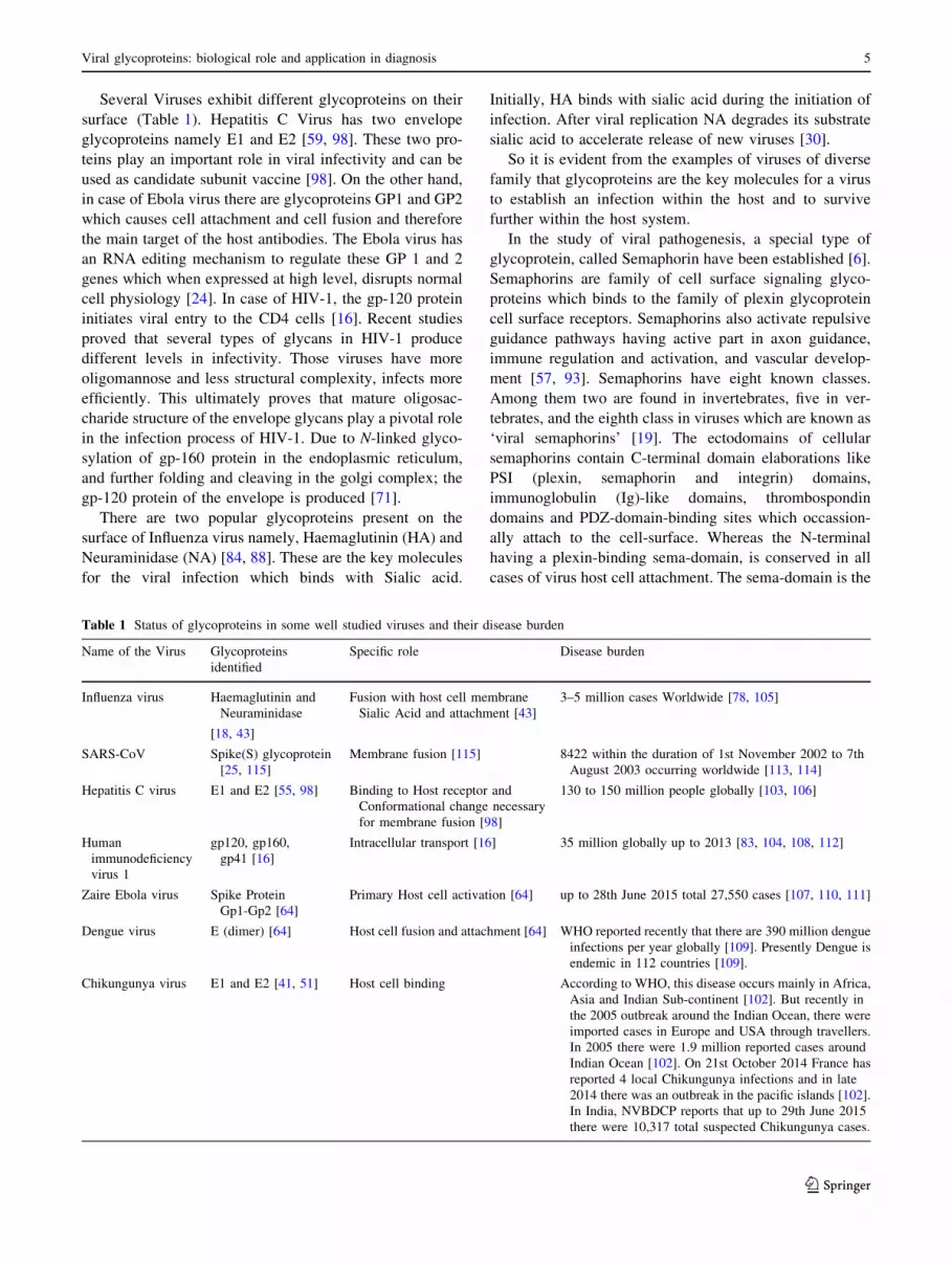
Several Viruses exhibit different glycoproteins on their
surface (Table 1). Hepatitis C Virus has two envelope
glycoproteins namely E1 and E2 [59, 98]. These two pro-
teins play an important role in viral infectivity and can be
used as candidate subunit vaccine [98]. On the other hand,
in case of Ebola virus there are glycoproteins GP1 and GP2
which causes cell attachment and cell fusion and therefore
the main target of the host antibodies. The Ebola virus has
an RNA editing mechanism to regulate these GP 1 and 2
genes which when expressed at high level, disrupts normal
cell physiology [24]. In case of HIV-1, the gp-120 protein
initiates viral entry to the CD4 cells [16]. Recent studies
proved that several types of glycans in HIV-1 produce
different levels in infectivity. Those viruses have more
oligomannose and less structural complexity, infects more
efficiently. This ultimately proves that mature oligosac-
charide structure of the envelope glycans play a pivotal role
in the infection process of HIV-1. Due to N-linked glyco-
sylation of gp-160 protein in the endoplasmic reticulum,
and further folding and cleaving in the golgi complex; the
gp-120 protein of the envelope is produced [71].
There are two popular glycoproteins present on the
surface of Influenza virus namely, Haemaglutinin (HA) and
Neuraminidase (NA) [84, 88]. These are the key molecules
for the viral infection which binds with Sialic acid.
Initially, HA binds with sialic acid during the initiation of
infection. After viral replication NA degrades its substrate
sialic acid to accelerate release of new viruses [30].
So it is evident from the examples of viruses of diverse
family that glycoproteins are the key molecules for a virus
to establish an infection within the host and to survive
further within the host system.
In the study of viral pathogenesis, a special type of
glycoprotein, called Semaphorin have been established [6].
Semaphorins are family of cell surface signaling glyco-
proteins which binds to the family of plexin glycoprotein
cell surface receptors. Semaphorins also activate repulsive
guidance pathways having active part in axon guidance,
immune regulation and activation, and vascular develop-
ment [57, 93]. Semaphorins have eight known classes.
Among them two are found in invertebrates, five in ver-
tebrates, and the eighth class in viruses which are known as
‘viral semaphorins’ [19]. The ectodomains of cellular
semaphorins contain C-terminal domain elaborations like
PSI (plexin, semaphorin and integrin) domains,
immunoglobulin (Ig)-like domains, thrombospondin
domains and PDZ-domain-binding sites which occassion-
ally attach to the cell-surface. Whereas the N-terminal
having a plexin-binding sema-domain, is conserved in all
cases of virus host cell attachment. The sema-domain is the
Table 1 Status of glycoproteins in some well studied viruses and their disease burden
Name of the Virus Glycoproteins
identified
Specific role Disease burden
Influenza virus Haemaglutinin and
Neuraminidase
[18, 43]
Fusion with host cell membrane
Sialic Acid and attachment [43]
3–5 million cases Worldwide [78, 105]
SARS-CoV Spike(S) glycoprotein
[25, 115]
Membrane fusion [115] 8422 within the duration of 1st November 2002 to 7th
August 2003 occurring worldwide [113, 114]
Hepatitis C virus E1 and E2 [55, 98] Binding to Host receptor and
Conformational change necessary
for membrane fusion [98]
130 to 150 million people globally [103, 106]
Human
immunodeficiency
virus 1
gp120, gp160,
gp41 [16]
Intracellular transport [16] 35 million globally up to 2013 [83, 104, 108, 112]
Zaire Ebola virus Spike Protein
Gp1-Gp2 [64]
Primary Host cell activation [64] up to 28th June 2015 total 27,550 cases [107, 110, 111]
Dengue virus E (dimer) [64] Host cell fusion and attachment [64] WHO reported recently that there are 390 million dengue
infections per year globally [109]. Presently Dengue is
endemic in 112 countries [109].
Chikungunya virus E1 and E2 [41, 51] Host cell binding According to WHO, this disease occurs mainly in Africa,
Asia and Indian Sub-continent [102]. But recently in
the 2005 outbreak around the Indian Ocean, there were
imported cases in Europe and USA through travellers.
In 2005 there were 1.9 million reported cases around
Indian Ocean [102]. On 21st October 2014 France has
reported 4 local Chikungunya infections and in late
2014 there was an outbreak in the pacific islands [102].
In India, NVBDCP reports that up to 29th June 2015
there were 10,317 total suspected Chikungunya cases.
Viral glycoproteins: biological role and application in diagnosis 5
123

only component found in viruses. Crystallographic studies
by Bowden et al. [10] have revealed that human Sema3A
and mouse Sema4D semadomains comprises of structurally
conserved homodimer of seven-bladed b-propellers [6, 50,65, 69, 77]. The immune-regulatory semaphorins like
Sema3A, 4A, 4D, and 7A helps in B cell mediated
immunity (Sema4D), T cell activation as well as differ-
entiation (Sema4A, Sema3A, and Sema4D), and inflam-
mation (Sema7A) [93]. These semaphorins provide a
molecular basis for how viruses can optimize their own
proteins to override normal physiological interactions.
A work by Shirato H as ‘Norovirus and histo-blood
group antigens’ in the journal Jpn J Infect Dis.
(2011;64(2):95–103) describes that NoroVirus (NoV)
causes viral gastroenteritis and interestingly bind to histo-
blood group antigens (HBGAs), like ABH antigens and
Lewis antigens. It has been shown epidemiologically that
persons with different ABH phenotypes are infected with
NoV strains in a genotype-dependant fashion. An in vitro
binding assay using NoV virus-like particles (VLPs)
showed a uniform recognition pattern for type 1 and 2 core
structures of histo blood group antigens. NoV VLPs bind
more tightly to type 1 carbohydrates than to type 2. Type 1
carbohydrates are found to be expressed at the surface of
the small intestine and targeted by NoV. This property
speaks about NoV tissue specificity.
So it is evident that glycoproteins perform a major
and active role in viral pathogenesis and disease
progression.
Glycoproteins provide tissue tropism to the virus. Some
virues used to infect the respiratory system whereas some
affects the liver. The cause is the type of glycoprotein with
which the virus binds to accelerate its invasion.
In a study by Raska et al. [81], it has been proved that
there are differential glycosylation in viruses like HIV1
depending upon the cells which produce the virus. N-gly-
cosylation of reconbinant gp120 of HIV1 is varied and
affected the recognition by serum antibodies. Glycosyla-
tion of gp120 protein of HIV1 affects its recognition by
neutralizing and non neutralizing monoclonal antibodies.
This study also says that this glycosylation is cell specific.
Another study by Lin et al. [64] stated that there are
C-type lectins expressed on the Dendritic Cell surface
known as DC-SIGN(Dendritic Cell-Specific Intercellular
adhesion molecule-3-Grabbing Non-integrin) or CD 209
and DC-SIGNR which binds to HIV1 and transmit to T
cells through the viral envelope Env glycoprotein. But
interestingly other highly glycosylated Viruses failed to
interact with DC-SIGNR [64]. Lin et al. showed that DC-
SIGN (R) or CD 209 selectively binds with HIV1 Env and
Zaire Ebola Virus glycoproteins containing more high-
mannose. By modulating N-glycans on Env or glycoprotein
during virus production in different primary cells or in the
presence of the mannosidase I inhibitor deoxymannojir-
imycin affected DC-SIGN(R) infectivity enhancement.
They also predict that viruses containing glycoproteins
with a high amount of high-mannose N-glycans effectively
interact with DC-SIGN(R), but those viruses having only
complex N-glycans cannot effectively react with DC-
SIGN(R). So it is evident that virus-producing cell type is a
crucial factor in depicting both N-glycan status and virus
interactions with DC-SIGN(R), which establishes virus
tropism and infection within the human body [64].
Liu et al. [66] described in their study that sialic acid
present on cell surface is essential for Human EnteroVirus
D 68 (EV-D68) entry. Crystallographic studies showed that
EV-D68 with sialylated glycan receptor analogues binds on
the viral surface. Sialic acid receptor induces a cascade of
conformational changes within the virus to secrete a fatty-
acid-like molecule which regulates the stability of the
virus. So, it is evident that binding of virus to a sialic acid
receptor and to immunoglobulin-like receptors facilitates
viral entry in enteroviruses.
Application of viral glycoproteins in diagnostics
Glycan based viral immunodiagnostics usually have high
sensitivity and specificity. Glycoprotein based IgM serol-
ogy was developed for the diagnosis of recent primary
rubella virus infections and significant sensitivity and
specificity was obtained. Similarly, Glycoprotein based
serology tests to detect antibodies to herpes simplex virus
glycoproteins G-1 and G-2, which evoke a type-specific
antibody response have also been developed. These tests
are used to confirm a diagnosis of genital herpes, and also
to establish diagnosis of HSV infection in patients with
atypical complaints, to identify asymptomatic carriers, and
identify persons at risk for acquiring HSV. Glycan based
immunodiagnostics have also been developed for the rapid
identification of different strains of Influenza Virus. A
novel peptide based ELISA which has sensitivity and
specificity of 96.55 and 74.4 % respectively is very
promising. On the other hand, the main diagnostic chal-
lenge related to SARS is to diagnose it differently with
atypical pneumonia [68, 79]. The key diagnostic tools are
immunofluorescent staining, ELISA and RT-PCR [67]. But
all these techniques are extremely sophisticated and of little
use in case of epidemics; especially in the developing
world. SARS Coronavirus possess a spike(S) glycoprotein,
which itself performs the membrane fusion for the entry of
the virion and its fusion with host cell (Fig. 1b) [115]. IgG
based diagnostics against this S protein has been developed
[114]. Indirect ELISA test has been developed by using
recombinant SARS ‘S’ protein and the N (nucleoprotein)
protein. The sensitivity of SARS ‘S’ and ‘N’ proteins are
6 N. Banerjee, S. Mukhopadhyay
123

100 and 96.7 % respectively whereas the specificity of
SARS ‘S’ and ‘N’ are 98.55 and 98.4 % [114].
Similarly, as acute Hepatitis C infection is asymp-
tomatic, so it is difficult to diagnose early. Generally
patients come to the clinic with damaged liver. Initially
patients are screened by anti-HCV antibody test and further
confirmed by testing Viral RNA. There are two glycopro-
teins E1 and E2 in HCV. Standardised PCR system is
available in HCV diagnostics but a Core antigen test is also
in the market. In 1995 Tanaka et al. [21] suggested that the
HCV core proteins can be used as antigens for the chronic
stage. Studies for developing Algorithms confirms 99.05 %
sensitivity in case of RT-PCR whereas 98.10 % for the
Core Antigen Test [82]. Another novel glycoproteomic
serum biomarker has been identified which can diagnose
HCV along with progressive liver cirrhosis is Wisteria
floribunda agglutinin positive Mac-2-binding protein
(WFA?-M2BP) [29]. The diagnostic threshold for cut-off
index values of this protein is 1.435 and 4.615 in HCV
negative and HCV positive patients showing progressive
liver cirrhosis [39].
In case of HIV1, ELISA and PCR are two Gold standard
tests. Type specific conformational epitopes of the gp160
and gp 41 glycoproteins of the HIV envelope are used for
the recognition of ‘early HIV antibodies’ [14]. Enzyme
Immuno assay (EIA) and RT-PCR are used in these assays.
These tests are very specific but they fail to diagnose early
infection [14]. The most challenging part in HIV diagnosis
is to diagnose the acute stage and differentiate ‘‘Window
Phase’’ patients from the serologically positive patients
[87]. Popular Serological tests fail to diagnose all patients
of HIV as they exploit the GAG proteins which literally
decrease after disease progression [60, 74]. There are three
glycoproteins present in HIV; namely gp 120, gp 160 and
gp 41. All these are encoded by the ENV gene [76]. The
HIV envelope glycoprotein gp120 contains nine disulphide
bridges and is highly glycosylated, carrying on average 24
N-linked glycans (Fig. 1d) [73]. Experiments proved that
the glycan part of the gp 41 protein has important role in
the efficient intracellular transport of another glycoprotein
gp 160. Those gp 160 proteins lack gp41 are arrested in
golgi complex after their biosynthesis [27]. As stated
above, there are limitations of the popular GAG antigen
based serological tests which cannot diagnose HIV patients
of different clinical stage. But antibodies against precursor
gp160 ENV protein and final ENV proteins gp 120 and gp
41 can detect all clinical stages of HIV [74]. The Sensi-
tivity of current available diagnostic system is 38 % at
\7 days, 97 % at 7–41 days and 95 % at 42–93 days [74]
The Specificity is almost 95 % [90].
In case of Zaire Ebola Virus, initially there was con-
troversy about the role of glycoproteins in the pathogenesis
of EBV. But later on, scientific researches proved that the
primary host cell activation by the EBV is mediated by
GP1-2 [100]. An antigen capture ELISA has been devel-
oped in Zaire EBOV using mAbs [85]. It has been reported
that these tests have both high sensitivity and specificity
[85].
Similarly, Dengue (DENV) NS1 is a highly conserved
glycoprotein, expressed as both membrane-associated and
secreted forms [33, 36, 94]. Secreted NS1 has been
detected ranging from 2–0.04 lg/mL in the serum of
dengue-infected patients during the early stages of the
disease. A high NS1 level has been demonstrated to cir-
culate as early as 1 day after onset of symptoms up to early
convalescences thus provides an alternative to virus culture
or PCR for early dengue diagnosis when IgM or IgG
antibodies are not present yet in dengue infected patients
[42, 49]. Circulating dengue NS1 in sera can be detected
either using ELISA assay or lateral flow based RDTs [31].
Thus, glycan based Viral Immunodiagnostics or Glyco-
Immunodiagnostics are helpful in early diagnosis of
patients with viral infection [28].
Current diagnosis scenario in Chikungunya is IgM and
IgG based ELISA and Nucleic Acid detection by RT-PCR
[12, 51]. But there is no Antigen based ELISA. This makes
the condition crucial as the primary health care providers in
the Virus affected countries do not have RT-PCR facilities.
It is not recommended to maintain RT-PCR facilities in
Primary Health care centre by policy. It is extremely costly
and demands expertise. It is not possible to provide such
facility in the densely populated tropical countries. As
Chikungunya causes short duration fever, often the patients
are not diagnosed properly. The joint pain generally per-
sists for some days but can be present for a year [102].
There is a study which shows even multi organ failure in
Chikungunya infected patients [45]. CHIKV has two
envelope glycoproteins, namely E1 and E2 (Fig. 1g) [11].
Recombinant CHIKV E1 and E2 glycoprotein based
ELISA showed a sensitivity of 77.5 and 90 % respectively
whereas the specificity for both cases was 100 % [11, 54]
highlighting the potential for these two glycoproteins in the
diagnosis.
Conclusion
Viral glycoproteins are integral parts of enveloped viruses
and they actively take part in their pathogenesis. Exploting
glycoproteins, viruses enter into their host and combat with
host immune system. Recent advances in technology
deciphers different role of glycoproteins which are
dependent on their structures.
Different viruses have different mode of pathogenesis
and glycoproteins directly takes part in the host binding
and entry. During maturation from host cell viruses have
Viral glycoproteins: biological role and application in diagnosis 7
123

host glycoproteins on their surface to avoid the immunity
of the host. So, to detect viruses and to decide for devel-
oping vaccines [37], glycoproteins always play a key role.
Antibody production is a prominent feature of the
immune response in patients with viral infection, and
particular isotypes correlate with resistance or susceptibil-
ity to infection [101]. A substantial proportion of the
antibodies detected in patients with acute or chronic
infections is directed against viral glycan epitopes. As
levels of anti glycan antibody are high and specific for each
viral infection this permits diagnostic discrimination
between the different viral infections. Taken together, viral
glycoproteins have important functions in pathogenesis and
can be exploited to develop viral diagnostics.
Acknowledgments Mr. Nilotpal Banerjee is a recipient of Junior
Research Fellowship from Department of Biotechnology [File No.
232/BT(Estt.)/RD-24/2014], Government of West Bengal, India.
References
1. About HIV/AIDS, HIV BASICS, CDC. http://www.cdc.gov/hiv/
basics/whatishiv.html. Accessed on 1st June 2015.
2. Adams EW, Ratner DM, Bokesch HR, McMahon JB, O’Keefe
BR, Seeberger PH. Oligosaccharide and glycoprotein microar-
rays as tools in HIV glycobiology; glycan-dependent gp120/
protein interactions. Chem Biol. 2004;11(6):875–81.
3. Alonso WJ, Laranjeira BJ, Pereira SAR, Florencio CMGD,
Moreno EC, Miller MA, Gigilo R, Schuch-Paim C, Moura FEA.
Comparative dynamics, morbidity and mortality burden of
pediatric viral respiratory infections in an equatorial city. Pediatr
Infect Dis J. 2012;31:e9–14. doi:10.1097/INF.
0b013e31823883be.
4. Amat-ur-Rasool H, Saghir A, Idrees M. Computational predic-
tion and analysis of envelop glycoprotein epitopes of DENV-2
and DENV-3 Pakistani isolates: a first step towards dengue
vaccine development. Samuel JE, ed. PLoS One. 2015;10:
e0119854.
5. Amon R, Reuven EM, Ben-Arye SL, Padler-Karavani V. Gly-
cans in immune recognition and response. Carbohydr Res.
2014;7(389):115–22.
6. Antipenko A, Himanen JP, van Leyen K, Nardi-Dei V, Lesniak
J, Barton WA, Rajashankar KR, Lu M, Hoemme C, Puschel
AW, Nikolov DB. Structure of the semaphorin-3A receptor
binding module. Neuron. 2003;39:589–98.
7. Barrientos LG, Martin AM, Pierre W, Rollin E. Secreted gly-
coprotein from live zaire ebolavirus—infected cultures: prepa-
ration, structural and biophysical characterization, and
thermodynamic stability. J Infect Dis. 2007;196(Supplement
2):S220–31.
8. Baumgarten A. Viral immunodiagnosis. Yale J Biol Med.
1980;53(1):71–83.
9. Biernat B, Stanczak J, Szostakowska B, Wroczynska A, Kuna A,
Nahorski WL, Racewicz M. Different serotypes of dengue virus
(DENV) imported by Polish travellers from dengue endemic
areas to Poland. Int Marit Health. 2015;66:72–6.
10. Bowden TA, Jones EY, Stuart DI. Cells under siege: viral gly-
coprotein interactions at the cell surface. J Struct Biol.
2011;175:120–6.
11. Byungki C, Jeon BY, Kim J, Noh J, Kim J, Park M, Park S.
Expression and evaluation of Chikungunya virus E1 and E2
envelope proteins for serodiagnosis of Chikungunya virus
infection. Yonsei Med J. 2008;49:828–35.
12. Caglioti C, Lalle E, Castilletti C, Carletti F, Capobianchi MR,
Bordi L. Chikungunya virus infection: an overview. New
Microbiol. 2013;36:211–27.
13. CDC (2000), MMWR, Interpretation and Use of the Western
Blot Assay for Serodiagnosis of Human Immunodeficiency
Virus Type 1 Infections, http://www.cdc.gov/mmwr/preview/
mmwrhtml/00001431.htm.
14. Chen J, Wang L, Chen JJ, Sahu GK, Tyring S, Ramsey K,
Indrikovs AJ, Petersen JR, Paar D, Cloyd MW. Detection of
antibodies to human immunodeficiency virus (HIV) that rec-
ognize conformational epitopes of glycoproteins 160 and 41
often allows for early diagnosis of HIV infection. J Infect Dis.
2002;186:321–31.
15. Chikungunya Virus, Centers for disease control and prevention.
http://www.cdc.gov/chikungunya/.
16. Chirmule N, Pahwa S. Envelope glycoproteins of human
immunodeficiency virus type 1: profound influences on immune
functions. Microbiol Rev. 1996;60:386–406.
17. Cifuentes-Munoz N, Salazar-Quiroz N, Tischler ND. Hantavirus
Gn and Gc envelope glycoproteins: key structural units for virus
cell entry and virus assembly. Viruses. 2014;6:1801–22.
18. Collins JK, Knight CA. Purification of the influenza hemag-
glutinin glycoprotein and characterization of its carbohydrate
components. J Virol. 1978;26:457–67.
19. Comeau MR, Johnson R, DuBose RF, Petersen M, Gearing P,
VandenBos T, Park L, Farrah T, Buller RM, Cohen JI, Strock-
bine LD, Rauch C, Spriggs MK. A poxvirus-encoded sema-
phorin induces cytokine production from monocytes and binds
to a novel cellular semaphorin receptor. VESPR. Immunity.
1998;8:473–82.
20. Das R. Ebola to have a global burden of $5.9 Billion By End Of
2016, Forbes, Pharma & Healthcare, Nov 12, 2014 @ 03:14 PM.
http://www.forbes.com/sites/reenitadas/2014/11/12/ebola-to-
have-a-global-burden-of-5-9-billion-by-end-of-2016/. Accessed
on 1st June 2015.
21. Dawson GJ. Detection of HCV core antigen, Abbott http://www.
cdc.gov/hepatitis/resources/mtgsconf/hcvsymposium2011-pdfs/
7_dawson.pdf.
22. De Haan CAM, Vennema H, Rottier PJM. Assembly of the
coronavirus envelope: homotypic Interactions between the M
proteins. J Virol. 2000;74:4967–78.
23. Dengue: Guidelines for Diagnosis, Treatment, Prevention and
Control: New Edition. Geneva: World Health Organization;
2009. 4, Laboratory diagnosis and diagnostic tests. Available
from: http://www.ncbi.nlm.nih.gov/books/NBK143156/.
24. Dolnik O, Volchkova VA, Escudero-Perez B, Lawrence P,
Klenk HD, Volchkov VE. Shedding of Ebola virus surface
glycoprotein is a mechanism of self-regulation of cellular
cytotoxicity and has a direct effect on virus infectivity. J Infect
Dis. 2015;212(suppl_2):S322–8.
25. Dong PH, Kima HG, Kima Y, Poonb LLM, Cho MW. Devel-
opment of a safe neutralization assay for SARS-CoV and
characterization of S-glycoprotein. Virology. 2004;326:140–9.
26. Feldmann H, Volchkov VE, Volchkova VA, Stroher U, Klenk
HD. Biosynthesis and role of filoviral glycoproteins. J Gen
Virol. 2001;82:2839–48.
27. Fenouillet E, Jones IM. The glycosylation of human immun-
odeficiency virus type 1 transmembrane glycoprotein (gp41) is
important for the efficient intracellular transport of the envelope
precursor gp160. J Gen Virol. 1995;76:1509–14.
28. Flamand M, Megret F, Mathieu M, Lepault J, Rey FA, Deubel
V. Dengue virus type 1 nonstructural glycoprotein ns1 is
8 N. Banerjee, S. Mukhopadhyay
123

secreted from mammalian cells as a soluble hexamer in a gly-
cosylation-dependent fashion. J Virol. 1999;73(7):6104–10.
29. Fujiyoshi M, Kuno A, Gotoh M, Fukai M, Yokoo H, Kamachi
H, Kamiyama T, Korenaga M, Mizokami M, Narimatsu H,
Taketomi A. Clinicopathological characteristics and diagnostic
performance of Wisteria floribunda agglutinin positive Mac-2-
binding protein as a preoperative serum marker of liver fibrosis
in hepatocellular carcinoma. J Gastroenterol.
2015;50(11):1134–44.
30. Gamblin SJ, Skehel JJ. Influenza hemagglutinin and neu-
raminidase membrane glycoproteins. J Biol Chem.
2010;285:28403–9.
31. Gelanew T, Poole-Smith BK, Hunsperger E. Development and
characterization of mouse monoclonal antibodies against
monomeric dengue virus non-structural glycoprotein 1 (NS1).
J Virol Methods. 2015;222:214–23.
32. Gelderblom HR. Structure and classification of viruses. In:
Baron S, editor. Medical microbiology. 4th ed. Galveston (TX):
University of Texas Medical Branch at Galveston; 1996.
Chapter 41.
33. Ghosh A, Dar L. Dengue vaccines: challenges, development,
current status and prospects. Indian J Med Microbiol.
2015;33:3–15.
34. Giller HR, Stanley W, Charles G. Cellular and humoral immu-
nity to varicella zoster virus glycoproteins in immune and sus-
ceptible human subjects. J Infect Dis. 1989;160(6):919–28.
35. Gubler DJ. Human arbovirus infections worldwide. Ann N Y
Acad Sci. 2001;951:13–24.
36. Gurugama P, Garg P, Perera J, Wijewickrama A, Seneviratne
SL. Dengue viral infections. Indian J Dermatol. 2010;55:68–78.
37. Hamborsky J, Kroger A, Wolfe C. Influenza, epidemiology and
prevention of vaccine-preventable diseases. In: The Pink Book:
course textbook. 13th ed. (2015), Centers for Disease Control
and Prevention, April 2015. http://www.cdc.gov/vaccines/pubs/
pinkbook/downloads/flu.pdf.
38. Hansen JE. Carbohydrates of human immunodeficiency virus.
APMIS Suppl. 1992;27:96–108.
39. Harvey David J. Analysis of carbohydrates and glycoconjugates
by matrix-assisted laser desorption/ionization mass spectrome-
try: an update for 2007–2008. Mass Spectrom Rev.
2012;31(2):183–311.
40. Heimburg-Molinaro J, Tappert M, Song X, Lasanajak Y, Air G,
Smith DF, Cummings RD. Probing virus-glycan interactions
using glycan microarrays. Methods Mol Biol. 2012;808:251–67.
41. Her Z, Kam YW, Lin RT, Ng LF. Chikungunya: a bending
reality. Microbes Infect. 2009;11:1165–76.
42. Hermann LL, Thaisomboonsuk B, Poolpanichupatam Y. Eval-
uation of a dengue NS1 antigen detection assay sensitivity and
specificity for the diagnosis of acute dengue virus infection.
Harris E, ed. PLoS Negl Trop Dis. 2014;8:e3193.
43. Hidari KIPJ, Suzuki T. Glycan receptor for influenza virus.
Open Antimicrob Agents J. 2010;2:26–33.
44. Hobson-Peters J. Approaches for the development of rapid
serological assays for surveillance and diagnosis of infections
caused by zoonotic flaviviruses of the japanese encephalitis
virus serocomplex. J Biomed Biotechnol. 2012;2012:15, Article
ID 379738.
45. Hoz JM, Bayona B, Viloria S, Accini JL, Juan-Vergara HS,
Viasus D. Fatal cases of Chikungunya virus infection in
Colombia: diagnostic and treatment challenges. J Clin Virol.
2015;69:27–9.
46. Hussain M, Idrees M, Afzal S. Development of global consensus
of dengue virus envelope glycoprotein for epitopes based vac-
cine design. Curr Comput Aided Drug Des. 2015;11:84–97.
47. Influenza Symptoms and the Role of Laboratory Diagnostics,
Health Professionals, Seasonal. Laboratory diagnostic
procedures for testing for influenza (flu)—CDC, http://www.
cdc.gov/flu/professionals/diagnosis/labrolesprocedures.htm.
Accessed on 1st June 2015.
48. Institute of Medicine (US) Forum on Microbial Threats.
Microbial evolution and co-adaptation: A tribute to the life and
scientific legacies of joshua lederberg: workshop summary.
Washington (DC): National Academies Press (US); 2009. 5,
Infectious disease emergence: past, present, and future. Avail-
able from: http://www.ncbi.nlm.nih.gov/books/NBK45714/.
49. Jain A, Shah AN, Patel P, Desai M, Somani S, Parikh P, Singhal
R, Joshi D. A clinico-hematological profile of dengue outbreak
among healthcare Professionals in a tertiary care hospital of
Ahmedabad with analysis on economic impact. Nat J Comm
Med. 2013;4:286–90.
50. Janssen BJ, Robinson RA, Perez-Branguli F, Bell CH, Mitchell
KJ, Siebold C, Jones EY. Structural basis of semaphorin-plexin
signalling. Nature. 2010;467:1118–22.
51. Kam Y-W, Lee WWL, Simarmata D. unique epitopes recog-
nized by antibodies induced in Chikungunya virus-infected non-
human primates: implications for the study of immunopathology
and vaccine development. Lu S, ed. PLoS One. 2014;
9(4):e95647.
52. Kamata T, Natesan M, Warfield K, Aman MJ, Ulrich RG.
Determination of specific antibody responses to the six species
of ebola and marburg viruses by multiplexed protein microar-
rays. Clin Vaccine Immunol. 2014;21:1605–12.
53. Kamena F, Tamborrini M, Liu X, Kwon Y, Thompson F. Syn-
thetic GPI array to study antitoxic malaria response. Nat Chem
Biol. 2008;4:238–40.
54. Komarova AV, Combredet C, Meyniel-Schicklin L, Chapelle
M, Caignard G, Camadro J-M, Lotteau V, Vidalain P-O, Tangy
F. Proteomic analysis of virus-host interactions in an infectious
context using recombinant viruses. Mol Cell Proteomics.
2011;10(12):M110.007443.
55. Kong L, Kadam RU, Giang E, Ruwona TB, Nieusma T, Culhane
JC, Stanfield RL, Dawson PE, Wilson IA, Law M. Structure of
hepatitis C virus envelope glycoprotein E1 antigenic site
314–324 in complex with antibody IGH526. J Mol Biol.
2015;427(16):2617–28.
56. Kroschewski H, Sagripanti J, Davidson A. Identification of
amino acids in the dengue virus type 2 envelope glycoprotein
critical to virus infectivity. J Gen Virol. 2009;90:2457–61.
57. Kruger RP, Aurandt J, Guan KL. Semaphorins command cells to
move. Nat Rev Mol Cell Biol. 2005;6:789–800.
58. Kuhn RJ, Zhang W, Rossmann MG. Structure of dengue virus:
implications for flavivirus organization, maturation, and fusion.
Cell. 2002;108:717–25.
59. Lagging LM, Meyer K, Owens RJ, Ray R. Functional role of
hepatitis C virus chimeric glycoproteins in the infectivity of
pseudotyped virus. J Virol. 1998;72:3539–46.
60. Lange JMA, Coutinho RA, Krone WJA. Distinct IgG recogni-
tion patterns during progression of subclinical and clinical
infection with lymphadenopathy associated virus/human T
lymphotropic virus. Brit Med J. 1986;292:228–30.
61. Lavie M, Goffard A, Dubuisson J. HCV glycoproteins: assembly
of a functional E1–E2 Heterodimer. In: Tan SL, editor. Hepatitis
C viruses: genomes and molecular biology. Norfolk (UK):
Horizon Bioscience; 2006. Chapter 4.
62. Lee N, Lui GCY, Wong KT, Li TCM, Tse ECM, Chan JYC, Yu
J, Wong SSM, Choi KW, Wong RYK, Ngai KLK, Hui DSC,
Chan PKS. High morbidity and mortality in adults hospitalized
for respiratory syncytial virus infections. Clin Infect Dis.
2013;57:1069–77.
63. Leroy EM. Diagnosis of Ebola haemorrhagic fever by RT-PCR
in an epidemic setting. J Med Virol. 2000;60:463–7.
Viral glycoproteins: biological role and application in diagnosis 9
123

64. Lin G, Simmons G, Pohlmann S, et al. Differential N-linked
glycosylation of human immunodeficiency virus and ebola virus
envelope glycoproteins modulates interactions with DC-SIGN
and DC-SIGNR. J Virol. 2003;77(2):1337–46.
65. Liu H, Juo ZS, Shim AH, Focia PJ, Chen X, Garcia KC, He X.
Structural basis of semaphorin-plexin recognition and viral
mimicry from Sema7A and A39R complexes with PlexinC1.
Cell. 2010;142:749–61.
66. Liu Y, Sheng J, Fokine A, et al. Structure and inhibition of EV-
D68, a virus that causes respiratory illness in children. Science
(New York, NY). 2015;347(6217):71–4.
67. Lo M, Ng EK. Molecular diagnosis of severe acute respiratory
syndrome. Methods Mol Biol. 2006;336:163–75.
68. Loughlin J, Poulios N, Napalkov P, Wegmuller Y, Monto AS. A
study of influenza and influenza-related complications among
children in a large US health insurance plan database. Pharma-
coeconomics. 2003;21:273–83.
69. Love CA, Harlos K, Mavaddat N, Davis SJ, Stuart DI, Jones EY,
Esnouf RM. The ligand-binding face of the semaphorins
revealed by the high-resolution crystal structure of SEMA4D.
Nat Struct Biol. 2003;10:843–8.
70. Mahony JB, Richardson S. Molecular diagnosis of severe acute
respiratory syndrome. JMD. 2005;7:551–9.
71. Malaspina A, Collins BS, Dell A, Alter G, Onami TM. Con-
ference report: ‘‘functional glycomics in HIV type 1 vaccine
design’’ workshop report, Bethesda, Maryland, April 30–May 1,
2012. AIDS Res Hum Retroviruses. 2013;29(11):1407–17.
doi:10.1089/aid.2013.0102.
72. Masalova OV, Klimova RR. Development of monoclonal anti-
bodies to highly pathogenic avian influenza H5N1 virus and
their application to diagnostics, prophylaxis, and therapy. Acta
Virol. 2011;55:3–14.
73. Mathys L, Balzarini J. Several N-glycans on the HIV envelope
glycoprotein gp120 preferentially locate near disulphide bridges
and are required for efficient infectivity and virus transmission.
Paxton WA, ed. PLoS One. 2015;10:e0130621.
74. McDougal JS, Kennedy MS, Nicholson JKA. Antibody response
to human immunodeficiency virus in homosexual men: relation
of antibody specificity, titer, and isotype to clinical status,
severity of immunodeficiency and disease progression. J Clin
Invest. 1987;80:316–24.
75. Mirazimi A. Ebola virus disease: societal challenges and new
treatments. J Intern Med. 2015;278:227–37.
76. Mushahwar IK. Human immunodeficiency viruses: molecular
virology, pathogenesis, diagnosis and treatment. Elsevier Book,
Perspect Med Virol. 2007;13:75–87.
77. Nogi T, Yasui N, Mihara E, Matsunaga Y, Noda M, Yamashita
N, Toyofuku T, Uchiyama S, Goshima Y, Kumanogoh A,
Takagi J. Structural basis for semaphorin signalling through the
plexin receptor. Nature. 2010;467:1123–7.
78. Pandemic (H1N1) 2009—update 103. Disease Outbreak News.
World Health Organization. 2010-06-04. Retrieved 2010-10-16.
Accessed on 1st June 2015.
79. Poon L, Poon LLM, Guan Y, Nicholls JM, Yuen KY, Peiris
JSM. Review, The aetiology, origins, and diagnosis of severe
acute respiratory syndrome. Lancet Infect Dis.
2004;4(11):663–71.
80. Radhakrishnan A, Yeo D, Brown G, Myaing MZ, Iyer LR, Fleck
R, Boon-Huan TI, Aitken J, Sanmun D, Tang K, Yarwood K,
Brink J, Sugrue RJ. Protein analysis of purified respiratory
syncytial virus particles reveals an important role for heat shock
protein 90 in virus particle assembly. Mol Cell Proteomics.
2010;9(9):1829–48.
81. Raska M, Czernekova L, Moldoveanu Z, et al. Differential
glycosylation of envelope gp120 is associated with differential
recognition of HIV-1 by virus-specific antibodies and cell
infection. AIDS Res Ther. 2014;11:23.
82. Reyes-Mendez MA, Juarez-Figueroa L, Iracheta-Hernandez P,
Medina-Islas Y, Ruiz-Gonzalez V. Comparison of two diag-
nostic algorithms for the identification of patients with HCV
viremia using a new HCVantigen test. Ann Hepatol.
2014;13:337–42.
83. Robinson EK, Evans BG. Oral sex and HIV transmission. AIDS.
1999;13:737–8.
84. Russell RJ, Kerry PS, Stevens DJ, Steinhauer DA, Martin SR,
Gamblin SJ, Skehel JJ. Structure of influenza hemagglutinin in
complex with an inhibitor of membrane fusion. Proc Natl Acad
Sci USA. 2008;105:17736–41.
85. Saijo M, Niikura M, Ikegami T, Kurane I, Kurata T, Morikawa
S. laboratory diagnostic systems for Ebola and Marburg hem-
orrhagic fevers developed with recombinant proteins. Clin
Vaccine Immunol. 2006;13:444–51.
86. Saldova R, Wormald MR, Dwek RA, Rudd PM. Glycosylation
changes on serum glycoproteins in ovarian cancer may con-
tribute to disease pathogenesis. Dis Mark. 2008;25(4–5):219–32.
87. Schito ML, D’Souza MP, Owen SM, Busch MP. Supplementary
Article, Challenges for rapid molecular HIV diagnostics. JID
2010:201(Suppl 1:S1–6).
88. Shtyrya YA, Mochalova LV, Bovin NV. Influenza virus neu-
raminidase: structure and function. Acta Naturae. 2009;1:26–32.
89. Silva LA, Khomandiak S, Ashbrook AW, Weller R, Heise MT,
Morrison TE, Dermody TS. A single-amino-acid polymorphism
in Chikungunya virus E2 glycoprotein influences glycosamino-
glycan utilization. J Virol. 2014;88:2385–97.
90. Simonds RJ. Sensitivity and specificity of a qualitative RNA
detection assay to diagnose HIV infection in young infants.
AIDS. 1998;12:1545–9.
91. Storch GA. Diagnostic virology. Clin Infect Dis.
2000;31(3):739–51.
92. Sullivan N, Yang ZY, Nabel GJ. Ebola virus pathogenesis:
implications for vaccines and therapies. J Virol.
2003;77:9733–7.
93. Suzuki K, Kumanogoh A, Kikutani H. Semaphorins and theirreceptors in immune cell interactions. Nat Immunol.
2008;9:17–23.
94. Tan BH, Fu JL, Sugrue RJ. Characterization of the dengue virus
envelope glycoprotein expressed in Pichia pastoris. Methods
Mol Biol. 2007;379:163–76.
95. Types of Influenza Viruses, Seasonal Influenza (Flu), CDC
Three Types of Influenza Viruses—CDC. http://www.cdc.gov/
flu/about/viruses/types.htm. Accessed on 1st June 2015.
96. Upadhyay RK. Biomarkers in Japanese encephalitis: a review.
BioMed Res Int 2013; 2013:24, Article ID 591290.
97. Van-Gorp EC, Suharti C, Mairuhu AT, Dolmans WM, van Der
Ven J, Demacker PN. Changes in the plasma lipid profile as a
potential predictor of clinical outcome in dengue hemorrhagic
fever. Clin Infect Dis. 2002;34:1150–3.
98. Varki A, Cummings RD, Esko JD, editors. Essentials of gly-
cobiology. 2nd ed. Cold Spring Harbor (NY): Cold Spring
Harbor Laboratory Press; 2009.
99. Wahl-Jensen V, Kurz SK, Hazelton PR. Role of Ebola virus
secreted glycoproteins and virus-like particles in activation of
human macrophages. J Virol. 2005;79:2413–9.
100. Wahl-Jensen VM, Afanasieva TA, Seebach J, Stroher U, Feld-
mann H, Schnittler H-J. Effects of Ebola Virus glycoproteins on
endothelial cell activation and barrier function. J Virol.
2005;79:10442–50.
101. Wandingera KP, Steinhagena S, Schepera T, Meyera W, Bar-
teltb U, Endersb G. Diagnosis of recent primary rubella virus
infections: Significance of glycoprotein-based IgM serology,
10 N. Banerjee, S. Mukhopadhyay
123

IgG avidity and immunoblot analysis. J Virol Method.
2011;174(1–2):85–93.
102. WHO, Chikungunya, Chikungunya: WHO fact sheet on
Chikungunya providing key facts and information on scope of
the problem, who is at risk, prevention, WHO response. http://
www.who.int/mediacentre/factsheets/fs327/en/. Accessed on 1st
June 2015.
103. WHO, Hepatitis C, Hepatitis C is a liver disease caused by the
hepatitis C virus: the virus can cause both acute and chronic
hepatitis infection, ranging in severity from a mild illness lasting
a few weeks to a serious, lifelong illness. http://www.who.int/
mediacentre/factsheets/fs164/en/. Accessed on 1st June 2015.
104. WHO, HIV/AIDS WHO fact sheet on HIV/AIDS with key facts
and information on signs and symptoms, transmission, risk
factors, testing and counselling, prevention, treatment and WHO
response. who.int http://www.who.int/mediacentre/factsheets/
fs360/en/. Accessed on 1st June 2015.
105. WHO, Influenza (Seasonal) WHO fact sheet on influenza:
includes key facts, definition, symptoms, transmission, seasonal
epidemics, effects, prevention, WHO response. http://www.who.
int/mediacentre/factsheets/fs211/en/. Accessed on 1st June
2015.
106. WHO, Prevention and Control of Viral Hepatitis Infection:
Framework for Global Action who.int http://www.who.int/hiv/
pub/hepatitis/Framework/en/. Accessed on 1st June 2015.
107. WHO, Urgently needed: rapid, sensitive, safe and simple Ebola
diagnostic tests, The goal of interrupting chains of Ebola virus
transmission depends heavily on laboratory support. http://www.
who.int/mediacentre/news/ebola/18-november-2014-diag
nostics/en/. Accessed on 1st June 2015.
108. WHO, data and statistics. http://www.who.int/hiv/data/en/data
and statistics Accessed on 1st June 2015.
109. WHO, Dengue and severe dengue, WHO fact sheet dengue and
severe dengue provides key facts, definition, provides
information on global burden, transmission, characteristics,
treatment, prevention and control and WHO response. http://
www.who.int/mediacentre/factsheets/fs117/en/. Accessed on 1st
June 2015.
110. WHO, Ebola Situation Reports, http://apps.who.int/ebola/ebola-
situation-reports. Accessed on 1st June 2015.
111. WHO, Ebola virus disease, WHO fact sheet on Ebola: key facts,
definition, transmission, symptoms, diagnosis, treatment, pre-
vention, WHO response. http://www.who.int/mediacentre/fact
sheets/fs103/en/. Accessed on 1st June 2015.
112. WHO, HIV/AIDS, WHO launches new guidelines on HIV
testing services. http://www.who.int/hiv/en/. Accessed on 1st
June 2015.
113. WHO, Middle East respiratory syndrome coronavirus (MERS-
CoV) :Summary of Current Situation, Literature Update and
Risk Assessment–as of 5 Feb 2015. http://www.who.int/csr/dis
ease/coronavirus_infections/mers-5-february-2015.pdf. Acces-
sed on 1st June 2015.
114. WHO, Summary table of SARS cases by country, 1 Nov 2002–7
August 2003 http://www.who.int/csr/sars/country/country2003_
08_15.pdf. Accessed on 1st June 2015.
115. Xiao X, Dimitrov DS. The SARS-CoV S glycoprotein. Cell Mol
Life Sci. 2004;61:2428–30.
116. Yuan J, Zhang Y, Li J, Zhang Y, Wang L-F, Shi Z. Serological
evidence of ebolavirus infection in bats, China. Virol J.
2012;9:236.
117. Zhao N, Martin BE, Yang C-K, Luo F, Wan X-F. Association
analyses of large-scale glycan microarray data reveal novel host-
specific substructures in influenza A virus binding glycans. Sci
Rep. 2015;5:15778. doi:10.1038/srep15778.
118. Zheng J, Tan BH, Sugrue R, Tang K. Current approaches on
viral infection: proteomics and functional validations. Front
Microbiol. 2012;3:393.
Viral glycoproteins: biological role and application in diagnosis 11
123