identification and quantification of glycoproteins by affinity electrophoresis
TRANSCRIPT
AKALYTICAI. RIOCHEMISTRY 81, 78-87 (1977)
Identification and Quantification of Glycoproteins by Affinity Electrophoresis
T. C. BOG-HANSEN, 0. J. BJERRUM, AND C.-H. BROCREN
The Protein Lahorczroty. lJnit,ersify of Copenhagen. Sigurdsgade 34, 2200, Copenhagen N, Denmark
Received July 26. 1976: accepted March 14, 1977
Affinity electrophoresis of lectin-binding glycoproteins in a gel containing lectin can be used for quantitation in analogy with crossed or rocket immunoelec- trophoresis (electroimmunoassay). A correlation is found between the amount of glycoprotein applied and the rocket height. When glycoproteins are extensively denatured by heat, acid, or detergent, the rocket height is a measure of the amount of glycoprotein irrespective of the degree of denaturation.
The main advantage of quantitative immunoelectrophoretic methods is that they permit detailed analysis and quantitation of proteins even in crude protein mixtures (l-3), but a prerequisite is that the antigen must be in a molecular state which allows antibodies to react with the surface of the protein. Unfortunately, the well-defined structure of antigenic sites is often lost partially or totally during preparation and separation of proteins from their tissue source or during storage and analysis, either unavoidably or purposely by use of denaturing agents. However, since the carbohydrate part of glycoproteins is considerably more resistant to denaturation than the polypeptide part, the carbohydrate part can form a basis for alternative analytical methods for denatured glycoproteins. The interaction of carbohydrates or glycoproteins with lectins appears to be similar to the interaction between antigens and antibodies (4-6), and lectins have been termed “paraimmunological tools” (7) [for reviews cf. Refs. (8,9)].
It has been our object to show how rocket-affinity electrophoresis of glycoproteins into thin layers of agarose gel containing various lectins can be used in analogy with quantitation by rocket immunoelectrophoresis, and, even when glycoproteins are extensively denatured by heat, acid, or detergent, we find that rocket-affinity electrophoresis can be used for quantitation, irrespective of the degree of denaturation.
MATERIALS AND METHODS
Proteins. Human serum from healthy donors (stored at -20°C) was used as a source for serum proteins. Concanavalin A (con A)-binding serum
78
ISSN 0003-2697
QUANTITATIVE AFFINITY ELECTROPHORESIS 79
proteins were prepared by affinity chromatography on con A-Sepharose (Pharmacia Fine Chemicals, Uppsala, Sweden) essentially as described in Ref. (10). The bound proteins were eluted in a sharp peak with 20% methyl a-D-glucopyranoside, dialyzed, and adjusted to a final concentration corresponding to the original serum. Human low-density lipoprotein (LDL = P-lipoprotein) was prepared from fresh serum according to Ref. (11). Highly purified human serum cholinesterase (ChE) was prepared as described in Ref. (12). Purified human a,-macroglobulin (a2-M), prepared by salting out and ion-exchange chromatography, was kindly provided by P. Larsen and N. M. G. Harboe. Human erythrocyte membrane proteins were solubilized with the nonionic detergent Berol EMU-043 (an ethylene oxide adduct of fatty alcohols, Modokemi, Stenungsund, Sweden) as described in Ref. (13), and the major M, N-glycoprotein (glycophorin) was prepared as in Ref. (14).
Lrctins. Con A, wheat germ agglutinin (WGA), soy bean agglutinin (SBA), and protein A were from Pharmacia Fine Chemicals, Uppsala, Sweden.
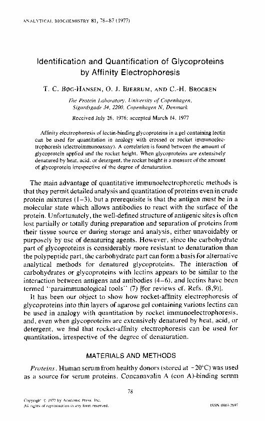
FIG. 1. Crossed-affinity electrophoresis of 1000 pg of serum proteins ( 15 ~1 of serum). First-dimensionelectrophoresis: 60min. Second-dimension electrophoresia was performedin a gel containing 130 pg of con A/cm?. Anode to the right cl+, first dimension) and to the top (2+. second-dimension electrophoresis). Inserted for visual comparison is another first-dimension gel fixed and stained immediately. Bar = 1 cm.
80 BBC&HANSEN, BJERRUM. AND BROGREN
AnMmdies. Antibodies against serum proteins (Code No. 100SF) were obtained from DAK0 Immunoglobulins A/S, Copenhagen, Denmark.
Elecfrophoreses. These were performed in 1 S-mm layers of 1% (w/v) agarose (Litex, Glostrup, Denmark) according to methods previously outlined (2,3). Proteins were electrophoresed into gel containing antibodies (immunoelectrophoresis) or various lectins (affinity electrophoresis).
The buffer was either Verona1 buffer (92 mM 5Jdiethylbarbiturate, pH 8.6) or Tris-Verona1 [73 mM Tris 7-9 (Sigma, St. Louis, MO.), 24.5 mM 5.5-diethylbarbiturate, 0.36 mM calcium lactate, and 0.2 mM sodium azide pH 8.61. In some experiments, 0.2% (w/v) Berol was included.
2a
LDL
LDL
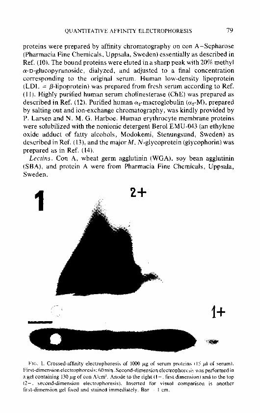
FIG. 2. Crossed-affinity electrophoresis (to be compared with Fig. 1). One hundred micrograms of serum proteins were electrophoresed into gel containing: (a) 130 pg of con A/cm2 and (b) 13 pg of con Aicm”. Ig, immunoglobulins: LDL, P-lipoprotein; M. a,-macroglobulin.
QUANTITATIVE AFFINITY ELECTROPHORESIS 81
Electrophoresis took place in a water-cooled (16°C) immunoelec- trophoresis cell from Holm-Nielsen Analyseudstyr. Soborg, Denmark, and rocket electrophoreses and fused-rocket immunoelectrophoreses were performed overnight at 1.5 V/cm. For crossed electrophoreses, the first-dimension electrophoreses were performed at 10 V/cm for 30-90 min. and the second-dimension electrophoreses were performed overnight at 1.5 V/cm. The gels were pressed, washed, dried, and stained (Coomassie brillant blue R 250) as in Refs. (2,3). Serum cholinesterase was stained histochemically with I-naphthyl acetate and fast red TR salt (15).
Heat dermrlrmtiorz. Con A-binding serum proteins were heat-denatured with protein diluted 10 times with 10X-diluted Tris-Verona1 (cf. above) at 85°C in closed vials. Samples were taken after indicated intervals and were frozen immediately at -20°C for later analysis.
Acid dermturcltiorz. Acid denaturation was performed analogously with 0.1 M glycine-HCI buffer, 0.2% merthiolate, pH 3.6, at 37°C.
Detergent demtwcrtion. Detergent denaturation of LDL was carried out by addition of Berol or sodium dodecyl sulfate (SDS, British Drug Houses, London) with subsequent incubation for 2 hr at room temperature.
RESULTS
Crossed-affinity electrophoresis of human serum proteins into con A is shown in Fig. 1. Characteristic of the pattern is the blurred appearance of many overlapping precipitates. Figure 2 shows the quantitative aspect of the enlarged precipitates with the low concentration of con A, and, in addition, this figure demonstrates that a certain protein concentration is
3a
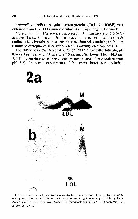
FIG. 3. Analysis of purified proteins. (a) Crossed-affinity electrophoresis of 675 pg of purified human serum a,-macroglobulin into con A (30 Fgicm’t. (b) Rocket-affinity electrophoresis of 900, 675, 540. 450. 360. and 275 pg of a,-macroglobulin into con A (40 pgicm’). (c) Rocket-affinity electrophoresis of I1 wg of purified human erythrocyte M. N-glycoprotein (glycophorin) into wheat germ agglutinin (WGA; SO pgicm’). Crossed electrophoresis as in Fig. I and I. Rocket electrophoresis: anode to the top. Each bar = I cm.
82 BOG-HANSEN, BJERRUM. AND BROGREN
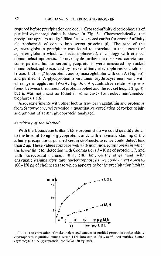
required before precipitation can occur. Crossed-affinity electrophoresis of purified a,-macroglobulin is shown in Fig. 3a. Characteristically, the precipitate appears totally “filled” as was noted earlier for crossed-affinity electrophoresis of con A into serum proteins (6). The area of the cu,-macroglobulin precipitate was found to correlate to the amount of cw,-macroglobulin which was electrophoresed, in analogy with crossed immunoelectrophoresis. To investigate further the observed correlation, some purified human serum glycoproteins were measured by rocket immunoelectrophoresis and by rocket-affinity electrophoresis: cholines- terase, LDL = j%lipoprotein, and cY,-macroglobulin with con A (Fig. 3b); and purified M, N-glycoprotein from human erythrocyte membrane with wheat germ agglutinin (WGA, Fig. 3~). A quantitative relationship was found between the amount of protein applied and the rocket height (Fig. 4), but is was not linear as found in some cases for rocket immunoelec- trophoresis (16).
Also, experiments with other lectins (soy bean agglutinin and protein A from Stuphylococcus) revealed a quantitative correlation of rocket height and amount of serum glycoprotein analyzed.
Sensitivity of the Method
With the Coomassie brilliant blue protein stain we could quantify down to the level of 10 ng of glycoprotein, and, with enzymatic staining of the affinity precipitate of purified serum cholinesterase, we could detect less than 2 ng. These values compare well with immunoelectophoresis in which the lower limit for detection with Coomassie is 3- 10 ng of protein (17) and with micrococcal mannan, 10 ng (18); but, on the other hand, with enzymatic staining after immunoelectrophoresis, we could detect down to IOO- 150 pg of cholinesterase which appears to be the precipitation limit in
FIG. 4. The correlation of rocket height and amount of purified protein in rocket-affinity electrophoresis: purified human serum LDL into con A (30 ~gicrn?) and purified human erythrocyte M. N-glycoprotein into WGA (50 pg/cm”).
QUANTITATIVE AFFINITY ELECTROPHORESIS
0’ I' 2’ 4’ 8’
0’ 1’ 2’ 4’ 8’
83
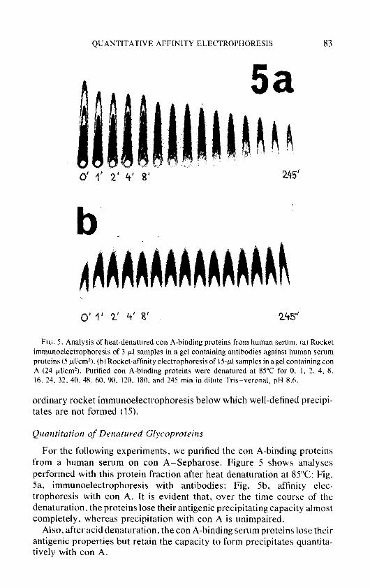
FIG. 5. Analysis of heat-denatured con A-binding proteins from human serum. (a) Rocket immunoelectrophoresis of 3.~1 samples in a gel containing antibodies against human serum proteins (5 plicm’). (b) Rocket-affinity electrophoresis of 1%~1 samples in a gel containing con A (24 ~l/cm*). Purified con A-binding proteins were denatured at 85°C for 0. 1, 2, 4, 8, 16, 24. 32. 40, 48, 60. 90. 120, 180, and 245 min in dilute Tri-veronal, pH 8.6.
ordinary rocket immunoelectrophoresis below which well-defined precipi- tates are not formed ( 15).
Quuntitation of Denatured Glycoproteins
For the following experiments, we purified the con A-binding proteins from a human serum on con A-Sepharose. Figure 5 shows analyses performed with this protein fraction after heat denaturation at 85°C: Fig. 5a, immunoelectrophoresis with antibodies; Fig. 5b, affinity elec- trophoresis with con A. It is evident that. over the time course of the denaturation, the proteins lose their antigenic precipitating capacity almost completely, whereas precipitation with con A is unimpaired.
Also, after acid denaturation, the con A-binding serum proteins lose their antigenic properties but retain the capacity to form precipitates quantita- tively with con A.
84 BOG-HANSEN. BJERRUM, AND BROGREN
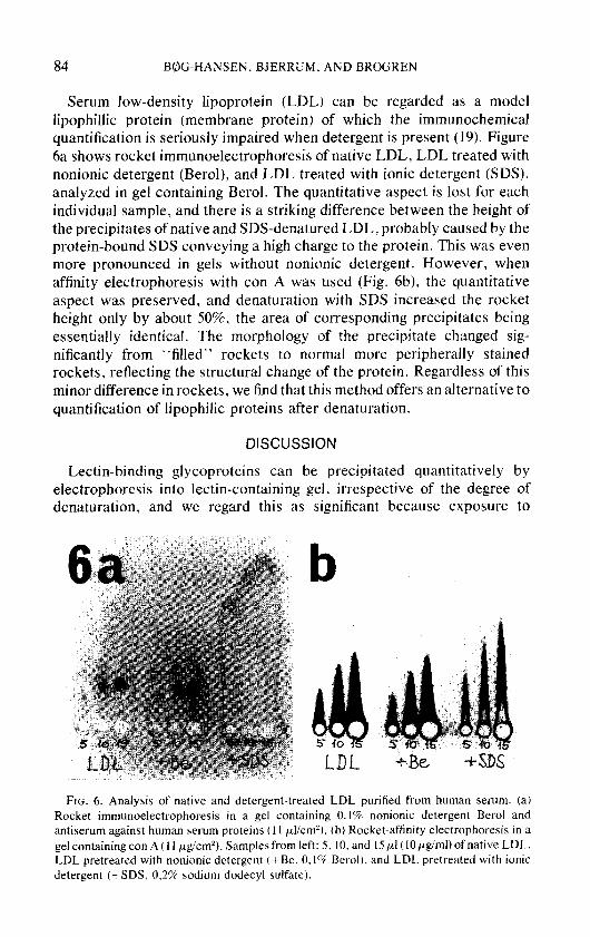
Serum low-density lipoprotein (LDL) can be regarded as a model lipophillic protein (membrane protein) of which the immunochemical quantification is seriously impaired when detergent is present (19). Figure 6a shows rocket immunoelectrophoresis of native LDL, LDL treated with nonionic detergent (Berol), and LDL treated with ionic detergent (SDS), analyzed in gel containing Berol. The quantitative aspect is lost for each individual sample, and there is a striking difference between the height of the precipitates of native and SDS-denatured LDL, probably caused by the protein-bound SDS conveying a high charge to the protein. This was even more pronounced in gels without nonionic detergent. However, when affinity electrophoresis with con A was used (Fig. 6b), the quantitative aspect was preserved, and denaturation with SDS increased the rocket height only by about 50%. the area of corresponding precipitates being essentially identical. The morphology of the precipitate changed sig- nificantly from “filled” rockets to normal more peripherally stained rockets, reflecting the structural change of the protein. Regardless of this minor difference in rockets, we find that this method offers an alternative to quantification of lipophilic proteins after denaturation.
DISCUSSION
Lectin-binding glycoproteins can be precipitated quantitatively by electrophoresis into lectin-containing gel, irrespective of the degree of denaturation, and we regard this as significant because exposure to
b
FIG. 6. Analysis of native and detergent-treated LDL purified from human serum. (a) Rocket immunoelectrophoresis in a gel containing 0. I 74 nonionic detergent Berol and antiserum against human serum proteins t 11 ~Vcm*). (b) Rocket-affinity electrophoresis in a gel containing con A t 1 I &cm*). Samples from left: 5. 10, and 15 /*I ( 10 &ml) of native LDL. LDL pretreated with nonionic detergent t +Be. 0,174 Berol), and LDL pretreated with ionic detergent (+SDS. 0.2% sodium dodecyl sulfate).
QUANTITATIVE AFFINITY ELECTROPHORESIS 85
denaturing agents, such as detergents and urea, is often used purposely during preparation and analysis of tissue proteins. As long as the antigenic structure is lost partially or totally by such treatment, immunoelec- trophoresis cannot be used for specific analysis and quantification, since the principle of immunochemical quantitation is based upon the absolute immunochemical identity and molecular integrity of the protein samples to be measured or compared. Therefore, even small changes in the protein structure cannot be tolerated when quantitative immunoelectrophoresis is used, and, as a consequence. alternative methods must be used. However, in some cases, it may be possible to renature proteins treated with charged detergents by inclusion of nonionic detergent in the medium (19,201.
In our model systems with purified lectin-binding proteins and with the
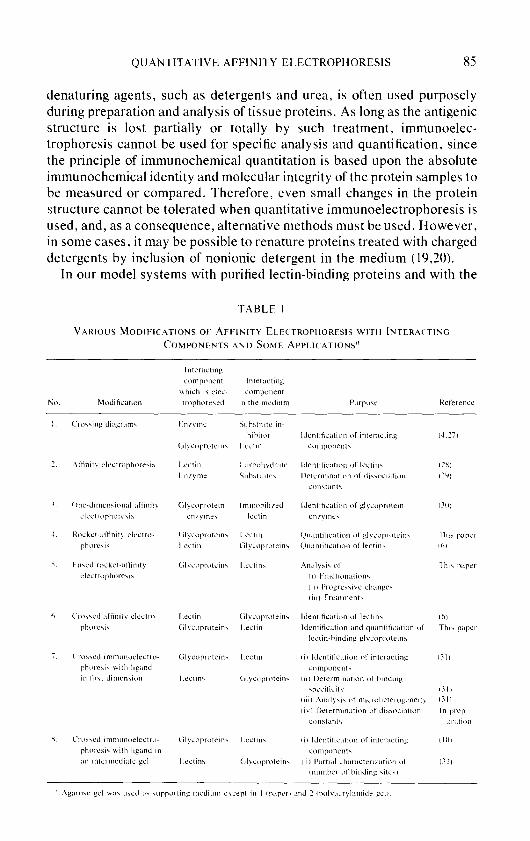
TABL,E I
VARIOUS MODIFICATIONS OF AFFINITY ELECTRO~HORESIS WITH INTERA<-TING
COMPONENTS AFU’D SOME APPLICATIONS”
-
86 BOG-HANSEN, BJERRUM. AND BROGREN
con A-binding human serum proteins taken as a group, we found that, within limits in which quantitation by rocket immunoelectrophoresis (electroimmunoassay) is impossible, affinity electrophoresis with lectin in the gel yields affinity rockets, the heights of which reflect the amount of denatured protein. Table 1 gives further suggestions for application of affinity electrophoresis for specific purposes. Due to the similarity of the carbohydrate part of serum proteins, crossed-affinity electrophoresis is of limited value in quantification of individual serum glycoproteins. but it may be useful for the study of changes in glycosylation of proteins known to occur in pathological conditions (21.22).
Quantification of SDS-denatured lectin-binding proteins after polyacryl- amide gel electrophoresis seems possible since most of the technical problems of the combined method (crossed electrophoresis with polyacryl- amide and agarose gels) have been solved (23-25). In this connection, it seems noteworthy that at least some lectins seem to be stable and fully reactive in detergent [cf. above and Ref. (26)].
ACKNOWLEDGMENTS
Ms. B. Bardenfleth. P. Jensen, L. Muir, M. Not-d, A. Thomsen, and I. Workare thanked for their skillful technical and secretarial assistance. This work was supported by Danish Medical Research Council Grant No. 5126563.
Note added in proof Based upon similar electrophoresis experiments of streptococcal lipoteichoic acids with con A and of glycoproteins (the I blood group substance and desialylated bovine and pig mucins) with wheat germ agglutinin. soybean agglutinin, and peanut agglutinin, Owen and coworkers have concluded recently that lectins provide an excellent complement and alternative to immune sera for the study of carbohydrate and glycoprotein preparations (P. Owen, J. D. Oppenhein. M. S. Nachbar. and R. E. Kessler. personal communication).
REFERENCES I. Laurel]. C. -B. (1972) Electrophoretic and Electra-lmmunochemical. Analysis of
Proteins, Universitetsforlaget. Oslo: Scund. J. C/in. Luh. Im)rst. 2, Suppl 124. 2. Axelsen, N. H.. Kroll, J.. and Weeke. B. (1973). A Manual of Quantitative
Immuno-Electrophoresis. Methods and Applications. Universitetsforlaget, Oslo; Scnnd. .I. Immunol. 2, Suppl. 1.
3. Axelsen. N. H. (1975). Developments in Quantitative Immunoelectrophoresis. Univer- sitetsforlaget. Oslo: Scund. J. It~7mrrnol. 4, Suppl. 2.
4. Nakamura, S., Tanaka, K.. and Murakawa, S. (1960) Nrlfure (London) 188, 144. 5. Doyle, R. J., and Cronholm, L. t 1969) J. Chem. Educ. 45, 868-869. 6. Bog-Hansen. T. C.. and Nord, M. (1974j.I. I?&/. Educ. 8, 167-173. 7. Uhlenbruck. G.. Rothe. A.. and Prokop, 0. (1974) if7 Connective tissues, Biochemistry
and Pathophysiology (Friche, R., and Hartmann, F.. eds.), pp. 201-220, Springer, Berlin.
8. Sharon, N.. and Lis, H. (1972) Sc,irnce 177. 949-959. 9. Lis. H., and Sharon, N. (1973). At7nrc. Rel,. Bkd7rm. 42, 541.
IO. Bog-Hansen, T. C. (1973) Annl. Biocl7ern. .56,480-488.
QUANTITATIVE AFFINITY ELECTROPHORESIS 87
I I. Havel, R. J., Eder, H. A., and Bragdon, J. H. (1955) J. C/in. Invest. 34, 1345-1353. 12. Brogren. C. H., and Just Svendsen, P. (1975) in Protides of the Biological Fluids.
Proceedings of the Twenty-second Colloquim. Brugge 1974 (Peeters. H.. ed.), Vol. 22. pp. 685-689. Pergamon, Oxford.
13. Bjerrum, 0. J., and Bog-Hansen, T. C. (1976) Biochim. Biophys. Actcr 4.55, 66-89. 14. Marchesi, V. T. (1972) in Methods in Enzymology (Ginsburg, V., ed.). Vol. 28, pp.
252-254. Academic Press, New York. 15. Brogren. C. H.. and Bog-Hansen. T. C. Sc~nd. J. Immunol. 4, Suppl. 2, 37-51. 16. Weeke. B. (1973) Stand. J. Zmm~nol. 2, Suppl. I. 17. Johansson. B. G., and Malmquist. J. (1971) Swnd. J. C/in. Lob. Znr*e.st. 27, 255-261. IS. Owen, P., and Salton, M. R. J. (1976) AMI/. Biochem. 72, 20-26. 19. Nielsen. C. S.. and Bjerrum. 0. J. (1975) Scund. J. Immuno~. 4. Suppl. 2. 73-80. 20. Bjerrum, 0. J., Bhakdi. S.. Bog-Hansen. T. C.. Kniifermann, H., and Wallach, D. F. H.
(1975) Biochim. Riophys. Acta 406, 489-504. 2 I. Schmid, K., Burke. J. F.. Debray-Sachs, M., and Tokita. K. ( 1964) Nature (London 1204,
75-76. 22. Winzler. R. J. (197i)in Glycoproteins of Blood Cells and Plasma (Jamieson. G. A., and
Greenwalt. T. J.. eds.). pp. 204-213, Lippincott, Philadelphia. 23. Skude, G.. and Jepsson. J. 0. (1972) Scund. J. Clin. Lab. Invest 29, Suppl. 124. 55-58. 24. Soderholm, J., Smyth. C. J.. and Wadstrom, T. (1975) Sctrnd. J. I,nmtrrro/. 2, Suppl. 1.
lO7- 113. 25. Converse. C. A., and Papermaster, D. S. (1975) Science 189.469-472. 26. Vestergaard. B. F., and Bog-Hansen. T. C. (1975) Scnnd. J. Imm~nol. 4, Suppl. 2,
211-215. 27. Nakamura, S. (1966) Cross Electrophoresis. Its Principles and Applications, Igaku Shoin,
Tokyo and Elsevier, Amsterdam. 28. Horejsi. V.. and Kocourek. J. (1974) Biochim. Biophys. Acttr 336, 338-343. 29. Takeo. K.. and Nakamura. S. (1972) Arch. Biochem. Biophys. 153, I. 30. Bog-Hansen. T. C.. Brogren. C. H., and McMurrough. I. (1974) J. Irut. Brpw.. 80,
443-446. 31. Bog-Hansen, T. C.. Bjerrum, 0. J.. Ramlau, J. (1975) Stand. Immrrrzol. 4, Suppl. 2,
141-147. 32. Bog-Hansen, T. C.. and Brogren, C. H. (1975)Scurrd. .I. Immunol. 4, Suppl. 2. 135-139.