tuberculosis (mycobacterium microti) in wild field vole populations
TRANSCRIPT
Tuberculosis (Mycobacterium microti) in wild field volepopulations
S. BURTHE1,2, M. BENNETT2, A. KIPAR2, X. LAMBIN3, A. SMITH1,2, S. TELFER1,2, and M.BEGON1,*
1 School of Biological Sciences, University of Liverpool, Crown Street, Liverpool L69 7ZB2 National Centre for Zoonosis Research, Faculty of Veterinary Science, University of Liverpool,Leahurst, Chester High Road, Neston CH64 7TE3 School of Biological Sciences, University of Aberdeen, Tillydrone Avenue, Aberdeen AB24 2TZ
SUMMARYVole tuberculosis (TB; Mycobacterium microti) is an understudied endemic infection. Despiteprogressing slowly, it causes severe clinical pathology and overt symptoms in its rodent host. TBwas monitored for 2 years in wild field voles in Kielder Forest, UK. The prevalence ofcharacteristic cutaneous TB lesions was monitored longitudinally at 4 sites, with individuals live-trapped and repeatedly monitored. A prevalence of 5·2% of individuals with lesions was recorded(n=2791). In a cross-sectional study, 27 sites were monitored bi-annually, with TB assessed bypost-mortem examination for macroscopic lesions, and by culture and histopathology. Seventy-nine voles (10·78%; n=733) were positive for mycobacteria, with the highest prevalence in spring(13·15%; n=327). TB prevalence varied, with between 0% and 50% of voles infected per site.Prevalence increased with age (mass), and apparent seasonality was due to a higher proportion ofolder animals in spring. Survival analysis supported this result, with cutaneous lesions onlymanifesting in the advanced stages of infection, and therefore only being found on older voles.The body condition of individuals with lesions declined at the time when the lesion was firstrecorded, when compared to individuals without lesions, suggesting there may be an acute phaseof infection during its advanced stage. Although predicted survival following the appearance of acutaneous lesion was lower than for uninfected individuals, this was not significant.
Keywordsendemic pathogen; Microtus agrestis; longitudinal; TB; survival; wildlife disease
INTRODUCTIONThe majority of studies investigating wildlife disease have focussed on epidemic pathogenscausing high levels of mortality, whereas most pathogens are endemic, persist in hostpopulations and show relatively small fluctuations in prevalence (Anderson and May, 1979).This study focuses on endemic vole tuberculosis (TB), Mycobacterium microti, in itsreservoir host, the field vole, Microtus agrestis L., in Kielder Forest, northern England. M.microti is a member of the Mycobacterium tuberculosis complex, which includes M.tuberculosis and M. bovis (van Soolingen et al. 1997, 1998). Despite being the first endemic
© 2007 Cambridge University Press* Corresponding author: School of Biological Sciences, The University of Liverpool, Biosciences Building, Crown Street, Liverpool,L69 7ZB, UK. Tel: +44 151 795 4525. Fax: +44 151 795 4408. [email protected].
Europe PMC Funders GroupAuthor ManuscriptParasitology. Author manuscript; available in PMC 2010 November 01.
Published in final edited form as:Parasitology. 2008 March ; 135(3): 309–317. doi:10.1017/S0031182007003940.
Europe PM
C Funders A
uthor Manuscripts
Europe PM
C Funders A
uthor Manuscripts
infection to be investigated thoroughly in wild rodents (Wells and Oxon, 1937; Wells, 1946;Chitty, 1954), with up to a third of sampled field voles recorded as infected (Wells, 1946;Cavanagh et al. 2004), very little is known about the effects of vole TB on wild populations.TB is also an interesting disease of wild rodents because, unusually for an endemicinfection, it is a chronic infection causing obvious and severe pathology in its host in its laterstages, with potential implications for host survival and increased susceptibility to furtherinfections. This also enhances its potential to exhibit delayed host density dependence in theprevalence of (serious) late-stage infections. Moreover, vole TB is a zoonotic infection. M.microti infections have been recorded in immunosuppressed and immunocompetent humans(van Soolingen et al. 1998; Niemann et al. 2000; Horstkotte et al. 2001), and prevalence ofinfection in humans is likely to be higher than has been currently reported (van Soolingen etal. 1998).
The present study, the first to examine the temporal dynamics of M. microti infection in awildlife population, investigates the pathology and duration of M. microti infection in wildfield voles by monitoring the presence of characteristic, externally visible cutaneous lesions.Wells (1946) found these especially in the subcutaneous tissue of the inter-scapular region,often with ulceration through the skin. It also examines temporal patterns in the prevalenceof these lesions, and assesses whether they affected body condition or survival. However, asdiagnosis of M. microti infection on the basis of external lesions alone underestimates M.microti prevalence (Cavanagh et al. 2004), patterns in prevalence were also evaluated in across-sectional study of voles examined post-mortem that included diagnosis of M. microtiinfection by the culture of material taken from potentially infected organs.
MATERIALS AND METHODSStudy area
The study took place in Kielder Forest, a man-made spruce forest occupying 620 km2,situated on the English-Scottish border (55°13′N, 2°33′W). Kielder Forest can be dividedinto 3 main catchments, lying in adjacent watersheds: Redesdale 10–15 km north-east ofKielder, Kielder, and Kershope 10–15 km to the southwest. Field voles inhabit grassy clear-cuts that represent 16–17% of the total area, but are completely absent from forested areas.Clear-cuts range in size from 5–100 ha. Field vole populations at Kielder fluctuate cyclicallywith a 3–4 year period (Lambin et al. 2000). Populations situated close together fluctuate insynchrony, but populations further apart are out of synchrony (Lambin et al. 1998; Biermanet al. 2006).
Longitudinal studyTrapping methods—Voles were trapped in 4 similar-sized clear-cuts, in 2 areas of theforest approximately 12 km apart, between May 2001 and July 2003. Kielder Site (KCS)and Plashett’s Jetty (PLJ) were situated 4 km apart within the Kielder catchment, with volepopulations at low to increasing density during the study. Black Blake Hope (BHP) andRob’s Wood (ROB) were 3·5 km apart within the Redesdale catchment, with voles atincreasing and peak density.
Populations were trapped in primary sessions every 28 days from March to November, andevery 56 days from November to March. Each site had a permanent 0·3 ha live-trapping gridconsisting of 100 Ugglan Special Mousetraps (Grahnab, Marieholm, Sweden), in optimalhabitat dominated by Deschampsia caespitosa Beauv., Agrostis tenuis Sibth., and Juncuseffusus L. Traps were set at 5 m intervals and baited with wheat and carrots. Traps were pre-baited with a slice of carrot and a few grams of oats 3 days before each trapping session, set
BURTHE et al. Page 2
Parasitology. Author manuscript; available in PMC 2010 November 01.
Europe PM
C Funders A
uthor Manuscripts
Europe PM
C Funders A
uthor Manuscripts
at approximately 18:00 on the first day and checked 5 times (‘secondary sessions’) atroughly 12-h intervals at dawn and dusk.
Individuals were identified using subcutaneous microchip transponders (AVID plc, EastSussex, UK) injected into the skin at the back of the neck. Mass and sex were recorded atthe time of first capture in each primary session. Animals with juvenile fur or in their firstmoult were classed as juveniles (Graham and Lambin, 2002). Animals were assigned toreproductive classes according to the external appearance of reproductive organs. A bodycondition (BC) ordinal score was calculated for each animal by estimating the degree of fatcover over the vertebral column and dorsal pelvic bones. Each area was scored on a scalebetween 1 and 5, and scores summed to give an overall BC score from 2 to 10 (Cavanagh,2001).
Vole density estimates for each primary session were calculated via the closed populationmodel MTH in the program CAPTURE (Otis et al. 1978) and using the estimator of Chaoand Lee (1991).
Diagnosis of TB—The presence, size and location of characteristic tuberculous skinlesions denoting late-stage TB were noted in all animals. Lesions are known to be mostcommonly sited in the scapular region (Wells, 1946; Cavanagh et al. 2002, 2004), and thusin order to undertake a conservative estimate of TB prevalence, individuals were onlyrecorded as positive for TB in the field if the lesion was observed between or on theshoulder blades and was > 1 mm in diameter, or if it was > 5 mm and situated else-where onthe body. Lesions that did not meet these criteria, or which appeared atypical (i.e. resembledbite wounds, or were not ulcerated through the skin) were recorded as ‘suspect’ in the field,and their size and location noted, but because of their suspect nature, and because a swab forbacteriological analysis could not be taken without potentially damaging the animalconcerned, they were omitted from analyses. Any effects of TB on voles are therefore likelyto be an underestimate.
Effect of TB on survival—The goodness of fit (GOF) of a ‘global’ (most fullyparameritized) model was assessed, as the capture-mark-recapture (CMR) models usedassume that: (1) every marked animal in the population immediately after time (i) has thesame probability of surviving to time (i+1); and (2) every marked animal present in thepopulation at time (i) has the same probability of recapture (pi). Individual capture historieswere classified by sex and site. GOF was assessed using tests implemented in the programRELEASE (Burnham et al. 1987) using a standard Cormack-Jolly Seber model applied tothe each group. ‘Test 2′ in RELEASE tests for violation of assumption (2), and ‘Test 3′tests for violation of assumption (1).
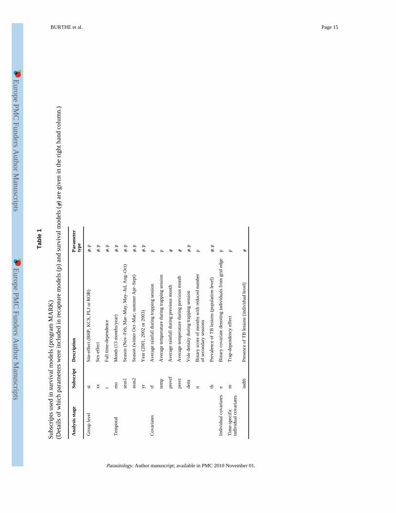
In examining the effect of TB on survival, the first step was to derive an optimal modelaccounting for individual variation in recapture and survival. The analysis was undertakenfollowing Lebreton et al. (1992) using program MARK (White and Burnham, 1999). Fulldetails have been given by Burthe et al. (2007), and the subscripts used for modelparameters are listed in Table 1. Additive effects are denoted by a plus (+) sign, andinteraction terms by an asterisk (*).
The first captures of all individuals were removed (1514 in total) in order to overcomebiases caused by transient individuals. This reduced the risk of confounding emigration andmortality as a cause of disappearance, but precluded the inclusion of age (juvenile/adult) as aparameter in model selection. The reduced data set of 1275 individual capture historiesyielded a more robust analysis and was used to estimate the best ‘base’ recapture andsurvival model, with which possible effects of TB could be investigated. First, recapture was
BURTHE et al. Page 3
Parasitology. Author manuscript; available in PMC 2010 November 01.
Europe PM
C Funders A
uthor Manuscripts
Europe PM
C Funders A
uthor Manuscripts
modelled in 3 stages. The first examined whether recapture rate varied with time, site or sex.The second investigated whether the temporal component of the recapture model could beadequately described by month, season or year. Effects of average rainfall and temperatureover the 3-day trapping period, and of reduced trapping effort in some primary sessions(caused by inclement weather) were also investigated. The third stage then examinedindividual level covariates such as trap dependency and ‘edge’. An edge animal had ≥75%of its captures at the edge of the trapping grid. Trap dependency was a time-dependentindividual covariate determined by whether or not an individual had been caught in thepreceding primary session. The fits of the models were assessed by Akaike’s InformationCriterion corrected for small samples (AICc) (Hurvich and Tsai, 1989). Models with adifference of AICc (ΔAICc) of less than 2 may be considered similar in their ability toaccount for the data (Sakamoto et al. 1986). According to the principle of parsimony, if 2alternative models had indistinguishable AICc values (ΔAICc<2), the model with fewerparameters was selected.
Survival was then modelled similarly using the best recapture model. To investigate theeffect of climatic variables on survival probabilities, the average daily temperature or theaverage daily rainfall over the entire 28-day period was included as weather variables.Finally, this best model of recapture and survival (the ‘base’ model) was used to investigatewhether TB affected host survival and recapture probabilities.
Cutaneous lesions are reported to represent an advanced stage of TB, and to confirm this TBwas included first as a binary individual covariate (tb) depending on whether that individualwas ever recorded positive for a cutaneous lesion (i.e. TB individuals vs individuals thatwere never infected). In a second separate analysis, TB was modelled as a time-varyingindividual covariate of lesion presence (indtb) reflecting the detection of lesion in a giventrapping occasion in order to investigate differential survival over the next trapping interval.
Effect of TB on body condition—The change in BC between primary sessions for volespositive for TB (n=47) was compared to voles without lesions (not including individualssuspect for lesions), by randomly assigning each positive individual a negative, controlindividual matched for trap session, sex and site for comparison, since Cavanagh (2001)found that only sex, site and season influenced individuals’ changes in BC. Individuals withTB lesions were included in the analysis if BC scores were available for the primary sessionwhen the lesion was first observed, and the preceding primary session. From within theappropriate control sets (size range 1–36, mean 13·6 per session/site/sex), individuals wereselected at random. Forty-seven matched TB and non-TB changes in BC were comparedusing a 2-tailed Mann-Whitney U-test, and this was repeated 100 times with replacement.The median change in score for the TB group was also compared to the distribution ofmedians of the 100 control groups.
Statistical analysis of the risk of infection—Factors influencing whether anindividual was positive for external tuberculous lesions were investigated using GeneralisedLinear Mixed Models (GLMM) with a logit link and binomial errors. Analyses wereconducted using the lme4 (Bates and Sarkar, 2006) and Matrix (Bates and Maechler, 2006)packages in the R software available under the GNU license at http://www.r-project.org. Asis common with parasitological data, individual observations were non-independent(individuals being sampled from the same site at the same time), and so site*trap-sessionwas included as a random effect (Paterson and Lello, 2003). The same individuals could alsobe sampled repeatedly over time, potentially leading to pseudo-replication. In this study,however, individuals appeared in the data only 1·38 times on average. Consequently thevariance component due to vole id was not estimable as a random effect in the GLMMs.
BURTHE et al. Page 4
Parasitology. Author manuscript; available in PMC 2010 November 01.
Europe PM
C Funders A
uthor Manuscripts
Europe PM
C Funders A
uthor Manuscripts
Population level covariates included the month, season, site and year of capture. Theindividual level covariates considered were sex and mass (as a crude proxy for age). Mass2
was also fitted in order to test for non-linear relationships between risk of TB and age.Biologically meaningful two-way interactions between individual level covariates andbetween individual level covariates and season were also included. Models were constructedfollowing a step-down procedure, eliminating interactions first. The models were restrictedusing the AIC, with models with a ΔAIC of less than 2 considered similar in their ability toaccount for the data (Sakamoto et al. 1986).
Cross-sectional trappingTrapping methods—Twenty-seven clear-cut sites were trapped: 6 in Redesdale, 12 inKielder and 9 in Kershope, with 3 traps placed within a 1 m radius of each corner of a 15 mby 15 m trapping grid. Grids were trapped bi-annually (March and September) betweenSeptember 2001 and March 2003. These times were chosen to reflect differences inpopulation structure: March samples consisting of over-wintered, adult animals, andSeptember samples occurring during the breeding season and including juveniles. Trapswere pre-baited with a slice of carrot and a few grammes of oats 3 days before each trappingsession, set overnight and then checked daily over 3 days. Animals were euthanized in thefield by isoflurane inhalation.
Post-mortem examination of field voles—Mass, sex, BC and reproductive status ofindividuals were recorded post-mortem. The presence, size and location of characteristiccutaneous lesions or gross internal lesions denoting TB (Wells, 1946; Cavanagh et al. 2002)were recorded during dissection. Field voles from surveys 2–4 (March 2002–March 2003)with internal signs of TB, or abnormally large lymph nodes, had all organs and any lesionmaterial removed and divided for culture and histology. Field voles with no visible signs ofTB had their lymph nodes, particularly those from the facial and scapular regions, removedfor culture in March 2002 and had their lungs and lymph nodes removed for culture inSeptember 2002 and March 2003. No organs were cultured from the September 2001survey. All suspect lesions were tested to confirm TB by PCR on clinical material for survey1, by PCR on cultured material from subsequent surveys (Magdalena et al. 1998), and byhistology. Samples for histology were fixed in 10% buffered formalin, processed bystandard procedures for embedding in paraffin, and stained with haematoxylin-eosin (HE),Auramine-rhodamine (AR) and Ziehl-Neelson (ZN) stains.
Decontamination and culture methods—Lymph nodes were pooled for culture if theanimal had no sign of TB, and cultured separately if macroscopic lesions were observed. Allother organ samples were cultured separately. To decontaminate, the tissue samples werehomogenized in an equal volume of sterile PBS. The sample was agitated at roomtemperature overnight, with an equal volume of CPC decontaminant (5 g cetyl-pryridiniumchloride, 10 g NaCl/1000 ml H2O). Decontaminated samples were centrifuged at 3000 g for15 min. Sediment was re-suspended in 250 μl of DD-H2O, and 200 μl of this used toinoculate Lowenstein-Jensen pyruvate based culture slopes (SGS Laboratory Supplies).Slopes were incubated at 37 °C for a minimum of 12 weeks. Negative slopes were kept for afurther 4 weeks for a final examination. Slopes with potential TB colonies were subculturedand subject to ZN staining and PCR (Magdalena et al. 1998) to confirm the presence of M.microti. The PCR is specific to the M. tuberculosis complex, and the size of the amplifiedDNA fragments detected varies amongst the different strains, with M. microti being 560 bpin size. In order to further confirm that the species was M. microti, positive cultures weresubject to spoligotyping (Kamerbeek et al. 1997) and IS6110 RFLP typing (van Embden etal. 1993).
BURTHE et al. Page 5
Parasitology. Author manuscript; available in PMC 2010 November 01.
Europe PM
C Funders A
uthor Manuscripts
Europe PM
C Funders A
uthor Manuscripts
Statistical analysisFactors influencing whether an individual was positive for M. microti were investigatedusing GLMMs with a logit link and binomial errors, with site*survey included as a randomeffect. As for the longitudinal data (above) the analyses were conducted in a 2-stagestepwise selection procedure. Population level covariates were catchment or site. Theindividual level covariates were sex, age (juvenile or adult, or mass as a proxy for age),maturity and BC score.
RESULTSPatterns of external TB lesion prevalence (longitudinal study)
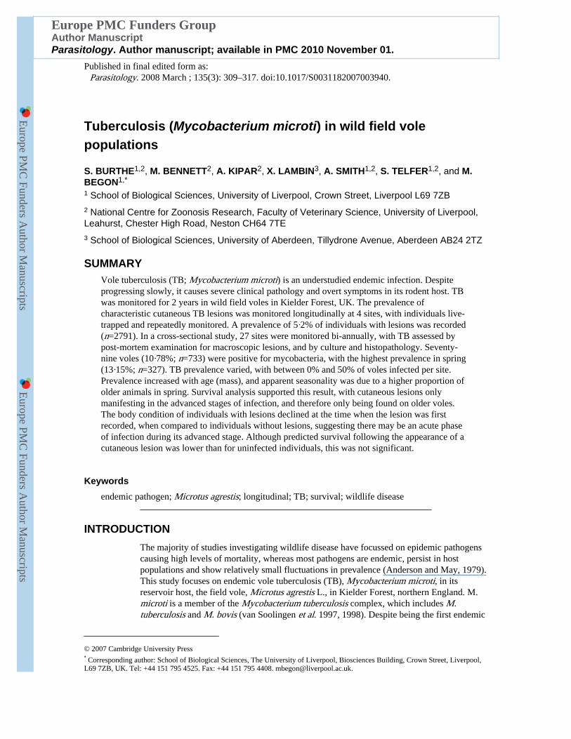
A total of 2791 voles were caught over the 2-year study period, with 108 individuals beingpositive for externally visible cutaneous TB lesions at some stage in their lives (prevalence3·87%; 95% CI: 3·18–4·65). A further 60 individuals had suspect lesions, 58 of which werenot captured again and hence could not be confirmed as having positive lesions and were notincluded in subsequent analyses. No juveniles (<18 g) were observed with external lesions,and prevalence was very low (1·31%, n=2287, 95% CI:0·89–1·87) for individuals of 18–25g. Overall prevalence of lesions in adults was 7·01% in 2001 (4·90–9·66, n=485), 5·52% in2002 (4·23–7·07, n=1081) and 5·43% in 2003 (4·06–7·10, n=948). Prevalence showed aseasonal pattern, with peaks of infection in late spring and early summer. April had thehighest monthly prevalence when the 4 sites were considered together (Fig. 1). However,month and season fell out of the model once mass and year were included, reflectingcovariation between season and the relative prevalence of animals of mass >18 g.
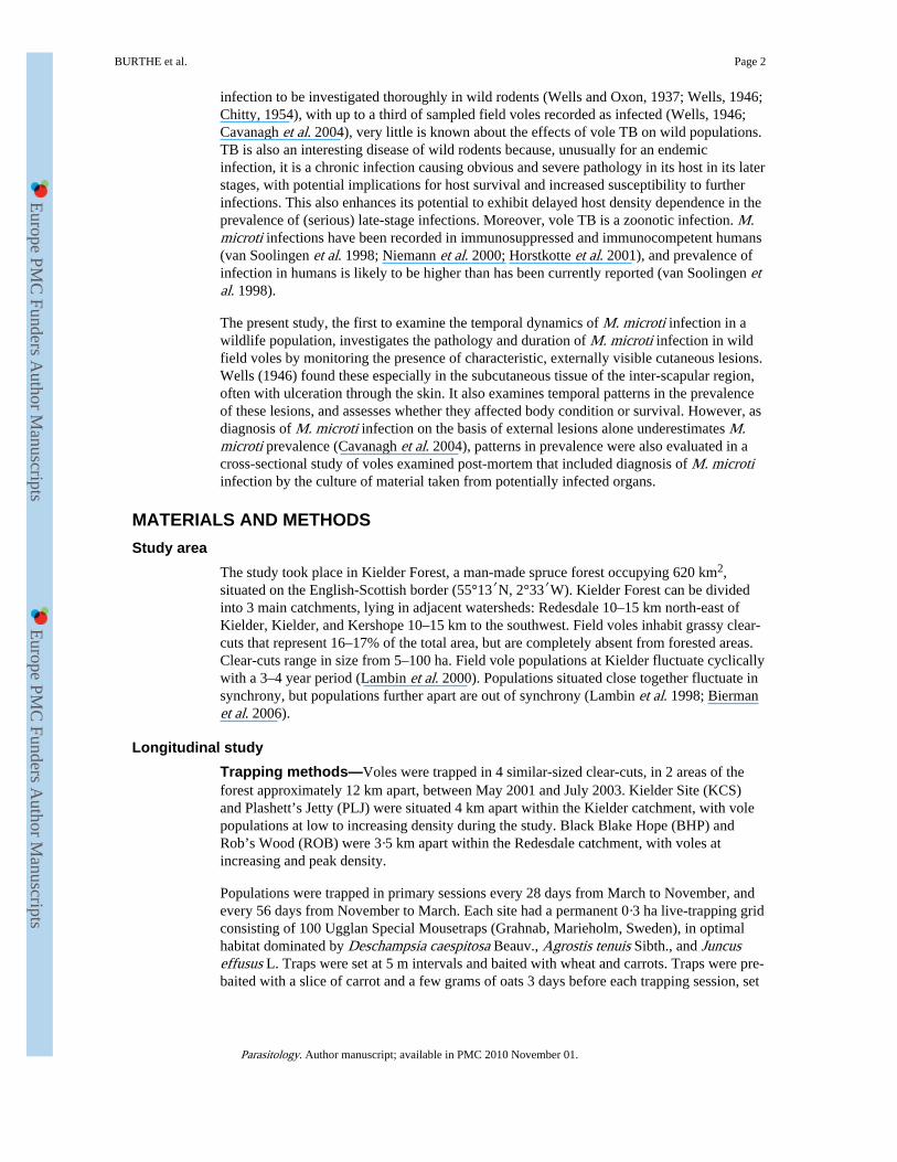
The model with the lowest AIC included mass and mass2 of individuals, and year. TB lesionprevalence increased with the mass of individuals (b=0·500, S.E.=0·099, z=5·066, P<0.0001).However, the risk of TB decelerated with increasing weight, as indicated by a negativerelationship with the squared mass of individuals (b=−0·007, S.E.=0·002, z=−4·377,P<0·0001) (Fig. 2). The prevalence of lesions also varied by year, with prevalence lowest in2003 (b=−0·712, S.E.=0·262, z=−2·719, P<0.007). There was no significant difference inprevalence between males and females (ΔAIC=1 between the model with the lowest AICand one including sex), or between sites (ΔAIC=1 between the model with the lowest AICand one including site).
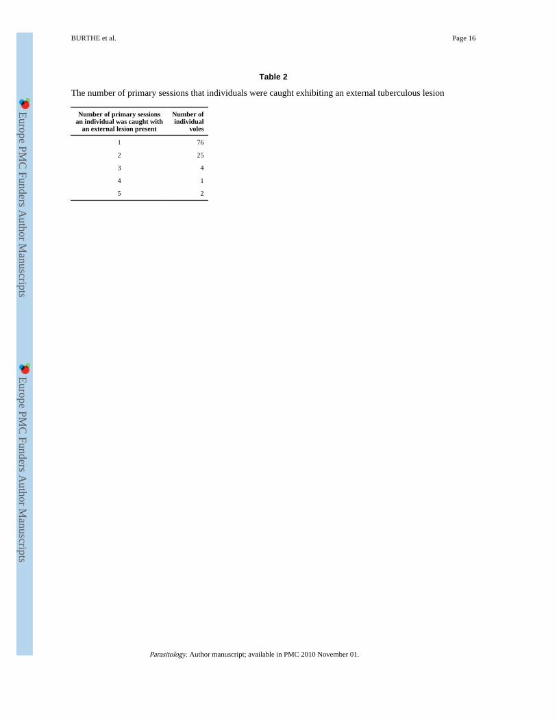
Effect of TB on vole survivalNineteen individuals caught on only 1 occasion had a TB lesion. These might have beentransient animals, and hence no conclusion can be drawn regarding their fate. Of theremaining 89 infected voles, 41 were never caught again subsequent to having been firstcaught with a lesion. Based on the frequency of voles infected per number of trap sessions,the median length of time that an infected individual was caught with a cutaneous TB lesionpresent was 1 month, and the maximum was 6 months (5 primary sessions due to 56 daysbetween winter trap sessions) (Table 2). Twenty-nine of the 108 individuals (26·85%; 95%CI: 18·78–36·24%) caught with positive external lesions were subsequently caught negativefor lesions at a later primary session. Of these, 6 were subsequently caught positive forlesions again, after having been recorded as negative for several primary sessions.
Individuals positive for a cutaneous lesion (i.e. that had a lesion at some stage in theircapture history) had higher overall survival than uninfected individuals. The untransformedβ parameter estimate on the logit scale was 0·72 (95% CI: 0·44–1·01), significantly greaterthan zero. This indicates that only those with good lifetime survival can exhibit these lesions(i.e. it confirms that cutaneous lesions are an advanced stage of the disease).
BURTHE et al. Page 6
Parasitology. Author manuscript; available in PMC 2010 November 01.
Europe PM
C Funders A
uthor Manuscripts
Europe PM
C Funders A
uthor Manuscripts
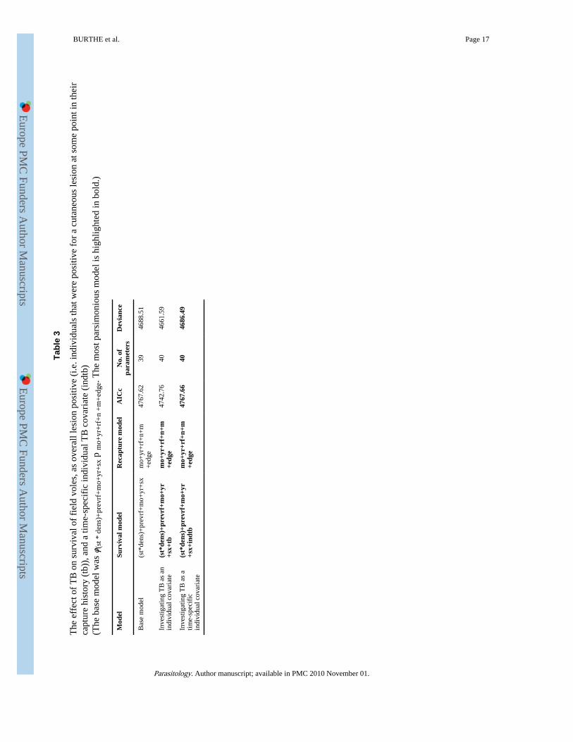
In spite of a drop in deviance, the AICc of a model including the effect of the presence of alesion in a primary session on immediately subsequent survival did not differ from that of asimpler model excluding this effect. Thus the hypothesis of an effect of TB on survival wasnot supported (Table 3). Nonetheless, predicted survival of individuals following theappearance of a cutaneous lesion was lower. The untransformed β parameter estimate on thelogit scale was −0·36 (95% CI: −0·93–0·20) equating to a drop of 8·05% in monthly survivalfor individuals with TB compared to uninfected individuals for 1 month with mediansurvival rates.
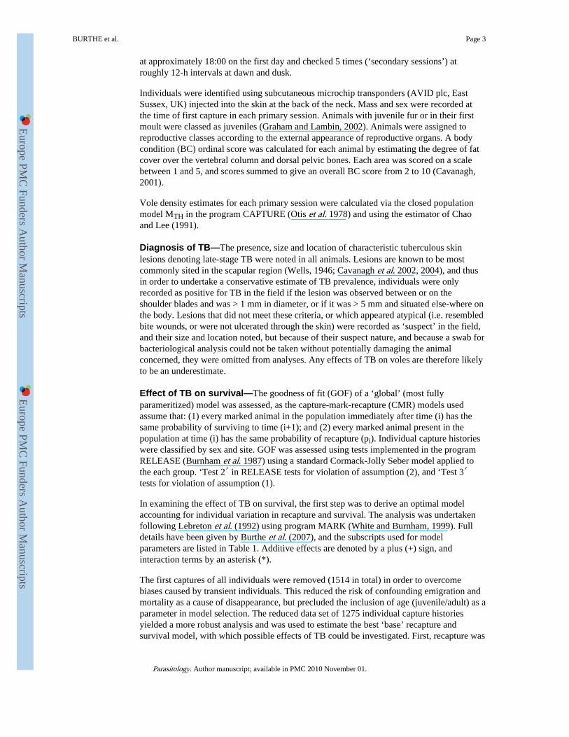
Effect of TB on body conditionForty-seven infected individuals had BC information for the primary session when firstcaught with a cutaneous lesion, and for the preceding session. These showed a significantdecline in BC between the primary session preceding infection and the primary session atwhich the lesions were first recorded, when compared to individuals of the same sex andfrom the same primary session and site. The median change in BC score was −1 for TBpositive individuals and 0 for negative individuals. Of the 100 Mann-Whitney U-tests, 87were significant, with 58 significant at the P<0·01 level. The median change in score for thepositive individuals was less than all but 5 of the changes in the 100 control groups (Fig. 3).
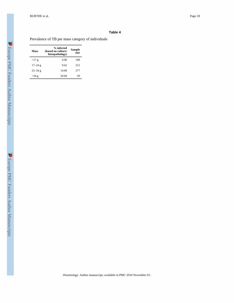
Risk of TB: cross-sectional studyThe average prevalence (based on post-mortem examination for lesions and on culture oforgans) for all sites and all surveys was 9·09% (n=733) and ranged between 1·78%(September 2001; n=169) and 13·15% (March 2003; n=327). However, prevalence variedbetween sites, with between 0% and 50% of voles infected. No voles of mass <17 g wereever positive regardless of the detection method used (Table 4).
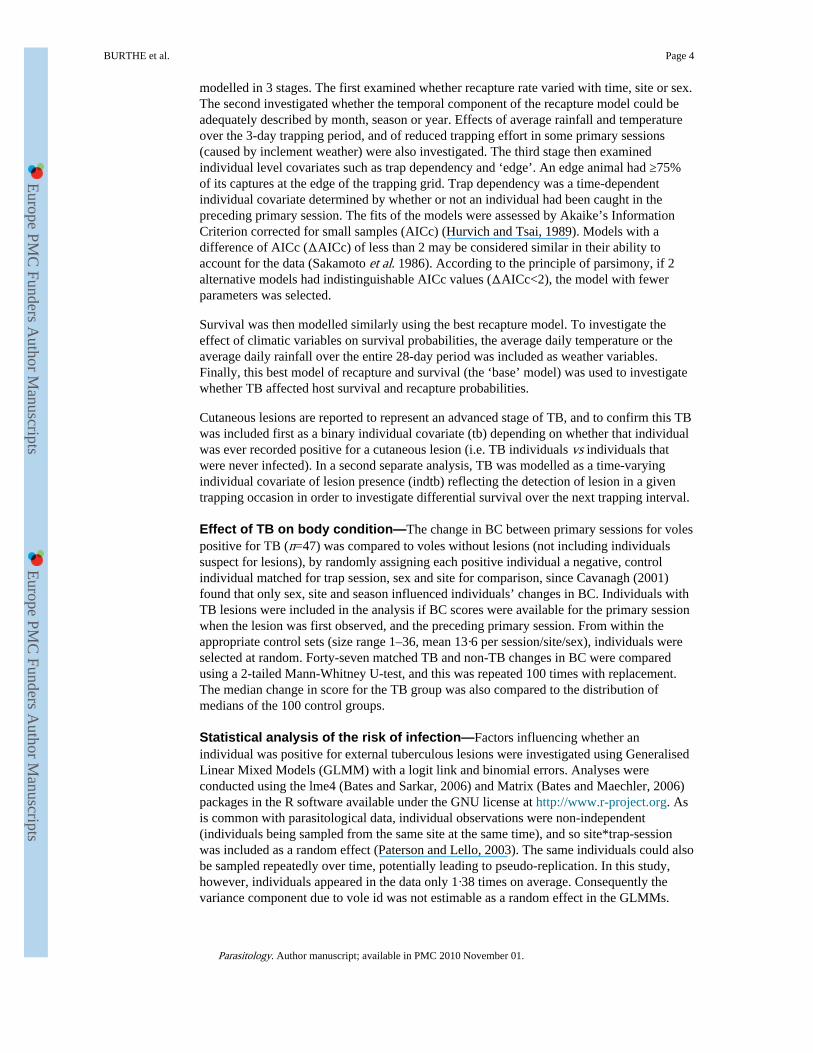
The model with the lowest AIC included mass and mass2 of individuals (AIC=461·4). Theprobability of TB increased with the mass of individuals (b=0·509, S.E.=0·199, z=2·554,P=0·011) however the risk of TB decelerated with increasing weight, as indicated by anegative relationship with the squared mass of individuals (b−0·008, S.E.=0·004, z= −2·172,P=0.030). There were also significant relationships between mass and mass2 and theprobability of TB for the different methods of diagnosis (i.e. based on external lesions,external and internal macro-lesions or on being culture positive but lesion negative) but theslopes of these relationships varied (Fig. 4). The slope for external lesions, indicative of late-stage infection, was steepest and differed from the slope for individuals that were culturepositive but lesion negative (with earlier stage infection). Season was not retained in themodel once mass was included (AIC=461·4). No other covariates, or two-way interactions,were significant at the P<0·01 level.
DISCUSSIONThe prevalence of vole TB averaged 3·87% from the longitudinal study, and 9·09% from thecross-sectional study at Kielder Forest. Significant variation in lesion prevalence wasobserved between the 3-study years of the longitudinal survey, with the highest prevalenceoccurring in 2001 (7·01% in 2001; 5·52% in 2002; 5·43% in 2003). There was considerablespatial variation in prevalence from the cross-sectional surveys, with local site prevalence ashigh as 50%.
The range of prevalence of cutaneous lesions observed is comparable to that reported byCavanagh et al. (2002) in the same area, where overall prevalence of external signs wasapproximately 2%, but reached levels of 8% in April. As shown by the cross-sectional study,the diagnosis of TB based on external lesions vastly underestimates TB prevalence. Wells
BURTHE et al. Page 7
Parasitology. Author manuscript; available in PMC 2010 November 01.
Europe PM
C Funders A
uthor Manuscripts
Europe PM
C Funders A
uthor Manuscripts
(1946) recorded an overall prevalence of infection of 20·5% based on post-mortemexamination for macro-lesions, and also found that prevalence was highest in spring andearly summer, and varied between sites.
External lesions were only found on adult voles of mass >17 g, with prevalence generallyincreasing with the age of individuals. Indeed, apparent seasonality in TB prevalenceactually reflected differences in the proportion of older animals present in the population,reaching up to 13·2% in March 2002. However, due to low numbers, and hence broadconfidence intervals, for individuals of mass >30 g in weight, it is not possible to confirmwhether risk of TB increases in the heaviest age classes. The relationship between age ofindividuals and the prevalence of macro-lesions (based on post-mortem) was stronger thanthat between age and prevalence for individuals that were lesion negative and culturepositive. This suggests that older animals are slightly more likely than younger animals tohave early stage infections (with no cutaneous lesions that have ulcerated through the skin),but are much more likely to have advanced stage TB. Although experimental work isnecessary to elucidate this further, it does suggest that external lesions only manifestthemselves during the more advanced stages of the disease, primarily encountered in olderindividuals.
Once mass was accounted for there was no significant difference in prevalence betweenmales or females. This is counter to the hypothesis that one route of infection of TB in fieldvoles is via skin abrasions caused by aggressive encounters, as might be expected based onthe appearance of cutaneous lesions, and which would be expected to be more prevalent inmales due to their more aggressive nature.
This study found evidence that TB appears to have a negative impact on body condition ofindividuals, providing further evidence that the decrease in survival following thedevelopment of a cutaneous lesion is likely to be significant. Positive individuals showed asignificantly greater reduction in BC between primary sessions than non-infected voles.Such a decline in BC associated with a chronic disease such as TB is not surprisingconsidering the extent of pathological damage observed in individuals with advancedinfection. Although laboratory experiments involve unnatural dosage levels andtransmission routes, Manabe et al. (2002) found that Microtus species voles infected via anintraperitoneal injection of M. microti were visibly ill and moribund by 8-weeks post-infection, and all showed pathological signs of advanced disease. Cavanagh et al. (2002)found 1 field vole with advanced TB to have had all of its lung lobes containing tuberculousnodules of up to 10 mm in diameter. Evidence of heavy levels of infection in lungs andlymphatic tissue is further supported by the results of the cross-sectional part of this study.
In general, duration of advanced disease appeared to be very short, with the majority ofindividuals only caught once with an external TB lesion. There were some exceptions with 2individuals being positive for 6 months. The survival analysis supported the finding that TBrisk increases with age, with only individuals that have survived for a relatively long timereaching the advanced stage of TB where lesions are visible. Survival rates following theprimary session where cutaneous lesions were present were lower compared to uninfectedindividuals, suggesting that TB reduces survival in the advanced stages of infection, but theresult must be interpreted with caution, due to low numbers of infected animals reducing thepower of the analysis to detect survival differences. However, any negative effect of TB onfield vole survival is likely to have been under-estimated, due to individuals dying beforebeing caught with overt cutaneous symptoms and being recorded as negative in the survivalanalyses, and due to individuals only caught once with TB lesions not being included in theanalysis. Moreover, some positive individuals were subsequently recorded as being negativefor cutaneous lesions, suggesting that lesions may be a transient stage in advanced
BURTHE et al. Page 8
Parasitology. Author manuscript; available in PMC 2010 November 01.
Europe PM
C Funders A
uthor Manuscripts
Europe PM
C Funders A
uthor Manuscripts
infections. Classifying such individuals as being negative for TB would not onlyunderestimate the effect of TB on survival in infected animals, but also lead to lowersurvival probabilities for apparently uninfected individuals that were actually infected.
In conclusion, utilizing complementary cross-sectional and longitudinal surveys reveals thatthis disease appears to adversely affect the condition and perhaps the survival of infectedindividuals. Further study of TB in natural populations, in conjunction with the developmentof superior diagnostic methods and laboratory studies, would provide further valuableinsights into this relatively under-studied, and potentially zoonotic, disease.
AcknowledgmentsThis study was funded by the Natural Environmental Research Council (Studentship number NER/S/A/2000/03445awarded to S.B.) and the Wellcome Trust (075202/Z/04/Z) and was licensed under Home Office project licensePPL40/1813. The Forestry Commission provided access to sites. Gordon Brown, David Carslake, JonathanFairbairn, Matt Oliver, Laura Taylor, Gill Telford, David Tidhar, Rachel Yeates and many others providedfieldwork assistance. Richard Birtles and Richard Fox assisted with post-mortems. Dick van Soolingen, Annemarievan den Brandt, Petra de Haas and Kristin Kremer at RIVM in the Netherlands undertook spoligotyping and RFLPtyping of the TB cultures.
REFERENCESAnderson RM, May RM. Population biology of infectious diseases: Part 1. Nature, London. 1979;
280:361–367. [PubMed: 460412]
Bates D, Maechler M. Matrix: a Matrix Package for R. R Package Version 0.995-5. 2006
Bates D, Sarkar D. lme4: Linear Mixed-Effects Models using S4 Classes. R Package Version 0.995-2.2006
Bierman SM, Fairbairn JP, Petty SJ, Elston DA, Tidhar D, Lambin X. Changes over time in thespatiotemporal dynamics of cyclic populations of field voles (Microtus agrestis L.). AmericanNaturalist. 2006; 167:583–590.
Burnham, KP.; Anderson, DR.; White, GC.; Brownie, C.; Pollock, KH. Design and Analysis of FishSurvival Experiments Based on Release-Recapture Data. American Fisheries Society, Monograph5; Bethesda, MD, USA: 1987.
Burthe SJ, Telfer S, Begon M, Bennett M, Smith A, Lambin X. Cowpox virus infection in natural fieldvole, Microtus agrestis, populations: significant negative impacts on survival. Journal of AnimalEcology. 2007 (in the Press).
Cavanagh, R. Ph.D. thesis. University of Liverpool; UK: 2001. Interactions between populationdynamics, body condition and infectious diseases (cowpox virus and Mycobacterium microti) ofwild rodents.
Cavanagh R, Begon M, Bennett M, Ergon T, Graham IM, de Haas PEW, Hart CA, Koedam M,Kremer K, Lambin X, Roholl P, van Soolingen D. Mycobacterium microti infection (voletuberculosis) in wild rodent populations. Journal of Clinical Microbiology. 2002; 40:3281–3285.[PubMed: 12202566]
Cavanagh R, Lambin X, Ergon T, Bennett M, Graham IM, van Soolingen D, Begon M. Diseasedynamics in cyclic populations of field voles (Microtus agrestis): cowpox virus and voletuberculosis (Mycobacterium microti). Proceedings of the Royal Society of London, B. 2004;271:859–867.
Chao, A.; Lee, S. Technical Report 91-C-01. Institute of Statistics, National Tsing Hua University;Hsin-Chu, Taiwan, Republic of China: 1991. Estimating population size for continuous timecapture-recapture models via sample coverage.
Chitty D. Tuberculosis among wild voles: with a discussion of other pathological conditions amongcertain mammals and birds. Ecology. 1954; 35:227–237.
Graham IM, Lambin X. The impact of weasel predation on cyclic field-vole survival: the specialistpredator hypothesis contradicted. Journal of Animal Ecology. 2002; 71:946–956.
BURTHE et al. Page 9
Parasitology. Author manuscript; available in PMC 2010 November 01.
Europe PM
C Funders A
uthor Manuscripts
Europe PM
C Funders A
uthor Manuscripts
Horstkotte MA, Sobottka I, Schewe CK, Schafer P, Laufs R, Rusch-Gerdes S, Niemann S.Mycobacterium microti llama-type infection presenting as pulmonary tuberculosis in a humanimmunodeficiency virus-positive patient. Journal of Clinical Microbiology. 2001; 39:406–407.[PubMed: 11136815]
Hurvich CM, Tsai CL. Regression and time series model selection in small samples. Biometrika. 1989;76:297–307.
Kamerbeek J, Schouls L, Kolk A, van Agterveld M, van Soolingen D, Kuijper S, Bunschoten A,Molhuizen H, Shaw R, Goyal M, van Emden J. Simultaneous detection and strain differentiationof Mycobacterium tuberculosis for diagnosis and epidemiology. Journal of Clinical Microbiology.1997; 35:907–914. [PubMed: 9157152]
Lambin X, Petty SJ, MacKinnon JL. Cyclic dynamics in field vole populations and generalistpredation. Journal of Animal Ecology. 2000; 69:106–118.
Lambin X, Elston DA, Petty SJ, MacKinnon JL. Spatial asynchrony and periodic travelling waves incyclic populations of field voles. Proceedings of the Royal Society of London, B. 1998; 265:1491–1496.
Lebreton JD, Burnham KP, Clobert J, Anderson DR. Modelling survival and testing biologicalhypotheses using marked animals: a unified approach with case studies. Ecological Monographs.1992; 62:67–118.
Magdalena J, Vachee A, Supply P, Locht C. Identification of a new DNA region specific for membersof Mycobacterium tuberculosis complex. Journal of Clinical Microbiology. 1998; 36:937–943.[PubMed: 9542912]
Manabe YC, Scott CP, Bishai WR. Naturally attenuated, orally administered Mycobacterium microtias a tuberculosis vaccine is better that subcutaneous Mycobacterium bovis BCG. Infection andImmunity. 2002; 70:1566–1570. [PubMed: 11854245]
Niemann S, Harmsen D, Rusch-Gerdes S, Richter E. Differentiation of clinical Mycobacteriumtuberculosis complex isolates by gyrB DNA sequence polymorphism analysis. Journal of ClinicalMicrobiology. 2000; 38:3231–3234. [PubMed: 10970363]
Otis D, Burnham K, White G, Anderson D. Statistical inference from capture data on closed animalpopulations. Wildlife Monographs. 1978; 62:1–133.
Paterson S, Lello J. Mixed models: getting the best use of parasitological data. Trends in Parasitology.2003; 19:370–375. [PubMed: 12901939]
Sakamoto, Y.; Ishiguro, M.; Kitagawa, G. Akaike Information Criterion Statistics. KTK ScientificPublishers; Tokyo: 1986.
van Embden JDA, Cave MD, Crawford JT, Dale JW, Eisenach KD, Gicquel B, Hermans P, Martin C,McAdam R, Shinnick TM, Small PM. Strain identification of Mycobacterium tuberculosis byDNA fingerprinting: recommendations for a standardized methodology. Journal of ClinicalMicrobiology. 1993; 31:406–409. [PubMed: 8381814]
van Soolingen D, Hoogenboezem T, de Haas PEW, Hermans PWM, Koedam MA, Teppema KS,Brennan PJ, Besra GS, Portaels F, Top J, Schouls LM, van Emden JDA. A novel pathogenic taxonof the Mycobacterium tuberculosis complex, Canetti: characterization of an exceptional isolatefrom Africa. International Journal of Systematic Bacteriology. 1997; 47:1236–1245. [PubMed:9336935]
van Soolingen D, van der Zanden AGM, de Haas PEW, Noordhoek GT, Kiers A, Foudraine NA,Portaels F, Kolk AHJ, Kremer K, van Emden JDA. Diagnosis of Mycobacterium microtiinfections among humans by using novel genetic markers. Journal of Clinical Microbiology. 1998;36:1840–1845. [PubMed: 9650922]
Wells AQ. The murine type of tubercle bacillus (the vole acid-fast bacillus). Special Report Series inMedicine, Council of London. 1946; 259:1–48.
Wells AQ, Oxon DM. Tuberculosis in wild voles. Lancet. 1937; I:1221.
White GC, Burnham KP. Program MARK: survival estimation from populations of marked animals.Bird Study. 1999; 46:120–138.
BURTHE et al. Page 10
Parasitology. Author manuscript; available in PMC 2010 November 01.
Europe PM
C Funders A
uthor Manuscripts
Europe PM
C Funders A
uthor Manuscripts
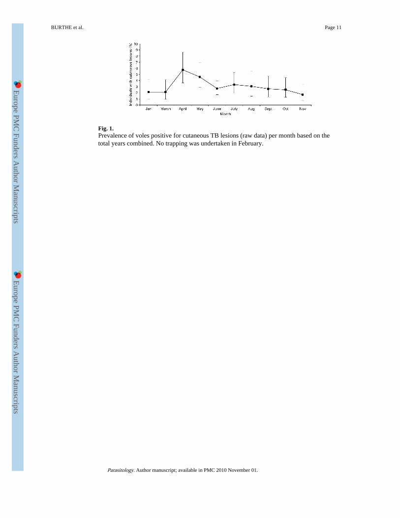
Fig. 1.Prevalence of voles positive for cutaneous TB lesions (raw data) per month based on thetotal years combined. No trapping was undertaken in February.
BURTHE et al. Page 11
Parasitology. Author manuscript; available in PMC 2010 November 01.
Europe PM
C Funders A
uthor Manuscripts
Europe PM
C Funders A
uthor Manuscripts
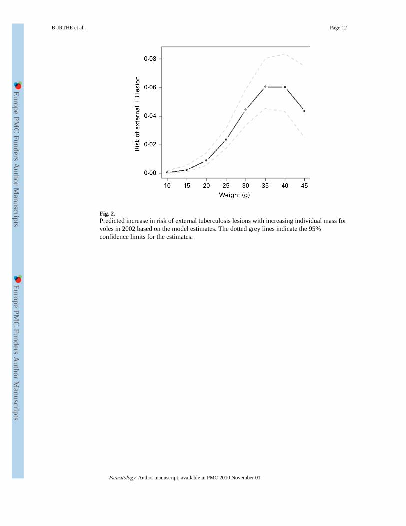
Fig. 2.Predicted increase in risk of external tuberculosis lesions with increasing individual mass forvoles in 2002 based on the model estimates. The dotted grey lines indicate the 95%confidence limits for the estimates.
BURTHE et al. Page 12
Parasitology. Author manuscript; available in PMC 2010 November 01.
Europe PM
C Funders A
uthor Manuscripts
Europe PM
C Funders A
uthor Manuscripts
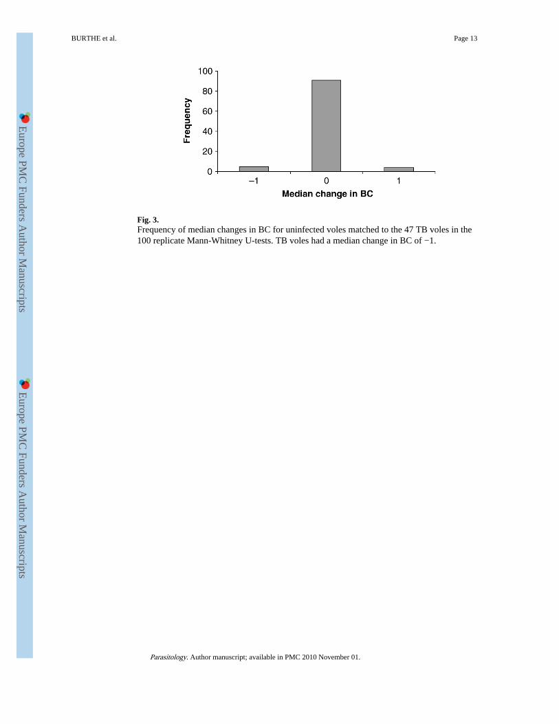
Fig. 3.Frequency of median changes in BC for uninfected voles matched to the 47 TB voles in the100 replicate Mann-Whitney U-tests. TB voles had a median change in BC of −1.
BURTHE et al. Page 13
Parasitology. Author manuscript; available in PMC 2010 November 01.
Europe PM
C Funders A
uthor Manuscripts
Europe PM
C Funders A
uthor Manuscripts
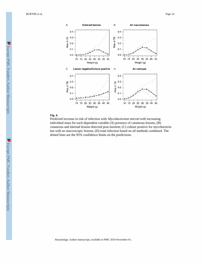
Fig. 4.Predicted increase in risk of infection with Mycobacterium microti with increasingindividual mass for each dependent variable (A) presence of cutaneous lesions; (B)cutaneous and internal lesions detected post-mortem; (C) culture positive for mycobacteriabut with no macroscopic lesions; (D) total infection based on all methods combined. Thedotted lines are the 95% confidence limits on the predictions.
BURTHE et al. Page 14
Parasitology. Author manuscript; available in PMC 2010 November 01.
Europe PM
C Funders A
uthor Manuscripts
Europe PM
C Funders A
uthor Manuscripts
Europe PM
C Funders A
uthor Manuscripts
Europe PM
C Funders A
uthor Manuscripts
BURTHE et al. Page 15
Tabl
e 1
Subs
crip
ts u
sed
in s
urvi
val m
odel
s (p
rogr
am M
AR
K)
(Det
ails
of
whi
ch p
aram
eter
s w
ere
incl
uded
in r
ecap
ture
mod
els
(p)
and
surv
ival
mod
els
(φ)
are
give
n in
the
righ
t han
d co
lum
n.)
Ana
lysi
s st
age
Subs
crip
tD
escr
ipti
onP
aram
eter
type
Gro
up le
vel
stSi
te-e
ffec
t (B
HP,
KC
S, P
LJ
or R
OB
)φ,
p
sxSe
x-ef
fect
φ, p
tFu
ll tim
e-de
pend
ence
φ, p
Tem
pora
lm
oM
onth
(13
mon
ths/
year
)φ,
p
seas
1Se
ason
(N
ov–F
eb, M
ar–M
ay, M
ay–J
ul, A
ug–O
ct)
φ, p
seas
2Se
ason
(w
inte
r O
ct–M
ar; s
umm
er A
pr–S
ept)
φ, p
yrY
ear
(200
1, 2
002
or 2
003)
φ, p
Cov
aria
tes
rfA
vera
ge r
ainf
all d
urin
g tr
appi
ng s
essi
onp
tem
pA
vera
ge te
mpe
ratu
re d
urin
g tr
appi
ng s
essi
onp
prev
rfA
vera
ge r
ainf
all d
urin
g pr
evio
us m
onth
φ
prev
tA
vera
ge te
mpe
ratu
re d
urin
g pr
evio
us m
onth
φ
dens
Vol
e de
nsity
dur
ing
trap
ping
ses
sion
φ, p
nB
inar
y sc
ore
of m
onth
s w
ith r
educ
ed n
umbe
rof
sec
onda
ry s
essi
ons
p
tbPr
eval
ence
of
TB
lesi
ons
(pop
ulat
ion
leve
l)φ,
p
Indi
vidu
al c
ovar
iate
se
Bin
ary
cova
riat
e de
notin
g in
divi
dual
s fr
om g
rid
edge
p
Tim
e-sp
ecif
icin
divi
dual
cov
aria
tes
mT
rap-
depe
nden
cy e
ffec
tp
indt
bPr
esen
ce o
f T
B le
sion
s (i
ndiv
idua
l lev
el)
φ
Parasitology. Author manuscript; available in PMC 2010 November 01.
Europe PM
C Funders A
uthor Manuscripts
Europe PM
C Funders A
uthor Manuscripts
BURTHE et al. Page 16
Table 2
The number of primary sessions that individuals were caught exhibiting an external tuberculous lesion
Number of primary sessionsan individual was caught with
an external lesion present
Number ofindividual
voles
1 76
2 25
3 4
4 1
5 2
Parasitology. Author manuscript; available in PMC 2010 November 01.
Europe PM
C Funders A
uthor Manuscripts
Europe PM
C Funders A
uthor Manuscripts
BURTHE et al. Page 17
Tabl
e 3
The
eff
ect o
f T
B o
n su
rviv
al o
f fi
eld
vole
s, a
s ov
eral
l les
ion
posi
tive
(i.e
. ind
ivid
uals
that
wer
e po
sitiv
e fo
r a
cuta
neou
s le
sion
at s
ome
poin
t in
thei
rca
ptur
e hi
stor
y (t
b)),
and
a ti
me-
spec
ific
indi
vidu
al T
B c
ovar
iate
(in
dtb)
(The
bas
e m
odel
was
φ(s
t * d
ens)
+pr
evrf
+m
o+yr
+sx
p m
o+yr
+rf
+n
+m
+ed
ge. T
he m
ost p
arsi
mon
ious
mod
el is
hig
hlig
hted
in b
old.
)
Mod
elSu
rviv
al m
odel
Rec
aptu
re m
odel
AIC
cN
o. o
fpa
ram
eter
sD
evia
nce
Bas
e m
odel
(st*
dens
)+pr
evrf
+m
o+yr
+sx
mo+
yr+
rf+
n+m
+ed
ge47
67.6
239
4688
.51
Inve
stig
atin
g T
B a
s an
indi
vidu
al c
ovar
iate
(st*
dens
)+pr
evrf
+mo+
yr+s
x+tb
mo+
yr+r
f+n+
m+e
dge
4742
.76
4046
61.5
9
Inve
stig
atin
g T
B a
s a
time-
spec
ific
indi
vidu
al c
ovar
iate
(st*
dens
)+pr
evrf
+mo+
yr+s
x+in
dtb
mo+
yr+r
f+n+
m+e
dge
4767
.66
4046
86.4
9
Parasitology. Author manuscript; available in PMC 2010 November 01.
Europe PM
C Funders A
uthor Manuscripts
Europe PM
C Funders A
uthor Manuscripts
BURTHE et al. Page 18
Table 4
Prevalence of TB per mass category of individuals
Mass% infected
(based on culture/histopathology)
Samplesize
<17 g 0.00 109
17–24 g 9.62 312
25–34 g 14.08 277
>34 g 20.69 29
Parasitology. Author manuscript; available in PMC 2010 November 01.