transfection of rat or mouse neurons by biolistics or electroporation
TRANSCRIPT

Transfection of rat or mouse neurons by biolistics orelectroporationSulayman D Dib-Hajj1–3, Jin Sung Choi1–3, Lawrence J Macala1–3, Lynda Tyrrell1–3, Joel A Black1–3,Theodore R Cummins4 & Stephen G Waxman1–3
1Department of Neurology, School of Medicine, Yale University, New Haven, Connecticut, USA. 2Center for Neuroscience and Regeneration Research, School ofMedicine, Yale University, New Haven, Connecticut, USA. 3Rehabilitation Research Center, Veterans Affairs Connecticut Healthcare System, West Haven, Connecticut,USA. 4Department of Pharmacology & Toxicology, Stark Neurosciences Institute, Indiana University School of Medicine, Indianapolis, Indiana, USA. Correspondenceshould be addressed to S.G.W. ([email protected]).
Published online 9 July 2009; doi:10.1038/nprot.2009.90
Properties of ion channels are affected by the background of the cells in which they are expressed. Thus, it is important for
investigators interested in neuronal function to study these proteins in post-mitotic neurons. However, post-mitotic neurons, and
many cell lines, are difficult to transfect by standard methods. Here we provide detailed protocols for two different procedures,
biolistic and electroporation, which have been used to transfect peripheral sensory neurons from mice or rats with expression
constructs of voltage-gated sodium channels. Neurons can be prepared, transfected and currents recorded within 48 h. Using these
methods, primary sensory neurons can be transfected with an efficiency of 5–20%, which has permitted studying biophysical
properties of sodium channels and their naturally occurring mutants in a native neuronal cell background. Although we have used
sodium channels for the examples that we show here, these methods can also be used to study other types of molecules.
INTRODUCTIONHere we describe two protocols that we have used in our labora-tories to transfect post-mitotic peripheral dorsal root ganglion(DRG) neurons. These methods provide a basis for functionalanalysis of voltage-gated sodium channels in their native cell back-ground and permit assessment of the contribution of these channelsto the firing properties of these neurons1–12. Although each target ofinvestigation poses its own challenges, in principle it should bepossible to express any channel or receptor in any of a broad rangeof neurons or other post-mitotic cell types, or other cell types thatare difficult to transfect by conventional methods using theseprotocols. Once cells are transfected using these protocols, userscan carry out immunohistochemical or electrophysiological studies,such as those described here and in the accompanying protocol13.
Overview of voltage-gated sodium channelsSodium channels are heterotrimeric glycoprotein complexes,which consist of a large pore-forming a-subunit and auxiliaryb-subunits14. The initial identification, cloning and functionalexpression of cDNAs encoding the pore-forming a-subunits(which will be referred to as sodium channels hereinafter)15–17
presaged the identification of nine genes (SCN1A–5A andSCN8A–11A) that encode distinct a-subunits (Nav1.1–Nav1.9) inmammalian genomes, which are expressed in tissue- and develop-ment-specific patterns14. These channels are broadly distinguishedpharmacologically by their sensitivity to the neurotoxin, tetrodo-toxin, as sensitive (TTX-S, blocked by nanomolar concentrations ofTTX) and resistant (TTX-R, blocked by 100- to 1,000-fold higherconcentrations of TTX)14. Molecular pathophysiological aspects ofsodium channelopathies that underlie many human diseases18–23
have been elucidated by cloning various sodium channels andfunctionally characterizing them in expression systems. Untilrecently, however, expression of sodium channels in native neuronshas been hampered by the low efficiency of transfection ofpost-mitotic neurons.
Overview of other transfection/expression systemsEarly functional studies showed that injecting transcripts ofa-subunits alone into Xenopus oocytes is sufficient to producevoltage-gated sodium channels16,17,24–26; however, the resultingsodium currents inactivate with slower kinetics and show depolar-ized voltage dependence compared with native currents in neuronsor muscles. Co-injection of low-molecular-weight RNA encodingb-subunits into Xenopus oocytes increases current density andalters the voltage dependence and inactivation properties of thesechannels24,25,27,28. Nevertheless, the different composition of theamphibian cell membrane, cell-type-specific differences in post-translational modifications of proteins and the absence of neuron-specific channel partners limit the utility of oocyte expressionsystems for extrapolation of results to native mammalian neurons.
Mammalian cell lines that can be readily transfected withrecombinant channel constructs using calcium phosphate orlipid-based techniques, and which can be used to derive stablytransfected cell lines, provide one alternative to the Xenopus oocyteexpression system. For example, transfection of sodium channelsNav1.2 or Nav1.4 into Chinese hamster ovary (CHO) or intohuman embryonic kidney 293 (HEK 293) cells produces sodiumcurrents that are qualitatively similar to native currents without theco-expression of the b1-subunit29–31. However, the currentsrecorded in these heterologous expression systems do not alwaysquantitatively reproduce those recorded in native neurons; forexample, co-expression of b1 and b2 subunits, together withNav1.3-HEK 293 stable cell line, produces significant effects onthe biophysical properties of the channel2. Nav1.6 and Nav1.8 alsodisplay different physiological properties when expressed in differ-ent types of neurons3,32–34. These studies show that the cell back-ground in different expression systems can have a significant effecton the channel density and biophysical properties of sodiumchannels. Thus, it is imperative to develop methods and protocolsto introduce sodium channel constructs into the different neurons
p
uor
G g
n ih si l
bu
P eru ta
N 900 2©
nat
ure
pro
toco
ls/
moc.er
ut an.
ww
w//:ptt
h
1118 | VOL.4 NO.8 | 2009 | NATURE PROTOCOLS
PROTOCOL
in which they are normally present for the voltage-clamp analysis ofchannel functional properties and for the current-clamp assess-ment of the contribution of these channels to the firing behavior ofthe neuronal type of interest13.
As almost all types of neurons express multiple isoforms ofsodium channels whose currents have similar, but not identical,kinetics and voltage dependencies, recording of the current fromthe channel of interest within most cell types presents a challenge.The challenge can be met by mutating the channel of interest tomake it resistant to TTX (TTX-R) and then using TTX to block theother endogenous TTX-sensitive (TTX-S) sodium channels in thecell. The situation is more complex in peripheral neurons, some ofwhich express TTX-R sodium channels (Nav1.8 and Nav1.9)14. Asnoted below, however, it is possible to assess electrophysiologicallythe current produced by sodium channels of interest, in isolation,even in these cells.
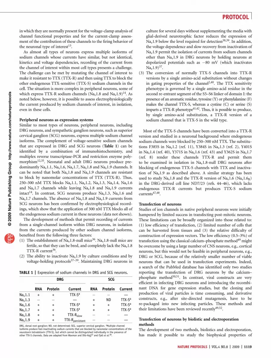
Peripheral neurons as expression systemsSimilar to most types of neurons, peripheral neurons, includingDRG neurons, and sympathetic ganglion neurons, such as superiorcervical ganglion (SCG) neurons, express multiple sodium channelisoforms. The complement of voltage-sensitive sodium channelsthat are expressed in DRG and SCG neurons (Table 1) can beidentified by a combination of immunohistochemistry, andmultiplex reverse transcriptase-PCR and restriction enzyme poly-morphism12,35. Neonatal and adult DRG neurons produce pre-dominantly Nav1.1, Nav1.6, Nav1.7, Nav1.8 and Nav1.9 channels. Itcan be noted that both Nav1.8 and Nav1.9 channels are resistantto block by nanomolar concentrations of TTX (TTX-R). Thus,250–300 nM TTX blocks Nav1.1, Nav1.2, Nav1.3, Nav1.4, Nav1.6and Nav1.7 channels while leaving Nav1.8 and Nav1.9 currentsintact14. In contrast, SCG neurons produce Nav1.3, Nav1.6 andNav1.7 channels. The absence of Nav1.8 and Nav1.9 currents fromSCG neurons has been confirmed by electrophysiological record-ings, which show that the application of 300 nM TTX blocks all ofthe endogenous sodium current in these neurons (data not shown).
The development of methods that permit recording of currentsfrom a single-channel isoform within DRG neurons, in isolationfrom the currents produced by other sodium channel isoforms,benefited from the following three factors:(1) The establishment of Nav1.8-null mice36. Nav1.8-null mice are
fertile, so that they can be bred, and completely lack the Nav1.8TTX-R current36.
(2) The ability to inactivate Nav1.9 by culture conditions and byvoltage-holding protocols37–39. Maintaining DRG neurons in
culture for several days without supplementing the media withglial-derived neurotrophic factor reduces the expression ofNav1.9 below the level required for detection38,39. In addition,the voltage dependence and slow recovery from inactivation ofNav1.9 permit the isolation of currents from sodium channelsother than Nav1.9 in DRG neurons by holding neurons atdepolarized potentials such as �80 mV (which inactivatesNav1.9)37.
(3) The conversion of normally TTX-S channels into TTX-Rversions by a single amino-acid substitution without changesin gating properties of the channel2,40. The TTX sensitivityphenotype is governed by a single amino-acid residue in thesecond re-entrant segment of the S5–S6 linker of domain I: thepresence of an aromatic residue, tyrosine (Y) or phenylalanine (F)makes the channel TTX-S, whereas a cystine (C) or serine (S)imparts a TTX-R phenotype41,42. Thus, it is possible to produce,by single amino-acid substitution, a TTX-R version of asodium channel that is TTX-S in the wild type.
Most of the TTX-S channels have been converted into a TTX-Rversion and studied in a neuronal background where endogenoussodium channels were blocked by 250–300 nM TTX. The substitu-tions F385S in Nav1.2 (ref. 11), Y384S in Nav1.3 (ref. 2), Y401Sin Nav1.4 (ref. 40), Y371S in Nav1.6 (ref. 43) and Y362S in Nav1.7(ref. 8) render these channels TTX-R and permit themto be examined in isolation in Nav1.8-null DRG neurons afterblocking of endogenous TTX-S channels with TTX and inactiva-tion of Nav1.9 as described above. A similar strategy has beenused to study Nav1.8 and the TTX-R version of Nav1.6 (Nav1.6R)in the DRG-derived cell line ND7/23 (refs. 44–46), which lacksendogenous TTX-R currents but produces TTX-S sodiumcurrents47,48.
Transfection of neuronsStudies of ion channels in native peripheral neurons were initiallyhampered by limited success in transfecting post-mitotic neurons.These limitations can be broadly organized into those related to:(1) low efficiency of transfection, (2) limited number of cells thatcan be harvested from tissues and (3) the relative difficulty ofconstruction of expression vectors. The low efficiency (0.5–3%) oftransfection using the classical calcium–phosphate method49 mightbe overcome by using a large number of CNS neurons, e.g., corticalneurons, but this would not be feasible in peripheral neurons, e.g.,DRG or SCG, because of the relatively smaller number of viableneurons that can be used in transfection experiments. Indeed,a search of the PubMed database has identified only two studiesreporting the transfection of DRG neurons by the calcium–phosphate method50,51. In contrast, viral constructs are veryefficient in infecting DRG neurons and introducing the recombi-nant DNA for gene expression studies, but the cloning andproduction of viral particles is time consuming, and derivativeconstructs, e.g., after site-directed mutagenesis, have to bere-packaged into new infecting particles. These methods andtheir limitations have been reviewed recently49,52.
Transfection of neurons by biolistic and electroporationmethodsThe development of two methods, biolistics and electroporation,has made it possible to study the biophysical properties of
p
uor
G g
n ih si l
bu
P eru ta
N 900 2©
nat
ure
pro
toco
ls/
moc.er
ut an.
ww
w//:ptt
h
TABLE 1 | Expression of sodium channels in DRG and SCG neurons.
DRG SCG
RNA Protein Current RNA Protein Current
Nav1.1 + + TTX-Sa � � —Nav1.3 � � — + ND TTX-Sa
Nav1.6 + + TTX-Sa + + TTX-Sa
Nav1.7 + + TTX-Sa + + TTX-Sa
Nav1.8 + + TTX-Rslow � � —Nav1.9 + + TTX-Rpersistent � � —
DRG, dorsal root ganglion; ND, not determined; SCG, superior cervical ganglion. aMultiple channelisoforms produce fast-inactivating sodium currents that are blocked by nanomolar concentrations of theneurotoxin tetrodotoxin (TTX-S), but which cannot be distinguished individually in the presence ofother TTX-S channels. Data are adapted from Waxman and Dib-Hajj22 and Fjell et al.38.
NATURE PROTOCOLS | VOL.4 NO.8 | 2009 | 1119
PROTOCOL

recombinant sodium channels in DRG neurons and to examine thefiring properties of these neurons1–12. Biolistic transfection is theintroduction of naked DNA into cells by discharging of goldparticle projectiles covered with DNA, under high pressure, intothe nucleus of post-mitotic neurons that have been cultured oncoverslips for several days. Transfected neurons are identified by thepresence of the gold particle lodged within the cell and by theexpression of reporter proteins, e.g., green fluorescence protein(GFP). Electroporation is the introduction of naked DNA into cellsby applying a strong electric current pulse to a mixture of cells andDNA. Transfection of neurons by electroporation is accomplishedby forming a suspension of freshly isolated cells, lipid-basedtransfection media and DNA in a cuvette and subjecting themixture to high-voltage pulse. The DNA, both target and reporter,constructs are driven inside the cell by the electric force of the pulse,and transfected neurons are identified by the expression of thereporter protein, e.g., GFP. Each of these methods reproduciblyachieves transfection efficiencies that permit high-resolution sin-gle-cell studies, e.g., imaging studies of recombinant proteins andelectrophysiological recordings of ionic conductances, as will bediscussed next.
Advantages and limitationsNeuronal transfection by biolistics or electroporation has signifi-cantly advanced our understanding of sodium channel physiologyand has permitted an assessment of the impact of specific channelson the firing properties of neurons. These methods could be used totransfect neurons with other targets of investigation. However, thereare advantages and limitations to both methods that need to beconsidered in deciding which one to use in a particular situation.Also, the experimental conditions in each of these methods influencethe interpretation of the outcomes, as will be discussed below.
As already discussed, biolistic transfection of neurons entailsshooting of gold particle projectiles covered with DNA into thenucleus of post-mitotic neurons cultured on coverslips. There arethree major advantages of biolistic transfection. First, the methoddoes not require a large number of neurons to obtain viabletransfected cells. Second, the gold particles can be pre-coatedwith multiple different plasmid DNAs, thus facilitating the trans-fection of individual neuron with multiple constructs. Usingbiolistic transfection of DRG neurons, we have shown earlier thatup to three independent plasmids can be functionally expressed in asingle DRG neuron10. Third, maintenance of rat DRG neurons inculture without supplementing the media with trophic factorsmimics the effect of axotomy and causes the downregulation ofNav1.9 channels38,39,53, thus permitting the electrophysiologicalstudies of the TTX-R versions of sodium channels as describedearlier.
The major disadvantage of biolistic transfection is the need touse neurons that have been in culture for several days (3–5 d).Neurons have to be firmly attached to the substrate in orderto withstand the force of the gold projectiles without lifting offthe coverslip. Clearly, neurons that have been kept in culturefor several days undergo significant time-dependent cellularchanges compared with acutely isolated neurons or neurons inculture for a short period of time; neurons in culture for a weekacquire features of axotomized neurons54. Another disadvantage
is the deterioration in potency of the cartridge with time. Althoughit is possible to store these cartridges under vacuum for a fewweeks, the efficiency of successful transfection deteriorates withstorage time, so valuable full-length channel constructs frequentlyare wasted.
Transfection of neurons by electroporation has gained popularitybecause it permits transfection of acutely isolated neurons and thusenables studies of neurons that are closer to their native state. Forexample, this method makes it feasible to study neuronal firingafter the expression of wild-type or mutant channels as it isexpected that the complement of ionic conductances undergoesless time-dependent change after 24–48 h of isolation, comparedwith neurons in culture for a week. In addition, we have obtainedroutine transfection efficiencies of 15–20% that provide ample cellnumbers for immunocytochemical and electrophysiological stu-dies. The use of neurons from younger animals can improve theefficiency of transfection, but an adequate yield of transfected cellscan be obtained from adult animals. Transfection by electropora-tion has produced the most efficient transfection of DRG neuronsby far, including the documentation of the functional expression ofthree different constructs (two sodium channels and an enhancedGFP, EGFP) at the same time12.
The major limitation of electroporation of DRG neurons is theneed for a very large number of viable neurons to survive theelectric pulse. The protocol optimally requires 1–2 � 106 cells pertransfection, which necessitates harvesting of at least 10 pairs ofDRGs for a single experiment. The challenge, however, is to obtainthis tissue in a very short period of time (10–20 min); otherwise, theviability of the cells significantly deteriorates. The usage of fewercells for transfection causes more cell death, with the survivingtransfected neurons not healthy enough for electrophysiologicalrecordings. Another limitation is the extensive neurite outgrowth oftransfected neurons that degrades space clamp and makes voltage-clamp studies more challenging. Modification of the attachmentsubstrate might reduce neurite outgrowth and improve whole-cellpatch clamp studies.
Further applicationsThe increased transfection efficiency of primary neurons by biolis-tics or electroporation enables studies of electrophysiology, channelbiogenesis and subcellular distribution. Further details of how tocarry out voltage-clamp and current-clamp recordings from trans-fected DRG neurons can be found in the accompanying paper13.Incorporation of chromogenic tags into ion channels facilitatestracking the trafficking of these channels through cytoplasmiccompartments. For example, Nav1.5 and Nav1.8 channels havebeen successfully tagged with GFP55–57, and in principle, otherchannels could also be produced as GFP-fusion proteins. GFP-tagged Nav1.8 or other channels that are expressed in peripheralneurons can be used to study trafficking of these membraneproteins among subcellular organelles and regulation of theirdifferential plasma membrane localization. In addition, fluorescentresonance energy transfer has been used to study the interaction offluorescently tagged Nav1.5 and b1 subunit in HEK 293 cells58;similar approaches can be used to investigate protein–proteininteractions between a sodium channel and its partners in trans-fected neurons.
p
uor
G g
n ih si l
bu
P eru ta
N 900 2©
nat
ure
pro
toco
ls/
moc.er
ut an.
ww
w//:ptt
h
1120 | VOL.4 NO.8 | 2009 | NATURE PROTOCOLS
PROTOCOL
MATERIALSREAGENTS.Male Sprague–Dawley albino rats (Harlan) or mice, e.g., Nav1.8-null
C57/BL6 transgenic mice36 ! CAUTION Use of rodents must conform toappropriate national and institutional regulations.
.Borate buffer (15 mM, pH 8.4; Sigma-Aldrich)
.Collagenase A (Roche)
.Collagenase D (Roche)
.Coverslips coated with poly-D-lysine/laminin (BD BioCoat, or see REAGENTSETUP) m CRITICAL Coverslips need to be sterilized with UV light for10 min before plating cells. UV sterilization is optional for the commerciallyavailable pre-coated coverslips.
.DNA for transfection, e.g., closed, circular DNA that encodes a sodiumchannel in a mammalian expression plasmid (e.g., pcDNA3.1, Invitrogen),and GFP encoded by the plasmid pEGFP (Clontech)
.D-MEM/F-12 (1:1) (Invitrogen)
.Fetal bovine serum-characterized (HycloneThermo)
.Gold particles (1 mm); for biolistic transfection only
.Ketamine/xylazine
.Laminin solution (5 mg ml�1; Sigma)
.Liberase Blendzyme 3 (Roche)
.Liberase Blendzyme 4 (Roche)
.Mouse neuron Nucleofector Kit (Lonza, cat. no. VPG-1001); forelectroporation only
.Papain (Worthington Biochemical)
.Penicillin/streptomycin (Invitrogen)
.Poly-L-ornithine hydrobromide, molecular weight (MW) 30–70k(Sigma-Aldrich, cat. no. P-3655)
.Rat neuron Nucleofector Kit (Lonza, cat. no. VPG-1003); for electroporationonly
.Polyvinylpyrrolidone (PVP) (Bio-Rad Laboratories); for biolistic transfectiononly
.Spermidine (Sigma); for biolistic transfection only
.Trypsin inhibitor (Sigma)EQUIPMENT.Cylinder of compressed helium, grade 4.5 or higher, with regulator; for
biolistic transfection only.Cylinder of compressed nitrogen, grade 4.8 or higher, with regulator; for
biolistic transfection only.Helios Gene Gun System (Bio-Rad Laboratories); for biolistic transfection
only.Inverted microscope with fluorescence, e.g., Nikon TE2000 with fluorescence
(Nikon).Microcentrifuge.Nutator.Nucleofector II (Lonza); for electroporation only.Nylon mesh, 70-mm pores (Small Parts); for biolistic transfection only..Surgical equipment (Fine Science Tools).Dumont #4 Forceps, straight, standard tip 0.13 � 0.08 mm, Dumoxel, 11 cm;.Dumont #5 Forceps, straight, standard tip 0.1 � 0.06 mm, Inox, 11 cm;.Dumont #5 Forceps, straight, Biologie tip 0.05 � 0.01 mm, Inox, 11 cm;.Dumont #7 Forceps, curved, standard tip, 0.17 � 0.1 mm, Dumoxel, 11.5 cm;.Malleus nipper, straight, 11 cm, 9-mm cutting edge;. Pearson Micro Rongeurs with extra fine tips; shallow bend, 1.2-mm cup
width, 19 cm (the length is a personal preference);. Spring scissors, straight, sharp/sharp, 10 cm, 0.2-mm-tip diameter; and.Vannas-Tubingen spring scissors, straight, sharp/sharp, 8.5 cm, 0.1–mm-tip
diameter.Syringes (2 � 10 ml) fitted with 6-inch silicone adaptor tubing; for biolistic
transfection-only.Vortex (Vortex-Genie 2, VWR, West Chester, PA)REAGENT SETUPComplete saline solution (CSS) (1 liter) This comprises 137 mM NaCl,5.3 mM KCl, MgCl2-6H2O, 25 mM Sorbitol, 10 mM HEPES, 3 mM CaCl2.
To prepare, first combine 8 g NaCl (MW 58.44), 0.395 g KCl (MW 74.56),0.203 g MgCl2-6H2O (MW 203.30), 4.555 g Sorbitol (MW 182.2), 2.382 gHEPES (MW 238.31) and 0.441 g CaCl2 (MW 147.0). Add double-distilledwater to reach a final volume of 1 liter and adjust pH to 7.2 with NaOH.Filter-sterilize and store at 4 1C.Poly-L-ornithine hydrobromide Dissolve 50 mg of poly-L-ornithinehydrobromide in 500 ml of 15 mM borate buffer (pH 8.4) and divide thesolution into 1-ml aliquots for long-term storage at �20 1C.DRG medium Mix 89% (vol/vol) DMEM/F-12, 10% (vol/vol) fetal bovineserum, 1% (vol/vol) penicillin/streptomycin.Preparation of DRG dissociation solutions BSA/TI: Add 5 ml DRG mediumto 7.5 mg bovine serum albumin (BSA) plus 7.5 mg trypsin inhibitor (TI).Vortex, warm at 37 1C and filter (0.2-mm pore size). Store at 4 1C (up to 5 d)until ready to use.Preparation of DRG dissociation enzymatic solutions for rat Collagenase Aand D: For rat DRG dissociation, weigh 15 mg collagenase A and 15 mgcollagenase D in separate 15 ml conical tubes. Oxygenate CSS for 10 min in a50-ml conical tube. Add 10 ml oxygenated CSS into collagenase A andcollagenase D tubes, then add 120 ml of 50 mM EDTA to each tube. Add papain(30 U ml�1) to a collagenase D tube. Vortex, warm at 37 1C (to ensure completesolubilization of the components), filter (0.2 mm pore size) in 15-ml tubes, andthen keep on ice until use. Warm the enzyme dissociation solutions at 37 1C for10–20 min before they are ready to be used. m CRITICAL Working solutions ofthese enzymes must be prepared on the day of the experiment. m CRITICALCollagenase A and D are crude preparations and their enzymatic activitiescan vary widely with each lot. We have found that rat DRG can tolerate thesevariations but mouse DRG cannot, giving dissociated neurons of variable quality.Preparation of DRG dissociation enzymatic solutions for mice LiberaseBlendzyme 3 and 4: Reconstitute the enzymes in sterile water and store 5-Ualiquots at �20 1C; volume of the aliquot varies with the specific activity of theenzymes that may vary with different lots. Use single 5-U aliquots of LiberaseBlendzyme 4 and 3 for mouse DRG dissociation. Oxygenate CSS for 10 min in a50-ml conical tube. Add an aliquot of Liberase Blendzyme 4 to a tube containing10 ml CSS and add an aliquot of Liberase Blendzyme 3 to a tube containing 10ml CSS. Then add 120 ml of 50 mM EDTA to each tube. Add papain (30 U ml�1)to Liberase Blendzyme 3 tube. Vortex, warm at 37 1C (to ensure completesolubilization of the components), filter (0.2 mm pore size) into 15-ml tubes, andthen keep on ice until use. Warm the enzyme dissociation solutions at 37 1C for10–20 min before they are ready to be used. m CRITICAL Working solutions ofthese enzymes must be prepared on the day of the experiment. m CRITICALLiberase are purified enzymes with less lot-to-lot variability than collagenases.Substituting Liberase Blendzyme 4 (20 min incubation at 37 1C) for CollagenaseA, and Liberase Blendzyme 3 (10 min incubation at 37 1C) for Collagenase D,gives reproducibly healthy, dissociated mouse DRG neurons.0.05 M Spermidine (for biolistic transfection only) Store aliquots of 1-Mspermidine stock solution at �20 1C. Prepare 0.05 M spermidine on the day ofexperiment. m CRITICAL Dilutions must be prepared on the day of experiment.0.05 mg ml�1 PVP (in Bio-Rad kit; for biolistic transfection only) Prepare20 mg ml�1 PVP in ethanol. Aliquot and store at �20 1C. Dilute an aliquotto 0.05 mg ml�1 on the day of experiment. m CRITICAL Dilutions must beprepared on the day of experiment.EQUIPMENT SETUPPreparation of coated coverslips This is an alternative to using commerciallyavailable pre-coated poly-D-lysine/laminin coverslips (BioCoat, BD Biosciences).First, clean and store 12-mm circular glass coverslips in 95% (vol/vol) ethanol.Flame coverslips to burn off the alcohol and place them in wells of a 24-well plate.Coat each coverslip with 100 ml of poly-L-ornithine solution (0.1 mg ml�1 in 15mM borate buffer, pH 8.4) and leave at room temperature (20–22 1C) for 20 min to1 h. Aspirate the poly-L-ornithine solution and rinse coverslips twice with sterile de-ionized water. Allow coverslips to dry completely under the hood (at least 15 minor overnight). Spread 2.5 ml laminin solution (5 mg ml�1) on each coverslip with aflattened (by warming) 200-ml plastic pipette tip and allow the coverslips to drycompletely. Sterilize coverslips with UV light for 10 min before plating cells.
PROCEDUREDRG dissection and dissociation1| Prepare surgery table and warm the collagenase A tube (for rat DRG dissociation) or Liberase Blendzyme 4 (for mouse DRGdissociation) at 37 1C. This, and harvesting, must be performed 3 d before biolistic transfection or on the day of electroporation.
p
uor
G g
n ih si l
bu
P eru ta
N 900 2©
nat
ure
pro
toco
ls/
moc.er
ut an.
ww
w//:ptt
h
NATURE PROTOCOLS | VOL.4 NO.8 | 2009 | 1121
PROTOCOL
2| Anesthetize adult rats (4–8-weeks-old) with CO2 narcosis and then decapitate. Anesthetize neonatal rats (P0–P5), andadult (4–8-weeks-old) and neonatal mice (P2–P6) with ketamine/xylazine (rats: 75:10 mg kg�1, i.p.; mice: 100:10 mg/kg, i.p.)and then decapitate.
3| If using an adult rat, cut with a scalpel through skin down the center of back and pull the skin away to expose vertebralcolumn, and then cut through muscle on either side of the vertebral column. Remove muscle tissue, vertebral lamina andpedicles with a rongeur to expose the spinal cord. Remove the spinal cord with forceps. Further, trim away bone to locate DRGon either side of the vertebral column. For adult and neonatal mouse and neonatal rat, use Malleus nippers instead of rongeurs.Alternatively, use a 10-cm spring scissors for cutting away the vertebral column in neonates and for adult mouse.
4| Cut through central-projecting roots using 10-cm spring scissors (for mouse and for neonates use 8.5 cm spring scissors),remove DRG using fine forceps, and place DRG in oxygenated CSS (in a 35-mm dish on ice). Electroporation experiments requireat least 20 DRGs from an adult mouse and from a neonatal rat or a mouse to reach a minimum number of cells (800,000:includes neurons, glia and fibroblasts) for transfection. Fewer adult rat ganglia will be needed to obtain a similar number ofcells (neurons, glia and fibroblasts). Typically, the L4–L5 DRG pairs from a single animal can be cultured on 12–16 coverslipsand used for biolistic transfection.
5| De-sheath adult rat DRG using fine forceps (#5 standard tip or #5 Biologie tip) before enzymatic dissociation. De-sheathingis not necessary for mice or for neonatal rats.
6| Place DRG tissue into a collagenase A tube for rat or Liberase Blendzyme 4 for mouse (# 7 curved forceps are useful for‘scooping’ the DRG out of the CSS) and incubate at 37 1C for 20 min on Nutator. Meanwhile, warm the collagenase D tube(for rat) or Liberase Blendzyme 3 tube (for mouse) at 37 1C.
7| After incubation, centrifuge DRG tissue for 2–3 min (100g). Remove the supernatant and add collagenase D solution forrat DRG and incubate for 20 min at 37 1C on Nutator; add Liberase Blendzyme 3 solution for mouse DRG and incubate for10 min at 37 1C on Nutator. Warm the BSA/TI solution and, for electroporation only, calcium-free DMEM at 37 1C.
8| After incubation, centrifuge for 2–3 min (100g). Remove the supernatant and add a volume of BSA/TI solution.m CRITICAL STEP A volume of BSA/TI solution depends on the desired final seeding density of neurons. For biolistic transfection(Step 10, option A) use B0.5 to 1 ml; for electroporation (Step 10, option B) use 0.5 ml.
9| Triturate the DRG using a 1-ml pipetman.m CRITICAL STEP Harsh trituration will damage the cells and reduce viability; under-trituration will lead to reduced number ofisolated cells for downstream application. To ensure a good outcome, triturate gently with uniform pressure and speed. DRGfrom adult rats are more easily dissociated and should typically be triturated with 6–8 strokes, whereas DRG from adult mouseand neonatal rat require 12 strokes. Healthy neurons are characterized, using Hoffman modulation contrast microscopy, by asmooth cell membrane (no blebbing), a distinct nucleus and a nucleolus, and by some cells that retain short segments of theiraxons, whereas harshly triturated neurons are devoid of these characteristics. The presence of vacuoles is also an indication ofunhealthy cells.
10| To transfect cells using the biolistic method, grow the cells for 3 d then follow option A. For electroporation (option B),use DRG neurons immediately after isolation.(A) Transfection of DRG neurons by biolistics using the Helios gene gun and DNA-coated gold particles
(i) Precipitation of DNA onto the gold particles (20 � 0.5 inch cartridges). Weigh 15 mg of 1-mm-diameter gold particles andadd 50 ml of 0.05 M spermidine in an Eppendorf tube. Vortex and sonicate for 15 s in an ultrasound bath. Add 10 mg ofsodium channel DNA, 5 mg of EGFP DNA and vortex.
(ii) Add 50 ml of 1 M CaCl2 to the DNA–gold particle mixture dropwise while vortexing gently (setting 4 on Vortex-Genie 2).Allow the DNA mixture to co-precipitate onto the gold particles at room temperature for 10 min.
(iii) Touch-spin the mixture in a microcentrifuge for 5 s at top speed (18,000g). Discard the supernatant and vortex the pellet.Wash the DNA–gold suspension three times in 1 ml of 100% (vol/vol) ethanol and touch-spin the mixture each time topellet the gold particles. Discard the ethanol supernatants.m CRITICAL STEP Use a fresh bottle of 100% ethanol for all ethanol steps.? TROUBLESHOOTING
(iv) Resuspend the mixture in 200 ml of 0.05 mg ml�1 PVP in ethanol and transfer to a 15-ml falcon tube. Rinse the contentsof the Eppendorf tube with 0.05 mg ml�1 PVP in ethanol (1.5 ml) and transfer to the 15-ml falcon tube; 1.7 ml is asufficient volume to coat the inner wall of 10 inch of Tefzel tubing in the Bio-Rad Tubing Prep Station.
p
uor
G g
n ih si l
bu
P eru ta
N 900 2©
nat
ure
pro
toco
ls/
moc.er
ut an.
ww
w//:ptt
h
1122 | VOL.4 NO.8 | 2009 | NATURE PROTOCOLS
PROTOCOL

(v) Preparation of gold-coated cartridges using the Tubing Prep Station. Ensure the apparatus is level, the syringes are cleanand the nitrogen cylinder is attached.
(vi) Open the nitrogen cylinder to 2 psi and adjust at the apparatus to 0.3 lpm. Turn off at the apparatus.(vii) Cut 30 inch tubing, supplied in the Cartridge kit, and position in the instrument without insertion into the O-ring.(viii) Use a syringe to rinse tubing with 100% (vol/vol) ethanol.
m CRITICAL STEP Keep the ethanol within the tubing.(ix) On removal of the ethanol, insert tubing into O-ring and dry thoroughly by blowing N2 at a pressure of 0.3–0.4 lpm
for B10 min.(x) Take out the tubing and position on the desk.(xi) Vortex the gold/PVP slurry in the falcon tube (the solution prepared in Step 10A(iv)).(xii) Attach a syringe to one end of the tubing and pull the suspended gold into the other end immediately after vortexing.
Continue to pull the slurry until it is positioned about 8–10 inches from the tubing ends.m CRITICAL STEP Avoid drawing in air bubbles.
(xiii) Carefully thread the tubing back into the apparatus without inserting into the O-ring. Allow the gold to settle for3–5 min.
(xiv) Carefully pull off the ethanol with a syringe at a rate of 1 inch s�1. Quickly rotate the tube 1801 and hold for 10 s.Then begin rotating the tubing for 30 s.
(xv) Push the tubing into the O-ring and blow N2 at 0.3–0.4 lpm pressure to dry the tube and contents (B5 min).? TROUBLESHOOTING
(xvi) Slice the coated tubing into 20 cartridges and store desiccated under vacuum at room temperature. This process resultsin a density of 0.75 mg of gold particles and 0.75 mg of total DNA per cartridge.’ PAUSE POINT Cartridges are stable under vacuum at room temperature for at least 1 month.
(xvii) Operation of Gene Gun. Sterilize the cartridge holder, barrel liner and mesh screens in ethanol and dry.(xviii) Load cartridges into their holder placing a blank cartridge in position 1.(xix) Insert the holder into the gun.(xx) Adjust helium pressure to 120 psi.(xxi) Advance the cartridge holder to position 1 and fire the blank cartridge over an empty well to make sure the pressure
is stable.(xxii) Remove the medium from one well of DRG neurons and position the mesh screen over the top of the well.(xxiii) Position the barrel liner directly over the screen, engage the interlock and fire. Quickly replace the medium.
m CRITICAL STEP The barrel of the gun must be held vertical to the wells, and the use of the 70-mm nylon screen allowsa uniform distribution of gold particles. Minimize the time the cells are without medium. Helium pressure is used by thegene gun to shoot the gold particles into the cell nucleus. The final distribution of gold particles is critical to an effectivetransfection. Too high of a gold particle density can cause excessive cell death. Too low of a gold particle density canresult in low transfection yield. A good density will yield 5–10 transfected cells per coverslip.? TROUBLESHOOTING
(xxiv) Incubate the cells at 37 1C in 95% O2-5% CO2.(xxv) Assess cell viability and the expression of recombinant proteins 24–48 h after shooting. Transfected cells are identified
by the green fluorescence from EGFP. Electrophysiological studies have shown that 99% of the cells that express GFP alsoexpress recombinant TTX-R sodium currents2.? TROUBLESHOOTING
(B) Transfection of DRG neurons by electroporation(i) If an exact cell count is not required (a reproducible yield becomes possible after practice), proceed directly to next
step. If an exact cell number is required, mix an equal volume (20 ml) of cell suspension and 0.4% (wt/vol) trypan bluesolution (20 ml) and apply to a hemocytometer for counting. At least 800,000 cells per transfection are required(optimal cell number ¼ 2 million cells).
(ii) Centrifuge cells for 2–3 min (100g) and remove the supernatant.(iii) Gently, resuspend cells at room temperature in 100 ml Nucleofector solution.(iv) Mix 100 ml cell suspension with DNA (2 mg EGFP + 10 mg channel DNA for an adult rat or a mouse; 1 mg EGFP +
5 mg channel DNA for a neonatal rat).(v) Transfer cell suspension into a cuvette provided in the Nucleofector kit (Lonza; cat no. VPG-1001 or VPG-1003).
Make sure that the sample covers the bottom of the cuvette and avoid air bubbles while pipetting. Close the cuvette withthe cap.
(vi) Insert the cuvette into its holder in the Nucleofector II electroporater. Use program G-013 for rat DRG and programO-003 for mouse DRG. Press the start ‘X’ key to start the program.
p
uor
G g
n ih si l
bu
P eru ta
N 900 2©
nat
ure
pro
toco
ls/
moc.er
ut an.
ww
w//:ptt
h
NATURE PROTOCOLS | VOL.4 NO.8 | 2009 | 1123
PROTOCOL

(vii) After transfection, immediately add 500 ml of pre-warmed (37 1C) calcium-free DMEM to the transfected cells using theplastic pipette provided in the kit, and then gently transfer the cells from the cuvette into 1.5-ml tube.m CRITICAL STEP Transfected cells are susceptible to calcium-mediated cytotoxicity; hence, recovery in Ca2+-free mediaenhances viability of the transfected neurons.
(viii) Incubate for 5 min at 37 1C in humidified 95% O2–5% CO2 incubator.(ix) After incubation, add 400 ml of pre-warmed BSA/TI solution and seed 80 ml per coverslip (time: 5 min).
m CRITICAL STEP Dilute to the desired seeding density with the BSA/TI solution, depending on the downstream assay,and aliquot samples onto coverslips. Low cell density is optimal for electrophysiological applications13, whereas celldensity is less important for immunocytochemical assays.
(x) Add DRG medium to a final volume of 1 ml per well, 30 min after seeding, and incubate cells at 37 1C in 95% O2-5% CO2.(xi) To perform current-clamp recordings13, supplement the DRG medium with neurotrophic growth factor (NGF) (50 ng ml�1)
and glial-derived neurotrophic factor (50 ng ml�1).m CRITICAL STEP As endogenous Nav1.8 and Nav1.9 sodium channels will be downregulated in the absence of trophicfactor support, these factors should not be added if the endogenous Nav1.9 TTX-R currents in Nav1.8�/� DRG neurons areto be run down in order to study recombinant channels that have been rendered TTX-R by whole-cell voltage-clamprecordings.
(xii) Assess cell viability and the expression of recombinant proteins 24–48 h after plating. Transfected cells should beidentifiable by the green fluorescence from EGFP. Electrophysiological studies have shown that 99% of the cells thatexpress EGFP also express recombinant TTX-R sodium currents1,9.? TROUBLESHOOTING
� TIMINGDRG dissection (Steps 1–4): 10–20 minNeuron isolation (Steps 5–9): 60 minTransfection by biolistics: precipitation of DNA onto the gold particles (Step 10A(i–iv)): 30 minTransfection by biolistics: preparation of gold coated cartridges (Step 10A(v–xvi)): 30 minTransfection by biolistics: operation of gene gun (Step 10(vii–xxv)): 5 min to set up plus 30–60 s per targetTransfection by electroporation (Step 10B(i–xii)): 25 min
? TROUBLESHOOTINGTroubleshooting advice can be found in Table 2.
p
uor
G g
n ih si l
bu
P eru ta
N 900 2©
nat
ure
pro
toco
ls/
moc.er
ut an.
ww
w//:ptt
h
TABLE 2 | Troubleshooting table.
Step Problem Solution
Biolistic transfection10A(iii) Gold particles aggregate after coating with DNA Lower the DNA loading ratio
10A(xv) Poor distribution of gold in tubing Complete dehydration of the DNA/gold mixture is crucial, and all traces ofwater must be eliminated. Therefore, use a fresh bottle of 100% ethanol inthe preparation of cartridges and if necessary replace the stock PVPAvoid working in humid environmentAfter drawing off the ethanol, quickly turn the tubing 1801 before rotating
10A(xxiii) Too few gold particles on coverslip Check that the helium pressure has remained stableIncrease pressure by 20 psiUse larger gold particles
10A(xxv) Poor transfection Efficiency of transfection decreases with longer storage periods of thecartridgesIncrease the DNA loading ratio
Too many green cells without sodium channel Alter the channel to EGFP ratio without changing the total DNA
Too many gold particles causing excessive cell death Decrease the pressure by 20 psiDecrease the amount of gold per shotMinimize the time the cells are left without medium (30 s is reasonabletime)
(continued)
1124 | VOL.4 NO.8 | 2009 | NATURE PROTOCOLS
PROTOCOL

ANTICIPATED RESULTSUsing these methods, it is our experience that we have been able to achieve transfection efficiencies of 5–20% depending onage and species of the animals used. We have found that using younger animals (P0–P5) produces better transfectionefficiencies than using adult animals. We have also found that DRG neurons from the Nav1.8�/� mice are more fragile thanthose from wild-type mice, thus resulting in lower efficiency of transfection and viability. Transfection by electroporationinvariably produces higher efficiency of transfection than biolistic transfection.
Biolistic transfection of mouse DRG neuronsFigure 1 shows recordings from DRG neurons transfected using the Helios Gene Gun with recombinant sodium channels thathave been rendered TTX-R2,8. For these experiments, DRG neurons from Nav1.8-null mouse were kept under standard tissueculture conditions for 3–5 d before biolistic transfections. Some Nav1.8-null DRG neurons express Nav1.9 persistent TTX-Rsodium currents37, but because these currents fall below 1 nA after several days in culture and run down quickly in whole-cellrecording configuration, they were not significant source of contamination under the recording conditions used in these studies.Neurons were co-transfected with EGFP, and electrophysiological studies are conducted 18–48 h after transfection; most of thecells that express EGFP also express fast-inactivating TTX-R sodium currents (Fig. 1).
Electroporation of sodium channels into DRG neuronsDRG neurons from Nav1.8-null mice (Nav1.8�/�)36 can be transfected by electroporation with the TTX-R version of any sodiumchannel, Nav1.8, or any of their mutant derivatives. We usually conduct immunohistochemical and electrophysiological studies
p
uor
G g
n ih si l
bu
P eru ta
N 900 2©
nat
ure
pro
toco
ls/
moc.er
ut an.
ww
w//:ptt
h
30
400 pA20 ms
500 pA
5 nA10 ms
5 nA
a b c
d
e f g
10 ms
20 ms
–30–60
–90–120
0
Electroporation transfection10B(xii) Low cell viability Increase total number of cells that are pulsed for electroporation
Reduce the time to harvest the DRG tissue after euthanizing the animalUse younger animals that yield more robust cellsUse an alternate program that delivers a weaker electric pulse
10B(xii) Low transfection efficiency Quality of the starting culture (see above) is a critical factor in yieldinghigher transfection efficiencyUse an alternate program to deliver a stronger electric pulse to reach theoptimal combination of cell viability and the number of transfected cells
10B(xii) Space-clamp problems Minimize time in culture to reduce distribution of recombinant andendogenous channels to neuritesElectroporation causes extensive neurite outgrowth; hence, electrophy-siological recordings are better carried out earlier than later aftertransfection
EGFP, enhanced green fluorescent protein; PVP, polyvinylpyrrolidone.
TABLE 2 | Troubleshooting table (continued).
Step Problem Solution
Figure 1 | Expression of recombinant Nav1.3 and Nav1.6 channels in
Nav1.8-null mouse DRG neurons. Voltage-clamp recordings of recombinant
sodium channels in DRG neurons. Transfection of Nav1.8-null neurons with
Nav1.3R channels. (a) Photomicrograph of Nav1.8-null neurons after biolistic
transfection with GFP plus Nav1.3R plasmid. Scale bar, 20 mm. Gold particles
(B1 mm black particles) are visible. Only one of the neurons in this field
was transfected, indicated by the white arrowhead, and this neuron exhibited
GFP fluorescence (b). (c) Family of current traces from representative
Nav1.8-null DRG neurons after biolistic transfection with GFP alone.
(d) Family of sodium current traces from a representative Nav1.8-null DRG
neuron expressing Nav1.3R channels after biolistic transfection. For panels cand d, the extracellular solution contained 500 nM TTX to block endogenous
TTX-S currents. (e) Resurgent sodium currents were recorded from B60% of
Nav1.8-null DRG neurons expressing Nav1.6R sodium channels. The voltage
protocol is shown in panel g. (f) Not all DRG neurons expressing Nav1.6R
currents exhibited resurgent currents. The peak transient Nav1.6R currents in
panels e and f are truncated so that resurgent currents are clearly observed.
Photomicrographs of panels a–d are reprinted with permission of Cummins
et al.2. Photomicrographs of panels e–g are reprinted with permission of
Cummins et al.3.
NATURE PROTOCOLS | VOL.4 NO.8 | 2009 | 1125
PROTOCOL

24–52 h after transfection. Using this methodology, 99% of the cells that expressed EGFP also expressed Nav1.8 (Fig. 2).Confirming that most of the cells that expressed EGFP also were co-transfected successfully with the recombinant TTX-R channelconstructs, these green fluorescent cells also produced TTX-R sodium currents that were not observed in untransfectedNav1.8�/� neurons36,37, or in Nav1.8�/� neurons transfected with EGFP alone1,2,8,11.
ACKNOWLEDGMENTS We thank Dr. Mark Estacion, Shujun Liu, Rachel Blackman,Bart Toftness and other members of S.G.W. laboratory for valuable assistancein development and refinement of the techniques described in this paper.Work in S.G.W. laboratory is supported in part by grants from the NationalMultiple Sclerosis Society and from the Rehabilitation Research and DevelopmentService and Medical Research Service, Department of Veterans Affairs. T.R.C. wassupported by research Grant NS053422 from the National Institutes of Health.The Center for Neuroscience and Regeneration Research is a collaboration ofthe Paralyzed Veterans of America and the United Spinal Association withYale University.
Published online at http://www.natureprotocols.com/.Reprints and permissions information is available online at http://npg.nature.com/reprintsandpermissions.
1. Choi, J., Hudmon, A., Waxman, S. & Dib-Hajj, S. Calmodulin regulates currentdensity and frequency-dependent inhibition of sodium channel Nav1.8 in DRGneurons. J. Neurophysiol. 96, 97–108 (2006).
2. Cummins, T.R. et al. Nav1.3 sodium channels: rapid repriming and slowclosed-state inactivation display quantitative differences after expression in amammalian cell line and in spinal sensory neurons. J. Neurosci. 21, 5952–5961(2001).
3. Cummins, T.R., Dib-Hajj, S.D., Herzog, R.I. & Waxman, S.G. Nav1.6 channelsgenerate resurgent sodium currents in spinal sensory neurons. FEBS Lett. 579,2166–2170 (2005).
4. Dib-Hajj, S.D. et al. Gain-of-function mutation in Nav1.7 in familialerythromelalgia induces bursting of sensory neurons. Brain 128, 1847–1854(2005).
5. Dib-Hajj, S.D. et al. Paroxysmal extreme pain disorder M1627K mutation in humanNav1.7 renders DRG neurons hyperexcitable. Mol. Pain 4, 37 (2008).
6. Estacion, M. et al. Nav1.7 gain-of-function mutations as a continuum: A1632Edisplays physiological changes associated with erythromelalgia and paroxysmalextreme pain disorder mutations and produces symptoms of both disorders.J. Neurosci. 28, 11079–11088 (2008).
7. Harty, T.P. et al. NaV1.7 mutant A863P in erythromelalgia: effects of alteredactivation and steady-state inactivation on excitability of nociceptive dorsal rootganglion neurons. J. Neurosci. 26, 12566–12575 (2006).
8. Herzog, R.I., Cummins, T.R., Ghassemi, F., Dib-Hajj, S.D. & Waxman, S.G. Distinctrepriming and closed-state inactivation kinetics of Nav1.6 and Nav1.7 sodiumchannels in mouse spinal sensory neurons. J. Physiol. (Lond.) 551, 741–750(2003).
9. Hudmon, A. et al. Phosphorylation of sodium channel Nav1.8 by p38mitogen-activated protein kinase increases current density in dorsal rootganglion neurons. J. Neurosci. 28, 3190–3201 (2008).
10. Liu, C. et al. CAP-1A is a novel linker that binds clathrin and the voltage-gatedsodium channel Na(v)1.8. Mol. Cell. Neurosci. 28, 636–649 (2005).
11. Rush, A.M., Dib-Hajj, S.D. & Waxman, S.G. Electrophysiological properties oftwo axonal sodium channels, Nav1.2 and Nav1.6, expressed in mouse spinalsensory neurons. J. Physiol. (Lond.) 564, 803–815 (2005).
12. Rush, A.M. et al. A single sodium channel mutation produces hyper- orhypoexcitability in different types of neurons. Proc. Natl. Acad. Sci. USA 103,8245–8250 (2006).
13. Cummins, T., Rush, A., Estacion, M., Dib-Hajj, S. & Waxman, S. Voltage-clamp andcurrent-clamp recording from mammalian DRG neurons. Nat. Protoc. (in press)doi: 10.1038/nprot.2009.91 (2009).
14. Catterall, W.A., Goldin, A.L. & Waxman, S.G. International Union of Pharmacology.XLVII. Nomenclature and structure–function relationships of voltage-gatedsodium channels. Pharmacol. Rev. 57, 397–409 (2005).
15. Noda, M. et al. Primary structure of Electrophorus electricus sodium channeldeduced from cDNA sequence. Nature 312, 121–127 (1984).
16. Noda, M. et al. Expression of functional sodium channels from cloned cDNA.Nature 322, 826–828 (1986).
17. Goldin, A.L. et al. Messenger RNA coding for only the alpha subunit of the ratbrain Na channel is sufficient for expression of functional channels in Xenopusoocytes. Proc. Natl. Acad. Sci. USA 83, 7503–7507 (1986).
18. Cannon, S.C. Pathomechanisms in channelopathies of skeletal muscle and brain.Annu. Rev. Neurosci. 29, 387–415 (2006).
19. George, A.L. Inherited disorders of voltage-gated sodium channels. J. Clin. Invest.115, 1990–1999 (2005).
20. Meisler, M.H. & Kearney, J.A. Sodium channel mutations in epilepsy and otherneurological disorders. J. Clin. Invest. 115, 2010–2017 (2005).
21. Moss, A.J. & Kass, R.S. Long QT syndrome: from channels to cardiac arrhythmias.J. Clin. Invest. 115, 2018–2024 (2005).
22. Waxman, S.G. & Dib-Hajj, S. Erythermalgia: molecular basis for an inherited painsyndrome. Trends Mol. Med. 11, 555–562 (2005).
23. Waxman, S.G. Channel, neuronal and clinical function in sodium channelopathies:from genotype to phenotype. Nat. Neurosci. 10, 405–409 (2007).
24. Auld, V.J. et al. A rat brain Na+ channel alpha subunit with novel gatingproperties. Neuron 1, 449–461 (1988).
25. Trimmer, J.S. et al. Primary structure and functional expression of a mammalianskeletal muscle sodium channel. Neuron 3, 33–49 (1989).
26. Cribbs, L.L., Satin, J., Fozzard, H.A. & Rogart, R.B. Functional expression of therat heart I Na+ channel isoform. Demonstration of properties characteristic ofnative cardiac Na+ channels. FEBS Lett. 275, 195–200 (1990).
27. Krafte, D.S. et al. Inactivation of cloned Na channels expressed in Xenopusoocytes. J. Gen. Physiol. 96, 689–706 (1990).
28. Isom, L.L. et al. Primary structure and functional expression of the beta 1 subunitof the rat brain sodium channel. Science 256, 839–842 (1992).
29. Scheuer, T. et al. Functional properties of rat brain sodium channels expressed ina somatic cell line. Science 247, 854–858 (1990).
30. West, J.W., Scheuer, T., Maechler, L. & Catterall, W.A. Efficient expression ofrat brain type IIA Na+ channel alpha subunits in a somatic cell line. Neuron 8,59–70 (1992).
31. Ukomadu, C., Zhou, J., Sigworth, F.J. & Agnew, W.S. muI Na+ channels expressedtransiently in human embryonic kidney cells: biochemical and biophysicalproperties. Neuron 8, 663–676 (1992).
32. Raman, I.M., Sprunger, L.K., Meisler, M.H. & Bean, B.P. Altered subthresholdsodium currents and disrupted firing patterns in Purkinje neurons of Scn8amutant mice. Neuron 19, 881–891 (1997).
33. Raman, I.M. & Bean, B.P. Resurgent sodium current and action potentialformation in dissociated cerebellar Purkinje neurons. J. Neurosci. 17, 4517–4526(1997).
34. Choi, J.S., Dib-Hajj, S.D. & Waxman, S. Differential slow inactivation and use-dependent inhibition of Nav1.8 channels contribute to distinct firing propertiesin IB4+ and IB4� DRG neurons. J. Neurophysiol. 97, 1258–1265 (2007).
35. Dib-Hajj, S.D., Tyrrell, L., Black, J.A. & Waxman, S.G. NaN, a novel voltage-gatedNa channel, is expressed preferentially in peripheral sensory neurons and down-regulated after axotomy. Proc. Natl. Acad. Sci. USA 95, 8963–8968 (1998).
36. Akopian, A.N. et al. The tetrodotoxin-resistant sodium channel SNS has aspecialized function in pain pathways. Nat. Neurosci. 2, 541–548 (1999).
p
uor
G g
n ih si l
bu
P eru ta
N 900 2©
nat
ure
pro
toco
ls/
moc.er
ut an.
ww
w//:ptt
h
Nav1.8 GFP 25 µm
Figure 2 | Expression of Nav1.8 channels in Nav1.8-null mouse DRG neurons.
This figure shows an example of DRG neurons that were cultured from
Nav1.8�/� mice and co-transfected with constructs for rat Nav1.8 and GFP.
Cultures were probed with an anti-Nav1.8 antibody and the primary antibody
detected with goat anti-rabbit Cy3 (1:2,000; Amersham). The transfected
neuron exhibiting GFP fluorescence (green) displays Nav1.8 immunolabeling
(red), whereas non-transfected Nav1.8�/� neurons do not show Nav1.8
immunolabeling. Fluorescent labeling is overlaid on Nomarski bright field
image. Reprinted with permission of Hudmon et al.9.
1126 | VOL.4 NO.8 | 2009 | NATURE PROTOCOLS
PROTOCOL

37. Cummins, T.R. et al. A novel persistent tetrodotoxin-resistant sodium current inSNS-null and wild-type small primary sensory neurons. J. Neurosci. 19, RC43 (1999).
38. Fjell, J. et al. Differential role of GDNF and NGF in the maintenance of twoTTX-resistant sodium channels in adult DRG neurons. Mol. Brain Res. 67, 267–282(1999).
39. Leffler, A. et al. GDNF and NGF reverse changes in repriming of TTX-sensitive Na(+)currents following axotomy of dorsal root ganglion neurons. J. Neurophysiol. 88,650–658 (2002).
40. Favre, I., Moczydlowski, E. & Schild, L. Specificity for block by saxitoxin anddivalent cations at a residue which determines sensitivity of sodium channelsubtypes to guanidinium toxins. J. Gen. Physiol. 106, 203–229 (1995).
41. Satin, J. et al. A mutant of TTX-resistant cardiac sodium channels withTTX-sensitive properties. Science 256, 1202–1205 (1992).
42. Sivilotti, L., Okuse, K., Akopian, A.N., Moss, S. & Wood, J.N. A single serineresidue confers tetrodotoxin insensitivity on the rat sensory-neuron-specificsodium channel SNS. FEBS Lett. 409, 49–52 (1997).
43. Herzog, R.I., Liu, C., Waxman, S.G. & Cummins, T.R. Calmodulin binds to theC terminus of sodium channels Nav1.4 and Nav1.6 and differentially modulatestheir functional properties. J. Neurosci. 23, 8261–8270 (2003).
44. Wittmack, E.K. et al. Fibroblast growth factor homologous factor 2B: associationwith Nav1.6 and selective colocalization at nodes of Ranvier of dorsal root axons.J. Neurosci. 24, 6765–6775 (2004).
45. Wittmack, E.K., Rush, A.M., Hudmon, A., Waxman, S.G. & Dib-Hajj, S.D. Voltage-gated sodium channel Nav1.6 is modulated by p38 mitogen-activated proteinkinase. J. Neurosci. 25, 6621–6630 (2005).
46. Choi, J.S., Tyrrell, L., Waxman, S.G. & Dib-Hajj, S.D. Functional role of theC-terminus of voltage-gated sodium channel Na(v)1.8. FEBS Lett. 572, 256–260(2004).
47. John, V.H. et al. Heterologous expression and functional analysis of rat Na(V)1.8(SNS) voltage-gated sodium channels in the dorsal root ganglion neuroblastomacell line ND7-23. Neuropharmacology 46, 425–438 (2004).
48. Zhou, X., Dong, X.W., Crona, J., Maguire, M. & Priestley, T. Vinpocetine is apotent blocker of rat NaV1.8 TTX-resistant sodium channels. J. Pharmacol.Exp. Ther. 306, 498–504 (2003).
49. Craig, A.M. Transfecting Cultured Neurons (MIT Press, Cambridge, Massachusetts,1998).
50. Watson, A. et al. A minimal CGRP gene promoter is inducible by nerve growthfactor in adult rat dorsal root ganglion neurons but not in PC12phaeochromocytoma cells. Eur. J. Neurosci. 7, 394–400 (1995).
51. Watson, A. & Latchman, D. Gene delivery into neuronal cells by calciumphosphate-mediated transfection. Methods 10, 289–291 (1996).
52. Washbourne, P. & McAllister, A.K. Techniques for gene transfer into neurons.Curr. Opin. Neurobiol. 12, 566–573 (2002).
53. Cummins, T.R., Black, J.A., Dib-Hajj, S.D. & Waxman, S.G. Glial-derivedneurotrophic factor upregulates expression of functional SNS and NaN sodiumchannels and their currents in axotomized dorsal root ganglion neurons.J. Neurosci. 20, 8754–8761 (2000).
54. Black, J.A., Langworthy, K., Hinson, A.W., Dib-Hajj, S.D. & Waxman, S.G. NGFhas opposing effects on Na+ channel III and SNS gene expression in spinalsensory neurons. Neuroreport 8, 2331–2335 (1997).
55. Zimmer, T. et al. Functional expression of GFP-linked human heart sodium channel(hH1) and subcellular localization of the a subunit in HEK293 cells and dogcardiac myocytes. J. Membr. Biol. 186, 1–12 (2002).
56. Hallaq, H. et al. Quantitation of protein kinase A-mediated trafficking of cardiacsodium channels in living cells. Cardiovasc. Res. 72, 250–261 (2006).
57. Schofield, G.G., Puhl, H.L. III & Ikeda, S.R. Properties of wild-type andfluorescent protein-tagged mouse tetrodotoxin-resistant sodium channel(NaV.18) heterologously expressed in rat sympathetic neurons. J. Neurophysiol.99, 1917–1927 (2008).
58. Biskup, C., Zimmer, T. & Benndorf, K. FRET between cardiac Na(+) channelsubunits measured with a confocal microscope and a streak camera.Nat. Biotechnol. 22, 220–224 (2004).
p
uor
G g
n ih si l
bu
P eru ta
N 900 2©
nat
ure
pro
toco
ls/
moc.er
ut an.
ww
w//:ptt
h
NATURE PROTOCOLS | VOL.4 NO.8 | 2009 | 1127
PROTOCOL