serum-free transfection of cho-cells with tailor-made unilamellar vesicles
TRANSCRIPT

ORIGINAL RESEARCH
Serum-free transfection of CHO-cells with tailor-madeunilamellar vesicles
Hannes Reisinger Æ Eva Sevcsik ÆKarola Vorauer-Uhl Æ Karl Lohner ÆHermann Katinger Æ Renate Kunert
Received: 24 November 2006 / Accepted: 28 March 2007 / Published online: 13 July 2007
� Springer Science+Business Media B.V. 2007
Abstract At present, a number of transfection
techniques are available to introduce foreign DNA
into cells, but still minimal intrusion or interference
with normal cell physiology, low toxicity, reproduc-
ibility, cost efficiency and successful creation of
stable transfectants are highly desirable properties for
improved transfection techniques.
For all previous transfection experiments done in
our labs, using serum-free cultivated host cell lines,
an efficiency value of *0.1% for selection of stable
cell lines has not been exceeded, consequently we
developed and improved a transfection system based
on defined liposomes, so-called large unilamellar
vesicles, consisting of different lipid compositions to
facilitate clone selection and increase the probability
for creation of recombinant high-production clones.
DNA and DOTAP/DOPE or CHEMS/DOPE interact
by electrostatic means forming so-called lipoplexes
(Even-Chen and Barenholz 2000) and the lipofection
efficiency of those lipoplexes has been determined
via confocal microscopy.
In addition, the expression of the EGFP was
determined by FACS to investigate transient as well
as stable transfection and the transfection efficiency
of a selection of different commercially available
transfection reagents and kits has been compared to
our tailor-made liposomes.
Keywords CHO-cells � EGFP � Liposomes � SAXS
analysis � Serum-free transfection � Transient
transfection
Abbreviations
a.u. Arbitrary units
CHEMS Cholesteryl hemisuccinate
CHO Chinese hamster ovary
DLS Dynamic light scattering
DOPE 1,2-Dioleoyl-sn-glycero-3-
phosphoethanolamine
DOTAP 1,2-Dioleoyl-3-trimethylammonium-
propane
DOTMA N-(1–2,3-Dioleyloxypropyl)-N,N,N-
triethylammonium
EGFP Enhanced green fluorescence protein
FACS Fluorescence activated cell sorter
HII phase Inverted hexagonal phases
H. Reisinger (&) � K. Vorauer-Uhl �H. Katinger � R. Kunert
Institute of Applied Microbiology, University of Natural
Resources and Applied Life Sciences Vienna, Muthgasse
18, 1190 Vienna, Austria
e-mail: [email protected]
H. Katinger
Polymun Scientific Immunbiologische Forschung GmbH,
Nußdorfer Lande 11, 1190 Vienna, Austria
e-mail: [email protected]
E. Sevcsik � K. Lohner
Institute of Biophysics and Nanosystems Research,
Austrian Academy of Sciences, Schmiedlstrabe 6, 8042
Graz, Austria
123
Cytotechnology (2007) 54:157–168
DOI 10.1007/s10616-007-9070-7

HBS Hepes buffered saline
N-Rh-PE 1,2-Dioleoyl-sn-glycero-3-
phosphoethanolamine-N-lissamine
rhodamine B sulfonyl
PBS Phosphate buffered saline
PDI Polydispersity index
Pn3m Cubic phase
rfu Relative fluorescence units
SAXS Small angle X-ray scattering
LB Luria Bertani
LUV Large unilamellar vesicles
Introduction
Modern mammalian cell culture technology has
gained importance based on the constant growing
demand for recombinant proteins in technological
and clinical application (Muller et al. 2005). For
generation of the recombinant protein producing cell
lines a broad variety of techniques like calcium
phosphate precipitation (Graham and van der Eb
1973; Loyter et al. 1982), liposome fusion (Cudd
et al. 1984), retroviruses (Cepko et al. 1984),
microinjection (Graessmann and Graessmann 1983),
electroporation (Neumann et al. 1982), cationic
polymers (Boussif et al. 1995) and protoplast fusion
(Schaffner 1980) have been described in literature.
Successful gene expression depends on the prerequi-
site that intact DNA can cross the endosomal or
lysosomal membrane and thus reaches the cytoplasm
and finally enters the nucleus (Wattiaux et al. 2000). To
avoid lysosomal degradation of the DNA an early
escape from this compartment is of primary importance.
One option is that the cationic lipids act destabilizing on
the lipid bilayer and thus enable the DNA to escape
(Wattiaux et al. 1997). Wrobel and et al. were the first to
demonstrate the fusion between the cationic liposomes
and endocytic vesicles in cell cultures (Wrobel et al.
1995), but still the exact mechanism of the endosomal
membrane crossing (Zuhorn et al. 2005) and the DNA
transfer across the nuclear membrane remain unre-
solved (Escriou et al. 2001).
The resulting three-dimensional structures of the
lipoplexes represent another crucial parameter after
the addition of DNA to the liposomes. Especially the
inverted hexagonal phase shows high transfection
efficiencies (Boomer and Thompson 1999; Rejman
et al. 2005). A close correlation could be found
between the occurrence of a lamellar to inverted
hexagonal phase transition and the presence of helper
lipids like DOPE (Smisterova et al. 2001) which
strongly promotes formation of such inverted lipid
structures (Lohner 1996). Therefore, cationic lipids in
combination with DOPE represent attractive DNA
vehicles. However, it was not possible to predict the
DNA delivery from the lipoplex structure alone
(Zuidam and Barenholz 1998), since a variety of
additional parameters e.g. the cell line, plasmid size
and DNA property are of equal importance.
Despite different companies, e.g. Avanti Polar
Lipids offer transfection kits based on DOTAP/
DOPE or premixed compositions, those reagents are
more expensive than the crude lipids moreover they
have to be extruded and characterized.
Qbiogene has developed a CHO-K1 transfection
kit based on DOTAP/DOPE, but the liposome
cocktail and the transfection solutions are not chem-
ically defined and the ratio between DOTAP/DOPE is
not specified.
The intention was to create a cheap and efficient
method that combines a protein-free adapted host cell
line with a chemically defined transfection method
using an easy detectable model protein to compare
transfection efficiencies of different transfection
methods.
Thus, we generated tailor-made liposomes con-
sisting of DOTAP/DOPE or CHEMS/DOPE and
characterized their structure in a dual way by
SAXS (Krishnaswamy et al. 2006) and DLS
analysis (Martin et al. 2005). Further we investi-
gated their toxicity and applicability for stable as
well as transient transfection experiments. Finally
we compared them to a selection of commercially
available systems to determine their suitability and
potency.
Material and methods
DOTAP was obtained from Merck Eprova AG
(Darmstadt, Germany). DOPE was purchased from
Lipoid (Ludwigshafen, Germany). CHEMS and
genomic calf thymus DNA were obtained from
Sigma-Aldrich (Sigma-Aldrich Handels GmbH,
Vienna, Austria). The fluorescence lipid N-Rh-PE
was purchased from Avanti Polar Lipids (Alabaster,
158 Cytotechnology (2007) 54:157–168
123

AL, USA). The plasmid pBSKII used for SAXS
analysis and pEGFP-N3 used for transfection exper-
iments were purchased from Stratagene (Cedar
Creek, TX, USA) and Clontech (Palo Alto, CA,
USA).
DNA
Plasmid DNA from transformed E.coli Top10 cells
cultivated in 500 mL LB ampicilin medium over
night was purified using a Qiagen Maxi-prep kit.
Isolated plasmids were stored in water to avoid
interactions between buffer salts and cationic lipo-
somes during the lipoplex formation. The plasmid
concentration and purity was determined by measur-
ing the absorption at 260 nm spectrophotometrically
(Biophotometer, Eppendorf). The A260/A280 ratio was
typically between 1.7 and 1.8.
Lipid assembly preparation
Hydrated unilamellar lipid vesicles were prepared
from lyophilized lipid mixtures as described (Zuidam
and Barenholz 1997). DOTAP/DOPE (Simberg et al.
2001) and CHEMS/DOPE (Slepushkin et al. 1997)
were used in 1:1 and 4:6 molar ratios, respectively.
About 0.0262 g DOTAP and 0.028 g DOPE were
dissolved in 500 mL 96% (v/v) ethanol and incubated
for 2 h at 37 ± 2 �C. About 0.0122 g CHEMS and
0.028 g DOPE were dissolved in 500 mL 96% (v/v)
ethanol and incubated for 2 h at 55 ± 2 �C. These
solutions were injected into 5 mL HBS-buffer
(10 mM HEPES and 150 mM NaCl, pH 7.5) with a
Hamilton syringe to generate liposomes with a
concentration of either 7.52 mM DOTAP and
7.52 mM DOPE or 5.02 mM CHEMS and 7.52 mM
DOPE in HBS. The produced liposomes had a
concentration of 11 mg mL�1 for the DOTAP/DOPE
and 12 mg mL�1 for the CHEMS/DOPE preparation.
The next day, these liposomes were downsized and
homogenized by extrusion through a 100 nm poly-
carbonate filter membrane by repeated extrusion
cycles (10 times). Finally, the extruded liposomes
were characterized by DLS analysis using the Mal-
vern Zetasizer Nano to determine the mean diameter
and quality of the liposomes. To follow the uptake
and internalization of the lipoplexes by CHO-cells,
0.5 mol% N-Rh-PE was added to the lipid solution
before injection.
For the SAXS analysis liposome suspensions with
a concentration of 100 mg mL�1 were used. For these
preparations the tenfold amount of lipid was dis-
solved in the tenfold volume of ethanol and injected
into 50 mL HBS. Afterwards, 30 mL of the prepa-
ration were concentrated in a stirred cell 1:10 to a
final volume of 3 mL using a 100 kDa regenerated
cellulose-membrane, followed by an extrusion step.
Finally the liposome quality was determined using
DLS analysis with respect to size distribution and
homogeneity.
DLS
A Zetasizer Nano NS 3600 (Malvern, UK) was used
for DLS analysis to determine the mean diameter
and distribution of particle size. The laser within the
Zetasizer is a nominal 5 mW Helium Neon contin-
uous power model with a wavelength of 633 nm,
whereby the Zetasizer measures the 171� backscat-
ter. To generate significant and reproducible results
series of three measurements were performed at
25 ± 2 �C.
SAXS
For the SAXS measurements two different DNA
preparations were utilized to determine potential
differences between lipoplex associated genomic calf
thymus DNA and plasmid DNA. Liposomes and
DNA were mixed to generate lipoplexes with a final
lipid concentration of 5 mg per 100 mL. Utilized
liposome to DNA ratios were 10:1 for pBSKII, 10:1
and 5:1 for genomic DNA.
X-ray scattering experiments were performed on a
SWAX camera (HECUS X-ray systems, Graz, Austria)
as described previously (Laggner and Mio 1992; Pozo
Navas et al. 2005). Ni-filtered Cu Ka-radiation
(k = 0.1542 nm) originating from a sealed-tube X-ray
generator (Seifert, Ahrensburg, Germany) with a
power of 2 kW. The camera was equipped with a
Peltier-controlled variable-temperature cuvette (tem-
perature resolution 0.1 K) and two linear one-dimen-
sional position sensitive detectors OED 50-M
(MBraun, Garching, Germany), where q = 4 = 4psin h/k is the scattering vector. Programmable temper-
ature and time controller (HECUS X-ray systems,
Graz, Austria) were utilized for temperature control
and data acquisition. After equilibrating the samples
Cytotechnology (2007) 54:157–168 159
123

rested for 10 min at the respective temperature and
diffractograms were recorded for 3600 s. Background
corrected data were evaluated in terms of a global
analysis model that has been reviewed recently (Pabst
2006).
Cell culture
Dihydrofolate-reductase deficient CHO-cells (ATCC;
CRL-9096) were used for transient and stable trans-
fection experiments. The cultivation medium con-
sisted of Dulbecco’s modified Eagle’s medium
(Biochrom KG, Berlin, Germany) containing 4 mM
L-glutamine (Life Technologies, Grand Island, NY,
USA), 0.25% Soya-peptone/UF (HY-SOY/UF Quest
International GmbH, Erfstadt-Lechenich, Germany),
0.1% Pluronic-F68 (Sigma-Aldrich Handels GmbH,
Vienna, Austria), protein-free supplement (Polymun
Scientific, Vienna, Austria) and HT (Hypoxanthine;
Thymidine: Sigma-Aldrich Handels GmbH, Vienna,
Austria). The cells were cultivated at 37 �C/7% CO2
and split 1:5 twice a week.
Lipoplex preparation and transfection
experiments
Liposomes and plasmid were diluted in HBS to a
total volume of 100 mL for the formation of
lipoplexes. First 11 mg DOTAP/DOPE or 12 mg
CHEMS/DOPE and 1 mg pEGFP-N3 were added to
HBS and at least incubated at 22 ± 2 �C for 30 min,
resulting in the formation of lipoplexes with a molar
charge ratio of 2.5:1 (cationic lipid:DNA) (Regelin
et al. 2000). About 5 · 105 CHO-cells were seeded
into each well of a 12-well plate (Nunc; Amex
Export-Import GmbH, Vienna, Austria). The lipo-
plexes were added to the cells by gentle pipetting and
incubated at 37 ± 2 �C for 4 h, before the cultivation
medium was exchanged. After 72 h the cells were
screened for the EGFP-reporter gene expression by
FACS analysis in transient experiments. Viability and
cell number were determined via trypan blue vital
stain using a Burker-Turk chamber. During the stable
transfection experiments we started selection after
72 h with G418 (cultivation medium +0.5 mg mL�1
G418), expanded the transfectants to T-25 Nunclon
delta Flask (Nunc; Amex Export-Import GmbH,
Vienna, Austria) and cultivated them for 12 weeks.
Comparison of transfection efficiency
Our cationic liposomal transfection systems were
further compared to several commercially available
transfection systems like Lipofectin reagent (Invitro-
gen), Lipofectamin 2000 (Invitrogen Corporation,
Austria), DMRIE-C reagent (Gibco) and the Nucle-
ofector CHO-cell optimized Kit T with the transfec-
tion program H14 (Amaxa). The transfection H14
shows the best ratio between viability and transfec-
tion efficiency for CHO dhfr� cells. The transfections
with these systems were carried out according to the
manufacturers instructions and transient expression
of EGFP was analyzed after 72 h.
Microscopic analysis
To analyze the DNA transfer into the nucleus,
PicoGreen (Invitrogen) was diluted 1:200 according
to the manual in 100 mL TE-buffer (10 mM Tris and
1 mM EDTA, pH 8.0) and mixed 1:1 with either
DNA and/or liposomes diluted in 100 mL HBS to a
total volume of 200 mL. The benefits of PicoGreen
include reduced interference with DNA condensation,
a 10-times lower dye concentration compared to
propidium iodide or ethidium bromide and no
interference with the diffusion coefficient (Hof
et al. 2005).
The final transfection cocktail consisting of Pico-
Green (kex 488 nm, kem523 nm) stained DNA and N-
Rh-PE (kex 550 nm, kem 590 nm) stained liposomes
was mixed and incubated for 30 min at 22 ± 2 �C in
the dark and finally added to the cells, which were
transfected for 4 h at 37 �C/7% CO2. For microscopic
analysis 20 mL of the cell suspension was pipetted
onto the adhesion slides (Bio-Rad Clin. Div. Mun-
chen-Herkules, CA, USA) and incubated for 10 min
at 37 ± 2 �C, washed with PBS and dried. Afterwards,
the cells were fixed by incubation with 3% parafor-
maldehyde (Merck, Germany) for 10 min and washed
with PBS. Finally the slides were mounted with
Vectashield H1000 (Vector Laboratories Inc., Bur-
lingame, CA 94010) and covered with a cover slip.
Confocal slides were prepared 4 and 72 h after
transfection. DNA and lipid uptake was visualized on
a Leica TCS SP2 confocal microscope with an Ar-
laser (488 nm) and a 63 · water objective in the XYZ
mode with 400 Hz.
160 Cytotechnology (2007) 54:157–168
123
The Olympus IMT-2 was either used as a light
microscope or as an UV microscope. Pictures were
taken with an Olympus DP 11 camera 72 h after
transfection to determine the EGFP expression.
Fluorescence activated cell sorting
The relative fluorescence units were determined with
a FACS-Calibur flow cytometer (Becton Dickinson,
NJ, USA). Cells were centrifuged and resuspended in
PBS and 10,000 counts were measured in each
sample (kex 488 nm, kem 530 nm). Viable single cells
were gated by size (FSC-H) and granularity (SSC)
using the Cell Quest Pro software to eliminate dead
cells and the cell debris from further analysis. These
viable single cells were again gated to determine the
EGFP expressing cells. Since FACS-analyses do not
result in absolute fluorescence values and we did not
use any standard curve, the samples of one experi-
ment were collected and the measured results were
immediately compared with similar FACS settings.
Results
Liposome characterization
Liposome preparations were characterized by DLS
using the Malvern DTS Zetasizer Nano NS3600 to
determine the size-distribution and the quality of the
liposomes by PDI and the intercept. The Z-average
mean is the average vesicle size in nm. The PDI
describes the particle dispersion within the sample.
Optimal values are between 0.1–0.3 and indicate a
normal size distribution of the liposomes. The
intercept is a coefficient for the correctness of a
measurement, influenced by the dilution of the
sample. Values between 0.95 and 1 indicate a
reproducible result, as defined by the protocol. The
two liposome preparations used for the transfection
experiments were DOTAP/DOPE with a PDI of
0.196 and CHEMS/DOPE with a PDI of 0.099,
indicating a proper particle size distribution. The
average vesicle size was estimated to be 147 nm for
DOTAP/DOPE and 96.6 nm for CHEMS/DOPE. In
both cases a high reproducibility of the preparations
could be deduced from the intercept value, being 0.96
for the former and 0.94 for the latter. For SAXS
analysis separate liposome preparations were
produced to gain liposome concentrations of
100 mg mL�1, necessary for SAXS analysis. PDI,
Z-average and intercept displayed a homogenous
dispersion of vesicles with the expected average size
of 120–134 nm similar to the more diluted liposome
preparations.
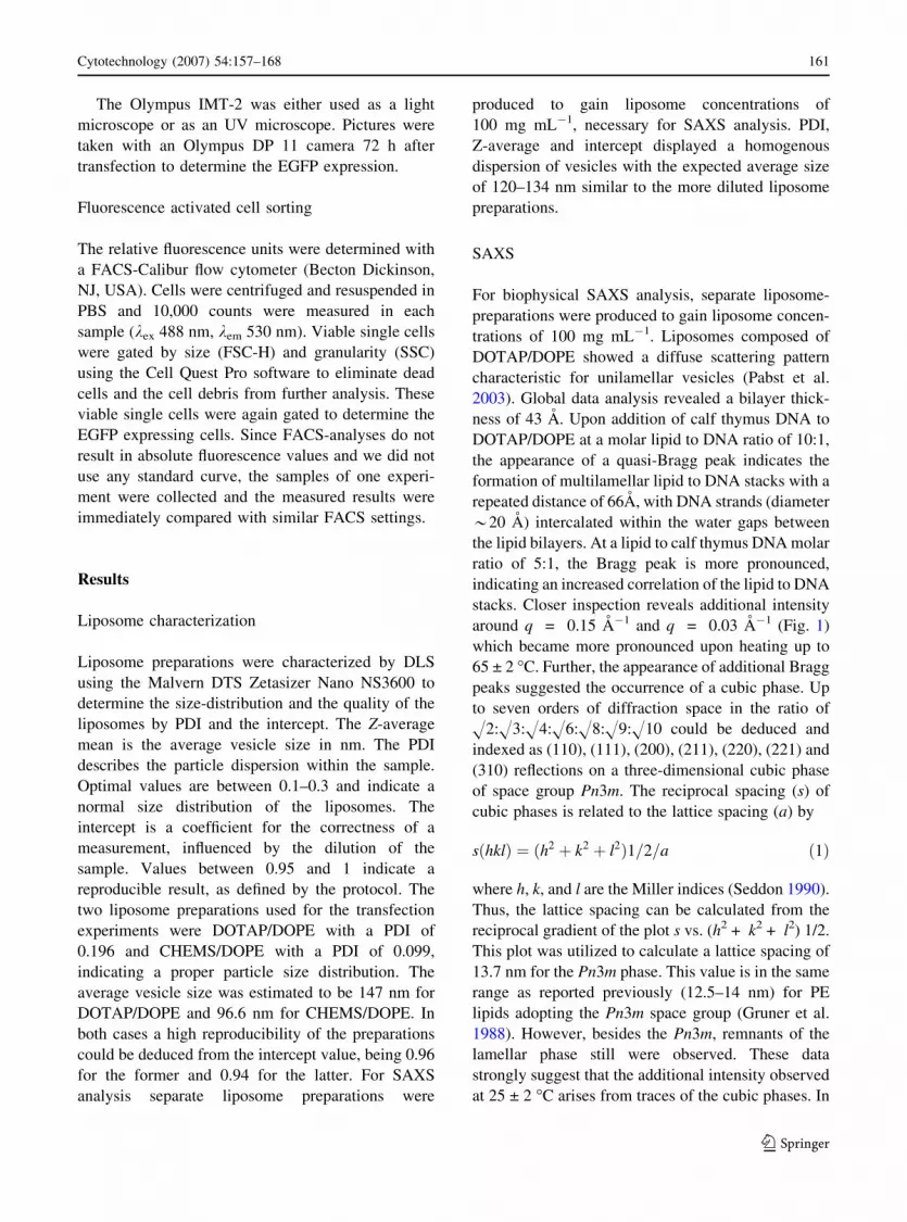
SAXS
For biophysical SAXS analysis, separate liposome-
preparations were produced to gain liposome concen-
trations of 100 mg mL�1. Liposomes composed of
DOTAP/DOPE showed a diffuse scattering pattern
characteristic for unilamellar vesicles (Pabst et al.
2003). Global data analysis revealed a bilayer thick-
ness of 43 A. Upon addition of calf thymus DNA to
DOTAP/DOPE at a molar lipid to DNA ratio of 10:1,
the appearance of a quasi-Bragg peak indicates the
formation of multilamellar lipid to DNA stacks with a
repeated distance of 66A, with DNA strands (diameter
*20 A) intercalated within the water gaps between
the lipid bilayers. At a lipid to calf thymus DNA molar
ratio of 5:1, the Bragg peak is more pronounced,
indicating an increased correlation of the lipid to DNA
stacks. Closer inspection reveals additional intensity
around q = 0.15 A�1 and q = 0.03 A�1 (Fig. 1)
which became more pronounced upon heating up to
65 ± 2 �C. Further, the appearance of additional Bragg
peaks suggested the occurrence of a cubic phase. Up
to seven orders of diffraction space in the ratio of
H2:H3:H4:H6:H8:H9:H10 could be deduced and
indexed as (110), (111), (200), (211), (220), (221) and
(310) reflections on a three-dimensional cubic phase
of space group Pn3m. The reciprocal spacing (s) of
cubic phases is related to the lattice spacing (a) by
sðhklÞ ¼ ðh2 þ k2 þ l2Þ1=2=a ð1Þ
where h, k, and l are the Miller indices (Seddon 1990).
Thus, the lattice spacing can be calculated from the
reciprocal gradient of the plot s vs. (h2 + k2 + l2) 1/2.
This plot was utilized to calculate a lattice spacing of
13.7 nm for the Pn3m phase. This value is in the same
range as reported previously (12.5–14 nm) for PE
lipids adopting the Pn3m space group (Gruner et al.
1988). However, besides the Pn3m, remnants of the
lamellar phase still were observed. These data
strongly suggest that the additional intensity observed
at 25 ± 2 �C arises from traces of the cubic phases. In
Cytotechnology (2007) 54:157–168 161
123

the presence of plasmid DNA, the Bragg peak
indicating the formation of lamellar stacks is barely
visible, probably due to the lower DNA concentration.
In the CHEMS/DOPE mixture, diffuse scattering was
observed before and after addition of either calf
thymus DNA or plasmid DNA indicating that neither
cubic phases nor lamellar stacks were formed in the
presence of DNA (Fig. 2).
Cellular uptake of lipoplex and EGFP expression
Intracellular location and distribution of the liposomes
and DNA after transfection was investigated by
rhodamine labeled liposomes and PicoGreen stained
DNA in the confocal microscope. Merely, the DNA
entered the nucleus while the liposomes stayed in the
cytoplasm (Fig. 3A, B) 4-h post-transfection. After
72 h the lipoplexes were degraded in both transfections
(Fig. 3C, D) by the cellular environment indicated by
alleviated staining. Panel 3E shows the EGFP expres-
sion of the DOTAP/DOPE transfected CHO-cells,
while panel 3F shows the reporter gene expression of
the CHEMS/DOPE transfected CHO-cells indicating
an overall weaker staining compared to the DOTAP/
DOPE transfection. Additionally, we quantified all
samples at the same time points by FACS. The data
revealed a better EGFP expression of DOTAP/DOPE
compared to CHEMS/DOPE transfection, confirming
our confocal data. Comparison between panel 3A and
3B reveals that DNA transported by DOTAP/DOPE
lipoplexes enters the nucleus more efficiently com-
pared to CHEMS/DOPE lipoplexes causing a higher
expression of the reporter gene. To ensure PicoGreen
does not permeate the cell and the nucleus, negative
controls were performed including the addition of free
PicoGreen, PicoGreen together with N-Rh-PE labeled
liposomes and merely stained liposomes to the cells.
The controls showed no staining of cellular DNA,
confirming that the fluorescence in Fig. 3A–D resulted
from introduced plasmid DNA (data not shown).
Toxicity
CHO-cells were transfected with increasing amounts
of liposomes ranging from 11 to 240 mg depending
on the liposome composition and with a constant
amount of 1 mg pEGFP-N3. Cell numbers and
viability were determined after 48 h using a Burker-
Turk chamber and trypan blue as vital stain
(Table 1). Three independent experiments were
performed to determine the cytotoxicity of the
self-tailored liposomes.
Cells transfected with DOTAP/DOPE were able to
tolerate 110 mg liposomes while cells exposed to the
CHEMS/DOPE formulation died almost completely
at 120 mg liposomes after 48 h. Higher liposome
concentrations resulted in 100% lethality in both lipid
q [Å-1]
0.1 0.2 0.3 0.4
].u .a[ ytisnetnI
0
1000
2000
3000
4000
5000
6000
A
B
C
D
E
Fig. 1 Small-angle scattering pattern of DOTAP/DOPE (A),
DOTAP/DOPE + genomic calf thymus DNA 10:1 (B), DO
TAP/DOPE + genomic calf thymus DNA 5:1 (C), DOTAP/
DOPE + genomic calf thymus DNA 5:1 (65 �C ±2) (D) and
DOTAP/DOPE + pBSKII 10:1 (E). Data were recorded at
25 ± 2 �C, if not indicated otherwise. Arrows indicate Bragg
peaks corresponding to a Pn3m phase. Circle indicates the
remnant lamellar phase
q [Å-1]
0.1 0.2 0.3 0.4
].u .a[ ytisnetnI
0
500
1000
1500
2000
2500
3000
3500
A
B
C
Fig. 2 Small-angle scattering pattern of CHEMS/DOPE (A),
CHEMS/DOPE + genomic calf thymus DNA 10:1 (B) and
CHEMS/DOPE + pBSKII 10:1 (C). Data were recorded at
25 ± 2 �C
162 Cytotechnology (2007) 54:157–168
123

formulations. The measured viabilities declined from
96 via 80 down to 0% for DOTAP/DOPE and 93 via
6 down to 0% for CHEMS/DOPE. These results
suggest that both formulations are toxic, but the
CHO-cells were able to survive higher amounts of
DOTAP/DOPE.
Fig. 3 Intracellular uptake and dissociation of lipoplexes
consisting of DOTAP/DOPE and CHEMS/DOPE liposomes
labeled with N-Rh-PE (red) and PicoGreen stained pEGFP-N3
(green). Transfected cells were analyzed by confocal micros-
copy (Leica TCS SP2) after 4 h (A, B) and 72 h (C, D). Panels
E, F were shot 72-h post-transfection with an UV-microscope.
Lipoplexes composed of DOTAP/DOPE/N-Rh-PE (A, C and
E) and CHEMS/DOPE/N-Rh-PE (B, D and F). (A, B) The
liposomes were located in the cytoplasm while the DNA
moved into the nucleus and was visualized by PicoGreen.
Panels C & D show alleviated staining of cells after 72 h due to
degradation of the lipid/rhodamine complex. Panel E & F show
the EGFP expressing cells after 72 h in UV-excitation in
parallel with transmitted light to localize the EGFP expressing
cells. PicoGreen, cells with PicoGreen and liposomes or cells
served as controls (data not shown)
Table 1 The toxicity of the liposomes was determined by increasing liposome amount
Liposome composition Liposome [mg] Cell number (105cells mL�1)a SDa Viability (%)a SDa
DOTAP/DOPE 11 15.1 ±4.3 96 ±3.1
DOTAP/DOPE 110 8.4 ±4.5 80 ±3.4
DOTAP/DOPE 220 1.2 ±1.1 0 ±0
CHEMS/DOPE 12 10.9 ±0.86 93 ±0.6
CHEMS/DOPE 120 2.47 ±0.4 6 ±10.1
CHEMS/DOPE 240 1.71 ±0.2 0 ±0
Control – 13.3 ±4.3 98 ±3.1
The cells were transfected with 1 mg pEGFP-N3 and varying liposome concentration for 4 h. Cell number and viability were detected
after 48 h; seeding density was 5 · 105 cells per mL. Mean values and standard deviations were obtained by three different
experimentsa 48 h post-transfection
Cytotechnology (2007) 54:157–168 163
123

Transfection optimization
To optimize transfection efficiency we varied the
DNA (0.2–5 mg) and liposome concentration (2.2–
60 mg) in transient transfection experiments. About
2.2, 11 and 55 mg DOTAP/DOPE were combined
with 0.2, 1 and 5 mg DNA. FACS analyses were
performed to determine the transient EGFP expres-
sion after 72 h (Table 2). At each liposome concen-
tration 1 mg DNA generated the highest transfection
rates ranging from 12 to 16% positive transfectants
after 72 h. Increase from 2.2 to 11 mg DOTAP/DOPE
improved the relative number of transfectants by one
quarter and showed a fluorescence intensity of
113 rfu, further increase of lipid to 55 and 1 mg
DNA per transfection resulted in reduced fluores-
cence intensity of 24 rfu. Similar results were
observed with the highest DNA concentration
(5 mg) and 55 mg liposomes resulting in a fluores-
cence of 31 rfu. The transfection efficiency was not
improved upon increase of liposome concentration
and the relative fluorescence intensity dropped to
approximately 50% of the initial fluorescence units
reached with 11 mg. In parallel the viability of all
transfections was above 90% indicating that this
method is suitable for the generation of stable cell
lines. All transfections with CHEMS/DOPE showed
poor transfection efficiencies at low percentage range
with only about one fifth of fluorescence intensity
(data not shown). However, cellular viability as well
as cell concentration was not distinguished. There-
fore, we decided to test both liposome preparations in
stable clone development.
Stable transfection
The process of generating stable recombinant cell
lines with DOTAP/DOPE or CHEMS/DOPE was
investigated by transfecting CHO-cells and long time
cultivation of the cells in selective medium. The
percentage of transfected cells was routinely deter-
mined every second to third passage using FACS
analysis (Fig. 4) and EGFP expression intensity was
quantified by the rfu. In both experiments cell
numbers increased from 5 · 105 to 1 · 106 cells
per mL after two weeks of cultivation in G418. In
contrast to transient experiments, transfection with
CHEMS/DOPE showed similar results like DOTAP/
DOPE. After 37 days of transfection a homogenous
cell population could be selected consisting of 75%
EGFP expressing cells in the population. The number
of positive transfectants increased continuously until
a homogenous population was established 70 days
after transfection. Furthermore, the EGFP expression
rate increased continuously during the selection
process. As already shown during transient transfec-
tion, the initial efficiency of CHEMS/DOPE was
lower but stable clones reached similar expression
titers indicated by the rfu (Fig. 4).
Table 2 Optimization of the transfection approaches by varying the amount of DNA and liposomes
DNA
(mg)
DOTAP/DOPE
(mg)
Transfectants
(%)aFluorescence (relative
units)aCell number
(1 · 105cells mL�1)aViability
(%)a
0.2 2.2 0 31 9.84 96
1 12 138 7.8 93
5 2 103 1.18 92
0.2 11 1 28 6.66 97
1 16 113 6.42 98
5 4 126 10.2 91
0.2 55 2 19 5.3 99
1 14 24 6.48 98
5 16 31 8.04 91
– 11 0 16 12.5 96
Ascending DNA concentrations (0.2, 1 and 5 mg) were mixed with different DOTAP/DOPE concentrations ranging from 2.2 to
55 mg. As negative control cells were merely incubated with DOTAP/DOPE. Seeding density was 5 · 105 cells per mLa 72-h post-transfection
164 Cytotechnology (2007) 54:157–168
123

Comparison of different transfection methods
To evaluate different transfection methods regarding
their potential for transient protein expression we
compared several commercially available products
(lipofectin, lipofectamin 2000, DMRIE-C reagent and
the nucleofector) with our tailor-made liposomes
(Fig. 5). The nucleofector kit showed the highest
transient transfection efficiency with 55% EGFP
positive transfectants and 147 rfu. CHEMS/DOPE
and lipofectamin 2000 showed the lowest with 6 and
2% positive cells. DMRIE-C showed a solid perfor-
mance of 11%, but a low fluorescence of 9 rfu,
whereas DOTAP/DOPE and lipofectin transfected 18
and 15% of the cells with nearly the same fluores-
cence of 66 and 64 rfu, respectively. Viability of all
cell populations was acceptable with values above
90%. However, the nucleofector transfected cells
showed a rather coarse structure correlating with cell
death, resulting in a viability of 80% after 24 h and
85% after 72 h (data not shown).
Discussion
We generated DOTAP/DOPE and CHEMS/DOPE
liposomes and characterized them by DLS and SAXS
analysis. The appearance of a quasi-Bragg peak
indicates the formation of lamellar DOTAP/DOPE
lipoplex stacks. Upon heating to 65 ± 2 �C, a Pn3m
was formed, while still remnants of the lamellar
phase were present. Consequently we suggest that the
additional intensity observed at 25 ± 2 �C results
from traces of the cubic phase. Our resulting obser-
vation is in disagreement with the data described by
Simberg and co-workers suggesting DOTAP/DOPE
liposome composition to form HII phases (Simberg
et al. 2001). The CHEMS/DOPE liposomes neither
formed Pn3m nor lamellar stacks in the presence of
DNA (Fig. 2). Thus, it is plausible that the transfec-
0
20
40
60
80
100
21 37 44 50 56 70 84
days
tra
nsfe
cta
nts
[%
]
1
10
100
1000
10000
rfu
Fig. 4 Stable transfection experiments along with the tailor-
made liposomes. CHO-cells were either transfected with 11 mg
DOTAP/DOPE or with 12 mg CHEMS/DOPE. Selection
started 24 h after transfection with G418. EGFP expressing
cells were quantified at the indicated days post-transfection by
detecting the percentage of positive cells (% transfectants) and
the relative expression titer indicated by the rfu. (-•-) [%]
DOTAP/DOPE transfectants, (-�-) [%] CHEMS/DOPE trans-
fectants, (-m-) fluorescent DOTAP/DOPE transfected cells and
(-D-) fluorescent CHEMS/DOPE transfected cells
0
20
40
60
80
100
EP
OD/
PA
TO
D
EP
OD/
SM
EH
C
nitcefopiL
eni
matcefopiL0 002
rotcefoelcuN
C-EI
RM
D
lortnoc
%[ s tnatcefs
nart]
0
20
40
60
80
100
120
140
160
ufr
Fig. 5 Comparison of the transient transfection efficiencies of
six methods. The CHO-cells were transfected according to the
manual and cultivated in 12-well plates. Bars indicate the
standard deviation derived from three independent experi-
ments, columns are the positive transfectants [%] and
diamonds (-(-} -)-) are relative fluorescence
Cytotechnology (2007) 54:157–168 165
123

tion mode of these vesicular structures differs from
the DOTAP/DOPE lipoplex stacks due to the pro-
pensity of the latter system to form non-lamellar
structures. Such Pn3m can be intermediate structures
between a lamellar to inverse hexagonal phase
transition (Seddon 1990) and thus enhance membrane
fusion (Siegel and Epand 1997), which is described to
play an essential role in the transfection mechanism
of DOTAP/DOPE liposomes (Boomer and Thompson
1999; Rejman et al. 2005).
To follow the fate of lipoplexes within the cell we
incorporated N-Rh-PE into the liposomes, labeled the
plasmid DNA with PicoGreen and transfected CHO-
cells with these lipoplexes. Via rhodamine stained
cells confocal microscopy revealed the internaliza-
tion of the complexes 4-h post-transfection (Fig.3A,
B) and the DNA could be visualized in the nucleus
with PicoGreen. In case of CHEMS/DOPE at least
part of the rhodamine stained cells did not show any
PicoGreen signal while the cytoplasmic rhodamine
staining has been found weaker and nuclear Pico-
Green staining was increased in DOTAP/DOPE
transfected cells. Thus, we assume that the initial
uptake of the lipoplex presents a prerequisite, but the
expression rate of the transgen is determined by the
optimal DNA uptake into the nucleus reflected by
much higher relative fluorescence of DOTAP/DOPE
transfectants (Figs. 3 and 5). The exclusion of the
liposomes from the nucleus is another necessity for
efficient expression (Xu and Szoka 1996). This is
supported by Zabner et al. (1995), who observed very
low expression rates when the lipoplexes were
microinjected into the nucleus directly.
In a different experiment we tested the influence of
the lipid concentration in the transfection cocktail.
The increase of liposomes resulted in a reduced
viability of the cells (Table 1) but did not correlate
with transfection efficiency (Table 2) despite posi-
tively charged lipoplexes enable efficient binding to
the cell surface (Scarzello et al. 2005), an overload of
lipoplexes enhances the cell-toxicity (Templeton
et al. 1997). EGFP expression was merely driven by
total DNA uptake (Farhood et al. 1995). Best results
were generated by transfection with 1 mg pEGFP-N3
independent of the liposome concentration. Further
increase of plasmid DNA did not improve the
transfection efficiency (Table 2). Comparison of
transient transfection with stable clone development
showed that the selection of high production clones
does not correlate with the initial transfection
efficiency in case of intracellular EGFP expression.
Despite transient DOTAP/DOPE expression resulted
in two to three fold higher transfection efficiencies,
CHEMS/DOPE transfectants behaved similar after
three weeks regarding EGFP productivity and clone
homogeneity. The percentage of transfectants and the
expression titers converged in both clones selected
during time progression and resulted in homogenous
recombinant cell populations with slightly higher
expression rates of CHEMS/DOPE transfectants
84 days after transfection (Fig.4).
For the evaluation of the potency of various
transfection techniques, four commercial systems
were selected and compared with DOTAP/DOPE
and CHEMS/DOPE. Lipofectin (DOTMA/DOPE),
DMRIE-C and DOTAP/DOPE belong to the family
of cationic lipids that release the associated DNA into
the cytoplasm by membrane destabilization. The
transfection data were evaluated via an one-way
ANOVA along with post-hoc differences calculated
with Tuckey honest significant difference test. Tran-
sient transfection experiments employing Lipofectin,
DMRIE-C and DOTAP/DOPE showed similar trans-
fection efficiencies, according to the statistical eval-
uation with no significant differences detectable.
CHEMS/DOPE showed a reduced transfection rate of
cells during transient transfection in contrast to the
nucleofector kit providing 55% transiently transfect-
ed cells after 72 h. The nucleofector kit showed a
significant difference (p < 0.001) with respect to the
number of transfectants and fluorescence intensity.
This might be ascribed to the direct DNA delivery
into the nucleus predicted by the supplier. However,
the lack of information about the transfection cocktail
makes it difficult to generate defined stable produc-
tion cell lines. Furthermore, the nucleofector bears
the disadvantages of significantly reduced cell via-
bility of 60–80% compared to the lipofections (data
not shown) with viabilities above 90%, the limited
scalability as well as the high cell concentrations
necessary for transfection.
Efficient transient gene expression gains increas-
ing importance to accumulate adequate amounts of
recombinant protein for research purpose and pre-
clinical studies in a short time schedule. Therefore
further studies concerning our lipoplexes in this
protein-free system will focus on the expression of
secreted recombinant proteins in transient ap-
166 Cytotechnology (2007) 54:157–168
123

proaches. The next step will be to evaluate the
suitability of our liposomes for transient transfection
in different host systems since the amount of
expressed recombinant protein mainly depends on
maximal cell density and viability of the culture in an
extended batch.
As a conclusion our chemically defined liposomes
are adaptable for transient large-scale production
since the costs of the transfection cocktail is one to
two orders of magnitude cheaper than commercial
DNA vehicles. Further the toxicity is lower compared
to lipofectin, nucleofector, lipofectamin 2000 and
DMRIE-C.
Among a set of different parameters like vesicle
size, mixing rate, order of addition, ionic strength of
the mixing buffer and lipid to DNA charge ratio
represent quite a few parameters affecting lipoplex
characteristics (Zuhorn and Hoekstra 2002). Further-
more, the high variability in lipid composition leaves
room for the improvement of transfection efficiency
of the tailored liposomes, which may be greatly
enhanced upon understanding of the details of the
molecular mechanism of this process.
We already started the optimization of transfection
protocols for protein-free adapted host cell lines a
couple of years ago mainly using lipofection and the
Amaxa nucleofector. These methods enabled us to
isolate recombinant clones with different transfection
efficiency and quality. Besides, we tested the self
made DOTAP/DOPE lipoplexes for stable transfec-
tion and antibody expression. In a series of experi-
ments carried out with the same monoclonal antibody
and identical expression plasmids our tailor-made
liposomes reduced the number of required transfec-
tions. We had to screen less growing wells for
generation of suitable stable clones and selection was
much faster. The transfection efficiency was more
reproducible and primary transfectants were easier to
select and adapt to MTX pressure with an outcome of
more IgG producing clones.
Acknowledgement This research was part of the Pharma-
Planta Project (LSHB-CT-2003–503565), kindly funded by an
EU FP6 program.
References
Boomer JA, Thompson DH (1999) Synthesis of acid-labile
diplasmenyl lipids for drug and gene delivery applica-
tions. Chem Phys Lipids 99:145–153
Boussif O, Lezoualch F, Zanta MA, Mergny MD, Scherman D,
Demeneix B, Behr JP (1995) A versatile vector for gene
and oligonucleotide transfer into cells in culture and
in vivo: polyethylenimine. Proc Natl Acad Sci USA
92:7297–7301
Cepko CL, Roberts BE, Mulligan RC (1984) Construction and
applications of a highly transmissible murine retrovirus
shuttle vector. Cell 37:1053–1062
Cudd A, Labbe H, Gervais M, Nicolau C (1984) Liposomes
injected intravenously into mice associate with liver
mitochondria. Biochim Biophys Acta 774:169–180
Escriou V, Carriere M, Bussone F, Wils P, Scherman D (2001)
Critical assessment of the nuclear import of plasmid
during cationic lipid-mediated gene transfer. J Gene Med
3:179–187
Even-Chen S, Barenholz Y (2000) DOTAP cationic liposomes
prefer relaxed over supercoiled plasmids. Biochim Bio-
phys Acta 1509:176–188
Farhood H, Serbina N, Huang L (1995) The role of dioleoyl
phosphatidylethanolamine in cationic liposome mediated
gene transfer. Biochim Biophys Acta 1235:289–295
Graessmann M, Graessmann A (1983) Microinjection of tissue
culture cells. Methods Enzymol 101:482–492
Graham FL, van der Eb AJ (1973) A new technique for the
assay of infectivity of human adenovirus 5 DNA. Virol-
ogy 52:456–467
Gruner SM, Tate MW, Kirk GL, So PT, Turner DC, Keane DT,
Tilcock CP, Cullis PR (1988) X-ray diffraction study of
the polymorphic behavior of N-methylated dioleoylphos-
phatidylethanolamine. Biochemistry 27:2853–2866
Hof M, Kral T, Langner M, Adjimatera N, Blagbrough IS
(2005) DNA condensation characterized by Fluorescence
correlation spectroscopy (FCS). Cell Mol Biol Lett 10:23–
25
Krishnaswamy R, Pabst G, Rappolt M, Raghunathan VA,
Sood AK (2006) Structure of DNA-CTAB-hexanol
complexes. Phys. Rev E Stat Nonlin Soft Matter Phys
73(031904)
Laggner P, Mio H (1992) SWAX—a dual-detector camera for
simultaneous small- and wide-angle X-ray diffraction in
polymer and liquid crystal research. Nucl Instr Meth Phys
Res A323:86–90
Lohner K (1996) Is the high propensity of ethanolamine
plasmalogens to form non-lamellar lipid structures mani-
fested in the properties of biomembranes? Chem Phys
Lipids 81:167–184
Loyter A, Scangos GA, Ruddle FH (1982) Mechanisms of
DNA uptake by mammalian cells: fate of exogenously
added DNA monitored by the use of fluorescent dyes.
Proc Natl Acad Sci USA 79:422–426
Martin B, Sainlos M, Aissaoui A, Oudrhiri N, Hauchecorne M,
Vigneron JP, Lehn JM, Lehn P (2005) The design of
cationic lipids for gene delivery. Curr Pharm Des 11:375–
394
Muller N, Girard P, Hacker DL, Jordan M, Wurm FM (2005)
Orbital shaker technology for the cultivation of mam-
malian cells in suspension. Biotechnol Bioeng 89:400–
406
Neumann E, Schaefer-Ridder M, Wang Y, Hofschneider PH
(1982) Gene transfer into mouse lyoma cells by electro-
poration in high electric fields. Embo J 1:841–845
Cytotechnology (2007) 54:157–168 167
123

Pabst G (2006) Global properties of biomimetic membranes:
prespectives on molecular features. Biophys Rev Lett
1:57–84
Pabst G, Koschuch R, Pozo-Navas B, Rappolt M, Lohner K,
Laggner P (2003) Structural analysis of weakly ordered
membrane stacks. J Appl Cryst 36:1378–1388
Pozo Navas B, Lohner K, Deutsch G, Sevcsik E, Riske KA,
Dimova R, Garidel P, Pabst G (2005) Composition
dependence of vesicle morphology and mixing properties
in a bacterial model membrane system. Biochim Biophys
Acta 1716:40–48
Regelin AE, Fankhaenel S, Gurtesch L, Prinz C, von Kied-
rowski G, Massing U (2000) Biophysical and lipofection
studies of DOTAP analogs. Biochim Biophys Acta
1464:151–164
Rejman J, Bragonzi A, Conese M (2005) Role of clathrin- and
caveolae-mediated endocytosis in gene transfer mediated
by lipo- and polyplexes. Mol Ther 12:468–474
Scarzello M, Smisterova J, Wagenaar A, Stuart MC, Hoekstra
D, Engberts JB, Hulst R (2005) Sunfish cationic amphi-
philes: toward an adaptative lipoplex morphology. J Am
Chem Soc 127:10420–10429
Schaffner W (1980) Direct transfer of cloned genes from
bacteria to mammalian cells. Proc Natl Acad Sci USA
77:2163–2167
Seddon JM (1990) Structure of the inverted hexagonal (HII)
phase, and non-lamellar phase transitions of lipids. Bio-
chim Biophys Acta 1031:1–69
Siegel DP, Epand RM (1997) The mechanism of lamellar-to-
inverted hexagonal phase transitions in phosphatidyleth-
anolamine: implications for membrane fusion mecha-
nisms. Biophys J 73:3089–3111
Slepushkin VA, Simoes S, Dazin P, Newman MS, Guo LS,
Pedroso de Lima MC, Duzgunes N (1997) Sterically
stabilized pH-sensitive liposomes. Intracellular delivery
of aqueous contents and prolonged circulation in vivo.
J Biol Chem 272:2382–2388
Simberg D, Danino D, Talmon Y, Minsky A, Ferrari ME,
Wheeler CJ, Barenholz Y (2001) Phase behavior, DNA
ordering, and size instability of cationic lipoplexes. Rel-
evance to optimal transfection activity. J Biol Chem
276:47453–47459
Smisterova J, Wagenaar A, Stuart MC, Polushkin E, ten Brinke
G, Hulst R, Engberts JB, Hoekstra D (2001) Molecular
shape of the cationic lipid controls the structure of cat-
ionic lipid/dioleylphosphatidylethanolamine-DNA com-
plexes and the efficiency of gene delivery. J Biol Chem
276:47615–47622
Templeton NS, Lasic DD, Frederik PM, Strey HH, Roberts
DD, Pavlakis GN (1997) Improved DNA: liposome
complexes for increased systemic delivery and gene
expression. Nat Biotechnol 15:647–652
Wattiaux R, Jadot M, Warnier-Pirotte MT, Wattiaux-De Con-
inck S (1997) Cationic lipids destabilize lysosomal
membrane in vitro. FEBS Lett 417:199–202
Wattiaux R, Laurent N, Wattiaux-De Coninck S, Jadot M
(2000) Endosomes, lysosomes: their implication in gene
transfer. Adv Drug Deliv Rev 41:201–208
Wrobel I, Collins D (1995) Fusion of cationic liposomes with
mammalian cells occurs after endocytosis. Biochim Bio-
phys Acta 1235:296–304
Xu Y, Szoka FC, Jr. (1996) Mechanism of DNA release from
cationic liposome/DNA complexes used in cell transfec-
tion. Biochemistry 35:5616–5623
Zabner J, Fasbender AJ, Moninger T, Poellinger KA, Welsh
MJ (1995) Cellular and molecular barriers to gene transfer
by a cationic lipid. J Biol Chem 270:18997–19007
Zuhorn IS, Bakowsky U, Polushkin E, Visser WH, Stuart MC,
Engberts JB, Hoekstra D (2005) Nonbilayer phase of
lipoplex-membrane mixture determines endosomal escape
of genetic cargo and transfection efficiency. Mol Ther
11:801–810
Zuhorn IS, Hoekstra D (2002) On the mechanism of cationic
amphiphile-mediated transfection. To fuse or not to fuse:
is that the question? J Membr Biol 189:167–179
Zuidam NJ, Barenholz Y (1997) Electrostatic parameters of
cationic liposomes commonly used for gene delivery as
determined by 4-heptadecyl-7-hydroxycoumarin. Biochim
Biophys Acta 1329:211–222
Zuidam NJ, Barenholz Y (1998) Electrostatic and structural
properties of complexes involving plasmid DNA and
cationic lipids commonly used for gene delivery. Biochim
Biophys Acta 1368:115–128
168 Cytotechnology (2007) 54:157–168
123