in vivo electroporation mediated gene delivery to the beating heart
TRANSCRIPT

In Vivo Electroporation Mediated Gene Delivery to theBeating HeartErick L. Ayuni1, Amiq Gazdhar2, Marie Noelle Giraud1, Alexander Kadner1, Mathias Gugger3, Marco
Cecchini4, Thierry Caus1, Thierry P. Carrel1, Ralph A. Schmid2*, Hendrik T. Tevaearai1
1 Department of Cardiovascular Surgery, University Hospital of Berne, Berne, Switzerland, 2 Division of General Thoracic Surgery, University Hospital of Berne, Berne,
Switzerland, 3 Department of Pathology, University of Berne, Berne, Switzerland, 4 Department of Urology, University Hospital of Berne, Berne, Switzerland
Abstract
Gene therapy may represent a promising alternative strategy for cardiac muscle regeneration. In vivo electroporation, aphysical method of gene transfer, has recently evolved as an efficient method for gene transfer. In the current study, weinvestigated the efficiency and safety of a protocol involving in vivo electroporation for gene transfer to the beating heart.Adult male rats were anesthetised and the heart exposed through a left thoracotomy. Naked plasmid DNA was injectedretrograde into the transiently occluded coronary sinus before the electric pulses were applied. Animals were sacrificed atspecific time points and gene expression was detected. Results were compared to the group of animals where no electricpulses were applied. No post-procedure arrhythmia was observed. Left ventricular function was temporarily altered only inthe group were high pulses were applied; CK-MB (Creatine kinase) and TNT (Troponin T) were also altered only in this group.Histology showed no signs of toxicity. Gene expression was highest at day one. Our results provide evidence that in vivoelectroporation with an optimized protocol is a safe and effective tool for nonviral gene delivery to the beating heart. Thismethod may be promising for clinical settings especially for perioperative gene delivery.
Citation: Ayuni EL, Gazdhar A, Giraud MN, Kadner A, Gugger M, et al. (2010) In Vivo Electroporation Mediated Gene Delivery to the Beating Heart. PLoS ONE 5(12):e14467. doi:10.1371/journal.pone.0014467
Editor: Pieter H. Reitsma, Leiden University Medical Center, Netherlands
Received May 24, 2010; Accepted November 10, 2010; Published December 30, 2010
Copyright: � 2010 Ayuni et al. This is an open-access article distributed under the terms of the Creative Commons Attribution License, which permitsunrestricted use, distribution, and reproduction in any medium, provided the original author and source are credited.
Funding: This work was supported by a grant from the Olga Mayenfisch Foundation, Zurich Switzerland and by two grants from the Swiss National ScienceFoundation (3200-068304- 2006 to RAS and 310000-118270-2007 to AK).The funders had no role in study design, data collection and analysis, decision to publish,or preparation of the manuscript.
Competing Interests: The authors have declared that no competing interests exist.
* E-mail: [email protected]
Introduction
Coronary artery disease continues to be a major cause of
morbidity and mortality worldwide. Currently, the medical
treatment and/or myocardial revascularization procedure, either
by percutaneous angioplasty or by coronary bypass surgery
(CABG) remains the standard therapy [1]. Regeneration of the
infracted myocardium, however, is the major challenge and its
lack remains the predominant cause of death. New therapeutic
modalities which may yield better and consistent results have to be
evaluated. Gene therapy, the transfer of nucleic acids to achieve
therapeutic benefits is one promising approach. However, progress
towards effective human gene therapy in cardiovascular diseases
has been hindered by a number of problems including vector
toxicity, poor targeting of diseased tissues, and host immune and
inflammatory activity [2,3,4]. Hence, safe and reproducible
methods have to be established. Electroporation a physical
method of gene transfer is one such promising technique.
It is based on the application of strong electric pulses for a very
short duration to enhance transfer of macromolecules like DNA
and proteins through cell membranes. Even though a number of
hypothesis regarding the cell membrane permeabilization have
been suggested [5,6], the exact mechanism remains unclear.
Nevertheless, electroporation has been shown to be one of the
most efficient gene transfer strategies in vivo [7,8] and was tested
in a broad range of target tissues and organs [9,10,11,12,13,14].
Ex vivo electroporation to the heart in experimental models has
also been reported [15,16] and showed a 100 to 1000 folds
increase in transgene expression as compared to direct DNA
injection. Consequently, the use of in vivo electroporation mediated
transfer may represent a promising method to overcome the
common limitations of gene therapy protocols using other types of
vectors. Regarding cardiac gene delivery more specifically, direct
injection into the cardiac muscle is the most common approach.
Gene therapy protocols also involve antegrade coronary injection
after aortic cross clamping, however, both approaches do not
result in global vector distribution in the heart. Furthermore, the
antegrade approach is also limited by heterogeneous and
inefficient distribution in the presence of coronary stenosis or
occlusion. To overcome this problem we present a retrograde
approach by injection via the coronary veins after transient
occlusion of the coronary sinus, to study the efficiency and the
effect of in vivo electroporation mediated gene transfer to the
beating heart.
Materials and Methods
PlasmidThe plasmids pCiKlux expresses firefly luciferase from the
CMV immediate early promoter/enhancer as described before
[13,17]. EGFP plasmid was from Add gene (USA). The
endotoxin-free plasmids were produced in large scale at Plasmid
Factory GmbH & Co (Bielefeld, Germany). For electroporation,
plasmids were suspended in endotoxin-free water.
PLoS ONE | www.plosone.org 1 December 2010 | Volume 5 | Issue 12 | e14467

AnimalsInbreed male Wistar rats (200–220 g) (Janvier, France) were
used and maintained on rodent feed and water in a air and
temperature controlled room. The experiments were performed in
compliance with the standards of the European convention of
animal care. The study protocols permission numbers 32/03 and
21/06, were approved by the University of Berne Animal Studies
Committee.
Gene Transfer techniqueAdult male rats were anesthetized by inhalation of 4%
isoflurane in a glass chamber before being intubated and
ventilated via a 14GA catheter (Insyte, Madrid, Spain) with
FIO2 = 1.0 and 1.5% isoflurane to maintain anesthesia, a
breathing frequency of 100/min, and a tidal volume of 10 ml/
kg body weight with a rodent ventilator (model 683 Harvard
Apparatus, South Natick MA).
A left thoracotomy in the 4th intercostal space was performed
and the heart was immobilized, the apex was lifted using an apical
7-0 prolene suture (Ethicon) and a 6-0 tourniquet was placed
around the distal coronary vein to allow temporary coronary sinus
occlusion (Figure 1a), immediately followed by the retrograde
injection of plasmid solution in a volume of 150 ml (plasmid
concentration 1 mg/ml). Animals in all groups below were injected
with equal volume of plasmid. Plate electrodes (2 cm61 cm) were
placed on each side of the heart followed by the application of 8
pulses of 20 ms and 200 V/cm,1 Hz using the pulse generator
(Inovio, San Diego) (Figure 1b). The distance between the two
plates of electrode was 1.2 mm. The tourniquet was then
immediately released. After the procedure, the heart was observed
for 2 minutes before a small chest drain was inserted into the left
hemithorax and the thoracotomy was closed with four layers of
continuous sutures (4/0 prolene). The drain was removed after the
animals restored spontaneous breathing, followed by extubation.
Experimental designThree sets of experiments were performed.
First, to establish the safety of in vivo electroporation as method
of gene transfer to the beating heart. The animals underwent gene
transfer as described, with different pulsing parameters of 8, 16
and 32 pulses. Animals were evaluated for different parameters of
toxicity at different time points.
Second, to establish in vivo electroporation as a method of gene
transfer to the beating heart. Two groups were studied (n = 5). All
animals underwent gene transfer as described. In the control
group, the entire procedure was identical with the exception that
the animals were not exposed to the electric pulses. Firefly
luciferase under control of CMV immediate early promoter/
enhancer was used and the animals were sacrificed 1 day after
gene transfer. One animal was injected with 150 ml EGFP
expression plasmid and electroporated as described above to
localize the gene expression.
Third, to evaluate transgene expression over time. All animals
underwent gene transfer as described and subgroups were
sacrificed at day 1, 3, 5 and 7 after gene transfer (n = 4 per
subgroup).As reporter gene firefly luciferase under control of
CMV immediate early promoter/enhancer was used.
Analysis of ToxicityHaemodynamic Measurements. Rats were anaesthetized
with isoflurane. The right carotid artery was exposed and a 1.4-
French pressure micro-catheter (MillarH, Mikro-TipH, Millar
Instruments, Houston, TX, USA) was inserted into the left
ventricle (LV). LV pressure curves were recorded and analyzed
(PowerLab system and its Chart 5.2 software, AD Instruments,
Spechbach, Germany). Data were recorded before the
electroporation procedure (basal), then continuously over a 60
minutes period. Thereafter, in a subgroup of animals, (n = 4) data
were measured 6 hours following the electroporation.
Cardiac Enzyme measurements. Two early cardiac
specific biomarkers, CK-MB (Creatine kinase) and TnT
(Troponin T) were used as indicators of myocardial injury. Blood
samples were collected 6 hours following the electroporation,
centrifuged and the serum was frozen at 220uC. Serum TnT and
CK-MB were measured using commercial kits (respectively Roche
Diagnostics, Basel Switzerland and Meia, Abbott, Baar,
Switzerland) according to manufacturer’s instructions.
Histological analysis at each time point. Immediately
following euthanasia, the heart was placed in a container with 4%
Figure 1. The in vivo electroporation mediated gene transfer to the heart is a two step procedure. (a) First, the coronary sinus is occludedwith a 6-0 prolene tourniquet and the plasmid is injected into the coronary vein. (b) The heart is then positioned between the plate electrodes andthe electric field is applied.doi:10.1371/journal.pone.0014467.g001
Gene Therapy to Beating Heart
PLoS ONE | www.plosone.org 2 December 2010 | Volume 5 | Issue 12 | e14467

Gene Therapy to Beating Heart
PLoS ONE | www.plosone.org 3 December 2010 | Volume 5 | Issue 12 | e14467

formalin for 12 hrs. After paraffin embedding the sections where
cut and routine haematoxylin and eosin staining was performed.
The sections were blinded and reviewed by an experienced
pathologist. Gene expression was analyzed at day 1, 3, 5, and 7
following the in vivo electroporation procedure.
Bioluminescent reporter imaging (BLI). The animals
were anesthetized with thiopental (50 mg/kg Penthotal, Abbot
AG, Baar, Switzerland). Monosodium luciferin (750 ml of an
80 mg/ml solution, Molecular Probes, The Netherlands) was
injected i.p, 15 min before the heart was removed and imaged in
an intensified charge-coupled device (CCD) camera (C2400-32,
Hamamatsu), fitted with a 50 mm Nikkor lens (Nikon, Japan) and
an image processor (Argus 20, Hamamatsu). An imaging system
similar to that described by Contag et al [18] was employed for
these studies as previously described [19].
Quantification of the Bioluminescent Signal. The lucif-
erase activity was measured by counting the photon emission in
the defined region of interest (ROI) with the aid of the open lab
software (Improvision, Coventry, UK) [19].
Measurement of reporter gene expression. Slices of the
hearts were frozen in liquid nitrogen immediately after imaging.
The tissue was grinded and suspended in 1 ml of lysis buffer
(Promega, Madison, WI, USA) and homogenized immediately.
Samples were thawed at room temperature and vortexed for
30 sec. Subsequently they were refrozen in liquid nitrogen. Three
freeze-thaw cycles were performed by alternating between liquid
nitrogen and room temperature water bath. Debris was removed
by centrifugation. Luciferase activity was then measured and
expressed as RLU/mg protein, using the slow glow assay system
(Promega, Madison WI) in MiniLumat LB 9506 luminometer
(Berthold Technologies, Switzerland).
EGFP reporter gene expression and confocal micro-
scopy. At day one after electroporation mediated gene
transfer, the animal was sacrificed and the heart was fixed in
O.C.T compound, the cryosections were made at the 10 mm
thickness and sections were observed under LSM 510 exciter
confocal microscope from Carl Zeiss (Germany).
Statistics. Data are presented as mean6SEM. The non
parametric Mann Whitney U test was performed to compare the
groups using GraphPad Prism version 4.00 for Windows,
GraphPad Prism version 4.00 for Windows, GraphPad Software,
San Diego California USA, www.graphpad.com. p,0.05 was
taken as the level of significance.
Results
Electroporation mediated gene transfer to the beatingheart is safe
Electroporation mediated gene transfer to the beating heart is
safe Fifty-eight animals were used in the entire study. Overall
mortality was 3.4% (2/58), due to bleeding from the coronary
vein. Therefore, mortality was never related to the electroporation
procedure itself. Especially, mortality remained null among the
two groups of animals treated with the higher electroporation
doses of 16 and 32 pulses.
Nevertheless, a very transient asystole followed the electropo-
ration in 2/8 animals in the 32 pulses protocol. The asystole
resumed spontaneously within 5 seconds. No fibrillation or other
rhythm alteration was recorded.
LV contractility function was analyzed over the first hour
following the electroporation procedure. Contractility (LVdp/dt
max) and relaxation (LVdP/dt min) as well as the developed LVP
(dLVP) were altered after the electroporation procedure only in
the group of animals with the maximum pulses regimen of 32
pulses (Figure 2); However, these functional alterations were
rapidly reversible and complete normalization occurred within
Figure 2. Left ventricular contractile function of heart following electroporation mediated gene transfer using 8, 16 and 32 pulsesprotocols (n = 5 each group). Indices of contractility and relaxation determined respectively by the maximum (dP/dt max) (a) and the minimum(dP/dt min) (b) and developed LVP (dLVP) (c) of the first derivative of ventricular pressure with respect to time. These parameters remainedunchanged before (time 0) and after electroporation except for the 32 pulses group where the values are transiently altered. A completenormalization of the values occurred within 10–20 minutes. No abnormality was detected in the group in which invivo electroporation was notperformed.doi:10.1371/journal.pone.0014467.g002
Figure 3. The two early cardiac enzymes for injury TnT and CK-MB were measured; both enzymes remained within normal rangeafter in vivo electroporation mediated gene transfer with 8 and 16 pulses, but the values were altered with the 32 pulses protocol.(a) TnT levels (b) CK-MB levels. The levels were below detection levels in the group in which invivo electroporation was not performed.doi:10.1371/journal.pone.0014467.g003
Gene Therapy to Beating Heart
PLoS ONE | www.plosone.org 4 December 2010 | Volume 5 | Issue 12 | e14467

10–20 minutes and persisted thereafter. Conversely, contractile
function remained within a normal range in animals’ electropo-
rated with either 8 or 16 pulses.
Six hours post-electroporation TNT (Figure 3a) and CK-MB
(Figure 3b) values also remained in the normal range in animals
treated with 8 and 16 pulses; Values were altered only when a 32
pulses regimen was tested (30.666.3 mg/l for TnT, and
0.6660.318 mg/l for CK-MB). The levels in the control group
without electroporation, were below detection value. Histological
analysis performed at each time point, revealed no signs of fibrosis
or necrosis however very moderate blood stasis, and very few
interstitial lymphocytes, were observed after 8 pulses. For 16 pulses
and 32 pulses slightly more blood stasis was observed with some
interstitial and perivascular lymphocytes, but no signs of necrosis
or fibrosis was seen (Figure 4).
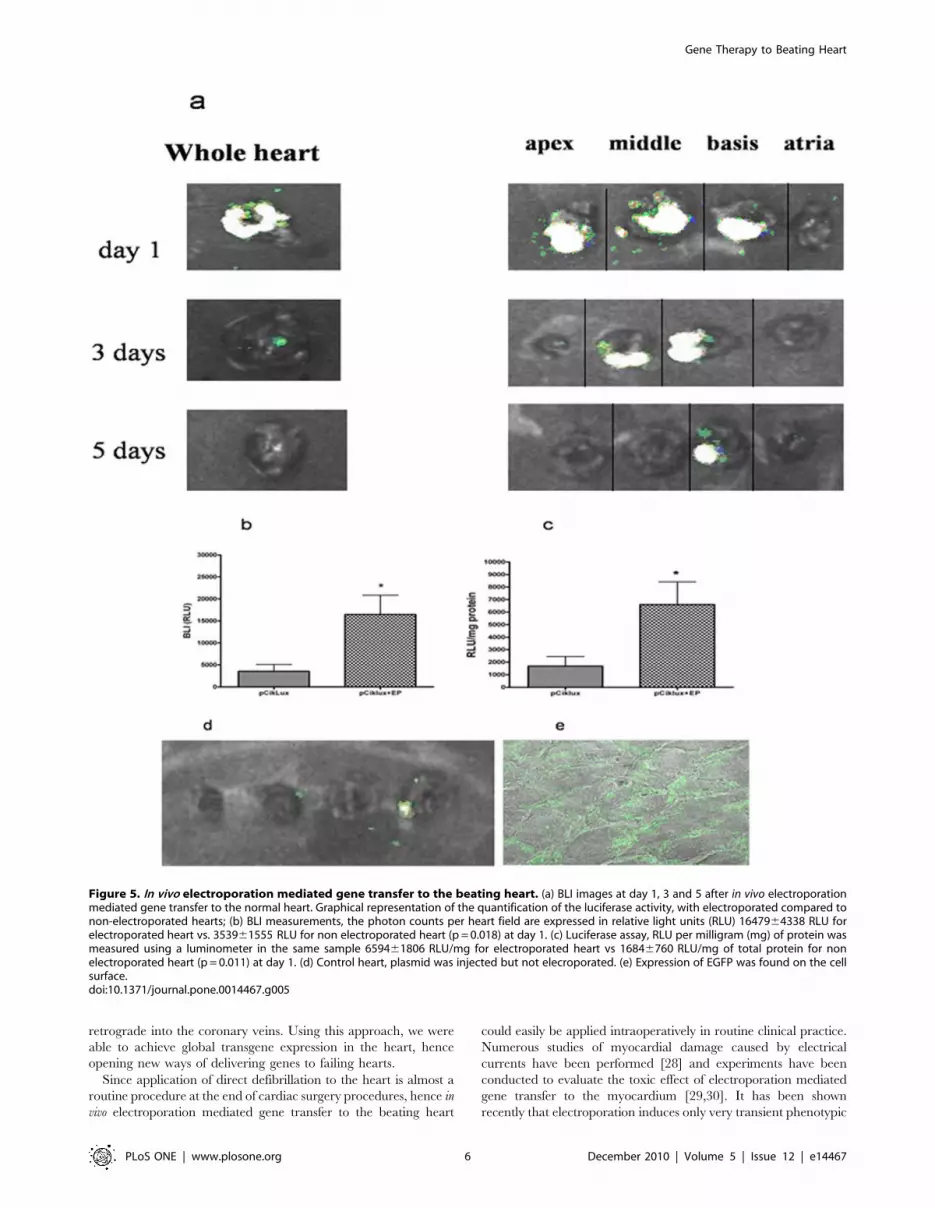
In vivo electroporation mediated gene transfer to theheart increases gene expression
When 150 ml of plasmid DNA pCiklux was injected without
electroporation very low transgene expression was detected by BLI
imaging (Figure 5 d). In contrast, when field strength of 200 V/cm
and 8 pulses was applied after injection of 150 mg of plasmid
pCiklux the mean lux activity was easily detected with BLI
imaging at day 1 (Figure 5a). The luciferase activity was measured
by counting the photon emission in the defined region of interest
(ROI), RLU measured were 1647964338 RLU for electroporated
heart vs. 353961555 RLU for non electroporated heart
(p = 0.018) (Figure 5b). These findings were further confirmed
with conventional luciferase assay in the same samples. Luciferase
activity observed on day 1 after electroporation mediated gene
transfer was highest; 659461806 RLU/mg for electroporated
heart vs 16846760 RLU/mg of total protein for non electropo-
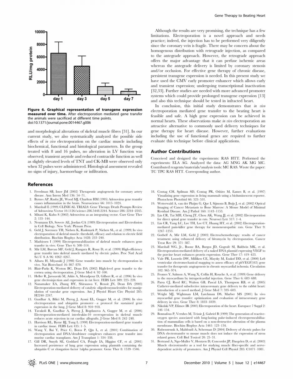
rated heart (p = 0.011) (Figure 5c); however, the activity showed a
gradual decrease, and at day 7 no transgene expression was seen
(Figure 6). The confocal images showed the expression of the
EGFP on the cell surface after invivo electroporation mediated gene
transfer of the EGFP plasmid (Figure 5 e).
Discussion
Regenerative medicine is a rapidly evolving field and efficient
and reliable methods are needed to expedite the process of
regeneration and healing. Current study demonstrates that in vivo
electroporation mediated gene transfer to the beating heart is a
feasible novel approach; it is also safe, quick, efficient and
reproducible. After retrograde injection of plasmid DNA into the
transiently occluded coronary sinus and immediate local applica-
tion of a series of 8 electric pulses, a high gene expression can be
achieved with no obvious adverse effect.
With the shortcomings of viral vectors and the inefficiency of the
other non-viral vectors, the physical method of electroporation
mediated gene transfer has emerged as a promising tool. Recently
the use of this technique in vivo has gained increasing acceptance
and has been used successfully in many tissues and organs
[8,11,20], under various conditions [12,14,21,22]. Even though
interest has been shown towards this promising approach, yet its
use in the cardiac muscles is limited. Few prior investigations
reported the use of electroporation mediated gene transfer to
cardiac tissue ex-vivo. Harrisson et al [15] electroporated chick
embryonic hearts ex vivo by placing the heart in a bath containing
the plasmid DNA, and showed high GFP and luciferase expression
after 48 hrs. Similarly in the mice heterotopic heart transplant
model Wang et al [16] demonstrated that ex vivo electroporation
mediated gene transfer of the graft before its implantation, allowed
significant gene expression. Very recently this technique was also
applied on the pig heart [23]. Therefore, it was with no doubt that
electroporation would enhance gene expression in the cardiac
muscle. In the current study, however we demonstrate the
applicability of this technique with ease and safety on beating
hearts of normal rats.
Various routes of administration of the plasmids in the heart
have been reported, but none of the protocols could achieve
satisfactory global distribution in the heart, especially not in
animals with heart failure. Some studies have evaluated the easy
direct injection of plasmid into the heart muscles as an efficient
method. It is however limited by local needle injury and time
consuming electromechanical mapping techniques [24]. The
epicardial delivery is another approach where the natural cavity
formed by the pericardial pouch was thought to be an advantage.
The higher permeability of the pericardium as compared to the
epicardium, lets the injected solution diffuse more eccentrically
than concentrically [25]. Most recently the percutaneous translu-
minal approach gathered a lot of attention as specially designed
ventricular catheters allow direct injection within the myocardium
[26], however, the problem of local needle injury and adequate
distribution still remains. In laboratory experiments the coronary
delivery approach has been largely used by cross clamping of the
ascending aorta and immediate injection of the gene solution to
the left ventricular cavity, the solution being delivered to perfuse
both coronaries [27]. Unfortunately limitations of this technique
involve an acute elevation of the afterload that will further reduce
LV function following aortic cross clamping. In the present study
we evaluated a retrograde approach where the coronary sinus is
clamped transiently before the plasmid solution is injected
Figure 4. For histological analysis the hearts were evaluated at 24 hrs post gene transfer. The following criteria were considered: vascularcongestion, infiltration and polymorphonuclear infiltrates. No haemorrhage or infiltration was noticed with any pulsing protocol. (a) 8 pulses (b) 16pulses (c) 32 pulses (Magnification =6200).doi:10.1371/journal.pone.0014467.g004
Gene Therapy to Beating Heart
PLoS ONE | www.plosone.org 5 December 2010 | Volume 5 | Issue 12 | e14467
retrograde into the coronary veins. Using this approach, we were
able to achieve global transgene expression in the heart, hence
opening new ways of delivering genes to failing hearts.
Since application of direct defibrillation to the heart is almost a
routine procedure at the end of cardiac surgery procedures, hence in
vivo electroporation mediated gene transfer to the beating heart
could easily be applied intraoperatively in routine clinical practice.
Numerous studies of myocardial damage caused by electrical
currents have been performed [28] and experiments have been
conducted to evaluate the toxic effect of electroporation mediated
gene transfer to the myocardium [29,30]. It has been shown
recently that electroporation induces only very transient phenotypic
Figure 5. In vivo electroporation mediated gene transfer to the beating heart. (a) BLI images at day 1, 3 and 5 after in vivo electroporationmediated gene transfer to the normal heart. Graphical representation of the quantification of the luciferase activity, with electroporated compared tonon-electroporated hearts; (b) BLI measurements, the photon counts per heart field are expressed in relative light units (RLU) 1647964338 RLU forelectroporated heart vs. 353961555 RLU for non electroporated heart (p = 0.018) at day 1. (c) Luciferase assay, RLU per milligram (mg) of protein wasmeasured using a luminometer in the same sample 659461806 RLU/mg for electroporated heart vs 16846760 RLU/mg of total protein for nonelectroporated heart (p = 0.011) at day 1. (d) Control heart, plasmid was injected but not elecroporated. (e) Expression of EGFP was found on the cellsurface.doi:10.1371/journal.pone.0014467.g005
Gene Therapy to Beating Heart
PLoS ONE | www.plosone.org 6 December 2010 | Volume 5 | Issue 12 | e14467
and morphological alterations of skeletal muscle fibres [31]. In our
current study, we also systematically analyzed the possible side
effects of in vivo electroporation on the cardiac muscle including
biochemical, functional and histological parameters. In the group
treated with 8 and 16 pulses, no alteration in LV function was
observed; transient asystole and reduced contractile function as well
as slightly elevated levels of TNT and CK-MB were observed only
when 32 pulses were administered. Histological assessment revealed
no signs of injury, haemorrhage or infiltration.
Although the results are very promising, the technique has a few
limitations. Electroporation is a novel approach and needs
practice; indeed, the injection has to be performed very diligently
since the coronary vein is fragile. There may be concern about the
homogenous distribution with retrograde injection, as compared
to the antegrade approach. However, the retrograde approach
offers the major advantage that it can perfuse ischemic areas
whereas the antegrade delivery is limited by coronary stenosis
and/or occlusion. For effective gene therapy of chronic disease,
persistent transgene expression is needed. In this present study we
have used the CMV early promoter enhancer which allows early
and transient expression; undergoing transcriptional inactivation
[32,33]. Further studies are needed with more advanced promoter
systems which could provide prolonged transgene expression [13]
and also this technique should be tested in infracted heart.
In conclusion, this initial study demonstrates that in vivo
electroporation mediated gene transfer to the beating heart is
feasible and safe. A high gene expression can be achieved in
normal hearts. These observations make in vivo electroporation an
attractive alternative to commonly used delivery techniques for
gene therapy for heart disease. However, further evaluations
including the use of functional genes are required to further
evaluate this technique before clinical applications.
Author Contributions
Conceived and designed the experiments: RAS HTT. Performed the
experiments: ELA AG. Analyzed the data: AG MNG AK MG MC.
Contributed reagents/materials/analysis tools: MC RAS. Wrote the paper:
TC TPC RAS HTT. Corresponding author.
References
1. Freedman SB, Isner JM (2002) Therapeutic angiogenesis for coronary arterydisease. Ann Intern Med 136: 54–71.
2. Byrnes AP, Rusby JE, Wood MJ, Charlton HM (1995) Adenovirus gene transfercauses inflammation in the brain. Neuroscience 66: 1015–1024.
3. Marshall E (1999) CLINICAL TRIALS: Gene Therapy Death Prompts Review
of Adenovirus Vector 10.1126/science.286.5448.2244. Science 286: 2244–2245.
4. Mitani K, Kubo S (2002) Adenovirus as an integrating vector. Curr Gene Ther
2: 135–144.
5. Neumann ES, Sowers AE, Jordan CA (1989) Electroporation and Electrofusionin Cell Biology. New York: Plenum Press.
6. Gehl J, Sorensen TH, Nielsen K, Raskmark P, Nielsen SL, et al. (1999) In vivo
electroporation of skeletal muscle: threshold, efficacy and relation to electric fielddistribution. Biochim Biophys Acta 1428: 233–240.
7. Mathiesen I (1999) Electropermeabilization of skeletal muscle enhances genetransfer in vivo. Gene Ther 6: 508–514.
8. Mir LM, Bureau MF, Gehl J, Rangara R, Rouy D, et al. (1999) High-efficiency
gene transfer into skeletal muscle mediated by electric pulses. Proc Natl AcadSci U S A 96: 4262–4267.
9. Aihara H, Miyazaki J (1998) Gene transfer into muscle by electroporation in
vivo. Nat Biotechnol 16: 867–870.
10. Blair-Parks K, Weston BC, Dean DA (2002) High-level gene transfer to the
cornea using electroporation. J Gene Med 4: 92–100.
11. Heller R, Jaroszeski M, Atkin A, Moradpour D, Gilbert R, et al. (1996) In vivogene electroinjection and expression in rat liver. FEBS Lett 389: 225–228.
12. Nunamaker EA, Zhang HY, Shirasawa Y, Benoit JN, Dean DA (2003)Electroporation-mediated delivery of catalytic oligodeoxynucleotides for manip-
ulation of vascular gene expression. Am J Physiol Heart Circ Physiol 285:
H2240–2247.
13. Gazdhar A, Bilici M, Pierog J, Ayuni EL, Gugger M, et al. (2006) In vivo
electroporation and ubiquitin promoter—a protocol for sustained gene
expression in the lung. J Gene Med 8: 910–918.
14. Tavakoli R, Gazdhar A, Pierog J, Bogdanova A, Gugger M, et al. (2006)
Electroporation-mediated interleukin-10 overexpression in skeletal musclereduces acute rejection in rat cardiac allografts. J Gene Med 8: 242–248.
15. Harrison RL, Byrne BJ, Tung L (1998) Electroporation-mediated gene transfer
in cardiac tissue. FEBS Lett 435: 1–5.
16. Wang Y, Bai Y, Price C, Boros P, Qin L, et al. (2001) Combination of
electroporation and DNA/dendrimer complexes enhances gene transfer into
murine cardiac transplants. Am J Transplant 1: 334–338.
17. Gill DR, Smyth SE, Goddard CA, Pringle IA, Higgins CF, et al. (2001)
Increased persistence of lung gene expression using plasmids containing theubiquitin C or elongation factor 1alpha promoter. Gene Ther 8: 1539–1546.
18. Contag CH, Spilman SD, Contag PR, Oshiro M, Eames B, et al. (1997)
Visualizing gene expression in living mammals using a bioluminescent reporter.
Photochem Photobiol 66: 523–531.
19. Wetterwald A, van der Pluijm G, Que I, Sijmons B, Buijs J, et al. (2002) Optical
Imaging of Cancer Metastasis to Bone Marrow: A Mouse Model of Minimal
Residual Disease. Am J Pathol 160: 1143–1153.
20. Lin CR, Tai MH, Cheng JT, Chou AK, Wang JJ, et al. (2002) Electroporation
for direct spinal gene transfer in rats. Neurosci Lett 317: 1–4.
21. Lin CR, Yang LC, Lee TH, Lee CT, Huang HT, et al. (2002) Electroporation-
mediated pain-killer gene therapy for mononeuropathic rats. Gene Ther 9:
1247–1253.
22. Gothelf A, Mir LM, Gehl J (2003) Electrochemotherapy: results of cancer
treatment using enhanced delivery of bleomycin by electroporation. Cancer
Treat Rev 29: 371–387.
23. Marshall WG, Jr., Boone BA, Burgos JD, Gografe SI, Baldwin MK, et al.
Electroporation-mediated delivery of a naked DNA plasmid expressing VEGF to
the porcine heart enhances protein expression. Gene Ther 17: 419–423.
24. Vale PR, Losordo DW, Milliken CE, Maysky M, Esakof DD, et al. (2000) Left
ventricular electromechanical mapping to assess efficacy of phVEGF(165) gene
transfer for therapeutic angiogenesis in chronic myocardial ischemia. Circulation
102: 965–974.
25. Fromes Y, Salmon A, Wang X, Collin H, Rouche A, et al. (1999) Gene delivery
to the myocardium by intrapericardial injection. Gene Ther 6: 683–688.
26. Parsa CJ, Reed RC, Walton GB, Pascal LS, Thompson RB, et al. (2005)
Catheter-mediated subselective intracoronary gene delivery to the rabbit heart:
introduction of a novel method. J Gene Med 7: 595–603.
27. Wright MJ, Wightman LM, Latchman DS, Marber MS (2001) In vivo
myocardial gene transfer: optimization and evaluation of intracoronary gene
delivery in vivo. Gene Ther 8: 1833–1839.
28. Nikolski VP, Efimov IR (2005) Electroporation of the heart. Europace 7 Suppl 2:
146–154.
29. Bonnafous P, Vernhes M, Teissie J, Gabriel B (1999) The generation of reactive-
oxygen species associated with long-lasting pulse-induced electropermeabilisa-
tion of mammalian cells is based on a non-destructive alteration of the plasma
membrane. Biochim Biophys Acta 1461: 123–134.
30. Rubenstrunk A, Mahfoudi A, Scherman D (2004) Delivery of electric pulses for
DNA electrotransfer to mouse muscle does not induce the expression of stress
related genes. Cell Biol Toxicol 20: 25–31.
31. Bertrand A, Ngo-Muller V, Hentzen D, Concordet JP, Daegelen D, et al. (2003)
Muscle electrotransfer as a tool for studying muscle fiber-specific and nerve-
dependent activity of promoters. Am J Physiol Cell Physiol 285: C1071–1081.
Figure 6. Graphical representation of transgene expressionmeasured over time. After electroporation mediated gene transferthe animals were sacrificed at different time points.doi:10.1371/journal.pone.0014467.g006
Gene Therapy to Beating Heart
PLoS ONE | www.plosone.org 7 December 2010 | Volume 5 | Issue 12 | e14467

32. Baskar JF, Smith PP, Nilaver G, Jupp RA, Hoffmann S, et al. (1996) The
enhancer domain of the human cytomegalovirus major immediate-earlypromoter determines cell type-specific expression in transgenic mice. J Virol
70: 3207–3214.
33. Loser P, Jennings GS, Strauss M, Sandig V (1998) Reactivation of the previously
silenced cytomegalovirus major immediate-early promoter in the mouse liver:
involvement of NFkappaB. J Virol 72: 180–190.
Gene Therapy to Beating Heart
PLoS ONE | www.plosone.org 8 December 2010 | Volume 5 | Issue 12 | e14467