thesis updated repub.indd
TRANSCRIPT
Peripheral Projections of the Trigeminovascular System
as Antimigraine Target
Alejandro Labastida-Ramírez
Peripheral Projections of the Trigeminovascular System as Antimigraine Target
ISBN: 978-94-6375-527-6
Cover: Xiomara, by BecaLayout: A. Labastida-RamírezPrinting: Ridderprint BV | www.ridderprint.nlPrinted in recycled paper
© A. Labastida-Ramírez 2019, Rotterdam, The Netherlands.
All rights reserved. No part of this thesis may be reproduced, stored in a retrieval of any nature, or transmitted in any form or means, without written permission of the author, or when appropriate, of the publishers of the publications.
The Anatomical Plates from Part I-IV were kindly provided by the Thomas Fisher Rare Book Library, University of Toronto. https://anatomia.library.utoronto.ca/
Peripheral Projections of the Trigeminovascular System
as Antimigraine Target
Perifere projecties van het trigeminovasculaire systeem als antimigraine doelwit
Thesis
ter verkrijging van de graad van doctor aan de Erasmus Universiteit Rotterdam
op gezag van de rector magnificusProf.dr. R.C.M.E. Engels
en volgens het besluit van het College voor Promoties.
De openbare verdediging zal plaatsvinden op
woensdag 25 september 2019 om 15.30 uur
Alejandro Labastida Ramírez
geboren te Mexico City, Mexico
Doctoral committee
Promotor : Prof. dr. A.H.J. Danser
Other members : Prof. dr. A.M.J.M. van den Maagdenberg : Prof. dr. C.M. Villalón : Prof. dr. M.A. Frens
Co-promotor : Dr. A. Maassen van den Brink
The author received a grant to pursue his doctoral studies at the Erasmus Medical Center from the Consejo Nacional de Ciencia y Tecnología-CONACYT (Grant No. 410778; Mexico)
Printing of this thesis was generously supported by:
European Headache Federation
Nederlandse Hoofdpijn Vereniging

I am not accostumed to saying anything with certainty after only one or two observations - Andreas Vesalius

TABLE OF CONTENTS
Part I. Introduction
Chapter 1 Current understanding of meningeal and cerebral 1 vascular function underlying migraine headache
Chapter 2 Aims of the Thesis 23 Part II. Vascular (side) effects of antimigraine drugs
Chapter 3 Triptans and calcitonin gene-related peptide (CGRP) 27 blockade – impact on the cranial vasculature
Chapter 4 Pharmacological analysis of the increases in heart rate 39 and diastolic blood pressure produced by (S)-isometheptene and (R)-isometheptene in pithed rats
Chapter 5 Effects of two new isometheptene enantiomers in isolated 53 human blood vessels and rat middle meningeal artery – potential antimigraine efficacy
Part III. Dural neurovascular pharmacology
Chapter 6 Characterization of the trigeminovascular actions of 67 several adenosine A2A receptor antagonists in an in vivo rat model of migraine
Chapter 7 Exploration of purinergic receptors as potential antimigraine 81 targets using established pre-clinical migraine models
Chapter 8 Lasmiditan inhibits CGRP release in the rodent 103 trigeminovascular system
Chapter 9 Trigeminovascular CGRP function in Cacna1a 117 R192Q-mutated knock-in mice
Part IV. Summary, conclusions and future perspectives
Chapter 10 Summary, conclusions and future perspectives 133 Nederlandse Samenvatting 138 Toekomstperspectief 139
Appendices:Acknowledgements 146PhD Portfolio 148Publication List 151Curriculum Vitae 152
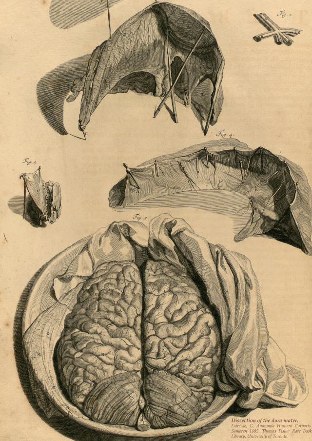
Dissection of the dura mater.Lairesse, G. Anatomia Humani Corporis. Someren 1685. Thomas Fisher Rare Book Library, University of Toronto.
Part I. Introduction
Chapter 1
Current understanding of meningeal and cerebral vascular function underlying migraine headache
Based on: Cephalalgia, 2018, In press.
Levy D, Labastida-Ramírez A and MaassenVanDenBrink A.
Abstract
Background: The exact mechanisms underlying the onset of a migraine attack are not completely understood. It is, however, now well accepted that the onset of the excruciating headache of migraine is mediated by the activation and increased mechanosensitivity (i.e. sensitization) of trigeminal nociceptive afferents that innervate the cranial meninges and their related large blood vessels.
Objective: To provide a critical summary of current understanding of the role that the cranial meninges, their associated vasculature, and immune cells play in meningeal nociception and the ensuing migraine headache.
Methods: We discuss the anatomy of the cranial meninges, their associated vasculature, innervation and immune cell population. We then debate the meningeal neurogenic inflammation hypothesis of migraine and its putative contribution to migraine pain. Finally, we provide insights into potential sources of meningeal inflammation and nociception beyond neurogenic inflammation and their potential contribution to migraine headache.
Background
Migraine is a complex, multifactorial neurological disorder affecting about 10% of the adult population worldwide [1]. It is the second most prevalent neurological disorder [1] and the first cause of disability in under 50s [2, 3]. While the exact mechanisms underlying the onset of a migraine attack remain unclear, it is now accepted that the development of the excruciating throbbing headache of migraine requires the initial activation and increased mechanosensitivity (i.e. sensitization) of trigeminal nociceptive afferents that innervate the cranial meninges and their related large blood vessels [4–7]. The goal of this review is to summarize current knowledge and understanding of the role that the cranial meninges and their related vasculature and cellular constituents play in the meningeal nociceptive processes underlying the onset of migraine headache.
(i) Anatomical features of the cranial meninges and their associated vasculature
The cranial meninges are comprised of two main distinct layers: the dura mater, or pachymeninx, a thick layer of connective tissue apposing to the cranium, and the leptomeninges. The latter can be further separated into the arachnoid and pia mater. The dura mater can be divided anatomically into three layers, the endosteal (periosteal) layer, the inner meningeal layer, and the dural border cell layer, or subdural neurothelium [8]. The dural layers are fused in most places, but separate to form the venous sinuses and at the Falx cerebri. The dural border cell layer is attached to the outer layer of the arachnoid – the arachnoid barrier cell layer – by occasional cell junctions, or desmosomes. Under various pathological conditions, such as in the case of subdural hematoma, damage to the dural border cell layer can lead to separation of the dura from the arachnoid and the formation of a ‘‘subdural space’’ [8]. The arachnoid layer connects to the thin piamater via collagen trabeculae to form the subarachnoid space, which is filled with cerebrospinal fluid (CSF). The pia mater, the most inner meningeal layer, abuts a cortical barrier layer made of astrocyte endfoot processes (i.e. the glia limitans, Figure 1(a)). Blood vessels within the subarachnoid space are coated by a pia mater layer (hence the name pial vessels). As arteries penetrate the brain, a single-layered sheath of pial cells
2
Chapter 1
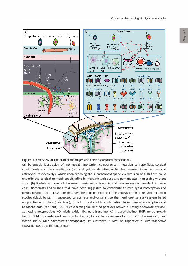
Figure 1. Overview of the cranial meninges and their associated constituents.(a) Schematic illustration of meningeal innervation components in relation to superficial cortical constituents and their mediators (red and yellow, denoting molecules released from neurons and astrocytes respectively), which upon reaching the subarachnoid space via diffusion or bulk flow, could underlie the cortical to meninges signaling in migraine with aura and perhaps also in migraine without aura. (b) Postulated crosstalk between meningeal autonomic and sensory nerves, resident immune cells, fibroblasts and vessels that have been suggested to contribute to meningeal nociception and headache and receptor systems that have been (i) implicated in the genesis of migraine pain in clinical studies (black font), (ii) suggested to activate and/or sensitize the meningeal sensory system based on preclinical studies (blue font), or with questionable contribution to meningeal nociception and headache pain (red font). CGRP: calcitonin gene-related peptide; PACAP: pituitary adenylate cyclase-activating polypeptide; NO: nitric oxide; NA: noradrenaline; ACh: acetylcholine; NGF: nerve growth factor; BDNF: brain-derived neurotrophic factor; TNF-a: tumor necrosis factor; IL-1: interleukin-1; IL-6: interleukin 6; ATP: adenosine triphosphate; SP: substance P; NPY: neuropeptide Y; VIP: vasoactive intestinal peptide; ET: endothelin.
3
Current understanding of migraine headache
Chap
ter
1
4
Chapter 1
is maintained for a short distance and separates between the vessel wall and the glia limitans [9, 10]. Upon entering the cortex, the pial layer that surrounds the arterioles becomes fenestrated. Penetrating veins lack a continuous pial layer [9]. The dura mater is highly vascularized. The arterial supply of the supratentorial dura mater comes primarily from branches of the posterior, middle (the largest), and anterior meningeal arteries, which arise from the occipital, maxillary, and ophthalmic arteries respectively. Meningeal arteries lie predominantly in the endosteal layer. The intracranial middle meningeal artery, which enters the cranium through the foramen spinosum, runs in grooves in the inner table of the calvarium, surrounded almost on three sides by bone [11]. A dense capillary network occurs in the inner meningeal layer of the dura. The major dural veins, which run primarily parallel to the path of the meningeal arteries, drain into efferent vessels in the periosteal layer or the dural venous sinuses. The dural venous sinuses absorb CSF from the subarachnoid space via arachnoid granulations. Studies in rodents also identified a dural lymphatic vascular network alongside blood vessels, primarily the middle meningeal artery, superior sagittal sinus, and transverse sinuses [12-15]. A recent study in non-human primates and humans has shown that the dural lymphatic system drains macromolecules from the dura [15]. In rodents, dural lymphatics drain CSF and parenchymal interstitial fluid [13, 14], but whether such lymphatic drainage also occurs in humans remains to be determined [16].
Innervation of the cranial meninges
The cranial meninges are innervated by sensory and autonomic nerves (Figure 1(b)). The sensory innervation of the dura mater originates primarily in the ophthalmic branch of the trigeminal ganglion but also from the mandibular and maxillary branches. Studies on samples obtained from humans, cats, and rodents have demonstrated nerve fibers in the supratentorial dura, along arteries, as well as at the tentorium cerebelli, and the venous sinuses [12, 17–20]. Using immunohistochemistry in animal and human tissue, axons innervating the cranial dura, cranial arteries and venous sinuses have been shown to express the sensory neuropeptides CGRP (the most abundant) and substance P [19, 21–23]. Pituitary adenylate cyclase-activating polypeptide (PACAP) was recently found to be co-expressed with CGRP in some dural nerve fibers [24]. Glutamate was also identified in trigeminal ganglion cells of rats, albeit primarily in neurons that do not express CGRP [25]. While there is no available data to suggest expression of glutamate in meningeal afferents, a recent retrograde tracing study in the mouse identified vesicular glutamate transporter 3 in a subpopulation of small diameter dural afferents [26]. The dura mater is also innervated by autonomic fibers that express PACAP, neuronal nitric oxide synthase (nNOS), vasoactive intestinal polypeptide (VIP), tyrosine hydroxylase (TH), acetylcholine (ACh), and neuropeptide Y (NPY) [18, 21–24, 27–31]. Ultrastructural studies conducted in the rat localized some peptidergic fibers’ termination in the vicinity, or walls, of dural blood vessels including lymphatics; axon terminations on collagen bundles in the dural connective tissue have been shown to be more abundant, however [12, 18]. Within the leptomeningeal, which lack their own blood supply, the cerebral (pial) arteries are the primary sites that receive sensory and autonomic innervations by fibers that originate from sympathetic, parasympathetic, and sensory trigeminal ganglia (Figure 1(a)).
Immunohistochemistry studies together with denervation and tracing studies identified trigeminal nerves expressing substance P and CGRP, as well as autonomic nerves that express NPY, VIP, nNOS, ACh, and tryptophan-5-hydroxylase [28, 32–39]. The sensory and autonomic innervation of cerebral pial vessels is primarily localized to the subarachnoid space and does not follow the vessels when they penetrate the cortex; the loss of pial sheath in the penetrating arteriole is also accompanied by the loss of perivascular innervation. Ultra-structurally, cerebral pial arteries are innervated by peptidergic sensory afferents that terminate either in the outer layer of the adventitia (substance P) or in the inner layer of the adventitia (CGRP), close to arterial smooth muscle cells [40].
Meningeal immune cells
Cytochemical and immunohistochemical studies in naive rats and mice localized resident immune cells to the dura mater (Figure 1(b)). Macrophages, which express numerous antigens (e.g. CD163, CD68) [41, 42] can be found along dural vessels, but also remotely from large vessels. In rodents, resident macrophages that express the chemokine receptor CX3CR1 were also localized to the dura and near pial vessels [43, 44]. The dura mater also harbors a sizeable population of mast cells (MCs) [19, 45–47]. Dural MCs are found in the endosteal/periosteal layer and are associated with blood vessels, dural sensory axons expressing CGRP, or substance P, and sympathetic fibers [27, 47, 48]. In the rat, dural MCs can also be found in a distinctive ‘‘linear arrays’’ arrangement [19, 46]. In the rat, a unique and much denser population of dural MCs is also found immediately caudal to the transverse sinus and medial to the superior cerebellar veins, which empty into the sinus, on either side of the midline [19]. Studies on human dural tissue also identified perivascular MCs in the periosteal dura [49]. In rats and mice, there is also a small population of MCs localized to the pia [50, 51]. Meningeal MCs can be classified based on their histochemical properties, using alcian blue and safranin staining [50, 52, 53]. Using these staining properties, it has been shown that the densities of subsets of dural MCs undergo dynamic changes in female rats during the estrous cycle that are estrogen-dependent [53]. Dendritic cells – antigen presenting cells expressing MHC class II and CD11c markers – are localized to the inner layer and connective tissue of the dura, in the arachnoid membrane, and pia mater layer [41, 42, 44, 54, 55]. Studies in naive mice demonstrated a small number of T lymphocytes also in the dura, with females showing higher numbers [13, 54, 55]. Memory CD4+ and CD3 T cells can also be found in the subarachnoid space [56, 57].
(ii) The meningeal neurogenic inflammation hypothesis of migraine
In vivo electrophysiological studies provided important information about the basic response properties of trigeminal dural afferents [58]. Knowledge about the response properties of leptomeningeal afferents is poor, however. Furthermore, knowledge of the endogenous processes that drive the activation and increased sensitivity of both dural and leptomeningeal afferents during a clinically occurring headache attack such as that of migraine is also limited. Tissue injury associated with local inflammation is a major driver of nociceptors’ activation, sensitization and pain. However, frank tissue injury or pathology has yet to be demonstrated in migraine (or any other primary headache condition). Nevertheless, a major hypothesis implicates local sterile meningeal inflammation as a key event that mediates the prolonged activation and sensitization of meningeal afferents and the origin of migraine
5
Current understanding of migraine headache
Chap
ter
1
headache [59, 60] (see below for further discussion on the contribution of meningeal neurogenic inflammation to meningeal nociception). Numerous clinical findings gathered over the years provided key, yet indirect, support for this inflammatory hypothesis of migraine. Among those are increased levels of inflammatory mediators in the cephalic venous outflow [61, 62] and the ability of corticosteroids and non-steroidal-anti-inflammatory drugs to abort migraine pain [63, 64]. Landmark preclinical studies in rodents provided indirect support for this hypothesis by showing that meningeal afferents are inflammatory sensors and can become persistently activated and sensitized to mechanical stimuli following local stimulation with mediators found in inflammatory exudates [59, 65–69]. The origin of such meningeal inflammatory response in primary headaches, and particularly in migraine, remains nevertheless elusive. In their hypothesis paper, Moskowitz and colleagues [70] proposed that ‘‘the headache phase of migraine may develop as a result of an abnormal interaction (and perhaps an abnormal release) of vasoactive neurotransmitters from the terminals of the trigeminal nerve with large intracranial and extracranial blood vessels’’. The meningeal process implicated in this hypothesis was neurogenic inflammation, a peripheral response comprised of (i) increased capillary permeability leading to plasma protein extravasation (PPE), (ii) arterial vasodilatation, and (iii) activation of resident immune cells. Neurogenic inflammation results from activity-dependent release of vasoactive substances, in particular substance P and CGRP from peripheral nerve endings of primary afferent nociceptors: this release occurs through an ‘‘axon reflex’’ process, where action potentials from acutely activated afferents are conducted antidromically and invade peripheral end branches [71]. The finding that dural and pial blood vessels are innervated by sensory nerves that express these vasoactive neuropeptides (see above) provided key support for the neurogenic inflammation hypothesis of migraine, which further led to the conceptualization of the trigeminovascular system and its role in migraine headache [72].
Increased meningeal vascular permeability
An early study in animals described the development of meningeal PPE in the dura mater following electrical stimulation of the trigeminal ganglion [73]. The subsequent findings that antimigraine drugs, including ergot alkaloids and triptans, could block this experimental meningeal PPE [74, 75] provided additional indirect support for the role of meningeal neurogenic inflammation in migraine headache. The inability to resolve the meningeal tissue and its vasculature in humans using imaging techniques remains a major hurdle in assessing meningeal PPE during migraine headache. However, one imaging study, conducted on a single migraine patient, suggested an increase in meningeal vascular permeability during an attack [76]. Current technical improvements in meningeal imaging [15] may be able to provide more clues into this process. In agreement with studies on non-cranial tissues [77], animal studies also implicated substance P and its neurokinin 1 receptor (NK1-R) in mediating meningeal neurogenic PPE [78, 79]. However, available data does not support a role for NK1 signaling in migraine pain. In clinical trials, NK1-R antagonists failed to abort migraine headache [80, 81]. While such negative data argues against the involvement of substance P signaling and meningeal neurogenic PPE in migraine pain, the possibility that the doses of NK1-R antagonists used in those studies were suboptimal and thus did not reach biologically-active plasma levels were considered [81, 82]. The possibility that during migraine, substance P does play a role in mediating a meningeal PPE response, but only during the early stages of the attack, may also be entertained. However, a small study
6
Chapter 1
reported the absence of substance P release into the internal jugular vein prior to the onset of the headache phase of a migraine with aura attack induced by intracarotid Xenon-133 injection [83], suggesting lack of substance P involvement in the triggering mechanisms of migraine headache.
Meningeal vasodilatation
Arterial vasodilation – another major characteristic of experimental meningeal neurogenic inflammation – has also been advocated for many years as a key cause of migraine headache. The theory that vasodilatation plays a role in migraine headache was largely based on the early observations of Graham and Wolff [84], who described a close relationship between the decrease in pulsation amplitude of the temporal artery and the decline of headache intensity following treatment with the vasoconstrictor (and antimigraine drug) ergotamine. The later observations that intracranial arteries (mainly the dural branches of the middle meningeal artery) are sensitive to painful stimuli [85] extended the extracranial vascular hypothesis to the intracranial vasculature. Moreover, when distending these arteries, a throbbing headache accompanied with nausea was induced, while neither constriction of the artery lumen nor stimulation of the dura mater 2 mm away from such vessels produced headache pain. These findings have led to the notion that dilatation and distention of intracranial dural meningeal arteries are a major source of migraine headache [86]. Thus, it was hypothesized that selective cranial vasoconstrictors would be more efficient and safe antimigraine drugs than ergotamine, which has affinity for a wide array of receptors, including 5-HT and dopamine [87]. On this basis, in the beginning of the 1970s, Humphrey et al. aimed at identifying novel antimigraine agents capable of mimicking the beneficial effects of 5-HT without its side-effects [88]. As a result, more selective vasoconstrictors of the cranial extra-cerebral circulation were developed, which allowed the identification of the 5-HT1B receptor (at the time of the development designated as 5-HT1-like receptor) as responsible for this vasoconstriction. Subsequently, one of those agents, the antimigraine drug sumatriptan, has been shown to produce selective cranial vasoconstriction in dogs, and to display much less activity in other vascular beds [89]. In accordance with the hypothesis on which its development was based, migraine-related changes in the middle cerebral artery blood flow, congruent with vasodilation, were reversed by sumatriptan [83]. Unfortunately, the vasoconstrictor effects of sumatriptan were not entirely selective for the cranial circulation (i.e. 5-HT1B receptors were also localized in coronary arteries), suggesting that the antimigraine effect of sumatriptan may not be entirely related to meningeal arterial vasoconstriction. Whether meningeal vasodilatation plays a causative role in migraine, or is merely an epiphenomenon – a secondary event arising from the activation of intracranial trigeminal afferents and the ensuing meningeal release of vasodilatory neuropeptides – remains a hotly debated subject [90]. However, neuroimaging studies have revealed that triptans produce cranial vasoconstriction; this may well contribute to their antimigraine effects [91, 92]. Assuming that receptor density expression in the cranial vasculature and the trigeminal ganglion have a close similarity, it could indeed be that triptans’ antimigraine effects are not only mediated via the blood vessels, but via other structures, such as peripheral nerve endings of meningeal afferents [93] (but see also 94), their trigeminal ganglion cell bodies or central nerve endings in the trigemino-cervical complex [95]. According to the
7
Current understanding of migraine headache
Chap
ter
1
‘‘vascular theory’’, intracranial vasodilatation (but possibly also extracranial) leads to the activation of trigeminal afferents that innervate these vessels, with ensuing headache [96]. A key process that could hypothetically mediate the activation of meningeal afferents by arterial vasodilatation is the stimulation of mechanosensitive stretch receptors located within the dilated vessels’ walls. While electrophysiological studies indicate that meningeal afferents that innervate the dura mater are mechanosensitive [58, 59], anatomical studies in animals suggest that most of the sensory innervation of the dura mater terminates in the connective tissue, far from the vessels [18]. Animal studies showing that administration of vasoactive agents, including CGRP and NO, failed to activate afferents with perivascular dural receptive fields [97–99], which further suggests that dural vasodilation alone is not sufficient to activate mechanosensitive meningeal afferents. As indicated above, the sensory innervation of intracranial pial vessels, which may also be mechanosensitive, terminates in the outer layer of the adventitia (substance P) or in the inner layer of the adventitia (CGRP), close to arterial smooth muscle cells [40]; but it is unclear whether pial afferents can be directly activated by arterial dilatation per se. The finding of only a slight dilatation of intracranial arteries during migraine attacks that was not reduced by effective treatment with sumatriptan [100] argues against the activation of meningeal afferents in responses to such intracranial vasodilatation in migraine (and see also 101). Moreover, headache provocation studies in healthy subjects revealed that migraine-like headaches can be induced by oral sildenafil, a phosphodiesterase 5 inhibitor, while no cerebral arterial vasodilation was detected. On the other hand, experimentally-induced headache in migraine patients revealed that the vasodilator adrenomedullin did not induce migraine headaches or changes in mean blood flow velocity of the middle cerebral artery. To properly interpret these studies, it should be taken into account that, due to technical limitations, only extracranial parts of the meningeal vasculature [100] or intracerebral vessels [102, 103] were measured and thus potentially do not always exactly reflect changes that occur in the meningeal vasculature [104]. Further, although sildenafil is strictly not a vasodilator per se, it is quite likely that its phosphodiesterase 5 inhibitory activity may lead to vasodilatation depending on the levels of cGMP in a blood vessel. Indeed, in isolated meningeal arteries from rats or patients undergoing neurosurgery, VIP, SNP, CGRP and PACAP, as well as sildenafil, can promote vasorelaxation. Interestingly, infusion of PACAP38 causes headache and vasodilation in both healthy subjects and migraine patients. In contrast, infusion of VIP in people with migraine evoked a marked cephalic vasodilation, but not a migrainous headache [105], which is also congruent with the notion that a provoked intracranial vasodilation alone is not sufficient to activate meningeal afferents. On the other hand, the above-mentioned discrepancy between VIP and PACAP may also be assigned to the shorter-lasting vasodilatory response evoked by VIP compared to that by PACAP (M Ashina, personal communication). Studies investigating the effects of VIP infusions over a longer period of time may explain whether the discrepancy between VIP and PACAP is due to different pharmacokinetics, or whether provoked vasodilatation is indeed not sufficient to provoke a migraine-like headache. Intriguingly, the receptors mediating dilatation in the meningeal circulation closely resemble many of the receptors expressed on the trigeminal ganglion, hampering a dissection between pure vascular and trigeminal effects. Increased diameter of meningeal vessels, whether dural or pial, may however lead to meningeal tissues’ stretching that could activate mechanosensitive afferents, in particular during a sensitized state [59, 101, 106].
8
Chapter 1
Key studies in rodents have led researchers to suggest that trigeminal nerve endings of activated perivascular meningeal afferents release CGRP, and this is the primary driver of neurogenic meningeal vasodilation in migraine [107]. The view that cephalic vasodilatation in migraine is neurogenically mediated received strong support from the findings of Goadsby and colleagues [108, 109], who demonstrated elevated levels of CGRP in the extra-cerebral circulation during a migraine attack. While these findings could not be replicated in a later study [110], the validity of its methodology was questioned subsequently [111]. Despite the inconclusive findings of increased CGRP levels within the intracranial circulation during a migraine attack [83, 112], the finding that sumatriptan normalized the elevated CGRP levels observed in the extra-jugular vein, concomitant with headache relief [109] further promoted the notion that trigeminal release of CGRP and the ensuing cranial neurogenic vasodilatation contribute to migraine headache. Moreover, a human experimental model of neurogenic vasodilation, where capsaicin is applied to a trigeminal (V1) dermatome in the human forehead to release endogenous CGRP (via activation of TRPV1 channels), confirmed that sumatriptan, most likely via 5 -HT1D/1F receptors, pre-synaptically inhibits CGRP release from trigeminal nerve endings [113]. The key findings that exogenous CGRP infusions could trigger delayed migraine-like headaches accompanied by a unilateral dilatation of the middle meningeal artery, and no dilation of the middle cerebral artery [114, 115] suggested a peripheral role for CGRP and its related meningeal vasodilation in migraine headache, especially since CGRP, like substance P, is unlikely to pass readily into the brain, due to its large molecular weight. It should be noted, however, that in chronic migraine an elevated CGRP level was also detected in the CSF [116], pointing to the possibility that CGRP released also from cortical parenchymal cells [117] (or possibly from pial afferents) is also involved in migraine. In a recent preliminary study, peripheral administration of CGRP to mice overexpressing the CGRP receptor subunit RAMP1 in smooth muscle cells (including in vascular smooth muscle cells) promoted light aversion (a rodent behavior suggested to be linked to the photophobia phenomenon of migraine), pointing to the possibility that over-activation of dural arterial cells can promote migraine headache [118]. Whether such migraine-related response is mediated via enhanced meningeal vasodilatation or augmented release of algesic mediators from meningeal vascular smooth muscle cells (VSMCs, see also below) remain to be determined. Taken together, the abovementioned studies demonstrate a complex bidirectional cross-talk between cranial blood vessels and their trigeminal nerve endings in migraine pathogenesis.
Neurogenic activation of meningeal immune cells
Another major feature of neurogenic inflammation is the activation of immune cells [119]. Of particular interest to migraine are meningeal MCs [106]. Activated MCs are proinflammatory and release a host of pro-nociceptive mediators [120] that can lead to the activation and sensitization of meningeal afferents [66, 67, 121, 122]. Clinical studies reported elevated plasma levels of the MC mediator’s histamine, tryptase and TNF-alpha during migraine [61, 123, 124], supporting the involvement of MCs in migraine. A role for MCs in meningeal neurogenic inflammation was initially supported by the finding that stimulation of the trigeminal ganglion to produce dural PPE also promoted morphological changes in dural MCs suggestive of degranulation [52, 125]. The anatomical localization of dural MCs in close apposition to terminals of dural afferents that express substance P and CGRP provided
9
Current understanding of migraine headache
Chap
ter
1
indirect support for the ability of trigeminal axon reflex to activate intracranial dural MCs. The activation of MCs’ NK1-R is thought to promote their degranulation by substance P [126]. However, the presumed lack of involvement of NK1 signaling in migraine headache suggests that if dural MCs are degranulated in response to meningeal axon reflex, and this process contributes to the headache of migraine (see also below), it is unlikely to involve substance P receptor signaling. Release of CGRP from activated meningeal afferents may also promote MC degranulation in experimental animals, although with less potency than substance P [127]. In vitro stimulation of rodent’s meningeal MCs with CGRP induced 5-HT and histamine release [128, 129]. The MC degranulating effect of CGRP may be nonetheless rodent specific. Rodent MCs express the required components of the CGRP receptor system, calcitonin receptor-like receptor (CLR) and receptor activity-modifying protein 1 (RAMP1) [24, 130, 131]. Human dural MCs, however, express only RAMP1 [24]. Nonetheless, the possibility that CGRP signals via the calcitonin (CTR)/RAMP1 receptor complex [132] in these cells should not be ignored. Meningeal release of PACAP may also promote MC degranulation [133, 134]. A recent clinical study demonstrated the expression of the PACAP receptor VPAC1 on human skin MCs [135]. Whether PACAP can promote the degranulation of human dural MCs is currently unknown. Meningeal MCs have been shown to become activated after exposure to carbachol [47, 136, 137], suggesting that activation of meningeal parasympathetic efferents could promote meningeal neurogenic inflammation [138]. While cranial parasympathetic activation has been implicated in trigeminal autonomic cephalalgias [139], and may accompany migraine attacks in some patients [140], its role in promoting meningeal nociception and migraine headache is less clear. Taken together, whether meningeal neurogenic inflammation, either mediated by release of sensory or autonomic transmitters, promotes a sufficient dural MC degranulation response that can lead to the activation and sensitization of meningeal afferents with ensuing headache remains to be determined. Perivascular meningeal macrophages may play a role in meningeal nociception and potentially in headache. Macrophages can release a host of inflammatory and nociceptive mediators including prostaglandins, cytokines, chemokines, and high levels of NO that can act directly, or indirectly, on meningeal afferents to promote their activation and mechanical sensitization [68, 69, 97, 99, 121]. The finding that systemic administration of the headache and migraine trigger nitroglycerin upregulates proinflammatory cytokines and the inducible (and pro-inflammatory) isoform of nitric oxide synthase (iNOS) in rat dural macrophages, as well as promoting dural inflammation [141], further suggests a role for these immune cells in headache. While functional interactions between sensory or autonomic transmitters, and meningeal macrophages are yet to be defined, in other tissues CGRP rather inhibits proinflammatory macrophage function [142–144]. A role for resident DCs and circulating T cells in meningeal neurogenic inflammation remains unknown. While studies in other tissues suggest that CGRP downregulates the expression of the nociceptive cytokine TNF-alpha in DCs, activation of peptidergic nociceptive afferents can drive DC production of the proinflammatory and nociceptive cytokine IL-23 with subsequent tissue PPE [145, 146]. T cells have been shown to mediate neuropathic pain in rodent models [147], but a role for T cells in acute nociception, such as during episodic migraine attacks, is yet to be demonstrated.
10
Chapter 1
How might meningeal neurogenic inflammation be triggered during migraine?
One critical unknown aspect of the neurogenic inflammation hypothesis of migraine is the identity of the endogenous processes that lead to the initial activation of meningeal afferents and the ensuing release of neuropeptides. One event that has been hypothesized to trigger meningeal neurogenic inflammation is cortical spreading depression (CSD), a wave of neural and glial depolarizations (followed by neuronal silencing) that is thought to underlie the aura phase of migraine [72]. In rats and rabbits, induction of a single CSD event gives rise to a brief dilatation of pial arteries and increase in cerebral blood flow [148]. The induction of CSD-evoked cortical hyperemia in the mouse [148] (and also in human subjects [149]), however, is less clear. The dilation of pial vessels in response to CSD has been shown to be mediated in part by CGRP receptor signaling [150–152], suggesting a short-lasting activation of peptidergic leptomeningeal afferents. The mechanisms underlying the pial afferent response to CSD are incompletely understood, but presumably involve the release of small nociceptive molecules, such as nitric oxide, potassium ions and ATP in the superficial cortical parenchyma during the passage of the CSD wave, their diffusion or bulk flow into the subarachnoid space and subsequent action upon leptomeningeal afferent nerve endings [153] (Figure 1(a)). A key part of the meningeal neurogenic inflammation theory of migraine posits that leptomeningeal afferents have additional branches that terminate in the dura mater (Figure 1(a)), and that activation of leptomeningeal afferents can promote the release of proinflammatory peptides also in the dura mater with ensuing local sterile inflammatory responses [154]. Anatomical support for this hypothesis comes from rodent studies demonstrating a sizable number of single trigeminal ganglion neurons that project to both the middle cerebral, and middle meningeal arteries, or other dural sites [17], and a small number of dural fibers that issue collateral branches to the pia at the frontal part of the brain [155]. The finding of prolonged dural vasodilatation and PPE following a single CSD event that were dependent upon an intact trigeminal nerve and activation of NK1-R [154] provided further support for this theory. In that study, CSD evoked prolonged dilatation of the dural middle meningeal artery that was also dependent on parasympathetic outflow from sphenopalatine ganglion neurons, highlighting the additional contribution of trigemino-parasympathetic reflex to meningeal NI. More recently, an electrophysiological single unit recording in a rat model provided direct evidence for the acute activation of a subpopulation of dural afferents during the passage of the CSD wave [156, 157]. A direct link between CSD, meningeal neurogenic inflammation and the activation of dural or leptomeningeal afferents, remains to be established. CSD has been shown to promote meningeal MC degranulation [158] and conformational changes in meningeal macrophages, reminiscent of an inflammatory response [44]. CSD also leads to arrested migration of meningeal DCs, suggesting inflammatory activation [44]. It is unknown, however, whether these inflammatory changes occur in response to the activation of meningeal afferents (i.e. via the release of neuropeptides). Nonetheless, the finding that CSD (which likely contributes to the headache in migraine with aura) leads to meningeal inflammatory changes involving MCs and macrophages, similar to those provoked by other migraine triggers such as GTN and F38 (which trigger migraine without aura), suggest that meningeal inflammation (whether this involves the release of sensory neuropeptides or not) serves as a common mechanism of migraine headache onset.
11
Current understanding of migraine headache
Chap
ter
1
Can meningeal neurogenic inflammation actually promote meningeal nociception?
Despite the indirect evidence for meningeal neurogenic inflammation in animal models of migraine, a critical question remains as to whether this event actually contributes to meningeal nociception and headache. Previous studies examined the effect of acute stimulation of primary afferent neurons that innervate other tissues on the sensitivity of primary afferent nociceptive neurons, but have yielded conflicting data. For example, studies in monkeys [159] and rats [160] have shown that stimulation of cutaneous nociceptive afferents did not subsequently alter their ongoing activity, mechanosensitivity, or heat sensitivity. A study in rabbits, however, reported the development of heat sensitization of nociceptive afferents following antidromic afferent stimulation [161]. A rat study that employed local capsaicin stimulation to evoke acute excitation of cutaneous afferents documented a delayed and prolonged increase in the afferents’ ongoing activity and mechanosensitivity that were suggested to involve CGRP signaling [162]. Finally, we have shown recently that brief meningeal application with potassium levels, similar to that expected during CSD, can lead to prolonged activation of dural afferents [163]. The finding that in addition to their acute activation, CSD also gives rise to prolonged activation and enhanced mechanosensitivity of meningeal afferents [156, 157, 164] may point to neurogenic inflammation as a nociceptive event. Nevertheless, numerous finding suggests meningeal nociception in response to CSD is not directly mediated by neurogenic inflammation. For example, persistent meningeal afferent activation still occurs following excision of the parasympathetic sphenopalatine ganglion [156], whose activation was critical to the CSD-evoked meningeal neurogenic vasodilatation. In addition, persistent meningeal afferent activation following CSD can occur in the absence of acute NI-mediated dural MC degranulation, as was observed in craniotomized animals, a preparation in which the majority of dural MCs are already in a state of degranulation prior to the induction of CSD [165]. As indicated above, studies in naive rats suggest that acute activation of meningeal CGRP receptors is not sufficient to activate or sensitize meningeal afferents [98]. While there is some evidence in rats and humans for the expression of the CGRP receptors components CLR and RAMP1 in the cytoplasm of some thick myelinated dural axons [24], these receptors were also reported to be localized specifically to the axons’ Schwann cells [130]. It also remains unclear whether these CLR/RAMP1 expressing dural afferents play a role in meningeal nociception as they are not co-localized with CGRP [24]. The possibility that CGRP receptors are preferentially expressed on non-nociceptive, myelinated dural fast A fibers, which comprise about one-third of the myelinated axons in the nerves supplying the dura, and exhibit the highest thresholds and the lowest firing rates, as well as the lowest incidence of mechanosensitivity [12, 166] should be considered. Recent data further suggests that acute blockade of meningeal CGRP receptors, using systemic administration of olcegepant (BIBN4096), does not inhibit the prolonged activation and mechano-sensitization of meningeal afferents, in the wake of CSD, despite diminishing the related cerebral vasodilatation [163]. This data is congruent with the observations that CSD does not alter dural CGRP release [167] or leads to CGRP release into the external jugular vein [168]. However, the finding that a prolonged sequestering of CGRP, using a monoclonal antibody approach, can inhibit the prolonged activation of slow conducting A-delta afferents following CSD [169] points to the possibility that the basal CGRP level somehow modulates the responsiveness of a subpopulation of meningeal afferents.
12
Chapter 1
Such effects are likely to be restricted to the dura mater, as antibodies, due to their large molecular size, are unlikely to gain access to the leptomeningeal space (i.e. CSF). Finally, the finding that acute CGRP receptor blockade can inhibit prolonged meningeal afferent activation in response to their local stimulation with high physiological levels of potassium [163] further suggests that the indirect, pro-nociceptive effect of CGRP, likely mediated through its signaling on non-neural cells and in tandem with other nociceptive factors, may play a role in mediating meningeal nociception. However, whether the antimigraine prophylactic effects of monoclonal antibodies that target CGRP or its receptor are linked to acute inhibition of dural NI or other, unrelated peripheral effects of CGRP remains unknown. (iii) Potential sources of meningeal nociception beyond neurogenic inflammation
Activation of meningeal MCs
Beyond neurogenic inflammation, are there other mechanisms that could lead to prolonged activation of meningeal afferents and the ensuing headache? As indicated above, release of MC mediators in the vicinity of meningeal afferents could potentially lead to their activation and sensitization. While not directly related to meningeal neurogenic inflammation, a causative role for MCs in migraine headache was already considered more than 50 years ago [170]. In that study, injection of an MC degranulating agent, compound 48/80, into the cranial circulation gave rise to a headache resembling that of migraine. Levy et al. [165] have shown that degranulation of dural MCs, using compound 48/80 as in Sicuteri’s study, promoted persistent activation of the majority of meningeal afferents, as well as of nociceptive neurons in the trigeminal nucleus caudalis. This work suggested that extensive dural MC degranulation could serve as a powerful peripheral pro-nociceptive stimulus capable of triggering the activation of the peripheral and central components of the migraine pain pathway. The finding that activation of dural MCs was also associated with the development of cephalic tactile hypersensitivity [6] provided further indirect evidence for their role in meningeal nociception and migraine headache pain. The identity of non-neurogenic inflammation mechanisms that could lead to dural MC degranulation with ensuing enhanced meningeal nociception are unclear at present. Because there is no blood-dura barrier, it is conceivable that circulating factors, reaching the dural circulation, could lead to the degranulation of a sizable population of dural MCs. The finding that migraine is comorbid with MC-related conditions such as allergies and asthma [171, 172] points to a potential link between MC-related events and migraine. The involvement of peripherally-acting MCs in migraine is also congruent with the recent finding that migraine is genetically and environmentally similar to peripheral inflammatory conditions that also involve MCs, such as irritable bowel syndrome and cystitis [173]. Finally, it has been suggested that some migraine events may be triggered by various food ingredients acting upon MCs, a notion that gained support from studies showing potent migraine prophylactic action of the MC stabilizing agent cromoglycate in a subset of patients with food-related attacks [174, 175]. It remains unclear, however, what level of meningeal MC degranulation is actually nociceptive. A relatively low level of dural MC degranulation induced by systemic infusion of the migraine trigger nitroglycerin [141, 176] was not sufficient to promote activation of meningeal afferents in a rodent model [99]. However, in migraineurs, a higher density of meningeal MCs, potentially due to endocrine changes,
13
Current understanding of migraine headache
Chap
ter
1
such as fluctuation in female sex hormones [53] or increased propensity of these immune cells to become activated in response to a given trigger, could potentially result in a robust proinflammatory that promotes the activation of meningeal afferents and lead to headache.
Beyond vasodilatation: release of algesic mediators
Vascular endothelial cells (ECs) and VSMCs are potentially important sources of algesic mediators that can activate and sensitize meningeal afferents. The vascular endothelium has been suggested to promote peripheral enhancement in pain (i.e. hyperalgesia) through the release of endothelin-1 [177] and ATP [178]. While a direct role for endothelin-1 in migraine has been questioned based on the lack of efficacy of endothelin receptor antagonism [179], its release from leptomeningeal vessels could potentially trigger CSD [180], and thus indirectly promote meningeal nociception. The genetic association between migraine, endothelin expression and vascular mechanisms [181, 182], as well as with the endothelin A receptor [183], also suggests a rather complex role for endothelin in migraine. Local release of ATP from the meningeal vasculature (but also from other meningeal cells) could promote acute activation of meningeal afferents [157], potentially via activation of afferent P2X2 and P2X3 purinergic receptors [184]. Prolonged activation and/or sensitization of meningeal afferents mediated by these, and potentially other purinergic receptors localized to meningeal afferent nerve endings, remains, however, to be established. Indirect meningeal nociceptive effects of ATP, promoted by its action upon proinflammatory P2X7 receptors localized to resident meningeal immune cells, such as macrophages [185] may also be of importance to headache and migraine mechanisms. Release of NO and prostacyclin (PGI2) from the vascular endothelium may lead to meningeal nociception [66, 121]. The release of COX-2-derived prostanoids, as well as high levels of NO and proinflammatory cytokines, such as IL-1β from VSMCs could activate and sensitize meningeal afferents [68]. Finally, the finding that the migraine trigger nitroglycerin, which leads to delayed migraine attacks, evokes a delayed mechanical sensitization of meningeal afferents that depends upon inflammatory signaling (i.e. ERK phosphorylation) in meningeal arteries [99] further points to the role of non-vasodilating meningeal vascular mechanisms in migraine headache. Recent work suggests that dural fibroblasts, which serve as key building blocks of this meningeal tissue, can release algesic mediators such as IL-6 that can act upon meningeal afferents and promote migraine-like behaviors in rodents [186, 187] (Figure 1(b)).
The role of cortical mediators
The finding that CSD, an event that is primarily restricted to the cortex, can lead to the activation and mechanical sensitization of meningeal afferents points to the possibility that algesic factors, such as ATP, prostaglandins and NO that are released into the parenchymal interstitial fluid, gain access to the subarachnoid space, where they can act upon leptomeningeal afferents. The presence of the dura-arachnoid interface layer, however, is likely to impede the passage of these cortical mediators into the cranial dura mater and their nociceptive action on dural afferents. Are there any routes by which cortical-derived algesic factors could reach the dura and interact with its nociceptive afferents? CSF in the subarachnoid space enters the bloodstream in the dural venous sinuses, via arachnoid granulation. Afferents localized to the dural sinus walls, in particular to the sinus lumen [23], could potentially sense these mediators. However, this implies that these
14
Chapter 1
afferents also sample venous blood – an unlikely occurrence – since blood itself has been shown to exert a nociceptive effect [188]. Alternatively, sensory nerve fiber terminals in the arachnoid granulations [189], which protrude through the dura, may become activated by CSF-derived nociceptive mediators.
Conclusions and future perspectives
While migraine is considered as a brain disorder, peripheral meningeal components including the sensory innervation of the cranial meninges and immune and vascular cells are likely to play a major role. A better understanding of the brain dysfunction processes and the nature of the cortical to meningeal signaling cascades that promote the activation and increased responsiveness of trigeminal nociceptive afferents is essential for the development of effective antimigraine prophylactic therapies. The finding that, genetically and environmentally, migraine is much closer to immune system diseases such as irritable bowel syndrome and cystitis/urethritis rather than to central nervous system specific conditions [173] further implies that migraine has a peripheral inflammatory origin that is not nervous system-specific. The concept of neurogenic inflammation undoubtedly had a tremendous impact on migraine research and provided an important roadmap for the development of neuropeptide and receptor driven therapies for migraine. While meningeal neurogenic inflammation continues to be regarded as a causal factor in migraine headache, direct evidence for the occurrence of neurogenic inflammation during migraine and its role in meningeal nociception is limited at best. Future studies may provide better direct evidence for the presence of the various features of meningeal neurogenic inflammation or lack thereof during a migraine attack and, most importantly, whether they constitute active players in driving migraine pain rather than simply epiphenomena. The advent of monoclonal antibodies against CGRP or its receptor, which are too large in molecular size to cross the blood brain barrier, may serve as a pharmacological tool to decipher the role of peripheral inflammatory mechanisms that involve CGRPergic signaling. While the success of treatment with antibodies that curtail CGRP signaling in preventing migraine attacks points to a peripheral, likely meningeal origin of migraine, the failure of such treatments in a subpopulation of patients [190] may point to a larger role for trigeminal leptomeningeal afferents, which cannot be targeted with systemically-administered antibodies, in mediating migraine pain, or to CGRP-independent processes. Thus, the role of other substances, including neuropeptides such as PACAP but certainly also immuno-active substances and numerous meningeal receptor systems (Figure 1(b)), should be explored and selective pharmacological tools developed. The availability of such tools will help to further unravel the pathophysiology of migraine and thus to ultimately provide a more effective and safe treatment for migraine patients.
References1. GBD 2015 Neurological Disorders Collaborator Group. Global, regional, and national burden of neurological disor-ders during 1990–2015: A systematic analysis for the Global Burden of Disease Study 2015. Lancet Neurol 2017; 16: 877–897.2. GBD 2106 Disease and Injury Incidence and Prevalence Collaborators. Global, regional, and national incidence, prevalence, and years lived with disability for 328 diseases and injuries for 195 countries, 1990–2016: A systematic analysis for the Global Burden of Disease Study 2016. Lancet 2017; 390: 1211–1259.3. Steiner TJ, Stovner LJ, Vos T, et al. Migraine is first cause of disability in under 50s: Will health politicians now take notice? J Headache Pain 2018; 19: 17.
15
Current understanding of migraine headache
Chap
ter
1
4. Olesen J, Burstein R, Ashina M, et al. Origin of pain in migraine: Evidence for peripheral sensitisation. Lancet Neurol 2009; 8: 679–690. 5. Messlinger K. Migraine: Where and how does the pain originate? Exp Brain Res 2009; 196: 179–193.6. Levy D, Kainz V, Burstein R, et al. Mast cell degranulation distinctly activates trigemino-cervical and lumbosacral pain pathways and elicits widespread tactile pain hypersensitivity. Brain Behav Immun 2012; 26: 311–317. 7. Noseda R and Burstein R. Migraine pathophysiology: Anatomy of the trigeminovascular pathway and associated neurological symptoms, cortical spreading depression, sensitization, and modulation of pain. Pain 2013; 154: S44–S53. 8. Haines DE. On the question of a subdural space. Anat Rec 1991; 230: 3–21. 9. Zhang ET, Inman CB and Weller RO. Interrelationships of the pia mater and the perivascular (Virchow-Robin) spaces in the human cerebrum. J Anat 1990; 170: 111–123.10. Weller RO, Sharp MM, Christodoulides M, et al. The meninges as barriers and facilitators for the movement of fluid, cells and pathogens related to the rodent and human CNS. Acta Neuropathol 2018; 135: 363–385.11. Shevel E. Middle meningeal artery dilatation in migraine. Headache 2009; 49: 1541–1543. 12. Andres KH, von During M, Muszynski K, et al. Nerve fibres and their terminals of the dura mater encephali of the rat. Anat Embryol (Berl) 1987; 175: 289–301. 13. Louveau A, Smirnov I, Keyes TJ, et al. Structural and functional features of central nervous system lymphatic vessels. Nature 2015; 523: 337–341.14. Aspelund A, Antila S, Proulx ST, et al. A dural lymphatic vascular system that drains brain interstitial fluid and macromolecules. J Exp Med 2015; 212: 991–999. 15. Absinta M, Ha SK, Nair G, et al. Human and nonhuman primate meninges harbor lymphatic vessels that can be visualized noninvasively by MRI. Elife 2017; 6 pii: e29738. 16. Ringstad G, Vatnehol SAS and Eide PK. Glymphatic MRI in idiopathic normal pressure hydrocephalus. Brain 2017; 140: 2691–2705. 17. O’Connor TP and van der Kooy D. Pattern of intracranial and extracranial projections of trigeminal ganglion cells. J Neurosci 1986; 6: 2200–2207. 18. Messlinger K, Hanesch U, Baumgartel M, et al. Innervation of the dura mater encephali of cat and rat: Ultrastruc-ture and calcitonin gene-related peptide-like and substance P-like immunoreactivity. Anat Embryol (Berl) 1993; 188: 219–237. 19. Strassman AM, Weissner W, Williams M, et al. Axon diameters and intradural trajectories of the dural innervation in the rat. J Comp Neurol 2004; 473: 364–376. 20. Lee SH, Shin KJ, Koh KS, et al. Visualization of the tentorial innervation of human dura mater. J Anat 2017; 231: 683–689.21. Edvinsson L, Rosendal-Helgesen S and Uddman R. Substance P: Localization, concentration and release in cerebral arteries, choroid plexus and dura mater. Cell Tissue Res 1983; 234: 1–7. 22. Edvinsson L and Uddman R. Adrenergic, cholinergic and peptidergic nerve fibres in dura mater – involvement in headache? Cephalalgia 1981; 1: 175–179. 23. Sampaolo S, Liguori G, Vittoria A, et al. First study on the peptidergic innervation of the brain superior sagittal sinus in humans. Neuropeptides 2017; 65: 45–55.24. Eftekhari S, Warfvinge K, Blixt FW, et al. Differentiation of nerve fibers storing CGRP and CGRP receptors in the peripheral trigeminovascular system. J Pain 2013; 14: 1289–1303. 25. Eftekhari S, Salvatore CA, Johansson S, et al. Localization of CGRP, CGRP receptor, PACAP and glutamate in trigem-inal ganglion. Relation to the blood-brain barrier. Brain Res 2015; 1600: 93–109.26. Ren L, Chang MJ, Zhang Z, et al. Quantitative analysis of mouse dural afferent neurons expressing TRPM8, VGLUT3, and NF200. Headache 2018; 58: 88–101. 27. Keller JT and Marfurt CF. Peptidergic and serotoninergic innervation of the rat dura mater. J Comp Neurol 1991; 309: 515–534.28. O’Connor TP and van der Kooy D. Enrichment of a vasoactive neuropeptide (calcitonin gene related peptide) in the trigeminal sensory projection to the intracranial arteries. J Neurosci 1988; 8: 2468–2476. 29. von During M, Bauersachs M, Bohmer B, et al. Neuropeptide Y- and substance P-like immunoreactive nerve fibers in the rat dura mater encephali. Anat Embryol (Berl) 1990; 182: 363–373. 30. Artico M and Cavallotti C. Catecholaminergic and acetylcholine esterase containing nerves of cranial and spinal dura mater in humans and rodents. Microsc Res Tech 2001; 53: 212–220. 31. Csati A, Tajti J, Kuris A, et al. Distribution of vasoactive intestinal peptide, pituitary adenylate cyclase-activating peptide, nitric oxide synthase, and their receptors in human and rat sphenopalatine ganglion. Neuroscience 2012; 202: 158–168. 32. Mayberg M, Langer RS, Zervas NT, et al. Perivascular meningeal projections from cat trigeminal ganglia: Possible pathway for vascular headaches in man. Science 1981; 213: 228–230. 33. Uddman R, Edvinsson L, Owman C, et al. Perivascular substance P: Occurrence and distribution in mammalian pial vessels. J Cereb Blood Flow Metab 1981; 1: 227–232. 34. Liu-Chen LY, Han DH and Moskowitz MA. Pia arachnoid contains substance P originating from trigeminal neurons. Neuroscience 1983; 9: 803–808.
16
Chapter 1
35. Hanko J, Hardebo JE, Kahrstrom J, et al. Existence and coexistence of calcitonin gene-related peptide (CGRP) and substance P in cerebrovascular nerves and trigeminal ganglion cells. Acta Physiol Scand Suppl 1986; 552: 29–32. 36. McCulloch J, Uddman R, Kingman TA and Edvinsson L. Calcitonin gene-related peptide: functional role in cerebro-vascular regulation. Proc Natl Acad Sci USA 1986; 83: 5731–5735. 37. Edvinsson L. Innervation of the cerebral circulation. Ann NY Acad Sci 1987; 519: 334–348. 38. Kimura T, Yu JG, Edvinsson L and Lee TJ. Cholinergic, nitric oxidergic innervation in cerebral arteries of the cat. Brain Res 1997; 773: 117–124. 39. Cohen Z, Bovento G, Lacombe P, et al. Cerebrovascular nerve fibers immunoreactive for tryptophan-5-hydroxylase in the rat: Distribution, putative origin and comparison with sympathetic noradrenergic nerves. Brain Res 1992; 598: 203–214. 40. Nakakita K. Peptidergic innervation in the cerebral blood vessels of the guinea pig: An immunohistochemical study. J Cereb Blood Flow Metab 1990; 10: 819–826. 41. McMenamin PG. Distribution and phenotype of dendritic cells and resident tissue macrophages in the dura mater, leptomeninges, and choroid plexus of the rat brain as demonstrated in wholemount preparations. J Comp Neurol 1999; 405: 553–562. 42. McMenamin PG, Wealthall RJ, Deverall M, et al. Macrophages and dendritic cells in the rat meninges and choroid plexus: Three-dimensional localisation by environmental scanning electron microscopy and confocal microscopy. Cell Tissue Res 2003; 313: 259–269. 43. Coles JA, Myburgh E, Brewer JM, et al. Where are we? The anatomy of the murine cortical meninges revisited for intravital imaging, immunology, and clearance of waste from the brain. Prog Neurobiol 2017; 156: 107–148. 44. Schain AJ, Melo-Carrillo A, Borsook D, et al. Activation of pial and dural macrophages and dendritic cells by CSD. Ann Neurol 2018; 83: 508–521. 45. Orr EL. Dural mast cells: Source of contaminating histamine in analyses of mouse brain histamine levels. J Neuro-chem 1984; 43: 1497–1499. 46. Dimlich RV, Keller JT, Strauss TA, et al. Linear arrays of homogeneous mast cells in the dura mater of the rat. J Neurocytol 1991; 20: 485–503. 47. Rozniecki JJ, Dimitriadou V, Lambracht-Hall M, et al. Morphological and functional demonstration of rat dura mater mast cell-neuron interactions in vitro and in vivo. Brain Res 1999; 849: 1–15. 48. Keller JT, Marfurt CF, Dimlich RV, et al. Sympathetic innervation of the supratentorial dura mater of the rat. J Comp Neurol 1989; 290: 310–321. 49. Varatharaj A, Mack J, Davidson JR, et al. Mast cells in the human dura: Effects of age and dural bleeding. Childs Nerv Syst 2012; 28: 541–545. 50. Michaloudi H, Batzios C, Chiotelli M, et al. Developmental changes of mast cell populations in the cerebral menin-ges of the rat. J Anat 2007; 211: 556–566. 51. Sayed BA, Christy AL, Walker ME, et al. Meningeal mast cells affect early T cell central nervous system infiltration and blood-brain barrier integrity through TNF: A role for neutrophil recruitment? J Immunol 2010; 184: 6891–6900. 52. Dimitriadou V, Buzzi MG, Moskowitz MA, et al. Trigeminal sensory fiber stimulation induces morphological changes reflecting secretion in rat dura mater mast cells. Neuroscience 1991; 44: 97–112. 12 Cephalalgia 0(0) 53. Boes T and Levy D. Influence of sex, estrous cycle, and estrogen on intracranial dural mast cells. Cephalalgia 2012; 32: 924–931. 54. Coles JA, Myburgh E, Ritchie R, et al. Intravital imaging of a massive lymphocyte response in the cortical dura of mice after peripheral infection by trypanosomes. PLoS Negl Trop Dis 2015; 9: e0003714. 55. McIlvried LA, Cruz JA, Borghesi LA, et al. Sex-, stress-, and sympathetic post-ganglionic-dependent changes in identity and proportions of immune cells in the dura. Cephalalgia 2017; 37: 36–48. 56. Reboldi A, Coisne C, Baumjohann D, et al. C-C chemokine receptor 6-regulated entry of TH-17 cells into the CNS through the choroid plexus is required for the initiation of EAE. Nat Immunol 2009; 10: 514–523. 57. Derecki NC, Cardani AN, Yang CH, et al. Regulation of learning and memory by meningeal immunity: A key role for IL-4. J Exp Med 2010; 207: 1067–1080. 58. Strassman AM and Levy D. Response properties of dural nociceptors in relation to headache. J Neurophysiol 2006; 95: 1298–1306. 59. Strassman AM, Raymond SA and Burstein R. Sensitization of meningeal sensory neurons and the origin of head-aches. Nature 1996; 384: 560–564. 60. Levy D. Migraine pain and nociceptor activation – where do we stand? Headache 2010; 50: 909–916. 61. Perini F, D’Andrea G, Galloni E, et al. Plasma cytokine levels in migraineurs and controls. Headache 2005; 45: 926–931. 62. Sarchielli P, Alberti A, Baldi A, et al. Proinflammatory cytokines, adhesion molecules, and lymphocyte integrin ex-pression in the internal jugular blood of migraine patients without aura assessed ictally. Headache 2006; 46: 200–207. 63. Klapper J. The pharmacologic treatment of acute migraine headaches. J Pain Symptom Manage 1993; 8: 140–147. 64. Woldeamanuel YW, Rapoport AM and Cowan RP. The place of corticosteroids in migraine attack management: A 65-year systematic review with pooled analysis and critical appraisal. Cephalalgia 2015; 35: 996–1024. 65. Bove GM and Moskowitz MA. Primary afferent neurons innervating guinea pig dura. J Neurophysiol 1997; 77:299-308.
17
Current understanding of migraine headache
Chap
ter
1
66. Zhang XC, Strassman AM, Burstein R, et al. Sensitization and activation of intracranial meningeal nociceptors by mast cell mediators. J Pharmacol Exp Ther 2007; 322: 806–812. 67. Zhang XC and Levy D. Modulation of meningeal nociceptors mechanosensitivity by peripheral proteinase-activated receptor-2: The role of mast cells. Cephalalgia 2008; 28: 276–284. 68. Zhang X, Burstein R and Levy D. Local action of the proinflammatory cytokines IL-1beta and IL-6 on intracranial meningeal nociceptors. Cephalalgia 2012; 32: 66–72. 69. Yan J, Melemedjian OK, Price TJ, et al. Sensitization of dural afferents underlies migraine-related behavior follow-ing meningeal application of interleukin-6 (IL-6). Mol Pain 2012; 8: 6. 70. Moskowitz MA, Reinhard JF Jr., Romero J, et al. Neurotransmitters and the fifth cranial nerve: Is there a relation to the headache phase of migraine? Lancet 1979; 2: 883–885. 71. Holzer P. Local effector functions of capsaicin-sensitive sensory nerve endings: Involvement of tachykinins, calci-tonin gene-related peptide and other neuropeptides. Neuroscience 1988; 24: 739–768. 72. Moskowitz MA. The neurobiology of vascular head pain. Ann Neurol 1984; 16: 157–168. 73. Markowitz S, Saito K and Moskowitz MA. Neurogenically mediated leakage of plasma protein occurs from blood vessels in dura mater but not brain. J Neurosci 1987; 7: 4129–4136. 74. Markowitz S, Saito K and Moskowitz MA. Neurogenically mediated plasma extravasation in dura mater: Effect of ergot alkaloids. A possible mechanism of action in vascular headache. Cephalalgia 1988; 8: 83–91. 75. Buzzi MG, Moskowitz MA, Peroutka SJ, et al. Further characterization of the putative 5-HT receptor which medi-ates blockade of neurogenic plasma extravasation in rat dura mater. Br J Pharmacol 1991; 103: 1421–1428. 76. Knotkova H and Pappagallo M. Imaging intracranial plasma extravasation in a migraine patient: A case report. Pain Med 2007; 8: 383–387. 77. Lynn B and Shakhanbeh J. Neurogenic inflammation in the skin of the rabbit. Agents Actions 1988; 25: 228–230. 78. Shepheard SL, Williamson DJ, Hill RG, et al. The nonpeptide neurokinin1 receptor antagonist, RP 67580, blocks neurogenic plasma extravasation in the dura mater of rats. Br J Pharmacol 1993; 108: 11–12. 79. Polley JS, Gaskin PJ, Perren MJ, et al. The activity of GR205171, a potent non-peptide tachykinin NK1 receptor antagonist, in the trigeminovascular system. Regul Pept 1997; 68: 23–29. 80. Goldstein DJ, Wang O, Saper JR, et al. Ineffectiveness of neurokinin-1 antagonist in acute migraine: A crossover study. Cephalalgia 1997; 17: 785–790. 81. Diener HC and the RPR100893 Study Group. RPR100893, a substance-P antagonist, is not effective in the treatment of migraine attacks. Cephalalgia 2003; 23: 183–185. 82. Moskowitz MA and Mitsikostas DD. A negative clinical study in the search for a migraine treatment. Cephalalgia 1997; 17: 720–721. 83. Friberg L, Olesen J, Iversen HK, et al. Migraine pain associated with middle cerebral artery dilation: reversal by sumatriptan. Lancet 1991; 338: 13–17. 84. Graham JR and Wolff HG. Mechanism of migraine headache and action of ergotamine tartrate. Arch Neurol Psy-chiatry 1938; 39: 737–763. 85. Ray BS and Wolff HG. Experimental studies on headache: Pain sensitive structures of the head and their signifi-cance in headache. Arch Surg 1940; 41: 813–856. 86. Wolff HG. Headache and other head pain. In: Headache and other head pain. New York: Oxford University Press, 1963. 87. Villalón CM and MaassenVanDenBrink A. The role of 5-hydroxytryptamine in the pathophysiology of migraine Levy et al. 13 and its relevance to the design of novel treatments. Mini Rev Med Chem 2017; 17: 928–938. 88. Feniuk W, Humphrey PP, Perren MJ, et al. Rationale for the use of 5-HT1-like agonists in the treatment of migraine. J Neurol 1991; 238: S57–S61. 89. Villalón CM, Ramirez-San Juan E, Castillo C, et al. Pharmacological profile of the receptors that mediate external carotid vasoconstriction by 5-HT in vagosympathectomized dogs. Br J Pharmacol 1995; 116: 2778–2784. 90. Charles A. The evolution of a migraine attack – a review of recent evidence. Headache 2013; 53: 413–419.91. Thomaides T, Karagounakis D, Spantideas A, et al. Transcranial doppler in migraine attacks before and after treat-ment with oral zolmitriptan or sumatriptan. Headache 2003; 43: 54–58.92. Asghar MS, Hansen AE, Kapijimpanga T, et al. Dilation by CGRP of middle meningeal artery and reversal by su-matriptan in normal volunteers. Neurology 2010; 75: 1520–1526. 93. Baillie LD, Ahn AH and Mulligan SJ. Sumatriptan inhibition of N-type calcium channel mediated signaling in dural CGRP terminal fibres. Neuropharmacology 2012; 63: 362–367. 94. Strassman AM and Levy D. The anti-migraine agent sumatriptan induces a calcium-dependent discharge in menin-geal sensory neurons. Neuroreport 2004; 15: 1409–1412. 95. Levy D, Jakubowski M and Burstein R. Disruption of communication between peripheral and central trigeminovas-cular neurons mediates the antimigraine action of 5HT 1B/1D receptor agonists. Proc Natl Acad Sci USA 2004; 101: 4274–4279. 96. Vecchia D and Pietrobon D. Migraine: A disorder of brain excitatory-inhibitory balance? Trends Neurosci 2012; 35: 507–520.
18
Chapter 1
97. Levy D and Strassman AM. Modulation of dural nociceptor mechanosensitivity by the nitric oxide – cyclic GMP signaling cascade. J Neurophysiol 2004; 92: 766–772. 98. Levy D, Burstein R and Strassman AM. Calcitonin gene-related peptide does not excite or sensitize meningeal nociceptors: Implications for the pathophysiology of migraine. Ann Neurol 2005; 58: 698–705. 99. Zhang X, Kainz V, Zhao J, et al. Vascular extracellular signal-regulated kinase mediates migraine-related sensiti-zation of meningeal nociceptors. Ann Neurol 2013; 73: 741–750. 100. Amin FM, Asghar MS, Hougaard A, et al. Magnetic resonance angiography of intracranial and extracranial arteries in patients with spontaneous migraine without aura: A cross-sectional study. Lancet Neurol 2013; 12: 454–461. 101. Levy D and Burstein R. The vascular theory of migraine: Leave it or love it? Ann Neurol 2011; 69: 600–601. 102. Kruuse C, Thomsen LL, Jacobsen TB, et al. The phosphodiesterase 5 inhibitor sildenafil has no effect on cerebral blood flow or blood velocity, but nevertheless induces headache in healthy subjects. J Cereb Blood Flow Metab 2002; 22: 1124–1131.103. Petersen KA, Birk S, Kitamura K, et al. Effect of adrenomedullin on the cerebral circulation: Relevance to primary headache disorders. Cephalalgia 2009; 29: 23–30. 104. MaassenVanDenBrink A, Ibrahimi K and Edvinsson L. Intracranial and extracranial arteries in migraine. Lancet Neurol 2013; 12: 847–848. 105. Rahmann A, Wienecke T, Hansen JM, et al. Vasoactive intestinal peptide causes marked cephalic vasodilation, but does not induce migraine. Cephalalgia 2008; 28: 226–236. 106. Levy D. Migraine pain, meningeal inflammation, and mast cells. Curr Pain Headache Rep 2009; 13: 237–240. 107. Edvinsson L, Ekman R, Jansen I, et al. Calcitonin generelated peptide and cerebral blood vessels: Distribution and vasomotor effects. J Cereb Blood Flow Metab 1987; 7: 720–728. 108. Goadsby PJ, Edvinsson L and Ekman R. Vasoactive peptide release in the extracerebral circulation of humans during migraine headache. Ann Neurol 1990; 28: 183–187. 109. Goadsby PJ and Edvinsson L. The trigeminovascular system and migraine: Studies characterizing cerebrovascular and neuropeptide changes seen in humans and cats. Ann Neurol 1993; 33: 48–56. 110. Tvedskov JF, Lipka K, Ashina M, et al. No increase of calcitonin gene-related peptide in jugular blood during migraine. Ann Neurol 2005; 58: 561–568. 111. Edvinsson L, Ekman R and Goadsby PJ. Measurement of vasoactive neuropeptides in biological materials: Prob-lems and pitfalls from 30 years of experience and novel future approaches. Cephalalgia 2010; 30: 761–766. 112. Sarchielli P, Alberti A, Codini M, et al. Nitric oxide metabolites, prostaglandins and trigeminal vasoactive peptides in internal jugular vein blood during spontaneous migraine attacks. Cephalalgia 2000; 20: 907–918. 113. Ibrahimi K, Danser A, Terwindt GM, et al. A human trigeminovascular biomarker for antimigraine drugs: A ran-domised, double-blind, placebo-controlled, crossover trial with sumatriptan. Cephalalgia 2017; 37: 94–98. 114. Hansen JM, Hauge AW, Olesen J, et al. Calcitonin gene-related peptide triggers migraine-like attacks in patients with migraine with aura. Cephalalgia 2010; 30: 1179–1186. 115. Asghar MS, Hansen AE, Amin FM, et al. Evidence for a vascular factor in migraine. Ann Neurol 2011; 69: 635–645. 116. van Dongen RM, Zielman R, Noga M, et al. Migraine biomarkers in cerebrospinal fluid: A systematic review and meta-analysis. Cephalalgia 2017; 37: 49–63. 117. Warfvinge K and Edvinsson L. Distribution of CGRP and CGRP receptor components in the rat brain. Cephalalgia. Epub ahead of print 31 August 2017. DOI: 10.1177/0333102417728873. 118. Mason BN, Kuburas A, Kutschke WJ, et al. Vascular contributions of peripheral CGRP in migraine-like photophobia. In: Program No 57902/Y18; Neuroscience Meeting Planner. Washington DC: Society for Neuroscience, 2017.119. Chiu IM, von Hehn CA and Woolf CJ. Neurogenic inflammation and the peripheral nervous system in host defense and immunopathology. Nat Neurosci 2012; 15: 1063–1067.120. Mekori YA and Metcalfe DD. Mast cells in innate immunity. Immunol Rev 2000; 173: 131–140. 121. Zhang XC, Kainz V, Burstein R, et al. Tumor necrosis factor-alpha induces sensitization of meningeal nociceptors mediated via local COX and p38 MAP kinase actions. Pain 2010; 152: 140–149. 122. Yan J, Wei X, Bischoff C, et al. pH-evoked dural afferent signaling is mediated by ASIC3 and is sensitized by mast cell mediators. Headache 2013; 53: 1250–1261. 123. Heatley RV, Denburg JA, Bayer N, et al. Increased plasma histamine levels in migraine patients. Clin Allergy 1982; 12: 145–149. 124. Olness K, Hall H, Rozniecki JJ, et al. Mast cell activation in children with migraine before and after training in self-regulation. Headache 1999; 39: 101–107. 125. Dimitriadou V, Buzzi MG, Theoharides TC, et al. Ultrastructural evidence for neurogenically mediated changes in blood vessels of the rat dura mater and tongue following antidromic trigeminal stimulation. Neuroscience 1992; 48: 187–203. 126. Foreman JC. Peptides and neurogenic inflammation. Br Med Bull 1987; 43: 386–400. 127. Piotrowski W and Foreman JC. Some effects of calcitonin gene-related peptide in human skin and on histamine release. Br J Dermatol 1986; 114: 37–46. 128. Ottosson A and Edvinsson L. Release of histamine from dural mast cells by substance P and calcitonin generelated peptide. Cephalalgia 1997; 17: 166–174. 129. Rozniecki JJ, Letourneau R, Sugiultzoglu M, et al. Differential effect of histamine 3 receptor-active agents on brain, but not peritoneal, mast cell activation. J Pharmacol Exp Ther 1999; 290: 1427–1435.
19
Current understanding of migraine headache
Chap
ter
1
130. Lennerz JK, Ruhle V, Ceppa EP, et al. Calcitonin receptor-like receptor (CLR), receptor activity-modifying protein 1 (RAMP1), and calcitonin gene-related peptide (CGRP) immunoreactivity in the rat trigeminovascular system: Differ-ences between peripheral and central CGRP receptor distribution. J Comp Neurol 2008; 507: 1277–1299. 131. Rychter JW, Van Nassauw L, Timmermans JP, et al. CGRP1 receptor activation induces piecemeal release of pro-tease-1 from mouse bone marrow-derived mucosal mast cells. Neurogastroenterol Motil 2011; 23: e57–68. 132. Hay DL, Garelja ML, Poyner DR, et al. Update on the pharmacology of calcitonin/CGRP family of peptides: IUPHAR Review 25. Br J Pharmacol 2018; 175: 3–17. 133. Baeres FM and Moller M. Origin of PACAP-immunoreactive nerve fibers innervating the subarachnoidal blood ves-sels of the rat brain. J Cereb Blood Flow Metab 2004; 24: 628–635. 134. Baun M, Pedersen MH, Olesen J, et al. Dural mast cell degranulation is a putative mechanism for headache in-duced by PACAP-38. Cephalalgia 2012; 32: 337–345. 135. Seeliger S, Buddenkotte J, Schmidt-Choudhury A, et al. Pituitary adenylate cyclase activating polypeptide: An important vascular regulator in human skin in vivo. Am J Pathol 2010; 177: 2563–2575. 136. Reynier-Rebuffel AM, Callebert J, Dimitriadou V, et al. Carbachol induces granular cell exocytosis and serotonin release in rabbit cerebral arteries. Am J Physiol 1992; 262: R105–R111. 137. Dimitriadou V, Lambracht-Hall M, Reichler J, et al. Histochemical and ultrastructural characteristics of rat brain perivascular mast cells stimulated with compound 48/80 and carbachol. Neuroscience 1990; 39: 209–224. 138. Delepine L and Aubineau P. Plasma protein extravasation induced in the rat dura mater by stimulation of the parasympathetic sphenopalatine ganglion. Exp Neurol 1997; 147: 389–400. 139. Newman LC. Trigeminal autonomic cephalalgias. Continuum (Minneap Minn) 2015; 21: 1041–1057. 140. Avnon Y, Nitzan M, Sprecher E, et al. Different patterns of parasympathetic activation in uni- and bilateral mi-graineurs. Brain 2003; 126: 1660–1670. 141. Reuter U, Bolay H, Jansen-Olesen I, et al. Delayed inflammation in rat meninges: Implications for migraine patho-physiology. Brain 2001; 124: 2490–2502. 142. Nong YH, Titus RG, Ribeiro JM, et al. Peptides encoded by the calcitonin gene inhibit macrophage function. J Immunol 1989; 143: 45–49. 143. Feng Y, Tang Y, Guo J, et al. Inhibition of LPS-induced TNF-alpha production by calcitonin gene-related peptide (CGRP) in cultured mouse peritoneal macrophages. Life Sci 1997; 61: PL281–PL287. 144. Baliu-Pique M, Jusek G and Holzmann B. Neuroimmunological communication via CGRP promotes the development of a regulatory phenotype in TLR4-stimulated macrophages. Eur J Immunol 2014; 44: 3708–3716. 145. Kashem SW, Riedl MS, Yao C, et al. Nociceptive sensory fibers drive interleukin-23 production from CD301bþ dermal dendritic cells and drive protective cutaneous immunity. Immunity 2015; 43: 515–526. 146. Riol-Blanco L, Ordovas-Montanes J, Perro M, et al. Nociceptive sensory neurons drive interleukin-23- mediated psoriasiform skin inflammation. Nature 2014; 510: 157–161. 147. Vicuna L, Strochlic DE, Latremoliere A, et al. The serine protease inhibitor SerpinA3N attenuates neuropathic pain by inhibiting T cell-derived leukocyte elastase. Nat Med 2015; 21: 518–523. 148. Busija DW, Bari F, Domoki F, et al. Mechanisms involved in the cerebrovascular dilator effects of cortical spreading depression. Progress Neurobiol 2008; 86: 379–395. 149. Hinzman JM, Andaluz N, Shutter LA, et al. Inverse neurovascular coupling to cortical spreading depolarizations in severe brain trauma. Brain 2014; 137: 2960–2972. 150. Wahl M, Schilling L, Parsons AA, et al. Involvement of calcitonin gene-related peptide (CGRP) and nitric oxide (NO) in the pial artery dilatation elicited by cortical spreading depression. Brain Res 1994; 637: 204–210. 151. Colonna DM, Meng W, Deal DD, et al. Calcitonin gene-related peptide promotes cerebrovascular dilation Levy et al. 15 during cortical spreading depression in rabbits. Am J Physiol 1994; 266: H1095–H1102. 152. Reuter U, Weber JR, Gold L, et al. Perivascular nerves contribute to cortical spreading depression-associated hyperemia in rats. Am J Physiol 1998; 274: H1979–H1987. 153. Levy D. Endogenous mechanisms underlying the activation and sensitization of meningeal nociceptors: The role of immuno-vascular interactions and cortical spreading depression. Curr Pain Headache Rep 2012; 16: 270–277. 154. Bolay H, Reuter U, Dunn AK, et al. Intrinsic brain activity triggers trigeminal meningeal afferents in a migraine model. Nat Med 2002; 8: 136–142. 155. Kosaras B, Jakubowski M, Kainz V, et al. Sensory innervation of the calvarial bones of the mouse. J Comp Neurol 2009; 515: 331–348. 156. Zhang X, Levy D, Noseda R, et al. Activation of meningeal nociceptors by cortical spreading depression: Implica-tions for migraine with aura. J Neurosci 2010; 30: 8807–8814. 157. Zhao J and Levy D. Modulation of intracranial meningeal nociceptor activity by cortical spreading depression: A reassessment. J Neurophysiol 2015; 113: 2778–2785. 158. Karatas H, Erdener SE, Gursoy-Ozdemir Y, et al. Spreading depression triggers headache by activating neuronal Panx1 channels. Science 2013; 339: 1092–1095. 159. Meyer RA, Campbell JN and Raja SN. Antidromic nerve stimulation in monkey does not sensitize unmyelinated nociceptors to heat. Brain Res 1988; 441: 168–172.
20
Chapter 1
160. Reeh PW, Kocher L and Jung S. Does neurogenic inflammation alter the sensitivity of unmyelinated nociceptors in the rat? Brain Res 1986; 384: 42–50. 161. Fitzgerald M. The spread of sensitization of polymodal nociceptors in the rabbit from nearby injury and by anti-dromic nerve stimulation. J Physiol 1979; 297: 207–216. 162. Lin Q, Zou X and Willis WD. Adelta and C primary afferents convey dorsal root reflexes after intradermal injection of capsaicin in rats. J Neurophysiol 2000; 84: 2695–2698. 163. Zhao J and Levy D. The CGRP receptor antagonist BIBN4096 inhibits prolonged meningeal afferent activation evoked by brief local K(þ) stimulation but not cortical spreading depression-induced afferent sensitization. Pain Rep 2018; 3: e632. 164. Zhao J and Levy D. Cortical spreading depression promotes persistent mechanical sensitization of intracra-nial meningeal afferents: Implications for the intracranial mechanosensitivity of migraine. eNeuro 2016; 3(6): ENEURO.0287-16.2016. 165. Levy D, Burstein R, Kainz V, et al. Mast cell degranulation activates a pain pathway underlying migraine head-ache. Pain 2007; 130: 166–176. 166. Levy D and Strassman AM. Mechanical response properties of A and C primary afferent neurons innervating the rat intracranial dura. J Neurophysiol 2002; 88: 3021–3031. 167. Ebersberger A, Schaible HG, Averbeck B, et al. Is there a correlation between spreading depression, neurogenic inflammation, and nociception that might cause migraine headache? Ann Neurol 2001; 49: 7–13. 168. Piper RD, Edvins-son L, Ekman R, et al. Cortical spreading depression does not result in the release of calcitonin gene-related peptide into the external jugular vein of the cat: Relevance to human migraine. Cephalalgia 1993; 13: 180–183; discussion 49. 169. Melo-Carrillo A, Strassman AM, Nir RR, et al. Fremanezumab – a humanized monoclonal antiCGRP antibody – in-hibits thinly myelinated (Adelta) but not unmyelinated (C) meningeal nociceptors. J Neurosci 2017; 37: 10587–10596. 170. Sicuteri F. Mast cell and their active substances: Their role in the pathogenesis of migraine. Headache 1963; 3: 86–92. 171. Martin VT, Taylor F, Gebhardt B, et al. Allergy and immunotherapy: Are they related to migraine headache? Headache 2011; 51: 8–20. 172. Martin VT, Fanning KM, Serrano D, et al. Asthma is a risk factor for new onset chronic migraine: Results from the American migraine prevalence and prevention study. Headache 2016; 56: 118–131. 173. Wang K, Gaitsch H, Poon H, et al. Classification of common human diseases derived from shared genetic and environmental determinants. Nat Genet 2017; 49: 1319–1325. 174. Monro J, Brostoff J, Carini C, et al. Food allergy in migraine. Study of dietary exclusion and RAST. Lancet 1980; 2: 1–4. 175. Monro J, Carini C and Brostoff J. Migraine is a foodallergic disease. Lancet 1984; 2: 719–721. 176. Pedersen SH, Ramachandran R, Amrutkar DV, et al. Mechanisms of glyceryl trinitrate provoked mast cell degranulation. Cephalalgia 2015; 35: 1287–1297. 177. Khodorova A, Navarro B, Jouaville LS, et al. Endothelin-B receptor activation triggers an endogenous analgesic cascade at sites of peripheral injury. Nat Med 2003; 9: 1055–1061. 178. Joseph EK, Green PG, Bogen O, et al. Vascular endothelial cells mediate mechanical stimulation-induced enhance-ment of endothelin hyperalgesia via activation of P2X2/3 receptors on nociceptors. J Neurosci 2013; 33: 2849–2859. 179. May A, Gijsman HJ, Wallnofer A, et al. Endothelin antagonist bosentan blocks neurogenic inflammation, but is not effective in aborting migraine attacks. Pain 1996; 67: 375–378. 180. Dreier JP, Kleeberg J, Petzold G, et al. Endothelin-1 potently induces Leao’s cortical spreading depression in vivo in the rat: A model for an endothelial trigger of migrainous aura? Brain 2002; 125: 102–112. 181. Gormley P, Anttila V, Winsvold BS, et al. Meta-analysis of 375,000 individuals identifies 38 susceptibility loci for migraine. Nat Genet 2016; 48: 856–866. 182. Gupta RM, Hadaya J, Trehan A, et al. A genetic variant associated with five vascular diseases is a distal regulator of endothelin-1 gene expression. Cell 2017; 170: 522–533. 183. Tzourio C, El Amrani M, Poirier O, et al. Association between migraine and endothelin type A receptor 16 Cepha-lalgia 0(0) (ETA -231 A/G) gene polymorphism. Neurology 2001; 56: 1273–1277. 184. Yegutkin GG, Guerrero-Toro C, Kilinc E, et al. Nucleotide homeostasis and purinergic nociceptive signaling in rat meninges in migraine-like conditions. Purinergic Signal 2016; 12: 561–574. 185. Di Virgilio F, Dal Ben D, Sarti AC, et al. The P2X7 receptor in infection and inflammation. Immunity 2017; 47:15–31. 186. Wei X, Melemedjian OK, Ahn DD, et al. Dural fibroblasts play a potential role in headache pathophysiology. Pain 2014; 155: 1238–1244. 187. Wei X, Yan J, Tillu D, et al. Meningeal norepinephrine produces headache behaviors in rats via actions both on dural afferents and fibroblasts. Cephalalgia 2015; 35: 1054–1064. 188. Nozaki K, Boccalini P and Moskowitz MA. Expression of c-fos-like immunoreactivity in brainstem after meningeal irritation by blood in the subarachnoid space. Neuroscience 1992; 49: 669–680. 189. von During M and Andres KH. Sensory nerve fiber terminals in the arachnoid granulations of non-human primates. Neurosci Lett 1991; 127: 121–124. 190. Yuan H, Lauritsen CG, et al. CGRP monoclonal antibodies for migraine: Rationale and progress. BioDrugs 2017; 31: 487–501.
21
Current understanding of migraine headache
Chap
ter
1


Chapter 2
Aims of the Thesis

Chap
ter
2
Aims of the Thesis
1. Thirty years after the discovery of the triptans (5-HT1B/1D receptor agonists), and about fifteen years after the proof-of-concept that CGRP receptor antagonists are effective, their precise antimigraine site and mechanism of action remain unknown. In Chapter 3 an overview is given of the effects of these drugs on the cranial vasculature, focusing on their specific relevance to migraine treatment.
2. Migraine is a neurovascular disorder recently interconnected with an overall increased risk of cardiovascular disease, while current specific drugs used for its acute treatment activate vascular receptors, a fact that potentially contraindicates these drugs in patients with cardiovascular disease. Therefore, in Chapter 4 we investigated in rats the cardiovascular effects and the pharmacological profile of two novel isometheptene enantiomers developed for the acute treatment of migraine. In Chapter 5, we investigated the vasoactive effects of these drugs on human blood vessels and their modulation of CGRP-induced neurogenic dural vasodilation as a pharmacological mechanism of action.
3. Adenosine has been shown to increase in plasma during migraine attacks and to induce vasodilation; however, it remains unknown whether purinergic receptors can interact with the trigeminovascular system. Therefore, in Chapter 6 we evaluated the involvement of adenosine A2A receptors as prejunctional modulators of trigeminal CGRPergic sensory fibers. In Chapter 7 we explored additional purinergic (P2Y and P2X) receptors as potential antimigraine targets.
4. Some of the triptans display affinity for the 5-HT1F receptor, which is located in trigeminal afferent fibers and it is believed to contribute to their antimigraine efficacy. Hence, in Chapter 8 we assessed whether a selective 5-HT1F receptor agonist is capable of inhibiting CGRP release from the peripheral and central components of the trigeminovascular system of mice and rats.
5. Familial hemiplegic migraine type 1 (FHM1) is a rare migraine subtype. Whereas transgenic knock-in mice with the human pathogenic FHM1 mutation reveal overall neuronal hyperexcitability, the effects of this mutation on the trigeminovascular system and CGRP receptor are largely unknown. In Chapter 9, we compared the trigeminovascular system of wild-type and FHM1 mice.
Aims of the thesis
25
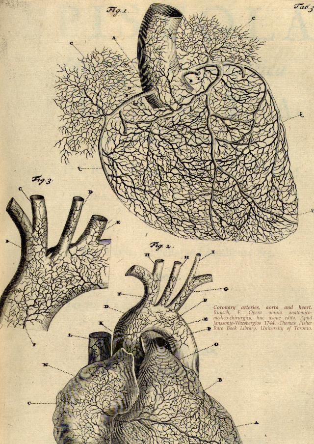
Coronary arteries, aorta and heart. Ruysch, F. Opera omnia anatomico-medico-chirurgica, huc usque edita. Apud Janssonio-Waesbergios 1744. Thomas Fisher Rare Book Library, University of Toronto.
Part II. Vascular (side) effects of antimigraine drugs
Chapter 3
Triptans and CGRP blockade – impact on the cranial vasculature
Based on: The Journal of Headache and Pain, 2017, 18: 103.
Benemei S*, Cortese F*, Labastida-Ramírez A*, Marchese F, Pellesi L, Romoli M, Vollesen AL, Lampl C, Ashina M and on behalf of the School of Advanced of the European Headache Federation. *Equal contributors.
Abstract
The trigeminovascular system plays a key role in the pathophysiology of migraine. The activation of this system causes release of various neurrotrasmitters and neuropeptides, including serotonin and calcitonin gene-related peptide (CGRP), which modulate nociceptive transmission and vascular tone. Thirty years after discovery of agonists for serotonin 5-HT1B
and 5-HT1D receptors (triptans) and less than fifteen after the proof-of-concept of the gepant class of CGRP receptor antagonists, we are still a long way from understanding their precise site and mode of action in migraine. The effect on cranial vasculature is relevant, because all specific antimigraine drugs and migraine pharmacological triggers may act in the perivascular space. This review reports the effects of triptans and CGRP blocking molecules on cranial vasculature in humans, focusing on their specific relevance to migraine treatment.
Background
Over the last century, controversies have raised around the vascular, neural or neurovascular origin of migraine [1]. From Galen original conjecture [2], with a meningeal involvement in the throbbing pain, several centuries passed before Willis, in 1672, hinted for the first time at a “vascular hypothesis” of migraine [3]. Throughout the 1930s and early 1940s headache science has emerged from studies by Graham, Ray and Wolff, who reported head pain after in vivo stimulation of dural and cerebral arteries, hypothesizing perivascular space as the possible site of migraine pain [4–7]. Pial, dural and extracranial vessels are part of the trigeminovascular system, a functional pathway that, on one side, releases vasoactive neuropeptides from perivascular nerve fibers and, on the other, reacts to them with nociception and vasodilation [8]. Pursuing the vascular hypothesis, several pharmacological triggers (such as glyceryl trinitrate (GTN), calcitonin gene-related peptide (CGRP) and pituitary adenylate cyclase-activating polypeptide-38 (PACAP38) were found to induce attacks phenotypically indistinguishable from spontaneous migraine in migraine patients [9–11]. The fact that all migraine-provoking molecules are vasoactive and sumatriptan constricts arteries [12, 13], further granted a key role of cranial vasculature in migraine pathophysiology [14]. Cranial arteries dilation has been shown, with different techniques, in both provocation and spontaneous migraine studies. Since the early 1990s, ultrasonography has been used to measure blood flow velocity in intracranial arteries [15] and extracranial artery diameter [16] during migraine attacks. Blood flow velocity correlates to vessel autoregulation and reactivity. Moreover, if cerebral blood flow does not change during an attack, blood flow velocity may be a surrogate marker of artery diameter (i.e. decreased blood flow velocity means increased middle cerebral artery lumen) [17]. In the last decade, investigation techniques have moved from ultrasonography to magnetic resonance angiography (MRA), allowing researchers to directly measure artery circumference [18–22]. MRA studies reported modest artery dilation during attacks, which was inhibited by triptans [12, 18, 21, 23]. Similar results, with prevention of superficial temporal artery dilation, were reported with the CGRP receptor antagonist olcegepant [24], hence suggesting that the modulation of cranial vasculature tone or perivascular nociception is of paramount importance in migraine treatment, too.
Chapter 3
28
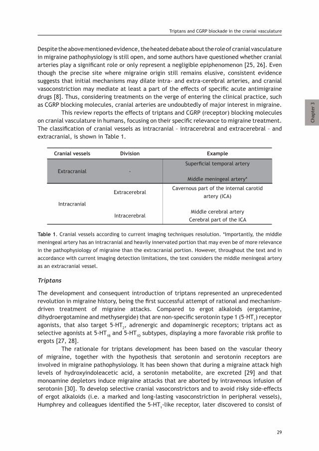
Chap
ter
3
Triptans and CGRP blockade in the cranial vasculature
29
Despite the above mentioned evidence, the heated debate about the role of cranial vasculature in migraine pathophysiology is still open, and some authors have questioned whether cranial arteries play a significant role or only represent a negligible epiphenomenon [25, 26]. Even though the precise site where migraine origin still remains elusive, consistent evidence suggests that initial mechanisms may dilate intra- and extra-cerebral arteries, and cranial vasoconstriction may mediate at least a part of the effects of specific acute antimigraine drugs [8]. Thus, considering treatments on the verge of entering the clinical practice, such as CGRP blocking molecules, cranial arteries are undoubtedly of major interest in migraine. This review reports the effects of triptans and CGRP (receptor) blocking molecules on cranial vasculature in humans, focusing on their specific relevance to migraine treatment. The classification of cranial vessels as intracranial – intracerebral and extracerebral – and extracranial, is shown in Table 1.
Cranial vessels Division Example
Extracranial -Superficial temporal artery
Middle meningeal artery*
Intracranial
Extracerebral
Intracerebral
Cavernous part of the internal carotid artery (ICA)
Middle cerebral artery Cerebral part of the ICA
Table 1. Cranial vessels according to current imaging techniques resolution. *Importantly, the middle meningeal artery has an intracranial and heavily innervated portion that may even be of more relevance in the pathophysiology of migraine than the extracranial portion. However, throughout the text and in accordance with current imaging detection limitations, the text considers the middle meningeal artery as an extracranial vessel.
Triptans
The development and consequent introduction of triptans represented an unprecedented revolution in migraine history, being the first successful attempt of rational and mechanism-driven treatment of migraine attacks. Compared to ergot alkaloids (ergotamine, dihydroergotamine and methysergide) that are non-specific serotonin type 1 (5-HT1) receptor agonists, that also target 5-HT2, adrenergic and dopaminergic receptors; triptans act as selective agonists at 5-HT1B and 5-HT1D subtypes, displaying a more favorable risk profile to ergots [27, 28]. The rationale for triptans development has been based on the vascular theory of migraine, together with the hypothesis that serotonin and serotonin receptors are involved in migraine pathophysiology. It has been shown that during a migraine attack high levels of hydroxyindoleacetic acid, a serotonin metabolite, are excreted [29] and that monoamine depletors induce migraine attacks that are aborted by intravenous infusion of serotonin [30]. To develop selective cranial vasoconstrictors and to avoid risky side-effects of ergot alkaloids (i.e. a marked and long-lasting vasoconstriction in peripheral vessels), Humphrey and colleagues identified the 5-HT1-like receptor, later discovered to consist of
30
Chapter 3
both the 5-HT1B and the 5-HT1D receptor subtypes, mostly located in cranial vessels, and then developed the first triptan, known as sumatriptan (GR43175) [31, 32]. Because of its efficacy and relative safety, sumatriptan has become a landmark in the treatment of migraine attacks [33]. Nevertheless, some peculiarities, such as the low oral bioavailability and short half-life [34], have favoured the development of new molecules, the so-called “second-generation” triptans (almotriptan, eletriptan, frovatriptan, naratriptan, rizatriptan), with an optimization of the pharmacokinetic profile [35]. Triptans are 5-HT1B/1D receptor agonists, most of them showing a moderate to high affinity for 5-HT1F receptors as well [35]. Immunohistochemical studies have shown that 5-HT1B receptors are mainly located within the smooth muscle and in the endothelium of human middle meningeal [36, 37] and cerebral [38] arteries. Importantly, in in vitro studies, triptans constrict these arteries [37–39]. The 5-HT1B receptors, together with the 5-HT1D
and 5-HT1F receptors, are also located within the trigeminal nerve endings and trigeminal nucleus, suggesting that their stimulation could inhibit the release of proinflammatory neuropeptides (e.g. CGRP) and consequently nociceptive transmission [40]. In a randomized placebo-controlled study, the administration of PNU142633, a selective 5-HT1D receptor agonist, failed to acutely alleviate migraine headache, suggesting a secondary role of the 5-HT1D receptors [41]. On the other side, selective non-vasoconstrictive 5-HT1F receptor agonists, LY334370 [42] and LY573144 (i.e. lasmiditan) [43] demonstrated clinical efficacy, even though it remains to be confirmed whether these molecules, at therapeutic concentrations, are devoid of any activity on 5-HT1B receptors. Interestingly, lasmiditan did not exert vasoactive effects in supratherapeutic concentrations [44]. Importantly, the high cranial (i.e. middle meningeal artery) 5-HT1B receptor density compared to peripheral (i.e. coronary artery) blood vessels probably renders the triptans relatively selective for producing cranial vasoconstriction [36, 45]. Human experimental data about vessel responses to triptans have refined our comprehension of triptan antimigraine effects and, indirectly, of the migraine mechanism. Differently from what observed in vitro [46], a placebo-controlled single-photon emission computed tomography (SPECT) study on healthy volunteers showed that sumatriptan infusion did not modify total and regional cerebral perfusion [47]. Interestingly, contrasting data in migraine patients have been initially reported about the correspondence between sumatriptan-related blood velocity modifications, measured by Doppler sonography, and the resolution of migraine attacks [12, 48, 49]. Importantly, combining the measurement of the regional cerebral blood flow and the blood velocity in the middle cerebral arteries, sumatriptan infusion has been shown to reverse the abnormal dilation of the middle cerebral artery in the headache side [12]. This finding suggests that the sumatriptan-induced vasoconstriction occurs only in the dilated vessels, without affecting normal ones.
Triptans and cranial vasculature
A key feature of migraine is that attacks can be provoked by pharmacological triggers, including glyceryl trinitrate (GTN) [50] and, as detailed below, provocation migraine models have provided important data on the role of cranial vasculature in migraine. The efficacy of triptans in GTN-induced headache was investigated in healthy volunteers in double-blind, placebo-controlled, crossover studies. Sumatriptan (6 mg) administered subcutaneously 20 min before GTN (0.12 μg/kg/min) infusion, relieved pain and decreased temporal artery diameter without affecting blood velocity of the middle cerebral artery (MCA) [51].
31
Triptans and CGRP blockade in the cranial vasculature
On the other hand, zolmitriptan (5 mg), administered orally during ongoing GTN infusion (0.2 μg/kg/min) had no effects on the induced-headache [52]. Oral triptans (rizatriptan 10 mg, sumatriptan 50 mg and zolmitriptan 2.5 mg) were also tested in migraine patients, in which they have been shown to both decrease diameter and increase resistance of temporal artery, although to a different extent [53]. More recently, the triptan effect was examined after the experimental administration of vasoactive neuropeptides such as CGRP, PACAP38 and VIP, in healthy volunteers and migraine patients. In a first study, 18 healthy volunteers were randomized to receive an intravenous infusion of human α-CGRP (1.5 μg/min) or placebo, for 20 min [18]. After 45 min, a single dose of subcutaneous sumatriptan (6 mg) was administered to each patient. A high-resolution MRA was performed at baseline, before and after the sumatriptan injection, to measure the changes in the circumference of the MCA and middle meningeal artery (MMA). Compared to placebo, CGRP caused significant dilation of MMA but not of MCA, and sumatriptan reduced the MMA circumference after CGRP pre-treatment by 25% and to a lesser extent on MCA, suggesting that sumatriptan exerts part of its antinociceptive effect primarily acting on the MMA. A second study was performed in 24 patients with migraine without aura [23], in which the CGRP intravenous infusion always resulted in a delayed headache, which fulfilled the criteria for migraine-like attacks in 18 patients (75%). MRA was performed in 15 of 18 patients, and of these, 10 (67%) patients reported unilateral head pain. MMA and MCA were dilated only on the painful side. The other 5 patients (33%) reported bilateral head pain, accompanied by a bilateral dilatation of both MMA and MCA. Sumatriptan subcutaneous injection reversed the dilatation of MMA and aborted the migraine attacks, without affecting the MCA circumference [23]. These data show that migraine is associated with dilatation of extracerebral and intracerebral arteries, but only the contraction of extracerebral arteries is associated with amelioration of headache. PACAP38 is a vasoactive neuropeptide that belongs to the secretin/glucagon/VIP family and it is used to provoke experimental headache and migraine [10]. PACAP38 is reported to cause delayed headache in healthy volunteers, associated with a significant and long-lasting dilatation of the MMA (up to 23%), but no change in the MCA circumference [19]. In comparison, sumatriptan induced a contraction of the MMA by 12.3% and reversed the delayed headache attack, but no effects on the MCA were observed. The role of PACAP38 was further investigated in a double-blind crossover study [22] conducted in 22 female migraine patients without aura. Sixteen patients (73%) after PACAP38 infusion, but only four patients (18%) after VIP infusion (8 pmol/kg/min), reported migraine-like attacks. Both peptides induced a marked dilatation of the extracranial arteries, but not of the intracranial arteries. The subcutaneous injection of sumatriptan reversed migraine attacks simultaneously to the constriction of the dilated extracerebral arteries, but not the intracerebral arteries. To date, only one MRA study has explicitly investigated cranial arteries during spontaneous migraine attacks [20]. Migraine attacks, with a median time from pain onset to scan of 5 h 45 min, are not accompanied by extracranial arterial dilation on the pain side, but only by slight dilation of intracerebral arteries, MCA and internal carotid (ICA). In addition, the dilatation of the intracerebral arteries persisted after subcutaneous injection of sumatriptan, which however relieved migraine pain and reduced the circumference of not-dilated extracranial arteries. These data suggested that the vasoconstrictor action of sumatriptan evident in extracranial arteries and in the cavernous portion of the ICA could
Chap
ter
3
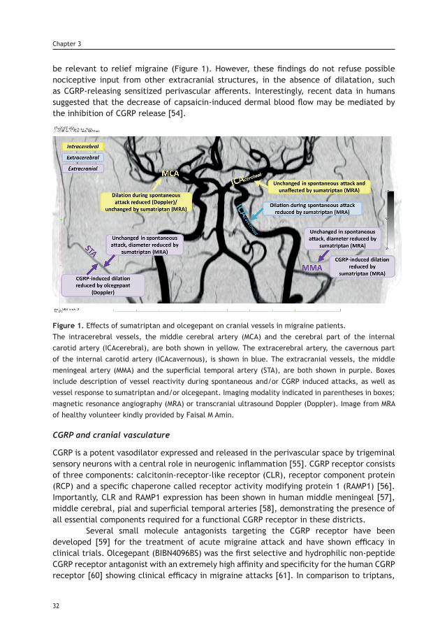
be relevant to relief migraine (Figure 1). However, these findings do not refuse possible nociceptive input from other extracranial structures, in the absence of dilatation, such as CGRP-releasing sensitized perivascular afferents. Interestingly, recent data in humans suggested that the decrease of capsaicin-induced dermal blood flow may be mediated by the inhibition of CGRP release [54].
Figure 1. Effects of sumatriptan and olcegepant on cranial vessels in migraine patients.The intracerebral vessels, the middle cerebral artery (MCA) and the cerebral part of the internal carotid artery (ICAcerebral), are both shown in yellow. The extracerebral artery, the cavernous part of the internal carotid artery (ICAcavernous), is shown in blue. The extracranial vessels, the middle meningeal artery (MMA) and the superficial temporal artery (STA), are both shown in purple. Boxes include description of vessel reactivity during spontaneous and/or CGRP induced attacks, as well as vessel response to sumatriptan and/or olcegepant. Imaging modality indicated in parentheses in boxes; magnetic resonance angiography (MRA) or transcranial ultrasound Doppler (Doppler). Image from MRA of healthy volunteer kindly provided by Faisal M Amin.
CGRP and cranial vasculature
CGRP is a potent vasodilator expressed and released in the perivascular space by trigeminal sensory neurons with a central role in neurogenic inflammation [55]. CGRP receptor consists of three components: calcitonin-receptor-like receptor (CLR), receptor component protein (RCP) and a specific chaperone called receptor activity modifying protein 1 (RAMP1) [56]. Importantly, CLR and RAMP1 expression has been shown in human middle meningeal [57], middle cerebral, pial and superficial temporal arteries [58], demonstrating the presence of all essential components required for a functional CGRP receptor in these districts. Several small molecule antagonists targeting the CGRP receptor have been developed [59] for the treatment of acute migraine attack and have shown efficacy in clinical trials. Olcegepant (BIBN4096BS) was the first selective and hydrophilic non-peptide CGRP receptor antagonist with an extremely high affinity and specificity for the human CGRP receptor [60] showing clinical efficacy in migraine attacks [61]. In comparison to triptans,
32
Chapter 3
Triptans and CGRP blockade in the cranial vasculature
33
which have been extensively studied in humans, most data on vascular effects of “gepants” come from preclinical studies and this has been previously reviewed [62]. Olcegepant, which inhibits dose-dependent relaxation of isolated human cerebral arteries [63], blocks MMA vasodilation following systemic administration of α-CGRP and β-CGRP, without significantly affecting pial artery dilation or the local cortical cerebral blood flow increase [64]. In contrast to the pial vessels, the meningeal arteries have no blood-brain barrier [65], suggesting that olcegepant likely acts outside of the blood-brain barrier [66]. In humans, olcegepant per se had no constrictor effect on the middle cerebral, radial, and superficial temporal artery [24], and no influence on global and regional cerebral blood flow [67]. Nevertheless, olcegepant effectively antagonizes the extracerebral vascular effect (e.g. temporal artery dilation) induced by CGRP intravascular administration [24]. A series of orally bioavailable small molecule CGRP receptor antagonists, including MK-0974 (telcagepant), have been then developed giving rise to the pharmacological class of “gepants”. Telcagepant has been shown to be able to abort CGRP-induced vasodilatation on human cerebral and meningeal arteries ex vivo [68]. However, notwithstanding efficacy in clinical trials [69], clinical development of early gepants has been discontinued [70], and accordingly their use in migraine models has been withdrawn. There are currently four monoclonal antibodies (mAb) in clinical development for migraine prophylaxis: three humanized mAb targeting CGRP (LY2951742/galcanezumab, Eli Lilly and Company; ALD403/eptinezumab, Alder Biopharmaceuticals; and TEV-48215/fremanezumab, TEVA Pharmaceuticals) and one fully human mAb targeting the CGRP receptor (AMG 334/erenumab, Amgen). These biological drugs have shown efficacy, tolerability and few adverse effects in phase 2 randomized control trials [71–76]. However, their exact site and mechanism of action is not completely understood. The new CGRP mAbs are macromolecules (around 150,000 Da) that are unlikely to cross the blood-brain barrier [66]. In line with this, few preclinical studies revealed that humanized CGRP mAb are (i) unable to penetrate the blood-brain barrier in the perfused MCA [77]; (ii) ineffective in inhibiting the responses to CGRP-induced neurogenic vasodilation of the pial artery [78]; and (iii) capable of inhibiting CGRP-induced neurogenic vasodilation of the MMA, which as mentioned above lacks blood-brain barrier [65, 79]. Importantly, it has been recently shown that there is no blood-brain barrier disruption during migraine attacks [80]. All these findings taken into account suggest a peripheral vascular site of action of the mAbs. A similar alternative under development for the preventive treatment of migraine is blocking CGRP-induced receptor activation through a RNA-Spiegelmer (NOX-C89). This single-strand mirror-image oligonucleotide binds to circulating CGRP and is highly resistant to endogenous nuclease degradation, hence inhibiting its function. Interestingly, this drug could not inhibit neurogenic vasodilation of pial arteries in vivo, which suggested that it is unlikely to penetrate the blood-brain barrier readily [78]. Targeting peripheral CGRP may reduce or prevent the phenomenon (i.e. vasodilation) that has been advocated as a mechanism of headache and associated symptoms, whereas long-term inhibition of CGRP outside of the blood-brain barrier induces modulation of central pathways remains unknown. Further studies are needed to fully clarify the exact antimigraine site of action of the CGRP mAbs and NOX-C89.
Chap
ter
3
34
Chapter 3
Conclusions
From Galen’s quote about meninges and vessels as mediators, together with other structures and mechanisms of migraine pain, long time has passed. In the last years, notwithstanding many detractors, cranial vasculature involvement in the pathogenesis of migraine pain has benefited from experimental data acquired by modern imaging techniques, such as MRA, and specific pharmacological tools, such as triptans. Recently developed human migraine models have suggested that attention should be paid to cranial extracerebral arteries (i.e. MMA) in addition to intracerebral vessels, which were the major focus at the dawn of vascular migraine research. On the basis of current knowledge [20, 23], future studies should investigate whether there are differences in how the perivascular nerves innervate the different sections of the MMA, including dural branches, because it is likely that it is activation or inhibition of the perivascular nerves that is associated with migraine pain relief. In addition, future advanced brain imaging methods will allow to investigate possible dilatation of dural branches of the MMA that are very difficult to visualise with current method, without injection of contrast agents [20]. The availability of innovative migraine-specific drugs, such as CGRP-targeted compounds [69], will further increase our ability to investigate the involvement of cranial vasculature in migraine pain, and will finally allow to properly balance the weight of vessel contribution to the neurovascular theory of migraine.
References1. Messlinger K. Migraine: where and how does the pain originate? Exp Brain Res. 2009; 196: 179–193. doi: 10.1007/s00221-009-1756-y.2. Isler H. The Galenic tradition and migraine. J Hist Neurosci. 1992; 1: 227–233. doi: 10.1080/09647049209525535.3. Willis T (1672) Two Discourses concerning the soul of brutes (De anima brutorum). Part 14. Graham JR, Wolff HG. Mechanism of migraine headache and action of ergotamine tartrate. Arch Neurol Psychiatr. 1938; 39: 737–763. doi: 10.1001/archneurpsyc.1938.02270040093005. 5. Wolff HG. Headache and cranial arteries. Trans Assoc Am Phys. 1938; 53: 193–198.6. Ray BS, Wolff HG. Experimental studies on headache: Pain-sensitive structures of the head and their significance in headache. Arch Surg. 1940; 41: 813–856. doi: 10.1001/archsurg.1940.01210040002001.7. Wolff HG, Tunis MM, Goodell H. Studies on headache: evidence of tissue damage and changes in pain sensitivity in subjects with vascular headaches of the migraine type. Trans Assoc Am Phys. 1953; 66: 332–341. 8. Olesen J, Burstein R, Ashina M, et al. Origin of pain in migraine: evidence for peripheral sensitisation. Lancet Neu-rol. 2009; 8: 679–690. doi: 10.1016/S1474-4422(09)70090-0. 9. Thomsen LL, Kruuse C, Iversen HK, et al. A nitric oxide donor (nitroglycerin) triggers genuine migraine attacks. Eur J Neurol. 1994; 1: 73–80. doi: 10.1111/j.1468-1331.1994.tb00053.x. 10. Schytz HW, Birk S, Wienecke T, et al. PACAP38 induces migraine-like attacks in patients with migraine without aura. Brain. 2009; 132: 16–25. doi: 10.1093/brain/awn307.11. Hansen JM, Hauge AW, Olesen J, et al. Calcitonin gene-related peptide triggers migraine-like attacks in patients with migraine with aura. Cephalalgia. 2010; 30: 1179–1186. doi: 10.1177/0333102410368444. 12. Thomaides T, Karagounakis D, Spantideas A, et al. Transcranial Doppler in migraine attacks before and after treatment with oral zolmitriptan or sumatriptan. Headache. 2003; 43: 54–58. doi: 10.1046/j.1526-4610.2003.03009.x. 13. Chan KY, Vermeersch S, de Hoon J, et al. Potential mechanisms of prospective antimigraine drugs: a focus on vascular (side) effects. Pharmacol Ther. 2011; 129: 332–351. doi: 10.1016/j.pharmthera.2010.12.001. 14. Thomsen LL, Iversen HK, Olesen J. Cerebral blood flow velocities are reduced during attacks of unilateral migraine without aura. Cephalalgia. 1995; 15: 109–116. doi: 10.1046/j.1468-2982.1995.015002109.x.15. Iversen HK, Nielsen TH, Olesen J, et al. Arterial responses during migraine headache. Lancet. 1990; 336: 837–839. doi: 10.1016/0140-6736(90)92339-J. 16. Markwalder TM, Grolimund P, Seiler RW, et al. Dependency of blood flow velocity in the middle cerebral artery on end-tidal carbon dioxide partial pressure--a transcranial ultrasound Doppler study. J Cereb Blood Flow Metab. 1984; 4: 368–372. doi: 10.1038/jcbfm.1984.54. 17. Asghar MS, Hansen AE, Kapijimpanga T, et al. Dilation by CGRP of middle meningeal artery and reversal by su-matriptan in normal volunteers. Neurology. 2010; 75: 1520–1526. doi: 10.1212/WNL.0b013e3181f9626a.
Triptans and CGRP blockade in the cranial vasculature
35
18. Amin FM, Asghar MS, Guo S, et al. Headache and prolonged dilatation of the middle meningeal artery by PACAP38 in healthy volunteers. Cephalalgia. 2012; 32: 140–149. doi: 10.1177/0333102411431333. 19. Amin FM, Asghar MS, Hougaard A, et al. Magnetic resonance angiography of intracranial and extracranial arter-ies in patients with spontaneous migraine without aura: a cross-sectional study. Lancet Neurol. 2013; 12: 454–461. doi: 10.1016/S1474-4422(13)70067-X. 20. Amin FM, Asghar MS, Ravneberg JW, et al. The effect of sumatriptan on cephalic arteries: A 3T MR-angiography study in healthy volunteers. Cephalalgia. 2013; 33: 1009–1016. doi: 10.1177/0333102413483374.21. Amin FM, Hougaard A, Schytz HW, et al. Investigation of the pathophysiological mechanisms of migraine attacks induced by pituitary adenylate cyclase-activating polypeptide-38. Brain. 2014; 137: 779–794. doi: 10.1093/brain/awt369. 22. Asghar MS, Hansen AE, Amin FM, et al. Evidence for a vascular factor in migraine. Ann Neurol. 2011; 69: 635–645. doi: 10.1002/ana.22292. 23. Petersen KA, Lassen LH, Birk S, et al. BIBN4096BS antagonizes human alpha-calcitonin gene relat-ed peptide-induced headache and extracerebral artery dilatation. Clin Pharmacol Ther. 2005; 77: 202–213. doi: 10.1016/j.clpt.2004.10.001. 24. Goadsby PJ, Holland PR, Martins-Oliveira M, et al. Pathophysiology of Migraine: A Disorder of Sensory Processing. Physiol Rev. 2017; 97: 553–622. doi: 10.1152/physrev.00034.2015. 25. Diener HC, Jansen JP, Reches A, et al. Efficacy, tolerability and safety of oral eletriptan and ergotamine plus caffeine (Cafergot) in the acute treatment of migraine: a multicentre, randomised, double-blind, placebo-controlled comparison. Eur Neurol. 2002; 47: 99–107. doi: 10.1159/000047960. 26. Sicuteri F, Testi A, Anselmi B. Biochemical Investigations in Headache: Increase in the Hydroxyindoleacetic Acid Excretion During Migraine Attacks. Int Arch Allergy. 1961 ;19: 55–58. doi: 10.1159/000229194. 27. Kimball RW, Friedman AP, Vallejo E. Effect of serotonin in migraine patients. Neurology. 1960;10:107–111. doi: 10.1212/WNL.10.2.107. 28. Humphrey PP, Feniuk W, Perren MJ, et al. The pharmacology of the novel 5-HT1-like receptor agonist, GR43175. Cephalalgia. 1989; 9(Suppl 9): 23–33. 29. Doenicke A, Brand J, Perrin VL. Possible benefit of GR43175, a novel 5-HT1-like receptor agonist, for the acute treatment of severe migraine. Lancet. 1988; 1: 1309–1311. doi: 10.1016/S0140-6736(88)92122-8.30. Humphrey PP. The discovery and development of the triptans, a major therapeutic breakthrough. Headache. 2008; 48: 685–687. doi: 10.1111/j.1526-4610.2008.01097.x. 31. Fowler PA, Lacey LF, Thomas M, et al. The clinical pharmacology, pharmacokinetics and metabolism of sumatriptan. Eur Neurol. 1991; 31: 291–294. doi: 10.1159/000116756.32. Tfelt-Hansen P, De Vries P, Saxena PR. Triptans in migraine: a comparative review of pharmacology, pharmacoki-netics and efficacy. Drugs. 2000; 60: 1259–1287. doi: 10.2165/00003495-200060060-00003.33. Razzaque Z, Pickard JD, Ma QP, et al. 5-HT1B-receptors and vascular reactivity in human isolated blood ves-sels: assessment of the potential craniovascular selectivity of sumatriptan. Br J Clin Pharmacol. 2002; 53: 266–274. doi: 10.1046/j.0306-5251.2001.01536.x.34. Nilsson T, Longmore J, Shaw D, et al. Contractile 5-HT1B receptors in human cerebral arteries: pharma-cological characterization and localization with immunocytochemistry. Br J Pharmacol. 1999; 128: 1133–1140. doi: 10.1038/sj.bjp.0702773. 35. van den Broek RW, Bhalla P, VanDenBrink AM, et al. Characterization of sumatriptan-induced contractions in human isolated blood vessels using selective 5-HT(1B) and 5-HT(1D) receptor antagonists and in situ hybridization. Cepha-lalgia. 2002; 22: 83–93. doi: 10.1046/j.1468-2982.2002.00295.x. 36. Buzzi MG, Carter WB, Shimizu T, et al. Dihydroergotamine and sumatriptan attenuate levels of CGRP in plasma in rat superior sagittal sinus during electrical stimulation of the trigeminal ganglion. Neuropharmacology. 1991; 30: 1193–1200. doi: 10.1016/0028-3908(91)90165-8.37. Gomez-Mancilla B, Cutler NR, Leibowitz MT, et al. Safety and efficacy of PNU-142633, a selective 5-HT1D agonist, in patients with acute migraine. Cephalalgia. 2001; 21: 727–732. doi: 10.1046/j.1468-2982.2001.00208.x.38. Goldstein DJ, Roon KI, Offen WW, et al. Selective serotonin 1F (5-HT(1F)) receptor agonist LY334370 for acute migraine: a randomised controlled trial. Lancet. 2001; 358: 1230–1234. doi: 10.1016/S0140-6736(01)06347-4. 39. Ferrari MD, Farkkila M, Reuter U, et al. Acute treatment of migraine with the selective 5-HT1F receptor agonist lasmiditan--a randomised proof-of-concept trial. Cephalalgia. 2010; 30: 1170–1178. doi: 10.1177/0333102410375512.40. Rubio Beltran E, Kristian H, Labastida A, et al. Lasmiditan and sumatriptan: comparison of in vivo vascular con-striction in the dog and in vitro contraction of human arteries. Cephalalgia. 2016; 36: 104–105.41. MaassenVanDenBrink A, van den Broek RW, de Vries R, et al. Craniovascular selectivity of eletriptan and su-matriptan in human isolated blood vessels. Neurology. 2000; 55: 1524–1530. doi: 10.1212/WNL.55.10.1524. 42. Feniuk W, Humphrey PP, Perren MJ. The selective carotid arterial vasoconstrictor action of GR43175 in anaesthe-tized dogs. Br J Pharmacol. 1989; 96: 83–90. doi: 10.1111/j.1476-5381.1989.tb11787.x.43. Scott AK, Grimes S, Ng K, et al. Sumatriptan and cerebral perfusion in healthy volunteers. Br J Clin Pharmacol. 1992; 33: 401–404. doi: 10.1111/j.1365-2125.1992.tb04058.x.
Chap
ter
3
36
Chapter 3
44. Friberg L, Olesen J, Iversen HK, et al. Migraine pain associated with middle cerebral artery dilatation: reversal by sumatriptan. Lancet. 1991; 338: 13–17. doi: 10.1016/0140-6736(91)90005-A.45. Ashina M, Hansen JM, Olesen J. Pearls and pitfalls in human pharmacological models of migraine: 30 years’ expe-rience. Cephalalgia. 2013; 33: 540–553. doi: 10.1177/0333102412475234. 46. Iversen HK, Olesen J. Headache induced by a nitric oxide donor (nitroglycerin) responds to sumatriptan. A human model for development of migraine drugs. Cephalalgia. 1996; 16: 412–418. doi: 10.1046/j.1468-2982.1996.1606412.x. 47. Tvedskov JF, Iversen HK, Olesen J, et al. Nitroglycerin provocation in normal subjects is not a useful human mi-graine model? Cephalalgia. 2010; 30: 928–932. doi: 10.1111/j.1468-2982.2009.02014.x. 48. de Hoon JN, Willigers JM, Troost J, et al. Vascular effects of 5-HT1B/1D-receptor agonists in patients with migraine headaches. Clin Pharmacol Ther. 2000; 68: 418–426. doi: 10.1067/mcp.2000.110502. 49. Ibrahimi K, Danser A, Terwindt GM, et al. A human trigeminovascular biomarker for antimigraine drugs: A ran-domised, double-blind, placebo-controlled, crossover trial with sumatriptan. Cephalalgia. 2017; 37: 94–98. doi: 10.1177/0333102416637833. 50. Durham PL. Calcitonin gene-related peptide (CGRP) and migraine. Headache. 2006; 46(Suppl 1): S3–S8. doi: 10.1111/j.1526-4610.2006.00483.x.51. Hay DL, Walker CS. CGRP and its receptors. Headache. 2017; 57: 625–636. doi: 10.1111/head.13064.52. Gupta S, Mehrotra S, Avezaat CJ, et al. Characterisation of CGRP receptors in the human isolated middle menin-geal artery. Life Sci. 2006; 79: 265–271. doi: 10.1016/j.lfs.2006.01.003. 53. Oliver KR, Wainwright A, Edvinsson L, et al. Immunohistochemical localization of calcitonin receptor-like receptor and receptor activity-modifying proteins in the human cerebral vasculature. J Cereb Blood Flow Metab. 2002; 22: 620–629. doi: 10.1097/00004647-200205000-00014.54. Moore EL, Salvatore CA. Targeting a family B GPCR/RAMP receptor complex: CGRP receptor antagonists and mi-graine. Br J Pharmacol. 2012; 166: 66–78. doi: 10.1111/j.1476-5381.2011.01633.x55. Doods H, Hallermayer G, Wu D, et al. Pharmacological profile of BIBN4096BS, the first selective small molecule CGRP antagonist. Br J Pharmacol. 2000; 129: 420–423. doi: 10.1038/sj.bjp.0703110. 56. Olesen J, Diener HC, Husstedt IW, et al. Calcitonin gene-related peptide receptor antagonist BIBN 4096 BS for the acute treatment of migraine. N Engl J Med. 2004; 350: 1104–1110. doi: 10.1056/NEJMoa030505. 57. Villalón CM, Olesen J. The role of CGRP in the pathophysiology of migraine and efficacy of CGRP receptor antag-onists as acute antimigraine drugs. Pharmacol Ther. 2009; 124: 309–323. doi: 10.1016/j.pharmthera.2009.09.003. 58. Edvinsson L, Alm R, Shaw D, et al. Effect of the CGRP receptor antagonist BIBN4096BS in human cerebral, coronary and omental arteries and in SK-N-MC cells. Eur J Pharmacol. 2002; 434: 49–53. doi: 10.1016/S0014-2999(01)01532-1. 59. Petersen KA, Birk S, Doods H, et al. Inhibitory effect of BIBN4096BS on cephalic vasodilatation induced by CGRP or transcranial electrical stimulation in the rat. Br J Pharmacol. 2004; 143: 697–704. doi: 10.1038/sj.bjp.0705966. 60. Edvinsson L. CGRP blockers in migraine therapy: where do they act? Br J Pharmacol. 2008; 155: 967–969. doi: 10.1038/bjp.2008.346. 61. Edvinsson L. CGRP receptor antagonists and antibodies against CGRP and its receptor in migraine treatment. Br J Clin Pharmacol. 2015; 80: 193–199. doi: 10.1111/bcp.12618. 62. Petersen KA, Birk S, Lassen LH, et al. The CGRP-antagonist, BIBN4096BS does not affect cerebral or systemic hae-modynamics in healthy volunteers. Cephalalgia. 2005; 25: 139–147. doi: 10.1111/j.1468-2982.2004.00830.x. 63. Edvinsson L, Chan KY, Eftekhari S, et al. Effect of the calcitonin gene-related peptide (CGRP) receptor antagonist telcagepant in human cranial arteries. Cephalalgia. 2010 ;30:1233–1240. doi: 10.1177/0333102410362122.64. Ho TW, Ferrari MD, Dodick DW, et al. Efficacy and tolerability of MK-0974 (telcagepant), a new oral antagonist of calcitonin gene-related peptide receptor, compared with zolmitriptan for acute migraine: a randomised, placebo-con-trolled, parallel-treatment trial. Lancet. 2008; 372: 2115–2123. doi: 10.1016/S0140-6736(08)61626-8. 65. Schuster NM, Rapoport AM. New strategies for the treatment and prevention of primary headache disorders. Nat Rev Neurol. 2016; 12: 635–650. doi: 10.1038/nrneurol.2016.143. 66. Dodick DW, Goadsby PJ, Silberstein SD, et al. Safety and efficacy of ALD403, an antibody to calcitonin gene-related peptide, for the prevention of frequent episodic migraine: a randomised, double-blind, placebo-controlled, explora-tory phase 2 trial. Lancet Neurol. 2014; 13: 1100–1107. doi: 10.1016/S1474-4422(14)70209-1. 67. Dodick DW, Goadsby PJ, Spierings EL, et al. Safety and efficacy of LY2951742, a monoclonal antibody to calcitonin gene-related peptide, for the prevention of migraine: a phase 2, randomised, double-blind, placebo-controlled study. Lancet Neurol. 2014; 13: 885–892. doi: 10.1016/S1474-4422(14)70128-0. 68. Bigal ME, Dodick DW, Rapoport AM, et al. Safety, tolerability, and efficacy of TEV-48125 for preventive treatment of high-frequency episodic migraine: a multicentre, randomised, double-blind, placebo-controlled, phase 2b study. Lancet Neurol. 2015; 14: 1081–1090. doi: 10.1016/S1474-4422(15)00249-5. 69. Bigal ME, Edvinsson L, Rapoport AM, et al. Safety, tolerability, and efficacy of TEV-48125 for preventive treatment of chronic migraine: a multicentre, randomised, double-blind, placebo-controlled, phase 2b study. Lancet Neurol. 2015; 14: 1091–1100. doi: 10.1016/S1474-4422(15)00245-8. 70. Sun H, Dodick DW, Silberstein S, et al. Safety and efficacy of AMG 334 for prevention of episodic mi-graine: a randomised, double-blind, placebo-controlled, phase 2 trial. Lancet Neurol. 2016; 15: 382–390. doi: 10.1016/S1474-4422(16)00019-3.
Triptans and CGRP blockade in the cranial vasculature
37
71. Tepper S, Ashina M, Reuter U, et al. Safety and efficacy of erenumab for preventive treatment of chronic migraine: a randomised, double-blind, placebo-controlled phase 2 trial. Lancet Neurol. 2017; 16: 425–434. doi: 10.1016/S1474-4422(17)30083-2. 72. Edvinsson L, Nilsson E, Jansen-Olesen I. Inhibitory effect of BIBN4096BS, CGRP(8-37), a CGRP antibody and an RNA-Spiegelmer on CGRP induced vasodilatation in the perfused and non-perfused rat middle cerebral artery. Br J Pharmacol. 2007; 150: 633–640. doi: 10.1038/sj.bjp.0707134. 73. Juhl L, Edvinsson L, Olesen J, et al. Effect of two novel CGRP-binding compounds in a closed cranial window rat model. Eur J Pharmacol. 2007; 567: 117–124. doi: 10.1016/j.ejphar.2007.04.004.74. Hougaard A, Amin FM, Christensen CE, et al. Increased brainstem perfusion, but no blood-brain barrier disruption, during attacks of migraine with aura. Brain 2007. 140: 1633–1642 75. Goadsby PJ. The vascular theory of migraine--a great story wrecked by the facts. Brain. 2009; 132: 6–7. doi: 10.1093/brain/awn321. 76. The Multinational Oral Sumatriptan and Cafergot Comparative Study Group A randomized, double-blind compari-son of sumatriptan and Cafergot in the acute treatment of migraine. The Multinational Oral Sumatriptan and Cafergot Comparative Study Group. Eur Neurol. 1991; 31: 314–322. 77. Longmore J, Razzaque Z, Shaw D, et al. Comparison of the vasoconstrictor effects of rizatriptan and sumatriptan in human isolated cranial arteries: immunohistological demonstration of the involvement of 5-HT1B-receptors. Br J Clin Pharmacol. 1998; 46: 577–582. doi: 10.1046/j.1365-2125.1998.00821.x. 78. Caekebeke JF, Ferrari MD, Zwetsloot CP, et al. Antimigraine drug sumatriptan increases blood flow velocity in large cerebral arteries during migraine attacks. Neurology. 1992; 42: 1522–1526. doi: 10.1212/WNL.42.8.1522. 79. Limmroth V, May A, Auerbach P, et al. Changes in cerebral blood flow velocity after treatment with sumatriptan or placebo and implications for the pathophysiology of migraine. J Neurol Sci. 1996; 138: 60–65. doi: 10.1016/0022-510X(95)00344-2. 80. Zeller J, Poulsen KT, Sutton JE, et a;. CGRP function-blocking antibodies inhibit neurogenic vasodilatation with-out affecting heart rate or arterial blood pressure in the rat. Br J Pharmacol. 2008; 155: 1093–1103. doi: 10.1038/bjp.2008.334.
Chap
ter
3

Chapter 4
Pharmacological analysis of the increases in heart rate and diastolic blood pressure produced by (S)-isometheptene
and (R)-isometheptene in pithed rats
Based on: The Journal of Headache and Pain, 2017, 18:52.
Labastida-Ramírez A, Rubio-Beltrán E, Hernández-Abreu O, Daugherty B,
MaassenVanDenBrink A and Villalón CM.
Abstract
Background: Isometheptene is a sympathomimetic drug effective in acute migraine treatment. It is composed of two enantiomers with diverse pharmacological properties. This study investigated in pithed rats the cardiovascular effects of (S)-isometheptene and (R)-isometheptene, and the pharmacological profile of the more potent enantiomer.
Methods: The effects of i.v. bolus injections (0.03, 0.1, 0.3, 1 and 3 mg/kg) of isometheptene racemate, (S)-isometheptene or (R)-isometheptene on heart rate and blood pressure were analysed in control experiments. The enantiomer producing more pronounced tachycardic and/or vasopressor responses was further analysed in rats receiving i.v. injections of prazosin (0.1 mg/kg), rauwolscine (0.3 mg/kg), propranolol (1 mg/kg) or intraperitoneal reserpine (5 mg/kg, -24 hr).
Results: Compared to (R)-isometheptene, (S)-isometheptene produced greater vasopressor responses, whilst both compounds equipotently increased heart rate. The tachycardic responses to (S)-isometheptene were abolished after propranolol, but remained unaffected by the other antagonists. In contrast, the vasopressor responses to (S)-isometheptene were practically abolished after prazosin. Interestingly, after reserpine, the tachycardic responses to (S)-isometheptene were abolished, whereas its vasopressor responses were attenuated and subsequently abolished by prazosin.
Conclusion: The different cardiovascular effects of the isometheptene enantiomers are probably due to differences in their mechanism of action, namely: (i) a mixed sympathomimetic action for (S)-isometheptene (a tyramine-like action and a direct stimulation of α1-adrenoceptors); and (ii) exclusively a tyramine like action for (R)-isometheptene. Thus, (R)-isometheptene may represent a superior therapeutic benefit as an antimigraine agent.
Background
Isometheptene racemate is a sympathomimetic amine that has been used for decades in the acute treatment of moderate primary headaches (i.e. migraine and tension-type headaches) [1–2], but its exact mechanisms of action have not been comprehensively explored. In this respect, our group has demonstrated in pithed rats [3] and anesthetized dogs [4] that isometheptene racemate behaves as a dual-acting sympathomimetic agent producing: (i) an indirect vasoconstrictor tyramine-like action (attenuated by reserpine) that involves the displacement of endogenous noradrenaline from sympathetic nerves; and (ii) a direct vasoconstriction that results in either rat vasopressor responses mediated by α1-adrenoceptors [3] or decreases in canine external carotid blood flow/conductance mediated by α2A/2C-adrenoceptors [4]. These properties have linked isometheptene’s therapeutic effectiveness with the vascular theory of migraine [5], so that its cranial vasoconstriction may reduce the phenomenon (i.e. vasodilatation) that has been advocated as a mechanism of headache and associated symptoms [6]. Interestingly, sumatriptan (a 5-HT1B/1D receptor agonist with acute antimigraine properties) seems to exert part of its antinociceptive action by constricting the human middle meningeal artery (MMA) [7, 8]. In contrast, isometheptene racemate failed to constrict the porcine MMA [9], but the fact that it produces arterial vasospasm [10-11]
40
Chapter 4
Chap
ter
4
supports its vasoactive action on dilated cranial and cerebral arterioles [12,13]. Tonix Pharmaceuticals has separated isometheptene racemate into its two enantiomers (R)-isometheptene and (S)-isometheptene, in order to explore their efficacy in the acute treatment of primary headaches [14]. Isometheptene racemate binds with high affinity (Ki=42 nM) to the I1-imidazoline receptor (I1-R), suggesting that this receptor, along with α-adrenoceptors, could be involved in its antimigraine action [15]. Moreover, (R)-isometheptene binds to the I1-R (Ki=18 nM) with about 60-fold greater affinity than (S)-isometheptene (Ki=1100 nM) [15]. Notably, other studies have described: (i) a decreased pain threshold in I1-R knockout mice [16]; and (ii) that treatment with (R)-isometheptene relieved trigeminal sensitivity in the inflammatory soup and in spontaneous trigeminal allodynia rat models, two different models of chronic migraine [17]. This study investigated the cardiovascular effects produced by these novel compounds in an experimental model predictive of systemic (cardio) vascular side-effects.
Materials and Methods
Animals
Seventy-four male normotensive Wistar rats (250-300 g, 8-10 weeks of age) were maintained at a 12/12-hr light-dark cycle (with light beginning at 07:00 hr) and housed in a special room at a constant temperature (22±2°C) and humidity (50%), with food and water ad libitum in their cages. All animal protocols of this investigation were approved by our Institutional Ethics Committee (CICUAL-Cinvestav; permission protocol number 507-12) and followed the regulations established by the Mexican Official Norm (NOM-062-ZOO-1999), in accordance with ARRIVE (Animal Research: Reporting In Vivo Experiments) reporting guidelines for the care and use of laboratory animals.
General Methods
After anaesthesia with sodium pentobarbital (60 mg/kg, i.p.) and cannulation of the trachea, all rats were pithed by inserting a stainless steel rod through the orbit and foramen magnum and down the vertebral foramen, as previously reported [18,19]. The animals were then artificially ventilated with room air using a model 7025 Ugo Basile pump (56 strokes/min.; stroke volume: 20 ml/kg), as previously established [20]. After cervical bilateral vagotomy, catheters were placed in: (i) the right femoral vein, for the administration of antagonists, isometheptene racemate, (R)-isometheptene or (S)-isometheptene; and (ii) the left carotid artery, connected to a Grass pressure transducer (P23XL), for the recording of arterial blood pressure. Heart rate was measured with a tachograph (7P4F, Grass Instrument Co., Quincy, MA, U.S.A.) triggered from the blood pressure signal. Both heart rate and blood pressure were recorded simultaneously by a model 7D Grass polygraph (Grass Instrument Co., Quincy, MA, U.S.A.). The body temperature of each pithed rat was maintained at 37°C by a lamp and monitored with a rectal thermometer.
Experimental protocols
After a stable hemodynamic condition during at least 30 min, baseline values of heart rate and diastolic blood pressure (a more accurate indicator of peripheral vascular resistance than mean blood pressure) were determined. Subsequently, the 74 rats were randomly assigned into three main sets (n=24, 40 and 10) for performing the following dose-response
41
Cardiovascular (side) effects of isometheptene
protocols. We a priori decided to investigate the pharmacological characteristics of the responses to the most potent enantiomer in more detail, since this enantiomer would most likely be responsible for the side-effects induced by isometheptene. In addition, we planned to study for the least potent enantiomer to which extent the cardiovascular responses were mediated by a direct or indirect, tyramine-like, mechanism of action.
Protocol I. Comparative analysis of the effects produced by the isometheptene enantiomers
The first set of animals (n=24) was divided into four groups (n=6 each) that received consecutive i.v. bolus injections (0.03, 0.1, 0.3, 1 and 3 mg/kg) of, respectively: (i) isometheptene racemate; (ii) (S)-isometheptene; (iii) (R)-isometheptene; and (iv) equivalent volumes of physiological saline (1 ml/kg given 5 consecutive times; vehicle for dissolving isometheptene). The peak effects produced by each dose of the above compounds on heart rate and diastolic blood pressure were measured and compared, as previously reported [3]. The dose-intervals between the different doses of isometheptene (racemate and enantiomers) ranged between 5 and 15 min, as in each case we waited with the administration of the next dose until heart rate and diastolic blood pressure had returned to baseline values.
Protocol II. Analysis of the pharmacological profile of the more potent enantiomer
The second set of animals (n=40) was divided into three subsets (n=25, 10 and 5). The first subset (untreated; n=25) received consecutive i.v. bolus injections of the more potent isometheptene enantiomer (0.03, 0.1, 0.3, 1 and 3 mg/kg). Then, this subset was divided into five groups (n=5 each) that received i.v. bolus injections of, respectively: (i) physiological saline (1 ml/kg; vehicle for dissolving the antagonists); (ii) prazosin (0.1 mg⁄kg; α1-adrenoceptor antagonist); (iii) rauwolscine (0.3 mg/kg; α2-adrenoceptor antagonist); (iv) the combination of prazosin (0.1 mg/kg) plus rauwolscine (0.3 mg/kg); and (v) propranolol (1 mg/kg; nonselective β-adrenoceptor antagonist). The doses of the antagonists used were sufficient to completely block their corresponding receptors, as previously reported [3]. Subsequently, the responses to the above doses of the isometheptene enantiomer were elicited again 15 min after administration of the aforementioned compounds. The second subset (reserpinized; n=10) was pretreated with reserpine (5 mg⁄kg, i.p.) 24 hr before the start of the experiments, as previously reported by our group [3], in order to eliminate the potential tyramine-like action exerted by the more potent isometheptene enantiomer. Then, this subset received sequential i.v. bolus injections of the isometheptene enantiomer (0.03-3 mg/kg) and was subsequently divided into two groups (n=5 each) that were given i.v. bolus injections of, respectively: (i) physiological saline (1 ml/kg); and (ii) prazosin (0.1 mg/kg). After 15 min, the responses to the above isometheptene enantiomer doses were elicited again. The third subset (sham; n=5) was pretreated with the vehicle of reserpine (1 ml⁄kg of 5% ascorbic acid; i.p.) 24 hr before the start of the experiments. Then, this subset received i.v. bolus injections of the isometheptene enantiomer as previously described.
Protocol III. Effects of reserpine pretreatment on the effects of the less potent enantiomer
Finally, the third set of animals (n=10) was divided into two groups (n=5 each) that were pretreated i.p. (24 hr before the start of the experiments) with, respectively: (i) 1 ml/kg
42
Chapter 4
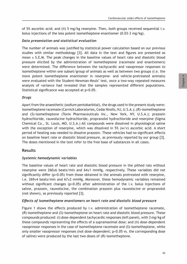
of 5% ascorbic acid; and (ii) 5 mg/kg reserpine. Then, both groups received sequential i.v. bolus injections of the less potent isometheptene enantiomer (0.03-3 mg/kg).
Data presentation and statistical evaluation
The number of animals was justified by statistical power calculation based on our previous studies with similar methodology [3]. All data in the text and figures are presented as mean ± S.E.M. The peak changes in the baseline values of heart rate and diastolic blood pressure elicited by the administration of isometheptene (racemate and enantiomers) were determined. The difference between the tachycardic and vasopressor responses to isometheptene within one subset/group of animals as well as between two groups (i.e. the more potent isometheptene enantiomer in reserpine- and vehicle-pretreated animals) were evaluated with the Student-Newman-Keuls’ test, once a two-way repeated measures analysis of variance had revealed that the samples represented different populations. Statistical significance was accepted at p<0.05.
Drugs
Apart from the anaesthetic (sodium pentobarbital), the drugs used in the present study were: isometheptene racemate (Carnick Laboratories, Cedar Knolls, NJ, U.S.A.); (R)-isometheptene and (S)-isometheptene (Tonix Pharmaceuticals Inc., New York, NY, U.S.A.); prazosin hydrochloride, rauwolscine hydrochloride, propranolol hydrochloride and reserpine (Sigma Chemical Co., St. Louis, MO, U.S.A.) All compounds were dissolved in physiological saline with the exception of reserpine, which was dissolved in 5% (w/v) ascorbic acid. A short period of heating was needed to dissolve prazosin. These vehicles had no significant effects on baseline heart rate or diastolic blood pressure, as previously reported by our group [3]. The doses mentioned in the text refer to the free base of substances in all cases.
Results
Systemic hemodynamic variables
The baseline values of heart rate and diastolic blood pressure in the pithed rats without reserpine were 260±6 beats/min and 64±1 mmHg, respectively. These variables did not significantly differ (p>0.05) from those obtained in the animals pretreated with reserpine, i.e. 269±4 beats/min and 67±2 mmHg. Moreover, these hemodynamic variables remained without significant changes (p>0.05) after administration of the i.v. bolus injections of saline, prazosin, rauwolscine, the combination prazosin plus rauwolscine or propranolol (not shown), as previously reported [3].
Effects of isometheptene enantiomers on heart rate and diastolic blood pressure
Figure 1 shows the effects produced by i.v. administration of isometheptene racemate, (R)-isometheptene and (S)-isometheptene on heart rate and diastolic blood pressure. These compounds produced: (i) dose-dependent tachycardic responses (left panel), with 3 mg/kg of these compounds representing the effects of a supramaximal dose; and (ii) dose-dependent vasopressor responses in the case of isometheptene racemate and (S)-isometheptene, while only smaller vasopressor responses (not dose-dependent; p<0.05 vs. the corresponding dose of saline) were produced by the last two doses of (R)-isometheptene.
43
Cardiovascular (side) effects of isometheptene
Chap
ter
4
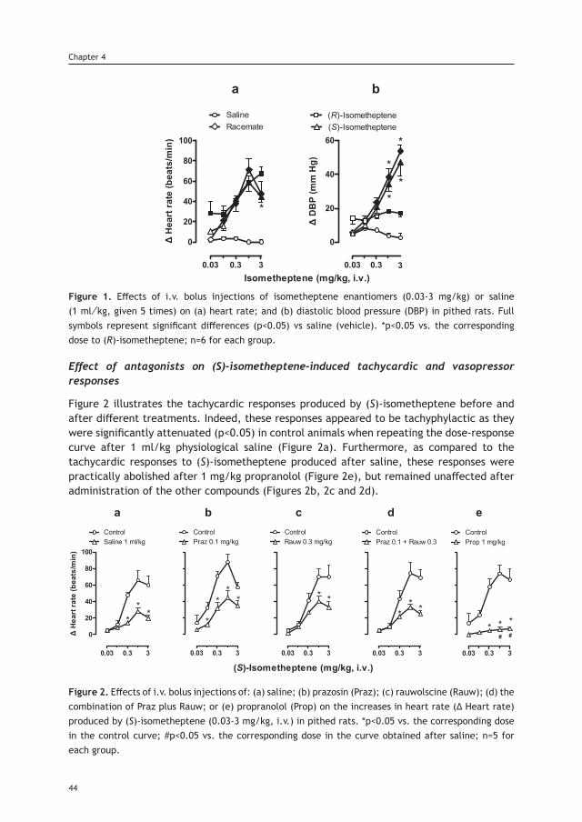
Figure 1. Effects of i.v. bolus injections of isometheptene enantiomers (0.03-3 mg/kg) or saline (1 ml⁄kg, given 5 times) on (a) heart rate; and (b) diastolic blood pressure (DBP) in pithed rats. Full symbols represent significant differences (p<0.05) vs saline (vehicle). *p<0.05 vs. the corresponding dose to (R)-isometheptene; n=6 for each group.
Effect of antagonists on (S)-isometheptene-induced tachycardic and vasopressor responses
Figure 2 illustrates the tachycardic responses produced by (S)-isometheptene before and after different treatments. Indeed, these responses appeared to be tachyphylactic as they were significantly attenuated (p<0.05) in control animals when repeating the dose-response curve after 1 ml/kg physiological saline (Figure 2a). Furthermore, as compared to the tachycardic responses to (S)-isometheptene produced after saline, these responses were practically abolished after 1 mg/kg propranolol (Figure 2e), but remained unaffected after administration of the other compounds (Figures 2b, 2c and 2d).
Figure 2. Effects of i.v. bolus injections of: (a) saline; (b) prazosin (Praz); (c) rauwolscine (Rauw); (d) the combination of Praz plus Rauw; or (e) propranolol (Prop) on the increases in heart rate (∆ Heart rate) produced by (S)-isometheptene (0.03-3 mg/kg, i.v.) in pithed rats. *p<0.05 vs. the corresponding dose in the control curve; #p<0.05 vs. the corresponding dose in the curve obtained after saline; n=5 for each group.
44
Chapter 4
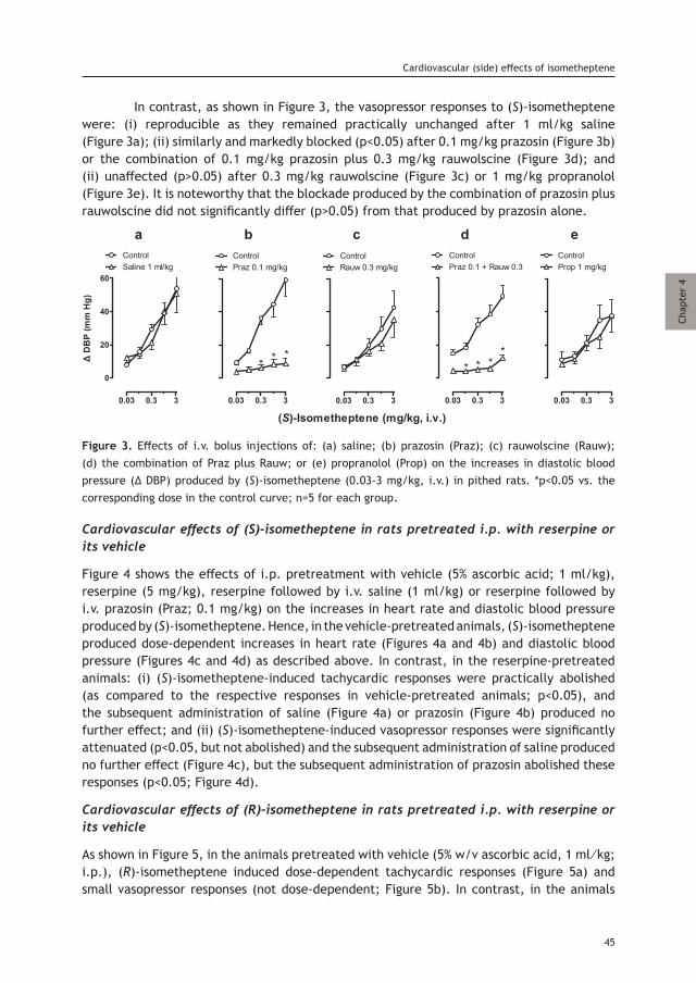
In contrast, as shown in Figure 3, the vasopressor responses to (S)-isometheptene were: (i) reproducible as they remained practically unchanged after 1 ml/kg saline (Figure 3a); (ii) similarly and markedly blocked (p<0.05) after 0.1 mg/kg prazosin (Figure 3b) or the combination of 0.1 mg/kg prazosin plus 0.3 mg/kg rauwolscine (Figure 3d); and (ii) unaffected (p>0.05) after 0.3 mg/kg rauwolscine (Figure 3c) or 1 mg/kg propranolol (Figure 3e). It is noteworthy that the blockade produced by the combination of prazosin plus rauwolscine did not significantly differ (p>0.05) from that produced by prazosin alone.
Figure 3. Effects of i.v. bolus injections of: (a) saline; (b) prazosin (Praz); (c) rauwolscine (Rauw); (d) the combination of Praz plus Rauw; or (e) propranolol (Prop) on the increases in diastolic blood pressure (∆ DBP) produced by (S)-isometheptene (0.03-3 mg/kg, i.v.) in pithed rats. *p<0.05 vs. the corresponding dose in the control curve; n=5 for each group.
Cardiovascular effects of (S)-isometheptene in rats pretreated i.p. with reserpine or its vehicle
Figure 4 shows the effects of i.p. pretreatment with vehicle (5% ascorbic acid; 1 ml/kg), reserpine (5 mg/kg), reserpine followed by i.v. saline (1 ml/kg) or reserpine followed by i.v. prazosin (Praz; 0.1 mg/kg) on the increases in heart rate and diastolic blood pressure produced by (S)-isometheptene. Hence, in the vehicle-pretreated animals, (S)-isometheptene produced dose-dependent increases in heart rate (Figures 4a and 4b) and diastolic blood pressure (Figures 4c and 4d) as described above. In contrast, in the reserpine-pretreated animals: (i) (S)-isometheptene-induced tachycardic responses were practically abolished (as compared to the respective responses in vehicle-pretreated animals; p<0.05), and the subsequent administration of saline (Figure 4a) or prazosin (Figure 4b) produced no further effect; and (ii) (S)-isometheptene-induced vasopressor responses were significantly attenuated (p<0.05, but not abolished) and the subsequent administration of saline produced no further effect (Figure 4c), but the subsequent administration of prazosin abolished these responses (p<0.05; Figure 4d).
Cardiovascular effects of (R)-isometheptene in rats pretreated i.p. with reserpine or its vehicle
As shown in Figure 5, in the animals pretreated with vehicle (5% w/v ascorbic acid, 1 ml⁄kg; i.p.), (R)-isometheptene induced dose-dependent tachycardic responses (Figure 5a) and small vasopressor responses (not dose-dependent; Figure 5b). In contrast, in the animals
45
Cardiovascular (side) effects of isometheptene
Chap
ter
4
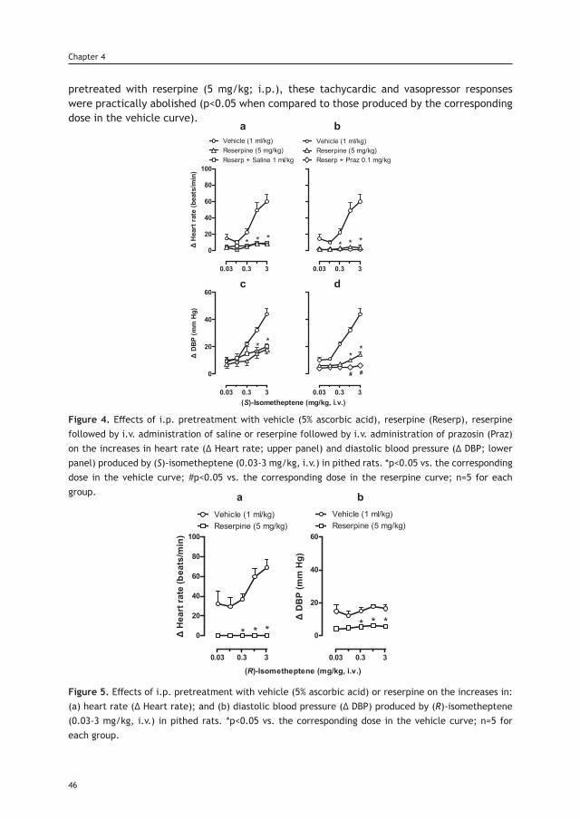
pretreated with reserpine (5 mg/kg; i.p.), these tachycardic and vasopressor responses were practically abolished (p<0.05 when compared to those produced by the corresponding dose in the vehicle curve).
Figure 4. Effects of i.p. pretreatment with vehicle (5% ascorbic acid), reserpine (Reserp), reserpine followed by i.v. administration of saline or reserpine followed by i.v. administration of prazosin (Praz) on the increases in heart rate (∆ Heart rate; upper panel) and diastolic blood pressure (∆ DBP; lower panel) produced by (S)-isometheptene (0.03-3 mg/kg, i.v.) in pithed rats. *p<0.05 vs. the corresponding dose in the vehicle curve; #p<0.05 vs. the corresponding dose in the reserpine curve; n=5 for each group.
Figure 5. Effects of i.p. pretreatment with vehicle (5% ascorbic acid) or reserpine on the increases in: (a) heart rate (∆ Heart rate); and (b) diastolic blood pressure (∆ DBP) produced by (R)-isometheptene (0.03-3 mg/kg, i.v.) in pithed rats. *p<0.05 vs. the corresponding dose in the vehicle curve; n=5 for each group.
46
Chapter 4
Discussion
General
Apart from the implications discussed below, our study demonstrates the importance of investigating separately the enantiomers of a given compound. Indeed, the (R)- and (S)-enantiomers of a racemate may behave pharmacologically different from each other [21]. Thus, it is appropriate to consider each enantiomer as a separate pharmacological entity with different properties, unless proven otherwise [21]. In our study, the enantiomers of isometheptene displayed clear pharmacodynamic differences in their cardiovascular effects, and these differences could lead to the future usage of these enantiomers in different conditions.
Systemic hemodynamic effects produced by the different treatments in pithed rats
The fact that the baseline values of diastolic blood pressure and heart rate remained without significant changes after prazosin, rauwolscine, the combination prazosin plus rauwolscine or propranolol (not shown), as previously reported [3], may be due to the fact that pithed rats are devoid of central and peripheral nervous influences. On this basis, it is most likely that the effect of any of these antagonists on the tachycardic and vasopressor responses to the isometheptene enantiomers involves a direct interaction with their corresponding receptors.
Differential cardiovascular effects of the isometheptene enantiomers
In agreement with the sympathomimetic properties of isometheptene racemate in pithed rats [3], both enantiomers and the racemate produced dose-dependent increases in heart rate, although the tachycardic response produced by 3 mg/kg was not strictly dose-dependent (Figure 1a) and apparently tachyphylactic. This finding implies a tyramine-like action (see below), as previously reported [3], and that such a high dose was supramaximal. In contrast, (R)-isometheptene produced only small vasopressor responses at 1 and 3 mg/kg (which were not dose-dependent), while (S)-isometheptene and isometheptene racemate produced dose-dependent (and equipotent) vasopressor responses (Figure 1b). The simplest interpretation of these findings indicates: (i) a differential pharmacological profile between (R)-isometheptene and (S)-isometheptene; and (ii) that the vasopressor effects of isometheptene racemate are mostly mediated by the (S)-enantiomer (see below). Since, overall, (S)-isometheptene displayed a higher potency to produce cardiovascular responses, we proceeded to analyse, in the first instance, the pharmacological profile of the receptors/mechanisms involved in the responses to (S)-isometheptene.
The role of a tyramine-like action producing indirect stimulation of cardiac β-adrenoceptors in the tachycardic responses to (S)-isometheptene
The tachycardic responses to isometheptene racemate in pithed rats are mediated by an indirect tyramine-like action involving stimulation of propranolol-sensitive β adrenoceptors, most likely of the β1-subtype [3]. In keeping with this finding, the tachycardic responses produced by (S)-isometheptene: (i) were highly tachyphylactic as they were not reproducible when repeating a second dose-response after saline (Figure 2a); (ii) are mainly mediated by activation of propranolol sensitive β-adrenoceptors (Figure 2e); and
47
Cardiovascular (side) effects of isometheptene
Chap
ter
4
(iii) do not involve activation of prazosin-sensitive α1-adrenoceptors and/or rauwolscine-sensitive α2-adrenoceptors (Figures 2b, 2c and 2d). Most notably, the fact that these responses were practically abolished after reserpine (Figure 4a) reinforces the role of an indirect tyramine-like action in (S)-isometheptene-induced tachycardic responses (i.e. by displacing noradrenaline from sympathetic nerves which, in turn, would subsequently stimulate cardiac β-adrenoceptors). It is noteworthy that this dose-schedule with reserpine (i.e. 5 mg/kg, i.p.; -24 hrs) has been shown to abolish in pithed rats the tachycardic and vasopressor responses to the typical indirect sympathomimetic agent, tyramine [3].
The role of both an indirect tyramine-like action and a direct stimulation of α1-adrenoceptors in the vasopressor responses to (S)-isometheptene
Unlike its tachycardic responses (see above), the vasopressor responses to (S)-isometheptene: (i) were reproducible as they remained without significant changes after saline (Figure 3a); and (ii) are mainly mediated by α1-adrenoceptors as these responses were markedly blocked after prazosin (Figure 3b), but not (p>0.05) after rauwolscine (Figure 3c) or propranolol (Figure 3e) in doses high enough to completely block their respective receptors mediating cardiovascular responses [3]. Consistent with this suggestion, the combination prazosin plus rauwolscine produced a blockade (Figure 3d) that did not significantly differ (p>0.05) from that produced by prazosin alone (Figure 3b). Furthermore, the fact that reserpine markedly attenuated (but did not abolish) these vasopressor responses (Figure 4c), and that the subsequent administration of prazosin practically abolished these responses (Figure 4d) suggests the involvement of a mixed effect of (S)-isometheptene, namely: (i) a major indirect (tyramine-like) mechanism; and (ii) a minor direct sympathomimetic mechanism mediated by stimulation of α1-adrenoceptors. In keeping with these findings, and as implied by other studies at the neuro-vascular junction [3, 22, 23]: (i) neuronally-displaced noradrenaline (i.e. by a tyramine-like action) primarily stimulates intrasynaptic α1-adrenoceptors; (ii) i.v. bolus of exogenous noradrenaline mainly activates extrasynaptic α2-adrenoceptors; and (iii) systemic vascular resistance (represented by diastolic blood pressure) is mainly modulated by α1-adrenoceptors. Interestingly, the fact that (R)-isometheptene-induced tachycardic (Figure 5a) and (weak) vasopressor (Figure 5b) responses were abolished by reserpine suggests the exclusive role of a tyramine-like action in both responses (with no role of direct sympathomimetic actions). This implies that (R)-isometheptene would produce stoichiometric displacement of noradrenaline from the sympathetic neurons innervating the heart and resistance blood vessels, resulting in tachycardic and vasopressor responses mediated by stimulation of cardiac β- and vascular α1-adrenoceptors [3, 22, 23].
Structure of the enantiomers and stereo-specificity of the α1-adrenoceptor
The different effects of the isometheptene enantiomers on the cardiovascular system in this study could also be explained by the chemical structures of these compounds. Indeed, there are some similarities between the molecular structures of isometheptene and noradrenaline, which may confer the capability of being recognized by the neuronal transporters, introduced into the synaptic vesicles and subsequently displace the stored catecholamines that, in turn, would interact with post-junctional adrenoceptors. The presence of an amine side chain in isometheptene provides the capability to stimulate directly the α1-adrenoceptor [23]. However, the presence of a chiral centre in the β carbon
48
Chapter 4
of the amine group could explain the differences in the pharmacological profile of both enantiomers, suggesting that (R)-isometheptene cannot interact with the α1-adrenoceptor, probably due to a steric impediment.
Study limitations
The pithed rat model is a useful preparation for investigating the cardiovascular (side) effects of new developed antimigraine drugs (i.e. isometheptene enantiomers) and their mechanism of action. Since migraine-specific agents, namely triptans, are contraindicated for acute migraine attacks in patients with cardiovascular risk factors, new antimigraine drugs should ideally have a beneficial cardiovascular safety profile. The pithed rat model is appropriate to assess peripheral cardiovascular effects, however, it should be kept in mind that it excludes central nervous system mechanisms, which might be relevant in the clinical situation. As migraine has a 2- to 3-fold higher prevalence in women, another limitation of our study is that we used only male rats. We deliberately chose to study male rats in this case since we wanted to limit experimental variation avoiding the effects of female hormones on vascular responsiveness, as it is known that noradrenaline vasopressor responses are altered in the presence of 17β-estradiol in this model [24]. Thus, future studies may focus on the antimigraine effects of the isometheptene enantiomers in female models during different hormonal cycle stages.
Conclusions
Our findings show that both isometheptene enantiomers (R and S) are equipotent in producing tachycardic responses, and that these responses are mediated by a tyramine-like action (abolished by reserpine). Moreover, (S)-isometheptene produced greater dose-dependent vasopressor responses, while those produced by (R)-isometheptene were not dose-dependent. These effects are probably due to differences in their mechanism of action, namely: (i) a mixed sympathomimetic action for (S)-isometheptene (a tyramine-like action and a direct stimulation of α1-adrenoceptors); and (ii) exclusively a tyramine-like action for (R)-isometheptene. Therefore, (R)-isometheptene may be responsible for the therapeutic action of isometheptene racemate and (S)-isometheptene (producing greater vasopressor responses) might be associated with the vasospasm described with the racemate. Accordingly, (R)-isometheptene may represent a superior therapeutic benefit as an antimigraine agent. Undoubtedly, further studies with (R)-isometheptene in human preparations and in animal models of trigeminovascular activation will shed further light on the potential role of imidazoline receptors in the pathophysiology of migraine.
References1. de Souza Carvalho D, Barea LM, et al. Efficacy and tolerability of combined dipyrone, isometheptene and caffeine in the treatment of mild-to-moderate primary headache episodes. Expert Rev Neurother 2012; 12: 159–167.2. Diamond S, Medina JL. Isometheptene a non-ergot drug in the treatment of migraine. Headache 1975; 15: 211–213.3. Valdivia LF, Centurión D, Perusquía M, et al. Pharmacological analysis of the mechanisms involved in the tachycardic and vasopressor responses to the antimigraine agent, isometheptene, in pithed rats. Life Sci 2004; 74: 3223–3234.4. Willems EW, Valdivia LF, Saxena PR, et al. Pharmacological profile of the mechanisms involved in the external carotid vascular effects of the antimigraine agent isometheptene in anaesthetised dogs. Naunyn Schmiedebergs Arch Pharmacol 2001; 364: 27–32.
49
Cardiovascular (side) effects of isometheptene
Chap
ter
4
5. Villalón CM, Centurión D, Valdivia LF, et al. Migraine: an updated review on pathophysiology, pharmacology, therapy and future trends. Curr Vasc Pharmacol 2003; 1: 71-84.6. Goadsby PJ, Lipton RB, Ferrari MD. Migraine--current understanding and treatment. N Engl J Med 2002; 346 :257–270.7. Asghar MS, Hansen AE, Kapijimpanga T, et al. Dilation by CGRP of middle meningeal artery and reversal by su-matriptan in normal volunteers. Neurology 2010; 75: 1520–1526. 8. Labruijere S, Chan KY, De Vries R, et al. Dihydroergotamine and sumatriptan in isolated human coronary artery, middle meningeal artery and saphenous vein. Cephalalgia 2015; 35: 182–189.9. Mehrotra S, Gupta S, Garrelds IM, et al. Effects of current and prospective antimigraine drugs on the porcine iso-lated meningeal artery. Naunyn Schmiedebergs Arch Pharmacol 2006; 374: 163–175. 10. Campos CR, Yamamoto FI. Intracerebral hemorrhage in postpartum cerebral angiopathy associated with the use of isometheptene. Int J Gynaecol Obstet 2006; 95: 151–152.11. Meschia JF, Malkoff MD, Biller J. Reversible segmental cerebral arterial vasospasm and cerebral infarction: possible association with excessive use of sumatriptan and Midrin. Arch Neurol 1998; 55: 712–714.12. Johnston JC. Life threatening intracerebral hemorrhage with isometheptene mucate, dichlorophenazine and acetaminophen combination therapy. J Forensic Leg Med 2009; 16: 489–491.13. Raroque HG, Tesfa G, Purdy P. Postpartum cerebral angiopathy. Is there a role for sympathomimetic drugs? Stroke 1993; 24: 2108–2110. 14. Lederman S, Daugherty B, Gershell LJ, et al. Isometheptene Isomer. US Patent 2014,0212486 A1, 31 Jul 2014. 15. Daugherty BL, et al.(R)-isometheptene (IMH) binds to the imidazoline-1 receptor and (S)-IMH increases blood pressure: potentially superior benefit to risk ratio for (R) IMH as an analgesic for headache. Headache 2015; June:55 (53) Abstract PS29.172.16. Zhang L, Zhao T-Y, Hou N, et al. Generation and primary phenotypes of imidazoline receptor antisera-selected (IRAS) knockout mice. CNS Neurosci Ther 2013; 19: 978-981.17. Fried NT, Oshinsky MI, Daugherty BL, et al. The (R) isomer of isometheptene decreases trigeminal sensitivity in a rat model of primary headache. Headache 2015; June:55 (53) Abstract PS58.184.18. Shipley RE, Tilden JH. A pithed rat preparation suitable for assaying pressor substances. Proc Soc Exp Biol Med 1947, 64: 453–455.19. Altamirano-Espinoza AH, González-Hernández A, Manrique-Maldonado G, et al. The role of dopamine D2, but not D3 or D4, receptor subtypes, in quinpirole-induced inhibition of the cardioaccelerator sympathetic outflow in pithed rats. Br J Pharmacol 2013, 170: 1102-1111.20. Kleinman LI, Radford EP. Ventilation Standards for Small Mammals. J Appl Physiol 1964; 19: 360–362.21. McConathy J, Owens MJ. Stereochemistry in Drug Action. Prim Care Companion J Clin Psychiatry 2003; 5: 70–73.22. Burn JH, Rand MJ. The action of sympathomimetic amines in animals treated with reserpine. J Physiol 1958; 144: 314–336.23. Brodde O-E, Michel MC. Adrenergic and Muscarinic Receptors in the Human Heart. Pharmacol Rev 1999; 51:651–690.24. Austin C, Chess-Williams R. The influence of 17-β-Oestradiol and the nautral oestrous cycle on α-adrenoceptor-me-diated responses of the cardiovascular system in the rat. J Pharm Pharmacol 1995; 47: 656-660.
50
Chapter 4


Chapter 5
Effects of two isometheptene enantiomersin isolated human blood vessels and rat middle meningeal artery
– potential antimigraine efficacy
Based on: The Journal of Headache and Pain, 2019, 20: 47.
Labastida-Ramírez A, Rubio-Beltrán E, Haanes KA, de Vries R, Dammers R, Bogers AJJC, van den Bogaerdt A, Daugherty BL, Danser AHJ, Villalón CM and MaassenVanDenBrink A.
Abstract
Background: Racemic isometheptene [(RS)-isometheptene] is an antimigraine drug that due to its cardiovascular side-effects was separated into its enantiomers, (R)- and (S)-isometheptene. This study set out to characterize the contribution of each enantiomer to its vasoactive profile. Moreover, rat neurogenic dural vasodilatation was used to explore their antimigraine mechanism of action.
Methods: Human blood vessel segments (middle meningeal artery, proximal and distal coronary arteries, and saphenous vein) were mounted in organ baths and concentration response curves to isometheptene were constructed. Calcitonin gene-related peptide (CGRP) induced neurogenic dural vasodilation was elicited in the presence of the enantiomers using a rat closed cranial window model.
Results: The isometheptene enantiomers did not induce any significant contraction in human blood vessels, except in the middle meningeal artery, when they were administered at the highest concentration (100 µM). Interestingly in rats, (S)-isometheptene induced more pronounced vasopressor responses than (R)-isometheptene. However, none of these compounds affected the CGRP-induced vasodilator responses.
Conclusion: The isometheptene enantiomers displayed a relatively safe peripheral vascular profile, as they failed to constrict the human coronary artery. These compounds do not appear to modulate neurogenic dural CGRP release, therefore, their antimigraine site of action remains to be determined.
Background
Migraine is a neurovascular disorder characterized by recurrent attacks of incapacitating unilateral headaches, recently interconnected with an overall increased risk of stroke and cardiovascular disease [1, 2]. Although its exact pathophysiology has not been elucidated completely, migraine headache has been associated with activation of the trigeminovascular system and increased release of calcitonin gene-related peptide (CGRP), resulting in dysfunctional nociceptive transmission and neurogenic dural vasodilatation [3]. The triptans, serotonergic agonists with selective affinity for 5-HT1B/1D/(1F) receptors, are specific drugs for the acute treatment. Their mechanism of action has been attributed to a dural perivascular inhibition of CGRP release, an inhibition of central nociception and⁄or a postjunctional constriction of (cranial) blood vessels [4-6]. Because of the latter, the triptans are contraindicated in patients with cardiovascular risk factors or a history of cardiovascular disease. Isometheptene is a sympathomimetic racemic drug available by prescription or over the counter in several countries, that has long been used for the acute treatment of primary headaches [7, 8]. Nevertheless, a few case reports of acute intracranial vasoconstriction after its use [9, 10] highlight its presumed vasoactive properties [11]. Given that the development of new antimigraine agents with a beneficial cardiovascular safety profile is crucial, Tonix Pharmaceuticals™ separated isometheptene racemate into its enantiomers, (S)-isometheptene and (R)-isometheptene, a mixed-acting (tyramine-like/minor direct α1-adrenoceptor) and an indirect-acting (tyramine-like) adrenergic receptor agonist, respectively. Additionally, (R)-isometheptene is an imidazoline I1 receptor agonist [12],
54
Chapter 5
Chap
ter
5
and previous studies have shown that: (i) imidazoline I1 receptor knockout mice have a potentiated nociceptive perception, suggesting that this receptor could be associated with an endogenous analgesia system [13]; (ii) (R)-isometheptene decreased trigeminal sensitivity in two rat models of chronic migraine [14]; and (iii) imidazoline I1 receptor agonists, like moxonidine and agmatine induced a prejunctional inhibition of the vasodepressor sensory CGRPergic outflow in pithed rats [15]. Together, these findings suggest that a potential antimigraine action of (R)-isometheptene could be mediated by inhibition of the trigeminal system. Hence, we hypothesized that the use of only (R)-isometheptene will maintain its antimigraine therapeutic effect, while the major side-effects associated with the racemate or (S)-isometheptene (i.e. cranial vasoconstriction) will be diminished [16]. On this basis, the present study set out to analyse the effects of the isometheptene enantiomers and the racemate on human isolated blood vessels (i.e. middle meningeal artery, proximal and distal coronary arteries, as well as saphenous vein) and trigeminal CGRP induced neurogenic dural vasodilation in anaesthetized rats (through a closed cranial window).
Materials and Methods
Human isolated blood vessels
Middle meningeal arteries [internal diameter (ID) 0.5-1.5 mm] were obtained from 11 patients (3 males, 8 females; mean age 53±5 years) who underwent neurosurgical interventions requiring a trepanation of the skull. During surgery, the dura mater together with a small piece of meningeal artery was collected in a sterile organ protecting solution and was immediately transported to the laboratory. The meningeal arteries were dissected and placed in a cold (4°C) oxygenated Krebs bicarbonate solution with the following composition (mmol/L): NaCl 119, KCl 4.7, CaCl2 1.25, MgSO4 1.2, KH2PO4 1.2, NaHCO2 25 and glucose 11.1; pH 7.4. Saphenous veins (ID 0.5-3 mm) were obtained from 11 patients (10 males, 1 female; mean age 71±2 years) who underwent coronary artery bypass surgery. Immediately after surgery, veins were placed in cold (4°C) oxygenated Krebs buffer solution with the following composition (mmol/L): NaCl 118, KCl 4.7, CaCl2 2.5, MgSO4 1.2, KH2PO4 1.2, NaHCO2 25 and glucose 8.3; pH 7.4. Proximal (ID 2-3 mm) and distal (ID 0.5-1.0 mm) coronary arteries were obtained from 10 heart valve donors (6 males, 4 females; mean age 40±5 years) who died of non-cardiac disorders: four traumatic brain injury, one benzodiazepine overdose, three anoxic encephalopathy and two cerebrovascular accident. The hearts were provided by the Heart Valve Bank Beverwijk (at that time still located in Rotterdam) from Dutch post-mortem donors, after donor mediation by The Dutch Transplantation Foundation (Leiden, The Netherlands), following removal of the aortic and pulmonary valves for homograft valve transplantation. All donors gave permission for research. Immediately after circulatory arrest, the hearts were stored at 4°C in a sterile organ protecting solution and were brought to the laboratory within the first 24 hours of death. The coronary arteries were dissected and placed in Krebs buffer with the same composition as the one used for the saphenous veins (see above). All blood vessels were used on the same day or stored overnight and used the following day for functional experiments.
55
Neurovascular effects of isometheptene
The middle meningeal arteries and the distal coronary arteries were cut into ring segments of 1-2 mm length and suspended in Mulvany myographs on two parallel steel wires. The tension was normalized to 90% of the estimated diameter at 100 mm Hg [17]. The proximal coronary arteries and saphenous veins were cut into ring segments of about 3-4 mm length and suspended on stainless steel hooks in 15-mL organ baths. The vascular rings were stretched to a stable pretension of 10-15 mN, the optimal pretension as determined earlier [17], and changes in tissue force were measured with an isometric force transducer (Harvard, South Natick, MA, U.S.A.) and recorded on a flatbed recorder (Servogor 124, Goerz, Neudorf, Austria). The buffer was aerated with 95% O2 and 5% CO2 and was maintained at 37°C. The segments were allowed to equilibrate for at least 30 min and were washed every 15 min.
In vitro experimental protocols
Initially, segments were exposed to 30 mM KCl, followed by 100 mM KCl to determine the reference contractile response in each segment. Cumulative concentration response curves were constructed to (S)-isometheptene, (R)-isometheptene, isometheptene racemate, sumatriptan and noradrenaline, using whole logarithmic steps (1 nM to 100 μM). Sumatriptan and noradrenaline were used as positive controls, as previously reported [17]. Finally, the functional integrity of the endothelium was assessed by observing the relaxation to substance P (10 nM) in arteries or bradykinin (10 µM) in saphenous veins after precontraction with the thromboxane A2 analogue U46619 (10-100 nM).
Animals
Twelve male Sprague-Dawley rats (300-350 g; 8-10 weeks of age), purchased from Harlan Netherlands (Horst, the Netherlands), were maintained at a 12/12 h light-dark cycle in a special room at constant temperature (22±2 °C) and humidity (50%), with food and water ad libitum. Only male rats were used to avoid crosstalk between CGRP and hormonal fluctuations of the oestrus cycle previously described in this model [18, 19]. Experimental protocols were approved by the Erasmus Medical Center’s institutional ethics committee (EMC permission protocol number 3393), in accordance with the European directive 2010/63/EU and the ARRIVE guidelines for reporting experiments in animals [20]. After anaesthesia with sodium pentobarbital (60 mg/kg i.p. followed by 18 mg/kg i.v. per hour), the trachea was cannulated and artificially ventilated (58 strokes/min.; small animal ventilator SAR 830 series, CWE Inc., Ardmore, PA, U.S.A). The adequacy of anaesthesia was judged by the absence of ocular reflexes and a negative tail flick test.End-tidal pCO2 was monitored with a capnograph (Capstar 100 CWE Inc., PA, U.S.A.) and kept between 35-45 mm Hg. The left femoral vein and artery were cannulated for i.v. administration of drugs and monitoring of mean arterial pressure (MAP), respectively. The animals’ body temperature was maintained at 37°C by a homeothermic blanket (Harvard Instruments, Edenbridge, Kent, U.K.). The head of each rat was fixed in a stereotaxic frame and the parietal bone overlying a segment of the dural middle meningeal artery was drilled thin, applying cold saline until the artery was clearly visible. As skull drilling induces vasodilation, animals were allowed to rest at least for 1 hour before the experimental protocol started. The artery diameter was recorded with an intravital microscopy setup (MZ16, Leica microsystem Ltd., Heerbrugg, Switzerland) using a cyan blue filter on a cold light source. A zoom lens (80-450x magnification) and a camera were used to display images on a standard PC monitor.
56
Chapter 5
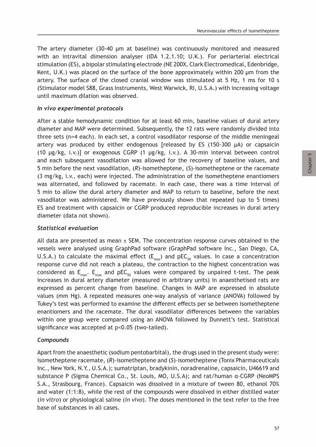
The artery diameter (30-40 µm at baseline) was continuously monitored and measured with an intravital dimension analyser (IDA 1.2.1.10; U.K.). For periarterial electrical stimulation (ES), a bipolar stimulating electrode (NE 200X, Clark Electromedical, Edenbridge, Kent, U.K.) was placed on the surface of the bone approximately within 200 µm from the artery. The surface of the closed cranial window was stimulated at 5 Hz, 1 ms for 10 s (Stimulator model S88, Grass Instruments, West Warwick, RI, U.S.A.) with increasing voltage until maximum dilation was observed.
In vivo experimental protocols
After a stable hemodynamic condition for at least 60 min, baseline values of dural artery diameter and MAP were determined. Subsequently, the 12 rats were randomly divided into three sets (n=4 each). In each set, a control vasodilator response of the middle meningeal artery was produced by either endogenous [released by ES (150-300 µA) or capsaicin (10 µg⁄kg, i.v.)] or exogenous CGRP (1 µg/kg, i.v.). A 30-min interval between control and each subsequent vasodilation was allowed for the recovery of baseline values, and 5 min before the next vasodilation, (R)-isometheptene, (S)-isometheptene or the racemate (3 mg/kg, i.v., each) were injected. The administration of the isometheptene enantiomers was alternated, and followed by racemate. In each case, there was a time interval of 5 min to allow the dural artery diameter and MAP to return to baseline, before the next vasodilator was administered. We have previously shown that repeated (up to 5 times) ES and treatment with capsaicin or CGRP produced reproducible increases in dural artery diameter (data not shown).
Statistical evaluation
All data are presented as mean ± SEM. The concentration response curves obtained in the vessels were analysed using GraphPad software (GraphPad software Inc., San Diego, CA, U.S.A.) to calculate the maximal effect (Emax) and pEC50 values. In case a concentration response curve did not reach a plateau, the contraction to the highest concentration was considered as Emax. Emax and pEC50 values were compared by unpaired t-test. The peak increases in dural artery diameter (measured in arbitrary units) in anaesthetised rats are expressed as percent change from baseline. Changes in MAP are expressed in absolute values (mm Hg). A repeated measures one-way analysis of variance (ANOVA) followed by Tukey’s test was performed to examine the different effects per se between isometheptene enantiomers and the racemate. The dural vasodilator differences between the variables within one group were compared using an ANOVA followed by Dunnett’s test. Statistical significance was accepted at p<0.05 (two-tailed).
Compounds
Apart from the anaesthetic (sodium pentobarbital), the drugs used in the present study were: isometheptene racemate, (R)-isometheptene and (S)-isometheptene (Tonix Pharmaceuticals Inc., New York, N.Y., U.S.A.); sumatriptan, bradykinin, noradrenaline, capsaicin, U46619 and substance P (Sigma Chemical Co., St. Louis, MO, U.S.A); and rat/human α-CGRP (NeoMPS S.A., Strasbourg, France). Capsaicin was dissolved in a mixture of tween 80, ethanol 70% and water (1:1:8), while the rest of the compounds were dissolved in either distilled water (in vitro) or physiological saline (in vivo). The doses mentioned in the text refer to the free base of substances in all cases.
57
Neurovascular effects of isometheptene
Chap
ter
5
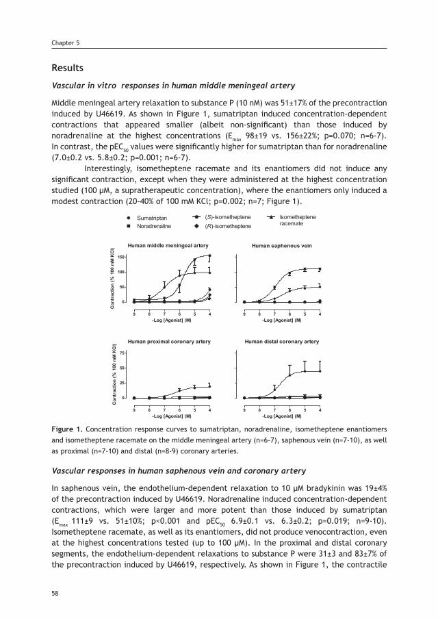
Results
Vascular in vitro responses in human middle meningeal artery
Middle meningeal artery relaxation to substance P (10 nM) was 51±17% of the precontraction induced by U46619. As shown in Figure 1, sumatriptan induced concentration-dependent contractions that appeared smaller (albeit non-significant) than those induced by noradrenaline at the highest concentrations (Emax 98±19 vs. 156±22%; p=0.070; n=6-7). In contrast, the pEC50 values were significantly higher for sumatriptan than for noradrenaline (7.0±0.2 vs. 5.8±0.2; p=0.001; n=6-7). Interestingly, isometheptene racemate and its enantiomers did not induce any significant contraction, except when they were administered at the highest concentration studied (100 µM, a supratherapeutic concentration), where the enantiomers only induced a modest contraction (20-40% of 100 mM KCl; p=0.002; n=7; Figure 1).
Figure 1. Concentration response curves to sumatriptan, noradrenaline, isometheptene enantiomers and isometheptene racemate on the middle meningeal artery (n=6-7), saphenous vein (n=7-10), as well as proximal (n=7-10) and distal (n=8-9) coronary arteries.
Vascular responses in human saphenous vein and coronary artery
In saphenous vein, the endothelium-dependent relaxation to 10 µM bradykinin was 19±4% of the precontraction induced by U46619. Noradrenaline induced concentration-dependent contractions, which were larger and more potent than those induced by sumatriptan (Emax 111±9 vs. 51±10%; p<0.001 and pEC50 6.9±0.1 vs. 6.3±0.2; p=0.019; n=9-10). Isometheptene racemate, as well as its enantiomers, did not produce venocontraction, even at the highest concentrations tested (up to 100 µM). In the proximal and distal coronary segments, the endothelium-dependent relaxations to substance P were 31±3 and 83±7% of the precontraction induced by U46619, respectively. As shown in Figure 1, the contractile
58
Chapter 5
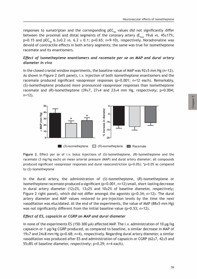
responses to sumatriptan and the corresponding pEC50 values did not significantly differ between the proximal and distal segments of the coronary artery (Emax 19±6 vs. 45±17%; p=0.15 and pEC50 6.3±0.2 vs. 6.2 ± 0.1; p=0.65; n=9-10), respectively. Noradrenaline was devoid of contractile effects in both artery segments; the same was true for isometheptene racemate and its enantiomers.
Effect of isometheptene enantiomers and racemate per se on MAP and dural artery diameter in vivo
In the closed cranial window experiments, the baseline value of MAP was 92±5 mm Hg (n=12). As shown in Figure 2 (left panel), i.v. injection of both isometheptene enantiomers and the racemate produced significant vasopressor responses (p<0.001; n=12 each). Remarkably, (S)-isometheptene produced more pronounced vasopressor responses than isometheptene racemate and (R)-isometheptene (39±7, 27±4 and 23±4 mm Hg, respectively; p=0.004; n=12).
Figure 2. Effect per se of i.v. bolus injections of (S)-isometheptene, (R)-isometheptene and the racemate (3 mg/kg each) on mean arterial pressure (MAP) and dural artery diameter; all compounds produced significant vasopressor responses and dural vasoconstriction (p<0.05); *p<0.05 as compared to (S)-isometheptene
In the dural artery, the administration of (S)-isometheptene, (R)-isometheptene or isometheptene racemate produced a significant (p<0.001, n=12) small, short-lasting decrease in dural artery diameter (12±2%, 13±2% and 10±2% of baseline diameter, respectively; Figure 2 right panel), which did not differ amongst the agonists (p=0.34; n=12). The dural artery diameter and MAP values restored to pre-injection levels by the time the next vasodilation was elucidated. At the end of the experiments, the value of MAP (88±5 mm Hg) was not significantly different from the initial baseline value (p=0.53; n=12).
Effect of ES, capsaicin or CGRP on MAP and dural diameter
In none of the experiments ES (150-300 μA) affected MAP. The i.v. administration of 10 µg/kg capsaicin or 1 µg/kg CGRP produced, as compared to baseline, a similar decrease in MAP of 19±7 and 24±8 mm Hg (p=0.68; n=4), respectively. Regarding dural artery diameter, a similar vasodilation was produced after ES and administration of capsaicin or CGRP (62±7, 42±5 and 55±8% of baseline diameter, respectively; p=0.29; n=4 each).
59
Neurovascular effects of isometheptene
Chap
ter
5
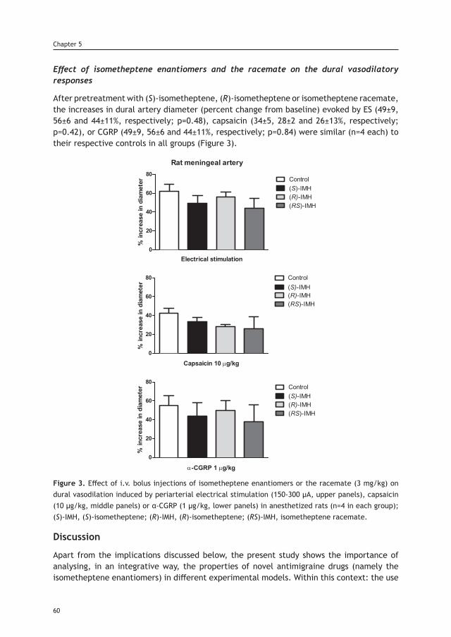
Effect of isometheptene enantiomers and the racemate on the dural vasodilatory responses
After pretreatment with (S)-isometheptene, (R)-isometheptene or isometheptene racemate, the increases in dural artery diameter (percent change from baseline) evoked by ES (49±9, 56±6 and 44±11%, respectively; p=0.48), capsaicin (34±5, 28±2 and 26±13%, respectively; p=0.42), or CGRP (49±9, 56±6 and 44±11%, respectively; p=0.84) were similar (n=4 each) to their respective controls in all groups (Figure 3).
Figure 3. Effect of i.v. bolus injections of isometheptene enantiomers or the racemate (3 mg/kg) on dural vasodilation induced by periarterial electrical stimulation (150-300 μA, upper panels), capsaicin (10 μg/kg, middle panels) or α-CGRP (1 μg/kg, lower panels) in anesthetized rats (n=4 in each group); (S)-IMH, (S)-isometheptene; (R)-IMH, (R)-isometheptene; (RS)-IMH, isometheptene racemate.
Discussion
Apart from the implications discussed below, the present study shows the importance of analysing, in an integrative way, the properties of novel antimigraine drugs (namely the isometheptene enantiomers) in different experimental models. Within this context: the use
60
Chapter 5
of different human isolated blood vessels allows us to discern possible vascular side-effects induced by potential antimigraine agents; and the rat closed cranial window is an in vivo neurovascular migraine model that focuses on the pathophysiological interaction of the trigeminal system with neurogenic dural vasodilation [21].
Human vascular (side) effects
A limitation of the current specific antimigraine drugs (i.e. triptans and ergots) is their theoretical risk of coronary vasoconstriction, consequently all vasoactive antimigraine agents are contraindicated in patients with cardiovascular risk factors or coronary artery disease [22]. With this in mind, we investigate the vasomotor effects of the isometheptene enantiomers in different human blood vessels, including the proximal and distal coronary arteries; additionally, the human saphenous vein was included as a positive control of peripheral venoconstriction that is sensitive to α-adrenergic stimulation [23]. Importantly, the isometheptene enantiomers and the racemate were devoid of vasoconstrictor properties in the proximal and distal coronary arteries, as well as the saphenous veins at all concentrations tested. Similarly, the isometheptene enantiomers were devoid of meningeal contractile effects, except when they were administered at the highest concentration (100 µM, Figure 1), a supratherapeutic concentration that would never be reached in the clinical situation. Regarding the possible mechanism of action of this meningeal vasoconstriction, it is tempting to speculate that it is mediated by an indirect (tyramine-like) action, resulting in noradrenaline displacement from perivascular sympathetic nerve terminals [24], as previously shown for (R)-isometheptene-induced vasopressor responses [25]. Admittedly, we did not test this hypothesis with experiments in the presence of neuronal reuptake inhibitors such as cocaine, because, as mentioned before, this phenomenon only happens at supratherapeutic concentrations and is thus unlikely to be clinically relevant. In contrast to the meningeal artery, there were no tyramine-like vasoconstrictor effects in coronary arteries, mainly because these vessels (via β2-adrenoceptors in vascular smooth muscle) normally dilate to (displaced) noradrenaline [26], as is evident from the lack of contraction after exogenous noradrenaline. Similarly, no tyramine-like responses were observed in saphenous veins; this may be attributed to a lesser sympathetic innervation (vs. arteries) and the possibility that an important amount of perivascular fibres [present in the loose connective tissue surrounding the vein, [27] were destroyed. Thus, on the basis of these results, the well-established antimigraine action of isometheptene racemate [7, 8, 11], and probably also of isometheptene enantiomers [in particular the antimigraine potential of (R)-isometheptene] would seem to be devoid of acute coronary side-effects.
In vivo effects of isometheptene on MAP
In this study, and in accordance with others [25, 28], isometheptene racemate and (S)-isometheptene are potent vasopressor compounds in rats. Their vascular responses are mediated by an indirect (tyramine-like action) and a minor direct stimulation of α1-adrenoceptors [28]. In contrast, vascular responses to (R)-isometheptene are exclusively indirect (tyramine-like action) and of less magnitude than its counterpart enantiomer [25]. Accordingly, (R)-isometheptene might produce fewer vascular side-effects as an antimigraine agent.
61
Neurovascular effects of isometheptene
Chap
ter
5
In vivo effects of isometheptene on dural artery diameter
In contrast to the lack of vasoconstriction in the isolated human middle meningeal artery, the isometheptene enantiomers and the racemate produced equipotent meningeal vasoconstrictor responses in vivo (Figure 2). This apparent in vitro/in vivo discrepancy suggests that isometheptene’s vasoconstriction is indeed mediated by a tyramine-like action mechanism, which is more evident when the perivascular sympathetic tone is higher (i.e. in vivo), whereas such neurogenic tone has been eliminated in vitro. Although it is believed that the marked dural vasoconstriction of ergots [17] and triptans [29], along with the inhibition of intracranial trigeminal afferents [21] contributes to the peripheral antimigraine mechanisms of these drugs, it is unlikely that isometheptene’s antimigraine action is related to this small, short-lasting decrease in dural artery diameter. Therefore, we proceeded to explore whether the attenuation of experimentally-activated trigeminovascular afferents could explain isometheptene antimigraine efficacy.
Modulation of perivascular CGRP release as antimigraine treatment
The rat closed cranial window method is a highly predictive model of antimigraine action, in which triptans [21] and CGRP receptor antagonists [gepants and CGRP (receptor)-binding antibodies [30] have shown its ability to inhibit neurogenic (CGRP-mediated) vasodilation of the dural middle meningeal artery as one of their pharmacological sites of action. It is noteworthy that the isometheptene enantiomers and the racemate did not reduce the dural vasodilation evoked by the release of endogenous CGRP (by ES or i.v. capsaicin) or exogenous CGRP (Figure 3). Hence, inhibition of trigeminal CGRP release, as one of the mechanisms associated with antimigraine action (or vasoconstriction), does not appear to explain isometheptene’s antimigraine efficacy. Interestingly, it has previously been shown that imidazoline I1 and α2-adrenoceptor agonists are capable of inhibiting prejunctionally the sensory vasodepressor CGRPergic outflow in pithed rats [25, 31]. However, (R)-isometheptene, an imidazoline I1 receptor agonist with extremely low affinity for α2-adrenoceptors [12], did not inhibit the neurogenic dural vasodilation induced by trigeminal stimulation; suggesting a differential receptor expression between sensory and trigeminal afferents, as previously shown for α2-adrenoceptors [32]. This suggests that in the rat closed cranial window model, imidazoline I1 and α2 receptors do not play a role as prejunctional modulators of CGRP release in the trigeminovascular system. Even though similar sample sizes have been used by two different research groups [32, 33], it could be argued that the statistical power of our in vivo experiments is low (and a limitation) due to the relatively small number of animals used per group. However, when comparing our current results with those of earlier findings using sumatriptan [21] and CGRP antagonists [30], the magnitude of inhibition of CGRP release is remarkably high (up to ca. 70%, and own experiments, data not shown). Thus, while we cannot categorically exclude that a higher number of animals could have produced statistically significant effects, such effects would be rather limited and, probably, devoid of clinical relevance, as an i.v. dose of 3 mg/kg isometheptene is already supramaximal in pithed rats [25].
Future perspectives for (R)-isometheptene
Isometheptene racemate as monotherapy or, as usual, combined with other drugs (e.g. analgesics), seems a cost-effective alternative (vs. the triptans) in some countries for the
62
Chapter 5
acute treatment of mild-to-moderate primary headache [8, 10]. Whereas (R)-isometheptene has been shown not to be effective in the treatment of episodic tension-type headache [34], its antimigraine efficacy has not yet been clinically tested. Overall, after considering the above pharmacological profile of (R)-isometheptene, it is not unreasonable to suggest that its potential clinical use as an antimigraine agent may have superior therapeutic advantages over either isometheptene racemate or (S)- isometheptene. Most importantly, the fact that (R)-isometheptene produced only a slight increase in MAP suggests that it is not directly associated with the intracranial vasospasm previously described with the racemate [9, 10]. As an imidazoline I1 receptor agonist, (R)-isometheptene should possess central antinociceptive properties, as previously shown for other imidazolines agonists [35]. This is supported by a preliminary study where high doses of (R)-isometheptene decreased trigeminal sensitivity in two rat models of chronic migraine, and this effect was associated with a reduced CGRP immunoreactivity in the trigeminal nucleus caudalis. Certainly, further experiments, falling beyond the scope of the present study, will be required to investigate whether: (i) (R)-isometheptene is capable of inducing central antinociception; (ii) activation of imidazoline receptors translates into acute or prophylactic antimigraine action; and (iii) selective imidazoline I1 receptor agonists can be developed as novel antimigraine agents.
Conclusion
It is noteworthy that the isometheptene racemate and its enantiomers displayed a relatively safe peripheral vascular profile, as they failed to constrict the human coronary artery. Isometheptene’s antimigraine action appears unrelated to modulation of the trigeminovascular system and CGRP release, but most likely involves central mechanisms. The exact site and mechanism for antinociceptive modulation still remains to be elucidated.
References1. Sacco S, Kurth T. Migraine and the risk for stroke and cardiovascular disease. Curr Cardiol Rep. 2014; 16: 524. doi: 10.1007/s11886-014-0524-12. Kurth T, Winter AC, Eliassen AH, et al. Migraine and risk of cardiovascular disease in women: prospective cohort study. Bmj. 2016; 353: i2610. doi: 10.1136/bmj.i26103. Levy D, Labastida-Ramirez A, MaassenVanDenBrink A. Current understanding of meningeal and cerebral vascular function underlying migraine headache. Cephalalgia. doi: 10.1177/03331024187713504. Benemei S, Cortese F, Labastida-Ramirez A, et al. Triptans and CGRP blockade - impact on the cranial vasculature. J Headache Pain. 2017; 18(1):103. doi: 10.1186/s10194-017-0811-55. Loder E. Triptan therapy in migraine. N Engl J Med. 2010; 363: 63-70. doi: 10.1056/NEJMct09108876. Rubio-Beltran E, Labastida-Ramirez A, Villalon CM, et al. Is selective 5-HT1F receptor agonism an entity apart from that of the triptans in antimigraine therapy? Pharmacol Ther. 2018. doi: 10.1016/j.pharmthera.2018.01.0057. de Souza Carvalho D, Barea LM, et al. Efficacy and tolerability of combined dipyrone, isometheptene and caffeine in the treatment of mild-to-moderate primary headache episodes. Expert Rev Neurother. 2012; 12: 159-67. doi: 10.1586/ern.11.1938. Freitag FG, Cady R, DiSerio F, et al. Comparative study of a combination of isometheptene mucate, dichloralphenazone with acetaminophen and sumatriptan succinate in the treatment of migraine. Headache. 2001; 41: 391-8. doi: 10.1046/j.1526-4610.2001.111006391.x9. Campos CR, Yamamoto FI. Intracerebral hemorrhage in postpartum cerebral angiopathy associated with the use of isometheptene. Int J Gynaecol Obstet. 2006; 95: 151-2. doi: 10.1016/j.ijgo.2006.06.01810. Johnston JC. Life threatening intracerebral hemorrhage with isometheptene mucate, dichlorophenazine and acetaminophen combination therapy. J Forensic Leg Med. 2009; 16: 489-91. doi: 10.1016/j.jflm.2009.07.00611. Ryan RE. A study of midrin in the symptomatic relief of migraine headache. Headache. 1974; 14: 33-42. https://doi.org/10.1111/j.1526-4610.1974.hed1401033.x
63
Neurovascular effects of isometheptene
Chap
ter
5
12. Daugherty BL GL and LS. (R)-isometheptene (IMH) binds to imidazoline-1 receptor and (S)-IMH increases blood pressure: potentially superior benefit to risk ratio for (R)-IMH as an analgesic for headache. Headache 2015, June: 55 (53) Abstract PS20.172. https://doi.org/10.1111/head.1260013. Zhang L, Zhao TY, Hou N, et al. Generation and primary phenotypes of imidazoline receptor antisera-selected (IRAS) knockout mice. CNS Neurosci Ther. 2013; 19: 978-81. doi: 10.1111/cns.1219214. Perino S, Fried N, Oshinsky, MI, et al. The (R) isomer of isometheptene decreases trigeminal sensitivity in a rat model of primary headache. Headache 2016, June: 56 Abstract PS62. https://doi.org/10.1111/head.1283215. Rubio-Beltran E, Labastida-Ramirez A, Hernandez-Abreu O, et al. Pharmacological analysis of the inhibition produced by moxonidine and agmatine on the vasodepressor sensory CGRPergic outflow in pithed rats. Eur J Pharmacol. 2017; 812: 97-103. doi: 10.1016/j.ejphar.2017.07.02016. Lederman S DB, Gershell LJ, et al. , inventor Isometheptene Isomer. United States 2014.17. Labruijere S, Chan KY, de Vries R, et al. Dihydroergotamine and sumatriptan in isolated human coronary artery, middle meningeal artery and saphenous vein. Cephalalgia. 2015; 35: 182-189. doi: 10.1177/033310241454497718. Labastida-Ramirez A, Rubio-Beltran E, Villalon CM, et al. Gender aspects of CGRP in migraine. Cephalalgia. 2017: 333102417739584. doi: 10.1177/033310241773958419. Gupta S, Villalon CM, Mehrotra S, et al. Female sex hormones and rat dural vasodilatation to CGRP, periarterial electrical stimulation and capsaicin. Headache. 2007; 47: 225-35. doi: 10.1111/j.1526-4610.2006.00526.x20. McGrath JC, Drummond GB, McLachlan EM, et al. Guidelines for reporting experiments involving animals: the ARRIVE guidelines. Br J Pharmacol. 2010; 160: 1573-6. doi: 10.1111/j.1476-5381.2010.00873.x21. Williamson DJ, Hargreaves RJ, Hill RG, et al. Sumatriptan inhibits neurogenic vasodilation of dural blood vessels in the anaesthetized rat--intravital microscope studies. Cephalalgia. 1997; 17: 525-31. doi: 10.1046/j.1468-2982.1997.1704525.x22. MaassenVanDenBrink A, Reekers M, Bax WA, et al. Coronary side-effect potential of current and prospective antimigraine drugs. Circulation. 1998; 98: 25-30.23. Giessler C, Wangemann T, Silber RE, et al. Noradrenaline-induced contraction of human saphenous vein and human internal mammary artery: involvement of different alpha-adrenoceptor subtypes. Naunyn Schmiedebergs Arch Pharmacol. 2002; 366: 104-9. doi: 10.1007/s00210-002-0582-624. Urquilla PR, Marco EJ, Balfagon G, et al. Adrenergic mechanisms in cerebral blood vessels: effect of tyramine on the isolated middle cerebral artery of the goat. Stroke. 1974; 5: 447-52.25. Labastida-Ramirez A, Rubio-Beltran E, Hernandez-Abreu O, et al. Pharmacological analysis of the increases in heart rate and diastolic blood pressure produced by (S)-isometheptene and (R)-isometheptene in pithed rats. J Headache Pain. 2017; 18(1):52. doi: 10.1186/s10194-017-0761-y26. Sun D, Huang A, Mital S, et al. Norepinephrine Elicits β<sub>2</sub>-Receptor–Mediated Dilation of Isolated Human Coronary Arterioles. Circulation. 2002; 106: 550-555.27. Loesch A, Dashwood MR. On the sympathetic innervation of the human greater saphenous vein: relevance to clinical practice. Curr Vasc Pharmacol. 2009; 7: 58-67. doi: 10.2174/15701610978735415028. Valdivia LF, Centurion D, Perusquia M, et al. Pharmacological analysis of the mechanisms involved in the tachycardic and vasopressor responses to the antimigraine agent, isometheptene, in pithed rats. Life Sci. 2004; 74: 3223-34. doi: 10.1016/j.lfs.2003.10.03329. Asghar MS, Hansen AE, Kapijimpanga T, et al. Dilation by CGRP of middle meningeal artery and reversal by sumatriptan in normal volunteers. Neurology. 2010; 75: 1520-6. doi: 10.1212/WNL.0b013e3181f9626a30. Zeller J, Poulsen KT, Sutton JE, et al. CGRP function-blocking antibodies inhibit neurogenic vasodilatation without affecting heart rate or arterial blood pressure in the rat. British Journal of Pharmacology. 2008; 155: 1093-103. doi: 10.1038/bjp.2008.33431. Villalon CM, Albarran-Juarez JA, Lozano-Cuenca J, et al. Pharmacological profile of the clonidine-induced inhibition of vasodepressor sensory outflow in pithed rats: correlation with alpha(2A/2C)-adrenoceptors. Br J Pharmacol. 2008; 154: 51-9. doi: 10.1038/bjp.2008.4932. Akerman S, Williamson DJ, Hill RG, et al. The effect of adrenergic compounds on neurogenic dural vasodilatation. European Journal of Pharmacology. 2001; 424: 53-8. https://doi.org/10.1016/S0014-2999(01)01111-633. Bhatt DK, Ploug KB, Ramachandran R, et al. Activation of PAR-2 Elicits NO-Dependent and CGRP-Independent Dilation of the Dural Artery. Headache: The Journal of Head and Face Pain. 2010; 50: 1017-30. doi: 10.1111/j.1526-4610.2010.01679.x34.https://www.tonixpharma.com/newsevents/pressreleases/detail/1004/tonix-pharmaceuticals-reports-top-line-results-from-phase-2 35. Fairbanks CA, Wilcox GL. Moxonidine, a selective alpha2-adrenergic and imidazoline receptor agonist, produces spinal antinociception in mice. J Pharmacol Exp Ther. 1999; 290: 403-12.
64
Chapter 5

Fig 1/2. Trigeminal nerve and ganglion.Roussin, E. Traité complet de l’anatomie de l’homme comprenant la medecine operatoire, par le docteur Bourgery. Delaunay, 1831-1854. Thomas Fisher Rare Book Library, University of Toronto.
Part III. Dural neurovascular pharmacology
Chapter 6
Characterization of the trigeminovascular actions of several adenosine A2A receptor antagonists
in an in vivo rat model of migraine
Based on: The Journal of Headache and Pain, 2018, 19: 41.
Haanes KA*, Labastida-Ramírez A*, Chan KY, de Vries R, Shook B,Jackson P, Zhang J, Flores CM, Danser AHJ, Villalón CM and MaassenVanDenBrink A. *Equal contributors.

Abstract
Background: Migraine is considered a neurovascular disorder, but its pathophysiological mechanisms are not yet fully understood. Adenosine has been shown to increase in plasma during migraine attacks and to induce vasodilation in several blood vessels; however, it remains unknown whether adenosine can interact with the trigeminovascular system. Moreover, caffeine, a non-selective adenosine receptor antagonist, is included in many over the counter anti-headache/migraine treatments.
Methods: This study used the rat closed cranial window method to investigate in vivo the effects of the adenosine A2A receptor antagonists with varying selectivity over A1 receptors; JNJ-39928122, JNJ-40529749, JNJ-41942914, JNJ 40064440 or JNJ-41501798 (0.3-10 mg⁄kg) on the vasodilation of the middle meningeal artery produced by either CGS21680 (an adenosine A2A receptor agonist) or endogenous CGRP (released by periarterial electrical stimulation).
Results: Regarding the dural meningeal vasodilation produced neurogenically or pharmacologically, all JNJ antagonists: (i) did not affect neurogenic vasodilation but (ii) blocked the vasodilation produced by CGS21680, with a blocking potency directly related to their additional affinity for the adenosine A1 receptor.
Conclusion: These results suggest that vascular adenosine A2A (and, to a certain extent, also A1) receptors mediate the CGS21680-induced meningeal vasodilation. These receptors do not appear to modulate prejunctionally the sensory release of CGRP. Prevention of meningeal arterial dilation might be predictive for antimigraine drugs, and since none of these JNJ antagonists modified per se blood pressure, selective A2A receptor antagonism may offer a novel approach to antimigraine therapy which remains to be investigated in clinical trials.
Background
Migraine is a neurovascular disorder associated with activation of the trigeminovascular system and release of calcitonin gene-related peptide (CGRP) from trigeminal sensory perivascular nerves, which results in cranial vasodilation and stimulation of sensory nerve transmission [1]. In line with these neurovascular mechanism: (i) plasma levels of CGRP, which increase during migraine, are normalized by triptans in parallel with amelioration of headache [2]; and (ii) CGRP receptor antagonists [1] and antibodies against CGRP or its receptor [3] are effective in migraine treatment. Although there seem to be some full-responders, the average reduction in migraine days compared to placebo is only in the excess of 1 day per month when administering any CGRP antibody [4]. This limited efficacy resulting from inhibiting CGRP effects suggests that the pathogenesis of migraine could involve additional mechanisms. Interestingly, adenosine (released centrally and peripherally as a breakdown product of ATP) is another neuromodulator that seems to play a role in migraine pathophysiology [5]. Indeed: (i) adenosine plasma levels have been reported to be increased during migraine attacks [6]; (ii) exogenous adenosine may trigger migraine attacks [7]; (iii) dipyridamole, an adenosine uptake inhibitor, may increase the frequency of migraine attacks [8]; and (iv) an adenosine gene haplotype has been associated with migraine with aura [9].
68
Chapter 6
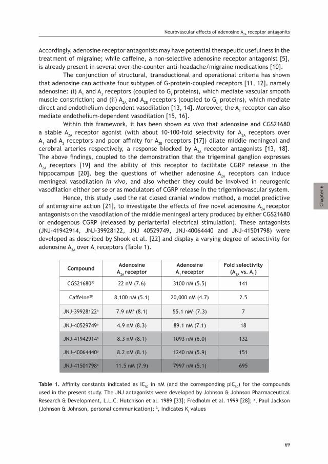
Chap
ter
6
Accordingly, adenosine receptor antagonists may have potential therapeutic usefulness in the treatment of migraine; while caffeine, a non-selective adenosine receptor antagonist [5], is already present in several over-the-counter anti-headache/migraine medications [10]. The conjunction of structural, transductional and operational criteria has shown that adenosine can activate four subtypes of G-protein-coupled receptors [11, 12], namely adenosine: (i) A1 and A3 receptors (coupled to Gi proteins), which mediate vascular smooth muscle constriction; and (ii) A2A and A2B receptors (coupled to Gs proteins), which mediate direct and endothelium-dependent vasodilation [13, 14]. Moreover, the A1 receptor can also mediate endothelium-dependent vasodilation [15, 16]. Within this framework, it has been shown ex vivo that adenosine and CGS21680 a stable A2A receptor agonist (with about 10-100-fold selectivity for A2A receptors over A1 and A3 receptors and poor affinity for A2B receptors [17]) dilate middle meningeal and cerebral arteries respectively, a response blocked by A2A receptor antagonists [13, 18]. The above findings, coupled to the demonstration that the trigeminal ganglion expresses A2A receptors [19] and the ability of this receptor to facilitate CGRP release in the hippocampus [20], beg the questions of whether adenosine A2A receptors can induce meningeal vasodilation in vivo, and also whether they could be involved in neurogenic vasodilation either per se or as modulators of CGRP release in the trigeminovascular system. Hence, this study used the rat closed cranial window method, a model predictive of antimigraine action [21], to investigate the effects of five novel adenosine A2A receptor antagonists on the vasodilation of the middle meningeal artery produced by either CGS21680 or endogenous CGRP (released by periarterial electrical stimulation). These antagonists (JNJ-41942914, JNJ-39928122, JNJ 40529749, JNJ-40064440 and JNJ-41501798) were developed as described by Shook et al. [22] and display a varying degree of selectivity for adenosine A2A over A1 receptors (Table 1).
Compound Adenosine A2A receptor
AdenosineA1 receptor
Fold selectivity (A2A vs. A1)
CGS2168033 22 nM (7.6) 3100 nM (5.5) 141
Caffeine28 8,100 nM (5.1) 20,000 nM (4.7) 2.5
JNJ-39928122a 7.9 nMλ (8.1) 55.1 nMλ (7.3) 7
JNJ-40529749a 4.9 nM (8.3) 89.1 nM (7.1) 18
JNJ-41942914a 8.3 nM (8.1) 1093 nM (6.0) 132
JNJ-40064440a 8.2 nM (8.1) 1240 nM (5.9) 151
JNJ-41501798a 11.5 nM (7.9) 7997 nM (5.1) 695
Table 1. Affinity constants indicated as IC50 in nM (and the corresponding pIC50) for the compounds used in the present study. The JNJ antagonists were developed by Johnson & Johnson Pharmaceutical Research & Development, L.L.C. Hutchison et al. 1989 [33]; Fredholm et al. 1999 [28]; a, Paul Jackson (Johnson & Johnson, personal communication); λ, Indicates Ki values
69
Neurovascular effects of adenosine A2A receptor antagonits
Materials and Methods
Intravital microscopy experiments
Animals
Fifty-seven normotensive male Sprague-Dawley rats (300-400 g), purchased from Harlan (Horst, The Netherlands), were maintained at a 12/12 h light dark cycle (with light beginning at 7 a.m.) and housed at a constant temperature (22±2 °C) and humidity (50%), with food and water ad libitum. Only male rats were used to avoid crosstalk between CGRP and hormonal fluctuations during the female estrus cycle [23]. The animals were anaesthetized with an intraperitoneal (i.p.) injection of sodium pentobarbital (60 mg/kg, followed by 18 mg/kg i.v. per hour when necessary). The adequacy of anesthesia was judged by a negative tail flick test and the absence of ocular reflexes, amongst others. All experimental protocols of this study were approved by our Institutional Ethics Committee [Erasmus MC; permission protocol number EMC 1931 (118-09-04], in accordance with the NIH guide for the Care and Use of Laboratory Animals in U.S.A. and the ARRIVE guidelines for reporting experiments in animals [24]. All rats were randomly assigned into the different experimental protocols (see experimental protocol section).
General methods
After anesthesia, the trachea was cannulated and connected to a pressure ventilator (small animal ventilator SAR-830 series, CWE Inc., Ardmore, PA, U.S.A.). End-tidal pCO2 was monitored (Capstar-100 CWE Inc., PA, U.S.A.) and kept between 35 and 48 mm Hg. The left femoral vein and artery were cannulated for intravenous (i.v.) administration of drugs and continuous monitoring of blood pressure, respectively. Two or three samples of blood (at the beginning and at the end of the experiment) were withdrawn via the femoral artery to monitor blood gases and other parameters, which were kept between normal values (pH: 7.35-7.48; pCO2: 35-48 mm Hg; pO2: 100-120 mm Hg). The body temperature of each rat was monitored via a rectal thermometer and maintained throughout the experiment (36.5-37.5°C) by a homeothermic blanket system for rodents (Harvard Instruments, Edenbridge, Kent, U.K.). The rats were placed in a stereotaxic frame and the parietal bone overlying a segment of the dural meningeal artery was carefully drilled thin, applying cold saline (4°C) until the artery was visible. Since skull drilling induces vasodilation, we allowed the animal to recover for 1 hour before the experimental protocol. The drilled area was covered with mineral oil to prevent drying and to facilitate visualization of the meningeal artery. The artery was captured with an intravital microscope (model MZ 16; Leica microsystem Ltd., Heerbrugg, Switzerland) using a cyan blue filter on a cold source of light. A zoom lens (80-450 × magnification) and a camera was used to display images with the blood vessel diameter (30-40 μm at baseline) being continuously monitored and measured with a video dimension analyzer (Living Systems Instrumentation Inc., Burlington, VT, U.S.A.). In rats where periarterial electrical stimulation was used to evoke dural vasodilation, a bipolar stimulating electrode (NE 200X, Clark Electromedical, Edenbridge, Kent, U.K.) was placed on the surface of the cranial window approximately within 200 µm from the vessel of interest. The cranial window surface was stimulated at 5 Hz, 1 ms for 10 s (Stimulator model S88, Grass Instruments, West Warwick, RI, U.S.A.). For neurogenic dural vasodilation, we initially
70
Chapter 6
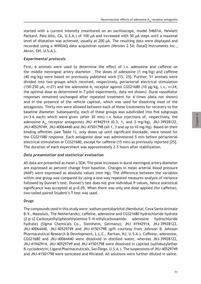
started with a current intensity (monitored on an oscilloscope, model 54601A, Hewlett Packard, Palo Alto, CA, U.S.A.) of 100 µA and increased with 50 µA steps until a maximal level of dilatation was achieved, usually at 200 µA. The resulting data were displayed and recorded using a WINDAQ data acquisition system (Version 2.54; DataQ Instruments Inc., Akron, OH, U.S.A.).
Experimental protocols
First, 6 animals were used to determine the effect of i.v. adenosine and caffeine on the middle meningeal artery diameter. The doses of adenosine (1 mg/kg) and caffeine (40 mg⁄kg) were based on previously published work [15, 25]. Further, 51 animals were divided into two groups which received, respectively, periarterial electrical stimulation (150-250 μA; n=27) and the adenosine A2 receptor agonist CGS21680 (10 μg/kg, i.v., n=24; the optimal dose as determined in 7 pilot experiments, data not shown). Dural vasodilator responses remained unchanged after repeated treatment for 4 times (data not shown) and in the presence of the vehicle captisol, which was used for dissolving most of the antagonists. Thirty min were allowed between each of these treatments for recovery to the baseline diameter. Subsequently, each of these groups was subdivided into five subgroups (n=3-6 each) which were given (after 30 min) i.v. bolus injections of, respectively, the adenosine A2A receptor antagonists JNJ 41942914 (0.3, 1, and 3 mg/kg), JNJ-39928122, JNJ-40529749, JNJ-40064440 and JNJ 41501798 (all 1, 3 and up to 10 mg/kg). Based on their binding affinities (see Table 1), only doses up until significant blockade, were tested for the CGS21580 response. Each antagonist dose was administered 5 min before periarterial electrical stimulation or CGS21680, except for caffeine (15 min) as previously reported [25]. The duration of each experiment was approximately 2.5 hours after stabilization.
Data presentation and statistical evaluation
All data are presented as mean ± SEM. The peak increases in dural meningeal artery diameter are expressed as percent change from baseline. Changes in mean arterial blood pressure (MAP) were expressed as absolute values (mm Hg). The difference between the variables within one group was compared by using a one-way repeated measures analysis of variance followed by Dunnet’s test. Dunnet’s test does not give individual P-values, hence statistical significance was accepted at p<0.05. When there was only one dose applied (for caffeine), two-tailed paired Student’s T-test was used.
Drugs
The compounds used in this study were: sodium pentobarbital (Nembutal; Ceva Sante Animale B.V., Maassluis, The Netherlands); caffeine, adenosine and CGS21680 hydrochloride hydrate (2-p-(2-Carboxyethyl)phenethylamino-5′-N-ethylcarboxamido adenosine hydrochloride hydrate) (Sigma Chemicals Co., Steinheim, Germany); JNJ 41942914, JNJ-39928122, JNJ-40064440, JNJ-40529749 and JNJ-41501798 (gift courtesy from Johnson & Johnson Pharmaceutical Research & Development, L.L.C., Raritan, NJ, U.S.A.). Caffeine, adenosine, CGS21680 and JNJ-40064440 were dissolved in distilled water, whereas JNJ-39928122, JNJ-41942914, JNJ-40529749 and JNJ-41501798 were dissolved in captisol (sulfobutylether β-cyclodextrin; Ligand Pharmaceuticals, San Diego, U.S.A.). The suspensions of JNJ-40529749 and JNJ-41501798 were sonicated and filtrated. All solutions were further diluted in saline.
71
Neurovascular effects of adenosine A2A receptor antagonits
Chap
ter
6
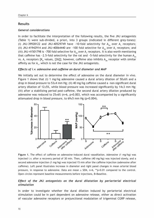
Results
General considerations
In order to facilitate the interpretation of the following results, the five JNJ antagonists (Table 1) were sub-divided, a priori, into 3 groups (indicated in different grey-tones): (i) JNJ-39928122 and JNJ-40529749 have ~10-fold selectivity for A2A over A1 receptors; (ii) JNJ-41942914 and JNJ-40064440 are ~100 fold selective for A2A over A1 receptors; and (iii) JNJ-41501798 is ~700 fold selective for A2A over A1 receptors. It is also worth mentioning that caffeine has ~2.5-fold selectivity for the rat and ~5-fold selectivity for the human A2A vs. A1 receptors [KD values, [26]]; however, caffeine also inhibits A2B receptor with similar affinity as for A1, which is not the case for the JNJ antagonists.
Effects of i.v. adenosine and caffeine on dural diameter and MAP
We initially set out to determine the effect of adenosine on the dural diameter in vivo. Figure 1 shows that (i) 1 mg/kg adenosine caused a dural artery dilation of 50±6% and a drop in blood pressure to 53±4 mm Hg; (ii) 40 mg/kg caffeine caused a non-significant dural artery dilation of 12±5%, while blood pressure was increased significantly by 14±3 mm Hg; (iii) after a stabilizing period post-caffeine, the second dural artery dilation produced by adenosine was reduced to 25±6% (n=6, p=0.003, which was accompanied by a significantly attenuated drop in blood pressure, to 69±5 mm Hg (p=0.004).
Figure 1. The effect of caffeine on adenosine-induced dural vasodilation. Adenosine (1 mg/kg) was injected i.v. after a recovery period of 30 min. Then, caffeine (40 mg/kg) was injected slowly, and a second adenosine injection (1 mg/kg) was injected 15 min after the caffeine injection (adenosine after caffeine). Left panel illustrates increase in diameter and right panel changes in mean arterial blood pressure, in response to adenosine. Data are mean ± SEM, n=6, **p<0.01 compared to the control. Open circles represent baseline measurements before injections, B=Baseline.
Effect of the JNJ antagonists on the dural dilatation by periarterial electrical stimulation
In order to investigate whether the dural dilation induced by periarterial electrical stimulation could be in part dependent on adenosine release, either as direct activation of vascular adenosine receptors or prejunctional modulation of trigeminal CGRP release,
72
Chapter 6
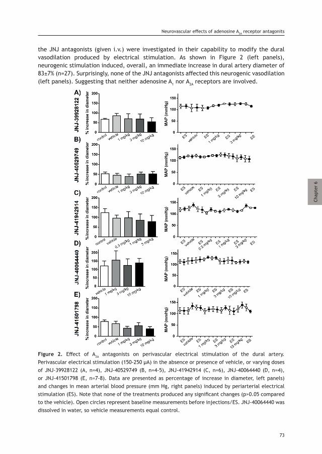
the JNJ antagonists (given i.v.) were investigated in their capability to modify the dural vasodilation produced by electrical stimulation. As shown in Figure 2 (left panels), neurogenic stimulation induced, overall, an immediate increase in dural artery diameter of 83±7% (n=27). Surprisingly, none of the JNJ antagonists affected this neurogenic vasodilation (left panels). Suggesting that neither adenosine A1 nor A2A receptors are involved.
Figure 2. Effect of A2A antagonists on perivascular electrical stimulation of the dural artery. Perivascular electrical stimulation (150-250 µA) in the absence or presence of vehicle, or varying doses of JNJ-39928122 (A, n=4), JNJ-40529749 (B, n=4-5), JNJ-41942914 (C, n=6), JNJ-40064440 (D, n=4), or JNJ-41501798 (E, n=7-8). Data are presented as percentage of increase in diameter, left panels) and changes in mean arterial blood pressure (mm Hg, right panels) induced by periarterial electrical stimulation (ES). Note that none of the treatments produced any significant changes (p>0.05 compared to the vehicle). Open circles represent baseline measurements before injections/ES. JNJ-40064440 was dissolved in water, so vehicle measurements equal control.
73
Neurovascular effects of adenosine A2A receptor antagonits
Chap
ter
6
The effect of the JNJ antagonists on MAP before and during neurogenic dural stimulation
As shown in Figure 2 (right panels), both periarterial electrical stimulation and the JNJ antagonists were devoid of any effect per se on MAP.
Effects of CGS21680 on dural artery diameter and MAP
Although adenosine A2A or A1 receptors did not appear to be important in the vasodilation observed after neurogenic dural stimulation, adenosine vasodilates dural arteries in vivo (Figure 2), most likely via both A2A and A2B receptors as previously reported ex vivo [13]. Since our study set out to study specifically the role of the adenosine A2A receptor, we continued our study using CGS21680, which is a more biologically stable, highly selective for A2A over A2B receptor agonist [17]. As shown in Figure 3, CGS21680 (10 µg/kg before administration of JNJ antagonists; n=24) mimicked adenosine in its capability to produce: (i) a marked dilation of the dural artery diameter (66±9%; left panels); and (ii) a drop in blood pressure (53±9 mm Hg; right panels) and hence excluding the involvement of A2B receptors.
The lower the selectivity (A2A over A1 receptors) the higher the potency of JNJ antagonists to block CGS21680-induced dural vasodilation
To further uncover the nature of the adenosine receptors in the dural vasculature, we explored the effect of the JNJ antagonist with varying selectivity (A2A over A1 receptors). Figure 3 (left panels) also shows that all JNJ antagonists significantly blocked the CGS21680-induced dural vasodilation with varying degrees of potency. Specifically, the vasodilation to CGS21680 was: (i) abolished by 1 mg/kg (1±2%) of JNJ-39928122 (Figure 3A); (ii) abolished at 1 mg/kg (-2±1%) of JNJ-40529749 (Figure 3B); (iii) significantly attenuated (but not abolished) by 3 mg/kg (21±11%) of JNJ-41942914 (Figure 3C); (iv) significantly attenuated by 3 mg/kg (23±15%) and abolished (1±3%) by 10 mg/kg of JNJ-40064440 (Figure 3D); and (v) dose-dependently blocked, and practically abolished by 10 mg/kg (5±4%) of JNJ-41501798 (Figure 3E). Clearly, the lower the selectivity of A2A vs. A1 (Table1) the higher the potency of JNJ antagonists to block CGS21680-induced dural vasodilation.
Effect of JNJ antagonists on CGS21680-induced vasodepressor responses
Similarly, the vasodepressor responses to CGS21680 were blocked by the JNJ antagonists as follows: (i) very potently by the less selective antagonists JNJ-39928122 and JNJ-40529749; and (ii) less potently by the highest doses of the more selective antagonists JNJ-41942914, JNJ-40064440 and JNJ-41501798, which display from low to very low affinity for the adenosine A1 receptor (Table 1).
74
Chapter 6
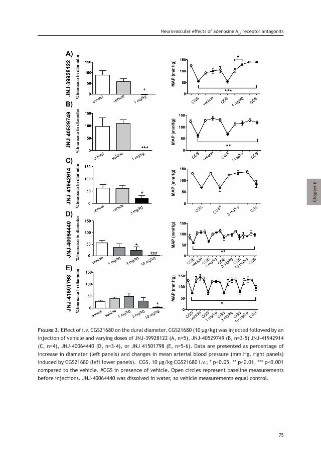
Figure 3. Effect of i.v. CGS21680 on the dural diameter. CGS21680 (10 µg/kg) was injected followed by an injection of vehicle and varying doses of JNJ-39928122 (A, n=5), JNJ-40529749 (B, n=3-5) JNJ-41942914 (C, n=4), JNJ-40064440 (D, n=3-4), or JNJ 41501798 (E, n=5-6). Data are presented as percentage of increase in diameter (left panels) and changes in mean arterial blood pressure (mm Hg, right panels) induced by CGS21680 (left lower panels). CGS, 10 µg/kg CGS21680 i.v.; * p<0.05, ** p<0.01, *** p<0.001 compared to the vehicle. #CGS in presence of vehicle. Open circles represent baseline measurements before injections. JNJ-40064440 was dissolved in water, so vehicle measurements equal control.
75
Neurovascular effects of adenosine A2A receptor antagonits
Chap
ter
6
Discussion
Comparison between in vivo and in vitro vascular responses to adenosine
The adenosine receptor antagonists SCH58261 [478-fold A2A over A1 selective [27]] and caffeine [non-selective A1/2A/2B [28]] have been shown to block the ex vivo adenosine-induced dilation of endothelium-denuded middle meningeal arteries [18]. In these experiments, not only did caffeine (50 µM) or SCH58261 (1 µM) prevent the dural dilation, but a vasoconstriction to adenosine was unmasked. Interestingly, this effect was not observed in vivo, which could be due to the fact that the artery used for the myograph (outer diameter ~ 100 µm) had a larger diameter than in this study (outer diameter ~ 35 µm) and that there potentially are less adenosine A3 receptors expressed in smaller vessels, as we see no indirect involvement of A3 (i.e. vasoconstriction) in the current experiments. These differences require further investigation, but it is known that receptor expression changes along different vascular beds [29].
General considerations
In addition to the implications discussed below, the present study shows that: (i) both adenosine and CGS21680 produced rat dural vasodilation in vivo; and (ii) for JNJ antagonists, the lower the selectivity (A2A over A1 receptors) the higher the potency to block the dural vasodilation and vasodepressor responses induced by CGS21680 (implying that blockade of adenosine A1 receptors is also necessary to completely block the dural vasodilation in vivo). The latter finding is most likely due to endothelial A1 receptors, as the main difference between the in vivo (present study) and the ex vivo studies [18] is the absence of endothelium. Indeed, Honey et al. [21] have shown the presence of adenosine A1 receptors mediating vasodilation in the rat middle meningeal artery in vivo.
The potential role of A2A and A1 receptors in the dural vasodilation as prejunctional modulators of neurogenic dural vasodilation or produced by CGS21680
The simplest interpretation of the fact that the JNJ antagonists had no effect on neurogenic dural vasodilation (Figure 2), which involves CGRP release [1], implies that: (i) adenosine is not released by periarterial electrical stimulation; (ii) A2A receptors do not constitute a positive feedback mechanism for CGRP release, as expected from its transductional properties [positive coupling to Gs proteins; [11]]; or (iii) cAMP increase, induced by CGRP, is so high that this could have masked the small increase in cAMP levels mediated by A2A receptors [26]. Interestingly, adenosine A1 receptors [coupled to Gi proteins; [11]] can produce a prejunctional inhibition of the neurogenic dural vasodilation in rats [21]. However, the weakly selective JNJ antagonists (JNJ-39928122 and JNJ-40529749), which would be theoretically expected to block (at least in part) this mechanism, did not increase neurogenic dural vasodilation (Figure 2). Several lines of evidence have previously shown in other systems that: (i) the vasodilation produced by adenosine and related agonists is mainly mediated by vascular and endothelial A2A receptors [13, 14] as well as by endothelial A1 receptors [16]; and (ii) the trigeminovascular system expresses adenosine A2A receptors [19]. In keeping with these findings, our results further demonstrate that the JNJ antagonists blocked CGS21680-induced dural vasodilation (Figure 3), with a different profile of blockade (dependent on
76
Chapter 6
A2A vs. A1 selectivity; see below). This reinforces the involvement of adenosine A2A and, probably to a lesser extent, of A1 receptors. In addition, based on the poor affinity of CGS21680 for the A2B receptors [17] and similar responses to adenosine, our data did not show any strong involvement of the A2B receptors.
Systemic effects of JNJ antagonists on A2A and A1 receptors
Caffeine is a non-selective adenosine A1, A2A and A2B receptor antagonist that does not affect adenosine A3 receptors at the doses used [28]. Accordingly, caffeine produced a slight increase in blood pressure (Figure 1), as previously reported [25]. Interestingly, the fact that none of the JNJ antagonists increased blood pressure (Figure 3, right panel), even at doses that blocked the dural vasodilation to CGS21680 (Figure 3, left panel) suggests that there is no strong “adenosine vascular tone”. In addition, it is worth emphasizing that adenosine A2B receptors are involved in the blood pressure effects of adenosine [30], which would explain the minor difference between caffeine and the JNJ antagonists in our study. It is well established that A2A receptor agonists lower blood pressure [12, 31]. The A1 receptor agonists GR79236 and N6-cyclopentyladenosine (CPA), although less studied, also decrease blood pressure with higher potency than CGS21680, and both cause direct production of endothelial NO [15, 16, 31]. Hence, the vasodepressor response to adenosine in A1 -/- mice is reduced [32]. In the present study, the less selective (JNJ-39928122 and JNJ-40529749) A2A vs. A1 antagonists potently blocked the decrease in blood pressure, whereas the more selective (JNJ-40064440 and JNJ-41501798) A2A antagonists were less potent, and only effective at 10 mg/kg. These high doses of JNJ-4006440 and JNJ-41501798 also induced inhibition of A1 receptors. Blockade of the adenosine A2A and A1 receptors prevents systemic vasodilation in response to adenosine, and therefore the block in blood pressure.
In vivo effects of CGS21680
In binding affinity studies, CGS21680 is 141-fold selective for A2A over A1 receptors [33]. However, our study raises the concern whether CGS21680 is a specific A2A receptor agonist in vivo in rats, as it appears that higher blocking affinities for the A1 receptor causes a more potent blockade of the vasodepressor and dural vasodilator responses. For the human adenosine receptors, the selectivity for A2A over A1 receptors is minimal [34]. The most obvious explanation for the apparent discrepancy between the binding affinity selectively and the in vivo effects, is the location of adenosine receptors, as A1 receptors are on the endothelium, whereas the A2A receptors are mainly located on vascular smooth muscle [12]; hence the endothelium will be directly exposed to an apparently higher concentration. In addition, there are opposing findings on the selectivity of CGS21680. For example CGS21680 binds with high affinity (around 1 nM) to adenosine A1 receptors in the hippocampus of A2A -/- mice [35], in contrast, in the same mice CGS21680 had no effect on blood pressure [36]. Comparing our findings with previous studies in rats, the vasodepressor response to CGS21680 (10 µg/kg) was completely blocked by 3 mg/kg of the A2A receptor antagonists ZM241385 [319-fold A2A over A1; [15, 27]] or CGS15943 [9 fold A2A over A1; [37]]. Clearly, ZM241385 has a higher A2A over A1 selectivity, but its Ki for A1 receptors is 255 nM. Since these binding data are similar to those of our less selective compounds, A2A and also A1 receptors would be blocked in these studies.
77
Neurovascular effects of adenosine A2A receptor antagonits
Chap
ter
6
Possible clinical implications
On the basis of the above lines of evidence, the antimigraine potential of selective adenosine A2A receptor antagonists would be of particular relevance in those patients whose adenosine plasma levels are markedly increased during a migraine attack. Although our findings indicate that adenosine is not released by perivascular electrical stimulation, inhibition of dural vasodilation is a shared mechanism of current (ergots and triptans) and prospective (CGRP (receptor) antagonists and antibodies) antimigraine drugs [1, 38]. Whether this (antimigraine) mechanism alone is sufficient to attenuate the trigeminal nociceptive transmission associated with migraine headache, remains to be determined. Additionally, other studies have shown that: (i) activation of A2A receptors facilitates the action of CGRP and VIP in the rat hippocampus [20]; (ii) A2A receptor knockout mice are hypoalgesic [36]; and (iii) A2A receptors are expressed in the rat trigeminovascular system [19] as well as in the rat trigeminal ganglion, together with A1, A2B and A3 receptors [18]. Furthermore, intra-articular administration of adenosine and N6-cyclohexyladenosine (CHA, an adenosine A1 receptor agonist), but not CGS21680, significantly increased ketorolac antinociception [39]. These findings, taken together: (i) argue in favor of selective blockade of adenosine A2 receptors as a potential antimigraine strategy; and (ii) imply that blockade of A1 receptors would be a disadvantage in antimigraine treatment. Obviously, further clinical studies should evaluate the JNJ antagonist(s) with the optimal oral bioavailability based on their pharmacokinetic properties.
Conclusion
In conclusion, all the JNJ antagonists were capable of blocking CGS21680-induced dural vasodilation without affecting neurogenic dural vasodilation (suggesting no modulation of trigeminal CGRP release). This blockade was more potent when showing lower A2A over A1 selectivity, and that both these receptors are involved in the dural artery vasodilation. On this basis, and considering that the JNJ antagonist were devoid of any effect per se on blood pressure, selective A2A receptor antagonism may offer a novel approach to antimigraine therapy that remains to be determined in clinical trials.
References1. Edvinsson L, Villalón CM, MaassenVanDenBrink A. Basic mechanims of migraine and its acute treatment. Pharmacol-ogy and Therapeutics 2012; 136: 319-333.2. Goadsby PJ, Edvinsson L. The trigeminovascular system and migraine: studies characterizing cerebrovascular and neuropeptide changes seen in humans and cats. Ann Neurol 1993; 33: 48-56.3. Mitsikostas DD, Reuter U. Calcitonin gene-related peptide monoclonal antibodies for migraine prevention: compar-isons across randomized controlled studies. Curr Opin Neurol 2017; 30: 272-280.4. Deen M, Correnti E, Kamm K, Kelderman T, Papetti L, Rubio-Beltrán E, et al. Blocking CGRP in migraine patients - a review of pros and cons. J Headache Pain 2017; 18:96.5. Shapiro RE. Caffeine and headaches. Neurol Sci 2007; 28 Suppl 2: S179-183.6. Guieu R, Devaux C, Henry H, et al. Adenosine and migraine. Can J Neurol Sci 1998; 25: 55-58.7. Brown SG, Waterer GW. Migraine precipitated by adenosine. Med J Aust 1995; 162: 389, 391.8. Hawkes CH. Dipyridamole in migraine. Lancet 1978; 2: 153.9. Hohoff C, Marziniak M, Lesch KP, et al. An adenosine A2A receptor gene haplotype is associated with migraine with aura. Cephalalgia 2007; 27: 177-181.10. Derry CJ, Derry S, Moore RA. Caffeine as an analgesic adjuvant for acute pain in adults. Cochrane Database Syst Rev 2014; 12: CD009281.11. Burnstock G. Purine and pyrimidine receptors. Cell Mol Life Sci 2007; 64: 1471-1483.12. Burnstock G, Ralevic V.Purinergic signaling and blood vessels in health and disease. Pharma Rev 2014; 66: 102-192.
78
Chapter 6
13. Ngai AC, Coyne EF, Meno JR, et al. Receptor subtypes mediating adenosine-induced dilation of cerebral arterioles. Am J Physiol Heart Circ Physiol 2001; 280: H2329-H2335.14. Shin HK, Park SN, Hong KW. Implication of adenosine A2A receptors in hypotension-induced vasodilation and cere-bral blood flow autoregulation in rat pial arteries. Life Sci 2000; 67: 1435-1445.15. Kirkup AJ, Eastwood C, Grundy D, et al. Characterization of adenosine receptors evoking excitation of mesenteric afferents in the rat. Br J Pharmacol 1998; 125: 1352-1360.16. Ray CJ, Marshall JM. The cellular mechanisms by which adenosine evokes release of nitric oxide from rat aortic endothelium. J Physiol 2006; 570: 85-96.17. Liang BT, Urso M, Zambraski E, et al. Adenosine A3 receptors in muscle protection. A3 Adenosine Receptors from Cell Biology to Pharmacology and Therapeutics: Springer; 2010. p. 257-280.18. Haanes KA, Edvinsson L. Expression and characterization of purinergic receptors in rat middle meningeal ar-tery-potential role in migraine. PLoS One 2014; 9:e108782.19. Lu W, Li B, Chen J, et al. Expression of calcitonin gene-related peptide, adenosine A2a receptor and adenosine A1 receptor in experiment rat migraine models. Biomed Rep 2016; 4: 379-383.20. Sebastiao AM, Macedo MP, Ribeiro JA. Tonic activation of A(2A) adenosine receptors unmasks, and of A(1) receptors prevents, a facilitatory action of calcitonin gene-related peptide in the rat hippocampus. Br J Pharmacol 2000; 129: 374-380.21. Honey AC, Bland-Ward PA, Connor HE, et al. Study of an adenosine A1 receptor agonist on trigeminally evoked dural blood vessel dilation in the anaesthetized rat. Cephalalgia 2002; 22: 260-264.22. Shook BC, Rassnick S, Hall D. Methylene amine substituted arylindenopyrimidines as potent adenosine A(2A)/A(1) antagonists. Bioorg Med Chem Lett 2010; 20: 2864-2867.23.Labastida-Ramírez A, Rubio-Beltrán E, Villalón CM, et al. Gender aspects of CGRP in migraine. Cephalalgia 2017; 333102417739584. https://doi.org/10.1177/033310241773958424. McGrath J, Drummond G, McLachlan, et al. Guidelines for reporting experiments involving animals: the ARRIVE guidelines. Br J Pharmacol 2010; 160: 1573-1576.25. Meno JR, Nguyen TS, Jensen EM, et al. Effect of caffeine on cerebral blood flow response to somatosensory stim-ulation. J Cereb Blood Flow Metab 2005; 25: 775-784.26. Arslan G, Kull B, Fredholm BB. Signaling via A2A adenosine receptor in four PC12 cell clones. Naunyn Schmiede-bergs Arch Pharmacol 1999; 359: 28-32.27. Ongini E, Dionisotti S, Gessi S, et al. Comparison of CGS 15943, ZM 241385 and SCH 58261 as antagonists at human adenosine receptors. Naunyn Schmiedebergs Arch Pharmacol 1999; 359: 7-10.28. Fredholm BB, Battig K, Holmen J, et al. Actions of caffeine in the brain with special reference to factors that contribute to its widespread use. Pharmacol Rev 1999; 51: 83-133.29. Haanes KA, Spray S, Syberg S, et al. New insights on pyrimidine signalling within the arterial vasculature - Different roles for P2Y2 and P2Y6 receptors in large and small coronary arteries of the mouse. J Mol Cell Cardiol 2016; 93: 1-11.30. Teng B, Fil D, Tilley SL, et al. Functional and RNA expression profile of adenosine receptor subtypes in mouse mesenteric arteries. J Cardiovasc Pharmacol 2013; 61: 70-76.31. Schindler CW, Karcz-Kubicha M, Thorndike EB, et al. Role of central and peripheral adenosine receptors in the cardiovascular responses to intraperitoneal injections of adenosine A1 and A2A subtype receptor agonists. Br J Phar-macol 2005; 144: 642-650.32. Koeppen M, Eckle T, Eltzschig HK. Selective deletion of the A1 adenosine receptor abolishes heart-rate slowing effects of intravascular adenosine in vivo. PLoS One 2009; 4: e6784.33. Hutchison AJ, Webb RL, Oei HH, et al. CGS 21680C, an A2 selective adenosine receptor agonist with preferential hypotensive activity. J Pharmacol Exp Ther 1989; 251: 47-55.34. de Lera Ruiz M, Lim YH, Zheng J. Adenosine A2A receptor as a drug discovery target. J Med Chem 2014; 57: 3623-3650.35. Halldner L, Lopes LV, Dare E, et al. Binding of adenosine receptor ligands to brain of adenosine receptor knock-out mice: evidence that CGS 21680 binds to A1 receptors in hippocampus. Naunyn Schmiedebergs Arch Pharmacol 2004; 370: 270-278.36. Ledent C, Vaugeois JM, Schiffmann SN, et al. Aggressiveness, hypoalgesia and high blood pressure in mice lacking the adenosine A2a receptor. Nature 388: 674-678.37. Patel M, Sheehan MJ, Strong P. Failure of CGS15943A to block the hypotensive action of agonists acting at the adenosine A3 receptor. Br J Pharmacol 1994; 113: 741-748.38. Rivera-Mancilla E, Aviles-Rosas VH, Manrique-Maldonado G, et al. The role of alpha1- and alpha2-adrenoceptor subtypes in the vasopressor responses induced by dihydroergotamine in ritanserin-pretreated pithed rats. J Headache Pain 2017; 18: 104.39. Aguirre-Banuelos P, Castaneda-Hernandez G, Lopez-Munoz FJ, et al. Effect of coadministration of caffeine and either adenosine agonists or cyclic nucleotides on ketorolac analgesia. Eur J Pharmacol 1999; 377: 175-182.
79
Neurovascular effects of adenosine A2A receptor antagonits
Chap
ter
6

Chapter 7
Exploration of purinergic receptors as potential antimigraine targets using established pre-clinical migraine models
Based on: Cephalalgia, 2019, In press.
Haanes KA, Labastida-Ramírez A, Blixt FW, Rubio-Beltrán E, Dirven CM, Danser AHJ, Edvinsson L and MaassenVanDenBrink A.
Abstract
Background: The current understanding of mechanisms behind migraine pain has been greatly enhanced with the recent therapies targeting calcitonin gene-related peptide (CGRP) and its receptor. The clinical efficacy of CGRP-blocking drugs indicates that, at least in a considerable proportion of patients, CGRP is a key molecule in migraine pain. There are several receptors and molecular pathways that can affect the release of and response to CGRP. One of these could be purinergic receptors that are involved in nociception, but are greatly understudied with respect to migraine.
Objective: We aimed to explore purinergic receptors as potential antimigraine targets.
Methods: We used the human middle meningeal artery as a proxy for the trigeminal system to screen for possible antimigraine candidates. The human findings were followed by intravital microscopy and CGRP release measurements in rodents.
Results: We show that purinergic P2Y13 receptor fulfils all the features of a potential antimigraine target. The P2Y13 receptor is expressed in both the human trigeminal ganglion and middle meningeal artery and activation of this receptor causes: i) middle meningeal artery contraction in vitro; ii) reduced dural artery dilation following periarterial electrical stimulation in vivo and iii) a reduction of CGRP release from both the dura and the trigeminal ganglion in situ. Furthermore, we show that P2X3 receptor activation of the trigeminal ganglion causes CGRP release and middle meningeal artery dilation.
Conclusion: Both an agonist directed at the P2Y13 receptor and an antagonist of the P2X3 receptor seem to be viable potential antimigraine therapies.
Background
The therapies for migraine patients have recently made a huge step forward with the arrival of the monoclonal antibodies targeting calcitonin gene-related peptide (CGRP) or its receptor [1]. Apart from directly scavenging CGRP or blocking its receptor, future therapies could also be directed at regulating the cellular effect of CGRP or the neuronal release of CGRP or other neuropeptides. Currently available, acutely acting antimigraine drugs, the triptans, have also been demonstrated to inhibit CGRP release [2] which could be one of their most important therapeutic actions. Neither the origin of a migraine attack, nor the cause of migraine pain is fully understood. In contrast, the involvement of the CGRPergic system in migraine has been clearly recognized [1]. For example, CGRP is released during a migraine attack, and mitigation of the migraine pain after the use of triptans is accompanied by a normalization of CGRP levels [3]. Hence, molecular targets that can prevent CGRP release or counteract the effects of CGRP are viable targets for antimigraine therapy, as exemplified by the fact that all CGRP-targeted therapies thus far have been successful [4]. An alternative approach to affect the CGRPergic system may be targeting the purinergic receptors because: i) purinergic receptors are involved in pain, ii) a subgroup of purinergic receptors initiates similar signaling pathways as the triptans and iii) in other systems, purinergic receptors have been shown to modulate neurotransmitter release. There are, in all, 19 purinergic receptors known today (18 in rodents), and there is increasing evidence for the involvement of these receptors as modulators or tuners of
82
Chapter 7
Chap
ter
7
cellular responses [5]. Purinergic receptors are divided into two main groups: the adenine nucleoside receptors (A/P1 receptors) and the purine/pyrimidine nucleotide receptors (P2 receptors). Furthermore, the P2 receptors are divided into P2X receptors (P2X1-7), which are ligand-gated ion channels, and P2Y receptors (P2Y1,2,4,6,12-14), which are G protein coupled receptors [5]. The purinergic field is relatively new, which is exemplified by the presence of only one marketed drug that targets purinergic receptors, namely the anti-coagulant clopidogrel [6]. Purinergic research in migraine has mainly been focusing on ATP and the role in neuronal sensitisation by acting on the low threshold ligand gated P2X3 receptor ion channel [7]. However, there is also data on the high ATP threshold P2X7 receptor in migraine models [8]. Focusing on the P2X3 receptor, increased expression in trigeminal ganglion (TG) neurons was observed in vitro after exposure to CGRP and in vivo following induction of trigeminal-associated pain [9, 10]. Activation of the P2X receptors might explain the painful sensation of ATP [11]. In contrast, the more modulatory P2Y receptors have been given little attention and to our knowledge only one in vitro study, investigating calcium signalling in primary neuron-glial trigeminal cultures, exists on the role of P2Y receptors [12]. Activation of three purinergic receptors, the P2Y12, P2Y13 and P2Y14 receptors, leads to a decrease in intracellular cAMP [13], as these receptors are coupled to Gi proteins. This is similar to the activation of 5-HT1B/1D receptors, which are the pharmacological target of the triptans [14]. Activation of the 5-HT1B/1D receptors leads to vasoconstriction of the middle meningeal artery (MMA) and reduces the release of CGRP from peripheral C-fibers [15]. In contrast to the 5-HT1B/1D receptors, with an abundance of data available, there is limited evidence for the negative modulation of presynaptic transmission of sympathetic [16] and cholinergic nerves [17] following ADP-induced activation of the P2Y12 or P2Y13 receptors. It is not clear yet whether a similar action could exist for trigeminal nerves, which would have implications for the release of CGRP. We therefore embarked on a study with the aim to uncover purinergic targets in line with the neurovascular theory of migraine. We hypothesized that P2Y receptors could play an important modulatory role in the pathophysiology of migraine and that P2X receptors might be important in the initiation of a migraine attack. Using the human MMA (hMMA), a potential direct target for antimigraine medication, as well as proxy for the trigeminal system, combined with intravital microscopy and CGRP release in rodents, we studied both a direct vasoactive effect, as well as a modulatory effect on CGRP release, by purinergic receptor stimulation in the trigeminal system.
Materials and Methods
All studies on animals were performed between 09:00 and 18:00, either on explanted organs or in the laboratory under anesthesia (see below).
Myograph studies in hMMA
Samples of dura mater were perioperatively obtained from 10 patients (two males and eight females; 48-69 years) undergoing neurosurgical procedures. Tissues were collected in a sterile organ-protecting solution and were immediately brought to the laboratory. Subsequently, hMMAs (internal diameter 0.5–1.5 mm) were dissected and used the same day or stored overnight in cold oxygenated Krebs solution of the following composition (mM):
Purinergic receptors as pontential antimigraine targets
83
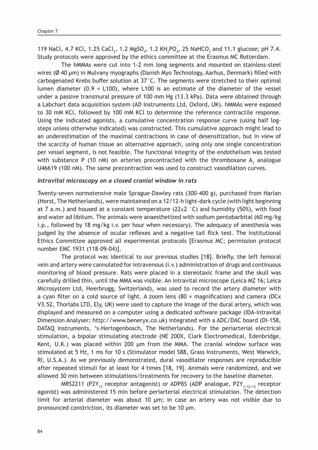
119 NaCl, 4.7 KCl, 1.25 CaCl2, 1.2 MgSO4, 1.2 KH2PO4, 25 NaHCO3 and 11.1 glucose; pH 7.4. Study protocols were approved by the ethics committee at the Erasmus MC Rotterdam. The hMMAs were cut into 1-2 mm long segments and mounted on stainless-steel wires (Ø 40 μm) in Mulvany myographs (Danish Myo Technology, Aarhus, Denmark) filled with carbogenated Krebs buffer solution at 37°C. The segments were stretched to their optimal lumen diameter (0.9 × L100), where L100 is an estimate of the diameter of the vessel under a passive transmural pressure of 100 mm Hg (13.3 kPa). Data were obtained through a Labchart data acquisition system (AD Instruments Ltd, Oxford, UK). hMMAs were exposed to 30 mM KCl, followed by 100 mM KCl to determine the reference contractile response. Using the indicated agonists, a cumulative concentration response curve (using half log-steps unless otherwise indicated) was constructed. This cumulative approach might lead to an underestimation of the maximal contractions in case of desensitization, but in view of the scarcity of human tissue an alternative approach, using only one single concentration per vessel segment, is not feasible. The functional integrity of the endothelium was tested with substance P (10 nM) on arteries precontracted with the thromboxane A2 analogue U46619 (100 nM). The same precontraction was used to construct vasodilation curves.
Intravital microscopy on a closed cranial window in rats
Twenty-seven normotensive male Sprague-Dawley rats (300-400 g), purchased from Harlan (Horst, The Netherlands), were maintained on a 12/12-h light-dark cycle (with light beginning at 7 a.m.) and housed at a constant temperature (22±2 °C) and humidity (50%), with food and water ad libitum. The animals were anaesthetized with sodium pentobarbital (60 mg⁄kg i.p., followed by 18 mg/kg i.v. per hour when necessary). The adequacy of anesthesia was judged by the absence of ocular reflexes and a negative tail flick test. The Institutional Ethics Committee approved all experimental protocols [Erasmus MC; permission protocol number EMC 1931 (118-09-04)]. The protocol was identical to our previous studies [18]. Briefly, the left femoral vein and artery were cannulated for intravenous (i.v.) administration of drugs and continuous monitoring of blood pressure. Rats were placed in a stereotaxic frame and the skull was carefully drilled thin, until the MMA was visible. An intravital microscope (Leica MZ 16; Leica Microsystem Ltd, Heerbrugg, Switzerland), was used to record the artery diameter with a cyan filter on a cold source of light. A zoom lens (80 × magnification) and camera (DCx V3.52, Thorlabs LTD, Ely, UK) were used to capture the image of the dural artery, which was displayed and measured on a computer using a dedicated software package (IDA-Intravital Dimension Analyser; http://www.beneryx.co.uk) integrated with a ADC/DAC board (DI-158, DATAQ instruments, ‘s-Hertogenbosch, The Netherlands). For the periarterial electrical stimulation, a bipolar stimulating electrode (NE 200X, Clark Electromedical, Edenbridge, Kent, U.K.) was placed within 200 μm from the MMA. The cranial window surface was stimulated at 5 Hz, 1 ms for 10 s (Stimulator model S88, Grass Instruments, West Warwick, RI, U.S.A.). As we previously demonstrated, dural vasodilator responses are reproducible after repeated stimuli for at least for 4 times [18, 19]. Animals were randomized, and we allowed 30 min between stimulations/treatments for recovery to the baseline diameter. MRS2211 (P2Y13 receptor antagonist) or ADPβS (ADP analogue, P2Y1/12/13 receptor agonist) was administered 15 min before periarterial electrical stimulation. The detection limit for arterial diameter was about 10 μm; in case an artery was not visible due to pronounced constriction, its diameter was set to be 10 μm.
84
Chapter 7
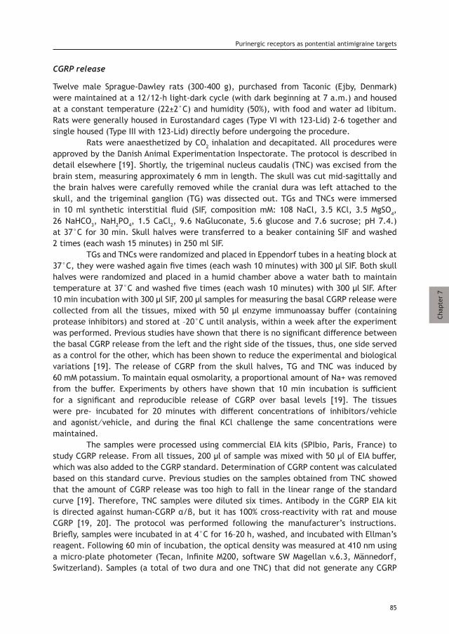
CGRP release
Twelve male Sprague-Dawley rats (300-400 g), purchased from Taconic (Ejby, Denmark) were maintained at a 12/12-h light-dark cycle (with dark beginning at 7 a.m.) and housed at a constant temperature (22±2°C) and humidity (50%), with food and water ad libitum. Rats were generally housed in Eurostandard cages (Type VI with 123-Lid) 2-6 together and single housed (Type III with 123-Lid) directly before undergoing the procedure. Rats were anaesthetized by CO2 inhalation and decapitated. All procedures were approved by the Danish Animal Experimentation Inspectorate. The protocol is described in detail elsewhere [19]. Shortly, the trigeminal nucleus caudalis (TNC) was excised from the brain stem, measuring approximately 6 mm in length. The skull was cut mid-sagittally and the brain halves were carefully removed while the cranial dura was left attached to the skull, and the trigeminal ganglion (TG) was dissected out. TGs and TNCs were immersed in 10 ml synthetic interstitial fluid (SIF, composition mM: 108 NaCl, 3.5 KCl, 3.5 MgSO4, 26 NaHCO3, NaH2PO4, 1.5 CaCl2, 9.6 NaGluconate, 5.6 glucose and 7.6 sucrose; pH 7.4.) at 37°C for 30 min. Skull halves were transferred to a beaker containing SIF and washed 2 times (each wash 15 minutes) in 250 ml SIF. TGs and TNCs were randomized and placed in Eppendorf tubes in a heating block at 37°C, they were washed again five times (each wash 10 minutes) with 300 μl SIF. Both skull halves were randomized and placed in a humid chamber above a water bath to maintain temperature at 37°C and washed five times (each wash 10 minutes) with 300 μl SIF. After 10 min incubation with 300 μl SIF, 200 μl samples for measuring the basal CGRP release were collected from all the tissues, mixed with 50 μl enzyme immunoassay buffer (containing protease inhibitors) and stored at –20°C until analysis, within a week after the experiment was performed. Previous studies have shown that there is no significant difference between the basal CGRP release from the left and the right side of the tissues, thus, one side served as a control for the other, which has been shown to reduce the experimental and biological variations [19]. The release of CGRP from the skull halves, TG and TNC was induced by 60 mM potassium. To maintain equal osmolarity, a proportional amount of Na+ was removed from the buffer. Experiments by others have shown that 10 min incubation is sufficient for a significant and reproducible release of CGRP over basal levels [19]. The tissues were pre- incubated for 20 minutes with different concentrations of inhibitors/vehicle and agonist⁄vehicle, and during the final KCl challenge the same concentrations were maintained. The samples were processed using commercial EIA kits (SPIbio, Paris, France) to study CGRP release. From all tissues, 200 μl of sample was mixed with 50 μl of EIA buffer, which was also added to the CGRP standard. Determination of CGRP content was calculated based on this standard curve. Previous studies on the samples obtained from TNC showed that the amount of CGRP release was too high to fall in the linear range of the standard curve [19]. Therefore, TNC samples were diluted six times. Antibody in the CGRP EIA kit is directed against human-CGRP α/β, but it has 100% cross-reactivity with rat and mouse CGRP [19, 20]. The protocol was performed following the manufacturer’s instructions. Briefly, samples were incubated in at 4°C for 16–20 h, washed, and incubated with Ellman’s reagent. Following 60 min of incubation, the optical density was measured at 410 nm using a micro-plate photometer (Tecan, Infinite M200, software SW Magellan v.6.3, Männedorf, Switzerland). Samples (a total of two dura and one TNC) that did not generate any CGRP
Purinergic receptors as pontential antimigraine targets
85
Chap
ter
7

release in response to 60 mM potassium, and one TG with baseline over 100 pg/ml CGRP was not included in the analysis. Since data were paired, both the left and right side were excluded in the above cases.
Immunohistochemistry
Human TGs were collected at autopsy, within 48 h post-mortem, from four patients (two males and two females; 68-82 years). The patients were without disorders related to the peripheral and central nervous system. The specimens were fixed after removal in 2% paraformaldehyde (PFA) and 0.2% picric acid in 0.1 mol/L phosphate buffer, pH 7.2, tyrode solution. The tissue samples were kept at −80 °C until embedding and cryo-sectioning. The study followed the guidelines of the European Communities Council (86/609/ECC) in accordance with the Szeged University Medical School guidelines for ethics in human tissue experiments and was approved by the local Hungarian Ethics Committee. The hMMA was obtained from the same patients that were used for the myograph studies. Study protocols were approved by the ethics committee at the Erasmus MC, Rotterdam. Human TGs and MMAs were cryosectioned. Details on immunohistochemistry are described elsewhere [21]. In short, slides with 10 μm sections were washed in PBS containing 2.5% Triton (PBS-T) for 15 min. Next, the primary antibody, rabbit anti-P2Y13 receptor (Alomone, APR#017, 1:50), rabbit anti-P2X3 receptor (Alomone, APR#026, 1:200) or mouse anti-CGRP (Abcam Ab81887, 1:100) was added. Following incubation overnight in moisturized chambers (+8°C), cryosections were washed in PBS-T twice for 15 min and incubated for 1 hour with secondary antibodies (FITC anti-mouse, Jackson Immunoresearch 1:200, and Cy3 anti-rabbit, Jackson Immunoresearch, 1:300) in the dark. After three washes for 15 min, the slides were mounted with Vectashield mounting medium containing DAPI (4′,6-diamidino-2-phenylindole, Vector Laboratories, Burlingame CA, USA). Negative controls were performed omitting the primary antibody. Immunofluorescence was observed using an epifluorescence microscope (Nikon 80i, Tokyo, Japan) combined with a Nikon DS-2MV camera. The images were then processed using Adobe Photoshop CS3 (v10.0 Adobe 3 Systems, Mountain View, CA, USA). Potential co-localization was determined by superimposing the images taken with different wavelength filters.
Data presentation
All quantitative data are presented as mean ± SEM. The n-number represents one rat or one human. The peak increases in dural meningeal artery diameter, as well as changes in mean arterial blood pressure (MAP, in vivo experiment) were expressed as percent change from baseline. The difference between two concentration response curves was determined by two-way ANOVA and the Bonferroni post-test. The difference between the variables within one group was compared by using a one-way repeated measures analysis of variance followed by Dunnet’s test. When only two variables were compared, a paired Student’s t-test was used.
Compounds
The compounds used in this study were: sodium pentobarbital (Nembutal; Ceva Sante Animale B.V., Maassluis, The Netherlands); MRS2211, αβmetATP, olcegepant (Tocris); ADPβS, UDPβS, UTPγS, ATPγS (Biolog.de) and CGRP (NeoMPS S.A., Strasbourg, France). All other chemicals were from Sigma Chemicals Co. (Steinheim, Germany). All compounds were
86
Chapter 7
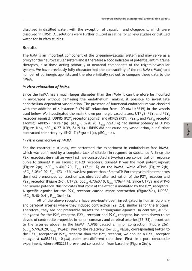
dissolved in distilled water, with the exception of capsaicin and olcegepant, which were dissolved in DMSO. All solutions were further diluted in saline for in vivo studies or distilled water for in vitro studies.
Results
The MMA is an important component of the trigeminovascular system and may serve as a proxy for the neurovascular system and is therefore a good indicator of potential antimigraine therapies, also those acting primarily at neuronal components of the trigeminovascular system. We have previously fully characterized the contractility of the rat MMA (rMMA) to a number of purinergic agonists and therefore initially set out to compare these data to the hMMA.
In vitro relaxation of hMMA
Since the hMMA has a much larger diameter than the rMMA it can therefore be mounted in myographs without damaging the endothelium, making it possible to investigate endothelium-dependent vasodilation. The presence of functional endothelium was checked with the addition of substance P (79±8% relaxation from 100 nM U46619) in the vessels used below. We investigated the main known purinergic vasodilators, UTPγS (P2Y2 and P2Y4 receptor agonist), UDPβS (P2Y6 receptor agonist) and ADPβS (P2Y1, P2Y12, and P2Y13 receptor agonist). ADPβS (Figure 1(a), pEC50 6.82±0.28, Emax 72±10 %) had similar potency as UTPγS (Figure 1(b), pEC50 6.27±0.39, 84±9 %). UDPβS did not cause any vasodilation, but further contracted the artery by 45±21 % (Figure 1(c), pEC50 ~ 4).
In vitro contraction of hMMA
For the contractile studies, we performed the experiment in endothelium-free hMMA, which was confirmed by a complete lack of dilation in response to substance P. Since the P2X receptors desensitize very fast, we constructed a two-log step concentration response curve to αβmetATP, an agonist at P2X receptors. αβmetATP was the most potent agonist (Figure 2(a), pEC50 6.40±0.20, Emax 117±11 %) on the hMMA, while ATPγS (Figure 2(b), pEC50 5.05±0.09, Emax 173± 47 %) was less potent than αβmetATP. For the pyrimidine receptors the most pronounced contraction was observed after activation of the P2Y2 receptor and P2Y4 receptor (Figure 2(c), UTPγS, pEC50 4.73±0.10, Emax 170±44 %). Since UTPγS and ATPγS had similar potency, this indicates that most of the effect is mediated by the P2Y2 receptors. A specific agonist for the P2Y6 receptor caused minor contraction (Figure2(d), UDPβS, pEC50 5.48±0.41, Emax 36±14%). All of the above receptors have previously been investigated in human coronary and cerebral arteries where they induced contraction [22, 23], similar as for the triptans. Therefore, they are not preferable targets for antimigraine agonists. In contrast, ADPβS, an agonist for the P2Y1 receptor, P2Y12 receptor and P2Y13 receptor, has been shown to be devoid of contractile properties in human coronary and cerebral arteries [22, 23]. In contrast to the arteries above, in the hMMA, ADPβS caused a minor contraction (Figure 2(e), pEC50 5.99±0.20, Emax 19±4%). Due to the relatively low EC50 value, corresponding better to the P2Y12 receptor or P2Y13 receptor than the P2Y1 receptor, we applied a P2Y13 receptor antagonist (MRS2211, 10 μM) under two different conditions. First, in a pure contractile experiment, where MRS2211 prevented contraction from baseline (Figure 2(e)).
Purinergic receptors as pontential antimigraine targets
87
Chap
ter
7
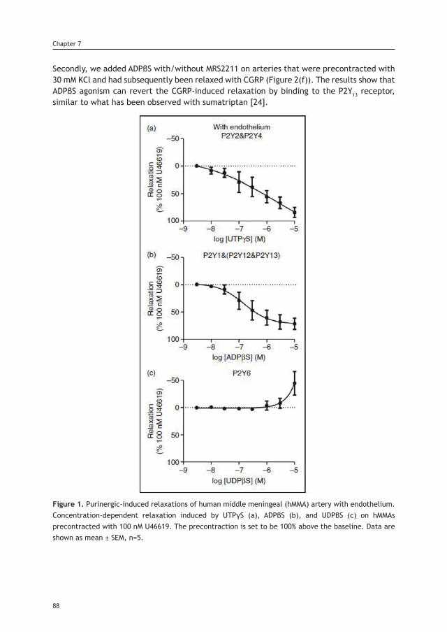
Secondly, we added ADPβS with/without MRS2211 on arteries that were precontracted with 30 mM KCl and had subsequently been relaxed with CGRP (Figure 2(f)). The results show that ADPβS agonism can revert the CGRP-induced relaxation by binding to the P2Y13 receptor, similar to what has been observed with sumatriptan [24].
Figure 1. Purinergic-induced relaxations of human middle meningeal (hMMA) artery with endothelium. Concentration-dependent relaxation induced by UTPγS (a), ADPβS (b), and UDPβS (c) on hMMAs precontracted with 100 nM U46619. The precontraction is set to be 100% above the baseline. Data are shown as mean ± SEM, n=5.
88
Chapter 7
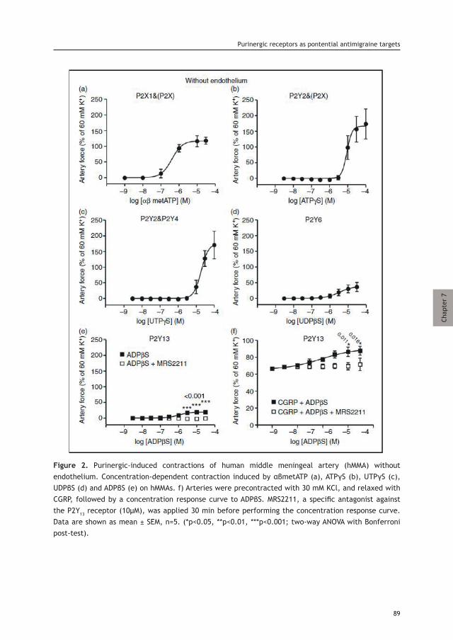
Figure 2. Purinergic-induced contractions of human middle meningeal artery (hMMA) without endothelium. Concentration-dependent contraction induced by αβmetATP (a), ATPγS (b), UTPγS (c), UDPβS (d) and ADPβS (e) on hMMAs. f) Arteries were precontracted with 30 mM KCl, and relaxed with CGRP, followed by a concentration response curve to ADPβS. MRS2211, a specific antagonist against the P2Y13 receptor (10μM), was applied 30 min before performing the concentration response curve. Data are shown as mean ± SEM, n=5. (*p<0.05, **p<0.01, ***p<0.001; two-way ANOVA with Bonferroni post-test).
Purinergic receptors as pontential antimigraine targets
89
Chap
ter
7
Effects on in vivo/in situ CGRP release in rat
Intravital microscopy – involvement of the P2Y13 receptor
Our in vitro data show that P2Y13 receptor stimulation induces contraction in the hMMA. To confirm that a P2Y13 receptor agonist also can have effects in vivo, we continued with intravital microscopy in rats, a common method to assess the antimigraine potential of a compound that is supposed to act on the trigeminovascular system. Upon intravenous injection of ADPβS (330 μg/kg), we observed a transient decrease in blood pressure (Figure 3(a), 65±3%), which most likely was caused by stimulation of endothelial P2Y1 receptors followed by desensitization [25]. Subsequently, a long-lasting increase in blood pressure was observed (Figure 3(a), 23±4%), which, in contrast to the vasodilation, was dependent on the P2Y13 receptor, since the increase was only 7±4% in the presence of MRS2211 (1 mg/kg). MRS2211 did not have any significant effect the blood pressure per se. The direct effect of ADPβS on the artery was similar as observed for the blood pressure, with an acute dilation (56±21%) that returned to baseline. In contrast to the blood pressure, we did not observe any direct contraction following the ADPβS administration (data not shown). Using periarterial electrical stimulation (ES), which releases CGRP [19, 26], the rMMA dilated (Figure 3(b), 69±11%), according to our expectations, and vehicle did not affect this vasodilation (74±10%). In contrast, ADPβS caused a significant inhibition of the dilation (Figure 3(b), 37±7%). Furthermore, MRS2211 (1 mg/kg), which had no significant effects per se (Figure 3(c)), prevented the inhibitory effect of ADPβS and normalized the ES-induced dilation (Figure 3(c), 62±7%). Blood pressure could be of importance in relation to interpreting the diameter increase during electrical stimulation. There were no significant absolute differences during the third ES after ADPβS (130±4 mmHg) compared to ADPβS + MRS2211 (123±5 mmHg).
CGRP release from dura and TG – involvement of the P2Y13 receptor To dissect the potential site of the P2Y13 inhibitory actions, we measured CGRP release ex vivo after individual stimulation of the trigeminal fibers in the dura mater and in the TG isolated from rats. ADPβS significantly inhibited CGRP release from both the dura mater (Figure 4(a) left panels, 172±15 vs 265±21 pg/ml, p=0.0003) and from the TG (Figure 4(a) right panels, 162±13 vs 278±36 pg/ml, p=0.0141). MRS2211 (10 μM) was without inhibitory effect per se (Figure 4(b)), but abolished the inhibitory response of ADPβS, illustrating that this is a P2Y13-dependent process (Figure 4(c)). Although not within the focus of the current study, ADPβS also inhibited CGRP release from the TNC (data not shown)
90
Chapter 7
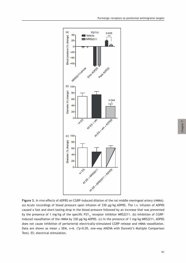
Figure 3. In vivo effects of ADPβS on CGRP-induced dilation of the rat middle meningeal artery (rMMA). (a) Acute recordings of blood pressure upon infusion of 330 μg/kg ADPβS. The i.v. infusion of ADPβS caused a fast and short-lasting drop in the blood pressure followed by an increase that was prevented by the presence of 1 mg/kg of the specific P2Y13 receptor inhibitor MRS2211. (b) Inhibition of CGRP-induced vasodilation of the rMMA by 330 μg/kg ADPβS. (c) In the presence of 1 mg/kg MRS2211, ADPβS does not cause inhibition of periarterial electrically-stimulated CGRP release and rMMA vasodilation. Data are shown as mean ± SEM, n=6. (*p<0.05, one-way ANOVA with Dunnett’s Multiple Comparison Test). ES: electrical stimulation.
Purinergic receptors as pontential antimigraine targets
91
Chap
ter
7
Intravital microscopy – involvement of the P2X3 receptor
Previous research has shown that P2X3 receptors are co-expressed with CGRP in the trigeminal ganglion [9], and that P2X3 receptor-positive neurons innervate the dura mater [27]. Since purinergic research has been mainly focused on the P2X receptors, we continued to investigate the effect of the P2X receptor agonist αβmetATP on the rMMA in vivo, exemplified in a raw trace (Figure 5(a)). αβmetATP (150 μg/kg) caused a strong constriction of the rMMA (Figure 5(b), -70±7%), followed by a recovery dilation (Figure 5(b), 54±10%). This dilation was completely prevented by the addition of the CGRP antagonist olcegepant (Figure 5(b), 1±4%, 100 μg/kg). Furthermore, olcegepant, as expected, prevented dilation both in response to capsaicin (10 μg/kg) and exogenous CGRP (1 μg/kg), but not to substance P (1 μg/kg). For the blood pressure data, we observed a similar response, with an initial increase in blood pressure (Figure 5(c), 34±7%), followed by a reduction of 23±7% (Figure 5(c)). In contrast to the data from the rMMA, the drop in blood pressure was not inhibited by olcegepant. In addition, these data show that the response to exogenous CGRP on the rMMA is unaffected by the presence of ADPβS, unlike the endogenous release (Figure 3(b)). Since our data indicate that αβmetATP could trigger CGRP release, we hypothesized that ADPβS, via activation of the P2Y13 receptor, might be inhibitory also on the αβmetATP-induced CGRP release. Surprisingly, we did not observe any significant inhibition on the maximum dilation (Figure 5(b), 35±13% vs 54±10%). However, there was a significant delay in the time to peak dilation, which is also visible in the illustrative raw-trace (Figure 5(a), 216±27 sec vs. 147±8 sec, p=0.0307), illustrating the potential involvement of the P2Y13 receptor in αβmetATP-induced CGRP release.
CGRP release from dura mater and TG – involvement of the P2X3 receptor
The dilation observed after constriction of the rMMA could be caused by: i) CGRP release triggered at the synapse or ii) antidromic CGRP release triggered by P2X receptors in the TG. We therefore investigated the effect of αβmetATP on CGRP release in the dura mater and the TG. We observed that αβmetATP did not cause any CGRP release from the dura mater above baseline values (Figure 6 left panel, 31±5 vs 32±5 pg/ml), in contrast to when it was applied to the TG (Figure 6 right panel, 58±7 vs 102±15 pg/ml, p=0.0073).
Immunohistochemistry on human TG and hMMA: localization of P2Y13 receptor and P2X3 receptor, co-localization with CGRP
We have here presented strong functional evidence for the involvement of the P2Y13 receptor and P2X3 receptor in in vivo and in situ rat models. Similar experiments are not possible in humans, but we had access to human tissue. Although the samples were obtained post mortem and could have been subject to potential changes in protein expression, we investigated whether similar receptor components are present in the human trigeminovascular system. Although sparsely expressed, we did detect neurons expressing both the P2Y13 receptor and CGRP in the human trigeminal ganglion (Figure 7(a)). In contrast, the P2X3 receptor is strongly expressed together with CGRP (Figure 7(b)). In the hMMA, CGRP and the P2Y13 receptor co-localized, in what appear to be C-fiber nerve endings (Figure 8(a), white arrows), whereas the P2X3 receptor is sparsely, if at all, present in the hMMA (Figure 8(b)). In one of the samples of hMMA, we observed a meningeal nerve (Figure 8(c)). Here it is even
92
Chapter 7
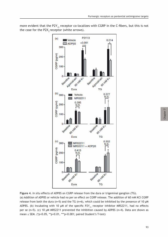
more evident that the P2Y13 receptor co-localizes with CGRP in the C-fibers, but this is not the case for the P2X3 receptor (white arrows).
Figure 4. In situ effects of ADPβS on CGRP release from the dura or trigeminal ganglion (TG).(a) Addition of ADPβS or vehicle had no per se effect on CGRP release. The addition of 60 mM KCl CGRP release from both the dura (n=5) and the TG (n=6), which could be inhibited by the presence of 10 µM ADPβS. (b) Incubating with 10 µM of the specific P2Y13 receptor inhibitor MRS2211, had no effects per se (n=5). (c) 10 µM MRS2211 prevented the inhibition caused by ADPβS (n=4). Data are shown as mean ± SEM. (*p<0.05, **p<0.01, ***p<0.001; paired Student’s T-test)
Purinergic receptors as pontential antimigraine targets
93
Chap
ter
7
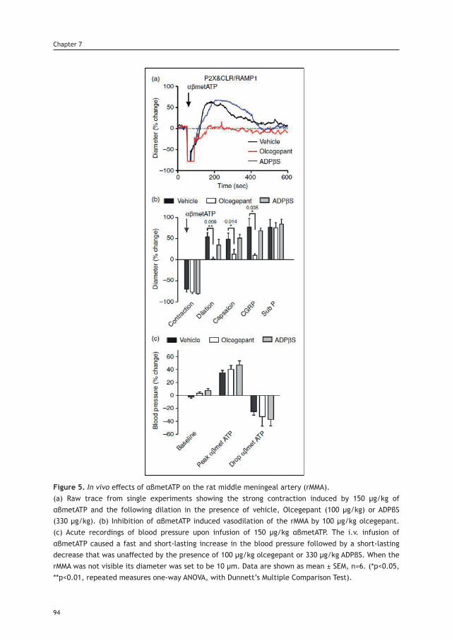
Figure 5. In vivo effects of αβmetATP on the rat middle meningeal artery (rMMA). (a) Raw trace from single experiments showing the strong contraction induced by 150 μg/kg of αβmetATP and the following dilation in the presence of vehicle, Olcegepant (100 μg/kg) or ADPβS (330 μg/kg). (b) Inhibition of αβmetATP induced vasodilation of the rMMA by 100 μg/kg olcegepant. (c) Acute recordings of blood pressure upon infusion of 150 μg/kg αβmetATP. The i.v. infusion of αβmetATP caused a fast and short-lasting increase in the blood pressure followed by a short-lasting decrease that was unaffected by the presence of 100 μg/kg olcegepant or 330 μg/kg ADPβS. When the rMMA was not visible its diameter was set to be 10 μm. Data are shown as mean ± SEM, n=6. (*p<0.05, **p<0.01, repeated measures one-way ANOVA, with Dunnett’s Multiple Comparison Test).
94
Chapter 7
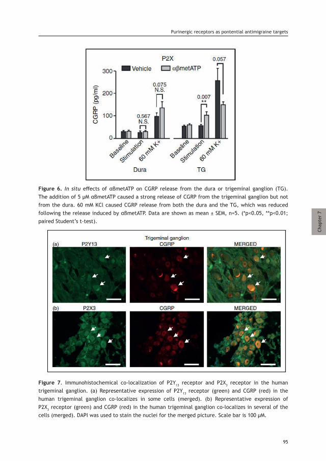
Figure 6. In situ effects of αβmetATP on CGRP release from the dura or trigeminal ganglion (TG). The addition of 5 μM αβmetATP caused a strong release of CGRP from the trigeminal ganglion but not from the dura. 60 mM KCl caused CGRP release from both the dura and the TG, which was reduced following the release induced by αβmetATP. Data are shown as mean ± SEM, n=5. (*p<0.05, **p<0.01; paired Student’s t-test).
Figure 7. Immunohistochemical co-localization of P2Y13 receptor and P2X3 receptor in the human trigeminal ganglion. (a) Representative expression of P2Y13 receptor (green) and CGRP (red) in the human trigeminal ganglion co-localizes in some cells (merged). (b) Representative expression of P2X3 receptor (green) and CGRP (red) in the human trigeminal ganglion co-localizes in several of the cells (merged). DAPI was used to stain the nuclei for the merged picture. Scale bar is 100 μM.
Purinergic receptors as pontential antimigraine targets
95
Chap
ter
7
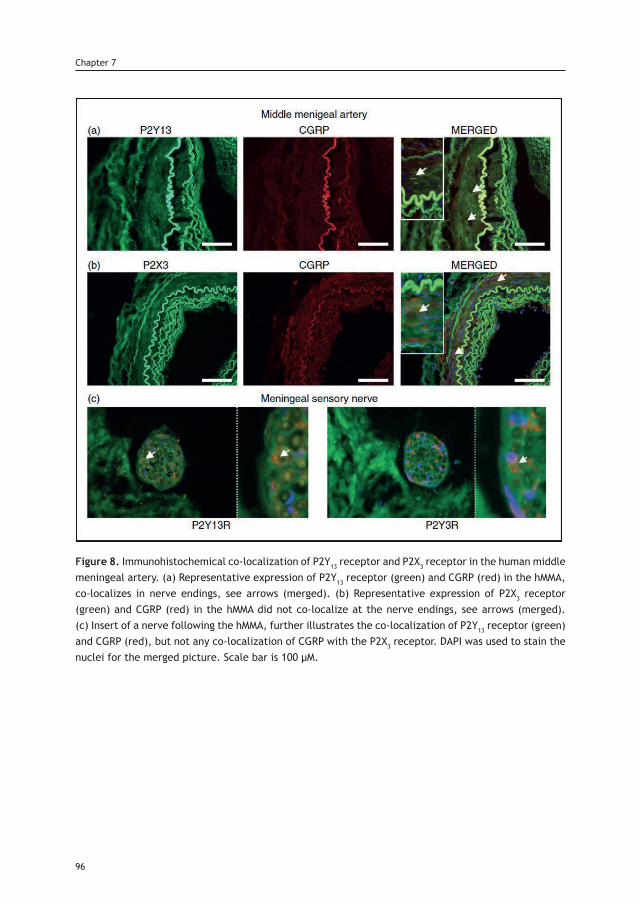
Figure 8. Immunohistochemical co-localization of P2Y13 receptor and P2X3 receptor in the human middle meningeal artery. (a) Representative expression of P2Y13 receptor (green) and CGRP (red) in the hMMA, co-localizes in nerve endings, see arrows (merged). (b) Representative expression of P2X3 receptor (green) and CGRP (red) in the hMMA did not co-localize at the nerve endings, see arrows (merged). (c) Insert of a nerve following the hMMA, further illustrates the co-localization of P2Y13 receptor (green) and CGRP (red), but not any co-localization of CGRP with the P2X3 receptor. DAPI was used to stain the nuclei for the merged picture. Scale bar is 100 μM.
96
Chapter 7
Discussion
Direct effects on the MMA
Until CGRP receptor antagonists were developed, all acutely acting specific antimigraine drugs induced direct vasoconstriction. Although direct vasoconstriction of the MMA may not be an essential requirement in treating migraine [1], it still might play a relevant role [28]. In addition, the MMA may serve as a proxy of the trigeminal system, where potential antimigraine compounds can be tested. In the current study, we started with a characterization of purinergic receptors on the hMMA. The data show that purinergic receptors have important effects. Particularly, their activation on the endothelium may induce strong dilation (Figure 1), while their activation in smooth muscle cells may induce a strong contraction (Figure 2). Only the contractile responses can be compared to our previously obtained data in the rat, as there were no endothelial responses in the rMMA due to their limited diameter [29].
Vasoactive properties of purinergic ligands on the hMMA
The P2X receptor agonist αβmetATP caused the most potent contraction in the hMMA (pEC50 6.40±0.20, Figure 2(a)). This is expected as the P2X1 receptor is known to mediate potent contractions in smooth muscle cells [30]. The main pyrimidine receptor in the hMMA is the P2Y2 receptor (Figure 1(b) and Figure 2(b)/(c)). When activated in the endothelium of the hMMA, it causes a strong vasodilation, with higher potency than in the human internal mammary artery [31]. The P2Y2 receptor is also the receptor that leads to the strongest contraction of the MMA, similar to human coronary and omental arteries [22, 23]. Activation of the P2Y6 receptor caused a pure contractile effect (Figure1(c) and Figure 2(d)). The Emax was not very high, which is similar to the human omental artery [22]. In human coronary arteries, there was no contraction to UDPβS [23], which contrasts to the cerebral arteries, where UDPβS is a strong constrictor [22]. ADPβS was a strong vasodilator (Figure 1(a)), which is typically attributed to the P2Y1 receptor [31]. The contraction to ADPβS in the hMMA was potent, but with a relatively low Emax (Figure 2(e)). In conclusion, the functional purinergic profile of the hMMA is most similar to human omental arteries and shares strong similarities to the coronary arteries. The profile in hMMA contrasts with that in human cerebral arteries, where the main difference is strong contractility to UDPβS (P2Y6 receptor agonist). Contractions to ADPβS are not described in any of other human arteries, making the hMMA unique.
Focus on the P2Y13 receptor in the hMMA
We were further particularly interested in ADP and its stable analogue ADPβS, since it is known that this does not cause contraction in the human coronary or cerebral vasculature [22, 23]. The side-effect of coronary vasoconstriction is a current caveat of the current acutely acting antimigraine treatments such as the triptans [32]. The contractile response to ADPβS in hMMA is small but significant. Furthermore, the response to ADPβS on the hMMA seems to be mediated purely by the P2Y13 receptor, as it is blocked by its antagonist MRS2211 (Figure 2(e)/(f)), at a concentration that should be devoid of effects on the P2Y1 and P2Y12 receptors [33]. Although the contraction to ADPβS was minor, ADPβS and P2Y13 receptor activation could reverse the dilation caused by CGRP (Figure 2(f)). This has a strong link
Purinergic receptors as pontential antimigraine targets
97
Chap
ter
7
98
Chapter 7
to antimigraine potential, and the data are very similar to what has been observed for the triptans [24].
Comparison between hMMA and rMMA
In our previous study on rMMA, ADPβS caused a strong contraction compared to the current data on the hMMA. We did not explore the functional pharmacological identity of the ADPβS response in the rat study, but we detected a strong P2Y13 receptor signal in the rMMA by PCR, combined with some P2Y1 receptor expression, but no expression of the P2Y12 receptor [29]. Regarding the difference in contraction between rMMAs and hMMAs, it appears that in both human and rat, fast acting and short-lasting effects are mediated via ATP on P2X receptors. In humans, long-term effects of purines are effectuated by ATP on the P2Y2 receptor, in contrast to rats where ADP is acting on P2Y1 receptor [29]. For pyrimidines, UTP through P2Y2 receptor activation is important in humans, while the breakdown product UDP acts on the P2Y6 receptor in rodents. It therefore seems that the human artery is more sensitive to ATP. In conclusion, the purinergic contractions in hMMA is caused by ATP/UTP, in contrast to the rMMA, which contracts to their breakdown-products UDP/ADP. Nevertheless, in vivo, the response is most likely similar, as the breakdown of UTP/ATP into UDP/ADP is nearly instantaneous [34], exemplified by studies on P2Y2 receptor and P2Y6 receptor knock out mice [35] and functional data from rat meningeal arteries [36].
Trigeminovascular effects
In the light of the recent advances in migraine research, investigating, the in vivo effects of potential antimigraine compounds on CGRP release is a highly relevant approach. We therefore used an established method to study the in vivo effects of ADPβS and αβmetATP, which activate P2Y13 and P2X3 receptors, respectively
Inhibitory effects of P2Y13 receptor agonism
Our data show that ADPβS, through activation of the P2Y13 receptor, inhibits vasodilation caused by periarterial electrical stimulation (Figure 3(b)/(c)). Periarterial stimulation has been shown to cause in vivo release of CGRP [19, 26]. Therefore, our experiments are the first to show that P2Y receptors, coupled to Gi proteins, can have prejunctional inhibitory effects on CGRP release in vivo. Similar observations have been made on other prejunctional nerve endings, where P2Y13 receptor activation was demonstrated to inhibit noradrenaline release from sympathetic neurons [16] and cholinergic transmission on the neuromuscular junction [17]. Importantly, we also show the direct inhibitory effect of ADPβS on CGRP release from both the dura and the TG in situ (Figure 4(a)). ADPβS is activating the P2Y13 receptor, as the specific P2Y13 receptor inhibitor MRS2211 was without effects per se, but reversed the inhibitory effect of ADPβS (Figure 4(b)/(c)). This is similar to the experiments by Honey et al., where an adenosine A1 receptor antagonist (DPCPX) blocked the inhibitory effect of an adenosine A1 agonist (GR79236) on meningeal vasodilation, but had no effects per se [37]. Since inhibiting CGRP release (or CGRP signaling) is now believed to be the most important hallmark of all acute antimigraine treatments [1], our data clearly demonstrate the therapeutic potential of a specific P2Y13 receptor agonist.
Purinergic receptors as pontential antimigraine targets
Trigeminal activation by P2X receptor activation
αβmetATP injections caused an activation of the trigeminovascular system, starting with a strong, short-lasting contraction of the rMMA, followed by a relaxation (Figure 5(a)). The relaxation is mediated by CGRP, as demonstrated by the blockade induced by the CGRP receptor antagonist olcegepant (Figure 5(b)). This is highly similar to the experiment by McCulloch and colleagues that was performed over 30 years ago, who showed that trigeminal nerve lesion prevented the relaxation following a constriction of the meningeal artery [38]. Our data show that αβmetATP-induced activation of P2X receptors on the TG is the most likely mechanism of action, leading to CGRP release (Figure 6). Indeed, Yegutkin et al. showed that αβmetATP triggered trigeminal nerve fiber activity [36] and Eroli et al, has shown that αβmetATP evokes neuronal firing in trigeminal cultures [39]. Therefore, stimulation of P2X3 receptors using αβmetATP could be a model of trigeminal activation, particularly for antidromic CGRP release occurring at the MMA. This is supported by: i) the relative long time from stimulation until observable dilation (Figure 5(a)), as the signal to release CGRP might travel from the TG and ii) the ability of αβmetATP to trigger CGRP release only from the TG and not in the dura mater (Figure 6).
Expression in human TG and clinical relevance
We investigated whether the purinergic receptors found to be important in the rat trigeminovascular system are also expressed in the human TG. The P2X3 receptor was strongly expressed in CGRPergic neurons (Figure 7(a)), similar to what has been shown before in rats [9]. There are also neurons that are positive for both the P2Y13 receptor and CGRP, although we only sporadically observed these (Figure 7(b)). In addition, we investigated the hMMA and its innervation. Here we found that, in contrast to the TG, the P2Y13 receptor is co-localized with CGRP (Figure 8(a)), in contrast to the P2X3 receptor (Figure 8(b)). Combined with the findings in the TG, these data match the functional data that we observed in the rat. Both the functional data and the immunohistochemistry indicate that P2X3 receptor activation and expression mainly occurs in the TG (Figures 5-8), and that P2Y13 receptor inhibition and expression can both exist in the TG (Figures 3/4 and 7) and on the nerve endings at the dura/MMA (Figures 3/4 and 8). Combined, the above findings suggest that there are similarities in the purinergic aspects of the trigeminovascular system between humans and rats. The clinical applications and potential antimigraine effects need to be further investigated; for this purpose, there is a particular need for the development of specific agonists. The main concern regarding the use of agonists based on ADP is their potential thrombotic activity. ADP activating P2Y1 or P2Y12 receptors is considered a relatively weak platelet aggregation agonist by itself (we did not observe any acute thrombotic events in the current experiments). Unfortunately, ADP still works as an amplifier of platelet aggregators such as thrombin [40], which is not compatible with treatment of patients. This potential side-effect should not be an issue for a specific P2Y13 receptor agonist.
99
Chap
ter
7
Conclusion
In the current study, we performed a thorough investigation of the purinergic receptors in the hMMA and compared it to our previous data on the rMMA. Interestingly, the expression and regulation of contractile responses are mediated by different receptors in human and rat. P2Y2 receptors (activated by ATP/UTP) are mediating most of the G-protein coupled purinergic function in hMMA compared to a combined effect of the P2Y1 receptor (ADP) and P2Y6 receptor (UDP) in rMMA. The P2Y13 receptor was important in counteracting the effects of CGRP on the hMMA and P2Y13 receptor activation also inhibited CGRP release, both in vivo and in situ. Therefore, the P2Y13 receptor could therefore be a novel antimigraine target, particularly when considering the intracellular pathway activated by this receptor in light of the migraine pathophysiology. Furthermore, activation of the P2X3 receptor caused specific CGRP release originating from the TG and a consequent olcegepant-sensitive dilation at the rMMA. Our findings contribute to further understanding of the pathophysiology of migraine. To investigate the full clinical potential of targeting the P2Y13 receptor, a pharmacologically specific agonist is clearly needed.
References1. Edvinsson L, Haanes KA, Warfvinge K et al. CGRP as the target of new migraine therapies - successful translation from bench to clinic. Nat Rev Neurol 2018; 14: 338-350.2. Amrutkar DV, Ploug KB, Hay-Schmidt A et al. mRNA expression of 5-hydroxytryptamine 1B, 1D, and 1F receptors and their role in controlling the release of calcitonin gene- related peptide in the rat trigeminovascular system. Pain 2012; 153: 830-838.3. Goadsby PJ, Edvinsson L. The trigeminovascular system and migraine: studies characterizing cerebrovascular and neuropeptide changes seen in humans and cats. Ann Neurol 1993; 33: 48-56.4. Schuster NM, Rapoport AM. Calcitonin Gene-Related Peptide-Targeted Therapies for Migraine and Cluster Headache: A Review. Clin Neuropharmacol 2017; 40: 169-174.5. Burnstock G. Purine and pyrimidine receptors. Cell Mol Life Sci 2007; 64: 1471-1483.6. Burnstock G. Blood cells: an historical account of the roles of purinergic signalling. Purinergic Signal 2015; 11: 411-434.7. Cieslak M, Czarnecka J, Roszek K et al. The role of purinergic signaling in the etiology of migraine and novel antimi-graine treatment. Purinergic Signal 2015; 11: 307-316.8. Goloncser F, Sperlagh B. Effect of genetic deletion and pharmacological antagonism of P2X7 receptors in a mouse animal model of migraine. J Headache Pain 2014; 15: 24.9. Ambalavanar R, Moritani M, Dessem D. Trigeminal P2X3 receptor expression differs from dorsal root ganglion and is modulated by deep tissue inflammation. Pain 2005; 117: 280-291.10. Fabbretti E, D’Arco M, Fabbro A et al. Delayed upregulation of ATP P2X3 receptors of trigeminal sensory neurons by calcitonin gene-related peptide. J Neurosci 2006; 26: 6163-6171.11. Inoue K, Tsuda M, Koizumi S. ATP induced three types of pain behaviors, including allodynia. Drug Dev Res 2003; 59: 56-63.12. Ceruti S, Fumagalli M, Villa G et al. Purinoceptor-mediated calcium signaling in primary neuron-glia trigeminal cultures. Cell Calcium 2008; 43: 576-590.13. Erb L, Weisman GA. Coupling of P2Y receptors to G proteins and other signaling pathways. Wiley Interdiscip Rev Membr Transp Signal 2012; 1: 789-803.14. Haanes KA, Edvinsson L. Pathophysiological Mechanisms in Migraine and the Identification of New Therapeutic Targets. CNS Drugs 2019; Accepted [DOI:19 10.1007/s40263-019-00630-6].15. Ahn AH, Basbaum AI. Where do triptans act in the treatment of migraine? Pain 2005; 115: 1-4.16. Queiroz G, Talaia C, Goncalves J. ATP modulates noradrenaline release by activation of inhibitory P2Y receptors and facilitatory P2X receptors in the rat vas deferens. J Pharmacol Exp Ther 2003; 307: 809-815.17. Guarracino JF, Cinalli AR, Fernandez V et al. P2Y13 receptors mediate presynaptic inhibition of acetylcholine re-lease induced by adenine nucleotides at the mouse neuromuscular junction. Neuroscience 2016; 326: 31-44.18. Haanes KA, Labastida-Ramirez A, Chan KY et al. Characterization of the trigeminovascular actions of several aden-osine A2A receptor antagonists in an in vivo rat model of migraine. J Headache Pain 2018; 19: 41.
Chapter 7
100
19. Bhatt DK, Gupta S, Jansen-Olesen I et al. NXN-188, a selective nNOS inhibitor and a 5-HT1B/1D receptor agonist, inhibits CGRP release in preclinical migraine models. Cephalalgia 2013; 33: 87-100.20. Gupta S, Amrutkar DV, Mataji A et al. Evidence for CGRP re-uptake in rat dura mater encephali. Br J Pharmacol 2010; 161: 1885-1898.21. Blixt FW, Haanes KA, Ohlsson L et al. Increased endothelin-1-mediated vasoconstriction after organ culture in rat and pig ocular arteries can be suppressed with MEK/ERK1/2 inhibitors. Acta Ophthalmol 2018; 96: e619-e625.22. Malmsjo M, Hou M, Pendergast W et al. Potent P2Y6 receptor mediated contractions in human cerebral arteries. BMC Pharmacol 2003; 3: 4.23. Malmsjo M, Hou M, Harden TK et al. Characterization of contractile P2 receptors in human coronary arteries by use of the stable pyrimidines uridine 5’-O-thiodiphosphate and uridine 5’-O-3-thiotriphosphate. J Pharmacol Exp Ther 2000; 293: 755-760.24. Grande G, Labruijere S, Haanes KA et al. Comparison of the vasodilator responses of isolated human and rat middle meningeal arteries to migraine related compounds. J Headache Pain 2014; 15: 22.25. Rodriguez-Rodriguez R, Yarova P, Winter P et al. Desensitization of endothelial P2Y1 receptors by PKC-dependent mechanisms in pressurized rat small mesenteric arteries. Br J Pharmacol 2009; 158: 1609-1620.26. Messlinger K, Hanesch U, Kurosawa M et al. Calcitonin gene related peptide released from dural nerve fibers me-diates increase of meningeal blood flow in the rat. Can J Physiol Pharmacol 1995; 73: 1020-1024.27. Staikopoulos V, Sessle BJ, Furness JB et al. Localization of P2X2 and P2X3 receptors in rat trigeminal ganglion neurons. Neuroscience 2007; 144: 208-216.28. Levy D, Labastida-Ramirez A, MaassenVanDenBrink A. Current understanding of meningeal and cerebral vascular function underlying migraine headache. Cephalalgia 2018; 333102418771350.29. Haanes KA, Edvinsson L. Expression and characterization of purinergic receptors in rat middle meningeal ar-tery-potential role in migraine. PLoS One 2014; 9: e108782.30. Wang L, Karlsson L, Moses S et al. P2 receptor expression profiles in human vascular smooth muscle and endothelial cells. J Cardiovasc Pharmacol 2002; 40: 841-853.31. Wihlborg AK, Malmsjo M, Eyjolfsson A et al. Extracellular nucleotides induce vasodilatation in human arteries via prostaglandins, nitric oxide and endothelium-derived hyperpolarising factor. Br J Pharmacol 2003; 138: 1451-1458.32. Chan KY, Labruijere S, Ramirez Rosas MB et al. Cranioselectivity of sumatriptan revisited: pronounced contractions to sumatriptan in small human isolated coronary artery. CNS Drugs 2014; 28: 273-278.33. Kim YC, Lee JS, Sak K et al. Synthesis of pyridoxal phosphate derivatives with antagonist activity at the P2Y13 receptor. Biochem Pharmacol 2005; 70: 266-274.34. Kauffenstein G, Drouin A, Thorin-Trescases N et al. NTPDase1 (CD39) controls nucleotide-dependent vasoconstric-tion in mouse. Cardiovasc Res 2010; 85: 204-213.35. Haanes KA, Spray S, Syberg S et al. New insights on pyrimidine signalling within the arterial vasculature - Different roles for P2Y2 and P2Y6 receptors in large and small coronary arteries of the mouse. J Mol Cell Cardiol 2016; 93: 1-11.36. Yegutkin GG, Guerrero-Toro C, Kilinc E et al. Nucleotide homeostasis and purinergic nociceptive signaling in rat meninges in migraine-like conditions. Purinergic Signal 2016; 12: 561-574.37. Honey AC, Bland-Ward PA, Connor HE et al. Study of an adenosine A1 receptor agonist on trigeminally evoked dural blood vessel dilation in the anaesthetized rat. Cephalalgia 2002; 22: 260-264.38. McCulloch J, Uddman R, Kingman TA et al. Calcitonin gene-related peptide: functional role in cerebrovascular regulation. Proc Natl Acad Sci U S A 1986; 83: 5731-5735.39. Eroli F, Vilotti S, van den Maagdenberg AMJM et al. Hyperpolarization-activated current Ih in mouse trigeminal sensory neurons in a transgenic mouse model of familial hemiplegic migraine type-1. Neuroscience 2017; 351: 47-64.40. Gachet C. ADP receptors of platelets and their inhibition. Thromb Haemost 2001; 86: 222-232.
101
Purinergic receptors as pontential antimigraine targets
Chap
ter
7

Chapter 8
Lasmiditan inhibits calcitonin gene-related peptide release in the rodent trigeminovascular system
Submitted
Labastida-Ramírez A, Rubio-Beltrán E, Haanes KA, Garrelds IM, Danser AHJ, Villalón CM and MaassenVanDenBrink A.
Abstract
Migraine headache pathophysiology involves trigeminovascular system activation, calcitonin gene-related peptide (CGRP) release and dysfunctional nociceptive transmission. Triptans are 5-HT1B/1D/(1F) receptor agonists that inhibit prejunctionally trigeminal CGRP release, but their vasoconstrictor properties limit their use in migraine patients with cardiovascular disease. In contrast, lasmiditan is a novel antimigraine and selective 5-HT1F receptor agonist devoid of vasoconstrictor properties. On this basis, the present study has investigated the modulation of trigeminal CGRP release by lasmiditan. For this purpose, we have comparatively analysed the trigeminovascular inhibition induced by lasmiditan and sumatriptan through: (i) ex vivo KCl induced CGRP release from isolated dura mater, trigeminal ganglion and trigeminal nucleus caudalis of mice; and (ii) in vivo dural vasodilation in the rat closed cranial window model induced by endogenous (electrical stimulation, capsaicin) and exogenous CGRP. The ex vivo release of CGRP was similarly diminished by sumatriptan and lasmiditan in all trigeminovascular system components. While in vivo, i.v. lasmiditan or higher doses of sumatriptan significantly attenuated endogenous CGRP release, but not exogenous CGRP effects. These data suggest that lasmiditan inhibits prejunctionally CGRP release in peripheral and central trigeminal nerve terminals. Since lasmiditan is a lipophilic drug that crosses the blood-brain barrier, additional central sites of action remain to be determined.
Background
Migraine is a chronic neurovascular disorder characterized by recurrent severe attacks of throbbing headache, which can be accompanied by nausea, vomiting, photo- and phonophobia [1].Although migraine pathophysiology has not been elucidated completely, it has been proposed that its headache phase results from activation and sensitization of trigeminal afferents from meningeal nociceptors, neuropeptide release and dysfunctional nociceptive transmission [2]. Calcitonin gene-related peptide (CGRP) is a key neuropeptide, widely expressed in the peripheral and central components of the trigeminovascular system, involved in craniofacial nociceptive modulation [3-5]. In migraine patients, infusion of CGRP triggers headaches [6], and during spontaneous attacks this peptide is released in the extracerebral circulation [7]. Moreover, blocking CGRP or its receptor with monoclonal antibodies, which do not cross the blood-brain barrier, have revealed that migraine attacks can be prevented via peripheral blockade of CGRP. For almost 30 years the triptans, 5-HT1B/1D receptor agonists, of which some also display affinity for the 5-HT1F receptor [8], have been the specific treatment of choice for terminating migraine attacks. These drugs have the ability to normalize elevated CGRP levels by inhibiting further release from trigeminal afferents and consequently decreasing nociceptive transmission from the periphery to the central nervous system [9]. However, due to their peripheral vasoconstrictor potential, including coronary vasoconstriction, they are contraindicated in migraine patients with cardiovascular diseases [10]. This problem led to the search for antimigraine agents lacking vasoconstrictor activity that culminated in the development of lasmiditan. Lasmiditan is a novel high-affinity, lipophilic, highly selective 5-HT1F receptor agonist (“ditan”) that is currently under development for the acute treatment of migraine [11].
104
Chapter 8
Chap
ter
8
Phase III trials revealed clinical efficacy in migraine patients, and preclinical data showed that it is devoid of vasoconstrictor properties in several blood vessels including human isolated coronary arteries [12]. However, few studies have investigated its exact (antimigraine) mechanism(s) of action. Lasmiditan may possess central as well as peripheral antinociceptive effects, as it reduced c-Fos expression in the trigeminal nucleus caudalis and inhibited electrical stimulation of trigeminal ganglion-induced dural plasma protein extravasation (PPE), respectively [11]. However, the latter effect has not been demonstrated to occur in migraine patients, and several PPE inhibitors failed in clinical trials [13]. Therefore, we set out to analyze the effects of lasmiditan on the trigeminovascular CGRPergic system (using sumatriptan as a positive control), based on the neurobiology of migraine headache. For this purpose, we investigated CGRP release in the peripheral (dura mater and trigeminal ganglion) and central (trigeminal nucleus caudalis) trigeminal components of mice ex vivo, as well as dural vasodilation induced by endogenous (capsaicin and electrical stimulation) and exogenous CGRP in rats in vivo. Our results show that selective 5-HT1F receptor activation by lasmiditan inhibits CGRP release and, consequently, attenuates nociceptive transmission in the trigeminovascular system.
Materials and methods
Experimental animals
Experiments were performed in 36 male C57BL/6J (20 g; 13-14 weeks of age) mice and 54 male Sprague-Dawley (300-350 g; 8-10 weeks of age) rats, purchased from Charles River. Animals were housed under a 12-hr dark-light cycle in a special room at constant temperature (22±2°C) and humidity (50%), with free access to food and water. It is noteworthy that only male animals were used to avoid the well-known interaction of CGRP with cycling estrogen [13], previously reported in the cranial-window model [14]. All experimental protocols of the study were approved by the Erasmus University Medical Center’s institutional animal ethics committee [permission protocol numbers EMC 2891/3393], in accordance with the European directive 2010/63/EU and ARRIVE (Animal Research: Reporting In Vivo Experiments) reporting guidelines for the care and use of laboratory animals, with randomization throughout.
Ex vivo: CGRP release in trigeminal nucleus caudalis, trigeminal ganglion and dura mater
This technique has been previously reported [15], in brief, mice were anesthetized using intraperitoneal (i.p.) sodium pentobarbital (80 mg/kg) and decapitated at the atlanto-occipital joint. The scalp was retracted from the cranium. Firstly, the trigeminal caudal nuclei, which run 9-13 mm caudally from bregma, were isolated from the brainstem. The skull was divided into halves by a clear cut along the sagittal suture, the cerebral hemispheres were carefully removed while the cranial dura was left attached to the skull. Secondly, the trigeminal ganglia were harvested by dissection 1 mm proximal and distal to the point where the mandibular nerve branches off. Thirdly, the remaining brain tissues were extracted from the hemisected skulls without damaging the dura of the middle cranial fossa. Each trigeminal nucleus caudalis, trigeminal ganglion and hemisected skull with the dura mater were immersed and washed in carbogenated synthetic interstitial fluid, containing (mM): NaCl (108), KCl (3.48), MgSO4 (3.5), NaHCO3 (26), NaH2PO4 (11.7),
105
Lasmiditan inhibits CGRP release
CaCl2 (1.5), sodium gluconate (9.6), glucose (5.55) and sucrose (7.6) for 30 minutes at 37°C. The isolated tissues were randomized and placed in a 24 well plate containing 500 μL synthetic interstitial fluid. The 24-well plate was fixed in a water bath that formed a closed humid chamber of 37°C. Basal CGRP values were measured in each tissue; then CGRP release was induced by superfusion with 60 mM KCl. Vehicle (saline), sumatriptan or lasmiditan (30 μM each, concentration based on our earlier experiments with sumatriptan [15]), were applied 10 min prior to the challenge with 60 mM KCl. For every sample, including the baseline measurement, the solution was collected after 10 min incubation and mixed with aprotinin (500 KIU/mL). For the assessment of CGRP content, samples were stored at 80°C until processed with a commercial CGRP RIA kit according to the manual (Phoenix Pharmaceuticals, Burlingame, CA, USA). CGRP absorbance values (pg/mL) were calculated through an interpolation method using an equation derived from the standard curve. The assay has a detection level of ~0.1 pg/mL. Blank wells without CGRP were used as a control to exclude possible false-positive measurements, as previously described [16]. In vivo: Intravital microscopy and dural artery vasodilation
Rats were anaesthetized throughout the experiments using sodium pentobarbital (60 mg⁄kg, i.p. followed by 18 mg/kg i.v. per hour). The adequacy of anesthesia was judged by a negative tail flick test, mean arterial pressure levels and the absence of ocular reflexes, amongst others. Tracheal intubation was performed and a capnograph was used to monitor pCO2 levels. As previously described [17], the femoral vein and artery were cannulated for i.v. administration of drugs and for continuous monitoring of mean arterial pressure, respectively. During the experiment, the core temperature of the animal was maintained between 36.5 and 37.5°C using a homeothermic blanket system for rodents (Harvard Instruments, Edenbridge, UK). Subsequently, the rat was placed in a stereotaxic frame, and the parietal bone was drilled thin until the dural middle meningeal artery was clearly visible. As the rat skull is thin, care was taken to drill with constant application of ice-cold saline. The drilled area was covered with saline to prevent drying of the skull and to facilitate visualization of the artery. The dural artery diameter was captured with an intravital microscope (Leica MZ 16; Leica Microsystem Ltd, Heerbrugg, Switzerland), using a cyan filter on a cold light source. A zoom lens (80-400x magnification) and camera (DCx V3.52, Thorlabs LTD, Ely, UK) were used to capture the image of the dural artery, which was displayed and measured on a computer using a dedicated software package (IDA-Intravital Dimension Analyser; http://www.beneryx.co.uk) integrated with a ADC/DAC board (DI-158, DATAQ instruments, ‘s-Hertogenbosch, The Netherlands). Dural artery diameter was calculated from the area under the curve of the intensity measured and expressed in arbitrary units (AU). For periarterial electrical stimulation a bipolar stimulating electrode (NE 200X, Clark Electromedical, Edenbridge, Kent, UK) was placed on the surface of the bone approximately 200 µM from the artery. The surface of the cranial window was stimulated at 5 Hz, 1 ms for 10 s (Stimulator model S88, Grass Instruments, West Warwick, RI, USA) with a current intensity of 100 µA (monitored on an oscilloscope, model 54601A; Hewlett Packard, Palo Alto, CA, USA) and increasing with 50 µA steps until a maximal level of vasodilation was achieved, usually at 200 µA [18]. Data of dural artery diameter and mean arterial blood pressure were recorded using a Labchart data acquisition system (AD Instruments Ltd, Oxford, UK). Dural artery vasodilator responses produced by periarterial electrical stimulation (100-200 µA), capsaicin (10 µg/kg, i.v.) or
106
Chapter 8
α-CGRP (1 µg/kg, i.v.) were elicited before and after consecutive i.v. bolus injections of vehicle (saline), sumatriptan or lasmiditan (0.3, 1, 3 and 10 mg/kg each). These doses were based on clinically relevant doses previously calculated [7] as well as, for sumatriptan, previous studies performed in the same model [17]. In brief, for sumatriptan assuming a 25-100 mg oral dose in a 70-kg adult with a 15% bioavailability would be equivalent to i.v. doses of about 0.05-0.21 mg/kg. For lasmiditan, a 50-200 mg oral dose in a 70-kg adult with a 40% bioavailability would be equivalent to i.v. doses of about 0.28-1.14 mg/kg. Each dose of the aforementioned compounds was administered 5 min before a subsequent treatment with electrical stimulation, capsaicin or CGRP. The dural artery diameter and mean arterial pressure values restored to pre-injection levels by the time the next vasodilation were elucidated, and 30 min were allowed to elapse after each of the vasodilator treatments for the recovery of baseline diameter.
Statistical analysis
All data are expressed as mean ± SEM. CGRP release in the different trigeminal components of mice was expressed as relative stimulated CGRP release, which was calculated by the ratio of KCl-induced CGRP release and basal CGRP values, as previously described [19]. Statistical differences of the basal CGRP values, the relative stimulated CGRP release in the presence of vehicle and the % of CGRP inhibition between groups, were calculated using one-tailed Mann-Whitney test for unpaired observations. For comparing the relative stimulated CGRP release in the presence of vehicle to its corresponding treatment (sumatriptan or lasmiditan), a one-tailed Wilcoxon matched pairs test for non-parametric analysis of paired was used. The peak changes in dural artery diameter were expressed as percent change from baseline. Changes in mean arterial pressure were expressed in absolute values (mm Hg). The differences between the variables within one group of animals were compared by using a one-way repeated measures analysis of variance followed by Dunnet’s test. Statistical significance was accepted at p < 0.05.
Compounds
The compounds used in the present study (obtained from the sources indicated) were: rat α-CGRP (NeoMPS S.A., Strasbourg, France); sumatriptan succinate and capsaicin (Sigma Chemical Co., Steinheim, Germany); and lasmiditan hydrochloride (provided by Eli Lilly & Co., Indianapolis, IN, USA). CGRP, sumatriptan and lasmiditan were dissolved in physiological saline. Capsaicin (1 mg/mL) was dissolved in a mixture of Tween-80, ethanol 70% and water (1:1:8). The doses mentioned in the text refer to the free base of substances in all cases.
Results
Ex vivo: Basal CGRP levels and relative stimulated CGRP release after KCl stimulation
A total of 108 tissues were analyzed. Tissues with basal CGRP measurement errors (CGRP below detection limit, n=2) or that did not generate CGRP release in response to 60 mM KCl (n=14) were excluded. In addition, in one experiment the positive control (inhibition of CGRP by sumatriptan) failed (instead we measured an increase in CGRP release of >15 times), this measurement was a statistically significant outlier (p=0.016, Dixon’s outlier test) and was therefore also excluded. Since data were paired between the left and right side, in these cases both the left and right sides were excluded.
107
Lasmiditan inhibits CGRP release
Chap
ter
8
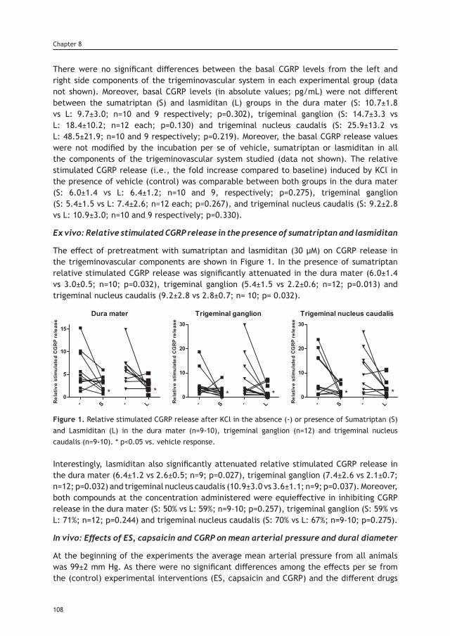
There were no significant differences between the basal CGRP levels from the left and right side components of the trigeminovascular system in each experimental group (data not shown). Moreover, basal CGRP levels (in absolute values; pg/mL) were not different between the sumatriptan (S) and lasmiditan (L) groups in the dura mater (S: 10.7±1.8 vs L: 9.7±3.0; n=10 and 9 respectively; p=0.302), trigeminal ganglion (S: 14.7±3.3 vs L: 18.4±10.2; n=12 each; p=0.130) and trigeminal nucleus caudalis (S: 25.9±13.2 vs L: 48.5±21.9; n=10 and 9 respectively; p=0.219). Moreover, the basal CGRP release values were not modified by the incubation per se of vehicle, sumatriptan or lasmiditan in all the components of the trigeminovascular system studied (data not shown). The relative stimulated CGRP release (i.e., the fold increase compared to baseline) induced by KCl in the presence of vehicle (control) was comparable between both groups in the dura mater (S: 6.0±1.4 vs L: 6.4±1.2; n=10 and 9, respectively; p=0.275), trigeminal ganglion (S: 5.4±1.5 vs L: 7.4±2.6; n=12 each; p=0.267), and trigeminal nucleus caudalis (S: 9.2±2.8 vs L: 10.9±3.0; n=10 and 9 respectively; p=0.330).
Ex vivo: Relative stimulated CGRP release in the presence of sumatriptan and lasmiditan
The effect of pretreatment with sumatriptan and lasmiditan (30 µM) on CGRP release in the trigeminovascular components are shown in Figure 1. In the presence of sumatriptan relative stimulated CGRP release was significantly attenuated in the dura mater (6.0±1.4 vs 3.0±0.5; n=10; p=0.032), trigeminal ganglion (5.4±1.5 vs 2.2±0.6; n=12; p=0.013) and trigeminal nucleus caudalis (9.2±2.8 vs 2.8±0.7; n= 10; p= 0.032).
Figure 1. Relative stimulated CGRP release after KCl in the absence (-) or presence of Sumatriptan (S) and Lasmiditan (L) in the dura mater (n=9-10), trigeminal ganglion (n=12) and trigeminal nucleus caudalis (n=9-10). * p<0.05 vs. vehicle response.
Interestingly, lasmiditan also significantly attenuated relative stimulated CGRP release in the dura mater (6.4±1.2 vs 2.6±0.5; n=9; p=0.027), trigeminal ganglion (7.4±2.6 vs 2.1±0.7; n=12; p=0.032) and trigeminal nucleus caudalis (10.9±3.0 vs 3.6±1.1; n=9; p=0.037). Moreover, both compounds at the concentration administered were equieffective in inhibiting CGRP release in the dura mater (S: 50% vs L: 59%; n=9-10; p=0.257), trigeminal ganglion (S: 59% vs L: 71%; n=12; p=0.244) and trigeminal nucleus caudalis (S: 70% vs L: 67%; n=9-10; p=0.275).
In vivo: Effects of ES, capsaicin and CGRP on mean arterial pressure and dural diameter
At the beginning of the experiments the average mean arterial pressure from all animals was 99±2 mm Hg. As there were no significant differences among the effects per se from the (control) experimental interventions (ES, capsaicin and CGRP) and the different drugs
108
Chapter 8
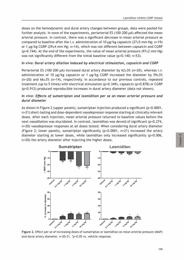
doses on the hemodynamic and dural artery changes between groups, data were pooled for further analysis. In none of the experiments, periarterial ES (100-200 μA) affected the mean arterial pressure. In contrast, there was a significant decrease in mean arterial pressure as compared to baseline after the i.v. administration of 10 µg/kg capsaicin (27±5 mm Hg; n=19) or 1 µg/kg CGRP (29±4 mm Hg; n=14), which was not different between capsaicin and CGRP (p=0.744). At the end of the experiments, the value of mean arterial pressure (97±2 mm Hg) was not significantly different from the initial baseline value (p=0.140; n=53).
In vivo: Dural artery dilation induced by electrical stimulation, capsaicin and CGRP
Periarterial ES (100-200 µA) increased dural artery diameter by 62±3% (n=20), whereas i.v. administration of 10 µg/kg capsaicin or 1 µg/kg CGRP increased the diameter by 59±3% (n=20) and 66±3% (n=14), respectively. In accordance to our previous controls, repeated treatment (up to 5 times) with electrical stimulation (p=0.349), capsaicin (p=0.878) or CGRP (p=0.912) produced reproducible increases in dural artery diameter (data not shown).
In vivo: Effects of sumatriptan and lasmiditan per se on mean arterial pressure and dural diameter
As shown in Figure 2 (upper panels), sumatriptan injection produced a significant (p<0.0001, n=21) short-lasting and dose-dependent vasodepressor response starting at clinically relevant doses. After each injection, mean arterial pressure returned to baseline values before the next vasodilation was elucidated. In contrast, lasmiditan was devoid of significant (p=0.274, n=20) vasodepressor responses at all doses tested. When considering dural artery diameter (Figure 2; lower panels), sumatriptan significantly (p<0.0001, n=21) increased the artery diameter starting at lower doses, while lasmiditan only increased significantly (p=0.006, n=20) the artery diameter after injecting the higher doses.
Figure 2. Effect per se of increasing doses of sumatriptan or lasmiditan on mean arterial pressure (MAP) and dural artery diameter. n=20-21. *p<0.05 vs. vehicle response.
109
Lasmiditan inhibits CGRP release
Chap
ter
8
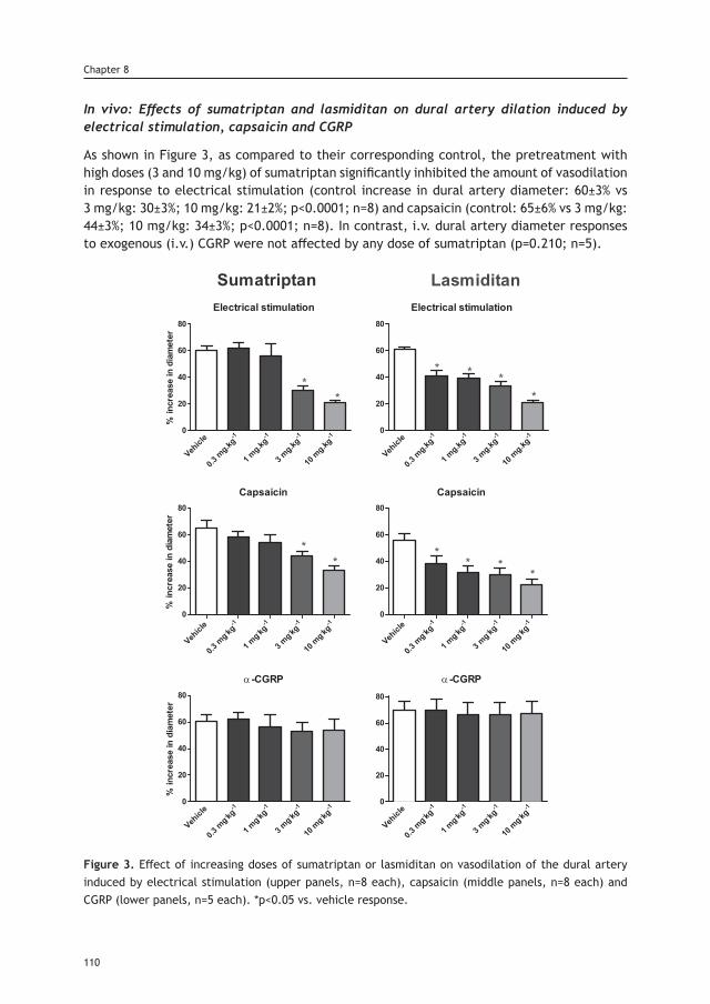
In vivo: Effects of sumatriptan and lasmiditan on dural artery dilation induced by electrical stimulation, capsaicin and CGRP
As shown in Figure 3, as compared to their corresponding control, the pretreatment with high doses (3 and 10 mg/kg) of sumatriptan significantly inhibited the amount of vasodilation in response to electrical stimulation (control increase in dural artery diameter: 60±3% vs 3 mg/kg: 30±3%; 10 mg/kg: 21±2%; p<0.0001; n=8) and capsaicin (control: 65±6% vs 3 mg/kg: 44±3%; 10 mg/kg: 34±3%; p<0.0001; n=8). In contrast, i.v. dural artery diameter responses to exogenous (i.v.) CGRP were not affected by any dose of sumatriptan (p=0.210; n=5).
Figure 3. Effect of increasing doses of sumatriptan or lasmiditan on vasodilation of the dural artery induced by electrical stimulation (upper panels, n=8 each), capsaicin (middle panels, n=8 each) and CGRP (lower panels, n=5 each). *p<0.05 vs. vehicle response.
110
Chapter 8
In contrast to sumatriptan, already lower doses of lasmiditan induced a significant dose-dependent attenuation of the vasodilator responses to electrical stimulation (control increase in dural artery diameter: 61±2% vs 0.3 mg/kg: 41±4%; 1 mg/kg: 39±3%; 3 mg/kg: 34±3%; 10 mg/kg: 21±2%; p<0.0001; n=8) and capsaicin (control: 56±5% vs 0.3 mg/kg: 38±7%; 1 mg/kg: 31±5%; 3 mg/kg: 30±5%; 10 mg/kg: 23±4%; p<0.0001; n=8) compared to their corresponding control. Similar as with sumatriptan, the exogenous CGRP responses were not affected by any dose of lasmiditan (p=0.911; n=5).
Discussion
In the current study, we investigated the potential antimigraine site of action of the selective 5-HT1F receptor agonist lasmiditan in relation to the trigeminovascular CGRPergic system, through inhibition of chemically-(KCl and capsaicin, via voltage-gated calcium and TRPV1 channel activation, respectively) and electrically-induced CGRP release from trigeminal sensory fibers. We compared our results with data obtained with sumatriptan, since this was the first triptan developed, and this class of drugs is the current gold standard for the specific acute treatment of migraine attacks. In accordance with our previous work [15], pretreatment with 30 µM sumatriptan attenuated KCl-relative stimulated CGRP release in the peripheral and central components of the trigeminovascular system of mice ex vivo (Figure 1). This inhibition has previously been shown to be mediated via activation of 5-HT1D and 5-HT1F receptors in rats, while a lower concentration (3 µM) of sumatriptan was ineffective [20]. Interestingly, 30 µM lasmiditan was equieffective in attenuating CGRP release in the dura mater, trigeminal ganglion as well as trigeminal nucleus caudalis; however, since theoretically multiple 5-HT1 (i.e. 5-HT1A and 5-HT1F) inhibitory-Gi protein receptor subtypes expressed in the mice trigeminovascular system [21] could be activated at the nonselective concentration used [22], we performed more in-depth experiments with a range of clinically relevant doses in the in vivo rat closed cranial window, an experimental neurovascular model of migraine [17]. In accordance with the ex vivo experiments, high doses (3 and 10 mg/kg) of sumatriptan were required to inhibited neurogenic dural vasodilation in response to i.v. capsaicin and periarterial electrical stimulation. These high dose inhibitory effects have been previously shown with sumatriptan, and also with rizatriptan, while low doses of both compounds (1 mg/kg) were ineffective in this model [17, 23]. Based on data available on receptor affinity profiles [8] and pharmacological antagonism [7, 24], the inhibition of CGRP release by triptans seems to be mediated via activation of 5-HT1D and possibly also 5-HT1F receptors; unfortunately, highly selective antagonists for the 5-HT1F receptors are not yet commercially available to further determine this. Remarkably, lasmiditan inhibited neurogenic dural vasodilation in response to i.v. capsaicin and periarterial electrical stimulation starting at lower, clinically relevant doses, this effect is most likely mediated by selective activation of the 5-HT1F receptor. In contrast, like sumatriptan, lasmiditan was unable to attenuate non-neurogenic dural vasodilation in responses to i.v. CGRP. Thus, we demonstrate that lasmiditan prejunctionally inhibits the release of CGRP from trigeminal sensory fibers innervating the dura vasculature. The activation of either 5-HT1B, 5-HT1D or 5-HT1F receptors on trigeminal fibers results in a direct inhibition of adenylyl cyclase and a subsequent decrease in the cAMP-
111
Lasmiditan inhibits CGRP release
Chap
ter
8
signaling pathway [25], which in turn alters the phosphorylation of TRPV1 channels [26] and decreases the release of CGRP, as evidenced by the inhibition of CGRP- and capsaicin-induced dural vasodilation by sumatriptan and lasmiditan. However, if both compounds act through the same second messenger cascade, why does agonism of the 5-HT1F receptor seem to be more effective? While the mRNA expression of the 5-HT1D and 5-HT1F receptors has been detected in the peripheral and central components of the rat trigeminovascular system [20], and both receptors have been found to be co-localized with CGRP [27, 28]; a small study (n=3) found that the 5-HT1D receptor is more abundantly expressed in the trigeminal ganglion, whereas the highest concentration of 5-HT1F receptors were found in the trigeminal nucleus caudalis, and this expression pattern could result in diverse inhibition profiles [20]. However, more in depth studies have found that the 5-HT1B, 5-HT1D and 5-HT1F receptors are equally represented in the trigeminal ganglion [29, 30]. Therefore, the discrepancy between sumatriptan and lasmiditan inhibition results does not seem to be explained by the expression/density profile of the 5-HT1B/1D/1F receptors. It is noteworthy that species differences have been described for the 5-HT1 receptor subtypes mediating the inhibition of trigeminal CGRP release by 5-HT1B and 5-HT1D receptor agonists (including sumatriptan). This inhibition has previously attributed to activation of 5-HT1B receptors in rat and mouse, and 5-HT1D receptors in the guinea pig, cats and humans [31]. Moreover, the rat and mouse 5-HT1B receptor has been considered to represent the counterpart to the 5-HT1D receptor in other species [32]. Thus, sumatriptan receptor affinity for the 5-HT1 receptors in rats (5-HT1D > 5-HT1B), correlates to the high doses required to inhibit CGRP release via 5-HT1B receptors and possibly also 5-HT1F receptors. It should be kept in mind that our rodent model is suitable to study inhibition of CGRP release, but not hemodynamic changes in response to 5-HT1 receptor stimulation. In support of this is the fact that lower doses of i.v. sumatriptan immediately activate systemic vascular 5-HT receptors before reaching the 5-HT1D (and later 5-HT1B) receptors in the trigeminal sensory fibers. Consequently, the injection of already clinically relevant doses of sumatriptan resulted in a significant drop in mean arterial pressure and dural artery vasodilation (Figure 2), which have been shown to be mediated via activation of 5-HT7 receptors in rodents [33]. However, since as we mentioned rodents are not suitable to study hemodynamic changes associated to 5-HT1 receptor activation, as receptor expression and vascular responses are not similar to humans, we have recently addressed the vasoconstrictor effects of sumatriptan in appropriate and translational (human isolated arteries and dog in vivo) models [11]. Similar to our previous findings, i.v. infusions of sumatriptan, via 5-HT1B receptor activation [34], increased systemic and pulmonary arterial pressure in humans [35], which represents a major limitation for triptans use in cardiovascular compromised patients. In contrast, lasmiditan was devoid of hemodynamics effects at all doses tested. Thus, clinically relevant doses of lasmiditan are not associated with changes in mean arterial pressure or dural artery diameter. These results suggest that the antimigraine efficacy of lasmiditan is unrelated to vasoconstrictive mechanisms. The 5-HT1F receptor is also widely express throughout the central nervous system and since lasmiditan is a lipophilic drug that crosses the blood-brain barrier (in contrast with sumatriptan), it has been associated with mild central side-effects [36]. The most common side-effects described in clinical trials are dizziness, paresthesia, somnolence, fatigue, nausea and lethargy. Given that lasmiditan inhibits the peripheral components of the
112
Chapter 8
trigeminovascular system, it is tempting to speculate what would happen to the side-effect profile or antimigraine efficacy of a selective 5-HT1F receptor agonist that does not enter the brain. Whether inhibiting the trigeminovascular system centrally as well as peripherally is more effective than just peripherally or centrally, or whether a mixed mechanism is associated with increased side-effects remains to be determined. Unfortunately, there are no hydrophilic (selective) 5-HT1F receptor agonists available yet. Additional (antimigraine) mechanisms of action described with previous 5-HT1F receptor agonists include modulation of glutamate release from trigeminal sensory fibers and mitochondrial biogenesis. Moreover, since CGRP receptor antagonism reduces the hyperemia associated with cortical spreading depression (CSD) [37], a key pathogenic event in migraine with aura, 5-HT1F receptor agonism could attenuate CSD via a central modulation (inhibition) of CGRP. Future experiments, beyond the scope of our study, are needed to determine whether: (i) lasmiditan can inhibit glutamatergic neurons in the central nervous system, (ii) it can enhance mitochondrial biogenesis, or (iii) it can attenuate CSD; and if all or none of these mechanisms are associated with its clinical antimigraine efficacy.
Conclusion
In conclusion, our results indicate that lasmiditan prejunctionally inhibits CGRP release in peripheral and central trigeminal nerve terminals. This effect may, at least partly, explain the clinical efficacy of lasmiditan. Since activation of 5-HT1F receptors is not associated with vasoconstriction, lasmiditan may represent a cardiovascular safety advantage over the vasoactive triptans.
References1. Lassen LH, Haderslev PA, Jacobsen VB, et al. Cgrp May Play A Causative Role in Migraine. Cephalalgia. 2002; 22: 54-61.2. Levy D, Labastida-Ramirez A and MaassenVanDenBrink A. Current understanding of meningeal and cerebral vascular function underlying migraine headache. Cephalalgia. 0: 0333102418771350.3. Eftekhari S, Warfvinge K, Blixt FW, et al. Differentiation of Nerve Fibers Storing CGRP and CGRP Receptors in the Peripheral Trigeminovascular System. The Journal of Pain. 14: 1289-303.4. Uddman R, Tajti J, Hou M, et al. Neuropeptide expression in the human trigeminal nucleus caudalis and in the cervical spinal cord C1 and C2. Cephalalgia. 2002; 22: 112-6.5. Hendrikse ER, Bower RL, Hay DL, et al. Molecular studies of CGRP and the CGRP family of peptides in the central nervous system. Cephalalgia. 2019; 39: 403-19.6. Goadsby PJ, Edvinsson L and Ekman R. Vasoactive peptide release in the extracerebral circulation of humans during migraine headache. Annals of Neurology. 1990; 28: 183-7.7. Rubio-Beltran E, Labastida-Ramirez A, Villalon CM, et al. Is selective 5-HT1F receptor agonism an entity apart from that of the triptans in antimigraine therapy? Pharmacol Ther. 2018.8. Goadsby PJ and Edvinsson L. The trigeminovascular system and migraine: studies characterizing cerebrovascular and neuropeptide changes seen in humans and cats. Ann Neurol. 1993; 33: 48-56.9. MaassenVanDenBrink A, Reekers M, Bax WA, et al. Coronary side-effect potential of current and prospective antimi-graine drugs. Circulation. 1998; 98: 25-30.10. Nelson DL, Phebus LA, Johnson KW, et al. Preclinical pharmacological profile of the selective 5-HT1F receptor agonist lasmiditan. Cephalalgia. 2010; 30: 1159-69.11. Rubio Beltran E, Kristian H, Labastida A, et al. Lasmiditan and sumatriptan: comparison of in vivo vascular con-striction in the dog and in vitro contraction of human arteries. Cephalalgia. 2016; 36: 104-5.12. Diener HC. Rpr100893, A Substance-P Antagonist, is Not Effective in the Treatment of Migraine Attacks. Cepha-lalgia. 2003; 23: 183-5.13. Labastida-Ramírez A, Rubio-Beltrán E, Villalón CM, et al. Gender aspects of CGRP in migraine. Cephalalgia. 2019; 39: 435-44.14. Gupta S, Villalon CM, Mehrotra S, et al. Female sex hormones and rat dural vasodilatation to CGRP, periarterial electrical stimulation and capsaicin. Headache. 2007; 47: 225-35.
113
Lasmiditan inhibits CGRP release
Chap
ter
8
15. Chan KY, Labastida-Ramirez A, Ramirez-Rosas MB, et al. Trigeminovascular calcitonin gene-related peptide func-tion in Cacna1a R192Q-mutated knock-in mice. J Cereb Blood Flow Metab. 2017: 271678X17725673.16. Gupta S, Amrutkar DV, Mataji A, et al. Evidence for CGRP re-uptake in rat dura mater encephali. British journal of pharmacology. 2010; 161: 1885-98.17. Williamson DJ, Hargreaves RJ, Hill RG, et al. Sumatriptan inhibits neurogenic vasodilation of dural blood vessels in the anaesthetized rat--intravital microscope studies. Cephalalgia. 1997; 17: 525-31.18. Haanes KA, Labastida-Ramírez A, Chan KY, et al. Characterization of the trigeminovascular actions of several aden-osine A2A receptor antagonists in an in vivo rat model of migraine. The Journal of Headache and Pain. 2018; 19: 41.19. Fioretti B, Catacuzzeno L, Sforna L, et al. Trigeminal ganglion neuron subtype-specific alterations of Ca(V)2.1 calcium current and excitability in a Cacna1a mouse model of migraine. J Physiol. 2011; 589: 5879-95.20. Amrutkar DV, Ploug KB, Hay-Schmidt A, et al. mRNA expression of 5-hydroxytryptamine 1B, 1D, and 1F receptors and their role in controlling the release of calcitonin gene-related peptide in the rat trigeminovascular system. Pain. 2012; 153: 830-8.21. Bonnavion P, Bernard JF, Hamon M, et al. Heterogeneous distribution of the serotonin 5-HT(1A) receptor mRNA in chemically identified neurons of the mouse rostral brainstem: Implications for the role of serotonin in the regulation of wakefulness and REM sleep. J Comp Neurol. 2010; 518: 2744-70.22. Kovalchin J, Ghiglieri A, Zanelli E, et al. Lasmiditan acts specifically on the 5-HT<inf>1F</inf> receptor in the central nervous system. Cephalalgia. 2016; 36: 103.23. Williamson DJ, Shepheard SL, Hill RG, et al. The novel anti-migraine agent rizatriptan inhibits neurogenic dural vasodilation and extravasation. Eur J Pharmacol. 1997; 328: 61-4.24. Bhatt DK, Gupta S, Jansen-Olesen I, et al. NXN-188, a selective nNOS inhibitor and a 5-HT1B/1D receptor agonist, inhibits CGRP release in preclinical migraine models. Cephalalgia. 2013; 33: 87-100.25. Adham N, Kao HT, Schecter LE, et al. Cloning of another human serotonin receptor (5-HT1F): a fifth 5-HT1 receptor subtype coupled to the inhibition of adenylate cyclase. Proceedings of the National Academy of Sciences. 1993; 90: 408-12.26. Bhave G, Zhu W, Wang H, et al. cAMP-dependent protein kinase regulates desensitization of the capsaicin receptor (VR1) by direct phosphorylation. Neuron. 2002; 35: 721-31.27. Ma Q-P, Hill R and Sirinathsinghji D. Colocalization of CGRP with 5-HT1B/1D receptors and substance P in trigeminal ganglion neurons in rats. European Journal of Neuroscience. 2001; 13: 2099-104.28. Ahn SK, Khalmuratova R, Jeon SY, et al. Colocalization of 5-HT1F receptor and calcitonin gene-related peptide in rat vestibular nuclei. Neurosci Lett. 2009; 465: 151-6.29. Classey JD, Bartsch T and Goadsby PJ. Distribution of 5-HT1B, 5-HT1D and 5-HT1F receptor expression in rat trigeminal and dorsal root ganglia neurons: Relevance to the selective anti-migraine effect of triptans. Brain Research. 2010; 1361: 76-85.30. Reuter U, Salomone S, Ickenstein GW, et al. Effects of Chronic Sumatriptan and Zolmitriptan Treatment on 5-HT1 Receptor Expression and Function in Rats. Cephalalgia. 2004; 24: 398-407.31. Williamson DJ and Hargreaves RJ. Neurogenic inflammation in the context of migraine. Microscopy research and technique. 2001; 53: 167-78.32. Hamblin MW, Metcalf MA, McGuffin RW, et al. Molecular cloning and functional characterization of a human 5-HT1B serotonin receptor: A homologue of the rat 5-HT1B receptor with 5-HT1D-like pharmacological specificity. Biochemical and Biophysical Research Communications. 1992; 184: 752-9.33. Terron JA and Martinez-Garcia E. 5-HT7 receptor-mediated dilatation in the middle meningeal artery of anesthe-tized rats. Eur J Pharmacol. 2007; 560: 56-60.34. Longmore, Razzaque, Shaw, et al. Comparison of the vasoconstrictor effects of rizatriptan and sumatriptan in human isolated cranial arteries: immunohistological demonstration of the involvement of 5-HT1B-receptors. British Journal of Clinical Pharmacology. 1998; 46: 577-82.35. MacIntyre PD, Bhargava B, Hogg KJ, et al. Effect of subcutaneous sumatriptan, a selective 5HT1 agonist, on the systemic, pulmonary, and coronary circulation. Circulation. 1993; 87: 401-5.36. Kuca B, Silberstein SD, Wietecha L, et al. Lasmiditan is an effective acute treatment for migraine: A phase 3 ran-domized study. Neurology. 2018; 91: e2222-e32.37. Tozzi A, de Iure A, Di Filippo M, et al. Critical role of calcitonin gene-related peptide receptors in cortical spread-ing depression. Proc Natl Acad Sci U S A. 2012; 109: 18985-90.
114
Chapter 8


Chapter 9
Trigeminovascular Calcitonin gene-related peptide function in Cacna1a R192Q-mutated knock-in mice
Based on: Journal of Cerebral Blood Flow & Metabolism, 2019, 39 718-729.
Chan KY, Labastida-Ramírez A, Ramírez-Rosas MB, Labruijere S, Garrelds IM, Danser AH, van den Maagdenberg AM and MaassenVanDenBrink A.
Abstract
Familial hemiplegic migraine type 1 (FHM1) is a rare migraine subtype. Whereas transgenic knock-in mice with the human pathogenic FHM1 R192Q missense mutation in the Cacna1a gene reveal overall neuronal hyperexcitability, the effects on the trigeminovascular system and calcitonin gene-related peptide (CGRP) receptor are largely unknown. This gains relevance as blockade of CGRP and its receptor are therapeutic targets under development. Hence, we set out to test these effects in FHM1 mice. We characterized the trigeminovascular system of wild-type and FHM1 mice through: (i) in vivo capsaicin- and CGRP-induced dural vasodilation in a closed cranial window; (ii) ex vivo KCl induced CGRP release from isolated dura mater, trigeminal ganglion and trigeminal nucleus caudalis; and (iii) peripheral vascular function in vitro. In mutant mice, dural vasodilatory responses were significantly decreased compared to controls. The ex vivo release of CGRP was not different in the components of the trigeminovascular system between genotypes, however, sumatriptan diminished the release in the trigeminal ganglion, trigeminal nucleus caudalis and dura mater but only in wild-type mice. Peripheral vascular function was similar between genotypes. These data suggest that the R192Q mutation might be associated with trigeminovascular CGRP receptor desensitization. Novel antimigraine drugs should be able to revert this complex phenomenon.
Background
Migraine is a disabling neurovascular brain disorder that is characterized by severe attacks of throbbing headache, which can be accompanied by nausea, vomiting, osmo-, photo- and phonophobia [1]. In one-third of patients, attacks may be preceded by neurological symptoms (migraine with aura), the likely consequence of a wave of neuronal and glial depolarization with subsequent temporary inactivity, called cortical spreading depression [2]. Migraine headache is associated with activation of the trigeminovascular system and calcitonin gene-related peptide (CGRP) release from sensory nerves [3]. Monogenic familial hemiplegic migraine, a rare subtype of migraine with transient hemiparesis during the aura, has shown validity as a genetic model of migraine [4]. Familial hemiplegic migraine type 1 (FHM1) is caused by specific missense mutations in the CACNA1A gene that encodes the α1A subunit of neuronal voltage-gated CaV2.1 (P/Q-type) calcium channels [5, 6]. A transgenic knock-in mouse model was generated by introducing the human FHM1 R192Q mutation into the mouse Cacna1a gene using a gene targeting approach [7]. The mutation results in a gain of CaV2.1 channel function with enhanced cortical glutamatergic neurotransmission [8], an increased susceptibility to cortical spreading depression [7, 9], and signs of spontaneous unilateral head pain, as evidenced by increased head grooming and eye blinking [10]. Few studies have investigated CGRP expression and function in the trigeminovascular system of FHM1 R192Q mice, however, the results are inconsistent and mostly only the trigeminal ganglion was investigated [11-13], making it difficult to understand the exact role of this mutation in the different components of the trigeminal system. Furthermore, the effect the R192Q mutation may have on perivascular CGRP release and CGRP-induced (dural artery) vasodilation has not been investigated. Also, it is not known whether the R192Q mutation could affect the peripheral vascular function. Hence, to study the effects of the R192Q mutation on the trigeminovascular system, we investigated in FHM1 R192Q mice: (i) trigeminovascular dural vasodilation induced
118
Chapter 9
Chap
ter
9
by endogenous and exogenous CGRP in vivo, (ii) CGRP release in the trigeminovascular components including the trigeminal nucleus caudalis, trigeminal ganglion and dura mater ex vivo, and (iii) peripheral vascular CGRP receptor function in vitro. We hypothesized that the R192Q mutation will increase dural vasodilation and CGRP release in the trigeminovascular system but will not disturb peripheral vascular function.
Material and Methods
Experimental animals
Experiments were performed in 13- to 14-week-old homozygous FHM1 R192Q mice (“R192Q”) and wild-type littermates (“wild-type”) of both sexes. Mice were backcrossed for at least five generations with C57BL/6J mice so that the genetic background is >97% C57BL/6J, as described before [7]. All mice were bred at the Leiden University Medical Center and transported to our animal facility at least 14 days before the start of the experiment to allow for sufficient equilibration time at the Erasmus MC. Animals were housed under a 12-hr dark-light cycle and given free access to food and water. All experiments were approved by the Erasmus University Medical Center’s institutional animal ethics committee and in accordance with the European directive 2010/63/EU and ARRIVE (Animal Research: Reporting In Vivo Experiments) reporting guidelines for the care and use of laboratory animals.
In vivo: Intravital microscopy and dural artery vasodilation
Animals (n=32; 13 wild-type and 19 R192Q) were anaesthetized throughout the experiment using intraperitoneal (i.p.) sodium pentobarbital (80 mg/kg, i.p. and then 20 mg/kg/h, i.p.). The trachea was cannulated and connected to a pressure ventilator (small animal ventilator SAR-830 series; CWE, Inc., Ardmore, PA, USA). The jugular vein was cannulated for intravenous (i.v.) administration of drugs and the femoral artery for continuous monitoring of mean arterial pressure. During the experiment, the core temperature of the animal was monitored via a rectal thermometer and maintained between 36.5 and 37.5°C using a homeothermic blanket system for rodents (Harvard Instruments, Edenbridge, UK). Subsequently, the mouse was placed in a stereotaxic frame, and the parietal bone was drilled thin until the dural artery was clearly visible. As the mouse skull is very thin, care was taken to drill with constant application of ice-cold saline. In four out of 32 mice, bleeding was observed underneath the skull, making visualization of the artery difficult; these animals were excluded from the study. The drilled area was covered with mineral oil to prevent drying of the skull and to facilitate visualization of the artery. The dural artery was captured with an intravital microscope (Leica MZ 16; Leica Microsystem Ltd, Heerbrugg, Switzerland), using a cyan filter on a cold source of light. A zoom lens (80x magnification) and camera (DCx V3.52, Thorlabs LTD, Ely, UK) were used to capture the image of the dural artery, which was displayed and measured on a computer using a dedicated software package (IDA-Intravital Dimension Analyser; http://www.beneryx.co.uk) integrated with a ADC/DAC board (DI-158, DATAQ instruments, ‘s-Hertogenbosch, The Netherlands). Data of dural artery diameter, mean arterial pressure and exhaled CO2 were recorded using Labchart data acquisition system (AD Instruments Ltd, Oxford, UK). As described in a previous study [14], mice dural arteries were first constricted with endothelin-1 (ET-1) before other pharmacological interventions, as it is not possible
119
Trigeminovascular CGRP function in R192Q mice
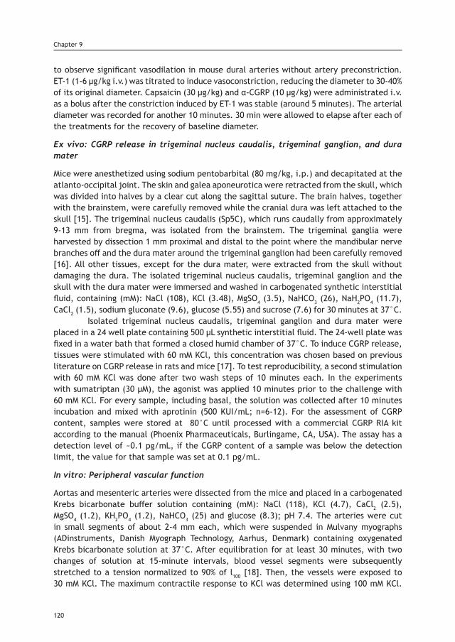
to observe significant vasodilation in mouse dural arteries without artery preconstriction. ET-1 (1-6 µg/kg i.v.) was titrated to induce vasoconstriction, reducing the diameter to 30-40% of its original diameter. Capsaicin (30 µg/kg) and α-CGRP (10 µg/kg) were administrated i.v. as a bolus after the constriction induced by ET-1 was stable (around 5 minutes). The arterial diameter was recorded for another 10 minutes. 30 min were allowed to elapse after each of the treatments for the recovery of baseline diameter.
Ex vivo: CGRP release in trigeminal nucleus caudalis, trigeminal ganglion, and dura mater
Mice were anesthetized using sodium pentobarbital (80 mg/kg, i.p.) and decapitated at the atlanto-occipital joint. The skin and galea aponeurotica were retracted from the skull, which was divided into halves by a clear cut along the sagittal suture. The brain halves, together with the brainstem, were carefully removed while the cranial dura was left attached to the skull [15]. The trigeminal nucleus caudalis (Sp5C), which runs caudally from approximately 9-13 mm from bregma, was isolated from the brainstem. The trigeminal ganglia were harvested by dissection 1 mm proximal and distal to the point where the mandibular nerve branches off and the dura mater around the trigeminal ganglion had been carefully removed [16]. All other tissues, except for the dura mater, were extracted from the skull without damaging the dura. The isolated trigeminal nucleus caudalis, trigeminal ganglion and the skull with the dura mater were immersed and washed in carbogenated synthetic interstitial fluid, containing (mM): NaCl (108), KCl (3.48), MgSO4 (3.5), NaHCO3 (26), NaH2PO4 (11.7), CaCl2 (1.5), sodium gluconate (9.6), glucose (5.55) and sucrose (7.6) for 30 minutes at 37°C. Isolated trigeminal nucleus caudalis, trigeminal ganglion and dura mater were placed in a 24 well plate containing 500 μL synthetic interstitial fluid. The 24-well plate was fixed in a water bath that formed a closed humid chamber of 37°C. To induce CGRP release, tissues were stimulated with 60 mM KCl, this concentration was chosen based on previous literature on CGRP release in rats and mice [17]. To test reproducibility, a second stimulation with 60 mM KCl was done after two wash steps of 10 minutes each. In the experiments with sumatriptan (30 μM), the agonist was applied 10 minutes prior to the challenge with 60 mM KCl. For every sample, including basal, the solution was collected after 10 minutes incubation and mixed with aprotinin (500 KUI/mL; n=6-12). For the assessment of CGRP content, samples were stored at 80°C until processed with a commercial CGRP RIA kit according to the manual (Phoenix Pharmaceuticals, Burlingame, CA, USA). The assay has a detection level of ~0.1 pg/mL, if the CGRP content of a sample was below the detection limit, the value for that sample was set at 0.1 pg/mL.
In vitro: Peripheral vascular function
Aortas and mesenteric arteries were dissected from the mice and placed in a carbogenated Krebs bicarbonate buffer solution containing (mM): NaCl (118), KCl (4.7), CaCl2 (2.5), MgSO4 (1.2), KH2PO4 (1.2), NaHCO3 (25) and glucose (8.3); pH 7.4. The arteries were cut in small segments of about 2-4 mm each, which were suspended in Mulvany myographs (ADinstruments, Danish Myograph Technology, Aarhus, Denmark) containing oxygenated Krebs bicarbonate solution at 37°C. After equilibration for at least 30 minutes, with two changes of solution at 15-minute intervals, blood vessel segments were subsequently stretched to a tension normalized to 90% of l100 [18]. Then, the vessels were exposed to 30 mM KCl. The maximum contractile response to KCl was determined using 100 mM KCl.
120
Chapter 9
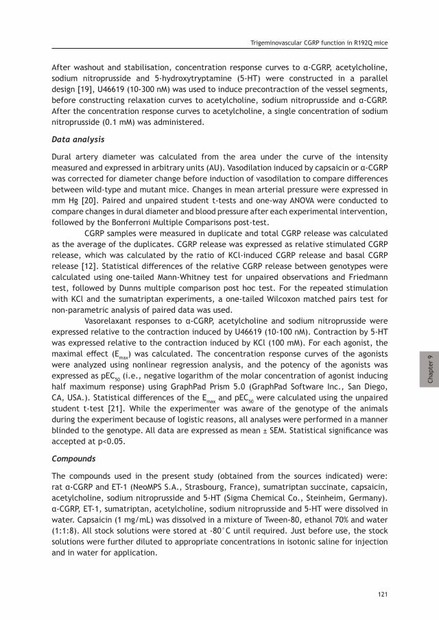
After washout and stabilisation, concentration response curves to α-CGRP, acetylcholine, sodium nitroprusside and 5-hydroxytryptamine (5-HT) were constructed in a parallel design [19], U46619 (10-300 nM) was used to induce precontraction of the vessel segments, before constructing relaxation curves to acetylcholine, sodium nitroprusside and α-CGRP. After the concentration response curves to acetylcholine, a single concentration of sodium nitroprusside (0.1 mM) was administered.
Data analysis
Dural artery diameter was calculated from the area under the curve of the intensity measured and expressed in arbitrary units (AU). Vasodilation induced by capsaicin or α-CGRP was corrected for diameter change before induction of vasodilation to compare differences between wild-type and mutant mice. Changes in mean arterial pressure were expressed in mm Hg [20]. Paired and unpaired student t-tests and one-way ANOVA were conducted to compare changes in dural diameter and blood pressure after each experimental intervention, followed by the Bonferroni Multiple Comparisons post-test. CGRP samples were measured in duplicate and total CGRP release was calculated as the average of the duplicates. CGRP release was expressed as relative stimulated CGRP release, which was calculated by the ratio of KCl-induced CGRP release and basal CGRP release [12]. Statistical differences of the relative CGRP release between genotypes were calculated using one-tailed Mann-Whitney test for unpaired observations and Friedmann test, followed by Dunns multiple comparison post hoc test. For the repeated stimulation with KCl and the sumatriptan experiments, a one-tailed Wilcoxon matched pairs test for non-parametric analysis of paired data was used. Vasorelaxant responses to α-CGRP, acetylcholine and sodium nitroprusside were expressed relative to the contraction induced by U46619 (10-100 nM). Contraction by 5-HT was expressed relative to the contraction induced by KCl (100 mM). For each agonist, the maximal effect (Emax) was calculated. The concentration response curves of the agonists were analyzed using nonlinear regression analysis, and the potency of the agonists was expressed as pEC50 (i.e., negative logarithm of the molar concentration of agonist inducing half maximum response) using GraphPad Prism 5.0 (GraphPad Software Inc., San Diego, CA, USA.). Statistical differences of the Emax and pEC50 were calculated using the unpaired student t-test [21]. While the experimenter was aware of the genotype of the animals during the experiment because of logistic reasons, all analyses were performed in a manner blinded to the genotype. All data are expressed as mean ± SEM. Statistical significance was accepted at p<0.05.
Compounds
The compounds used in the present study (obtained from the sources indicated) were: rat α-CGRP and ET-1 (NeoMPS S.A., Strasbourg, France), sumatriptan succinate, capsaicin, acetylcholine, sodium nitroprusside and 5-HT (Sigma Chemical Co., Steinheim, Germany). α-CGRP, ET-1, sumatriptan, acetylcholine, sodium nitroprusside and 5-HT were dissolved in water. Capsaicin (1 mg/mL) was dissolved in a mixture of Tween-80, ethanol 70% and water (1:1:8). All stock solutions were stored at -80°C until required. Just before use, the stock solutions were further diluted to appropriate concentrations in isotonic saline for injection and in water for application.
121
Trigeminovascular CGRP function in R192Q mice
Chap
ter
9
Results
General
There was no difference in body weight between the wild-type (22.4±1.0 g) and R192Q (24.6±1.2) mice included in our study (p=0.807). Also, in none of the experiments there was a difference between the results obtained in male and female animals. Therefore, the results from both sexes were pooled for further analysis.
In vivo: Effect on dural artery dilation induced by capsaicin and CGRP
Diameter changes induced by ET-1, capsaicin or CGRP are shown in Figure 1.
Figure 1. Trigeminovascular dural artery vasodilation.Diameter change (%) induced by endothelin-1 (ET-1), capsaicin or CGRP in wild-type (WT, white bars) and R192Q (grey bars) mice. ET-1 induced comparable dural vasoconstriction in all groups (upper left and right panels). Capsaicin induced significant dural vasodilation in both genotypes (upper left panel), while CGRP only induced dural vasodilation in the wild-type group (upper right panel). The vasodilation induced by capsaicin and CGRP, corrected for the ET-1 baseline, is significantly lower in the R192Q groups than in the wild-type groups (lower left and right panel). Data are expressed as mean ± SEM, n=9-16, *p<0.05 vs the corresponding dose to ET-1, #p<0.05 between genotypes.
Blood vessel baseline diameter before any pharmacological intervention was significantly higher in the wild type (721±48 AU; n=12) than mutant (560±53 AU; n=16, p<0.05) mice. Administration of ET-1 reduced the dural artery diameter in the capsaicin-treated group to 40±3% and 31±4% of its original diameter in wild-type and mutant mice, respectively. In the CGRP-treated group, ET-1 reduced the diameter to 37±6% of its original diameter in wild-type mice and to 34±9% of its original diameter in the mutant mice. There was no significant difference between the two genotypes in the dose of ET-1 required to reach this preconstriction (wild-type: 3.3±0.6 mg/kg; n=22 vs R192Q: 3.7±0.4 mg/kg; n=27; p=0.174).
122
Chapter 9
Capsaicin induced significant (p<0.001) dural vasodilation in wild-type (88±4%) and mutant (53±6%) mice compared to the preconstriction. Capsaicin-induced vasodilation corrected for the diameter change before induction of vasodilation, was significantly lower in the mutant mice (22±4%; n=16) than in the wild-type (48±5%; n=11; p<0.001). CGRP-induced dural vasodilation was significantly different compared to the preconstriction in wild-type (68±6%; n=11; p<0.01), showing a tendency for significance in mutant mice (46±9%; n=11; p=0.06). In accordance, CGRP-induced vasodilation corrected for the diameter change before induction of vasodilation, was also significantly lower in mutant mice (11±5%; n=11) than in the wild-type (31±7%; n=11; p<0.05).
In vivo: Effect on mean arterial pressure of capsaicin and CGRP
As shown in Figure 2, baseline mean arterial pressure was comparable between the two genotypes (wild-type: 80±3 mm Hg; n=10 vs R192Q: 72±4 mm Hg; n=14; p=0.11).
Figure 2. Effect of pharmacological intervention on mean arterial pressure.Mean arterial pressure (MAP; mm Hg) after administration of endothelin-1 (ET-1), capsaicin or CGRP in wild type (white bars) and R192Q (grey bars) mice. ET-1 increased the MAP compared to the baseline in both genotypes, and this increase was reverted after administration of only CGRP. Data are expressed as mean ± SEM, n=9-14, *p<0.05.
ET-1 increased the mean arterial pressure similarly in both genotypes (wild-type: 111±7 mm Hg; n=10 vs R192Q: 107±6 mm Hg; n=14; p<0.001 vs baselines). Capsaicin did not reverse the elevated blood pressure caused by ET-1 in either genotype (wild-type: 100±7 mm Hg; n=10 vs R192Q: 102±5 mm Hg; n=14). Administration of CGRP caused a normalization of the elevated blood pressure in both genotypes (wild-type: 80±5 mm Hg; n=9, vs R192Q: 89±5 mm Hg; n=10; p<0.001 vs baselines). Mean arterial pressure changes induced by ET-1, capsaicin or CGRP were not different between wild-type and mutant mice.
Ex vivo: Relative stimulated CGRP release after KCl stimulation
Basal CGRP release (in absolute values) in trigeminal caudal nucleus (wild-type: 4.9±1.0; n=11 vs R192Q: 6.2±1.3 n=12; p=0.203), trigeminal ganglion (wild-type: 1.3±0.3; n=11 vs R192Q: 1.3±0.2 n=12; p=0.367) and dura mater (wild-type: 0.9±0.6; n=12 vs R192Q: 1.0±0.2 n=12; p=0.321) was not different between wild-type and mutant mice. Relative stimulated CGRP release (measured as the ratio of KCl-induced CGRP release and CGRP release at
123
Trigeminovascular CGRP function in R192Q mice
Chap
ter
9
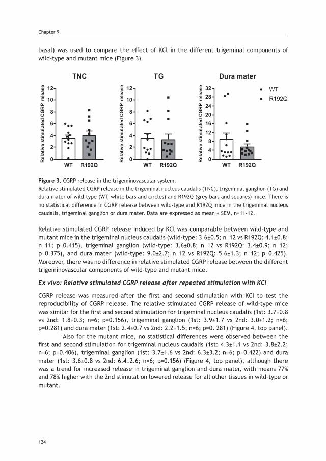
basal) was used to compare the effect of KCl in the different trigeminal components of wild-type and mutant mice (Figure 3).
Figure 3. CGRP release in the trigeminovascular system.Relative stimulated CGRP release in the trigeminal nucleus caudalis (TNC), trigeminal ganglion (TG) and dura mater of wild-type (WT, white bars and circles) and R192Q (grey bars and squares) mice. There is no statistical difference in CGRP release between wild-type and R192Q mice in the trigeminal nucleus caudalis, trigeminal ganglion or dura mater. Data are expressed as mean ± SEM, n=11-12.
Relative stimulated CGRP release induced by KCl was comparable between wild-type and mutant mice in the trigeminal nucleus caudalis (wild-type: 3.6±0.5; n=12 vs R192Q: 4.1±0.8; n=11; p=0.415), trigeminal ganglion (wild-type: 3.6±0.8; n=12 vs R192Q: 3.4±0.9; n=12; p=0.375), and dura mater (wild-type: 9.0±2.7; n=12 vs R192Q: 5.6±1.3; n=12; p=0.425). Moreover, there was no difference in relative stimulated CGRP release between the different trigeminovascular components of wild-type and mutant mice.
Ex vivo: Relative stimulated CGRP release after repeated stimulation with KCl
CGRP release was measured after the first and second stimulation with KCl to test the reproducibility of CGRP release. The relative stimulated CGRP release of wild-type mice was similar for the first and second stimulation for trigeminal nucleus caudalis (1st: 3.7±0.8 vs 2nd: 1.8±0.3; n=6; p=0.156), trigeminal ganglion (1st: 3.9±1.7 vs 2nd: 3.0±1.2; n=6; p=0.281) and dura mater (1st: 2.4±0.7 vs 2nd: 2.2±1.5; n=6; p=0. 281) (Figure 4, top panel). Also for the mutant mice, no statistical differences were observed between the first and second stimulation for trigeminal nucleus caudalis (1st: 4.3±1.1 vs 2nd: 3.8±2.2; n=6; p=0.406), trigeminal ganglion (1st: 3.7±1.6 vs 2nd: 6.3±3.2; n=6; p=0.422) and dura mater (1st: 3.6±0.8 vs 2nd: 6.4±2.6; n=6; p=0.156) (Figure 4, top panel), although there was a trend for increased release in trigeminal ganglion and dura mater, with means 77% and 78% higher with the 2nd stimulation lowered release for all other tissues in wild-type or mutant.
124
Chapter 9
WT R192Q0
2
4
6
8
10
12
TNC
Rela
tive
stim
ulat
edCG
RPre
leas
e
WT R192Q0
2
4
6
8
10
12
TG
Rela
tive
stim
ulat
edCG
RPre
leas
e
WT R192Q048
121620242832
Dura mater
Rela
tive
stim
ulat
edCG
RPre
leas
e WTR192Q
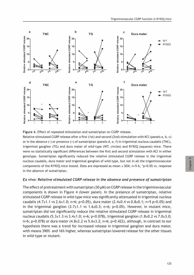
Figure 4. Effect of repeated stimulation and sumatriptan on CGRP release.Relative stimulated CGRP release after a first (1st) and second (2nd) stimulation with KCl (panels a, b, c) or in the absence (-) or presence (+) of sumatriptan (panels d, e, f) in trigeminal nucleus caudalis (TNC), trigeminal ganglion (TG) and dura mater of wild-type (WT, circles) and R192Q (squares) mice. There were no statistically significant differences between the first and second stimulation with KCl in either genotype. Sumatriptan significantly reduced the relative stimulated CGRP release in the trigeminal nucleus caudalis, dura mater and trigeminal ganglion of wild-type, but not in all the trigeminovascular components of the R192Q mice tested. Data are expressed as mean ± SEM, n=5-6, *p<0.05 vs. response in the absence of sumatriptan.
Ex vivo: Relative stimulated CGRP release in the absence and presence of sumatriptan
The effect of pretreatment with sumatriptan (30 µM) on CGRP release in the trigeminovascular components is shown in Figure 4 (lower panel). In the presence of sumatriptan, relative stimulated CGRP release in wild-type mice was significantly attenuated in trigeminal nucleus caudalis (4.7±1.1 vs 2.6±1.0; n=6; p<0.05), dura mater (2.4±0.4 vs 0.8±0.1; n=5 p<0.05) and in the trigeminal ganglion (3.7±1.1 vs 1.6±0.3; n=6; p<0.05). However, in mutant mice, sumatriptan did not significantly reduce the relative stimulated CGRP release in trigeminal nucleus caudalis (5.3±1.3 vs 3.4±1.0; n=6; p=0.078), trigeminal ganglion (1.8±0.2 vs 7.0±3.0; n=6; p=0.078) or dura mater (4.8±2.2 vs 5.6±3.2; n=6; p=0.422), although, in contrast to our hypothesis there was a trend for increased release in trigeminal ganglion and dura mater, with means 390% and 16% higher, whereas sumatriptan lowered release for the other tissues in wild-type or mutant.
125
Trigeminovascular CGRP function in R192Q mice
Chap
ter
9
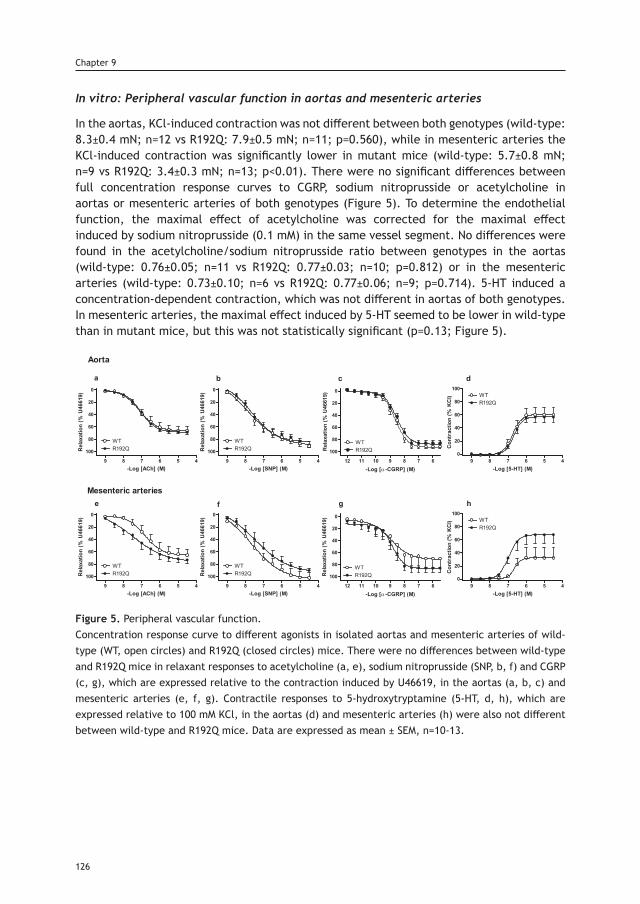
In vitro: Peripheral vascular function in aortas and mesenteric arteries
In the aortas, KCl-induced contraction was not different between both genotypes (wild-type: 8.3±0.4 mN; n=12 vs R192Q: 7.9±0.5 mN; n=11; p=0.560), while in mesenteric arteries the KCl-induced contraction was significantly lower in mutant mice (wild-type: 5.7±0.8 mN; n=9 vs R192Q: 3.4±0.3 mN; n=13; p<0.01). There were no significant differences between full concentration response curves to CGRP, sodium nitroprusside or acetylcholine in aortas or mesenteric arteries of both genotypes (Figure 5). To determine the endothelial function, the maximal effect of acetylcholine was corrected for the maximal effect induced by sodium nitroprusside (0.1 mM) in the same vessel segment. No differences were found in the acetylcholine/sodium nitroprusside ratio between genotypes in the aortas (wild-type: 0.76±0.05; n=11 vs R192Q: 0.77±0.03; n=10; p=0.812) or in the mesenteric arteries (wild-type: 0.73±0.10; n=6 vs R192Q: 0.77±0.06; n=9; p=0.714). 5-HT induced a concentration-dependent contraction, which was not different in aortas of both genotypes. In mesenteric arteries, the maximal effect induced by 5-HT seemed to be lower in wild-type than in mutant mice, but this was not statistically significant (p=0.13; Figure 5).
Figure 5. Peripheral vascular function.Concentration response curve to different agonists in isolated aortas and mesenteric arteries of wild-type (WT, open circles) and R192Q (closed circles) mice. There were no differences between wild-type and R192Q mice in relaxant responses to acetylcholine (a, e), sodium nitroprusside (SNP, b, f) and CGRP (c, g), which are expressed relative to the contraction induced by U46619, in the aortas (a, b, c) and mesenteric arteries (e, f, g). Contractile responses to 5-hydroxytryptamine (5-HT, d, h), which are expressed relative to 100 mM KCl, in the aortas (d) and mesenteric arteries (h) were also not different between wild-type and R192Q mice. Data are expressed as mean ± SEM, n=10-13.
126
Chapter 9
Discussion
The mechanisms underlying migraine pathophysiology are not completely elucidated. It seems that migraine is a disorder of brain sensory processing, characterized by a generalized neuronal hyperexcitability [22], while the head pain is thought to be the consequence of activation of the trigeminovascular system, CGRP release and sensitization of trigeminal nociceptors. Transgenic mice harboring the FHM1 R192Q mutation revealed an overall hyperexcitability phenotype and increased susceptibility to CSD [4], but whether this mutation could affect the normal physiology of the trigeminovascular system is unknown. Therefore, the functionality of the trigeminovascular system was investigated in transgenic mice that express the FHM1 R192Q missense mutation in the α1A subunit of voltage-gated CaV2.1 calcium channels that leads to a gain of function of these channels by studying: (i) dural artery vasodilation induced by endogenous and exogenous CGRP in vivo, (ii) CGRP release in various components of the trigeminovascular system ex vivo, and (iii) peripheral vascular function in vitro. Several aspects of the CGRP-related trigeminovascular function were abnormal in the mutant mice. First of all, the trigeminovascular dural artery vasodilation in vivo, either by endogenous (induced by capsaicin) or exogenous CGRP, was reduced. A priori we expected an increased vasodilation in mutant mice. After all, as CaV2.1 channels are also expressed on perivascular nerve endings [23], given the gain of function of the R192Q mutation on CaV2.1 channels [7], activation of the channels would lead to release of CGRP, a potent vasodilator, and increased vasodilation [24]. One possible explanation for the reduced effects on dural vasodilation, is that CGRP release may already be elevated at basal in mutant mice, due to the increased activity of mutant CaV2.1 channels as proposed in previous studies [11, 25]. Therefore, administration or release of CGRP from an exogenous or endogenous source, may no longer exert an effect in response to a stimulus. However, more complex mechanisms seem to be involved, since we observed a lower baseline dural diameter in the mutant mice in our in vivo study; and no genotypic differences in CGRP release, at basal or after KCl stimulation, in the trigeminovascular components in our ex vivo study. On the other hand, hyperactivity of mutant CaV2.1 channels, could potentially lead to CGRP depletion – already at baseline – as may be concluded from an immunohistochemistry study that showed reduced CGRP immunoreactivity in the trigeminal ganglia and trigeminocervical complex of these mice [13]. However, our data do not seem to support CGRP depletion, since no statistical differences in basal CGRP release or CGRP release after repeated stimulation between the genotypes were observed. Still, a reduction in response, in their case for intracellular Ca2+ response, was observed after somatosensory stimulation when studying the same R192Q mouse model, which was ascribed to reduced neurovascular coupling [26]. An alternative explanation for the in vivo reduced dural artery vasodilation response to capsaicin may be mediated by a change in CGRP receptor function or expression, since we also observed reduced dural vasodilation induced by exogenous CGRP in the mutant mice, which might suggest a desensitization or downregulation of this receptor. Receptor desensitization, as suggested before for CGRP-mediated ATP-gated P2X3 signaling in FHM1 R192Q mice [25] is more likely given that CGRP receptor expression on trigeminal ganglion neurons was shown to be similar between genotypes in culture and in situ [27].
127
Trigeminovascular CGRP function in R192Q mice
Chap
ter
9
Hypothetically, the different in vivo effects on dural vasodilation between wild-type and mutant mice could have been due to changes in mean arterial pressure associated with the administration of drugs. However, this is unlikely as changes in blood pressure during intravenous injection of ET-1, capsaicin or CGRP, are below the level affecting dural vasodilation in this experimental model [28] and, more importantly, they were not different between wild-type and mutant mice. When considering the present and previous studies on FHM1 R192Q mice, the basal release of CGRP and functionality of the CGRP receptor may change with age. It is noteworthy that in 11-day-old mutant mice there is an increased basal and KCl stimulated CGRP release from trigeminal ganglia [11]; in 2-week-old animals an increased basal CGRP release was identified [25]; whereas in 4-week-old mice an increased stimulated CGRP release but unaffected basal CGRP release was identified [12]. Remarkably, in the present study of 13- to 14-week-old mutant mice, which are young adult mice, neither an increase in basal nor in KCl-stimulated CGRP release in the components of the trigeminovascular system (including trigeminal ganglia) was observed, while in our in vivo experiments, CGRP-induced dural artery vasodilation was decreased. Moreover, a reduced CGRP expression was found in the trigeminal ganglion of 5 to 8 month old mutant mice in an immunohistochemistry study [13]. Therefore, it is tantalizing that the age of the mutant mice may be a determining factor that could reconcile the different findings of CGRP release in these studies. An alternative explanation for the apparent discrepancy between our FHM1 R192Q mice data and the previously studies mentioned, where an increased CGRP release was observed in the trigeminal ganglion [12], may be the concentration of KCl employed in the different experiments. While Fioretti et al. [12] used 35 mM KCl to stimulate CGRP release, we used 60 mM KCl based on studies showing that this concentration activates calcium channels (including the CaV2.1 channels) [29]; and this induces a significant increase in CGRP release without reaching the maximal effect in the trigeminal ganglion, trigeminal nucleus caudalis and dura mater [17], which are essential characteristics for our study. However, we cannot exclude that using lower concentrations, a difference in sensitivity to KCl between wild-type and mutant mice might have been unmasked. To assess systemic vascular CGRP receptor desensitization, we performed concentration response curves to CGRP in isolated aortas and mesenteric arteries, to understand the effect of the R192Q mutation on peripheral CGRP receptor function. Interestingly, no differences in the relaxant responses were found between genotypes, so it is unlikely that the R192Q mutation affects CGRP-induced vasodilation in these vessels. Though, if desensitization of CGRP receptors occurs due to a structurally elevated CGRP release (caused by the gain of function of the CaV2.1 calcium channel), it may only be occurring in dural arteries. Unfortunately, it is not possible to do functional studies with a Mulvany myograph, due to the small size of those vessels (outer diameter < 30 µm). The general vascular function of the aortas and mesenteric arteries was also investigated by performing concentration response curves to acetylcholine, sodium nitroprusside, 5-HT and a single dose of KCl. Between genotypes, there were no differences in the acetylcholine and sodium nitroprusside responses, or the acetylcholine/sodium nitroprusside ratio, suggesting that the R192Q mutation does not influence peripheral nitric oxide (NO) endothelium-dependent and -independent pathways, which seems in line with a lack of effect on the baseline mean arterial pressure in vivo.
128
Chapter 9
Instead, a decreased KCl-induced contraction in the mesenteric arteries of mutant mice was observed, which might suggest that the R192Q mutation (in the CaV2.1 calcium channel) indirectly influences other channels. This might also be the case with other voltage-dependent calcium channels, disturbing the levels of cytoplasmic [Ca2+], the activation of the Ca2+-calmodulin-dependent myosin light chain (MLC) kinase, MLC phosphorylation, and overall contraction of the smooth muscle cells [30].The latter seems to be supported by findings, although not confirmed by other research groups, that the CaV2.1 calcium channel may be present in smooth muscle cells of murine aortas, rat renal vessels and rabbit renal afferent arterioles [31, 32]; and may contribute to contraction by regulating intracellular calcium concentrations [32].The fact that this effect was only observed in mesenteric arteries and not in aortas, suggests that the R192Q mutation in CaV2.1 channels might play a more prominent role in resistance arteries than in conductance arteries. This could be related to the channel expression levels in different vascular beds. Since the 5-HT induced contraction was related to the KCl response in vitro, the trend of increased 5-HT induced contraction observed in the mesenteric arteries of the R192Q mice, might rather be due to biological differences in the KCl responses, than differences in the function of the 5-HT1B receptors, which is responsible for the 5-HT induced contraction [33]. The ex vivo CGRP release experiments demonstrated in wild-type mice, that the presence of sumatriptan significantly reduced CGRP release upon stimulation in the trigeminal nucleus caudalis, dura mater and in the trigeminal ganglion. This seems in line with a finding that naratriptan and sumatriptan prevented the induction of sensitization in central trigeminovascular neurons, but not from peripheral terminals innervating the dura mater [34].However, there was also a trend of lower CGRP release in the trigeminal nucleus caudalis of wild-type mice after a second stimulation with KCl (Figure 4, top panel), which might suggest that the effect of sumatriptan observed is a result of lower release in response to the second stimulation, rather than inhibition of CGRP release by sumatriptan. This is unlikely since delta CGRP release in the sumatriptan experiments (CGRP release in the presence of sumatriptan – CGRP release in the absence of sumatriptan) were significantly different from the hypothetical value of zero (p=0.05); which was not the case for the delta CGRP release in the repeated experiments (CGRP release after second stimulation – CGRP release after first stimulation), indicating that the lower CGRP release in the trigeminal nucleus caudalis of wild-type mice in the sumatriptan experiments is indeed an effect of sumatriptan. Notably, in the mutant mice, sumatriptan did not show a statistically significant change in CGRP release upon stimulation on the different components of the trigeminovascular system. This might be explained by the gain of function effect on the CaV2.1 calcium channels that influence the modulation properties of sumatriptan, since activation of the 5-HT1 receptor modulates calcium channels to control CGRP release [35].
Conclusion
In conclusion, our data do not support the a priori hypothesis that the FHM1 R192Q mutation increases dural artery vasodilation and CGRP release in the trigeminovascular system; instead, a decrease in dural vasodilation and no effect on CGRP release was observed, possibly involving trigeminovascular CGRP receptor desensitization. Thus, our data reinforce the findings of previous studies that the R192Q mutation does not only affect central aspects of migraine pathophysiology, but also the normal functioning of the trigeminovascular
129
Trigeminovascular CGRP function in R192Q mice
Chap
ter
9
system. Moreover, the effects of the mutation on CGRP release in the trigeminovascular system may not just be an ‘on’ or ‘off’ phenomenon but rather of a modulatory nature, involving multiple mechanisms. Although it is still undefined whether the trigeminovascular effects of the R192Q mutation are the consequence of a direct neurovascular effect, a central neuronal dysfunction, or a combination of both; our results clearly indicate that the FHM1 R192Q mice display trigeminovascular abnormalities, underlining their relevance for migraine research.
References1. Society. HCCotIH. The International Classification of Headache Disorders, 3rd edition (beta version). Cephalalgia 2013; 33: 629-808.2. Lauritzen M. Pathophysiology of the migraine aura. The spreading depression theory. Brain 1994; 117 ( Pt 1): 199-210.3. Karsan N and Goadsby PJ. Calcitonin gene-related peptide and migraine. Curr Opin Neurol 2015; 28: 250-254.4. Ferrari MD, Klever RR, Terwindt GM, Ayata C and van den Maagdenberg AM. Migraine pathophysiology: lessons from mouse models and human genetics. Lancet Neurol 2015; 14: 65-80.5. Ophoff RA, Terwindt GM, Vergouwe MN, et al. Familial hemiplegic migraine and episodic ataxia type-2 are caused by mutations in the Ca2+ channel gene CACNL1A4. Cell 1996; 87: 543-552.6. van den Maagdenberg AM, Haan J, Terwindt GM, et al. Migraine: gene mutations and functional consequences. Curr Opin Neurol 2007; 20: 299-305.7. van den Maagdenberg AM, Pietrobon D, Pizzorusso T, et al. A Cacna1a knockin migraine mouse model with increased susceptibility to cortical spreading depression. Neuron 2004; 41: 701-710.8. Tottene A, Conti R, Fabbro A, et al. Enhanced excitatory transmission at cortical synapses as the basis for facilitated spreading depression in Ca(v)2.1 knockin migraine mice. Neuron 2009; 61: 762-773.9. Eikermann-Haerter K, Dilekoz E, Kudo C, et al. Genetic and hormonal factors modulate spreading depression and transient hemiparesis in mouse models of familial hemiplegic migraine type 1. J Clin Invest 2009; 119: 99-109.10. Chanda ML, Tuttle AH, Baran I, et al. Behavioral evidence for photophobia and stress-related ipsilateral head pain in transgenic Cacna1a mutant mice. Pain 2013; 154: 1254-1262.11. Ceruti S, Villa G, Fumagalli M, et al. Calcitonin gene-related peptide-mediated enhancement of purinergic neu-ron/glia communication by the algogenic factor bradykinin in mouse trigeminal ganglia from wild-type and R192Q Cav2.1 Knock-in mice: implications for basic mechanisms of migraine pain. J Neurosci 2011; 31: 3638-3649.12. Fioretti B, Catacuzzeno L, Sforna L, et al. Trigeminal ganglion neuron subtype-specific alterations of Ca(V)2.1 calcium current and excitability in a Cacna1a mouse model of migraine. J Physiol 2011; 589: 5879-5895.13. Mathew R, Andreou AP, Chami L, et al. Immunohistochemical characterization of calcitonin gene-related peptide in the trigeminal system of the familial hemiplegic migraine 1 knock-in mouse. Cephalalgia 2011; 31: 1368-1380.14. Gupta S, Akerman S, van den Maagdenberg AM, et al. Intravital microscopy on a closed cranial window in mice: a model to study trigeminovascular mechanisms involved in migraine. Cephalalgia 2006; 26: 1294-1303.15. Ebersberger A, Averbeck B, Messlinger K, et al. Release of substance P, calcitonin gene-related peptide and pros-taglandin E2 from rat dura mater encephali following electrical and chemical stimulation in vitro. Neuroscience 1999; 89: 901-907.16. Eberhardt M, Hoffmann T, Sauer SK, et al. Calcitonin gene-related peptide release from intact isolated dorsal root and trigeminal ganglia. Neuropeptides 2008; 42: 311-317.17. Amrutkar DV, Ploug KB, Hay-Schmidt A, et al I. mRNA expression of 5-hydroxytryptamine 1B, 1D, and 1F receptors and their role in controlling the release of calcitonin gene-related peptide in the rat trigeminovascular system. Pain 2012; 153: 830-838.18. Mulvany MJ and Halpern W. Contractile properties of small arterial resistance vessels in spontaneously hyperten-sive and normotensive rats. Circ Res 1977; 41: 19-26.19. MaassenVanDenBrink A, Reekers M, Bax WA, et al. Coronary side-effect potential of current and prospective an-ti-migraine drugs. Circulation 1998; 98: 25-30.20. Chan KY, Gupta S, de Vries R, et al. Effects of ionotropic glutamate receptor antagonists on rat dural artery diam-eter in an intravital microscopy model. Br J Pharmacol 2010; 160: 1316-1325.21. Labruijere S, van Houten EL, de Vries R, et al. Analysis of the vascular responses in a murine model of polycystic ovary syndrome. J Endocrinol 2013; 218: 205-213.22. Akerman S, Romero-Reyes M and Holland PR. Current and novel insights into the neurophysiology of migraine and its implications for therapeutics. Pharmacology & Therapeutics 2017; 172: 151-170.23. Asakura K, Kanemasa T, Minagawa K, et al. α-Eudesmol, a P/Q-type Ca2+ channel blocker, inhibits neurogenic vas-odilation and extravasation following electrical stimulation of trigeminal ganglion. Brain Research 2000; 873: 94-101.
130
Chapter 9
131
24. Akerman S, Williamson DJ and Goadsby PJ. Voltage-dependent calcium channels are involved in neurogenic dural vasodilatation via a presynaptic transmitter release mechanism. British Journal of Pharmacology 2003; 140: 558-566.25. Hullugundi SK, Ferrari MD, van den Maagdenberg A, et al. The mechanism of functional up-regulation of P2X3 receptors of trigeminal sensory neurons in a genetic mouse model of familial hemiplegic migraine type 1 (FHM-1). PLoS One 2013; 8: e60677.26. Khennouf L, Gesslein B, Lind BL, et al. Activity-dependent calcium, oxygen, and vascular responses in a mouse model of familial hemiplegic migraine type 1. Annals of Neurology 2016; 80: 219-232.27. Vilotti S, Vana N, Van den Maagdenberg AM, et al. Expression and function of calcitonin gene-related peptide (CGRP) receptors in trigeminal ganglia of R192Q Cacna1a knock-in mice. Neuroscience Letters 2016; 620: 104-110.28. Petersen KA, Dyrby L, Williamson D, et al. Effect of hypotension and carbon dioxide changes in an improved gen-uine closed cranial window rat model. Cephalalgia 2004; 25: 23-29.29. Amrutkar DV, Ploug KB, Olesen J, et al. Role for voltage gated calcium channels in calcitonin gene-related peptide release in the rat trigeminovascular system. Neuroscience 2011; 172: 510-517.30. Ratz PH, Berg KM, Urban NH, et a;. Regulation of smooth muscle calcium sensitivity: KCl as a calcium-sensitizing stimulus. Am J Physiol Cell Physiol 2005; 288: C769-83.31. Andreasen D, Friis UG, Uhrenholt TR, et al. Coexpression of voltage-dependent calcium channels Cav1.2, 2.1a, and 2.1b in vascular myocytes. Hypertension 2006; 47: 735-741.32. Hansen PB, Jensen BL, Andreasen D, et al. Vascular smooth muscle cells express the alpha(1A) subunit of a P-/Q-type voltage-dependent Ca(2+)Channel, and It is functionally important in renal afferent arterioles. Circ Res 2000; 87: 896-902.33. Villalón CM and Centurión D. Cardiovascular responses produced by 5-hydroxytriptamine:a pharmacological update on the receptors/mechanisms involved and therapeutic implications. Naunyn-Schmiedeberg’s Arch Pharmacol 2007; 376: 45-63.34. Kageneck C, Nixdorf-Bergweiler BE, Messlinger K, et al. Release of CGRP from mouse brainstem slices indicates central inhibitory effect of triptans and kynurenate. J Headache Pain 2014; 15:7.35. Xiao Y, Richter JA and Hurley JH. Release of glutamate and CGRP from trigeminal ganglion neurons: Role of calcium channels and 5-HT1 receptor signaling. Mol Pain 2008; 16: 4-12.
Trigeminovascular CGRP function in R192Q mice
Chap
ter
9
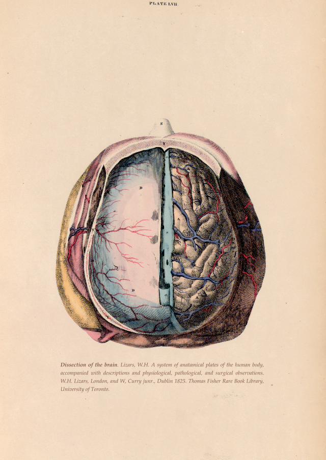
Dissection of the brain. Lizars, W.H. A system of anatamical plates of the human body, accompanied with descriptions and physiological, pathological, and surgical observations. W.H. Lizars, London, and W, Curry junr., Dublin 1825. Thomas Fisher Rare Book Library, University of Toronto.

Part IV. Summary, conclusions and future perspectives
Chapter 10
Summary, conclusions and future perspectives
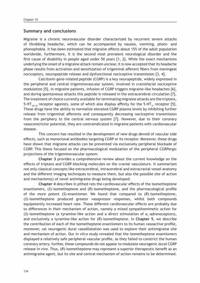
Summary and conclusions
Migraine is a chronic neurovascular disorder characterized by recurrent severe attacks of throbbing headache, which can be accompanied by nausea, vomiting, photo- and phonophobia. It has been estimated that migraine affects about 15% of the adult population worldwide, furthermore, it is the second most prevalent neurological disorder and the first cause of disability in people aged under 50 years [1, 2]. While the exact mechanisms underlying the onset of a migraine attack remain unclear, it is now accepted that its headache phase results from activation and sensitization of trigeminal afferent fibers from meningeal nociceptors, neuropeptide release and dysfunctional nociceptive transmission [3, 4]. Calcitonin gene-related peptide (CGRP) is a key neuropeptide, widely expressed in the peripheral and central trigeminovascular system, involved in craniofacial nociceptive modulation [5]. In migraine patients, infusion of CGRP triggers migraine-like headaches [6], and during spontaneous attacks this peptide is released in the extracerebral circulation [7]. The treatment of choice currently available for terminating migraine attacks are the triptans, 5-HT1B/1D receptor agonists, some of which also display affinity for the 5-HT1F receptor [5]. These drugs have the ability to normalize elevated CGRP plasma levels by inhibiting further release from trigeminal afferents and consequently decreasing nociceptive transmission from the periphery to the central nervous system [7]. However, due to their coronary vasoconstrictor potential, they are contraindicated in migraine patients with cardiovascular disease. This concern has resulted in the development of new drugs devoid of vascular side effects, such as monoclonal antibodies targeting CGRP or its receptor. Moreover, these drugs have shown that migraine attacks can be prevented via exclusively peripheral blockade of CGRP. This thesis focused on the pharmacological modulation of the peripheral CGRPergic projections of the trigeminovascular system. Chapter 3 provides a comprehensive review about the current knowledge on the effects of triptans and CGRP-blocking molecules on the cranial vasculature. It summarizes not only classical concepts like extracerebral, intracerebral and extracranial vessel anatomy and the different imaging techniques to measure them, but also the possible site of action and mechanism(s) of novel antimigraine drugs being developed. Chapter 4 describes in pithed rats the cardiovascular effects of the isometheptene enantiomers, (S)-isometheptene and (R)-isometheptene, and the pharmacological profile of the more potent (S)-enantiomer. We found that compared to (R)-isometheptene, (S)-isometheptene produced greater vasopressor responses, whilst both compounds equipotently increased heart rate. These different cardiovascular effects are probably due to differences in their mechanism of action, namely a mixed sympathomimetic action for (S)-isometheptene (a tyramine-like action and a direct stimulation of α1-adrenoceptors), and exclusively a tyramine-like action for (R)-isometheptene. In Chapter 5, we describe the contribution of each of the isometheptene enantiomers to its human vasoactive profile, moreover, rat neurogenic dural vasodilatation was used to explore their antimigraine site and mechanism of action. Our in vitro study revealed that the isometheptene enantiomers displayed a relatively safe peripheral vascular profile, as they failed to constrict the human coronary artery; further, these compounds do not appear to modulate neurogenic dural CGRP release in vivo. Thus, (R)-isometheptene may represent a superior therapeutic benefit as an antimigraine agent, but its site and central mechanism of action remains to be determined.
134
Chapter 10
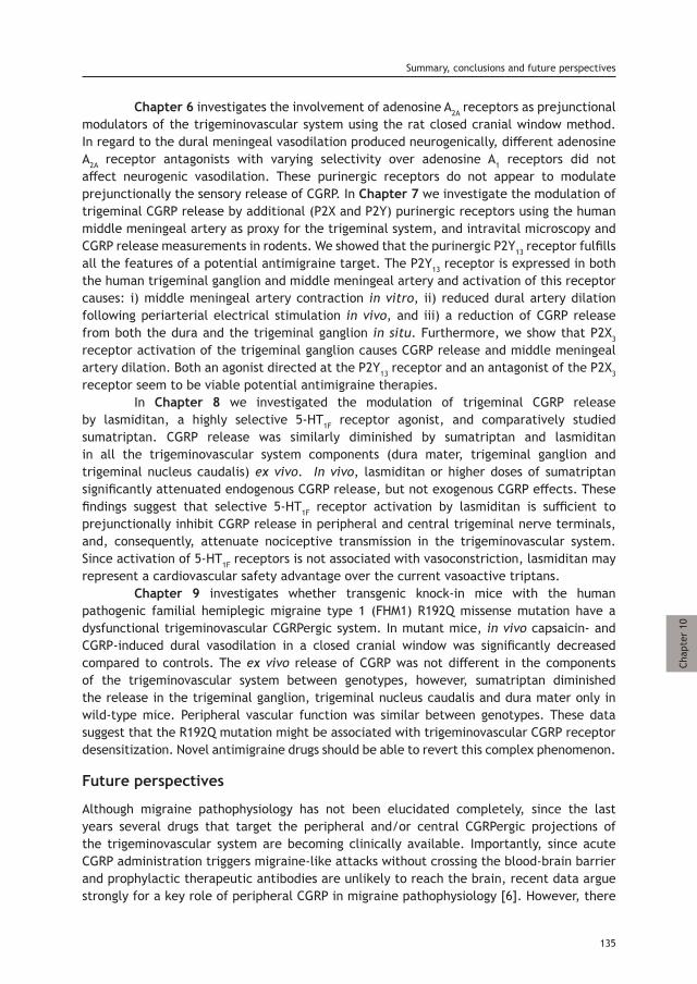
Chap
ter
10
Chapter 6 investigates the involvement of adenosine A2A receptors as prejunctional modulators of the trigeminovascular system using the rat closed cranial window method. In regard to the dural meningeal vasodilation produced neurogenically, different adenosine A2A receptor antagonists with varying selectivity over adenosine A1 receptors did not affect neurogenic vasodilation. These purinergic receptors do not appear to modulate prejunctionally the sensory release of CGRP. In Chapter 7 we investigate the modulation of trigeminal CGRP release by additional (P2X and P2Y) purinergic receptors using the human middle meningeal artery as proxy for the trigeminal system, and intravital microscopy and CGRP release measurements in rodents. We showed that the purinergic P2Y13 receptor fulfills all the features of a potential antimigraine target. The P2Y13 receptor is expressed in both the human trigeminal ganglion and middle meningeal artery and activation of this receptor causes: i) middle meningeal artery contraction in vitro, ii) reduced dural artery dilation following periarterial electrical stimulation in vivo, and iii) a reduction of CGRP release from both the dura and the trigeminal ganglion in situ. Furthermore, we show that P2X3 receptor activation of the trigeminal ganglion causes CGRP release and middle meningeal artery dilation. Both an agonist directed at the P2Y13 receptor and an antagonist of the P2X3 receptor seem to be viable potential antimigraine therapies. In Chapter 8 we investigated the modulation of trigeminal CGRP release by lasmiditan, a highly selective 5-HT1F receptor agonist, and comparatively studied sumatriptan. CGRP release was similarly diminished by sumatriptan and lasmiditan in all the trigeminovascular system components (dura mater, trigeminal ganglion and trigeminal nucleus caudalis) ex vivo. In vivo, lasmiditan or higher doses of sumatriptan significantly attenuated endogenous CGRP release, but not exogenous CGRP effects. These findings suggest that selective 5-HT1F receptor activation by lasmiditan is sufficient to prejunctionally inhibit CGRP release in peripheral and central trigeminal nerve terminals, and, consequently, attenuate nociceptive transmission in the trigeminovascular system. Since activation of 5-HT1F receptors is not associated with vasoconstriction, lasmiditan may represent a cardiovascular safety advantage over the current vasoactive triptans. Chapter 9 investigates whether transgenic knock-in mice with the human pathogenic familial hemiplegic migraine type 1 (FHM1) R192Q missense mutation have a dysfunctional trigeminovascular CGRPergic system. In mutant mice, in vivo capsaicin- and CGRP-induced dural vasodilation in a closed cranial window was significantly decreased compared to controls. The ex vivo release of CGRP was not different in the components of the trigeminovascular system between genotypes, however, sumatriptan diminished the release in the trigeminal ganglion, trigeminal nucleus caudalis and dura mater only in wild-type mice. Peripheral vascular function was similar between genotypes. These data suggest that the R192Q mutation might be associated with trigeminovascular CGRP receptor desensitization. Novel antimigraine drugs should be able to revert this complex phenomenon.
Future perspectives
Although migraine pathophysiology has not been elucidated completely, since the last years several drugs that target the peripheral and/or central CGRPergic projections of the trigeminovascular system are becoming clinically available. Importantly, since acute CGRP administration triggers migraine-like attacks without crossing the blood-brain barrier and prophylactic therapeutic antibodies are unlikely to reach the brain, recent data argue strongly for a key role of peripheral CGRP in migraine pathophysiology [6]. However, there
Summary, conclusions and future perspectives
135
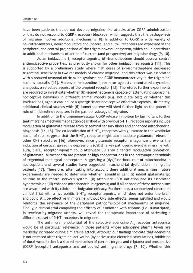
have been patients that do not develop migraine-like attacks after CGRP administration or that do not respond to CGRP (receptor) blockade, which suggests that the pathogenesis of migraine involves additional mechanisms [8]. In addition to CGRP, a wide variety of neurotransmitters, neuromodulators and (hetero- and auto-) receptors are expressed in the peripheral and central projections of the trigeminovascular system, which could contribute to additional mechanisms of action of current (and prospective) antimigraine drugs [9, 10]. As an imidazoline I1 receptor agonist, (R)-isometheptene should possess central antinociceptive properties, as previously shown for other imidazolines agonists [11]. This is supported by a preliminary study where high doses of (R)-isometheptene decreased trigeminal sensitivity in two rat models of chronic migraine, and this effect was associated with a reduced neuronal nitric oxide synthase and CGRP immunoreactivity in the trigeminal nucleus caudalis [12]. Moreover, imidazoline I1 receptor agonists potentiated oxycodone analgesia, a selective agonist of the µ-opioid receptor [13]. Therefore, further experiments are required to investigate whether (R)-isometheptene is capable of attenuating supraspinal nociceptive behaviors in different animal models (e.g. hot plate test) or whether this imidazoline I1 agonist can induce a synergistic antinociceptive effect with opioids. Ultimately, additional clinical studies with (R)-isometheptene will shed further light on the potential role of imidazoline receptors in the pathophysiology of migraine. In addition to the trigeminovascular CGRP release inhibition by lasmiditan, further (antimigraine) mechanisms of action described with previous 5-HT1F receptor agonists include modulation of glutamate release from trigeminal sensory fibers and enhanced mitochondrial biogenesis [14, 15]. The co-localization of 5-HT1F receptors with glutamate in the vestibular nuclei of rats, suggests that the 5-HT1F receptor might also modulate glutamate release in other CNS structures [16]. Moreover, since glutamate receptor antagonism prevents the induction of cortical spreading depressions (CSDs), a key pathogenic event in migraine with aura, 5-HT1F receptor agonism could attenuate CSDs via a central modulation (inhibition) of glutamate. Mitochondria are present at high concentrations in the presynaptic terminals of trigeminal meningeal nociceptors, suggesting a (dys)functional role of mitochondria in nociception; and several studies have suggested mitochondrial dysfunction in migraine patients [17]. Therefore, after taking into account these additional mechanisms, future experiments are needed to determine whether lasmiditan can: (i) inhibit glutamatergic neurons in the central nervous system, (ii) attenuate CSDs initiation and its associated hyperaemia or, (iii) enhance mitochondrial biogenesis; and if all or none of these mechanisms are associated with its clinical antimigraine efficacy. Furthermore, a randomized controlled clinical trial with a hydrophilic 5-HT1F receptor agonist, which does not enter the brain and could still be effective in migraine without CNS side effects, seems justified and would reinforce the relevance of the peripheral pathophysiological mechanisms of migraine. Finally, a clinical trial comparing the efficacy of lasmiditan with triptans (i.e. sumatriptan) in terminating migraine attacks, will reveal the therapeutic importance of activating a different subset of 5-HT1 receptors in migraine. The antimigraine potential of the selective adenosine A2A receptor antagonists would be of particular relevance in those patients whose adenosine plasma levels are markedly increased during a migraine attack. Although our findings indicate that adenosine is not released after trigeminal activation (by perivascular electrical stimulation), inhibition of dural vasodilation is a shared mechanism of current (ergots and triptans) and prospective (CGRP (receptor) antagonists and antibodies) antimigraine drugs [7, 10]. Whether this
136
Chapter 10
137
(antimigraine) mechanism alone is sufficient to attenuate the dysfunctional trigeminal nociceptive transmission associated with migraine headache remains to be determined. Obviously, further clinical studies are needed to evaluate the antimigraine potential of the JNJ adenosine receptor antagonists with the optimal doses based on their pharmacokinetic properties. Since our functional data and immunohistochemistry revealed that P2Y13 receptor expression and activation (inhibition of CGRP release) occurs in the trigeminal ganglion and on the nerve endings at the dura mater/middle meningeal artery, the clinical applications and potential anti-migraine effects need to be further investigated. For this purpose, there is a particular need for the development of specific agonists. The main concern regarding the use of agonists based on ADP is their potential thrombotic activity. ADP activating P2Y1 or P2Y12 receptors is considered a relatively weak platelet aggregation agonist by itself. Unfortunately, ADP still works as an amplifier of platelet aggregators such as thrombin [18], which is not compatible with the treatment of patients. This potential side-effect should not be an issue for a selective P2Y13 receptor agonist. The generation of transgenic knock-in mice with the human pathogenic FHM1 R192Q missense mutation in the Cacna1a gene affects the central aspects of migraine pathophysiology (overall neuronal hyperexcitability), but also results in peripheral CGRP-trigeminovascular desensitization. In addition to these effects, our study suggests that this mutation alters the peripheral vascular function of resistance blood vessels (e.g. reduced KCl-induced contraction of the mesenteric artery), which could contribute to the generalized vascular dysfunction that has been associated with migraine [19]. Further experiments in these mice are needed to elucidate the effects of the FHM1 mutation on the peripheral CGRPergic fibers at the neurovascular junction and if this contributes to peripheral vascular dysfunction. This could clarify whether this is one of the mechanisms that leads to the increased cardiovascular risk of migraineurs. Migraine is three times more prevalent in women of reproductive age than in men and previous studies have provided insight into the effects of female sex hormones on the pathophysiology of migraine, suggesting that the reactivity of the trigeminovascular system in migraine patients, in response to varying levels of female hormones, differs from that in healthy women without migraine [20]. Moreover, a crosstalk between CGRP and hormonal fluctuations of the estrus cycle has previously been described [21]. Therefore, in the present thesis the pharmacological characterizations were limited to male rodents. Future experiments should test whether there are any differences in CGRP release between male and female rodents, and how ovarian steroid hormones fluctuation modifies these responses during the different stages of the estrous cycle. It has recently been shown that application of CGRP to the cranial meninges causes behavioral responses consistent with headache, only in female rodents [22], supporting a thorough pharmacological characterization in female rodents during the different stages of the estrous cycle.
Summary, conclusions and future perspectives
Chap
ter
10
Nederlandse samenvatting
Migraine is een chronische neurovasculaire aandoening die bij circa 15% van de wereldwijde populatie volwassenen voorkomt [1]. Het is de tweede meest voorkomende neurologische aandoening en de meest invaliderende aandoening bij mensen in de leeftijd onder de 50 jaar [2]. Terwijl de exacte onderliggende mechanismen die het begin van een migraineaanval veroorzaken nog steeds onbekend zijn, wordt nu algemeen geaccepteerd dat de hoofdpijnfase veroorzaakt wordt door de activatie en sensitisatie van trigeminale afferente vezels vanuit meningeale nociceptoren, neuropeptide afgifte en verstoorde nociceptieve transmissie [3, 4]. Calcitonin gene-related peptide (CGRP) is een belangrijk neuropeptide, dat veel tot expressie komt in zowel het perifere als centrale trigeminovasculaire systeemen dat tijdens een migraine aanval vrijkomt in de extracerebrale circulatie [5]. Bovendien is aangetoond dat door blokkade van CGRP of de receptor met monoclonale antilichamen migraine aanvallen voorkomen kunnen worden door een perifere remming van CGRP. In dit proefschrift ligt de focus op de farmacologische regulatie van de perifere CGRP projecties van het trigeminovasculaire systeem. Hoofdstuk 3 geeft een uitgebreid overzicht van de huidige kennis over de effecten van triptanen en CGRP-blokkerende moleculen op de craniële vasculatuur. Het bevat niet alleen een samenvatting van de klassieke concepten als de extracerebrale, intracerebrale en extracraniële vasculaire anatomie en de verschillende beeldvormingstechnieken om deze te meten, maar ook de mogelijke plaats van activiteit en mechanisme(n) van nieuwe antimigrainemiddelen die in ontwikkeling zijn. Hoofdstuk 4 beschrijft de cardiovasculaire effecten van de isomethepteen enantiomeren, (S)-isomethepteen en (R)-isomethepteen in ratten onder anesthesie, en het farmacologisch profiel van het meest potente (S)-enantiomeer. Vergeleken met (R)-isomethepteen vonden we dat (S)-isomethepteen een grotere bloeddrukverhogende respons opleverde, terwijl beide stoffen in gelijke mate de hartslag verhoogden. Deze verschillende cardiovasculaire effecten worden waarschijnlijk veroorzaakt door verschillen in hun werkingsmechanismen, namelijk een gemengd sympathomimetische werking bij (S)-isomethepteen (een tyramine-achtige werking en een directe stimulatie van α1-adrenoceptoren), en een exclusief tyramine-achtige werking bij (R)-isomethepteen. In Hoofdstuk 5 beschrijven we bijdrage van elk van de isomethepteen enantiomeren aan het humane vasoactieve profiel. Daarnaast is het neurogene durale vasodilatatie rat model om hun antimigraine aangrijpingspunt en werkingsmechanisme te onderzoeken. Onze in vitro studie liet zien dat de isomethepteen enantiomeren een relatief veilig perifeer vasculair profiel vertoonden, aangezien zij geen vernauwing in de humane kransslagader veroorzaakten; bovendien lijken deze stoffen niet de durale afgifte van CGRP in vivo te reguleren. Samengevat lijkt (R)-isomethepteen het beste profiel te hebben als antimigraine middel, maar het locatie van het antimigraine effect en het centrale actiemechanisme dienen nog te worden vastgesteld. Hoofdstuk 6 onderzoekt de betrokkenheid van adenosine A2A receptoren als prejunctionele modulatoren van het trigeminovasculaire systeem, waarbij gebruik gemaakt is van de ‘closed cranial window’ methode in ratten. Verschillende adenosine A2A receptor antagonisten met variërende selectiviteit over adenosine A1 receptoren hadden geen invloed op de neurogeen ge:induceerde dural meningeal vaatverwijding. Deze purinerge receptoren lijken niet prejunctioneel de sensorische afgifte van CGRP te reguleren. In Hoofdstuk 7
138
Chapter 10
139
onderzochten we de regulatie van trigeminale CGRP afgifte door additionele (P2X and P2Y) purinerge receptoren, gebruik makend van de humane a. meningea media als graadmeter voor het trigeminale systeem, en intravitalemicroscopie en metingen van de CGRP afgifte bij ratten en muizen. We toonden aan dat de purinerge P2Y13 receptor alle kenmerken bezit van een mogelijk antimigraine doelwit. De P2Y13 receptor komt zowel in het humane trigeminale ganglion als in de a. meningea media tot expressie en activatie van deze receptor veroorzaakt: i) in vitro vernauwing van de a. meningea media, ii) verminderde verwijding van de durale arterie na peri-arteriële electrische stimulatie in vivo, en iii) een verminderde afgifte van CGRP vanuit zowel de dura als het trigeminale ganglion in situ. Voorts toonden we aan dat activatie van de P2X3 receptor in het trigeminale ganglion CGRP afgifte en verwijding van de a. meningea mediaveroorzaakt. Zowel een agonist gericht op de P2Y13 receptor als een antagonist van de P2X3 receptor lijken geschikte kandidaten voor antimigraine behandelingen te zijn. In Hoofdstuk 8 onderzochten we de regulatie van trigeminale CGRP afgifte door lasmiditan, een zeer selectieve 5-HT1F receptor agonist, en bestudeerden deze in vergelijking met sumatriptan. Ex vivo was CGRP afgifte was verminderd op gelijke wijze door zowel sumatriptan als lasmiditan in alle componenten van het trigeminovasculaire systeem (dura mater, trigeminale ganglion en trigeminale nucleus caudalis). In vivo zorgden lasmiditan of hogere doses sumatriptan voor een significant verminderde endogene CGRP afgifte, maar werd de respons op exogeen CGRP niet beïnvloed. Deze bevindingen suggereren dat selectieve 5-HT1F receptor activatie door lasmiditan voldoende is om CGRP afgifte prejunctioneel te remmen in perifere en centrale trigeminale zenuwuiteinden, waardoor als gevolg hiervan de nociceptieve transmissie in het trigeminovasculaire systeem afzwakt. Aangezien de activatie van 5-HT1F receptoren niet betrokken is bij vasoconstrictie, heeft lasmiditan mogelijk een cardiovasculair veiligheidsvoordeel ten opzichte van de bestaande vasoactieve triptanen. Hoofdstuk 9 is onderzocht of transgene knock-in muizen met de humane pathogene familiaire hemipleigische migraine type 1 (FHM1) R192Q missense mutatie een verstoordefunctie van het trigeminovasculaire CGRP systeem hebben. In mutante muizen was in vivo capsaïcine- en CGRP-geïnduceerde durale vaatverwijding in een het ‘closed cranial window’significant verminderd in vergelijking met controle muizen. Er was geen verschil tussen genotypen wat betreftj ex vivo afgifte van CGRP in de verschillende componenten van het trigeminovasculaire systeem. Sumatriptan verminderde echter de afgifte in het trigeminale ganglion, de trigeminale nucleus caudalis en dura mater in alleen de wild-type, en niet de mutante muizen. Perifere vasculaire functie was in alle genotypes hetzelfde. Deze uitkomsten wijzen erop dat de R192Q mutatie wellicht betrokken is bij desensitisatie van de trigeminovasculaire CGRP receptor. Nieuwe antimigraine middelen zouden dit complexe fenomeen moeten kunnen herstellen.
Toekomstperspectief
Hoewel de pathofysiologie van migraine nog niet volledig is opgehelderd, zijn de laatste jaren verscheidene middelen op de markt gekomen die aangrijpen op perifere en/of centrale CGRP-projecties van het trigeminovasculaire systeem. Aangezien acute toediening van CGRP een migraine-achtige aanval oproept zonder de bloed-hersenbarrière te passeren en het onwaarschijnlijk is dat profylactische therapeutische antilichamen de hersenen bereiken, pleiten recente onderzoeksresultaten sterk voor een belangrijke rol van perifeer
Dutch summary
Chap
ter
10
CGRP in migraine [6]. Er zijn echter patiënten die geen migraine aanval ontwikkelen na het toedienen van CGRP, of die niet reageren op de remming van CGRP (receptor), hetgeen er op wijst dat ook andere mechanismen dan CGRP betrokken zijn bij de pathogenese van migraine [7]. Naast CGRP komt een groot aantal neurotransmitters, neuromodulatoren en (hetero- en auto-) receptoren tot expressie in perifere en centrale projecties van het trigeminovasculaire systeem, die bij kunnen dragen aan aanvullende werkingsmechanismen voor huidige (en toekomstige) antimigraine middelen [8, 9]. Als een imidazoline I1 receptor agonist zou (R)-isomethepteen centrale anti-nociceptieve eigenschappen moeten bezitten, zoals eerder is aangetoond bij andere imidazoline agonisten [10]. Deze theorie wordt ondersteund door een voorstudie waarbij een hoge dosering van (R)-isometheptene de trigeminale gevoeligheid verminderde in twee rattenmodellen met chronische migraine, dit effect ging gepaard met een verminderde neuronale stikstof oxide synthase en CGRP immunoreactiviteit in de trigeminale nucleus caudalis [11]. Bovendien werken imidazoline I1 receptor agonisten versterkend op de analgesie geïndueerd door oxycodon, een selectieve agonist van de µ-opioïde receptor [12]. Om deze reden is nader onderzoek vereist om vast te stellen of (R)-isometheptene in staat is om supra-spinale nociceptieve gedragingen in verschillende diermodellen (bijvoorbeeld de hete plaattest) af te zwakken of dat de imidazoline I1 agonist een synergetisch anti-nociceptief effect teweeg kan brengen met opioïden. Naast de remming van trigeminovasculaire CGRP afgifte met lasmiditan worden andere (antimigraine) actiemechanismen beschreven met voorgenoemde 5-HT1F receptor agonist, waaronder modulatie van glutamaat afgifte vanuit de trigeminale sensorische vezels en verbeterde mitochondrische biogenese [13, 14]. De co-lokalisatie van 5-HT1F receptoren met glutamaat in de vestibulaire kernen van ratten suggereert dat de 5-HT1F receptor wellicht ook de glutamaat afgifte in andere structuren van het centrale zenuwstelsel reguleert [15]. Aangezien remming van de glutamaat receptor de opwekking van ‘cortical spreading depressions’ (CSDs), een belangrijke pathogeen fenomeen bij migraine met aura, voorkomt, zou remming van de 5-HT1F receptor CSDs kunnen verminderen door middel van een centrale regulatie (remming) van glutamaat. Mitochondriën zijn in hoge concentraties aanwezig in de presynaptische uiteinden van trigeminale meningeale nociceptoren, hetgeen wijst op een (ont)regelende rol van mitochondriën in nociceptie; inderdaad bevatten verschillende onderzoeken aanwijzingen voor een verstoring in de mitochondriën bij migrainepatiënten [16]. Deze aanvullende mechanismen in beschouwing nemend is meer onderzoek nodig om vast te stellen of lasmiditan in staat is om: (i) glutamatergec neuronen in het centraal zenuwstelsel te remmen, (ii) beginnende CSDs en de daarmee gepaard gaande hyperemie te verminderen of, (iii) mitochondriële biogenese te verbeteren; en of deze mechanismen samenhangen met hun klinische effectiviteit in het tegengaan van migraine. Bovendien lijkt een gerandomiseerde gecontroleerde klinische studie met een hydrofiele 5-HT1F receptor agonist, die niet de hersenen binnendringt en desondanks effectief kan zijn bij migraine zonder centrale bijwerkingen, gerechtvaardigd. Een dergelijke studie zou de inzichten in de relevantie van de perifere pathofysiologische mechanismen van migraine vergroten. Aangezien onze functionele data en immunohistochemie lieten zien dat P2Y13 receptorexpressie en -activatie (remming van de CGRP afgifte) plaatsvindt in het trigeminale ganglion en op de zenuwuiteinden van de dura mater/a. meningea media, dienen de klinische toepassingen en potentiële antimigraine effecten nader onderzocht te
140
Chapter 10
141
worden. Hiervoor is in het bijzonder de ontwikkeling van specifieke agonisten noodzakelijk. De voornaamste zorg met betrekking tot het gebruik van op ADP gebaseerde agonisten is hun potentiële trombotische activiteit. ADP dat P2Y1 of P2Y12 receptoren activeert, wordt op zichzelf als een relatief zwakke agonist beschouwd voor samenklontering van bloedplaatjes. Helaas werkt ADP nog steeds als een versterker van aggregatie van bloedplaatjes zoals trombine [17], wat niet verenigbaar is met de behandeling van patiënten. Deze mogelijke bijwerking zou geen probleem vormen bij het gebruik van een selectieve P2Y13 receptor agonist. Migraine komt drie keer zo vaak voor bij vrouwen in de reproductieve leeftijd dan bij mannen en eerdere studies hebben inzicht verschaft in de effecten van vrouwelijke geslachtshormonen op de pathofysiologie van migraine, en suggereren dat de reactiviteit van het trigeminovasculaire systeem bij migrainepatiënten in reactie op wisselende vrouwelijke hormoonspiegels, anders is dan bij gezonde vrouwen zonder migraine [18]. Daarnaast is in het verleden een wederzijdse beïnvloeding van CGRP en hormonale schommelingen in de vruchtbaarheidscyclus beschreven in de literatuur [19]. Om deze redenen zijn in dit proefschrift de farmacologische typeringen beperkt tot mannelijke knaagdieren. Toekomstig onderzoek zal moeten uitwijzen of er verschillen bestaan in de afgifte van CGRP bij mannelijke en vrouwelijke knaagdieren, en hoe de schommeling van steroïd hormonen in de eierstokken op deze reactie inwerkt tijdens de verschillende stadia van de vruchtbaarheidscyclus. Recentelijk is aangetoond dat het toedienen van CGRP in het hersenvlies gedragsreacties veroorzaakt die overeenkomen met hoofdpijn, wat alleen bij vrouwelijke knaagdieren plaatsvindt [6]. Dit vraagt om een grondige farmacologische typering bij vrouwelijke knaagdieren tijdens de verschillende stadia van de vruchtbaarheidscyclus.
References1. GBD 2015 Neurological Disorders Collaborator Group. Global, regional, and national burden of neurological disor-ders during 1990–2015: A systematic analysis for the Global Burden of Disease Study 2015. Lancet Neurol 2017; 16: 877–897.2. GBD 2106 Disease and Injury Incidence and Prevalence Collaborators. Global, regional, and national incidence, prevalence, and years lived with disability for 328 diseases and injuries for 195 countries, 1990–2016: A systematic analysis for the Global Burden of Disease Study 2016. Lancet 2017; 390: 1211–12593. Strassman AM and Levy D. Response properties of dural nociceptors in relation to headache. J Neurophysiol 2006; 95: 1298–1306. 4. Levy D. Migraine pain and nociceptor activation – where do we stand? Headache 2010; 50: 909–916. 5. Hendrikse ER, Bower RL, Hay DL and Walker CS. Molecular studies of CGRP and the CGRP family of peptides in the central nervous system. Cephalalgia. 2019; 39: 403-19.6. Hansen JM, Hauge AW, Olesen J, et al. Calcitonin gene-related peptide triggers migraine-like attacks in patients with migraine with aura Cephalalgia. 2010; 30: 1179-867. Goadsby PJ and Edvinsson L. The trigeminovascular system and migraine: Studies characterizing cerebrovascular and neuropeptide changes seen in humans and cats. Ann Neurol 1993; 33: 48–56.8. Ashina H, Schytz HW and Ashina M. CGRP in human models of migraine. Handbook of Experimental Pharmacology 2018; Springer, Berlin, Heideleberg.9. Russo AF, Overview of neuropeptides: awakening the senses? Headache 2017; 57: 37-4610. González-Hernández A, Marichal-Cancino BA, Lozano-Cuenca JZ et al., Heteroreceptors modulating CGRP release at neurovascular junction: potential therapeutic implications on some vascular-related diaseases. Biomed Research International 2016, 2056786. http://dx.doi.org/10.1155/2016/2056786.11. Fairbanks CA, Wilcox GL. Moxonidine, a selective alpha2-adrenergic and imidazoline receptor agonist, produces spinal antinociception in mice. J Pharmacol Exp Ther 1999;290(1):403-12. 12. Perino S, Fried N, Oshinsky, MI, Daugherty, BL Lederman S and Elliott M. The (R) isomer of isometheptene decreas-es trigeminal sensitivity in a rat model of primary headache. Headache 2016, June: 56 Abstract PS62. https://doi.org/10.1111/head.1283213. Gulati A, Bhalla S, Matwyshyn G et at., Determination of adrenergic and Imidazoline Receptor Involvement in Augmentation of Morphine and Oxycodone Analgesia by Clonidine and BMS182874. Pharmacology 2009; 83: 45-58.
Dutch summary
Chap
ter
10
14. Ma QP, Co-localization of 5-HT1B/1D/1F receptor and glutamate in trigeminal ganglia in rats. Neuroreport 2001; 13: 1589-91 15. Scholpa NE, Lynn MK, Corum D et al., 5-HT1F receptor-mediated mitochondrial biogenesis for the treatment of Parkinson’s disease. BJP 2017; 175: 348-358.16. SK A, Khalmuratova R, Jeon SY et al., Colocalization of 5-HT1F receptor and glutamate in neurons of the vestibular nuclei in rats. Neuroreport 2009; 20: 111-517. Lodi R, Iotti S, Cortelli P et al., Mitochondria Deficient energy metabolism is associated with low free magnesium in the brains of patients with migraine and cluster headache. Brain Res Bull 2001; 54: 437-4118. Gachet C. ADP receptors of platelets and their inhibition. Thromb Haemost 2001; 86: 222-232.19. Sacco S, Ripa P, Grassi D, et al. Peripheral vascular dysfunction in migraine: a review. J Headache Pain 2013; 1: 14:80.20. Ibrahimi K, van Oosterhout WP, van Dorp W et al., Reduced trigeminovascular cyclicity in patients with menstrually related migraine. Neurology 2015; 84: 125-3121. Labastida-Ramírez A, Rubio-Beltrán E, Villalón CM et at., Gender aspects of CGRP in migraine. Cephalalgia 2019, 39: 435-4448. 22. Avona A, Burgos-Vega C, Burton DM et al., Dural calcitonin gene-related peptide produces female-specific respons-es in rodent migraine models. J Neurosci 2019; 10. 1523/JNEUROSCI.0364-19.2019
142
Chapter 10


Appendices
Acknowledgements
PhD Portfolio
Publication list
Curriculum Vitae
146
Acknowledgements
Acknowledgements
The accomplishment of this thesis would not have been possible without the contribution of many of my colleagues who were involved during the different stages of my PhD. I take the opportunity to express my gratitude. To my co-promotor and daily supervisor Dr. Atoinette MaassenvandenBrink, thank you for always making yourself available when I needed, not only for work but also for personal counseling. I really enjoyed working together, learning each day from you and going to international conferences as a team. I am sure that we will be long-term collaborators and I will always be happy to visit you, the lab and Rotterdam. Viva la farmacalogie! To my promotor Prof. dr. Jan Danser, thank you for trusting my work and your great comments to the manuscripts. I admire your passion for your work and your efficiency when reviewing articles. Thank you also for the support you gave us for going to the Vancouver congress and for making me stick to the delivery schedule of the thesis. A mi mentor de México, el Prof. Dr. Carlos Villalón, le agradezco por todo el apoyo que me dió para hacer el doctorado en los Paises Bajos y por nuestra continua colaboración. I would like to extend my gratitude towards the members of my reading committee for reviewing my work, Prof. dr. A.M.J.M van den Maagdenberg, Prof. dr. C. Villalón and Prof. dr. M.A. Frens. With gratitude, I acknowledge Dr. Kayi Chan for introducing me to the intravital miscrocopy and CGRP release techniques and helping with the experiments. I learned a lot from you. I also really appreciate how you help us arranging our apartment. I would like to thank Dr. Kristian Haanes for his guidance during my first year of the PhD. Collaborating with you resulted in crucial chapters of this thesis. I really enjoyed doing experiments together, discussing science and visiting the Irish pub (in no particular order). It was also a pleasure teaching you how soccer is truly played ;) A mi maestro de farmacología, el Dr. Gutavo Liy, quien me introdujo al mundo de la Neurofarmacología y que sin su sabio consejo de hacer una maestría en el Cinvestav, esto jamás hubiera sido posible. Many thanks to Prof. Paolo Martelletti for creating the European Headache Federation-School of Advanced Studies and letting me join the first generation. This initiative was of great importance to one chapter of this thesis and also for my early scientific career. It was a pleasure to return twice to the Sapienza Guesthouse as faculty member. Grazie!I would like to thank Dr. Dan Levy for the chapter we co-authored for this thesis. I would also like to thank Prof. dr. Messoud Ashina and Dr. Cristian Lampl for being the supervisors of the two SAS review papers we co-authored. I decided to joined the clinical group to broaden my expertise and this was great experience. I hope this is the beginning of a long collaboration. My special thanks also for all the members of the Department of Pharmacology with whom I had the opportunity to work with: Anton Roks, Ingrid, Richard, Rene, Cathy, Keivan, Ehsan, Tessa, Eliana, Eloísa, Emilie, Michele, Liwei, Usha, Yuan, Alexandre, Martin and Usha. Thanks to: Anton for broadening my wine knowledge and always contributing to my presentations with relevant questions. Ingrid and Richard for always willing to help me. Eliana por compartir tu anticongelante checo, el secreto ancestral de cómo hacer arepas y por las buenas salidas a los bares junto con Paula. Cathy por salvarme del aburrimiento oficinal con diveridas pláticas y sabios consejos. Liwei and Yuan for your sincere friendship. Keivan and Ehsan for great talks during ‘normal people’ lunch time and also great conversations
Appe
ndic
es
147
during beer time. See you in Isfahan! Usha for having incredible talks, for worrying about us and for inviting us to your house to have a great time with your family. Emilie and Michelle for your Dutch lessons and friendship. Brigitte for always being willing to help and show me each time the way to the hidden university offices. Un agradecimiento especial para mis paraninfas: Anna Mas Capdevila y Eloísa Rubio Beltrán. Espero no desmayarme durante mi examen, pero si lo hago, se que defenderán mi tesis a muerte y si tienen que cortar la cabeza de un sinodal, se que sabrán por cual empezar... Un agradecimiento enorme para Alexandre ‘el boila’ Martini, por dos años fuiste mi familia en este país. Nunca olvidaré como intenté llevarte en mi bicicleta después del Irish Pub (¡bendita Murphy stout!), al menos ese día no estabas muito fofucho. Fue un verdadero placer viajar a Bélgica y Dublin juntos. Você é muito legal! Anna Mas, aunque solo estuviste seis meses en el lab, estoy muy agradecido de haberte conocido. Aprendí de ti que el ‘‘clima de mierda’’ no va a cambiar y es mejor sonreirle. Estaré esperando tu visita a México, practica el barrio bravo que te enseñe y nadie te hará pndj@ en Ciudad de México! Otro agradecimiento especial para mi amigo el psicólogo Jorge Sigg que ha jugado un papel muy importante en mi formación académica y emocional al estar prácticamente desde inicios de la carrera, maestría y ahora en el doctorado. Creo que desde la carrera he explotado tus habilidades psicológicas más de lo que hubiera querido, pero siempre que nos visitas tus sabios consejos me hacen retomar el buen camino. Serás necesario para el postdoctorado al igual que Coco Smg y sus terapias a distancia. Gulden draaks a su salud! Many kob kun krabs to Fon, I had a lot of fun hanging out together and taking care of Babao and Miboon. It was really nice from you that when we were super busy you visited us to cheering up. You can send Babao and Miboon to Mexico, I am more than happy to pay :) Estoy muy agradecido con cada uno de los miembros de mi familia que nos visitaron durante el doctorado: Wicha, Ram, Diana, Edgardon, Idalia, Melisa, Liuba, Ulises, Rebeca, Florian, Luisa, Arlen, Fer, Oscar, Maru y Cesar. Ver a cada uno de ustedes en Europa me llenó de alegría y energía. A Arlen “sillaman” por haber compartido lo bueno y los traumas de vivir en este país y aunque no pudimos vernos tanto como hubiera querido, disfrute mucho cada uno de nuestros encuentros. A Florian y Rebe por engordarnos cada vez que los visitabamos y por habernos invitado a vivir una navidad Francesa. Beca, muchas gracias por todo el tiempo que te alejaste de Luisa para hacer mi portada, te estaré siempre agradecido. A mi hermano Edgardon por estár siempre dispuesto a viajar juntos sin importar el lugar, por manejar con jet lag y por llevarme cada vez comida mexicana. A mi hermana Diana por haber apoyada a distancia y desde que tengo memoria. A mis padres que siempre me han apoyado en todos los aspectos durante mi formación académica. En especial por darme la oportunidad de explorar el mundo y aprender de diferentes culturas. Vivir en Italia fue una oportunidad única que cambió mi manera de pensar e influenció para hacer el doctorado en el extranjero. También por estar siempre al pendiente de nuestras vida profesional. Finalmente y en este caso si la más importante, mis más profundos agradecimientos para Amadoide, por siempre creer en mi, por haberme apoyado en los momentos más dificiles a nivel personal y laboral, por tenerme paciencia en el laboratorio, por haber aguntado mi mal humor durante los inviernos sin luz y por torcerme el brazo en el momento adecuado para que no me saliera de la maestría. Ha sido un verdadedero placer compartir esta experiencia contigo y ahora pensar juntos en el postdoctorado. Sin duda lo repetiría.
PhD Portfolio
Name PhD Student: Alejandro Labastida-Ramírez
PhD period: 2015-2019
Erasmus MC Department: Department of Internal Medicine, Division of Vascular Medicine and Pharmacology
Promotor: Prof. dr. A. H. J. Danser
Supervisor: Dr. A. Maassen van den Brink
Research School: Cardiovascular Research School Erasmus University Rotterdam (COEUR)
General academic and research skills (3 ECTS)
2016 Laboratory animal science, Erasmus MC, Rotterdam, The Netherlands.
In-depth Courses (10.8 ECTS)
2019 Sex, Gender & Pain, Amsterdam, The Netherlands.
2018 Homeostatic regulation and migraine, a view from the bench, London, England.
2018 iHead Meeting, The International Headache Society, London, England.
2018 Experimental Neurophysiology: Theory and Practice, Amsterdam, The Netherlands.
2017 Headache research for young scientists, Vancouver, Canada.
2017 1st School of Advanced Studies of the European Headache Federation, Rome, Italy.
2015-2019 COEUR Courses and Research Seminars, Erasmus MC, Rotterdam, The Netherlands.
Teaching activities (5.3 ECTS)
2015-2019 Pharmacology practical courses, scientific internship of Junior Med School students, Autonomic Nervous System practicum.
Presentations (5.9 ECTS)
2019 19th Congress of the International Headache Society, Dublin, Ireland. Trigeminovascular calcitonin gene-related peptide release in rapidly aging mice (Poster presentation).9th School of Advanced Studies of the European Headache Federation, Rome, Italy. Ditans and gepants in migriane (Oral presentation).
2018 12th European Headache Federation Congress, Florence, Italy. Sex differences in vascular responses to CGRP (Poster presentation).
17th Biennial Migraine Trust International Symposium, London, England. Lasmiditan inhibits dural CGRP release from the rat trigeminovascular system (Oral presentation).
5th School of Advanced Studies of the European Headache Federation, Rome, Italy. Emerging migraine treatments (Oral presentation).
148
PhD Portfolio
149
NVF Spring Meeting, Utrecht, The Netherlands. Lasmiditan inhibits CGRP release in the mouse trigeminovascular system (Poster presentation).
Wetenschapsdagen Interne Geneeskunde, Antwerpen, Belgium. Characterization of several adenosine A2A receptor antagonists using a pharmacological model of migraine (Poster presentation).
2017 18th Congress of the International Headache Society, Vancouver, Canada. Characterization of several adenosine A2A receptor antagonists using a pharmacological model of migraine (Poster presentation).
18th Congress of the International Headache Society, Vancouver, Canada. Lasmiditan inhibits CGRP release in the mouse trigeminovascular system (Poster presentation).
27th ADMA Annual Meeting, Norwich, England. Characterization of several adenosine A2A receptor antagonists using a pharmacological model of migraine (Oral presentation).
FIGON Dutch Medicine Days 2017, Ede, The Netherlands. Isometheptene enantiomers in human blood vessels and rat middle meningeal artery (Poster presentation).
Wetenschapsdagen Interne Geneeskunde, Antwerpen, Belgium. Isometheptene enantiomers in human blood vessels and rat middle meningeal artery (Poster presentation).
2016 FIGON Dutch Medicine Days 2016, Ede, The Netherlands. Isometheptene enantiomers in human blood vessels and rat middle meningeal artery (Poster presentation).
5th European Headache and Migraine Trust International Congress – EHMTIC, Glasgow, Scotland. Isometheptene enantiomers in human blood vessels and rat middle meningeal artery (Poster presentation).
26th ADMA Annual Meeting, Dordrecht, The Netherlands. Pharmacological analysis of the increases in diastolic blood pressure and heart rate produced by (S)-isometheptene and (R)-isometheptene in pithed rats (Oral presentation).
NVF Spring Meeting, Groningen, The Netherlands. Pharmacological analysis of the increases in diastolic blood pressure and heart rate produced by (S)-isometheptene and (R)-isometheptene in pithed rats (Poster presentation).
Wetenschapsdagen Interne Geneeskunde, Antwerpen, Belgium. Pharmacological analysis of the increases in diastolic blood pressure and heart rate produced by (S)-isometheptene and (R)-isometheptene in pithed rats (Poster presentation).
2015 FIGON Dutch Medicine Days 2016, Ede, The Netherlands. Pharmacological analysis of the increases in diastolic blood pressure and heart rate produced by (S)-isometheptene and (R)-isometheptene in pithed rats (Poster presentation).
PhD Portfolio
Appe
ndic
es
150
International conferences (8.1 ECTS)
2019 19th Congress of the International Headache Society, Dublin, Ireland.
2018 12th European Headache Federation Congress, Florence, Italy.
17th Biennial Migraine Trust International Symposium, London, England.
28th ADMA Annual Meeting, Zwolle, The Netherlands.
2017 11th European Headache Federation Congress, Rome, Italy.
18th Congress of the International Headache Society, Vancouver, Canada.
27th ADMA Annual Meeting, Norwich, England.
2016 5th European Headache and Migraine Trust International Congress – EHMTIC, Glasgow, Scotland.
26th ADMA Annual Meeting, Dordrecht, The Netherlands.
Grants and awards
Fellowship of the International Headache Society for the International Headache Academy (iHEAD) 2018 (London, 3-4 September).
IHS travel grant to attend the International Headache Congress 2017 (Vancouver, 7-10 September).
Travel grant for the 27th ADMA Annual Meeting 2017 (Norwich, 25-27 May).
Fellowship of the European Headache Federation for the 1st School of Advanced Studies 2017 (Rome, 7-9 April).
Travel grant for the 26th ADMA Annual Meeting 2016 (Dordrecht, 8-10 June).
PhD Portfolio
151
Publication list
Publication ListORCID iDhttps://orcid.org/0000-0003-2079-1156
Characterization of binding, functional activity and contractile responses of the selective 5-HT1F receptor agonist lasmiditan. Rubio-Beltrán E, Labastida-Ramírez A, Haanes KA, et al. BJP 2019; in press.
Characterisation of vasodilatory responses in the presence of the CGRP receptor antibody erenumab in human isolated arteries. Rubio-Beltrán E, Labastida-Ramírez A, Haanes KA, et al. Cephalalgia 2019; doi: 10.1177/0333102419863027. Exploration of purinergic receptors as potential anti-migraine targets using established pre-clinical migraine models. Haanes KA, Labastida-Ramírez A, Blixt FW, et al. Cephalalgia, doi: 10.1177/0333102419851810.
Effects of two isometheptene enantiomers in isolated human blood vessels and rat middle meningeal artery – potential antimigraine efficacy. Labastida-Ramírez A, Rubio-Beltrán E, Haanes KA, et al. J Headache Pain 2019; 20:47.
Migraine and cluster headache – the common link. Vollesen AL*, Benemei S*, Cortese F*, Labastida-Ramírez A*, et al . J Headache Pain 2018; 19: 89. *Equal contributors.
Current understanding of meningeal and cerebral vascular function underlying migraine headache. Levy D, Labastida-Ramírez A, MaassenVanDenBrink A. Cephalalgia 2018; doi: 10.1177/0333102418771350.
Characterization of the trigeminovascular actions of several adenosine A2A receptor antagonists in an in vivo rat model of migraine. Haanes KA*, Labastida-Ramírez A*, Chan KY, et al. J Headache Pain 2018; 19: 41.*Equal contributors.
Is selective 5-HT1F receptor agonism an entity apart from that of the triptans in antimigraine therapy? Rubio-Beltrán E*, Labastida-Ramírez A*, Villalón CM, MaassenVanDenBrink A. Pharmacology and Therapeutics 2018; 186: 88-97. *Equal contributors.
Gender aspects of CGRP in migraine. Labastida-Ramírez A*, Rubio-Beltrán E*, Villalón CM, MaassenVanDenBrink A. Cephalalgia 2019; 39: 435-444. Epub 2017. *Equal contributors.
Triptans and CGRP blockade – impact on the cranial vasculature. Benemei S*, Cortese F*, Labastida-Ramírez A*, et al; School of Advanced Studies of the European Headache Federation. J Headache Pain 2017; 18: 103.*Equal contributors.
Trigeminovascular calcitonin gene-related peptide function in Cacna1a R192Q-mutated knock-in mice. Chan KY, Labastida- Ramírez A, Ramírez-Rosas MB, et al. J Cereb Blood Flow Metab 2019; 39: 718-729. Epub 2017.
Pharmacological analysis of the inhibition produced by moxonidine and agmatine on the vasodepressor sensory CGRPergic outflow in pithed rats. Rubio-Beltrán E, Labastida-Ramírez A, Hernández-Abreu O, et al. Eur J Pharmacol 2017; 812: 97-103.
Pharmacological analysis of the increases in heart rate and diastolic blood pressure produced by (S)-isometheptene and (R)-isometheptene in pithed rats. Labastida-Ramírez A, Rubio-Beltrán E, Hernández-Abreu O, et al. J Headache Pain 2017; 18: 52.
Appe
ndic
es

Curriculum Vitae
Alejandro Labastida Ramírez was born on the 14th of June 1988 in Mexico City, Mexico. In 2013, Alejandro obtained his MD degree at the University Anahuac, Mexico City, Mexico. In 2015, he obtained his master degree (MSc) in Neuropharmacology and Experimental Therapeutics at the Center for Research and Advanced Studies CINVESTAV, Pharmacobiology Department, Mexico City, Mexico, under the supervision of Prof. Dr. Carlos Miguel Villalón Herrera. In 2015, he came to the Netherlands to do his PhD in Neurovascular Pharmacology in the Department of Pharmacology-Internal Medicine, Division of Vascular Medicine and Pharmacology, at Erasmus Medical Center, Rotterdam under the supervision of Prof. Jan Danser and Dr. Antoinette MaassenVanDenBrink. The results are displayed in the present thesis.
Contact: [email protected]
152
Curriculum Vitae