the lateral occipital cortex in the face perception network: an effective connectivity study
TRANSCRIPT

ORIGINAL RESEARCH ARTICLEpublished: 10 May 2012
doi: 10.3389/fpsyg.2012.00141
The lateral occipital cortex in the face perception network:an effective connectivity studyKrisztina Nagy 1,2, Mark W. Greenlee1 and Gyula Kovács1,2*
1 Institute of Psychology, University of Regensburg, Regensburg, Germany2 Department of Cognitive Science, Budapest University of Technology and Economics, Budapest, Hungary
Edited by:
Guillaume A. Rousselet, University ofGlasgow, UK
Reviewed by:
Michael P. Ewbank, MRC Cognitionand Brain Sciences Unit, UKMartin Lages, University of Glasgow,UK
*Correspondence:
Gyula Kovács, Institute of Psychology,University of Regensburg,Universitatstrasse 31, 93047Regensburg, Germany.e-mail: [email protected]
The perception of faces involves a large network of cortical areas of the human brain. Whileseveral studies tested this network recently, its relationship to the lateral occipital (LO) cor-tex known to be involved in visual object perception remains largely unknown. We usedfunctional magnetic resonance imaging and dynamic causal modeling (DCM) to test theeffective connectivity among the major areas of the face-processing core network and LO.Specifically, we tested how LO is connected to the fusiform face area (FFA) and occipitalface area (OFA) and which area provides the major face/object input to the network. Wefound that LO is connected via significant bidirectional connections to both OFA and FFA,suggesting the existence of a triangular network. In addition, our results also suggest thatface- and object-related stimulus inputs are not entirely segregated at these lower levelstages of face-processing and enter the network via the LO.These results support the roleof LO in face perception, at least at the level of face/non-face stimulus discrimination.
Keywords: dynamic causal modeling, face perception, effective connectivity, fusiform face area, lateral occipital
cortex, occipital face area
INTRODUCTIONThe neural processing of faces is a widely researched topic ofcognitive science. Based on functional imaging studies, single-cellrecordings, and neuropsychological research it has been suggestedthat face-processing is performed by a distributed network, involv-ing several cortical areas of the mammalian brain (Haxby et al.,2000; Marotta et al., 2001; Rossion et al., 2003a; Avidan et al.,2005; Sorger et al., 2007). While the extent of this face-processingnetwork is currently under intensive debate (Ishai, 2008; Wiggettand Downing, 2008) most researchers agree that there are numer-ous cortical areas activated by face stimuli. The most influentialmodel of face perception, based on the original model of Bruceand Young, 1986; Young and Bruce, 2011) proposes a distinctionbetween the representation of invariant and variant aspects offace perception in a relatively independent manner, separated intoa “core” and an “extended” part (Haxby et al., 2000). The mostimportant regions of the “core network” are areas of the occipitaland the lateral fusiform gyri (FG). The areas of these two anatom-ical regions seem to be specialized for distinct tasks: while theoccipital face area (OFA), located on the inferior occipital gyrus(IOG) seems to be involved in the structural processing of faces, thefusiform face area (FFA) processes faces in a higher-level manner,contributing for example to the processing of identity (Sergentet al., 1992; George et al., 1999; Ishai et al., 1999; Hoffman andHaxby, 2000; Rossion et al., 2003a,b; Rotshtein et al., 2005). Inaddition, the changeable aspects of faces (such as facial expres-sions, direction of eye–gaze, expression, lip movements (Perrettet al., 1985, 1990), or lip-reading (Campbell, 2011) seem to beprocessed in the superior temporal sulcus (STS; Puce et al., 1998;Hoffman and Haxby, 2000; Winston et al., 2004). The three above-mentioned areas (FFA, OFA, STS) form the so-called “core” of the
perceptual system of face-processing (Haxby et al.,2000; Ishai et al.,2005). While basic information about faces is processed by thiscore system complex information about the others’ mood, level ofinterest, attractivity, or direction of attention also adds informa-tion to face perception and is processed by an additional, so-called“extended” system (Haxby et al., 2000). This system contains brainregions with a large variety of cognitive functions related to theprocessing of changeable facial aspects (Haxby et al., 2000; Ishaiet al., 2005) and include areas such as the amygdala, insula, theinferior frontal gyrus as well as the orbitofrontal cortex (Haxbyet al., 2000, 2002; Fairhall and Ishai, 2007; Ishai, 2008).
Interactions of the above-mentioned areas are modeled in thepresent study using methods that calculate the effective connec-tivity among cortical areas. Dynamic causal modeling (DCM) is awidely used method to explore effective connectivity among brainregions. It is a generic approach for modeling the mutual influ-ence of different brain areas on each other, based on fMRI activity(Friston et al., 2003; Stephan et al., 2010) and to estimate theinter-connection pattern of cortical areas. DCMs are generativemodels of neural responses, which provide “a posteriori” estimatesof synaptic connections among neuronal populations (Fristonet al., 2003, 2007). The existence of the distributed network forface-processing was first confirmed by functional connectivityanalysis by Ishai (2008), who claimed that the central node offace-processing is the lateral FG, connected to lower-order areasof the IOG as well as to STS, amygdala, and frontal areas. Thefirst attempt to reveal the face-processing network in case of real-istic dynamic facial expressions was done by Foley et al. (2012).They confirmed the role of IOG, STS, and FG areas and foundthat the connection strength between members of the core net-work (OFA and STS) and of the extended system (amygdala) are
www.frontiersin.org May 2012 | Volume 3 | Article 141 | 1

Nagy et al. LO in face perception network
increased for processing affect-laden gestures. Currently, severalstudies elaborated our understanding on the face-processing net-work and revealed a direct link between amygdala and FFA and therole of this connection in the perception of fearful faces (Morriset al., 1996; Marco et al., 2006; Herrington et al., 2011). Finally, theeffect of higher cognitive functions on face perception was alsomodeled by testing the connections of the orbitofrontal cortex tothe core network (Li et al., 2010). It was found (Li et al., 2010) thatthe orbitofrontal cortex has an effect on the OFA, which furthermodulates the information processing of the FFA.
While prior effective connectivity studies revealed the detailsof the face-processing network related to various aspects of faceperception, they ignored the simple fact that faces can also beconsidered as visual objects. We know from a large body ofexperiments that visual objects are processed by a distributedcortical network, including early visual areas, occipito-temporal,and ventral–temporal cortices, largely overlapping with the face-processing network (Haxby et al., 1999, 2000, 2002; Kourtzi andKanwisher, 2001; Ishai et al., 2005; Gobbini and Haxby, 2006,2007; Haxby, 2006; Ishai, 2008). One of the major areas of visualobject processing is the lateral occipital cortex (LOC), which canbe divided into two parts: the anterior–ventral (PF/LOa) and thecaudal–dorsal part (LO; Grill-Spector et al., 1999; Halgren et al.,1999). The LOC was first described by Malach et al. (1995), whomeasured increased activity for objects, including famous faces aswell, when compared to scrambled objects (Malach et al., 1995;Grill-Spector et al., 1998a). Since then, the lateral occipital (LO)is considered primarily as an object-selective area, which is never-theless invariably found to have elevated activation for faces as well(Malach et al., 1995; Puce et al., 1995; Lerner et al., 2001), especiallyfor inverted ones (Aguirre et al., 1999; Haxby et al., 1999; Epsteinet al., 2005; Yovel and Kanwisher, 2005).
Thus, it is rather surprising that while several studies have dealtwith the effective connectivity of face-processing areas, none ofthem considered the role of the LO in the network. In a previ-ous fMRI study we found that LO has a crucial role in sensorycompetition for face stimuli (Nagy et al., 2011). The activity ofLO was reduced by the presentation of simultaneously presentedconcurrent stimuli and this response reduction, which reflects sen-sory competition among stimuli, was larger when the surroundingstimulus was a face when compared to a Fourier-phase random-ized noise image. This result also supported the idea that LOmay play a specific role in face perception. Therefore, here weexplored explicitly, using methods of effective connectivity, howLO is linked to FFA and OFA, members of the proposed core net-work of face perception (Haxby et al., 1999, 2000, 2002; Ishai et al.,2005).
MATERIALS AND METHODSSUBJECTSTwenty-five healthy participants took part in the experiment (11females, median: 23 years, min.: 19 years, max.: 35 years). All ofthem had normal or corrected to normal vision (self reported),none of them had any neurological or psychological diseases. Sub-jects provided their written informed consent in accordance withthe protocols approved by the Ethical Committee of the Universityof Regensburg.
STIMULISubjects were centrally presented by gray-scale faces, non-senseobjects, and the Fourier randomized versions of these stimuli,created by an algorithm (Nasanen, 1999) that replaces the phasespectrum with random values (ranging from 0˚ to 360˚), leav-ing the amplitude spectrum of the image intact, while removingany shape information. Faces were full-front digital images of 20young males and 20 young females. They were fit behind a roundshape mask (3.5˚ diameter) eliminating the outer contours of thefaces (see a sample image in Figure 1). Objects were non-sense,rendered objects (n = 40) having the same average size as the facemask. The luminance and contrast (i.e., the standard deviationof the luminance distribution) of the stimuli were equated bymatching the luminance histograms (mean luminance: 18 cd/m2)using Photoshop. Stimuli were back-projected via an LCD videoprojector (JVC, DLA-G20, Yokohama, Japan, 72 Hz, 800 × 600resolution) onto a translucent circular screen (app. 30˚ diame-ter), placed inside the scanner bore at 63 cm from the observer.Stimulus presentation was controlled via E-prime software (Psy-chological Software Tools, Pittsburgh, PA, USA). Faces, objects,and Fourier noise images were presented in subsequent blocks of20 s, interleaved with 20 s of blank periods (uniform gray back-ground with a luminance of 18 cd/m2). Stimuli were presentedfor 300 ms and were followed by an ISI of 200 ms (2 Hz) in arandom order. Each block was repeated five times. Participantswere asked to focus continuously on a centrally presented fixa-tion mark. These functional localizer runs were part of two otherexperiments of face perception, published elsewhere (Nagy et al.,2009, 2011).
DATA ACQUISITION AND ANALYSISImaging was performed using a 3-T MR Head scanner (SiemensAllegra, Erlangen, Germany). For the functional series we con-tinuously acquired images (29 slices, 10˚ tilted relative to axial,T2∗ weighted EPI sequence, TR = 2000 ms; TE = 30 ms; flipangle = 90˚; 64 × 64 matrices; in-plane resolution: 3 mm × 3 mm;slice thickness: 3 mm). High-resolution sagittal T1-weightedimages were acquired using a magnetization EPI sequence (MP-RAGE; TR = 2250 ms; TE = 2.6 ms; 1 mm isotropic voxel size)to obtain a 3D structural scan (For details, see Nagy et al.,2011).
FIGURE 1 | Sample stimuli of the experiment. All images weregray-scale, same in size, luminance, and contrast. Left panel shows a face,gender specific features (such as hair, jewelry etc.) was hidden behinds anoval mask. Middle panel shows a sample non-sense geometric object,while right panel shows the Fourier-phase randomized version of objects,used as control stimuli.
Frontiers in Psychology | Perception Science May 2012 | Volume 3 | Article 141 | 2
Nagy et al. LO in face perception network
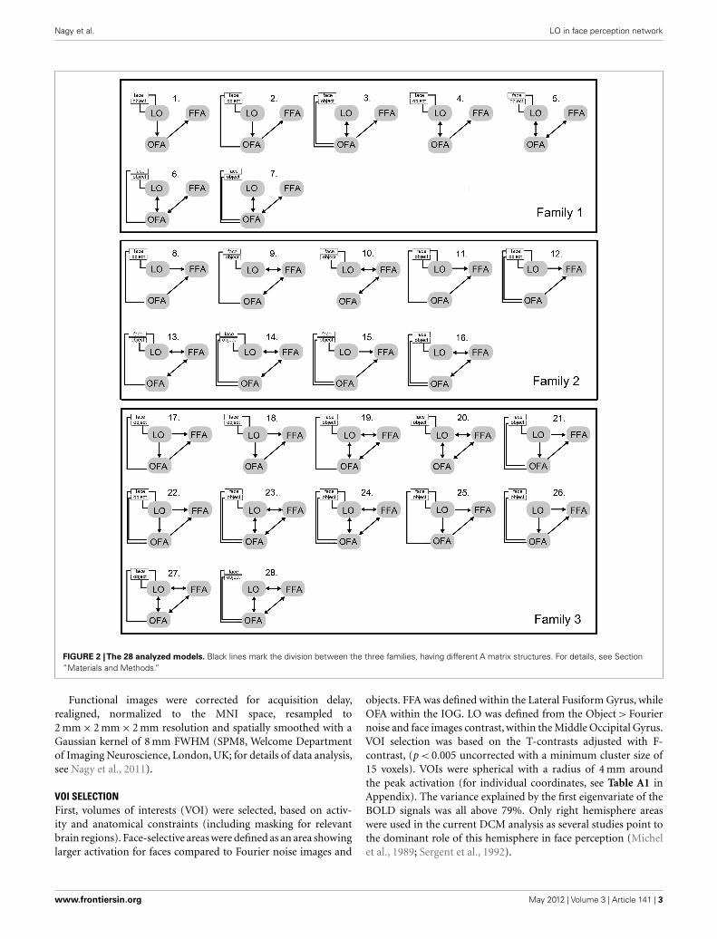
FIGURE 2 |The 28 analyzed models. Black lines mark the division between the three families, having different A matrix structures. For details, see Section“Materials and Methods.”
Functional images were corrected for acquisition delay,realigned, normalized to the MNI space, resampled to2 mm × 2 mm × 2 mm resolution and spatially smoothed with aGaussian kernel of 8 mm FWHM (SPM8, Welcome Departmentof Imaging Neuroscience, London, UK; for details of data analysis,see Nagy et al., 2011).
VOI SELECTIONFirst, volumes of interests (VOI) were selected, based on activ-ity and anatomical constraints (including masking for relevantbrain regions). Face-selective areas were defined as an area showinglarger activation for faces compared to Fourier noise images and
objects. FFA was defined within the Lateral Fusiform Gyrus, whileOFA within the IOG. LO was defined from the Object > Fouriernoise and face images contrast, within the Middle Occipital Gyrus.VOI selection was based on the T-contrasts adjusted with F-contrast, (p < 0.005 uncorrected with a minimum cluster size of15 voxels). VOIs were spherical with a radius of 4 mm aroundthe peak activation (for individual coordinates, see Table A1 inAppendix). The variance explained by the first eigenvariate of theBOLD signals was all above 79%. Only right hemisphere areaswere used in the current DCM analysis as several studies point tothe dominant role of this hemisphere in face perception (Michelet al., 1989; Sergent et al., 1992).
www.frontiersin.org May 2012 | Volume 3 | Article 141 | 3
Nagy et al. LO in face perception network
EFFECTIVE CONNECTIVITY ANALYSISEffective connectivity was tested by DCM-10, implemented inSPM8 toolbox (Wellcome Department of Imaging Neuroscience,London, UK), running under Matlab R2008a (The MathWorks,Natick, MA, USA). Models of DCM are defined with endogenousconnections, representing coupling between brain regions (matrixA), modulatory connections (matrix B), and driving input (matrixC). Here in the A matrix we defined the connections between theface-selective regions (FFA and OFA) and LO. Images of faces andobjects served as driving input (matrix C) and at this analysis stepwe did not apply any modulatory effects on the connections.
Model estimation aimed to maximize the negative free-energyestimates of the models (F) for a given dataset (Friston et al., 2003).This method ensures that the model fit uses the parameters in aparsimonious way (Ewbank et al., 2011). The estimated modelswere compared, based on the model evidences p(y |m), which isthe probability p of obtaining observed data y given by a partic-ular model m (Friston et al., 2003; Stephan et al., 2009). In thepresent study we apply the negative free-energy approximation(variational free-energy) to the log evidence (MacKay, 2003; Fris-ton et al., 2007). Bayesian Model Selection (BMS) was carried outon both the random (RFX) and fixed (FFX) effect designs (Stephanet al., 2009). BMS RFX is more resistant to outliers than FFX and itdoes not assume that the same model would explain the functionfor each participant (Stephan et al., 2009). In other words RFX isless sensitive to noise. In the RFX approach the output of the analy-sis is the exceedance probability of the model space, which is theextent of which one model is more likely to explain the measureddata than other models. The other output of the RFX analysis isthe expected posterior probability, which reflects the probabilitythat a model generated the observed data, allowing different dis-tributions for different models. Both of these parameter values arereduced by the broadening of the model space (i.e., by increasingthe number of models), therefore they behave in a relative mannerand models with shared features and implausible models may dis-tort the output of the analysis. Therefore, in addition to the directcomparison of the 28 created models we partitioned the modelspace into families, having similar connectivity patterns, using themethods of Penny et al. (2010).
Since several previous DCM studies point to the close bidirec-tional connection between FFA and OFA (Ishai, 2008; Gschwindet al., 2012) in our analysis these two areas were always linked toeach other and LO was connected to them in every biologicallyplausible way. The 28 relevant models were divided into threemodel-families based on structural differences (Penny et al., 2010;Ewbank et al., 2011). Family 1 contains models with linear con-nections among the three areas, supposing that information flowsfrom the LO to the FFA via the OFA. Family 2 contains modelswith a triangular structure where LO sends input directly to theFFA and the OFA is also directly linked to the FFA. Family 3 con-tains models, in which the three areas are interlinked, supposinga circular flow of information (Figure 2). In order to limit thenumber of models in this step of the analysis the inputs modu-lated solely the activity of their entry areas. The three families werecompared by a random design BMS.
Second, the models from the winner family were elabo-rated further by creating every plausible model with modulatory
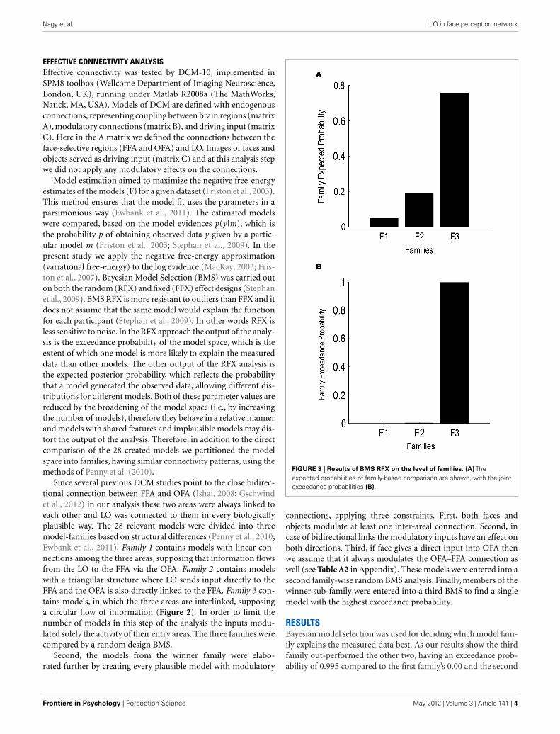
FIGURE 3 | Results of BMS RFX on the level of families. (A) Theexpected probabilities of family-based comparison are shown, with the jointexceedance probabilities (B).
connections, applying three constraints. First, both faces andobjects modulate at least one inter-areal connection. Second, incase of bidirectional links the modulatory inputs have an effect onboth directions. Third, if face gives a direct input into OFA thenwe assume that it always modulates the OFA–FFA connection aswell (see Table A2 in Appendix). These models were entered into asecond family-wise random BMS analysis. Finally, members of thewinner sub-family were entered into a third BMS to find a singlemodel with the highest exceedance probability.
RESULTSBayesian model selection was used for deciding which model fam-ily explains the measured data best. As our results show the thirdfamily out-performed the other two, having an exceedance prob-ability of 0.995 compared to the first family’s 0.00 and the second
Frontiers in Psychology | Perception Science May 2012 | Volume 3 | Article 141 | 4
Nagy et al. LO in face perception network
family’s 0.004 (Figures 3A,B). The winner model family (Fam-ily 3; Figure 2, bottom) contains 12 models, having connectionsbetween the LO and FFA, OFA and FFA, and LO and OFA as well,but differing in the directionality of the connections as well as inthe place of input to the network.
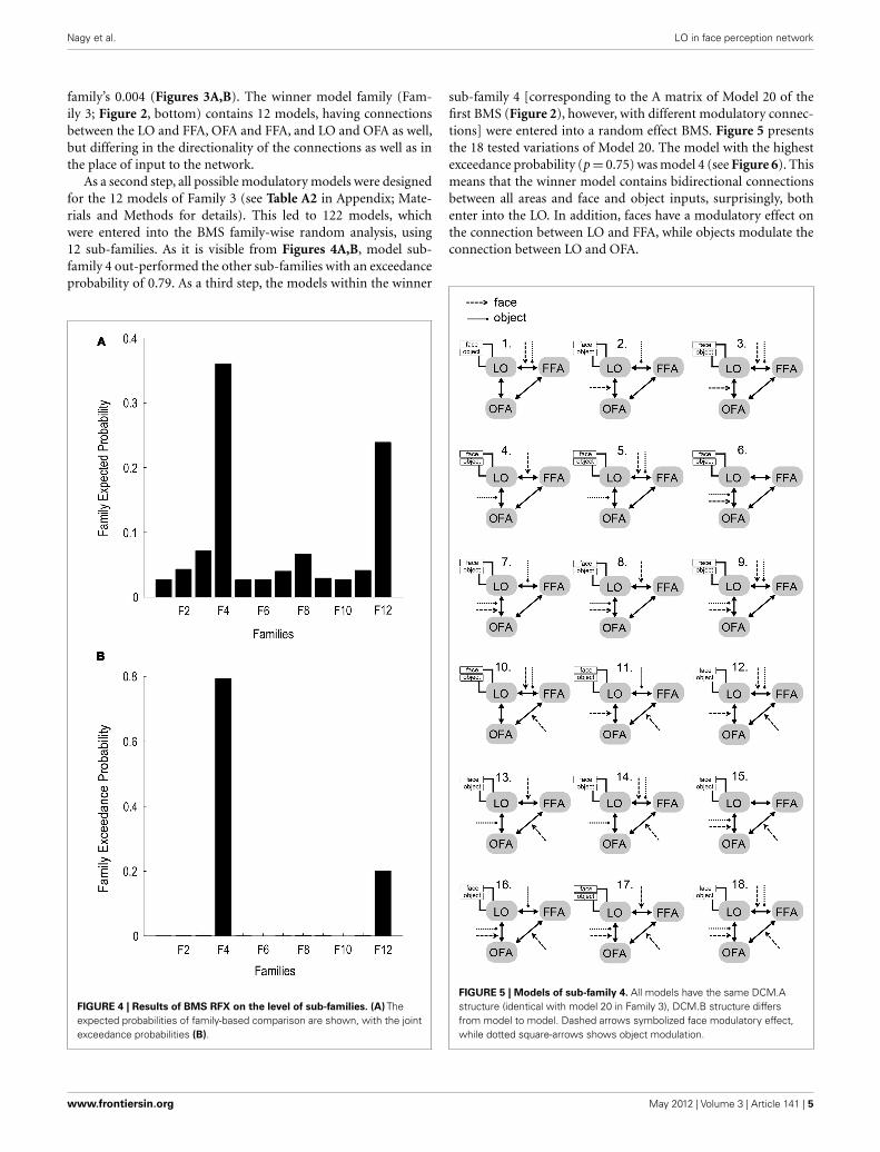
As a second step, all possible modulatory models were designedfor the 12 models of Family 3 (see Table A2 in Appendix; Mate-rials and Methods for details). This led to 122 models, whichwere entered into the BMS family-wise random analysis, using12 sub-families. As it is visible from Figures 4A,B, model sub-family 4 out-performed the other sub-families with an exceedanceprobability of 0.79. As a third step, the models within the winner
FIGURE 4 | Results of BMS RFX on the level of sub-families. (A) Theexpected probabilities of family-based comparison are shown, with the jointexceedance probabilities (B).
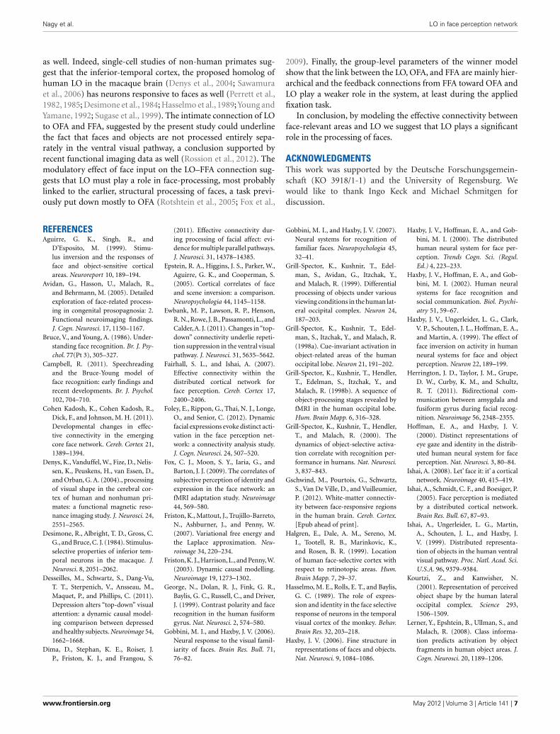
sub-family 4 [corresponding to the A matrix of Model 20 of thefirst BMS (Figure 2), however, with different modulatory connec-tions] were entered into a random effect BMS. Figure 5 presentsthe 18 tested variations of Model 20. The model with the highestexceedance probability (p = 0.75) was model 4 (see Figure 6). Thismeans that the winner model contains bidirectional connectionsbetween all areas and face and object inputs, surprisingly, bothenter into the LO. In addition, faces have a modulatory effect onthe connection between LO and FFA, while objects modulate theconnection between LO and OFA.
FIGURE 5 | Models of sub-family 4. All models have the same DCM.Astructure (identical with model 20 in Family 3), DCM.B structure differsfrom model to model. Dashed arrows symbolized face modulatory effect,while dotted square-arrows shows object modulation.
www.frontiersin.org May 2012 | Volume 3 | Article 141 | 5

Nagy et al. LO in face perception network
FIGURE 6 | Results of BMS within sub-family 4. (A) The expectedprobabilities of family-based comparison are shown, with the jointexceedance probabilities (B).
For analyzing parameter estimates of the winner model acrossthe group of subjects a random effect approximation was used. Allthe subject-specific maximum a posteriori (MAP) estimates wereentered into a t -test for single means and tested against 0 (Stephanet al., 2010; Desseilles et al., 2011). The results indexed with anasterisk on Figure 7 differed significantly from 0 (p < 0.05).
DISCUSSIONThe major result of the present effective connectivity study sug-gest that (a) LO is linked directly to the OFA–FFA face-processing
FIGURE 7 |The structure of the winner model. Simple lines signify theobject and face input stimuli to the system (DCM.C). Black arrows showinter-regional connections (DCM.A) while the red arrows stand for themodulatory connections (DCM.B): face modulation is signified with dashedarrows, while object modulation is signified with square-head dashedarrows. Group-level averages of MAP estimates and 95% confidenceintervals are illustrated. The averages were tested against 0 and significantresults are signified with ∗ if p < 0.05.
system via bidirectional connections to both areas; (b) non-faceand face inputs are intermixed at the level of occipito-temporalareas and enter the system via LO; (c) face input has a modulatoryeffect on LO and FFA connection, while object input modulatesthe LO and OFA connection significantly.
The role of LO in object perception is well-known from previ-ous studies (Malach et al., 1995; Grill-Spector et al., 1998a,b, 2000;Lerner et al., 2001, 2008). However, in spite previous studies usu-ally found an increased activity for complex objects, as well as forfaces in LO, the area is usually associated with objects and rela-tively less importance is attributed to its role in face-processing.In the present study, effective connectivity analysis positioned theLO in the core network of face perception. The direct link betweenOFA and FFA has been proven previously both functionally andanatomically (Gschwind et al., 2012). However, as there is no cur-rent data available regarding the role of LO in this system, welinked it to the other two regions in several plausible ways.
The first random BMS showed that in the winner model familyLO is interconnected with both FFA and OFA and the connec-tions are bidirectional. Therefore, it highlights that LO may havea direct structural connection to FFA. With modeling all the pos-sible modulatory effects we found that a sub-family of modelswon, where both object and face inputs enter the system viathe LO, supposing that the LO plays a general and importantinput region role. Since previous functional connectivity stud-ies all started the analysis of the face-processing network at thelevel of IOG, corresponding to OFA (Fairhall and Ishai, 2007;Ishai, 2008; Cohen Kadosh et al., 2011; Dima et al., 2011; Foleyet al., 2012) it is not surprising that they overlooked the signif-icant role of LO. However, faces are actually a distinct categoryof visual objects, suggesting that neurons sensitive to objects andshapes should be activated, at least to a certain degree, by faces
Frontiers in Psychology | Perception Science May 2012 | Volume 3 | Article 141 | 6
Nagy et al. LO in face perception network
as well. Indeed, single-cell studies of non-human primates sug-gest that the inferior-temporal cortex, the proposed homolog ofhuman LO in the macaque brain (Denys et al., 2004; Sawamuraet al., 2006) has neurons responsive to faces as well (Perrett et al.,1982, 1985; Desimone et al., 1984; Hasselmo et al., 1989;Young andYamane, 1992; Sugase et al., 1999). The intimate connection of LOto OFA and FFA, suggested by the present study could underlinethe fact that faces and objects are not processed entirely sepa-rately in the ventral visual pathway, a conclusion supported byrecent functional imaging data as well (Rossion et al., 2012). Themodulatory effect of face input on the LO–FFA connection sug-gests that LO must play a role in face-processing, most probablylinked to the earlier, structural processing of faces, a task previ-ously put down mostly to OFA (Rotshtein et al., 2005; Fox et al.,
2009). Finally, the group-level parameters of the winner modelshow that the link between the LO, OFA, and FFA are mainly hier-archical and the feedback connections from FFA toward OFA andLO play a weaker role in the system, at least during the appliedfixation task.
In conclusion, by modeling the effective connectivity betweenface-relevant areas and LO we suggest that LO plays a significantrole in the processing of faces.
ACKNOWLEDGMENTSThis work was supported by the Deutsche Forschungsgemein-schaft (KO 3918/1-1) and the University of Regensburg. Wewould like to thank Ingo Keck and Michael Schmitgen fordiscussion.
REFERENCESAguirre, G. K., Singh, R., and
D’Esposito, M. (1999). Stimu-lus inversion and the responses offace and object-sensitive corticalareas. Neuroreport 10, 189–194.
Avidan, G., Hasson, U., Malach, R.,and Behrmann, M. (2005). Detailedexploration of face-related process-ing in congenital prosopagnosia: 2.Functional neuroimaging findings.J. Cogn. Neurosci. 17, 1150–1167.
Bruce, V., and Young, A. (1986). Under-standing face recognition. Br. J. Psy-chol. 77(Pt 3), 305–327.
Campbell, R. (2011). Speechreadingand the Bruce-Young model offace recognition: early findings andrecent developments. Br. J. Psychol.102, 704–710.
Cohen Kadosh, K., Cohen Kadosh, R.,Dick, F., and Johnson, M. H. (2011).Developmental changes in effec-tive connectivity in the emergingcore face network. Cereb. Cortex 21,1389–1394.
Denys, K., Vanduffel, W., Fize, D., Nelis-sen, K., Peuskens, H., van Essen, D.,and Orban, G. A. (2004)., processingof visual shape in the cerebral cor-tex of human and nonhuman pri-mates: a functional magnetic reso-nance imaging study. J. Neurosci. 24,2551–2565.
Desimone, R., Albright, T. D., Gross, C.G., and Bruce, C. J. (1984). Stimulus-selective properties of inferior tem-poral neurons in the macaque. J.Neurosci. 8, 2051–2062.
Desseilles, M., Schwartz, S., Dang-Vu,T. T., Sterpenich, V., Ansseau, M.,Maquet, P., and Phillips, C. (2011).Depression alters “top-down” visualattention: a dynamic causal model-ing comparison between depressedand healthy subjects. Neuroimage 54,1662–1668.
Dima, D., Stephan, K. E., Roiser, J.P., Friston, K. J., and Frangou, S.
(2011). Effective connectivity dur-ing processing of facial affect: evi-dence for multiple parallel pathways.J. Neurosci. 31, 14378–14385.
Epstein, R. A., Higgins, J. S., Parker, W.,Aguirre, G. K., and Cooperman, S.(2005). Cortical correlates of faceand scene inversion: a comparison.Neuropsychologia 44, 1145–1158.
Ewbank, M. P., Lawson, R. P., Henson,R. N., Rowe, J. B., Passamonti, L., andCalder, A. J. (2011). Changes in“top-down” connectivity underlie repeti-tion suppression in the ventral visualpathway. J. Neurosci. 31, 5635–5642.
Fairhall, S. L., and Ishai, A. (2007).Effective connectivity within thedistributed cortical network forface perception. Cereb. Cortex 17,2400–2406.
Foley, E., Rippon, G., Thai, N. J., Longe,O., and Senior, C. (2012). Dynamicfacial expressions evoke distinct acti-vation in the face perception net-work: a connectivity analysis study.J. Cogn. Neurosci. 24, 507–520.
Fox, C. J., Moon, S. Y., Iaria, G., andBarton, J. J. (2009). The correlates ofsubjective perception of identity andexpression in the face network: anfMRI adaptation study. Neuroimage44, 569–580.
Friston, K., Mattout, J., Trujillo-Barreto,N., Ashburner, J., and Penny, W.(2007). Variational free energy andthe Laplace approximation. Neu-roimage 34, 220–234.
Friston, K. J., Harrison, L., and Penny,W.(2003). Dynamic causal modelling.Neuroimage 19, 1273–1302.
George, N., Dolan, R. J., Fink, G. R.,Baylis, G. C., Russell, C., and Driver,J. (1999). Contrast polarity and facerecognition in the human fusiformgyrus. Nat. Neurosci. 2, 574–580.
Gobbini, M. I., and Haxby, J. V. (2006).Neural response to the visual famil-iarity of faces. Brain Res. Bull. 71,76–82.
Gobbini, M. I., and Haxby, J. V. (2007).Neural systems for recognition offamiliar faces. Neuropsychologia 45,32–41.
Grill-Spector, K., Kushnir, T., Edel-man, S., Avidan, G., Itzchak, Y.,and Malach, R. (1999). Differentialprocessing of objects under variousviewing conditions in the human lat-eral occipital complex. Neuron 24,187–203.
Grill-Spector, K., Kushnir, T., Edel-man, S., Itzchak, Y., and Malach, R.(1998a). Cue-invariant activation inobject-related areas of the humanoccipital lobe. Neuron 21, 191–202.
Grill-Spector, K., Kushnir, T., Hendler,T., Edelman, S., Itzchak, Y., andMalach, R. (1998b). A sequence ofobject-processing stages revealed byfMRI in the human occipital lobe.Hum. Brain Mapp. 6, 316–328.
Grill-Spector, K., Kushnir, T., Hendler,T., and Malach, R. (2000). Thedynamics of object-selective activa-tion correlate with recognition per-formance in humans. Nat. Neurosci.3, 837–843.
Gschwind, M., Pourtois, G., Schwartz,S., Van De Ville, D., and Vuilleumier,P. (2012). White-matter connectiv-ity between face-responsive regionsin the human brain. Cereb. Cortex.[Epub ahead of print].
Halgren, E., Dale, A. M., Sereno, M.I., Tootell, R. B., Marinkovic, K.,and Rosen, B. R. (1999). Locationof human face-selective cortex withrespect to retinotopic areas. Hum.Brain Mapp. 7, 29–37.
Hasselmo, M. E., Rolls, E. T., and Baylis,G. C. (1989). The role of expres-sion and identity in the face selectiveresponse of neurons in the temporalvisual cortex of the monkey. Behav.Brain Res. 32, 203–218.
Haxby, J. V. (2006). Fine structure inrepresentations of faces and objects.Nat. Neurosci. 9, 1084–1086.
Haxby, J. V., Hoffman, E. A., and Gob-bini, M. I. (2000). The distributedhuman neural system for face per-ception. Trends Cogn. Sci. (Regul.Ed.) 4, 223–233.
Haxby, J. V., Hoffman, E. A., and Gob-bini, M. I. (2002). Human neuralsystems for face recognition andsocial communication. Biol. Psychi-atry 51, 59–67.
Haxby, J. V., Ungerleider, L. G., Clark,V. P., Schouten, J. L., Hoffman, E. A.,and Martin, A. (1999). The effect offace inversion on activity in humanneural systems for face and objectperception. Neuron 22, 189–199.
Herrington, J. D., Taylor, J. M., Grupe,D. W., Curby, K. M., and Schultz,R. T. (2011). Bidirectional com-munication between amygdala andfusiform gyrus during facial recog-nition. Neuroimage 56, 2348–2355.
Hoffman, E. A., and Haxby, J. V.(2000). Distinct representations ofeye gaze and identity in the distrib-uted human neural system for faceperception. Nat. Neurosci. 3, 80–84.
Ishai, A. (2008). Let’ face it: it’ a corticalnetwork. Neuroimage 40, 415–419.
Ishai, A., Schmidt, C. F., and Boesiger, P.(2005). Face perception is mediatedby a distributed cortical network.Brain Res. Bull. 67, 87–93.
Ishai, A., Ungerleider, L. G., Martin,A., Schouten, J. L., and Haxby, J.V. (1999). Distributed representa-tion of objects in the human ventralvisual pathway. Proc. Natl. Acad. Sci.U.S.A. 96, 9379–9384.
Kourtzi, Z., and Kanwisher, N.(2001). Representation of perceivedobject shape by the human lateraloccipital complex. Science 293,1506–1509.
Lerner, Y., Epshtein, B., Ullman, S., andMalach, R. (2008). Class informa-tion predicts activation by objectfragments in human object areas. J.Cogn. Neurosci. 20, 1189–1206.
www.frontiersin.org May 2012 | Volume 3 | Article 141 | 7
Nagy et al. LO in face perception network
Lerner, Y., Hendler, T., Ben-Bashat,D., Harel, M., and Malach, R.(2001). A hierarchical axis of objectprocessing stages in the humanvisual cortex. Cereb. Cortex 11,287–297.
Li, J., Liu, J., Liang, J., Zhang, H., Zhao,J., Rieth, C. A., Huber, D. E., Li,W., Shi, G., Ai, L., Tian, J., and Lee,K. (2010). Effective connectivities ofcortical regions for top-down faceprocessing: a dynamic causal mod-eling study. Brain Res. 1340, 40–51.
MacKay, D. J. C. (2003). InformationTheory, Inference, and Learning Algo-rithms. Cambridge: Cambridge Uni-versity Press.
Malach, R., Reppas, J. B., Benson, R. R.,Kwong, K. K., Jiang, H., Kennedy,W. A., Ledden, P. J., Brady, T.J., Rosen, B. R., and Tootell, R.B. (1995). Object-related activityrevealed by functional magnetic res-onance imaging in human occipitalcortex. Proc. Natl. Acad. Sci. U.S.A.92, 8135–8139.
Marco, G., de Bonis, M., Vrignaud, P.,Henry-Feugeas, M. C., and Peretti, I.(2006). Changes in effective connec-tivity during incidental and inten-tional perception of fearful faces.Neuroimage 30, 1030–1037.
Marotta, J. J., Genovese, C. R., andBehrmann, M. (2001). A functionalMRI study of face recognition inpatients with prosopagnosia. Neu-roreport 12, 1581–1587.
Michel, F., Poncet, M., and Signoret, J. L.(1989). Are the lesions responsiblefor prosopagnosia always bilateral?Rev. Neurol. (Paris) 145, 764–770.
Morris, J. S., Frith, C. D., Perrett, D. I.,Rowland, D.,Young,A. W., Calder,A.J., and Dolan, R. J. (1996). A differ-ential neural response in the humanamygdala to fearful and happy facialexpressions. Nature 383, 812–815.
Nagy, K., Greenlee, M. W., and Kovács,G. (2011). Sensory competition inthe face processing areas of thehuman brain. PLoS ONE 6, e24450.doi:10.1371/journal.pone.0024450
Nagy, K., Zimmer, M., Greenlee, M.W., and Kovács, G. (2009). ThefMRI correlates of multi-face adap-tation. Perception 38(ECVP AbstractSuppl.) 77.
Nasanen, R. (1999). Spatial frequencybandwidth used in the recognitionof facial images. Vision Res. 39,3824–3833.
Penny, W. D., Stephan, K. E., Dau-nizeau, J., Rosa, M. J., Friston,K. J., Schofield, T. M., and Leff,A. P. (2010). Comparing fami-lies of dynamic causal models.PLoS Comput. Biol. 6, e1000709.doi:10.1371/journal.pcbi.1000709
Perrett, D. I., Harries, M. H., Mistlin,A. J., Hietanen, L. K., and Bevan, R.(1990). Social signals analyzed at thesingle cell level: someone’s looking atme, something touched me, some-thing moved! Int. J. Comp. Psychol.4, 25–55.
Perrett, D. I., Rolls, E. T., and Caan,W. (1982). Visual neurones respon-sive to faces in the monkey temporalcortex. Exp. Brain Res. 47, 329–342.
Perrett, D. I., Smith, P. A., Potter, D. D.,Mistlin, A. J., Head, A. S., Milner, A.D., and Jeeves, M. A. (1985). Visualcells in the temporal cortex sensi-tive to face view and gaze direction.Proc. R. Soc. Lond. B Biol. Sci. 223,293–317.
Puce, A., Allison, T., Bentin, S., Gore, J.C., and McCarthy, G. (1998). Tem-poral cortex activation in humansviewing eye and mouth movements.J. Neurosci. 18, 2188–2199.
Puce, A., Allison, T., Gore, J. C., andMcCarthy, G. (1995). Face-sensitiveregions in human extrastriate cor-tex studied by functional MRI. J.Neurophysiol. 74, 1192–1199.
Rossion, B., Caldara, R., Seghier, M.,Schuller, A. M., Lazeyras, F., andMayer, E. (2003a). A networkof occipito-temporal face-sensitiveareas besides the right middlefusiform gyrus is necessary for nor-mal face processing. Brain 126(Pt11), 2381–2395.
Rossion, B., Schiltz, C., and Crom-melinck, M. (2003b). Thefunctionally defined right occip-ital and fusiform “face areas”discriminate novel from visuallyfamiliar faces. Neuroimage 19,877–883.
Rossion, B., Hanseeuw, B., and Dri-cot, L. (2012). Defining face per-ception areas in the human brain:a large-scale factorial fMRI facelocalizer analysis. Brain Cogn. 79,138–157.
Rotshtein, P., Henson, R. N. A., Treves,A., Driver, J., and Dolan, R. J. (2005).Morphing Marilyn into Maggie dis-sociates physical and identity facerepresentations in the brain. Nat.Neurosci. 8, 107–113.
Sawamura, H., Orban, G. A., and Vogels,R. (2006). Selectivity of neuronaladaptation does not match responseselectivity: a single-cell study of thefMRI adaptation paradigm. Neuron49, 307–318.
Sergent, J., Ohta, S., and MacDon-ald, B. (1992). Functional neu-roanatomy of face and object pro-cessing. A positron emission tomog-raphy study. Brain 115(Pt 1),15–36.
Sorger, B., Goebel, R., Schiltz, C., andRossion, B. (2007). Understand-ing the functional neuroanatomy ofacquired prosopagnosia. Neuroim-age 35, 836–852.
Stephan, K. E., Penny, W. D., Dau-nizeau, J., Moran, R. J., and Friston,K. J. (2009). Bayesian model selec-tion for group studies. Neuroimage46, 1004–1017.
Stephan, K. E., Penny, W. D., Moran,R. J., den Ouden, H. E., Dau-nizeau, J., and Friston, K. J. (2010).Ten simple rules for dynamiccausal modeling. Neuroimage 49,3099–3109.
Sugase, Y., Yamane, S., Ueno, S., andKawano, K. (1999). Global and fineinformation coded by single neuronsin the temporal visual cortex. Nature400, 869–873.
Wiggett, A. J., and Downing, P.E. (2008). The face network:overextended? (Comment on: “Let’sface it: it’s a cortical network”by Alumit Ishai). Neuroimage 40,420–422.
Winston, J. S., Henson, R. N. A., Fine-Goulden, M. R., and Dolan, R.J. (2004). fMRI-adaptation revealsdissociable neural representationsof identity and expression in faceperception. J. Neurophysiol. 92,1830–1839.
Young, A. W., and Bruce, V. (2011).Understanding person perception.Br. J. Psychol. 102, 959–974.
Young, M. P., and Yamane, S. (1992).Sparse population coding of facesin the inferotemporal cortex. Science256, 1327–1331.
Yovel, G., and Kanwisher, N. (2005).The neural basis of the behavioralface-inversion effect. Curr. Biol. 15,2256–2262.
Conflict of Interest Statement: Theauthors declare that the research wasconducted in the absence of any com-mercial or financial relationships thatcould be construed as a potential con-flict of interest.
Received: 01 February 2012; accepted: 20April 2012; published online: 10 May2012.Citation: Nagy K, Greenlee MWand Kovács G (2012) The lateraloccipital cortex in the face percep-tion network: an effective connectivitystudy. Front. Psychology 3:141. doi:10.3389/fpsyg.2012.00141This article was submitted to Frontiers inPerception Science, a specialty of Frontiersin Psychology.Copyright © 2012 Nagy, Greenlee andKovács. This is an open-access articledistributed under the terms of the Cre-ative Commons Attribution Non Com-mercial License, which permits non-commercial use, distribution, and repro-duction in other forums, provided theoriginal authors and source are credited.
Frontiers in Psychology | Perception Science May 2012 | Volume 3 | Article 141 | 8

Nagy et al. LO in face perception network
APPENDIX
Table A1 |The MNI coordinates of the respective maximal activations within FFA, OFA, and LO of our subject sample.
FFA (x, y, z) OFA (x, y, z) LO (x, y, z)
1. 42 −58 −14 36 −74 −18 54 −70 −2
2. 48 −48 −26 48 −76 −18 50 −74 −8
3. 40 −58 −20 38 −86 −8 40 −84 −8
4. 48 −48 −22 44 −76 −16 54 −70 −2
5. 48 −50 −20 46 −74 −10 52 −68 −4
6. 38 −50 −22 38 −72 −16 44 −76 4
7. 40 −48 −22 38 −86 −18 56 −68 −8
8. 42 −42 −24 38 −86 −18 56 −68 −8
9. 46 −58 −18 48 −76 −12 56 −70 −4
10. 40 −62 −20 44 −84 −8 44 −82 −10
11. 40 −56 −24 42 −84 −12 48 −80 −6
12. 44 −52 −20 40 −76 −8 46 −82 −2
13. 48 −62 −28 36 −84 −12 50 −80 4
14. 38 −54 −22 46 −82 −10 40 −78 2
15. 42 −64 −18 56 −66 −2 54 −68 −2
16. 52 −52 −30 46 −76 −20 46 −80 −4
17. 44 −50 −16 40 −80 −10 50 −72 2
18. 46 −60 −14 44 −78 −12 52 −76 −2
19. 48 −58 −20 42 −82 −14 40 −80 −4
20. 40 −50 −24 36 −74 −18 44 −82 −10
21. 40 −54 −22 44 −74 −14 54 −74 −6
22. 40 −58 −16 40 −78 −40 50 −76 −2
23. 46 −52 −18 42 −74 −12 48 −80 0
24. 42 −44 −26 44 −70 −16 34 −78 −2
25. 48 −48 −24 46 −74 −10 52 −70 −4
Mean 43.6 −53.44 −21.2 42.48 −77.68 −14.08 48.56 −75.44 −3.44
St. Dev. 3.9 5.8 4.1 4.7 5.4 6.9 5.9 5.3 4.0
www.frontiersin.org May 2012 | Volume 3 | Article 141 | 9

Nagy et al. LO in face perception network
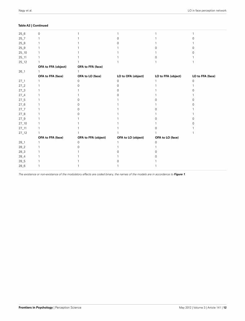
Table A2 | All possible modulatory models of Family 3.
LO to FFA (object) LO to OFA (object) OFA to FFA (face)
17_1 0 1 1
17_2 1 0 1
17_3 1 1 1
LO to OFA (object) LO to OFA (face) LO to FFA (object) LO to FFA (face) OFA to FFA (face)
18_1 0 0 1 1 0
18_2 0 1 1 0 0
18_3 0 1 1 1 0
18_4 1 0 0 1 0
18_5 1 0 1 1 0
18_6 1 1 0 0 0
18_7 1 1 1 0 0
18_8 1 1 0 1 0
18_9 1 1 1 1 0
18_10 0 0 1 1 1
18_11 0 1 1 0 1
18_12 0 1 1 1 1
18_13 1 0 0 1 1
18_14 1 0 1 1 1
18_15 1 1 0 0 1
18_16 1 1 1 0 1
18_17 1 1 0 1 1
18_18 1 1 1 1 1
OFA to FFA (face) LO to OFA (face) LO to OFA (object) LO to FFA (object)
19_1 1 0 0 1
19_2 1 0 1 1
19_3 1 1 0 1
19_4 1 1 1 1
19_5 1 0 0 1
19_6 1 0 1 1
19_7 1 1 0 1
19_8 1 1 1 1
LO to OFA (object) LO to OFA (face) LO to FFA (object) LO to FFA (face) OFA to FFA (face)
20_1 0 0 1 1 0
20_2 0 1 1 0 0
20_3 0 1 1 1 0
20_4 1 0 0 1 0
20_5 1 0 1 1 0
20_6 1 1 0 0 0
20_7 1 1 1 0 0
20_8 1 1 0 1 0
20_9 1 1 1 1 0
20_10 0 0 1 1 1
20_11 0 1 1 0 1
20_12 0 1 1 1 1
20_13 1 0 0 1 1
20_14 1 0 1 1 1
20_15 1 1 0 0 1
20_16 1 1 1 0 1
20_17 1 1 0 1 1
20_18 1 1 1 1 1
(Continued)
Frontiers in Psychology | Perception Science May 2012 | Volume 3 | Article 141 | 10

Nagy et al. LO in face perception network
Table A2 | Continued
LO to FFA (object) LO to OFA (object) OFA to FFA (face)
21_1 0 1 1
21_2 1 1 1
21_3 1 0 1
OFA to FFA (face) OFA to FFA (object) LO to FFA (object) LO to FFA (face)
22_1 1 0 1 0
22_2 1 0 1 1
22_3 1 1 0 0
22_4 1 1 1 0
22_5 1 1 0 1
22_6 1 1 1 1
OFA to FFA (face) OFA to LO (object) OFA to FFA (object) LO to FFA (object)
23_1 1 0 0 1
23_2 1 0 1 0
23_3 1 0 1 1
23_4 1 1 0 0
23_5 1 1 0 1
23_6 1 1 1 0
23_7 1 1 1 1
OFA to FFA (face) OFA to LO (face) OFA to LO (object) OFA to FFA (object) LO to FFA (object) LO to FFA (face)
24_1 1 0 0 0 1 0
24_2 1 0 0 0 1 1
24_3 1 0 0 1 0 0
24_4 1 0 0 1 1 0
24_5 1 0 0 1 0 1
24_6 1 0 0 1 1 1
24_7 1 1 0 0 1 0
24_8 1 1 0 0 1 1
24_9 1 1 0 1 0 0
24_10 1 1 0 1 1 0
24_11 1 1 0 1 0 1
24_12 1 1 0 1 1 1
24_13 1 0 1 0 0 0
24_14 1 0 1 0 1 0
24_15 1 0 1 0 0 1
24_16 1 0 1 0 1 1
24_17 1 0 1 1 0 0
24_18 1 0 1 1 1 0
24_19 1 0 1 1 0 1
24_20 1 0 1 1 1 1
24_21 1 1 1 0 0 0
24_22 1 1 1 0 1 0
24_23 1 1 1 0 0 1
24_24 1 1 1 0 1 1
24_25 1 1 1 1 0 0
24_26 1 1 1 1 1 0
24_27 1 1 1 1 0 1
24_28 1 1 1 1 1 1
LO to OFA (face) OFA to FFA (face) LO to OFA (object) LO to FFA (object) LO to FFA (face)
25_1 0 1 0 1 0
25_2 0 1 0 1 1
25_3 0 1 1 0 0
25_4 0 1 1 1 0
25_5 0 1 1 0 1
(Continued)
www.frontiersin.org May 2012 | Volume 3 | Article 141 | 11
Nagy et al. LO in face perception network
Table A2 | Continued
25_6 0 1 1 1 1
25_7 1 1 0 1 0
25_8 1 1 0 1 1
25_9 1 1 1 0 0
25_10 1 1 1 1 0
25_11 1 1 1 0 1
25_12 1 1 1 1 1
OFA to FFA (object) OFA to FFA (face)
26_1 1 1
OFA to FFA (face) OFA to LO (face) LO to OFA (object) LO to FFA (object) LO to FFA (face)
27_1 1 0 0 1 0
27_2 1 0 0 1 1
27_3 1 1 0 1 0
27_4 1 1 0 1 1
27_5 1 0 1 0 0
27_6 1 0 1 1 0
27_7 1 0 1 0 1
27_8 1 0 1 1 1
27_9 1 1 1 0 0
27_10 1 1 1 1 0
27_11 1 1 1 0 1
27_12 1 1 1 1 1
OFA to FFA (face) OFA to FFA (object) OFA to LO (object) OFA to LO (face)
28_1 1 0 1 0
28_2 1 0 1 1
28_3 1 1 0 0
28_4 1 1 1 0
28_5 1 1 0 1
28_6 1 1 1 1
The existence or non-existence of the modulatory effects are coded binary, the names of the models are in accordance to Figure 1.
Frontiers in Psychology | Perception Science May 2012 | Volume 3 | Article 141 | 12