the expansion of the microbiological spectrum of brain abscesses with use of multiple 16s ribosomal...
TRANSCRIPT

New Bacteria in Brain Abscesses • CID 2009:48 (1 May) • 1169
M A J O R A R T I C L E
The Expansion of the Microbiological Spectrumof Brain Abscesses with Use of Multiple 16SRibosomal DNA Sequencing
Mouhamad Al Masalma,1 Fabrice Armougom,1 W. Michael Scheld,4 Henri Dufour,2 Pierre-Hugues Roche,3
Michel Drancourt,1 and Didier Raoult1
1Pole des Maladies Infectieuses, Assistance Publique-Hopitaux de Marseille and URMITE, Centre National de la Recherche Scientifique, UniteMixte de Recherche 6236, Institut pour la Recherche et le Developpement 198, Universite de la Mediterranee, 2Service de Neurochirurgie,Hopital de la Timone, Assistance Publique-Hopitaux de Marseille, and 3Service de Neurochirurgie, Hopital Sainte-Marguerite, AssistancePublique-Hopitaux de Marseille, Marseille, France; and 4Department of Internal Medicine, Division of Infectious Diseasesand International Health, University of Virginia Health System, Charlottesville, Virginia
(See the editorial commentary by DiGiulio and Relman on pages 1179–81)
Background. Brain abscess is commonly treated using empirically prescribed antibiotics. Thus, a comprehensivestudy of bacterial organisms associated with brain abscess is essential to define the best empirical treatment forthis life-threatening condition.
Methods. We prospectively compared cultures to single and multiple sequenced 16S ribosomal DNA polymerasechain reaction amplifications (by cloning and/or pyrosequencing) of cerebral abscesses in 20 patients from 2hospitals in Marseilles, France, during the period January 2005 through December 2007.
Results. The obtained cultures identified significantly fewer types of bacteria (22 strains) than did moleculartesting (72 strains; , by analysis of variance test). We found that a patient could exhibit as many as 16P p .017different bacterial species in a single abscess. The obtained cultures identified 14 different species already knownto cause cerebral abscess. Single sequencing performed poorly, whereas multiple sequencing identified 49 species,of which 27 had not been previously reported in brain abscess investigations and 15 were completely unknown.Interestingly, we observed 2 patients who harbored Mycoplasma hominis (an emerging pathogen in this situation)and 3 patients who harbored Mycoplasma faucium, which, to our knowledge, has never been reported in literature.
Conclusions. Molecular techniques dramatically increased the number of identified agents in cerebral abscesses.Mycoplasma species are common and should be detected in this situation. These findings led us to question theaccuracy of the current empirical treatment of brain abscess.
Brain abscess is a life-threatening condition [1–4] with
frequent serious sequelae [3, 5, 6] for which medical
management remains empirical because of a lack of
comprehensive knowledge regarding the organisms re-
sponsible. Microbiological documentation of brain ab-
scess primarily relies on direct microscopic examination
and culturing of abscess pus specimens collected after
neurosurgical drainage [4, 7]. Unfortunately, this pro-
Received 8 July 2008; accepted 3 December 2008; electronically published 31March 2009.
Reprints or correspondence: Prof. Didier Raoult, URMITE, CNRS UMR 6236, IRD198, Universite de la Mediterranee, Faculte de Medecine, 27 Bd Jean Moulin,13005 Marseille, France ([email protected]).
Clinical Infectious Diseases 2009; 48:1169–78� 2009 by the Infectious Diseases Society of America. All rights reserved.1058-4838/2009/4809-0001$15.00DOI: 10.1086/597578
cedure produces inconclusive results in 9%–63% of
cases, depending on the quality of anaerobe culturing
[6].
PCR-amplified 16S ribosomal DNA (rDNA) se-
quencing was recently used to overcome the limitations
of culture-based bacteria detection in brain abscess pus
specimens, and it was demonstrated to be effective for
documentation of monomicrobial infection after an-
tibiotic treatment. Unfortunately, this procedure failed
to discriminate among mixed flora [8–11]. As such, we
suspect that the number of species associated with brain
abscess is much larger than previously expected.
The purpose of this investigation was to analyze and
evaluate the bacterial flora responsible for brain abscess
by comparing standard culture technique to the fol-
lowing 3 techniques using 16S rDNA amplification: (1)
direct sequencing (providing a single sequence), (2)
by guest on May 11, 2011
cid.oxfordjournals.orgD
ownloaded from
1170 • CID 2009:48 (1 May) • Al Masalma et al.
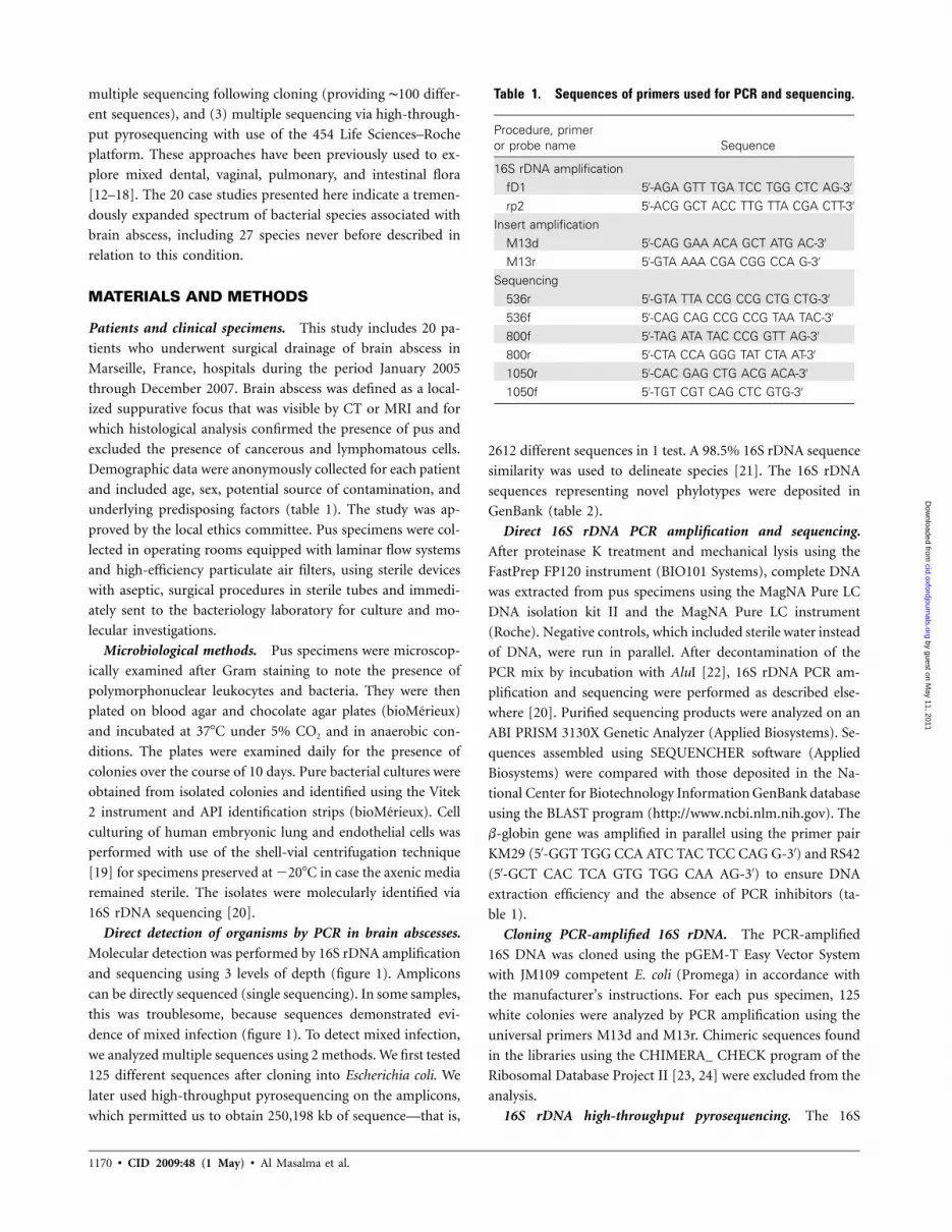
Table 1. Sequences of primers used for PCR and sequencing.
Procedure, primeror probe name Sequence
16S rDNA amplificationfD1 5′-AGA GTT TGA TCC TGG CTC AG-3′
rp2 5′-ACG GCT ACC TTG TTA CGA CTT-3′
Insert amplificationM13d 5′-CAG GAA ACA GCT ATG AC-3′
M13r 5′-GTA AAA CGA CGG CCA G-3′
Sequencing536r 5′-GTA TTA CCG CCG CTG CTG-3′
536f 5′-CAG CAG CCG CCG TAA TAC-3′
800f 5′-TAG ATA TAC CCG GTT AG-3′
800r 5′-CTA CCA GGG TAT CTA AT-3′
1050r 5′-CAC GAG CTG ACG ACA-3′
1050f 5′-TGT CGT CAG CTC GTG-3′
multiple sequencing following cloning (providing ∼100 differ-
ent sequences), and (3) multiple sequencing via high-through-
put pyrosequencing with use of the 454 Life Sciences–Roche
platform. These approaches have been previously used to ex-
plore mixed dental, vaginal, pulmonary, and intestinal flora
[12–18]. The 20 case studies presented here indicate a tremen-
dously expanded spectrum of bacterial species associated with
brain abscess, including 27 species never before described in
relation to this condition.
MATERIALS AND METHODS
Patients and clinical specimens. This study includes 20 pa-
tients who underwent surgical drainage of brain abscess in
Marseille, France, hospitals during the period January 2005
through December 2007. Brain abscess was defined as a local-
ized suppurative focus that was visible by CT or MRI and for
which histological analysis confirmed the presence of pus and
excluded the presence of cancerous and lymphomatous cells.
Demographic data were anonymously collected for each patient
and included age, sex, potential source of contamination, and
underlying predisposing factors (table 1). The study was ap-
proved by the local ethics committee. Pus specimens were col-
lected in operating rooms equipped with laminar flow systems
and high-efficiency particulate air filters, using sterile devices
with aseptic, surgical procedures in sterile tubes and immedi-
ately sent to the bacteriology laboratory for culture and mo-
lecular investigations.
Microbiological methods. Pus specimens were microscop-
ically examined after Gram staining to note the presence of
polymorphonuclear leukocytes and bacteria. They were then
plated on blood agar and chocolate agar plates (bioMerieux)
and incubated at 37�C under 5% CO2 and in anaerobic con-
ditions. The plates were examined daily for the presence of
colonies over the course of 10 days. Pure bacterial cultures were
obtained from isolated colonies and identified using the Vitek
2 instrument and API identification strips (bioMerieux). Cell
culturing of human embryonic lung and endothelial cells was
performed with use of the shell-vial centrifugation technique
[19] for specimens preserved at �20�C in case the axenic media
remained sterile. The isolates were molecularly identified via
16S rDNA sequencing [20].
Direct detection of organisms by PCR in brain abscesses.
Molecular detection was performed by 16S rDNA amplification
and sequencing using 3 levels of depth (figure 1). Amplicons
can be directly sequenced (single sequencing). In some samples,
this was troublesome, because sequences demonstrated evi-
dence of mixed infection (figure 1). To detect mixed infection,
we analyzed multiple sequences using 2 methods. We first tested
125 different sequences after cloning into Escherichia coli. We
later used high-throughput pyrosequencing on the amplicons,
which permitted us to obtain 250,198 kb of sequence—that is,
2612 different sequences in 1 test. A 98.5% 16S rDNA sequence
similarity was used to delineate species [21]. The 16S rDNA
sequences representing novel phylotypes were deposited in
GenBank (table 2).
Direct 16S rDNA PCR amplification and sequencing.
After proteinase K treatment and mechanical lysis using the
FastPrep FP120 instrument (BIO101 Systems), complete DNA
was extracted from pus specimens using the MagNA Pure LC
DNA isolation kit II and the MagNA Pure LC instrument
(Roche). Negative controls, which included sterile water instead
of DNA, were run in parallel. After decontamination of the
PCR mix by incubation with AluI [22], 16S rDNA PCR am-
plification and sequencing were performed as described else-
where [20]. Purified sequencing products were analyzed on an
ABI PRISM 3130X Genetic Analyzer (Applied Biosystems). Se-
quences assembled using SEQUENCHER software (Applied
Biosystems) were compared with those deposited in the Na-
tional Center for Biotechnology Information GenBank database
using the BLAST program (http://www.ncbi.nlm.nih.gov). The
b-globin gene was amplified in parallel using the primer pair
KM29 (5′-GGT TGG CCA ATC TAC TCC CAG G-3′) and RS42
(5′-GCT CAC TCA GTG TGG CAA AG-3′) to ensure DNA
extraction efficiency and the absence of PCR inhibitors (ta-
ble 1).
Cloning PCR-amplified 16S rDNA. The PCR-amplified
16S DNA was cloned using the pGEM-T Easy Vector System
with JM109 competent E. coli (Promega) in accordance with
the manufacturer’s instructions. For each pus specimen, 125
white colonies were analyzed by PCR amplification using the
universal primers M13d and M13r. Chimeric sequences found
in the libraries using the CHIMERA_ CHECK program of the
Ribosomal Database Project II [23, 24] were excluded from the
analysis.
16S rDNA high-throughput pyrosequencing. The 16S
by guest on May 11, 2011
cid.oxfordjournals.orgD
ownloaded from

New Bacteria in Brain Abscesses • CID 2009:48 (1 May) • 1171
Figure 1. 16S ribosomal DNA (rDNA) sequence-based molecular investigation of brain abscess flora using direct and multiple sequencing. FLEXwas manufactured by Life Science–Roche.
rDNA amplified from the specimen of one patient was purified
and sequenced using the GS FLX platform (454 Life Sciences-
Roche) on a PicoTiterPlate (PTP) with 8 regions40 � 75
(Roche) [25]. Reads exhibiting a sequence similarity �98.5%
and a 90% sequence coverage with any hit in the Ribosomal
Database Project II (http://rdp.cme.msu.edu/) using the BLAST
algorithm were identified at the species level; reads exhibiting
97%–98.5% sequence similarity and a sequence coverage �90%
were identified at the genus level; and reads exhibiting a se-
quence similarity !97% were not classified. The number of
reads assigned to a given species or genus was calculated. Only
species and genera that collected 110 reads were kept for primer
design and PCR amplification control. The Nucmer function
from the MUMmer 3.20 package [26] was used to map the
reads classified in a species onto the reference 16S rDNA. De-
fault parameters were used.
Literature search strategy and selection criteria. The
PubMed database was searched for articles published during
the period 1980 through April 2008 with the combined search
term “brain AND abscess.” Additional articles were identified
by hand-searching the bibliographies of selected papers. The
strategy was developed by splitting the review into its elemental
facets. Additional search terms included “microbiology,” “bac-
teriology,” “16S,” “molecular,” “detection,” “metagenomics,”
“Mycoplasma hominis,” “Mycoplasma faucium,” and “Myco-
plasma orale.” The publication language was restricted to En-
glish. The bibliographies of key references were later hand-
searched to identify articles missing in the database search.
RESULTS
Patients. Twenty patients were prospectively included in this
study (table 2), including 15 male patients (75%) and 5 female
patients (25%). The mean age of patients was 54.5 years (range,
14–76 years), and there were 2 children aged !15 years. Head-
ache was the most common clinical manifestation and was
observed in 10 patients; fever and motor weakness were ob-
served in 7 patients; vomiting and/or nausea, alterations in
consciousness, and aphasia were observed in 6 patients; visual
problems were observed in 4 patients; weight loss was observed
in 3 patients; other neurological manifestations were observed
in 3 patients; convulsions were observed in 2 patients; and
vertigo was observed in 2 patients. CT and MRI detected a
single brain abscess in 15 patients (75%) and multiple brain
abscesses in 5 patients (25%). A solitary abscess was localized
to the parietal lobe in 5 patients, to the frontal lobe in 4 patients,
to the occipital lobe in 2 patients, to the temporal lobe in 2
patients, to a frontoparietal location in 1 patient, and to a
parieto-occipital location in 1 patient. Brain abscess after neu-
rosurgery occurred in 4 patients (25%), whereas contiguous
infection after sinusitis or dental abscess was the second most
frequent source of brain abscess in 7 patients (35%). Aggre-
gatibacter aphrophilus pneumonia was a possible source of he-
matogenous spread in 1 patient, whereas no primary source
could be identified in 8 patients (40%). Underlying conditions
were noted for 9 patients. Direct microscopic examination of
pus revealed bacteria in 9 patients, whereas cultures yielded 1
by guest on May 11, 2011
cid.oxfordjournals.orgD
ownloaded from

1172
Tabl
e2.
Char
acte
ristic
sof
the
20pa
tient
sw
ithbr
ain
absc
ess
exam
ined
inth
isst
udy
and
bact
eria
iden
tified
,by
tech
niqu
e.
Mul
tiple
sequ
enci
ng
Pre
disp
osin
gfa
ctor
,pa
tient
Sex
/ag
e,ye
ars
Trea
tmen
tO
utco
me
Dur
atio
nof
follo
w-u
p,m
onth
sC
ultu
refin
ding
Sin
gle
sequ
enci
ng
Clo
ning
Pyr
oseq
uenc
ing
Ana
erob
icba
cter
ia
Aer
obic
bact
eria
GP
firm
icut
esG
Nba
cilli
Pos
tneu
rosu
rger
y
1aM
/52
Imif
ollo
wed
byor
alA
mox
Dea
th…
Ste
rile
Aci
neto
bact
erca
lcoa
cetic
usA
.ca
lcoa
cetic
us…
……
2F/
41D
oxy
Favo
rabl
e32
Ste
rile
Myc
opla
sma
hom
inis
M.
hom
inis
……
…
3M
/76
Imif
ollo
wed
byor
alA
mox
Dea
th…
Ser
ratia
mar
cesc
ens
S.
mar
cesc
ens
S.
mar
cesc
ens
……
…
4M
/55
Rif
and
aFQ
Favo
rabl
e13
Sta
phyl
ococ
cus
aure
usS
.au
reus
S.
aure
us…
……
Sin
usiti
sor
dent
alab
sces
ses
5M
/38
Imi
Favo
rabl
e23
Kle
bsie
llaox
ytoc
aB
acte
roid
esfr
agili
sM
ycop
lasm
afa
u-ci
um,
K.
oxyt
oca
…B
.fr
agili
s,Fu
soba
cter
ium
navi
-fo
rme,
uncu
lture
dba
cter
ium
1,un
cultu
red
bact
eriu
m2,
uncu
lture
dba
cter
ium
3
…
6F/
76Im
iFa
vora
ble
23S
trep
toco
ccus
cons
tella
tus,
Esc
heric
hia
coli
Pol
ymic
robi
alS
.co
nste
llatu
s,M
.fa
uciu
mM
icro
mon
asm
icro
s,P
epto
stre
ptoc
occu
sst
omat
is,
Mog
ibac
-te
rium
timid
um,
uncu
lture
dba
cte-
rium
6
Fuso
bact
eriu
mnu
clea
tum
,P
re-
vote
llain
term
edia
,P
revo
tella
oris
,P
revo
tella
tann
erae
,P
revo
tella
baro
niae
,un
cul-
ture
dba
cter
ium
7,un
cultu
-re
dba
cter
ium
8,un
cultu
red
bact
eriu
m9
…
7M
/47
Imif
ollo
wed
byor
alA
mox
Seq
uela
e30
Str
epto
cocc
usin
term
ediu
s,E
iken
ella
corr
oden
s
Pol
ymic
robi
alS
.in
term
ediu
s,E
.co
rrod
ens
M.
mic
ros
……
8M
/14
Imi
Favo
rabl
e31
S.
inte
rmed
ius
S.
inte
rmed
ius
S.
inte
rmed
ius
……
…
9M
/68
Imi
Favo
rabl
e30
S.
inte
rmed
ius
S.
inte
rmed
ius
S.
inte
rmed
ius,
M.
fauc
ium
…B
.fr
agili
s,un
cultu
red
bact
e-riu
m1
…
10M
/53
Imi
Seq
uela
e27
S.
cons
tella
tus,
Gem
ella
haem
olys
ans
Pol
ymic
robi
alS
.co
nste
llatu
s,G
.ha
emol
ysan
sM
.m
icro
s,E
ubac
teri-
umbr
achy
P.or
is,
Fuso
bact
eriu
mnu
clea
-tu
m,
Por
phyr
omon
asen
do-
dont
alis
,P
orph
yrom
onas
gin-
giva
lis,
uncu
lture
dba
cter
ium
4,un
cultu
red
bact
eriu
m5
S.
cons
tella
tus,
M.
mic
ros,
Eub
acte
rium
brac
hy,
F.nu
clea
tum
P.gi
ngiv
alis
,C
ampy
loba
cter
rect
us,
Trep
onem
asp
ecie
s,un
cultu
red
Eub
acte
rium
E1-
K13
,un
cultu
red
Eub
acte
rium
E1-
K9,
uncu
lture
dE
ubac
teriu
msp
ecie
s,B
ac-
tero
idal
esge
nom
osp
oral
clon
e,un
-cu
lture
dP
revo
tella
spec
ies,
Pre
vo-
tella
spec
ies
oral
clon
eD
O03
3
by guest on May 11, 2011
cid.oxfordjournals.orgD
ownloaded from
1173
11M
/14
Cep
h-Va
nc-M
etFa
vora
ble
20S
trep
toco
ccus
pneu
mon
iae
S.
pneu
mon
iae
S.
pneu
mon
iae
……
…
12M
/61
Cep
h-Va
nc-M
etFa
vora
ble
1S
.in
term
ediu
s,S
.ep
ider
mid
is
Pol
ymic
robi
alS
.in
term
ediu
s,E
.co
rrod
ens,
Cam
-py
loba
cter
grac
ilis
M.
mic
ros
Cap
nocy
toph
aga
spec
ies,
F.na
vifo
rme,
F.nu
clea
tum
…
13M
/66
…D
eath
…S
trep
toco
ccus
spec
ies
Pol
ymic
robi
alS
trep
toco
ccus
angi
-no
sus,
Gem
ella
mor
billo
rum
M.
mic
ros
Pre
vote
llasp
ecie
s,P.
inte
rme-
dia,
F.nu
clea
tum
…
Con
geni
tal
hear
tdi
seas
e:14
F/49
…D
eath
…S
.in
term
ediu
s,S
.ep
ider
mid
is
S.
inte
rmed
ius
S.
inte
rmed
ius,
M.
hom
inis
,E
.co
rrod
ens
M.
mic
ros
F.nu
clea
tum
…
Can
cer
15F/
69Im
iFa
vora
ble
9N
ocar
dia
spec
ies
Neg
ativ
e…
……
16M
/62
Cep
h-Va
nc-M
etLo
stto
follo
w-u
p…
Noc
ardi
acy
riaci
geor
gia
N.
cyria
cige
orgi
aN
.cy
riaci
geor
gia
……
…
17a
M/5
Imi
Favo
rabl
e19
Ste
rile
Nei
sser
iasp
ecie
sN
eiss
eria
spec
ies
……
…
Not
know
n
18M
/62
Cep
h-Va
nc-M
etD
eath
…A
ggre
gatib
acte
rap
hrop
hilu
sA
.ap
hrop
hilu
sA
.ap
hrop
hilu
s…
……
19M
/45
Imi
Favo
rabl
e29
S.
inte
rmed
ius
S.
inte
rmed
ius
S.
inte
rmed
ius
……
…
20M
/50
Imi
Favo
rabl
e28
S.
inte
rmed
ius
S.
inte
rmed
ius
S.
inte
rmed
ius
……
…
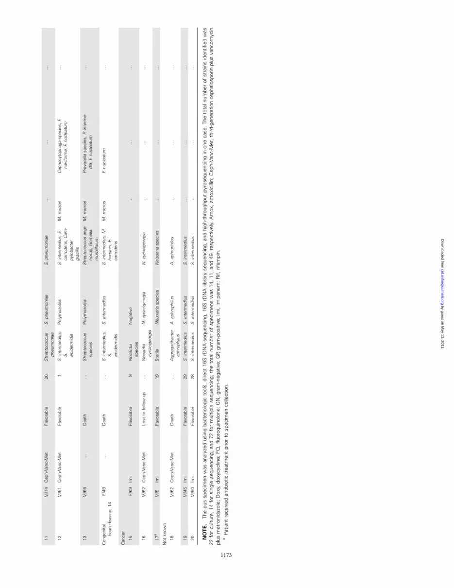
NO
TE
.Th
epu
ssp
ecim
enw
asan
alyz
edus
ing
bact
erio
logi
cto
ols,
dire
ct16
SrD
NA
sequ
enci
ng,
16S
rDN
Alib
rary
sequ
enci
ng,
and
high
-thr
ough
put
pyro
sequ
enci
ngin
one
case
.The
tota
lnum
ber
ofst
rain
sid
entifi
edw
as22
for
cultu
re,
14fo
rsi
ngle
sequ
enci
ng,
and
72fo
rm
ultip
lese
quen
cing
;th
eto
taln
umbe
rof
spec
imen
sw
as14
,11
,an
d49
,re
spec
tivel
y.A
mox
,am
oxic
illin
;C
eph-
Vanc
-Met
,th
ird-g
ener
atio
nce
phal
ospo
rinpl
usva
ncom
ycin
plus
met
roni
dazo
le;
Dox
y,do
xycy
clin
e;FQ
,flu
oroq
uino
lone
;G
N,
gram
-neg
ativ
e;G
P,gr
am-p
ositi
ve;
Imi,
imip
enem
;R
if,rif
ampi
n.a
Pat
ient
rece
ived
antib
iotic
trea
tmen
tpr
ior
tosp
ecim
enco
llect
ion.
by guest on May 11, 2011
cid.oxfordjournals.orgD
ownloaded from

1174 • CID 2009:48 (1 May) • Al Masalma et al.
Figure 2. Gram-stained brain abscess pus (A) for 1 patient indicating gram-positive cocci when high-throughput pyrosequencing demonstrated 13different bacterial species delineated in a phylogenetic tree (bootstrap values �90% are indicated at nodes; B).
bacterial species in 12 patients (60%), yielded 2 bacterial species
in 5 patients (25%), and were sterile in 3 patients (15%). The
negative controls remained negative in all PCR experiments.
Discrepancy between culture and molecular detection.
Direct 16S rDNA PCR amplification results were negative for
1 specimen and positive for 19 of 20 specimens, including 5
instances in which mixed flora were detected because of trou-
bled sequences. Altogether, direct 16S rDNA sequencing per-
formed marginally worse than cultures ( , by analysisP p .048
of variance). Among the 14 interpretable sequences, Strepto-
coccus intermedius was detected in 5 patients. Further identi-
fications included Acinetobacter calcoaceticus, M. hominis, Bac-
teroides fragilis, Serratia marcescens, Streptococcus pneumoniae,
Nocardia cyriacigeorgia, Neisseria species, A. aphrophilus, and
Staphylococcus aureus (1 patient each). In 9 specimens, the same
bacterial species were detected by culture and direct 16S rDNA
sequencing. One specimen that yielded negative results by 16S
rDNA PCR detection (the b-globin positive control yielded
positive results) exhibited the presence of Nocardia species by
cell culture. Three specimens that had negative culture results
yielded 1 bacterial species by direct 16S rDNA PCR. In 1 case,
culture yielded Klebsiella oxytoca, whereas direct 16S rDNA
sequencing yielded B. fragilis. For 4 specimens, cultures yielded
Streptococcus species mixed with another bacterial species,
which agreed with direct 16S rDNA sequencing that indicated
mixed flora. For 1 specimen, culture yielded S. intermedius and
Staphylococcus epidermidis, whereas direct 16S rDNA sequenc-
ing identified only S. intermedius. For 1 specimen, culture
yielded Streptococcus species, whereas direct 16S rDNA se-
quencing exhibited mixed flora.
by guest on May 11, 2011
cid.oxfordjournals.orgD
ownloaded from
New Bacteria in Brain Abscesses • CID 2009:48 (1 May) • 1175
Multiple sequencing of amplicons. A total of 2100 non-
chimeric inserts of ∼1500 bp were sequenced, corresponding
to 1100 clones for each pus specimen. In 11 specimens, only
1 bacterial species was detected that was identical to that de-
tected by direct 16S rDNA sequencing. In 8 patients, clone
library analysis identified bacteria that had not been found
using direct 16S rDNA sequencing (table 2). A total of 2612
reads, with a mean length of 95.78 bp, were obtained after the
pyrosequencing of patient 10. In this patient, 2 bacterial species
were found by culture, 10 were found by multiple sequencing
after cloning, and 16 were found by high-throughput multi-
ple sequencing (figure 2). Altogether, 72 bacterial strains were
identified, compared with the 22 species identified by cul-
ture ( , by analysis of variance test); 8 mixed infectionsP p .017
were identified, compared with 5 identified by culture (P p
).nonsignificant
DISCUSSION
The current knowledge base of bacteria that cause brain ab-
scesses relies on cultures of pus specimens collected after neu-
rosurgical drainage [1, 27, 28]. In this study, bacteria were
microscopically observed in 45% of patients, whereas culturing
on axenic media yielded bacteria in 80% of patients. No isolate
was obtained from patients who received antibiotics at the time
of pus collection (patients 1 and 17), as has been reported
elsewhere [2, 11, 27]. One Nocardia strain was isolated with
use of only a cell culture system. The spectrum of bacteria
isolated from brain abscess pus highly depends on the atmo-
sphere and culture media used to inoculate the specimen. In
particular, the prevalence of anaerobic bacteria is highly vari-
able, because these bacteria require adequate transport con-
ditions and special care in the laboratory, which were not sys-
tematically achieved in our study. In fact, no anaerobic
bacterium was cultivated in this study, in contrast with the
reported prevalence of anaerobes reported in other studies [29].
This fact limits the value of our comparison between culture
and molecular diagnostic tools. Molecular detection pointed to
mixed flora, including anaerobes, in patients with underlying
dental or sinus infection.
In this study, pus was collected into a sterile tube using sterile
devices and surgical procedures, virtually eliminating the risk
of perioperative contamination of the pus specimen. Absence
of intralaboratory contamination was interpreted by the neg-
ativity of negative controls and the general concordance of
various methods. S. epidermidis, which was isolated from 2
patients, was interpreted as a contaminant during processing
of the specimen. Indeed, S. epidermidis organisms were found
by culture only and were not detected by direct or multiple
16S rDNA gene sequencing. In both cases, other organisms,
including S. intermedius, were consistently found by culture
and molecular detection. Except for S. epidermidis, all organ-
isms herein reported were regarded as authentic—that is, in-
deed present in the pus specimen. The cloning method we used
provided a semiquantitative evaluation of the relative abun-
dance of each bacterial species, which all represented 11% of
the actual bacterial flora.
Direct 16S rDNA PCR amplification and sequencing have
seldom been used to circumvent the pitfalls associated with
culture-based techniques. Three studies have demonstrated that
the results obtained by direct PCR amplification and sequencing
for bacterial identification were more sensitive and precise than
was phenotypic identification [8, 10, 11]. In particular, the
detection of fastidious organisms, such as M. hominis, Gemella
morbillorum, A. aphrophilus, and Fusobacterium necrophorum,
was more rapid with molecular detection than with culture [8,
10]. In addition, molecular detection identified streptococci in
children who received antibiotics prior to brain abscess punc-
ture [11]. Unfortunately, single 16S rDNA PCR sequencing is
inadequate for clinical specimens that contain mixed flora; this
approach yielded mixed, uninterpretable sequences, as was pre-
viously described for brain abscess specimens [8, 10]. Therefore,
we used 2 complementary approaches to resolve this issue and
to allow for multiple sequencing: 16S rDNA clone library anal-
ysis and high-throughput pyrosequencing (in 2 cases). This
strategy yielded excellent results, whereas cultures recovered 22
bacteria species and single sequencing identified only 15 bac-
teria species and 5 mixed populations from 19 positive am-
plicons. This strategy also facilitated the identification of 1 par-
ticularly fastidious bacterium not usually observed by culturing
(i.e., M. hominis).
In contrast to single sequencing, multiple sequencing dra-
matically increased the number of identified bacteria (72 bac-
teria species, compared with 22 for culture [ ] and 14P p .017
for single sequencing [ ]). The power of multiple se-P p .048
quencing was demonstrated in patient 10, for whom cultures
yielded 2 bacterial species and high-throughput sequencing re-
covered 16 species. Three species were found by culture alone,
including 1 Nocardia species (obtained only by cell culture) and
2 S. epidermidis isolates. We suspect that the latter 2 cases
resulted from contamination during the inoculation procedure.
Multiple sequencing also identified 3 patients with mixed in-
fections that were not detected by direct sequencing or culture.
We detected anaerobes using only 16S rDNA amplification
in 40% of specimens. This potentially represents a flaw in our
laboratory, because others have recovered comparatively more
anaerobes by culture. Further developments in molecular bi-
ology may, in the future, help avoid this problem of fastidious
anaerobe isolation. We detected the presence of Mycoplasma
species in 25% of the specimens; in 4 of 5 cases, the Mycoplasma
species were part of mixed flora abscesses and were not detected
by direct sequencing. M. hominis should be regarded as an
emerging pathogen in brain abscesses; we detected this species
by guest on May 11, 2011
cid.oxfordjournals.orgD
ownloaded from

Table 3. The bacterial species detected in different patients by culture, 16S ribosomal DNA (rDNA) direct sequencing,and/or 16S rDNA multiple sequencing.
Species (GenBank accession number) Means of identification
Species commonly reported in brain abscessesStreptococcus intermedius Culture, 16S rDNA direct sequencing, and 16S rDNA multiple sequencingStreptococcus constellatus Culture, 16S rDNA direct sequencing, and 16S rDNA multiple sequencingStreptococcus pneumoniae Culture, 16S rDNA direct sequencing, and 16S rDNA multiple sequencingStreptococcus anginosus Culture, 16S rDNA direct sequencing, and 16S rDNA multiple sequencingStaphylococcus aureus Culture, 16S rDNA direct sequencing, and 16S rDNA multiple sequencingGemella haemolysans Culture, 16S rDNA direct sequencing, and 16S rDNA multiple sequencingGemella morbillorum 16S rDNA multiple sequencingEscherichia coli Culture, 16S rDNA direct sequencing, and 16S rDNA multiple sequencingAcinetobacter calcoaceticus 16S rDNA direct sequencing and 16S rDNA multiple sequencingEikenella corrodens Culture, 16S rDNA direct sequencing, and 16S rDNA multiple sequencingKlebsiella oxytoca Culture, 16S rDNA direct sequencing, and 16S rDNA multiple sequencingSerratia marcescens Culture, 16S rDNA direct sequencing, and 16S rDNA multiple sequencingAggregatibacter aphrophilus 16S rDNA direct sequencing and 16S rDNA multiple sequencingMicromonas micros 16S rDNA multiple sequencingFusobacterium nucleatum 16S rDNA multiple sequencingPorphyromonas endodontalis 16S rDNA multiple sequencingPorphyromonas gingivalis 16S rDNA multiple sequencingPrevotella oris 16S rDNA multiple sequencingPrevotella intermedia 16S rDNA multiple sequencingBacteroides fragilis 16S rDNA direct sequencing and 16S rDNA multiple sequencing
Species rarely reported in brain abscessesNocardia cyriacigeorgia Culture, 16S rDNA direct sequencing, and 16S rDNA multiple sequencingNocardia species CultureMycoplasma hominis 16S rDNA direct sequencing and 16S rDNA multiple sequencingFusobacterium naviforme 16S rDNA multiple sequencingStaphylococcus epidermidis Culture
Species never previously reported in brainabscessesMycoplasma faucium 16S rDNA multiple sequencingCampylobacter gracilis 16S rDNA multiple sequencingPeptostreptococcus stomatis 16S rDNA multiple sequencingEubacterium brachy 16S rDNA multiple sequencingMogibacterium timidum 16S rDNA multiple sequencingPrevotella tannerae 16S rDNA multiple sequencingPrevotella baroniae 16S rDNA multiple sequencingPrevotella species (EU663611) 16S rDNA multiple sequencingNeisseria species (EU663609) 16S rDNA multiple sequencingCapnocytophaga species (EU663610) 16S rDNA multiple sequencingCampylobacter rectus 16S rDNA multiple sequencingTreponema maltophilum 16S rDNA multiple sequencingUncultured bacterium 1 (EU663600) 16S rDNA multiple sequencingUncultured bacterium 2 (EU663601) 16S rDNA multiple sequencingUncultured bacterium 3 (EU663602) 16S rDNA multiple sequencingUncultured bacterium 4 (EU663603) 16S rDNA multiple sequencingUncultured bacterium 5 (EU663604) 16S rDNA multiple sequencingUncultured bacterium 6 (EU663605) 16S rDNA multiple sequencingUncultured bacterium 7 (EU663606) 16S rDNA multiple sequencingUncultured bacterium 8 (EU663607) 16S rDNA multiple sequencingUncultured bacterium 9 (EU66308) 16S rDNA multiple sequencingUncultured Eubacterium E1-K13 16S rDNA multiple sequencingUncultured Eubacterium E1-K9 16S rDNA multiple sequencingUncultured Eubacterium species 16S rDNA multiple sequencing
by guest on May 11, 2011
cid.oxfordjournals.orgD
ownloaded from

New Bacteria in Brain Abscesses • CID 2009:48 (1 May) • 1177
Table 3. (Continued.)
Species (GenBank accession number) Means of identification
Bacteroidales genomosp. oral clone 16S rDNA multiple sequencingUncultured Prevotella species 16S rDNA multiple sequencingPrevotella species oral clone DO033 16S rDNA multiple sequencing
in pus specimens obtained from 2 patients (10%), and detection
of this species was also noted in 3 previous reports [8, 30, 31].
M. faucium is even more fastidious [32] and has, to our knowl-
edge, never been reported in brain abscesses. The organisms
detected by PCR are most likely not false-positive results, be-
cause the negative controls remained negative in all PCR runs.
In addition, the detected organisms are not contemporarily
known to be general environmental contaminants, and they
were not detected in other clinical specimens analyzed at the
same time as the brain abscess pus specimens.
The observed increase in the repertoire of brain abscess–
causative organisms can be linked to molecular evidence for
fastidious—and yet-uncultured—organisms (table 3). We ob-
served 15 sequences that matched with contemporarily uncul-
tured bacteria that had previously been characterized among
anaerobic periodontal and intestinal flora. Fourteen sequences
belonged to the gram-negative bacilli Bacteroidetes phylum, and
1 sequence belonged to the gram-positive bacilli Firmicutes phy-
lum order Clostridiales. In a previous study of oral flora, it was
estimated that ∼50% of the present bacteria were uncultured
[33] and belonged to the Bacteroidetes and Firmicutes phyla.
High-throughput analyses of the bacteria associated with brain
abscess in our study revealed that the flora was even more
variable than expected, including 27 bacterial species found in
cerebral abscesses that have not been previously reported. Sim-
ilar results were observed in previous studies that used com-
parable tools and that investigated other infections, such as
endodontic infection, dental caries and periodontitis, and pul-
monary infection in patients with cystic fibrosis [16, 17, 34,
35]. Moreover, molecular approaches often permitted us to
associate M. faucium [32] with the development of brain ab-
scesses for the first time, because this species was observed in
3 patients. M. faucium is an inhabitant of the primate oro-
pharynx [36], and it was recently associated with chronic gas-
tritis in Korean patients [37]. Other species that were detected
in individual patients are normal inhabitants of the human oral
cavity, and they include Campylobacter gracilis [38], Mogibac-
terium timidum [39], Prevotella baroniae [40], Prevotella tan-
nerae [41], Peptostreptococcus stomatis [40], a new Neisseria
species [42], a new Capnocytophaga species that exhibits 99%
16S rDNA sequence similarity with 1 uncultured Capnocyto-
phaga species [38], and a new Prevotella species that exhibits
97.8% 16S rDNA sequence similarity with Prevotella shahii [43].
Compared with findings from the literature (table 2), 27 of the
49 species observed in our study have never before been re-
ported in brain abscesses.
In conclusion, our investigation has determined that the va-
riety of brain abscess–associated bacterial species is much larger
than has previously been reported, and it includes many an-
aerobes and uncultured bacteria from oral cavity flora. We have
confirmed the recently reported role of M. hominis, and we
report, for the first time, the role of M. faucium. Additional
studies and the data reported herein could potentially direct
modifications to current brain abscess antibiotic treatments.
Acknowledgments
We acknowledge the contribution of Prof. Herve Richet for statisticalanalyses.
Financial support. French National Programme Hospitalier de Re-cherche Clinique 2008 “Etude metagenomique des abces cerebraux.”
Potential conflicts of interest. All authors: no conflicts.
References
1. Roche M, Humphreys H, Smyth E, et al. A twelve-year review of centralnervous system bacterial abscesses; presentation and aetiology. ClinMicrobiol Infect 2003; 9:803–9.
2. Takeshita M, Kagawa M, Izawa M, Takakura K. Current treatmentstrategies and factors influencing outcome in patients with bacterialbrain abscess. Acta Neurochir (Wien) 1998; 140:1263–70.
3. Tonon E, Scotton PG, Gallucci M, Vaglia A. Brain abscess: clinicalaspects of 100 patients. Int J Infect Dis 2006; 10:103–9.
4. Yang SY, Zhao CS. Review of 140 patients with brain abscess. SurgNeurol 1993; 39:290–6.
5. Carpenter J, Stapleton S, Holliman R. Retrospective analysis of 49 casesof brain abscess and review of the literature. Eur J Clin Microbiol InfectDis 2007; 26:1–11.
6. Tseng JH, Tseng MY. Brain abscess in 142 patients: factors influencingoutcome and mortality. Surg Neurol 2006; 65:557–62.
7. Lu CH, Chang WN, Lin YC, et al. Bacterial brain abscess: microbio-logical features, epidemiological trends and therapeutic outcomes. QJM2002; 95:501–9.
8. Kupila L, Rantakokko-Jalava K, Jalava J, et al. Aetiological diagnosisof brain abscesses and spinal infections: application of broad rangebacterial polymerase chain reaction analysis. J Neurol Neurosurg Psy-chiatry 2003; 74:728–33.
9. Tatti KM, Shieh WJ, Phillips S, Augenbraun M, Rao C, Zaki SR. Mo-lecular diagnosis of Nocardia farcinica from a cerebral abscess. HumPathol 2006; 37:1117–21.
10. Tsai JC, Teng LJ, Hsueh PR. Direct detection of bacterial pathogens inbrain abscesses by polymerase chain reaction amplification and se-quencing of partial 16S ribosomal deoxyribonucleic acid fragments.Neurosurgery 2004; 55:1154–62.
11. Petti CA, Simmon KE, Bender J, et al. Culture-negative intracerebralabscesses in children and adolescents from Streptococcus anginosusgroup infection: a case series. Clin Infect Dis 2008; 46:1578–80.
by guest on May 11, 2011
cid.oxfordjournals.orgD
ownloaded from
1178 • CID 2009:48 (1 May) • Al Masalma et al.
12. Eckburg PB, Bik EM, Bernstein CN, et al. Diversity of the humanintestinal microbial flora. Science 2005; 308:1635–8.
13. Fredricks DN, Fiedler TL, Marrazzo JM. Molecular identification ofbacteria associated with bacterial vaginosis. N Engl J Med 2005; 353:1899–911.
14. de Lillo A, Ashley FP, Palmer RM, et al. Novel subgingival bacterialphylotypes detected using multiple universal polymerase chain reactionprimer sets. Oral Microbiol Immunol 2006; 21:61–8.
15. Dymock D, Weightman AJ, Scully C, Wade WG. Molecular analysisof microflora associated with dentoalveolar abscesses. J Clin Microbiol1996; 34:537–42.
16. Hutter G, Schlagenhauf U, Valenza G, et al. Molecular analysis ofbacteria in periodontitis: evaluation of clone libraries, novel phylotypesand putative pathogens. Microbiology 2003; 149:67–75.
17. Munson MA, Banerjee A, Watson TF, Wade WG. Molecular analysisof the microflora associated with dental caries. J Clin Microbiol2004; 42:3023–9.
18. Pace NR. Time for a change. Nature 2006; 441:289.19. Gouriet F, Fenollar F, Patrice JY, Drancourt M, Raoult D. Use of shell-
vial cell culture assay for isolation of bacteria from clinical specimens:13 years of experience. J Clin Microbiol 2005; 43:4993–5002.
20. Drancourt M, Berger P, Raoult D. Systematic 16S rRNA gene sequenc-ing of atypical clinical isolates identified 27 new bacterial species as-sociated with humans. J Clin Microbiol 2004; 42:2197–202.
21. Stackebrandt E, Ebers J. Taxonomic parameters revisited: tarnished goldstandards. Microbiology Today 2006; 152–5.
22. Rothman RE, Majmudar MD, Kelen GD, et al. Detection of bacteremiain emergency department patients at risk for infective endocarditisusing universal 16S rRNA primers in a decontaminated polymerasechain reaction assay. J Infect Dis 2002; 186:1677–81.
23. Maidak BL, Cole JR, Lilburn TG, et al. The RDP-II (Ribosomal Da-tabase Project). Nucleic Acids Res 2001; 29:173–4.
24. Cole JR, Chai B, Marsh TL, et al.; Ribosomal Database Project. TheRibosomal Database Project (RDP-II): previewing a new autoalignerthat allows regular updates and the new prokaryotic taxonomy. NucleicAcids Res 2003; 31:442–3.
25. Margulies M, Egholm M, Altman WE, et al. Genome sequencing inmicrofabricated high-density picolitre reactors. Nature 2005; 437:376–80.
26. Kurtz S, Phillipy A, Delcher AL, et al. Versatile and open software forcomparing large genomes. Genome Biol 2004; 5:R12.
27. Prasad KN, Mishra AM, Gupta D, Husain N, Husain M, Gupta RK.Analysis of microbial etiology and mortality in patients with brainabscess. J Infect 2006; 53:221–7.
28. Valarezo J, Cohen JE, Valarezo L, et al. Nocardial cerebral abscess:report of three cases and review of the current neurosurgical man-agement. Neurol Res 2003; 25:27–30.
29. Le Moal G, Landron C, Grollier G, et al. Characteristics of brain abscesswith isolation of anaerobic bacteria. Scand J Infect Dis 2003; 35:318–21.
30. Kupila L, Rantakokko-Jalava K, Jalava J, et al. Brain abscess caused byMycoplasma hominis: a clinically recognizable entity? Eur J Neurol2006; 13:550–1.
31. Zheng X, Olson DA, Tully JG, et al. Isolation of Mycoplasma hominisfrom a brain abscess. J Clin Microbiol 1997; 35:992–4.
32. Freundt EA, Taylor-Robinson D, Purcell RH, Chanock RM, Black FT.Proposal of Mycoplasma buccale nom. nov. and Mycoplasma fauciumnom. nov. for Mycoplasme orale ‘types’ 2 and 3, respectively. Int J SystBacteriol 1974; 24:252–5.
33. Socransky SS, Gibbons RJ, Dale AC, Bortnick L, Rosenthal E, Mac-donald JB. The microbiota of the gingival crevice area of man. I. Totalmicroscopic and viable counts and counts of specific organisms. ArchOral Biol 1963; 8:275–80.
34. Munson MA, Pitt-Ford T, Chong B, Weightman A, Wade WG. Mo-lecular and cultural analysis of the microflora associated with endo-dontic infections. J Dent Res 2002; 81:761–6.
35. Harris JK, De Groote MA, Sagel SD, et al. Molecular identification ofbacteria in bronchoalveolar lavage fluid from children with cystic fi-brosis. Proc Natl Acad Sci USA 2007; 104:20529–33.
36. Rawadi G, Dujeancourt-Henry A, Lemercier B, Roulland-Dussoix D.Phylogenetic position of rare human mycoplasmas, Mycoplasma fau-cium, M. buccale, M. primatum and M. spermatophilum, based on 16SrRNA gene sequences. Int J Syst Bacteriol 1998; 48:305–9.
37. Kwon HJ, Kang JO, Cho SH, et al. Presence of human mycoplasmaDNA in gastric tissue samples from Korean chronic gastritis patients.Cancer Sci 2004; 95:311–5.
38. Vandamme P, Vancanneyt M, van Belkum A, et al. Polyphasic analysisof strains of the genus Capnocytophaga and Centers for Disease Controlgroup DF-3. Int J Syst Bacteriol 1996; 46:782–91.
39. Nakazawa F, Sato M, Poco SE, et al. Description of Mogibacteriumpumilum gen. nov., sp. nov. and Mogibacterium vescum gen. nov., sp.nov., and reclassification of Eubacterium timidum (Holdeman et al.1980) as Mogibacterium timidum gen. nov., comb. nov. Int J Syst EvolMicrobiol 2000; 50:679–88.
40. Downes J, Sutcliffe I, Tanner AC, Wade WG. Prevotella marshii sp.nov. and Prevotella baroniae sp. nov., isolated from the human oralcavity. Int J Syst Evol Microbiol 2005; 55:1551–5.
41. Moore LV, Johnson JL, Moore WE. Descriptions of Prevotella tanneraesp. nov. and Prevotella enoeca sp. nov. from the human gingival creviceand emendation of the description of Prevotella zoogleoformans. Int JSyst Bacteriol 1994; 44:599–602.
42. Diaz PI, Chalmers NI, Rickard AH, et al. Molecular characterizationof subject-specific oral microflora during initial colonization of enamel.Appl Environ Microbiol 2006; 72:2837–48.
43. Sakamoto M, Suzuki M, Huang Y, Umeda M, Ishikawa I, Benno Y.Prevotella shahii sp. nov. and Prevotella salivae sp. nov., isolated fromthe human oral cavity. Int J Syst Evol Microbiol 2004; 54:877–83.
by guest on May 11, 2011
cid.oxfordjournals.orgD
ownloaded from