synchronous selection of homing peptides for multiple tissues by in vivo phage display
TRANSCRIPT
The FASEB Journal • FJ Express Summary
Synchronous selection of homing peptides for multipletissues by in vivo phage display
Mikhail G. Kolonin,* Jessica Sun,* Kim-Anh Do,‡ Claudia I. Vidal,* Yuan Ji,‡
Keith A. Baggerly,‡ Renata Pasqualini,*,†,1 and Wadih Arap*,†,1
*Department of Genitourinary Medical Oncology, †Department of Cancer Biology, and ‡Departmentof Biostatistics, The University of Texas M. D. Anderson Cancer, Houston, Texas, USA
To read the full text of this article, go to http://www.fasebj.org/cgi/doi/10.1096/fj.05–5186fje
SPECIFIC AIMS
The aim of the study was to streamline the procedurefor mapping vascular ligand-receptor pairs in patientswith in vivo phage display. Specifically, we tested theapproach for simultaneous screening of multiple or-gans with combinatorial phage display libraries in themouse model.
PRINCIPAL FINDINGS
1. A platform strategy combining advanced molecularbiology, biostatistics, and biochemistry has beenestablished that allows to quickly and efficientlyidentify vascular ligand-receptor pairs in multipleorgans
Selectivity of tissue expression for surface molecules ofcells lining the vasculature has been uncovered indisease and during normal development. Systematicprofiling of selectively expressed “vascular addresses”has revealed prospective molecular targets that may beused to direct therapies to specific tissues. In vivo phagedisplay is a technology used to reveal organ-specificvascular ligand-receptor systems in animal models and,recently, in patients, and to validate them as potentialtherapy targets. Because a single biopanning round of aphage library may not always sufficiently enrich fororgan-homing peptides, until now, phage displayscreens have encompassed recovery of phage from theorgan of interest in three to four rounds of libraryselection, utilizing one subject per each round. Thus,systematic characterization of the vasculature by usingthis “conventional” in vivo phage display screen setuphas been rate-limited by the need to separately performindividual multiround screens for motifs homing toeach tissue studied. To screen phage libraries in pa-tients more efficiently, we have initiated attempts toisolate tissue-homing peptides for several tissues in asingle screen. Here, we integrated a comprehensivestrategy to simultaneously screen phage display libraries
for peptides homing to any number of tissues withoutthe need for an individual subject for each target tissue.
2. We isolated peptides independently homing to sixdifferent mouse organs: pancreas, muscle, bowel,uterus, kidney, and brain
To establish the experimental framework for selectionof peptides independently homing to different organs,we performed a synchronous in vivo screen of a phage-displayed cyclic random peptide library on six mouseorgans: muscle, bowel, uterus, kidney, pancreas, andbrain. Three rounds of library selection were donewithout the step of whole-body perfusion of the vascu-lature, which was skipped in order to simulate screen-ing conditions to be used in patients. In each round,peptide-displaying phage were isolated from targetorgans, amplified, and pooled for the next round ofselection. To identify organ-homing motifs, we se-quenced the DNA corresponding to peptide insertsfrom phage clones recovered after each of the threerounds of selection for each of the six organs andanalyzed the sequences. Preferential cell binding ofspecifically homing peptides to differentially expressedreceptors results in enrichment, defined by the in-creased frequency of the peptide recovery in eachsubsequent round of the screen. Thus, we set out toprofile the differential distribution of library-encodedpeptides among the six organs, which could be used forthe subsequent identification of their targets.
3. Novel statistical algorithms have been integratedinto the library screening procedure that enable theidentification of peptide motifs enriched throughoutthe successive rounds of the screening
Based on the premise that three residue motifs (tri-peptides) provide a sufficient structure for peptide-
1 Correspondence: 1515 Holcombe Blvd., Houston, TX77030-4095, USA. E-mail: watap@mdanderson. ord;[email protected]
doi: 10.1096/fj.05–5186fje
9790892-6638/06/0020-0979 © FASEB

protein interactions, we performed a computer-assistedanalysis of the 7489 tripeptides contained in eachdirection within the phage-displayed peptides (2620from the first round; 2554 from the second round; and2315 from the third round). First, we evaluated theincrease in recovery frequency for individual tripep-tides in the three consecutive rounds of selection byusing the Bayesian Beta/Binomial model and for eachorgan found a number of tripeptides progressivelyenriched, thus suggesting their superior affinity and/orspecificity. Next, we assessed the significance of motifrepresentation increase on selection by using the Fisherexact test and identified within the enriched motifsthose with terminal frequencies higher than thosepresent in the phage library prior to selection. Weadapted the Monte Carlo algorithm to confirm that thestatistical cutoff for the Fisher exact test was set appro-priately and to demonstrate a progressive accumulationof tripeptides isolated with lower P values from the firstto the third round, consistent with enrichment of thecorresponding motifs. Finally, we adapted the Fisherexact test to analyze the motifs recovered from the thirdselection round for specificity of tissue homing byidentifying tripeptides that were enriched in one of thesix organs, but not in the rest of the organs studied.
4. We implemented a concept of performing asystematic “retro-basic local alignment search tool(BLAST)” analysis of the homing peptide sequencesagainst the prototype biological ligands of thecandidate peptide-bound receptors in order tovalidate the targeted receptors and to map sites ofligands involved in receptor interaction
To identify candidate biological ligands mimicked byhoming peptides, we chose an approach based on theprevious notion that peptide motifs binding to cellsurface receptors often mimic native ligands of thesereceptors. We used the online ClustalW software todetermine extended tripeptide-containing motifs re-
sponsible for organ homing and searched a nonredun-dant database of mouse proteins to identify regions ofsimilarity within proteins potentially mimicked by themotifs using the NCBI BLAST. This revealed 19 motifsas segments of extracellular signaling factors that hadbeen reported to regulate organ-dependent vasculargrowth or homeostasis and, in some cases, identifiedseveral motifs that homed to the same organ and thatmatched different domains within the same protein.For some of the organs, BLAST identified matches ofhoming tripeptides to different ligands that share areceptor with a functional role in vascular biology inthe target organ, which strongly suggests such a recep-tor as a vascular zip code.
To demonstrate the possibility of efficient character-ization of circulation-accessible receptors by synchro-nous biopanning, as a proof-of-principle, we chose tovalidate the PRL receptor (PRLR) as a peptide target inthe pancreas. PL-I and PLP-M identified by BLASTanalysis to contain pancreas-targeted peptide se-quences (ASVL, WSGL, and SWSG) belong to theconserved family of PRL-like peptidic hormones thathave been shown to function in the pancreas duringpregnancy. Because PRLR is the only known receptorfor these proteins, we proposed that the selected pep-tide motifs target PRLR in vivo by mimicking PRLfamily hormones. To validate a biochemical interactionof pancreas-homing motifs with PRLR, we screenedpancreas-homing phage (pooled clones recovered inrounds 2 and 3) against recombinant PRLR, as well asagainst PRLR expressed on the surface of COS-1 cells.As a result, we selected seven dominating phage-pep-tides that bound to PRLR but not to control proteins.Remarkably, computer-assisted “retro-BLAST” analysisof sequences revealed that all of the selected peptidescontained amino acid motifs similar to those present inproteins of PRL family. The cluster of matches identi-fied around one of the hormone domains that hadbeen shown to mediate receptor interaction supports
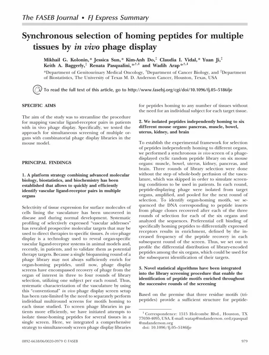
Figure 1. Schematic description of synchronous invivo phage display screening. In every selectionround, phage are intravenously (i.v.) administeredand simultaneously recovered from N target tis-sues, amplified, pooled, and used for the nextselection round. Increased recovery of phagetransforming units (TU) in the third round re-flects the selection of peptides preferentially hom-ing to the target organ.
980 Vol. 20 May 2006 KOLONIN ET AL.The FASEB Journal

our conclusion that the peptides selected mimic thePRLR-binding domain of placental lactogens.
5. To demonstrate the efficiency of the approach, wevalidated prolactin receptor (PRLR) as a target ofpancreas-homing peptides mimicking prolactin-likehormones and as a previously unrecognized vascularmarker
As a test case for the biochemical validation of PRLR asa pancreatic vascular marker, we chose CRVASVLPC:the peptide that contained a pancreas-enriched SVLtripeptide and was also recovered as a PRLR-bindingprolactin mimic. To demonstrate direct physical inter-action between CRVASVLPC and PRLR, we testedbinding of CRVASVLPC-phage to COS-1 cells trans-fected with PRLR and found it to be 9-fold higher thanits nonspecific binding to nontransfected COS-1 cellsthat served as a negative control. The CRVASVLPCmotifs bound to PRLR in both forward and reverseorientation in the context of phage, and alanine-scanning mutagenesis demonstrated that all of theresidues within the RVASVLP sequence were importantfor PRLR binding. To further demonstrate the specificaffinity of the CRVASVLPC motif for its receptor, weshowed that the PRL mimic specifically bound to cellsexpressing PRLR by using immunofluorescence: phagedisplaying either CRVASVLPC or CPLVSAVRC werefound bound and internalized specifically by cells ex-pressing PRLR, but not by nonexpressing control cells,whereas none of the CRVASVLPC mutants displayeddetectable PRLR-expressing cell binding and internal-ization. Finally, since the SVL tripeptide found withinthe PRL mimic CRVASVLPC was isolated from thepancreas, we evaluated weather the motif homes toPRLR in the pancreatic blood vessels. We showed thatthe previously reported pancreatic expression of mousePRLR protein in the vasculature and in the islets closelyresembles the in vivo distribution of phage displayingthe CRVASVLPC motif. Taken together, these dataindicate that the peptide CRVASVLPC binds to PRLRand suggests that it targets vasculature-exposed PRLRin the pancreas.
CONCLUSIONS AND SIGNIFICANCE
An important aspect of experimental biology is theidentification of molecular targets and the design ofmethods for their functional validation. Here, we testedthe approach for synchronous biopanning in mice byselecting homing peptide ligands for six different or-
gans in a single screen and prioritizing them by usingsoftware compiled for statistical validation of peptidebiodistribution specificity. Both Bayesian and frequen-tist statistics have led to the identification of largelyoverlapping populations of peptide sequences en-riched in the target organs, thus reinforcing the validityof the identified homing motifs. Based on similarity ofthe selected peptide motifs to mouse proteins, weidentified a number of motif-containing biological can-didates for ligands binding to organ-selective receptors.To demonstrate that this methodology can lead totargetable ligand-receptor systems, we validated one ofthe pancreas-homing peptides as a mimic peptide ofnatural prolactin receptor ligands. A concept to system-atically “retro-BLAST” the receptor-binding peptidesonto prototype ligands in order to map sites of ligandsinvolved in receptor interaction allowed us to identifymultiple additional PRL mimics. The successful valida-tion of PRLR as a PRL-mimicking peptide target estab-lishes this receptor as a previously unrecognized vascu-lar marker and demonstrates the viability of ourapproach.
The high-throughput targeting strategy bestowed bythe synchronous biopanning reported in this study willopen new possibilities for rapid and efficient identifi-cation and validation of vascular receptors and theirligand-directed targeting. This approach for screeningphage libraries in vivo enables highly efficient isolationof organ-homing ligands from combinatorial librariesand may streamline vascular mapping efforts, thusleading to a better understanding of the functionalprotein–protein interactions in the vasculature. Thesynchronous approach to in vivo phage display providesan advantage over the conventional approach becausemultiple organs internally control for organ selectivityof each other in the successive rounds of selection.Direct combinatorial screenings recently initiated inpatients have opened the possibility for systematicisolation of peptide ligands for therapeutic targeting.Synchronous selection of homing peptides for multipletissues will accelerate the process of phage display-based vascular mapping in vivo and may prove particu-larly relevant for patient studies, allowing efficienthigh-throughput selection of targeting ligands for mul-tiple organs in a single screening. In the future, the useof peptide libraries for probing the vasculature inindividual patients for diagnostic and therapeutic pur-poses may become a reality. In summary, the combina-torial methodological platform optimized here foridentification of differentially expressed vascular li-gand-receptor pairs will greatly advance the initiative inhuman vasculature mapping.
981SYNCHRONOUS SELECTION OF TISSUE-HOMING PEPTIDES
The FASEB Journal • FJ Express Full-Length Article
Synchronous selection of homing peptides for multipletissues by in vivo phage display
Mikhail G. Kolonin,* Jessica Sun,* Kim-Anh Do,‡ Claudia I. Vidal,* Yuan Ji,‡
Keith A. Baggerly,‡ Renata Pasqualini,*,†,1 and Wadih Arap*,†,1
*Department of Genitourinary Medical Oncology, †Department of Cancer Biology, and ‡Departmentof Biostatistics, The University of Texas M. D. Anderson Cancer, Houston, Texas, USA
ABSTRACT In vivo phage display is a technologyused to reveal organ-specific vascular ligand-receptorsystems in animal models and, recently, in patients, andto validate them as potential therapy targets. Here, wedevised an efficient approach to simultaneously screenphage display libraries for peptides homing to anynumber of tissues without the need for an individualsubject for each target tissue. We tested this approachin mice by selecting homing peptides for six differentorgans in a single screen and prioritizing them by usingsoftware compiled for statistical validation of peptidebiodistribution specificity. We identified a number ofmotif-containing biological candidates for ligands bind-ing to organ-selective receptors based on similarity ofthe selected peptide motifs to mouse proteins. Todemonstrate that this methodology can lead to tar-getable ligand-receptor systems, we validated one of thepancreas-homing peptides as a mimic peptide of natu-ral prolactin receptor ligands. This new comprehensivestrategy for screening phage libraries in vivo providesan advantage over the conventional approach becausemultiple organs internally control for organ selectivityof each other in the successive rounds of selection. Itmay prove particularly relevant for patient studies,allowing efficient high-throughput selection of target-ing ligands for multiple organs in a single screen.—Kolonin, M. G., Sun, J., Do, K.-A., Vidal, C. I., Ji, Y.,Baggerly, K. A., Pasqualini, R., Arap, W. Synchronousselection of homing peptides for multiple tissues by invivo phage display. FASEB J. 20, E99–E107 (2006)
Key Words: vascular � receptor � ligand � combinatorialpeptide library � phage display
Selectivity of tissue expression for surface mole-cules of cells lining the vasculature has been uncoveredin disease and during normal development (1–3).Systematic profiling of selectively expressed “vascularaddresses” has revealed prospective molecular targetsthat may be used to direct therapies to specific tissues(1, 4). Vascular mapping by in vivo phage display isbased on the preferential ability of short peptide li-gands from combinatorial libraries (displayed on thepIII protein of an M13-derived phage vector) (5) tohome to a specific organ after systemic administration
(1, 2, 4, 6). Peptides targeting several tissues anddisease states isolated in mouse models (1, 2, 7–9) andpatients (10, 11) have led to the identification of thecorresponding vascular receptors (7–12).
Because a single biopanning round of a large (�109
unique combinatorial sequences) phage library maynot always sufficiently enrich for organ-homing pep-tides, until now, phage display screens have encom-passed recovery of phage from the organ of interest inthree to four rounds of library selection, utilizing onesubject per each round (2, 4, 6). Thus, systematiccharacterization of the vasculature by using this “con-ventional” in vivo phage display screen setup has beenrate-limited by the need to separately perform individ-ual multiround screens for motifs homing to eachtissue studied. To screen phage libraries in patientsmore efficiently, we have initiated attempts to isolatetissue-homing peptides for several tissues in a singlescreen (10). The proof-of-principle screen involvedonly one round of selection and, therefore, requiredstatistical analysis of very large numbers of phage clones(thousands, as opposed to hundreds in a conventionalscreen) in order to identify homing peptide motifs.Although this effort yielded a prostate vasculature-selective ligand-receptor pair (10, 12, 13), for futuresystematic vascular mapping, there are practical limita-tions to the number of phage clones that can beroutinely processed, thus requiring further optimiza-tion of the methodology.
To meet such challenge of the setting where maximalinformation recovery is critical, here we integrated acomprehensive strategy to use phage display for syn-chronous identification of organ-homing peptides formultiple tissues in a single screen. This strategy allowsus to analyze an order of magnitude fewer peptidesequences than those surveyed for the initial single-round multi-organ screen (10). We evaluated our ap-proach in the mouse model by performing a screen to
1 Correspondence: Wadih Arap and Renata Pasqualini,1515 Holcombe, Blvd, Houston, TX 77030-4095, USA. E-mail:[email protected]; [email protected]
doi: 10.1096/fj.05-5186fje
E990892-6638/06/0020-0099 © FASEB

isolate peptides independently segregating to six differ-ent organs and developed a statistical analysis platformfor identification of motifs selectively enriched in tar-geted tissues. Finally, to demonstrate the efficiency ofthis approach, we biochemically validated one of theidentified pancreas-homing peptides as a mimic ofpeptide hormones that bind to prolactin receptor(PRLR).
MATERIALS AND METHODS
Synchronous screening of phage libraries in vivo
C57Bl/6 female mice were injected intravenously (i.v.) with1010 transducing units (TU) of previously described (6)library CX7C (round 1) or a mixture (109 TU per organ) ofamplified phage recovered from each of the organs studied(rounds 2 and 3). For each round, phages were allowed tocirculate for 15 min prior to organ recovery (without heartperfusion). After each round of selection, phage peptide-coding inserts were sequenced as described (6), amplified foreach organ individually, and subsequently pooled for the nextround of in vivo selection.
Statistical analysis of selected peptide motifs
Calculation of tripeptide motif frequencies in CX7C peptidesencountered in each target tissue (in both directions) wasdone by using a character pattern recognition program basedon SAS (version 8.1.2, SAS Institute) and Perl (version 5.8.1),as described (10). To identify tripeptides progressively en-riched from round 1 to round 3 of panning, a BayesianBeta/Binomial model was implemented by estimation of theposterior probability distribution for each tripeptide (14, 15);posterior distributions for the proportion of each tripeptidein rounds 1 through 3 were calculated by using Splus (version6). To determine the selectivity of tripeptide motif distribu-tion in tissues, we used Fisher’s exact test (one-tailed) andcalculated the P values for the count of each tripeptide in atarget tissue, as compared with its count within the 2210tripeptides of the unselected library (Table 1, middle col-umn) or the combined tripeptides from the other five tissues(Table 1, right column). Statistical uncertainty was furtherassessed by Monte Carlo simulations based on an establishedalgorithm (16, 17). Using MATLAB, all tripeptide sequences(unselected library and selected for each organ) were pooled,and the combined tripeptide pool was distributed in 1000simulations into permutated groups corresponding in size tothose analyzed in Table 1 for each organ and the pre-selection library. For each round, Fisher’s exact test wasperformed on the 1000 scrambled datasets, and distributionsof the 50 lowest P values generated in each simulated testwere compared with the distribution of 50 lowest P valuesfrom the actual experimental data (the most significant ofwhich are shown in Table 1, middle column).
High-throughput identification of peptide-mimickedproteins
To facilitate large-scale peptide sequence analysis, we con-structed an interactive peptide sequence management data-base based on MySQL. We developed a web-based peptidesequence retrieval and management software based on Com-mon Gateway Interface (CGI) and Perl, and integrated it with
the statistical analysis software. To identify candidate cellularproteins mimicked by selected peptides, we consolidated ourdatabase with on-line ClustlW software (http://www.ebi.ac.uk/clustalw/) to identify extended (4–7 residue long)motifs shared among multiple peptides homing to a specifictissue. Basic local alignment search tool (BLAST) (http://www.ncbi.nlm.nih.gov/basic local alignment search tool/)was used to identify proteins mimicked by the extendedhoming motifs by screening batches of ClustlW-identifiedpeptide motifs against sequences contained in on-line nonre-dundant databases of mouse proteins. To identify PRLRligand-matching motifs among phage-displayed pancreas-homing peptides binding to PRLR, a software was codified inPerl 5.8.1 and run against ClustlW-aligned protein sequencesfor sheep PL (oPL), and mouse mPL-I and mPRL. Eachseven-mer peptide sequence was aligned in each orientationagainst the protein sequences from N- to C-terminus inone-residue shifts. The peptide-protein similarity scores foreach residue were calculated based on a BLOSUM62 matrixmodified to identify peptide matches of four or more residuesin any position being identical to the corresponding aminoacid positions in any of the three PRL homologues aligned.
Phage-peptide binding to PRLR
To identify peptides binding to PRLR, phage clones isolatedfrom the pancreas in rounds 2 and 3 of the screening wereindividually amplified and pooled. For panning on immobi-lized PRLR, 109 TU of the mixed phage clones were incu-bated overnight at 4°C with 10 �g of purified recombinantrabbit PRLR (Protein Laboratories Rehovot, Israel), or BSAcontrol, immobilized on plastic. Unbound phage were exten-sively washed off with PBS, and then the bound phage wererecovered by infecting host K91 E. coli directly on the plate.For panning on cell surface-expressed PRLR, COS-1 cellsfrom American Type Culture Collection (ATCC, Manassas,VA) were transiently transfected with pECE-PRLR as de-scribed (18) and subjected to biopanning with the amplifiedpancreas-isolated phage using the BRASIL protocol (19). Ineach biopanning, bound phage were selected for tetracyclineresistance, quantified by infecting host K91 E. coli and se-quenced. Single amino acid substitutions and reversal of thePRL-matching motif displayed on phage was performed bypolymerase chain reaction (PCR)-directed mutagenesis of thepeptide-coding insert (TGTCGCGTGGCGAGCGTGCTGC-CGTGT) and cloning it into the fUSE5 vector (5). Phagedisplaying forward (CRVASVLPC), reverse CPLVSAVRC, andthe alanine point mutants were titered inparallel with insert-less fd-tet phage and tested for binding to COS-1 cellstransfected with PRLR by using the BRASIL method (19).COS-1 cells not transfected with PRLR served as a negativecontrol.
Immunolocalization
Immunofluorescent detection of PRLR expression in COS-1cells was performed by using anti-PRLR antibody (Ab) MA1–610 (Affinity Bioreagents) diluted to 20 �g/ml and a second-ary FITC-conjugated goat anti-mouse Ab F-0257 (Sigma) at1:100 dilution. For validating phage binding and internaliza-tion into COS-1 cells transfected with PRLR, 109 TU of phagedisplaying PRLR-binding or mutant peptides were subjectedto cell binding and internalization as described (12). Immu-nodetection of cell-associated phage was performed withanti-fd Ab B-7786 (Sigma) at 1:500 dilution and a secondaryCy3-conjugated donkey anti-rabbit Ab 711–165-152 (Jackson)
E100 Vol. 20 May 2006 KOLONIN ET AL.The FASEB Journal

TABLE 1. Peptide motifs homing to n different tissuesa
Tripeptides progressively enriched in Rounds 1–3 Tripeptides selected (vs. unselected library) Tripeptides specific (vs. other organs)
Targetorgan/motif
Motif frequency (%)Posterior
probability foldover baseline
Targetorgan/motif
Motiffrequency
(%)P
valueTarget
organ/motif
Motiffrequency
(%)P
valueRound
1Round
2Round
3
Muscle Muscle MuscleRSG 2.2 2.4 5.6 10.0 RSG 5.6 0.0301 RSG 5.6 0.0351SGA 0.0 1.2 5.6 7.0 SGA 5.6 0.0301 SGA 5.6 0.0132AIG 0.0 0.0 4.4 5.0 AIG 4.4 0.0006 AIG 4.4 0.0242IGS 0.0 1.2 5.6 7.0 IGS 5.6 0.0037 IGS 5.6 0.0010GSF 0.0 1.2 5.6 7.0 GSF 5.6 0.0072 GSF 5.6 0.0030AGG 1.1 1.2 4.4 7.0 AGG 4.4 0.0261 APA 2.2 0.0333ASR 0.0 0.0 3.3 4.0 APA 2.2 0.0253 DFS 4.4 0.0121DFS 0.0 0.0 4.4 5.0 DFS 4.4 0.0028 GDT 3.3 0.0210DGT 0.0 0.0 3.3 4.0 GDT 3.3 0.0040 GGT 5.6 0.0010DTG 0.0 0.0 3.3 4.0 GGT 5.6 0.0201 IAY 4.4 0.0011FRS 0.0 1.2 3.3 5.0 IAY 4.4 0.0006GDT 0.0 1.2 3.3 5.0GGT 0.0 0.0 5.6 6.0GWS 0.0 0.0 3.3 4.0IAY 0.0 0.0 4.4 5.0RRS 0.0 1.2 3.3 5.0SGV 0.0 1.2 3.3 5.0
Pancreas Pancreas PancreasSVL 1.1 3.3 4.8 9.0 SVL 4.8 0.0022 VSS 7.1 0.0012VSS 1.1 0.0 7.1 8.0 VSS 7.1 0.0006 WSG 6.0 0.0021WSG 1.1 0.0 6.0 7.0 WSG 6.0 0.0012 FGV 3.6 0.0371GWR 0.0 1.1 3.6 5.0 FGV 3.6 0.0033 GYN 3.6 0.0169GYN 0.0 0.0 3.6 4.0 GYN 3.6 0.0033 LTR 3.6 0.0048LTR 0.0 1.1 3.6 5.0 LTR 3.6 0.0118 TLV 3.6 0.0371TLV 0.0 0.0 3.6 4.0 TLV 3.6 0.0033
Brain Brain BrainLGG 1.1 0.0 5.8 8.0 LGG 5.8 0.0204 LGG 5.8 0.0427RGF 0.0 0.0 3.5 4.0 RGF 3.5 0.0337 RGF 3.5 0.0250ALG 2.1 0.0 3.5 6.0 DSY 2.3 0.0174 DSY 2.3 0.0215LLS 1.1 0.0 3.5 5.0 GFS 2.3 0.0174 GIW 2.3 0.0215
GIW 2.3 0.0174 HGL 2.3 0.0215HGL 2.3 0.0477 ALG 3.5 0.0446
LLS 3.5 0.0446Kidney Kidney Kidney
LGS 1.1 2.3 4.4 8.0 SLS 4.4 0.0004 SLS 4.4 0.0030SLS 1.1 1.1 4.4 7.0 DRG 3.3 0.0239 DRG 3.3 0.0151DRG 0.0 0.0 3.3 4.0 RRV 3.3 0.0239 FLS 2.2 0.0265RRV 0.0 0.0 3.3 4.0 SRV 4.4 0.0186DSG 0.0 1.1 3.3 5.0 FLS 2.2 0.0208LRV 0.0 1.1 3.3 5.0SRV 1.1 1.1 4.4 7.0
Uterus Uterus UterusGSS 1.2 0.0 4.9 6.0 GSS 4.9 0.0456 GPS 3.7 0.0157LLG 1.2 0.0 4.9 6.0 LLG 4.9 0.0308 GAA 4.9 0.0322GAA 0.0 1.1 4.9 6.0 GAA 4.9 0.0109GLL 1.2 0.0 4.9 6.0 GPS 3.7 0.0246ARG 1.2 3.2 3.7 8.0GAS 0.0 1.1 3.7 5.0GGL 2.4 1.1 4.9 8.0GPS 0.0 0.0 3.7 4.0
Bowel Bowel BowelAGV 0.0 0.0 4.7 5.0 AGV 4.7 0.0719 AGV 4.7 0.0344WRD 0.0 0.0 4.7 5.0 WRD 4.7 0.0023 WRD 4.7 0.0098FGG 0.0 0.0 4.7 5.0 FGG 4.7 0.0005 FGG 4.7 0.0199GGR 1.1 0.0 7.1 8.0 RWS 3.5 0.0275 RWS 3.5 0.0179GRV 0.0 1.2 3.5 5.0 VGV 3.5 0.0124RWS 0.0 0.0 3.5 4.0 GVG 3.5 0.0488VGV 0.0 0.0 3.5 4.0
Left column: using the Bayesian Beta/Binomial model, we ranked tripeptides according to posterior mean. Shown are tripeptides withposterior probability fold change of x3 or more over baseline (posterior probability for tripeptides not isolated in any of the three rounds).Middle column: tripeptide motifs contained in CX7C peptides isolated in round 3 from target organs with frequency significantly higher than thatobserved in the unselected phage library (Fisher Exact test, one-tailed; P�0.05). Right column: tripeptide motifs occurring in CX7C peptidesenriched in round 3 in a specific organ but not in other target organs analyzed (Fisher Exact test, one-tailed; P�0.05).
E101SYNCHRONOUS SELECTION OF TISSUE-HOMING PEPTIDES
at 3 �g/ml. For phage-peptide immunolocalization in situ,1010 TU of iv-injected phage were let circulate for 5 min.Immunohistochemistry on formalin-fixed, paraffin-embed-ded mouse tissue sections was performed as described (6, 10)by using anti-fd Ab B-7786 at 1:1,000 dilution and the LSAB�peroxidase kit (DAKO).
RESULTS
Synchronous phage library screening in vivo
We reasoned that peptide-displaying phage clones ofsystemically administered library would segregate in thebloodstream irrespective of each other and, thus, targetindividual organs independently. If this hypothesis iscorrect, independent enrichment of phage-peptidestargeting any number of organs should take place onsuccessive rounds of selection. To establish the experi-mental framework for synchronous selection of pep-tides independently homing to different organs, wescreened a phage-displayed cyclic random peptide li-brary CX7C (C, cysteine; X, any residue). Six organswere targeted in mice: muscle, bowel, uterus, kidney,pancreas, and brain (Fig. 1). Three rounds of libraryselection were performed based on previously de-scribed methodology (6), but without the step of whole-body perfusion of the vasculature, which was skipped inorder to simulate screening conditions used in patients(10). In each round, peptide-displaying phage wereisolated from target organs, amplified, and pooled forthe next round of selection. To identify organ-homingmotifs, we sequenced the DNA corresponding to pep-tide inserts from 96 recovered phage clones after eachof the three rounds of selection for each of the sixorgans and analyzed the sequences. Preferential cellbinding of specifically homing peptides to differen-tially-expressed receptors results in enrichment, de-fined by the increased frequency of the peptide recov-ery in each subsequent round of the screen (4, 6).Thus, we set out to profile the differential distributionof library-encoded peptides among the six organs,which could be used for the subsequent identificationof their targets.
To analyze the spectrum of the peptides resultingfrom the screening and to compare those amongdifferent organs, we adopted a combinatorial statistical
approach based on the premise that three residuemotifs (tripeptides) provide a sufficient structure forpeptide-protein interactions in the context of phagedisplay (10). Since a tripeptide within a CX7C sequencemay be responsible for receptor targeting in eitherorientation, we performed a computer-assisted analysisof the 7489 tripeptides contained in each directionwithin the CX7C inserts (2620 from the first round;2554 from the second round; and 2315 from the thirdround) successfully sequenced for the organ-recoveredclones. First, we evaluated the increase in recoveryfrequency for individual tripeptides in the three con-secutive rounds of selection by using the BayesianBeta/Binomial model (14, 15). For each organ, wefound a number of tripeptides progressively enriched(Table 1, left column), thus, suggesting their superioraffinity and/or specificity. Next, we surveyed the 2315motifs recovered from the third round of selection toidentify motifs with terminal frequencies higher thanthose present in the phage library prior to selection.The significance of motif representation increase onselection was assessed by the Fisher exact test (Table 1,middle column). To show that the P value of 0.05 forestablishment of selected tripeptides was chosen appro-priately, we adapted the Monte Carlo algorithm (16),and confirmed in the third selection round that the Pvalues of the actual data were smaller than at least 95%of the simulated P values (Fig. 2). Of note, Monte Carlosimulations showed a progressive accumulation of tri-peptides isolated with lower P values from the first tothe third round (Fig. 2), consistent with enrichment ofthe corresponding motifs identified by the BayesianBeta/Binomial model (Table 1, left column). Finally,we used the Fisher exact test to analyze the motifsrecovered from the third selection round for specificityof tissue homing by identifying tripeptides that wereenriched in one of the six organs, but not in the rest ofthe organs studied (Table 1, right column).
Identification of candidate biological ligandsmimicked by homing peptides
The majority of peptide motifs identified by statisticalanalysis enriched during the screen also showed speci-ficity of association with the organ from which theywere recovered (Table 1). For validation of potentially
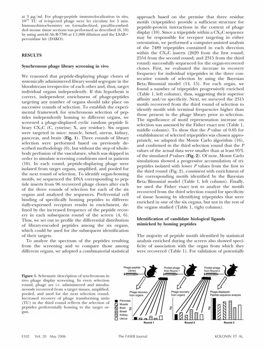
Figure 1. Schematic description of synchronous invivo phage display screening. In every selectionround, phage are i.v. administered and simulta-neously recovered from n target tissues, amplified,pooled, and used for the next selection round.Increased recovery of phage transforming units(TU) in the third round reflects the selection ofpeptides preferentially homing to the target or-gan.
E102 Vol. 20 May 2006 KOLONIN ET AL.The FASEB Journal

specific organ-homing motifs, we chose to focus ontripeptides that fulfilled the criteria of the statisticaltests applied (Table 1). Since peptide motifs binding tocell surface receptors have been previously found tomimic native ligands for these receptors (7, 10, 19), theClustalW software was used to determine whether thetripeptides represented parts of longer motifs respon-sible for organ homing, which would facilitate peptide/protein similarity search. For some of the tripeptides,this analysis identified extended (four to seven residue)motifs shared among multiple CX7C peptides isolatedfrom the given organ (Fig. 3). Each of these extendedmotifs was screened by using the BLAST algorithmagainst a nonredundant database of mouse proteins toidentify regions of similarity within proteins potentiallymimicked by the motifs (Table 2).
We systematically analyzed the BLAST output forselected motif similarities to extracellular signalingfactors that had been reported to regulate organ-dependent vascular growth or homeostasis. This re-vealed 19 motifs as segments of such proteins and, insome cases, identified several motifs that homed to thesame organ and that matched different domains within
the same protein (Table 2). For example, muscle-homing motifs GRSG�R and SGASAV, matched to twodifferent domains of the Jagged2-like protein (Table 2)that belongs to a family of ligands for Notch receptorsknown to regulate vascular development and function(20, 21). Similarly, pancreas-homing motifs ASVL (inthe reverse orientation) and WSGL showed close simi-larity to different domains of a placental lactogen (22).Interestingly, for muscle and pancreas, we also matchedhoming tripeptides to different ligands that share areceptor with a functional role in vascular biology inthe target organ (Table 2). Among skeletal muscle-homing motifs, tripeptides FSG and SGI were partiallyoverlapping in the extended DFSGIA� region of simi-larity to disintegrin family metalloproteinases ADAMand Spi 12, respectively, which cleave Notch receptors(23). In the pancreas, motif SWSG matched to prolac-tin (PRL)-like protein M (PLP-M), which belongs to thesame family as the placental lactogen PL-I (containingthe reverse ASVL and WSGL) and also binds to the PRLreceptor (22,24).
Biochemical validation of PRLR as the target forpancreas-homing peptides
To demonstrate the possibility of efficient characteriza-tion of circulation-accessible receptors by synchronousbiopanning, as a proof-of-principle, we chose to vali-date the PRL receptor (PRLR) as a peptide target in thepancreas. PL-I and PLP-M identified by BLAST analysisbelong to the conserved family of PRL-like peptidichormones that have been shown to function in thepancreas during pregnancy (22, 25). Because PRLR isthe only known receptor for these proteins (22, 24), weproposed that the ASVL, WSGL, and SWSG motifstarget PRLR in vivo by mimicking PRL family hor-mones.
To test this hypothesis, we attempted to reveal abiochemical interaction of pancreas-homing motifswith PRLR. We used the BRASIL (biopanning andrapid analysis of selective interactive ligands) method(19) to screen a pancreas-homing phage sublibrary(pooled clones recovered in rounds 2 and 3) againstPRLR expressed on the surface of COS-1 cells (18). Inparallel, we screened the same sublibrary on purifiedrecombinant PRLR (26). A single round of each selec-
Figure 2. Monte Carlo simulations to assess tripep-tide motif tissue homing. For each selectionround, the plot of 50 smallest P values (black line)determined by Fisher’s exact test for enrichedtripeptides (Table 1, middle column), was com-pared with the corresponding simulated datasetplots (colored lines). To generate simulated data-sets, all tripeptides isolated from the target organswere pooled with tripeptides isolated from theunselected CX7C library, and Fisher’s exact testwas performed on 1000 random permutations. For
every permutation, the pool of tripeptides was randomly distributed into groups corresponding to numbers of peptidesequences analyzed for each tissue (Table 1, middle column). Plotted are the 50 smallest P values (index number of P values1 through 50, ascending order) generated in each of the 1000 permutations.
Figure 3. Identification of extended motifs homing to mousetissues. Peptide sequences containing tripeptides enriched ina given tissue (Table 1) were aligned in clusters by using theClustalW software to obtain longer motifs shared amongdifferent peptides from each cluster. Similarity between pep-tides at the concentration of amino acid class is color-coded:red, hydrophobic; green, neutral and polar; purple, basic;blue, acidic. Original tripeptides are depicted in bold; ex-tended motifs are highlighted in yellow.
E103SYNCHRONOUS SELECTION OF TISSUE-HOMING PEPTIDES

tion for PRLR-binding phage-peptides resulted in over90% of the clones sequenced comprised by sevendifferent peptides, five of which were enriched on bothimmobilized and cell surface-expressed PRLR (Fig. 4A).Phage displaying these peptides had specific affinity toPRLR, as determined by subjecting the same sublibraryto binding of an immobilized BSA control (Fig. 4B).Remarkably, computer-assisted analysis of sequencesrevealed that all of the selected peptides containedamino acid motifs similar to those present in proteinsof PRL family (Fig. 4A). Furthermore, a clear cluster ofmatches was identified around one of the hormonedomains that had been shown (27) to mediate receptorinteraction (Fig. 4A). As a negative control, the samesimilarity search algorithm did not reveal such matchesfor the selected sequences to unrelated proteins such asinsulin, IL-11 and bZIP (data not shown).
Peptide motif CRVASVLPC recovered as a PRLR-
binding prolactin mimic (Fig. 4) contained a tripep-tide, SVL, also identified as one of those enriched inthe pancreas during the screen (Table 1). The CRVAS-VLPC motif matched one of the PL-I sites involved inPRLR interaction (27), as it had amino acids identicalto those found in one or more of the three aligned PRLhomologues in four out of seven positions (Fig. 4A).Similarity of this peptide in reverse orientation to a partof PL-I (Fig. 4A), initially identified by the BLASTanalysis (Table 2), was found by RasMol-assisted analysisof 3D protein structure to reside within the domainexposed on the surface of PRL family proteins (datanot shown). To demonstrate direct physical interactionbetween CRVASVLPC and PRLR, we tested binding ofCRVASVLPC-phage to COS-1 cells transfected withPRLR and found it to be 9-fold higher than its nonspe-cific binding to nontransfected COS-1 cells that servedas a negative control (Fig. 5A). To address the issue of
TABLE 2. Candidate mouse proteins mimicked by synchronously selected peptidesa
Extended motifMouse protein
containing motif Protein description Accession #
Muscle
DFSGIA�; DFSGIA� ADAM 10; Spi12 Notch Interactor; Serine protease inhibitor(serpin)
NP_031425; NP_035584
GRSG�R; SGASAV Syndactylism, Jagged2-similar
Notch ligand XP_192739
SG�GVF DPP IV Dipeptidylpeptidase active in musclevasculature
NP_034204
AGSF Fibrillin-1 Extracellular matrix protein, TGF�interactor
XP_192917
SLGSFP SPARC-related protein Extracellular matrix calcium-binding protein NP_071711
Pancreas
LVSA; WSGL Placental lactogen-I �(PL-I)
Pancreas-signaling peptide hormone AAN39710.1
GWSG PRL-like protein M Pancreas-signaling peptide hormone NP_064375�SVLTR Ecgf1, gliostatin Endothelial cell growth factor NP_612175.1
Brain
SLGG NGF-alpha; kallikreinK22
Nerve growth factor; NGF endopeptidase NP_035045; P15948
Kidney
GSLS Endothelin-convertingenzyme
Processing of peptidic vasoconstrictinghormones
XP_131743.2
LSLSL Thrombospondin Anti-angiogenic ECM protein, TGF�activator
AAA50611.1
Uterus
�PGSSF Pregnancy zone protein;�1 m
Differentially expressed endometrial LRPsubunit
NP_031402.1
GSS�WA Fractalkine; neurotactin Chemokine; small inducible cytokine NP_033168PGLL Luteinizing hormone � Pregnancy peptide hormone NP_032523
Bowel
AGVGV Fibrillin-2 Extracellular matrix protein, TGF�interactor
AAA74908
�CFGG� Prepronatriodilatin Atrial natriuretic factor: intestinal paracrineeffector
P05125
For sequence similarity search to mouse proteins, tripeptide-containing motifs (in either orientation) identified in Fig. 3 were screenedusing BLAST (NCBI). Examples of candidate proteins potentially mimicked by the peptides homing to mouse tissues are listed. Sequencescorrespond to the regions of 100% identity between the peptide selected and the candidate protein. Conserved amino acid substitutions areindicated as (�). Tripeptides shown in Table 1 are highlighted.
E104 Vol. 20 May 2006 KOLONIN ET AL.The FASEB Journal

a possible importance of the motif orientation forPRLR binding, we constructed phage displayingCPLVSAVRC, the PRL-mimicking motif in the oppositeorientation. Reversal of the motif did not significantlydecrease its binding to PRLR on the expressing cells(Fig. 5B). However, disruption of the motif by alanine-scanning mutagenesis of any amino acid significantlydecreased binding to PRLR-expressing cells (Fig. 5A),thus indicating cooperation of the RVASVLP residuescomprising the motif in the receptor recognition. Tofurther demonstrate the specific affinity of the CRVAS-VLPC motif for its receptor, we showed that the PRLmimic specifically bound to cells expressing PRLR byusing immunofluorescence (Fig. 5C). Phage displayingeither CRVASVLPC or CPLVSAVRC were found boundand internalized specifically by cells expressing PRLR,
but not by nonexpressing control cells, whereas none ofthe CRVASVLPC mutants displayed detectable PRLR-expressing cell binding and internalization (Figs. 5C–Dand data not shown).
Finally, since the SVL tripeptide found within the
Figure 4. Retro-BLAST analysis to identify PRLR ligand-matching motifs. A) Peptide sequences isolated from thepancreas-homing phage pool as those binding to PRLR werematched in each orientation to mature sheep (oPL) andmouse (mPL-I and mPRL) protein sequences (leader peptidesequence not included). Peptide matches of four or moreresidues in any position being identical to the correspondingamino acid positions in any of the three PRL homologues aredisplayed. Shaded protein sequences: published PRLR bind-ing sites. Peptide amino acid colors are coded according totheir relationship to the corresponding amino acid positionsof one or more of the three aligned PRLR ligands: red,identity; green, similarity. Motifs SGATGRA, SGPTGRA,QVHSSAY, VFSDYKR, and LPTLSLN were isolated by biopan-ning on both: in vitro immobilized and cell-surface expressedPRLR. Forward and reverse matches of the validated RVAS-VLP motif are underlined. B) Binding of pancreas-homingphage-peptides (recovered from synchronous biopanningrounds 2 and 3) to recombinant rabbit PRLR, as compared totheir binding to BSA control. TU: transforming units.
Figure 5. Validation of PRLR as a candidate receptor for aPRLR ligand mimic CRVASVLPC. A) Specific binding of theCRVASVLPC-phage, but not of the control phage (CYAIGS-FDC-displaying or insertless fd-tet) to COS-1 cells transfectedwith pECE-PRLR. Phage binding to COS-1 PRLR-transfectedcells (as compared to control nontransfected cells) was deter-mined by BRASIL (10, 19). B) Binding of phage displayingforward SVL-containing CRVASVLPC motif (right arrow), aswell as the reverse CPLVSAVRC motif (left arrow), to PRLR-transfected COS-1 cells, as compared to biding of the sixalanine-scan motif mutants (A1:CAVASVLPC, A2:CRAASV-LPC, A3:CRVAAVLPC, A4:CRVASALPC, A5:CRVASVAPC,and A6:CRVASVLAC). C–D) Specific binding to and internal-ization of CRVASVLPC-phage (C) and an alanine-scan mu-tant A4 (D) into COS-1 PRLR-transfected cells, detected byco-immunolocalization of CRVASVLPC-phage (red) withPRLR-expression (green), resulting in overlapping yellowsignal (C). E–H) Antiphage immunohistochemistry (brown)in paraffin sections of formalin-fixed pancreas (E, H) orskeletal muscle (F, G) from mice i.v. injected with CRVASV-LPC-phage (E, G), or control muscle-homing CYAIGSFDC-phage (F, H). Hematoxylin counterstaining is shown in blue.
E105SYNCHRONOUS SELECTION OF TISSUE-HOMING PEPTIDES

PRL mimetope CRVASVLPC was isolated from thepancreas, we evaluated weather the motif homes toPRLR in the pancreatic blood vessels. We showed thatthe previously reported pancreatic expression of mousePRLR protein in the vasculature and in the islets (28)closely resembles the in vivo distribution of phagedisplaying the CRVASVLPC motif (Fig. 5E–H). Immu-nohistochemistry of mouse tissues on i.v. CRVASVLPC-phage administration revealed localization of theCRVASVLPC motif to pancreatic blood vessels and thepancreatic islet cells (Fig. 5E), but not to skeletalmuscle (Fig. 5F). In contrast, a control in vivo–admin-istered phage clone displaying CYAIGSFDC sequencehoming to the skeletal muscle was found predomi-nantly in the vasculature of the skeletal muscle, but notin the pancreas (Fig. 5H). Taken together, these dataindicate that the peptide CRVASVLPC binds to PRLRand suggests that it targets vasculature-exposed PRLRin the pancreas.
DISCUSSION
Here, we present an advance in the in vivo phagedisplay methodology: simultaneous screening of pep-tide libraries on multiple organs. Parallel analysis offewer sequences (hundreds) in multiple biopanningrounds performed here, as opposed to thousands ofsequences required for the identification of homingmotifs in a single round, as reported preciously (10),provides control over the progressive organ-specificenrichment of individual motifs throughout the con-secutive screening rounds. Our approach enableshighly efficient isolation of organ-homing ligands fromcombinatorial libraries and may streamline vascularmapping efforts, thus leading to a better understandingof the functional protein–protein interactions in thevasculature.
Both Bayesian and frequentist statistics have led tothe identification of largely overlapping populations ofpeptide motifs enriched in the target organs, thusreinforcing the validity of the identified homing motifs.Organ-homing peptides were prioritized for furthervalidation based on their sequence similarity to do-mains of mouse proteins. Protein-peptide similarity-based survey failed to identify prototype proteins forsome of the homing tripeptides. This is not surprising,as the approach does not reveal peptide motifs thatmimic conformationally defined interacting sites withinproteins. Moreover, it is certainly not a given that everysimilarity-based lead will ultimately yield a biologicallysignificant interaction. Nevertheless, this strategy hasproven effective in the identification of receptor-ligandleads that might subsequently be validated directly byusing binding assays (12, 19).
As an example, we validated a pancreas-homingmotif CRVASVLPC as a peptide mimic of naturalligands of PRLR, which include PL, PRL, and a numberof other hormones from the same family. A conceptimplemented here is the ability to systematically “retro-
BLAST” the receptor-binding peptides onto prototypeligands in order to map sites of ligands involved inreceptor interaction. By such retro-targeting of thereceptor ligands with peptides derived from in vivoselections, we were able to identify multiple apparentPRL mimics, many of which had been missed in thestatistical survey of selected tripeptides. We show thatone of the PRL mimics, CRVASVLPC, specifically bindsto PRLR in the context of phage coat pIII protein, andthat the precise amino acid sequence of the motifappears to be essential for binding. We demonstratethat the CRVASVLPC motifs binds to PRLR in bothforward and reverse orientation, however it should benoted that such orientation independence may not bea general feature that is applicable to every ligand-receptor pair interaction. We also show that theCRVASVLPC peptide homes to pancreatic vasculaturein vivo, consistent with vascular PRLR expression andwith the established function of placental lactogens inthe pancreas during pregnancy (22, 24, 25, 28–30).These findings are also consistent with reported PRLRexpression in other organs, as neither VSL, nor othermotifs apparently mimicking biological PRLR ligands,qualified the statistical test of organ-specific motif dis-tribution (Table 1, right column).
In summary, synchronous selection of homing pep-tides for multiple tissues accelerates the process ofphage display-based vascular mapping in vivo. Directcombinatorial screenings in patients (10, 11, 13) haveopened the possibility of systematic isolation of peptideligands for therapeutic targeting. In the future, the useof peptide libraries for probing the vasculature inindividual patients for diagnostic and therapeutic pur-poses may become a reality. The high-throughput tar-geting strategy bestowed by the synchronous biopan-ning reported here will open new possibilities for rapidand efficient identification and validation of vascularreceptors and their ligand-directed targeting.
We thank Dr. Li-yuanYu-Lee for the plasmid pECE-PRLR.We also thank Xuemei Wang and Carlotta L. Cavazos fortechnical assistance. This work was supported by grants fromthe NIH (CA103030, DK67683, and CA90810 to W.A.;CA90270 to R.P.; and CA111853 to M.G.K.) and by awardsfrom the Gillson-Longenbaugh Foundation, the AngelWorksFoundation, and the V Foundation (to W.A. and R.P).
REFERENCES
1. Arap, W., Pasqualini, R., and Ruoslahti, E. (1998) Cancertreatment by targeted drug delivery to tumor vasculature in amouse model. Science 279, 377–380
2. Pasqualini, R., and Ruoslahti, E. (1996) Organ targeting in vivousing phage display peptide libraries. Nature 380, 364–366
3. Rajotte, D., Arap, W., Hagedorn, M., Koivunen, E., Pasqualini,R., and Ruoslahti, E. (1998) Molecular heterogeneity of thevascular endothelium revealed by in vivo phage display. J. Clin.Invest. 102, 430–437
4. Kolonin, M. G., Pasqualini, R., and Arap, W. (2001) Molecularaddresses in blood vessels as targets for therapy. Curr. Opin.Chem. Biol. 5, 308–313
5. Smith, G. P., and Scott, J. K. (1993) Libraries of peptides andproteins displayed on filamentous phage. Methods Enzymol. 217,228–257
E106 Vol. 20 May 2006 KOLONIN ET AL.The FASEB Journal

6. Pasqualini, R., Arap, W., Rajotte, D., and Ruoslahti, E. (2001) InVivo Selection of Phage-display Libraries. In Phage Display: ALaboratory Manual (Barbas, C., Burton, D., Silverman, G., andScott, J., eds) pp. 22.21–22.24, Cold Spring Harbor LaboratoryPress, New York, NY
7. Kolonin, M. G., Pasqualini, R., and Arap, W. (2002) Teratoge-nicity induced by targeting a placental immunoglobulin trans-porter. Proc. Natl. Acad. Sci. U. S. A. 99, 13055–13060
8. Pasqualini, R., Koivunen, E., Kain, R., Lahdenranta, J., Saka-moto, M., Stryhn, A., Ashmun, R. A., Shapiro, L. H., Arap, W.,and Ruoslahti, E. (2000) Aminopeptidase N is a receptor fortumor-homing peptides and a target for inhibiting angiogenesis.Cancer Res. 60, 722–727
9. Kolonin, M. G., Saha, P. K., Chan, L., Pasqualini, R., and Arap,W. (2004) Reversal of obesity by targeted ablation of adiposetissue. Nature Med. 10, 625–632
10. Arap, W., Kolonin, M. G., Trepel, M., Lahdenranta, J., Cardo-Vila, M., Giordano, R. J., Mintz, P. J., Ardelt, P. U., Yao, V. J.,Vidal, C. I., Chen, L., Flamm, A., Valtanen, H., Weavind, L. M.,Hicks, M. E., Pollock, R. E., Botz, G. H., Bucana, C. D.,Koivunen, E., Cahill, D., Troncoso, P., Baggerly, K. A., Pentz,R. D., Do, K. A., Logothetis, C. J., and Pasqualini, R. (2002)Steps toward mapping the human vasculature by phage display.Nature Med. 8, 121–127
11. Kolonin, M. G., Pasqualini, R., and Arap, W. (2003) Mappinghuman vascular heterogeneity by in vivo phage display. InGenetics of Angiogenesis (Hoying, J. B., ed), BIOS ScientificPublishers Ltd., Oxford
12. Zurita, A. J., Troncoso, P., Cardo-Vila, M., Logothetis, C. J.,Pasqualini, R., and Arap, W. (2004) Combinatorial screenings inpatients: the interleukin-11 receptor alpha as a candidate targetin the progression of human prostate cancer. Cancer Res. 64,435–439
13. Pentz, R. D., Flamm, A. L., Pasqualini, R., Logothetis, C. J., andArap, W. (2003) Revisiting ethical guidelines for research withterminal wean and brain-dead participants. Hastings Cent. Rep.33, 20–26
14. Lee, K. E., Sha, N., Dougherty, E. R., Vannucci, M., and Mallick,B. K. (2003) Gene selection: a Bayesian variable selectionapproach. Bioinformatics 19, 90–97
15. Carlin, B. P., and Sargent, D. J. (1996) Robust Bayesian ap-proaches for clinical trial monitoring. Stat. Med. 15, 1093–1106
16. Gelman, A., and Rubin, D. B. (1996) Markov chain Monte Carlomethods in biostatistics. Stat. Methods Med. Res. 5, 339–355
17. Zhang, L., Zhou, W., Velculescu, V. E., Kern, S. E., Hruban,R. H., Hamilton, S. R., Vogelstein, B., and Kinzler, K. W. (1997)Gene expression profiles in normal and cancer cells. Science 276,1268–1272
18. Wang, Y., O’Neal, K. D., and Yu-Lee, L. (1997) Multipleprolactin (PRL) receptor cytoplasmic residues and Stat1 medi-ate PRL signaling to the interferon regulatory factor-1 pro-moter. Mol. Endocrinol. 11, 1353–1364
19. Giordano, R. J., Cardo-Vila, M., Lahdenranta, J., Pasqualini, R.,and Arap, W. (2001) Biopanning and rapid analysis of selectiveinteractive ligands. Nature Med. 7, 1249–1253
20. Lindner, V., Booth, C., Prudovsky, I., Small, D., Maciag, T., andLiaw, L. (2001) Members of the Jagged/Notch gene families areexpressed in injured arteries and regulate cell phenotype viaalterations in cell matrix and cell-cell interaction. Am. J. Pathol.159, 875–883
21. Krebs, L. T., Xue, Y., Norton, C. R., Shutter, J. R., Maguire, M.,Sundberg, J. P., Gallahan, D., Closson, V., Kitajewski, J., Calla-han, R., Smith, G. H., Stark, K. L., and Gridley, T. (2000) Notchsignaling is essential for vascular morphogenesis in mice. GenesDev. 14, 1343–1352
22. Wiemers, D. O., Shao, L. J., Ain, R., Dai, G., and Soares, M. J.(2003) The mouse prolactin gene family locus. Endocrinology144, 313–325
23. Brou, C., Logeat, F., Gupta, N., Bessia, C., LeBail, O., Doedens,J. R., Cumano, A., Roux, P., Black, R. A., and Israel, A. (2000) Anovel proteolytic cleavage involved in Notch signaling: the roleof the disintegrin-metalloprotease TACE. Mol. Cell 5, 207–216
24. Goffin, V., Binart, N., Touraine, P., and Kelly, P. A. (2002)Prolactin: the new biology of an old hormone. Annu. Rev.Physiol. 64, 47–67
25. Freemark, M., Avril, I., Fleenor, D., Driscoll, P., Petro, A., Opara,E., Kendall, W., Oden, J., Bridges, S., Binart, N., Breant, B., andKelly, P. A. (2002) Targeted deletion of the PRL receptor:effects on islet development, insulin production, and glucosetolerance. Endocrinology 143, 1378–1385
26. Bignon, C., Sakal, E., Belair, L., Chapnik-Cohen, N., Djiane, J.,and Gertler, A. (1994) Preparation of the extracellular domainof the rabbit prolactin receptor expressed in Escherichia coli andits interaction with lactogenic hormones. J. Biol. Chem. 269,3318–3324
27. Elkins, P. A., Christinger, H. W., Sandowski, Y., Sakal, E.,Gertler, A., de Vos, A. M., and Kossiakoff, A. A. (2000) Ternarycomplex between placental lactogen and the extracellular do-main of the prolactin receptor. Nature Struct. Biol. 7, 808–815
28. Brelje, T. C., Svensson, A. M., Stout, L. E., Bhagroo, N. V., andSorenson, R. L. (2002) An immunohistochemical approach tomonitor the prolactin-induced activation of the JAK2/STAT5pathway in pancreatic islets of Langerhans. J. Histochem. Cyto-chem. 50, 365–383
29. Brelje, T. C., Scharp, D. W., Lacy, P. E., Ogren, L., Talamantes,F., Robertson, M., Friesen, H. G., and Sorenson, R. L. (1993)Effect of homologous placental lactogens, prolactins, andgrowth hormones on islet B-cell division and insulin secretion inrat, mouse, and human islets: implication for placental lactogenregulation of islet function during pregnancy. Endocrinology 132,879–887
30. Vasavada, R. C., Garcia-Ocana, A., Zawalich, W. S., Sorenson,R. L., Dann, P., Syed, M., Ogren, L., Talamantes, F., and Stewart,A. F. (2000) Targeted expression of placental lactogen in thebeta cells of transgenic mice results in beta cell proliferation,islet mass augmentation, and hypoglycemia. J. Biol. Chem. 275,15399–15406
Received for publication September 26, 2005.Accepted for publication November 1, 2005.
E107SYNCHRONOUS SELECTION OF TISSUE-HOMING PEPTIDES